orexin neurons express a functional pancreatic polypeptide y4 receptor
TRANSCRIPT
Orexin Neurons Express a Functional Pancreatic PolypeptideY4 Receptor
Rebecca E. Campbell,1,2 M. Susan Smith,1,2 Summer E. Allen,1 Bernadette E. Grayson,1 Jarlath M. H. ffrench-Mullen,3
and Kevin L. Grove1
1Department of Neuroscience, Oregon National Primate Research Center, Beaverton, Oregon 97006, 2Departments of Physiology and Pharmacology, OregonHealth and Science University, Portland, Oregon 97209, and 3Gene Logic Inc., Gaithersburg, Maryland 20878
The receptor subtypes that mediate the effects of neuropeptide Y (NPY) on food intake have not been clearly defined. The NPY Y4 receptorhas been identified recently as a potential mediator of the regulation of food intake. The purpose of the present study was to characterizethe central site of action of the Y4 receptor using a combination of neuroanatomical and physiological approaches. Using immunocyto-chemistry, Y4-like immunoreactivity was found to be colocalized with orexin cell bodies in the lateral hypothalamic area (LHA) andorexin fibers throughout the brain. In situ hybridization confirmed the expression of Y4 mRNA in orexin neurons. To determine the func-tional interaction between Y4 receptors and orexin neurons, we examined the effects of rat pancreatic polypeptide (rPP), a Y4-selective ligand,or NPY, a nonselective ligand, administered directly into the LHA on the stimulation of food and water intake and c-Fos expression. Both rPPand NPY significantly increased food and water intake when they were administered into the LHA, although NPY was a more potent stimula-tor of food intake. Furthermore, both NPY and rPP significantly stimulated c-Fos expression in the LHA. However, whereas rPP stimulatedc-Fos expression in orexin neurons, NPY did not. Neither rPP nor NPY stimulated c-Fos in melanin-concentrating hormone neurons, butboth activated neurons of an unknown phenotype in the LHA. These results suggest that a functional Y4 receptor is expressed on orexin neu-rons and that these neurons are activated in response to a ligand with high affinity for the Y4 receptor (rPP). Although these data suggest arole for central Y4 receptors, the endogenous ligand for this receptor has yet to be clearly established.
Key words: neuropeptide Y; pancreatic polypeptide; rat; c-Fos; orexin; feeding
IntroductionNeuropeptide Y (NPY) is a member of the pancreatic polypeptidehormone family that also includes pancreatic polypeptide (PP)and peptide YY (PYY). NPY is most notably characterized as apotent orexigenic agent, and exogenous NPY administrationcauses a robust increase in food intake and body weight (Mortonand Schwartz, 2001; Williams et al., 2001). In addition, NPY isalso an important modulator of the neuroendocrine reproduc-tive axis (Kalra and Horvath, 1998). It has been suggested thatNPY may act as an integrating factor between signals for energybalance and neuroendocrine reproductive function (Smith andGrove, 2002); however, the neuronal circuitry responsible forthese effects remains to be elucidated.
Five receptor subtypes have been characterized that bindNPY, PYY, and PP with different affinities; these subtypes havebeen distinguished pharmacologically as Y1, Y2, Y4, Y5, and Y6.They all belong to the superfamily of G-protein-coupled recep-tors and cause the inhibition of adenylate cyclase and, in someinstances, the activation of the phospholipase C/protein kinase Cpathway (Selbie et al., 1995; Parker et al., 1998). Currently, the Y1and Y5 receptor subtypes are implicated in mediating the effectsof NPY on food intake based on a variety of studies using partiallyselective pharmacological tools and antisense approaches. Both
Y1 and Y5 antagonists can inhibit nocturnal feeding as well asfasting and NPY-induced food intake (Crisioine et al., 1998; Ka-natani et al., 1999), and administration of a selective Y5 agonistsignificantly stimulates food intake (Cabrele et al., 2000). Like-wise, antisense oligonucleotides directed against both the Y1 andthe Y5 receptors reduce NPY-induced food intake (Lopez-Valpuesta et al., 1996; Schaffhauser et al., 1997; Campbell et al.,2001). However, knock-out studies have provided some unex-pected results. Mice lacking the Y1 or Y5 receptor gene exhibitlate-onset obesity and even hyperphagia rather than the expectedlean/hypophagic phenotype (Kushi et al., 1998; Marsh et al.,1998; Pedrazzini et al., 1998). In contrast, Y4 receptor knock-outmice do display the expected phenotype, because they are leanerand hypophagic (Sainsbury et al., 2002).
The Y4 receptor has been thought of primarily as a peripheralreceptor, because it binds with a much higher affinity to periph-erally produced PP than to NPY (Bard et al., 1995). However,both Y4 receptor mRNA and binding within the brain have beenreported, including hypothalamic areas involved in the regula-tion of food intake (Parker and Herzog, 1999; Dumont andQuirion, 2000). Also, administration of 1229U91, a Y4 agonist, orrat PP (rPP) into the third ventricle stimulates luteinizing hor-mone secretion, providing additional evidence for centrally ac-tive Y4 receptors (Jain et al., 1999; Raposinho et al., 2000). There-fore, although it is unclear whether the lean phenotype of the Y4knock-out mouse is attributable to peripheral or central mecha-nisms, it is reasonable to hypothesize that hypothalamic Y4 re-ceptors may play a role in the central regulation of food intake.The present study was designed using both anatomical and phys-
Received Sept. 10, 2002; revised Nov. 27, 2002; accepted Dec. 3, 2002.This work was supported by National Institutes of Health Grants HD-14643 and RR-00163. We thank Dr. Anda
Cornea for assistance with confocal microscopy.Correspondence should be addressed to Dr. Kevin L. Grove, Oregon National Primate Research Center, Oregon
Health and Science University, Beaverton, OR 97006. E-mail: [email protected] © 2003 Society for Neuroscience 0270-6474/03/231487-11$15.00/0
The Journal of Neuroscience, February 15, 2003 • 23(4):1487–1497 • 1487

iological approaches to investigate Y4 receptor expression in thehypothalamus and the potential role of Y4 receptors in the centralregulation of food intake.
Materials and MethodsAnimals and tissueAdult rats (Simonsen, Gilroy, CA) were maintained under a 12 hr light/dark cycle (lights on at 7:00 A.M.) and constant temperature (23 � 2°C).Food and water were available ad libitum. All animal procedures wereapproved by the Oregon National Primate Research Center InstitutionalAnimal Care and Use Committee.
For immunocytochemical studies, male and female rats were killedunder pentobarbital anesthesia by cardiac infusion with ice-cold PBSfollowed by ice-cold paraformaldehyde in NaPO4 buffer, pH 7.4. Thebrains and peripheral tissues then were removed, saturated in 25% su-crose, frozen in cooled isopentane, and stored at �80°C until sectionedon a microtome (25 �m). For Western blot analysis, in situ hybridization,and reverse transcriptase (RT)-PCR studies, animals were killed by rapiddecapitation. Brains and various peripheral tissues were removed, imme-diately frozen on crushed dry ice, and stored at �80°C until use. Tissuecollected for RT-PCR was stored in RNAlater (Ambion, Austin, TX).
Characterization of Y4 receptor antibodyProduction of the Y4 receptor antibody. Rabbit polyclonal antisera wereraised against a peptide derived from a sequence unique to the rat NPYY4 protein (FVTTRQKEKSNVTN). The Y4 receptor antibody was pro-duced by J. M. H. ffrench-Mullen. The Y4 synthetic peptide was cross-linked with the keyhole limpet carrier and injected into rabbits, and serawere collected from rabbits after five and six boosters.
Single-label immunocytochemistry. These methods have been de-scribed previously (Campbell et al., 2001). Briefly, tissue sections (n �3– 4, both male and female rats were used in each experiment; one seriesof sections from a 1:3 series) were removed from cryoprotectant andwashed in 0.05 M potassium PBS (KPBS) followed by preincubation inblocking buffer (KPBS plus 0.4% Triton X-100 plus 2% normal donkeyserum) for 30 min at room temperature. Sections were then incubatedin the Y4 primary antibody (1:10,000) in blocking buffer for 48 hr at4°C. After washes in KPBS, tissue was incubated for 1 hr in biotinylateddonkey anti-rabbit antibody (1:600; Jackson ImmunoResearch, WestGrove, PA), and then washed and incubated in avidin– biotin solution(Vectastain; Vector Laboratories, Burlingame, CA) for 1 hr. Y4-immunoreactive (-IR) signal was further amplified using a commercialkit (tyramide signal amplification-indirect kit; NEN Life Sciences Prod-ucts, Boston, MA) and was visualized with a chromagen label, 3,3�-diaminobenzidine enhanced with nickel chloride, or a fluorescent labelFITC conjugated to streptavidin (1:1000; Jackson ImmunoResearch).
To confirm antibody specificity, serial sections were stained eitherwith the primary antibody alone, as described above, or with the primaryantibody preadsorbed with control peptide (Phoenix Pharmaceuticals,Belmont, CA) for 24 hr at 4°C. Preadsorption with control peptide elim-inated specific antibody staining (data not shown).
Western blot analysis. Western blot analysis was used to investigate thecentral and peripheral distribution of the Y4 protein. Tissue was homog-enized in ice-cold hypotonic buffer and then centrifuged at 1500 rpm(200 � g) for 5 min at 4°C. The resulting pellet containing themembrane-bound protein was resuspended in sample buffer (50 mM
Tris, 150 mM NaCl, 1 mM EDTA, 0.1% SDS, pH 7.4), frozen, and storedat �80°C. For Western blot analysis, samples were thawed on ice and 25�g protein samples were further solubilized, separated by SDS-PAGEusing PAGEr Gold precast gels (BioWhittaker Molecular Applications,Rockland, ME), and then electroblotted onto polyvinylidene difluoridemembranes (Boehringer Mannheim, Indianapolis, IN). NPY Y4 proteinwas detected with the NPY Y4 antibody (1:5000) and visualized usingenhanced chemiluminescence (SuperSignal; Pierce, Rockford, IL) by ex-posure to sheet film (XOMAT; Eastman Kodak, Rochester, NY).
Production of NPY Y4 cDNA clone and RT-PCR. For generation of therat NPY Y4 cDNA clone, total RNA was collected and PCR was per-formed as described previously (Brogan et al., 2000). The followingprimers were used: NPY Y4 forward, 5�-GACTTGCTACCCATCCT-
CATA-3�; NPY Y4 reverse, 5�-ATCACCACCGCCTCATCTACA-3�. ThePCR product was cloned into pGemT (Promega, Madison, WI) andsequenced to confirm its identity. For the qualitative assessment of Y4mRNA in central and peripheral tissues, RT-PCR was performed on totalmRNA using the primers cited above.
Double-label immunofluorescenceTo characterize the phenotype of cells expressing the NPY Y4 receptor,double-label immunofluorescence (IF) and confocal microscopic analysiswere performed. The polyclonal orexin antibody made in the goat (SantaCruz Biotechnology, Santa Cruz, CA) was used at a concentration of1:10,000 in mixture with the Y4 primary antibody used at a concentrationof 1:10,000 as described above. In the double-label experiment, Y4-IR sig-nal was visualized using FITC conjugated to streptavidin (1:1000; JacksonImmunoResearch) after biotinylated tyramide enhancement, and orexinwas visualized with a donkey anti-goat antibody conjugated to an Alexafluorophore (Alexa 546; Molecular Probes, Eugene, OR).
Double-label IF was also used to investigate the phenotype of neuronsexpressing the NPY Y1 receptor subtype using a previously characterizedY1 antibody (Li et al., 1999, 2000). In these experiments, the polyclonalorexin antibody was used in a mixture with the polyclonal Y1 antibodymade in the rabbit (Center for Ulcer Research and Education/Gastroen-teric Biology Center, Antibody/RIA Core, University of California LosAngeles, Los Angeles, CA; used at a concentration of 1:7000). The Y1-IRsignal was visualized with a donkey anti-rabbit antibody conjugated toFITC after biotinylated tyramide enhancement, and orexin was visual-ized with a donkey anti-goat antibody conjugated to an Alexa fluoro-phore (Alexa 546; Molecular Probes).
Confocal laser microscopyConfocal laser microscopy was used to analyze the double-label IF im-ages as described previously (Campbell et al., 2001). The Leica (Nussloch,Germany) TSC SP confocal system, consisting of a Leica RBE invertedmicroscope, an argon laser producing light at 488 nm (for visualizationof FITC), and a krypton laser producing light at 568 nm (for visualizingrhodamine or Alexa 546), was used to scan the images. Various objectives(25�, numerical aperture 0.75; 40�, numerical aperture 1.25) were usedto scan and capture images. For each experiment, fluorophore signalswere checked individually for bleed-through to the apposing detector. Allbleed-through was eliminated by adjusting laser intensity and detectorwindow width. To assess colocalization of two signals, a series of contin-uous optical sections, at 0.5 �m intervals along the z-axis of the tissuesection, was scanned for each fluorescent signal. The signals were ob-tained for each fluorophore on one series of optical sections and werestored separately as a series of 512 � 512 pixel images. The stacks ofindividual optical slices (0.5 �m resolution) were analyzed using theMetaMorph Imaging System (Universal Imaging Corporation, WestChester, PA) to determine colocalization. The confocal images are pre-sented as projections of a stack of optical images. The brightness andcontrast of the images were adjusted in Photoshop (Adobe Systems, SanJose, CA) to match microscope visualization.
Double-label immunocytochemistry/in situ hybridization fororexin and Y4For additional confirmation of Y4 receptor expression in orexin neurons,double-labeling of Y4 mRNA and orexin-IR was performed. Initially,staining for orexin-IR was performed on floating sections throughout thehypothalamus. Sections were incubated in the orexin primary antibodymade in the goat (Santa Cruz Biotechnology) and used at a 1:5000 con-centration with 2 U/ml RNase inhibitor in KPBS plus 0.4% Triton X-100for 48 hr at 4°C. After washes in KPBS, tissue was incubated for 1 hr inbiotinylated donkey anti-rabbit antibody (1:600; Jackson Immuno-Research) with 10 U/ml RNase inhibitor and then washed and incubatedin avidin– biotin solution (Vectastain) for 1 hr. Orexin-IR was visualizedwith the chromagen 3,3�-diaminobenzidine enhanced with nickel chlo-ride. Sections then were rinsed and fixed in 4% paraformaldehyde,treated with a fresh solution containing 0.25% acetic anhydride in 0.1 M
triethanolamine, pH 8.0, and then rinsed in 2� SSC.The NPY Y4 cRNA probe was transcribed from a 400 bp cDNA in
which 100% of the UTP was 33P labeled. After the rinses above, tissue was
1488 • J. Neurosci., February 15, 2003 • 23(4):1487–1497 Campbell et al. • Y4 Receptor Activates Orexin Neurons

incubated in sense or antisense probe (20 million counts per milliliter oflabeled Y4 probe) overnight at 37°C. After incubation, tissue was washedin SSC that increased in stringency and RNase A at 37°C, and then 0.1�SSC at 55°C. After washing in 0.9% saline, tissue sections were mountedonto slides and air dried overnight. Slides then were quickly dehydratedthrough a series of alcohols and exposed to autoradiographic films for1 d. Slides then were dipped in Kodak NBT2 emulsion diluted 1:1 in 600mM ammonium acetate and stored in light-tight boxes for 5 d. Slides weredeveloped and counter-stained with cresyl violet, and the distribution ofsilver grains was analyzed by dark-field microscopy.
Effects of rPP or NPY injections into the lateral hypothalamic areaCannulation of the lateral hypothalamic area and peptide injections. Toprovide evidence for a functional interaction between Y4 receptors andorexin neurons, rPP, a Y4 selective ligand, or NPY was injected directlyinto the lateral hypothalamic area (LHA) using doses that have beenshown to induce a maximum stimulation of food intake. c-Fos expres-sion was used to assess whether there was a differential response to thedrugs with respect to neuronal populations activated in the LHA, with aspecific focus on orexin or melanin-concentrating hormone (MCH) cellpopulations.
Male rats (weighing 250 –275 gm), under isofluorane gas anesthesia,were implanted with permanent, stainless-steel guide cannulas (28gauge, 8.9 mm long; Plastics One, Wallingford, CT), stereotaxicallyplaced into the LHA according to the atlas of Paxinos and Watson (1998)[stereotaxic coordinates: anteroposterior (AP), �3.3; dorsoventral,�8.9; and lateral, 1.2; relative to bregma]. The incisor bar was set at �5mm to ensure that the animals’ skulls were level. The guide cannula wassecured to the skull by acrylic dental cement and anchored with smallstainless-steel screws. The guide cannula was plugged with an internaldummy cannula (28 gauge; Plastics One) until the time of injections.Only those rats displaying normal food intake and weight gain aftersurgery were used.
At 5– 6 d after cannulation of the LHA, freely moving rats were re-motely injected at 9:00 A.M. with rPP [2 �g (0.45 nmol) per rat in 0.5 �l],NPY [1 �g (0.23 nmol) per rat in 0.5 �l], or vehicle (0.5 �l sterile artificialCSF) using a Hamilton syringe and polyethylene tubing filled with saline(Grove and Smith, 1998). The injector cannula (33 gauge) extended 2mm beyond the guide cannula. The doses of rPP and NPY were extrap-olated from the literature as causing a maximal stimulation of food intakewhen given intracerebroventricularly (rPP) (Nakajima et al., 1994; Kat-suura et al., 2002) or directly into specific brain nuclei (NPY) (Currie andCoscina, 1995; Brown et al., 2000). Immediately after injections, pre-weighed rat chow and water were placed in cages. The amount of food
and water consumed was measured 1 and 3 hr after injection, and behav-ior was monitored. At 12:00 P.M., 3 hr after injection, animals were killedand their brains were processed for immunocytochemistry.
Immunohistochemistry for c-Fos and LHA neuronal phenotypes. Ani-mals were killed under pentobarbital anesthesia by cardiac infusion withice-cold PBS followed by ice-cold paraformaldehyde (4%) in NaPO4
buffer, pH 7.4. The brains then were removed, postfixed overnight in 4%paraformaldehyde at 4°C, and saturated in 25% sucrose. Subsequently,brains were frozen on crushed dry ice and stored at �80°C until beingsectioned on a microtome (25 �m) in a one-in-three series.
Triple-label immunohistochemistry was used to label c-Fos, orexin,and MCH to quantify c-Fos-positive staining within these two popula-tions of neurons in the LHA. All staining procedures were performed inone experiment, and a complete set of sections was used that representeda one-in-three series encompassing the entire rostrocaudal extent of theLHA. Briefly, floating tissue sections were removed from cryoprotectantand washed in 0.05 M KPBS. Tissue was incubated in 1% H2O2 in meth-anol for 10 min to eliminate endogenous peroxidase activity, followed bywashes in KPBS and incubation in blocking buffer for 20 min (KPBS plus0.4% Triton X-100 plus 2% normal donkey serum) to reduce back-ground. Sections were then incubated in a mixture of primary antibodies,including the c-Fos antibody made in the rabbit (SC-52; Santa CruzBiotechnology) (used at concentration of 1:25,000), the orexin antibodymade in the goat (Santa Cruz Biotechnology) (used at a concentration of1:10,000), and the MCH antibody made in the chicken (Bachem, Tor-rance, CA) (used at a concentration of 1:3000) for 48 hr at 4°C. Afterwashes in KPBS, tissue was incubated for 1 hr in biotinylated donkeyanti-rabbit antibody (1:600; Jackson ImmunoResearch) and thenwashed and incubated in avidin– biotin solution (Vectastain) for 1 hr.c-Fos-IR was visualized with the chromagen 3,3�-diaminobenzidine en-hanced with nickel chloride. The sections were then rinsed and incubatedwith fluorescent secondary antibodies. Orexin was visualized with FITCconjugated to a donkey anti-goat antibody (1:200; Jackson Immuno-Research), and MCH was visualized with rhodamine conjugated to adonkey anti-chicken antibody (1:200; Jackson ImmunoResearch). Sec-tions were mounted onto subbed slides, cover-slipped with glycerol, andsealed.
Quantification of c-Fos staining in orexin and MCH neurons. Only thoseanimals with proper guide cannula placement in the dorsal extent of theLHA were used in this study, thus placing the injector cannula, whichextended 2 mm beyond the guide cannula, near the level of the fornix.Cannula placement was easily identifiable by the cannula tract and area
Figure 1. Single-label hypothalamic distribution of NPY Y4-IR. A, Low-power photomicrograph example of low levels of Y4-IR expressed in small, round cells throughout the hypothalamus. Agreater density of Y4-IR cells can be seen in the arcuate nucleus, but the most intensely labeled cells were found in the LHA ( A). B, Higher-power view of Y4-IR cells in the LHA. fx, Fornix; 3v, thirdventricle.
Campbell et al. • Y4 Receptor Activates Orexin Neurons J. Neurosci., February 15, 2003 • 23(4):1487–1497 • 1489
of tissue damage surrounding the cannula (see Fig. 7). Blind to the treat-ment group, orexin and MCH neurons were counted as positive or neg-ative for c-Fos on both ipsilateral and contralateral sides, relative to theinjection site, using a Zeiss (Oberkochen, Germany) Axioskop 2 fluores-cence microscope with an adjustable xenon light intensity (AttoArc 2;Zeiss). Orexin- and MCH-positive neurons were counted from therostral-to-caudal extent of the LHA. On average, 680 orexin and 1226
MCH neurons were counted per rat brain, and there were 8 –10 animalsper group. Images from selected sections were captured using a LeicaTSC SP confocal system.
Statistical analysis. The food and water intake, cell count, and c-Fosdata were analyzed by one-way ANOVA using Prizm software (Graph-Pad Software, San Diego, CA) followed post hoc by Newman–Keulsmultiple-comparison test; p � 0.05 was considered significant.
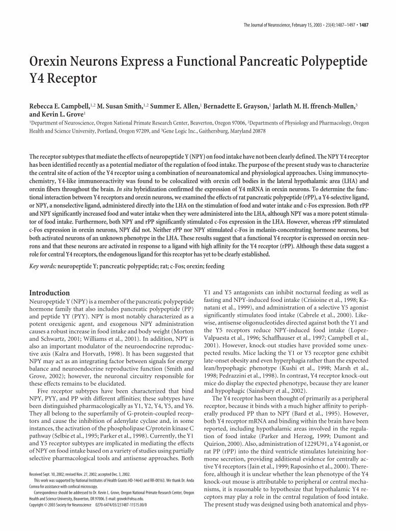
Figure 2. Correlation of Y4 mRNA and protein in central and peripheral tissues. Y4 mRNA was found by RT-PCR in the hypothalamus (HYP) and cortex (CTX ) ( A) and throughout peripheralstructures ( B ), including kidney (KID), liver (LIV ), duodenum (DUO), large intestine (LGI ), stomach (STO), and lung (LNG ). Western blot analysis shows Y4 protein in membrane ( M ) fractions of thehypothalamus and cortex (C ) and in all peripheral structures (D), but not in the soluble (S) fraction. Y4-IR was identified in cells of the hypothalamus (E ), the cortex (F ), and throughout peripheraltissues; the duodenum is shown as an example (G ).
1490 • J. Neurosci., February 15, 2003 • 23(4):1487–1497 Campbell et al. • Y4 Receptor Activates Orexin Neurons

ResultsY4 distributionAnalysis of the single-label distribution of chromagen Y4-IRdemonstrated low levels of staining throughout the hypothala-mus (Fig. 1A). Similar distribution patterns were observed inmale and female rats. The majority of staining appeared to be insmall, round cells. Unexpectedly, the most intensely labeledY4-IR cell bodies were found in the larger cells of the LHA (Fig.1A,B). Densely labeled Y4-IR cells were apparent throughout therostral-to-caudal extent of the LHA.
To provide additional characterization of the Y4 antibody andY4 distribution in both central and peripheral structures, we ex-amined the correlation of Y4 mRNA and protein expression us-ing PCR, Western blot analysis, and immunocytochemistry. Cen-trally, Y4 mRNA was present in both the hypothalamus and thecortex (Fig. 2A). In peripheral structures, all of the areas investi-gated, including the kidney, liver, duodenum, large intestine, co-lon, stomach, and lung, expressed Y4 mRNA (Fig. 2B). By West-ern blot analysis, the Y4 antibody identified Y4 protein withinmembrane extracts of both the hypothalamus and the cortex (Fig.2C) and all of the peripheral tissues tested (Fig. 2D). Finally, byimmunocytochemistry, Y4-IR was observed in both the hypo-thalamus and the cortex. The Y4 antibody also identified Y4-IR inperipheral tissues. Figure 2G shows an example of IF labeling inthe duodenum in which Y4-IR was observed within goblet cellsand along the basal lamina of intestinal villi. Collectively, thesedata suggest that the Y4 antibody recognizes the membrane-bound Y4 receptor in both peripheral and central tissues.
Y4 expression in orexin neuronsDouble-label IF experiments indicated that a majority of neuronsdisplaying Y4-IR observed in the LHA (Fig. 3A, green) were colo-
calized with orexin, whose cell bodies are also entirely restrictedto this region (Fig. 3B, red). Although there was dense overlap ofthese proteins in cell bodies, as shown in yellow, there were only afew orexin fibers that colocalized the Y4 receptor in this region(Fig. 3C). Y4-IR was also observed in sparse fibers throughout thehypothalamus (Fig. 3D, green), and all of these fibers wereidentified as colocalized with orexin (Fig. 3F, yellow). Double-labeling of Y4 mRNA by in situ hybridization and orexin bychromagen immunocytochemistry provided additional confir-mation for the expression of Y4 receptors on orexin neurons.Silver grain clusters are clearly colocalized with orexin neurons inthe LHA (Fig. 4A–D). However, there are also a number of non-orexin neurons in the LHA that express Y4 mRNA.
Y1 receptor subtype expression in the LHAAn additional double-label IF study investigated the expressionpattern of the NPY Y1 receptor subtype within the LHA and inrelation to orexin neurons. Y1-IR (Fig. 5A–D, green) was ob-served on numerous fibers and some cell bodies ( yellow arrows)within the LHA. Many Y1-IR fibers surrounded orexin neurons(red) and appeared to make close contact ( yellow), as indicatedby white arrows (Fig. 5A–D); however, Y1-IR was not colocalizedwith orexin neurons or fibers.
Stimulatory effects of rPP and NPY injected into the LHA
Food and water intakeBoth NPY and rPP caused a significant increase in food and waterintake in male rats (Fig. 6). NPY elicited a robust feeding re-sponse, with 3.06 � 0.23 and 4.69 � 0.71 gm of cumulative foodintake at 1 and 3 hr time points. rPP caused a more moderatefood-intake response, with 1.05 � 0.23 and 2.60 � 0.33 gm of
Figure 3. NPY Y4-IR colocalizes with orexin in the LHA. Double-label IF for Y4 (A, D, green) and orexin (B, E, red) is shown in LHA cell bodies ( A–C ) and a representative fiber in the paraventricularnucleus of the hypothalamus (PVH; D–F ). Their colocalization is indicated in yellow (C, F ). Arrows in F indicate orexin/Y4 double-label fibers.
Campbell et al. • Y4 Receptor Activates Orexin Neurons J. Neurosci., February 15, 2003 • 23(4):1487–1497 • 1491

cumulative food intake at 1 and 3 hr time points. Food intakestimulated by rPP was significantly greater than vehicle ( p � 0.01at 1 hr; p � 0.001 at 3 hr) and significantly less than that stimu-lated by NPY ( p � 0.001 at 1 hr; p � 0.01 at 3 hr) (Fig. 6A,B).Vehicle-injected controls did not consume any food during thisperiod, as would be expected during daylight hours. Althoughcontrols consumed a small amount of water, NPY and rPP wereequally effective at significantly stimulating water intake at both 1and 3 hr time points ( p � 0.05) (Fig. 6C,D).
Induction of c-Fos expressionc-Fos patterns were examined 3 hr after injections of either rPP orNPY. It is well established that c-Fos expression remains stablefor �3 hr after a stimulus (Hoffman et al., 1992). The resultsshowed that c-Fos expression was specifically induced only on theipsilateral side to the injection site (Fig. 7A–C). Both rPP andNPY stimulated c-Fos expression on the ipsilateral side, whereasvehicle injections caused a low level of c-Fos induction directlyadjacent to the injection site (Fig. 7A–C). On the contralateralside to the injection site, extremely low levels of c-Fos were ob-served in all groups (Fig. 7D–F). The lack of c-Fos activation onthe contralateral side provides strong evidence that c-Fos expres-sion on the treatment side was specific to the drugs and not
secondary to the induction of physiological responses, such asingestion of food. Any c-Fos expression resulting from food in-take would be expected to occur bilaterally. However, we cannoteliminate the possibility that neurons are being activated by acombination of the stimulus from the peptides and the ingestedmeal. Furthermore, confinement of c-Fos expression to the treat-ment side indicates that the peptides were not diffusing to thethird ventricle and causing responses via activation of some siteremote from the LHA.
Three hours after rPP injection, there was a significant induc-tion of c-Fos expression in orexin neurons on the ipsilateral side(59.3 � 8.5%; p � 0.001 compared with vehicle treatment) (Figs.8, 9). In contrast, NPY did not significantly activate c-Fos expres-sion in orexin cells when compared with vehicle-treated animals(18.5 � 4.7 and 21.7 � 5.4%, respectively) (Figs. 8, 9), althoughthere were numerous c-Fos-IR cells of unknown phenotype inthe area. On the contralateral side, very few orexin neurons dis-played c-Fos expression in any of the treatment groups (Fig. 8B).
The number of c-Fos-positive MCH neurons was extremelylow and was not significantly different among the treatmentgroups on either the ipsilateral or the contralateral side (Fig.10A,B). The number of ipsilateral MCH cells displaying c-Fos
Figure 4. Y4 mRNA is coexpressed in orexin neurons. Representative photomicrographs show Y4 mRNA at low ( A) and high ( B–D) power identified by silver grain clusters ( purple/black)throughout the LHA and double-labeled with orexin neurons (brown).
1492 • J. Neurosci., February 15, 2003 • 23(4):1487–1497 Campbell et al. • Y4 Receptor Activates Orexin Neurons

represents only a small percentage (�1%)of the number of MCH neurons analyzedper animal: 10.80 � 4.0, 12.29 � 4.43, and9.33 � 4.68 from vehicle-, rPP-, and NPY-injected animals, respectively.
DiscussionThe present study demonstrates for thefirst time that orexin neurons in the LHAcoexpress the NPY Y4 receptor subtypeand are activated by a Y4-specific ligand,rPP. Using a newly characterized NPY Y4antibody, a low level of Y4-IR was observedin small, round cells throughout the hypo-thalamus. However, the most intensely la-beled Y4-IR cell bodies in the hypothala-mus were observed in the LHA (Fig. 1A,B).The overall pattern of Y4-IR was in agree-ment with the previously reported in situhybridization pattern of Y4 mRNA (Parkerand Herzog, 1999) and the pattern of Y4receptor binding (Dumont and Quirion,2000). Additional characterization of theY4 antibody demonstrated a complete cor-relation between Y4 mRNA, as detected byRT-PCR, and protein expression, as de-tected by Western blot analysis and im-munocytochemistry, in both central andperipheral regions (Fig. 2 A–G), support-ing the specificity and sensitivity of thisantibody.
Interestingly, immunocytochemistry themost intense Y4-IR observed in the LHAwas colocalized with orexin neurons (Fig.3). The colocalization of Y4 mRNA expres-sion in orexin neurons supports this find-
ing (Fig. 4); however, Y4 mRNA was also expressed in a numberof non-orexin-expressing cells. Orexins have been found to reg-ulate arousal states, to influence ingestion (both drinking andfeeding), and to play a role in the modulation of neuroendocrineprocesses, including luteinizing hormone release and thermoreg-ulation (for review, see Beuckmann and Yanagisawa, 2002; Saku-rai, 2002; Sutcliffe and de Lecea, 2002). Central injection oforexin A stimulates food intake in satiated rats (Dube et al., 1999;Haynes et al., 1999), and orexin mRNA is upregulated in responseto fasting (Sakurai et al., 1998). However, although orexin Aincreases daytime feeding, overall 24 hr food intake is not in-creased, and repeated injections have no effect on body weight(Yamanaka et al., 1999). Therefore, orexin may be important inshort-term feeding episodes or in the initiation of feeding, but it isnot necessarily implicated in obesity. Alternatively, numerous stud-ies have shown that orexin is a key signal in arousal-associated be-havior (Sutcliffe et al., 2002). Thus, the orexin system might be re-sponsible for the complex coordination of behavioral andphysiological responses to an animal’s nutritional needs. De-termining the neuroanatomical pathways that influenceorexin activity will aid in the understanding of the relativeimportance of orexin in these physiological roles.
Although the neuroanatomical results presented abovestrongly support a direct role of the Y4 receptor on orexin neu-rons, this needed to be directly tested. Injection of rPP, a selectiveY4 receptor ligand, directly into the LHA of freely moving rats
Figure 5. NPY Y1-IR on fibers and cells of an unknown phenotype in the LHA. Double-label IF for Y1 ( green) and orexin(red) in the LHA ( A–D) shows Y1-IR on non-orexin cells ( yellow arrow) and on fibers surrounding orexin neurons. Y1-IR isnot colocalized with orexin neurons, but Y1-IR fibers appear to make close contact with orexin neurons (white arrows).
Figure 6. Intra-LHA administration of NPY and rPP stimulates food and water intake.Mean � SEM cumulative food intake and water intake 1 and 3 hr after intra-LHA injection ofvehicle (open bars), NPY (hatched bars), or rPP ( filled bars); n � 10 –11 per group. NPY causeda significant increase in food intake compared with both vehicle- and rPP-injected animals (**p �0.01; ***p � 0.001). rPP significantly increased food intake compared with vehicle (***p � 0.001).Both NPY and rPP significantly increased water intake compared with vehicle (*p � 0.05).
Campbell et al. • Y4 Receptor Activates Orexin Neurons J. Neurosci., February 15, 2003 • 23(4):1487–1497 • 1493

elicited a robust dipsogenic response and a moderate food-intakeresponse (Fig. 5). These results resemble previously reported re-sponses after orexin administration. Orexin A, administered in-tracerebroventricularly, increases water intake at a level compa-rable with angiotensin II, and deprivation of water upregulatesorexin mRNA (Kunii et al., 1999). As mentioned above, orexinhas been implicated in the regulation of food intake; however,orexin is a much less potent orexigenic agent than NPY (Edwardset al., 1999). Similar to other studies (Stanley et al., 1993), thepresent study demonstrated a robust increase in both food andwater intake after NPY injection into the LHA.
The differences between the abilities of NPY and the rPP toinduce food and water intake suggest that these peptides may beactivating different neurocircuits in eliciting ingestive behavior.
One factor that may contribute to the different responses is thegreatly different affinities of NPY or rPP for the various NPYreceptors. Whereas NPY has a relatively high affinity for the Y1,Y2, and Y5 receptors, rPP has a relatively low affinity for thesereceptors but has a 200-fold greater affinity for the Y4 receptor(Bard et al., 1995; Schober et al., 2000). The differences in c-Fosactivation of specific neuronal populations within the LHA alsosupport the idea that rPP and NPY may be activating differentneurocircuits in the induction of food intake. Although both rPPand NPY elicited significant food intake and stimulated a robustinduction of c-Fos expression in the area of the injection site, onlyrPP significantly induced c-Fos in orexin neurons. Althoughthese findings alone do not demonstrate whether the effects ofrPP are direct on orexin neurons, if they are coupled with theneuroanatomical evidence of Y4 protein and mRNA colocaliza-tion in orexin neurons, they strongly support this conclusion.However, we cannot eliminate the possibility that the stimulationof food intake by rPP occurs via actions on other populations ofneurons in the LHA that then activate orexin neurons. AlthoughrPP did not stimulate c-Fos expression in MCH neurons, it didactivate a population of LHA neurons distinct from orexin. Fur-thermore, whereas only low levels of Y4-IR were observed innon-orexin neurons within the LHA, an abundance of Y4 mRNAwas detected in non-orexin cells throughout this area (Fig. 4).These data suggest either that the Y4 antibody may not be sensi-tive enough to detect low levels of Y4 protein or that Y4 mRNA inthese cells may not be translated at a high rate under the presentconditions. Additional studies are necessary to identify the phe-notypes of this population of LHA neurons, and it remains to bedetermined whether rPP stimulates food and water intake di-rectly via orexin neurons or via this unidentified population.
Figure 7. Single-label c-Fos expression on ipsilateral and contralateral sides of intra-LHA injections. Representative photomicrographs show c-Fos expression in the LHA on the ipsilateral ( A–C )and contralateral ( D–F ) side of the injection site from animals receiving vehicle (A, D), rPP (B, E ), or NPY (C, F ). The black arrowhead indicates the ventral extent of the guide cannula. The injectorcannula extended 2 mm beyond the guide cannula. The images were taken at the approximate AP coordinates�3.5 according to the rat brain atlas of Paxinos and Watson (1998). fx, Fornix; 3V, thirdventricle.
Figure 8. Intra-LHA rPP, but not NPY, induces a robust increase in c-Fos expression in orexinneurons. Mean � SEM percentage of orexin cells displaying c-Fos 3 hr after intra-LHA injectionof vehicle (open bars), NPY (hatched bars), or rPP ( filled bars); n � 8 –10 per group. rPP injec-tion caused a robust induction of c-Fos expression ipsilateral to the injection site compared withvehicle and NPY ( A) (*p � 0.05). Very low levels of c-Fos expression were seen in orexinneurons contralateral to the injection site ( B) in all treatment groups.
1494 • J. Neurosci., February 15, 2003 • 23(4):1487–1497 Campbell et al. • Y4 Receptor Activates Orexin Neurons

Surprisingly, NPY injection into the LHA did not significantlystimulate c-Fos in either orexin or MCH neurons, although c-Fosexpression was stimulated in the LHA in a population of neuronswith an unknown phenotype. This is in contrast to previous stud-ies showing that NPY injection into the third ventricle causes theinduction of c-Fos in a small percentage of orexin neurons (Niimiet al., 2001). This discrepancy is most likely attributable to themode of administration. Injections into the third ventricle mayactivate a number of other systems that project to the LHA. Thus,the induction of c-Fos observed may be a second- or third-orderevent. The lack of significant c-Fos induction in MCH and orexinneurons was particularly unexpected considering the abundanceof NPY terminals that are in contact with orexin and MCH neu-rons (Broberger et al., 1998; Horvath et al., 1999). Reciprocalconnections of orexin with NPY and �-melanin-stimulating hor-
mone neurons in the hypothalamic arcuate nucleus also havebeen described previously (Elias et al., 1998), and it has beensuggested that these are key circuits in the maintenance of food-intake signals. However, NPY projections to the LHA may beworking through other NPY receptor subtypes, either presynap-tically or postsynaptically on different phenotypes of neurons.The lack of c-Fos induction in orexin and MCH neurons is sup-ported by the lack of Y1 receptor expression on orexin (Fig. 4)and MCH neurons (our unpublished observations). Y1-IR wasfound on fibers and cells of an unknown phenotype dispersedthroughout the LHA. Additionally, Y1-IR fibers were found inclose contact with orexin and MCH neurons, suggesting presyn-aptic NPY effects via the Y1 receptor. Future studies will attemptto identify the phenotypes of neurons expressing the Y1 receptor.The Y5 receptor also has been identified in the LHA (Campbell etal., 2001), although the phenotype of cells expressing the Y5 re-ceptor subtype also remains to be determined. In addition toMCH and orexin, the LHA is known to contain large numbers ofglucose-receptive neurons (Bernardis and Bellinger, 1996) of un-known phenotypes, as well as the orexigenic neuropeptide gala-nin (Melander et al., 1986). What is clear from these studies isthat orexin and MCH neurons do not express the Y1 receptor anddo not express c-Fos in response to NPY administered directly tothe LHA.
Although previous reports clearly illustrate the activation ofcentral Y4-like receptors (Jain et al., 1999; Raposinho et al., 2000),and the present study demonstrates physiological and c-Fos re-sponses to the peripheral Y4 agonist rPP, an endogenous ligandfor the centrally expressed Y4 receptor has not yet been defini-tively characterized. PP, the only known endogenous high-affinity ligand for the Y4 receptor, is produced in the islet cells of
Figure 9. Orexin neurons and c-Fos expression after intra-LHA injections of vehicle, rPP, or NPY. Representative images show orexin-IR ( green, A–C ) and c-Fos-IR (black, D–F ) in identical areasin the LHA from animals injected with vehicle (VEH; A, D), rPP (B, E ), or NPY (C, F ). The yellow arrowheads in B represent c-Fos colocalization with orexin-IR neurons and in E represent the same cellsshowing only c-Fos-IR. There was no c-Fos colocalization in the orexin cells shown in A (vehicle treated) or in C (NPY treated). The images were taken at the approximate AP coordinates �3.6according the rat brain atlas of Paxinos and Watson (1998).
Figure 10. MCH neurons do not express c-Fos in response to intra-LHA rPP or NPY. Mean �SEM number of MCH cells displaying c-Fos 3 hr after intra-LHA injection of vehicle (open bars),NPY (hatched bars), or rPP ( filed bars); n � 8 –10 per group. Very few MCH neurons on eitherside of the injection site displayed c-Fos (A, B).
Campbell et al. • Y4 Receptor Activates Orexin Neurons J. Neurosci., February 15, 2003 • 23(4):1487–1497 • 1495

the endocrine pancreas. Peripherally produced PP, however,does not appear to readily cross the blood– brain barrier. Earlystudies identified PP-like IR in the brain (Loren et al., 1979; Lund-berg et al., 1980; Olschowka et al., 1981; Inui et al., 1985); how-ever, later studies were not able to detect PP-like peptide in thebrain (Miyazaki and Funakoshi, 1988), leading to the view thatthe antibodies used in previous studies were probably cross-reacting with NPY or some other substance. Furthermore, Pieri-bone et al. (1992) also showed the lack of PP mRNA in the brain,using in situ hybridization with oligo probes specific for rPP. Incontrast, recent preliminary studies in our laboratory have iden-tified rPP mRNA in the hypothalamus and brainstem using RT-PCR (our unpublished observations), raising the possibility thatPP or a PP-like ligand for the Y4 receptor may be present in thebrain. Additional studies are required to confirm the presence ofthe Y4 ligand mRNA and protein in the brain as well as theanatomical localization of the PP-expressing cells. Armed withthis neuroanatomical information, it then will be possible tostudy the importance of the central rPP–Y4 system in the regula-tion of ingestive behavior.
ReferencesBard JA, Walker MW, Branchek TA, Weinshank RL (1995) Cloning and func-
tional expression of a human Y4 subtype receptor for pancreatic polypeptide,neuropeptide Y, and peptide YY. J Biol Chem 270:26762–26765.
Bernardis LL, Bellinger LL (1996) The lateral hypothalamic area revisited:ingestive behavior. Neurosci Biobehav Rev 20:189 –287.
Beuckmann CT, Yanagisawa M (2002) Orexins: from neuropeptides to en-ergy homeostatis and sleep/wake regulation. J Mol Med 80:329 –342.
Broberger C, De Lecea L, Sutcliffe JG, Hokfelt T (1998) Hypocretin/orexin-and melanin-concentrating hormone-expressing cells form distinct popula-tions in the rodent lateral hypothalamus: relationship to the neuropeptide Yand agouti gene-related protein systems. J Comp Neurol 402:460–474.
Brogan RS, Grove KL, Smith MS (2000) Differential regulation of leptinreceptor but not orexin in the hypothalamus of the lactating rat. J Neu-roendocrinol 12:1077–1086.
Brown CM, Coscina DV, Fletcher PJ (2000) The rewarding properties ofneuropeptide Y in perifornical hypothalamus vs. nucleus accumbens.Peptides 21:1279 –1287.
Cabrele C, Langer M, Bader R, Wieland HA, Doods HN, Zerbe O, Beck-Sickinger AG (2000) The first selective agonist for the neuropeptide YY5receptor increases food intake in rats. J Biol Chem 275:36043–36048.
Campbell RE, ffrench-Mullen JM, Cowley MA, Smith MS, Grove KL (2001)Hypothalamic circuitry of neuropeptide Y regulation of neuroendocrinefunction and food intake via the Y5 receptor subtype. Neuroendocrinol-ogy 74:106 –119.
Crisioine L, Gigollier P, Batzl-Hartman C, Rueger H, Stricker-Krongrad A,Wyss P, Brunner L, Whitebread S, Yamaguchi Y, Gerald C, Heurich RO,Walker MW, Chiesi M, Schilling W, Hofbauer KG, Levens N (1998)Food intake in free-feeding and energy deprived lean rats is mediated bythe neuropeptide Y Y5 receptor. J Clin Invest 102:2136 –2145.
Currie PJ, Coscina DV (1995) Dissociated feeding and hypothermia effectsof neuropeptide Y in the paraventricular and perifornical hypothalamus.Peptides 16:599 – 604.
Dube MG, Kalra SP, Kalra PS (1999) Food intake elicited by central admin-istration of orexins/hypocretins: identification of hypothalamic sites ofaction. Brain Res 842:473– 477.
Dumont Y, Quirion R (2000) [(125)I]-GR231118: a high affinity radioli-gand to investigate neuropeptide Y, Y(1), and Y(4) receptors. Br J Phar-macol 129:37– 46.
Edwards CM, Abusnana S, Sunter D, Murphy KG, Ghatei MA, Bloom SR(1999) The effect of the orexins on food intake: comparison with neu-ropeptide Y, melanin-concentrating hormone and galanin. J Endocrinol160:7–12.
Elias CF, Saper CB, Maratos-Flier E, Tritos NA, Lee C, Kelly J, Tatro JB,Hoffman GE, Ollmann MM, Barsh GS, Sakurai T, Yanagisawa M,Elmquist JK (1998) Chemically defined projections linking the medio-basal hypothalamus and the lateral hypothalamic area. J Comp Neurol402:442– 459.
Grove KL, Smith MS (1998) Resistance of the hippocampus in the lactatingrat to N-methyl-D-aspartate (NMDA)-mediated excitation is not due to anonfunctional receptor system. Brain Res 814:157–163.
Haynes AC, Jackson B, Overend P, Buckingham RE, Wilson S, Tadayyon M,Arch JRS (1999) Effects of single and chronic intracerebroventricularadministration of the orexins on feeding in the rat. Peptides 20:1099 –1105.
Hoffman GE, Smith MS, Fitsimmons MD (1992) Detecting steroidal effectson immediate early gene expression in the hypothalamus. Neuroproto-cols 1:52– 66.
Horvath TL, Diano S, van den Pol AN (1999) Synaptic interaction betweenhypocretin (orexin) and neuropeptide Y cells in the rodent and primatehypothalamus: a novel circuit implicated in metabolic and endocrineregulations. J Neurosci 19:1072–1087.
Inui A, Mizuno N, Ooya M, Suenaga K, Morioka H, Ogawa T, Ishida M, BabaS (1985) Cross-reactivities of neuropeptide Y and peptide YY with pan-creatic polypeptide antisera: evidence for the existence of pancreaticpolypeptide in the brain. Brain Res 330:386 –389.
Jain MR, Pu S, Kalra PS, Kalra SP (1999) Evidence that stimulation of twomodalities of pituitary luteinizing hormone release in ovarian steroid-primed ovariectomized rats may involve neuropeptide Y, Y1, and Y4receptors. Endocrinology 140:5171–5177.
Kalra SP, Horvath TL (1998) Neuroendocrine interactions between galanin,opioids, and neuropeptide Y in the control of reproduction and appetite.Ann NY Acad Sci 863:236 –240.
Kanatani A, Kanno T, Ishihara A, Hata M, Sakuraba A, Tanaka T, Tsuchiya Y,Mase T, Fukuroda T, Fukami T, Ihara M (1999) The novel neuropeptideY Y(1) receptor antagonist J-104870: a potent feeding suppressant withoral bioavailability. Biochem Biophys Res Commun 266:88 –91.
Katsuura G, Asakawa A, Inui A (2002) Roles of pancreatic polypeptide inregulation of food intake. Peptides 23:323–329.
Kunii K, Yamanaka A, Nambu T, Matsuzaki I, Goto K, Sakurai T (1999) Orex-ins/hypocretins regulate drinking behaviour. Brain Res 842:256–261.
Kushi A, Sasai H, Koizumi H, Takeda N, Yokoyama M, Nakamura M (1998)Obesity and mild hyperinsulinemia found in neuropeptide Y-Y1receptor-deficient mice. Proc Natl Acad Sci USA 95:15659 –15664.
Li C, Chen P, Smith MS (1999) Morphological evidence for direct interac-tion between arcuate nucleus neuropeptide Y (NPY) neurons andgonadotropin-releasing hormone neurons and the possible involvementof NPY Y1 receptors. Endocrinology 140:5382–5390.
Li C, Chen P, Smith MS (2000) Corticotropin releasing hormone (CRH)neurons in the paraventricular nucleus (PVH) are direct targets for neu-ropeptide Y (NPY) neurons in the arcuate nucleus (ARH): an anterogradetracing study. Brain Res 854:122–129.
Lopez-Valpuesta FJ, Nyce JW, Myers RD (1996) NPY-Y1 receptor antisenseinjected centrally in rats causes hyperthermia and feeding. NeuroReport7:2781–2784.
Loren I, Alumets J, Hakanson R, Sundler F (1979) Immunoreactive pancreaticpolypeptide (PP) occurs in the central and peripheral nervous system: pre-liminary immunocytochemical observations. Cell Tissue Res 200:179–186.
Lundberg JM, Hokfelt T, Anggard A, Kimmel J, Goldstein M, Markey K(1980) Coexistence of an avian pancreatic polypeptide (APP) immuno-reactive substance and catecholamine in some peripheral and central neu-rons. Acta Physiol Scand 110:107–109.
Marsh DJ, Hollopeter G, Kafer KE, Palmiter RD (1998) Role of the Y5 neu-ropeptide Y receptor in feeding and obesity. Nat Med 4:718 –721.
Melander T, Hokfelt T, Rokaeus A (1986) Distribution of galaninlike immu-noreactivity in the rat central nervous system. J Comp Neurol 248:475–517.
Miyazaki K, Funakoshi A (1988) A distribution of pancreatic polypeptide-like immunoreactivity in rat tissues. Regul Pept 21:37– 43.
Morton GJ, Schwartz MW (2001) The NPY/AgRP neuron and energy ho-meostasis. Int J Obes Relat Metab Disord 5:56 – 62.
Nakajima M, Inui A, Teranishi A, Miura M, Hirosue Y, Okita M, Himori N,Baba S, Kasuga M (1994) Effects of pancreatic polypeptide family pep-tides on feeding and learning behavior in mice. J Pharmacol Exp Ther268:1010 –1014.
Niimi M, Sato M, Taminato T (2001) Neuropeptide Y in central control offeeding and interactions with orexin and leptin. Endocrine 14:269 –273.
Olschowka JA, O’Donohue TL, Jacobowitz DM (1981) The distribution ofbovine pancreatic polypeptide-like immunoreactive neurons in rat brain.Peptides 2:309 –331.
1496 • J. Neurosci., February 15, 2003 • 23(4):1487–1497 Campbell et al. • Y4 Receptor Activates Orexin Neurons

Parker RMC, Herzog H (1999) Regional distribution of Y-receptor subtypemRNAs in rat brain. Eur J Neurosci 11:1431–1448.
Parker SL, Parker MS, Sweatman T, Crowley WF (1998) Characterization ofG protein and phospholipase C-coupled agonist binding to the Y1 neu-ropeptide Y receptor in the rat brain: sensitivity to G protein activatorsand inhibitors and to inhibitors of phospholipase C. J Pharmacol ExpTher 286:382–391.
Paxinos G, Watson C (1998) The rat brain in stereotaxic coordinates. NewYork: Academic.
Pedrazzini T, Seydoux J, Kunstner P, Aubert J-F, Grouzmann E, Beermann F,Brunner HR (1998) Cardiovascular response, feeding behavior and lo-comotor activity in mice lacking the NPY Y1 receptor. Nat Med4:722–726.
Pieribone VA, Brodin L, Friberg K, Dahlstrand J, Soderberg C, Larhammar D,Hokfelt T (1992) Differential expression of mRNAs for neuropeptideY-related peptides in rat nervous tissue: possible evolutionary conserva-tion. J Neurosci 12:3361–3371.
Raposinho PD, Broqua P, Hayward A, Akinsanya K, Galyean R, SchteingartC, Junien J, Aubert ML (2000) Stimulation of the gonadotropic axis bythe neuropeptide Y receptor Y1 antagonist/Y4 agonist 1229U91 in themale rat. Neuroendocrinology 71:2–7.
Sainsbury A, Schwarzer C, Couzens M, Jenkins A, Oakes SR, Ormandy CJ,Herzog H (2002) Y4 receptor knockout rescues fertility in ob/ob mice.Genes Dev 16:1077–1088.
Sakurai T (2002) Roles of orexin in regulation of feeding and wakefulness.NeuroReport 13:987–995.
Sakurai T, Amemiya A, Ishii M, Matsuzaki I, Chemelli RM, Tanaka H, Wil-liams SC, Richardson JA, Kozlowski GP, Wilson S, Arch JR, BuckinghamRE, Haynes AC, Carr SA, Annan RS, McNulty DE, Liu WS, Terrett JA,
Elshourbagy NA, Bergsma DJ, et al. (1998) Orexins and orexin recep-tors: a family of hypothalamic neuropeptides and G protein-coupled re-ceptors that regulate feeding behavior. Cell 92:573–585.
Schaffhauser AO, Stricker-Krongrad A, Brunner L, Cumin F, Gerald C,Whitebread S, Criscione L, Hofbauer KG (1997) Inhibition of food in-take by neuropeptide Y Y5 receptor antisense oligodeoxynucleotides. Di-abetes 46:1792–1798.
Schober DA, Gackenheimer SL, Heiman ML, Gehlert DR (2000) Pharmaco-logical characterization of 125I–1229U91 binding to Y1 and Y4 neuropeptideY/peptide YY receptors. J Pharmacol Exp Ther 2000 293:275–280.
Selbie LA, Darby K, Schmitz-Peiffer C, Browne CL, Herzog H, Shine J, BidenTJ (1995) Synergistic interaction of Y1-neuropeptide Y and alpha 1�-adrenergic receptors in the regulation of phospholipase C, protein kinaseC, and arachidonic acid production. J Biol Chem 270:11789 –11796.
Smith MS, Grove KL (2002) Integration of the regulation of reproductivefunction and energy balance: lactation as a model. Front Neuroendocri-nol 23:225–256.
Stanley BG, Magdalin W, Seirafi A, Thomas WJ, Leibowitz SF (1993) Theperifornical area: the major focus of (a) patchily distributed hypothalamicneuropeptide Y-sensitive feeding system(s). Brain Res 604:304 –317.
Sutcliffe JG, de Lecea L (2002) The hypocretins: setting the arousal thresh-old. Nat Rev Neurosci 3:339 –349.
Williams G, Bing C, Cai XJ, Harrold JA, King PJ, Liu XH (2001) The hypo-thalamus and the control of energy homeostasis: different circuits, differ-ent purposes. Physiol Behav 74:683–701.
Yamanaka A, Sakurai T, Katsumoto T, Yanagisawa M, Goto K (1999)Chronic intracerebroventricular administration of orexin-A to rats in-creases food intake in daytime, but has no effect on body weight. Brain Res849:248 –252.
Campbell et al. • Y4 Receptor Activates Orexin Neurons J. Neurosci., February 15, 2003 • 23(4):1487–1497 • 1497