organ pathology: inquiry, inspection, palpation,...
TRANSCRIPT

Examination of the patients with respiratory
organ pathology: inquiry, inspection,
palpation, percussion
Head of the Propedeutics
to Internal Medicine Department N1,
Basis of Bioethics and Biosafety
Professor T. Ashcheulova
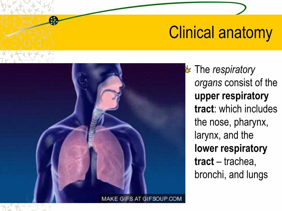
Clinical anatomy
The respiratory
organs consist of the
upper respiratory
tract: which includes
the nose, pharynx,
larynx, and the
lower respiratory
tract – trachea,
bronchi, and lungs

Topographic regions and lines of the
chest To describe an abnormality on the
chest, you need to locate it in two
dimensions: along vertical axis and
around the circumference of the chest.
To locate vertically, you
must be able to number
the ribs and interspaces
accurately
Note that an interspace
between two ribs is
numbered by the rib
above it.

Topographic regions and lines of the
chest Anterior view As the 1st rib is covered by clavicle, the 1st
interspace is below it. From here, using two
fingers, you can “walk down the interspaces”.
Do not try to count interspaces along the
lower edge of the sternum; the ribs here are
too close together. Note that the costal
cartilages of only the first seven ribs
articulate with the sternum. Those of the 8th,
9th, and 10th ribs articulate instead with the
costal cartilages just above them. The 11th
and 12th ribs, the so-called floating ribs, have
no anterior attachments. The cartilaginous tip
of the 11th rib can usually be felt laterally,
and the 12th rib may be felt posteriorly.
Costal cartilages are not distinguishable from
ribs by palpation.
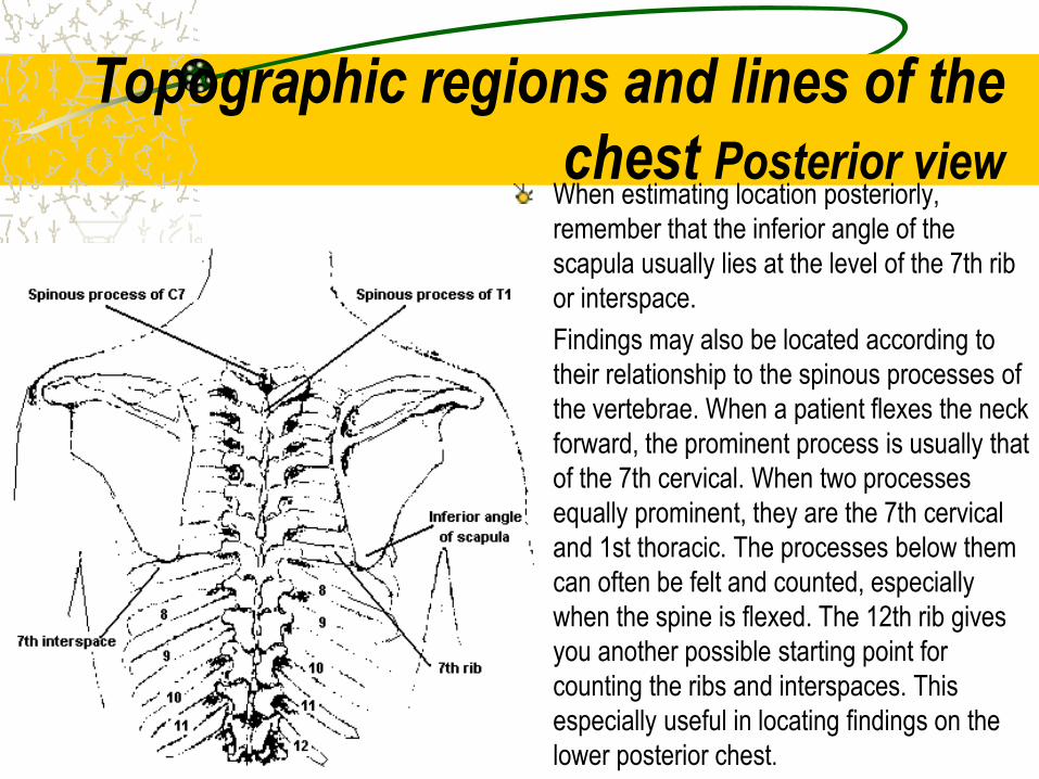
Topographic regions and lines of the
chest Posterior view When estimating location posteriorly,
remember that the inferior angle of the
scapula usually lies at the level of the 7th rib
or interspace.
Findings may also be located according to
their relationship to the spinous processes of
the vertebrae. When a patient flexes the neck
forward, the prominent process is usually that
of the 7th cervical. When two processes
equally prominent, they are the 7th cervical
and 1st thoracic. The processes below them
can often be felt and counted, especially
when the spine is flexed. The 12th rib gives
you another possible starting point for
counting the ribs and interspaces. This
especially useful in locating findings on the
lower posterior chest.
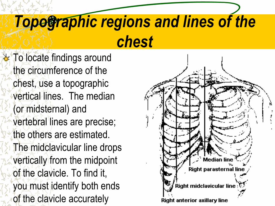
Topographic regions and lines of the
chest To locate findings around
the circumference of the
chest, use a topographic
vertical lines. The median
(or midsternal) and
vertebral lines are precise;
the others are estimated.
The midclavicular line drops
vertically from the midpoint
of the clavicle. To find it,
you must identify both ends
of the clavicle accurately
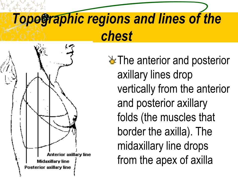
Topographic regions and lines of the
chest
The anterior and posterior
axillary lines drop
vertically from the anterior
and posterior axillary
folds (the muscles that
border the axilla). The
midaxillary line drops
from the apex of axilla
Topographic regions and lines of the
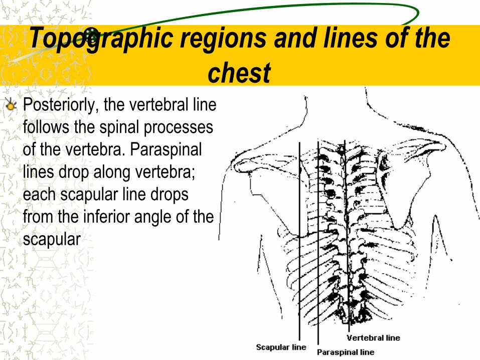
chest Posteriorly, the vertebral line
follows the spinal processes
of the vertebra. Paraspinal
lines drop along vertebra;
each scapular line drops
from the inferior angle of the
scapular

Topographic regions and lines of the
chest
The lungs lobes and fissures
can be outlined mentally on
the chest wall. Anteriorly, the
apex of the each lung rises
about 2 cm to 4 cm above
the inner third of the clavicle.
The lower border of the lung
passes the 6th rib at the
midclavicular line and 8th rib
at the midaxillary line.
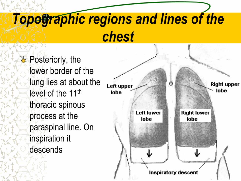
Topographic regions and lines of the
chest
Posteriorly, the
lower border of the
lung lies at about the
level of the 11th
thoracic spinous
process at the
paraspinal line. On
inspiration it
descends
Topographic regions and lines of the
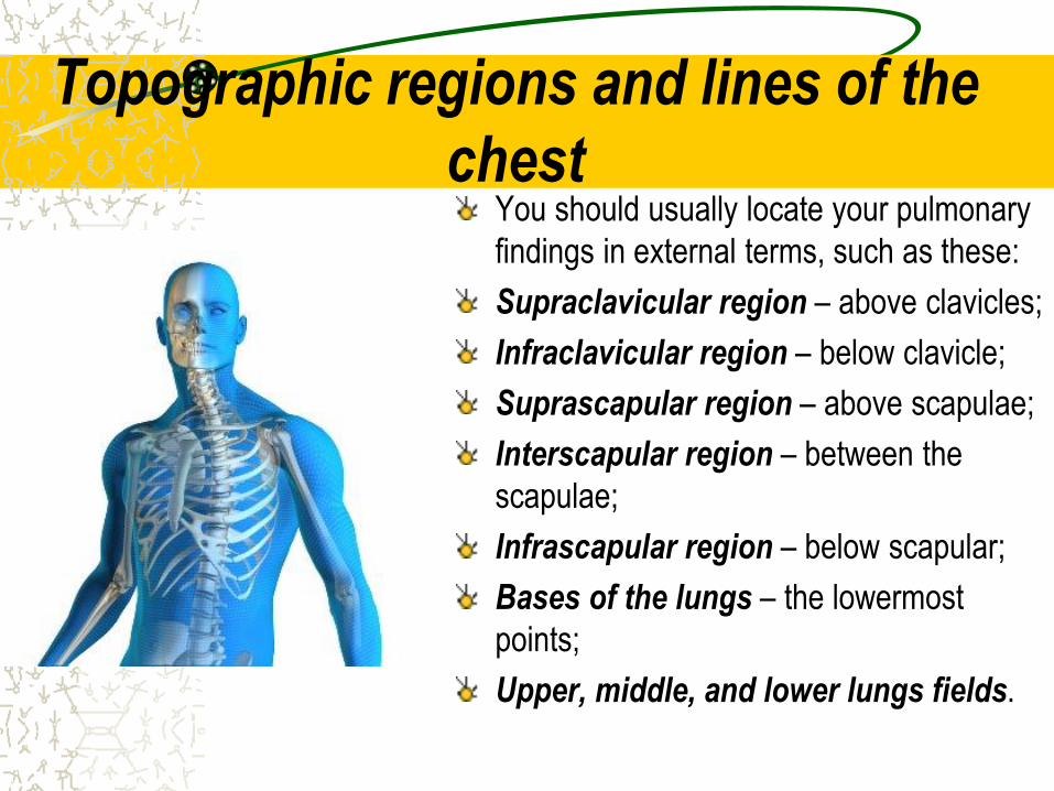
chest You should usually locate your pulmonary
findings in external terms, such as these:
Supraclavicular region – above clavicles;
Infraclavicular region – below clavicle;
Suprascapular region – above scapulae;
Interscapular region – between the
scapulae;
Infrascapular region – below scapular;
Bases of the lungs – the lowermost
points;
Upper, middle, and lower lungs fields.

THE BASIC METHODS OF
RESPIRATORY SYSTEM EXAMINATION I. Subjective
Inquiring;
Anamnesis morbi;
Anamnesis vitae;
II. Objective General inspection;
Inspection of the chest;
Palpation of the chest;
Percussion of the lungs: comparative and topographic;
Auscultation of the lungs;
III. Instrumental examination of the respiratory system
IV. Laboratory examination

SUBJECTIVE EXAMINATION
METHODS

INQUIRY
• The main complaints of the patients with disease
of the respiratory system are:
dyspnea (breathlessness),
cough,
chest pain.
INQUIRY
Dyspnea
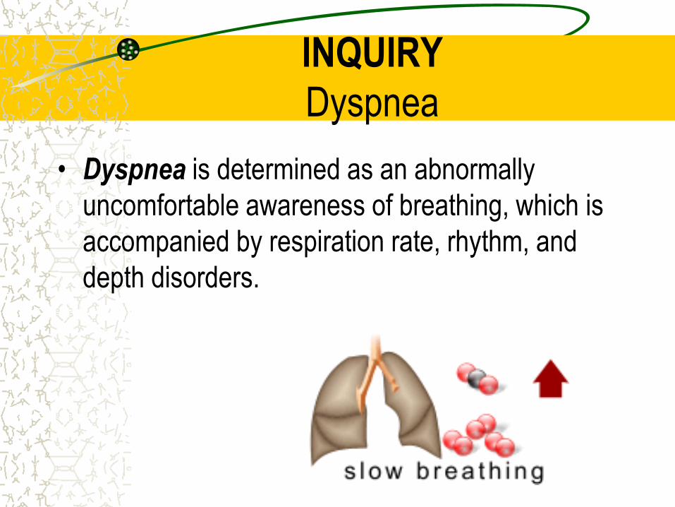
• Dyspnea is determined as an abnormally
uncomfortable awareness of breathing, which is
accompanied by respiration rate, rhythm, and
depth disorders.

INQUIRY
Dyspnea
• The mechanism of dyspnea is irritation of
the respiratory center structures caused
by higher pCO2 (hypercapnia) and the
accumulation in the blood of oxidized
products of lactic acid exchange
(acidosis), with a corresponding change
in the rate, rhythm and depth of
breathing, aimed at restoring gas
homeostasis

INQUIRY
Dyspnea classification І. Physiological
ІІ. Pathological
а) with respect to the patient:
subjective, objective, mixed;
b) according to appearance time:
constant, periodic, paroxysmal;
c) according to respiratory cycle:
inspiratory, expiratory, mixed;
d) according to appearance mechanism:
restrictive, obstructive, paroxysmal obstructive, vascular congestive.
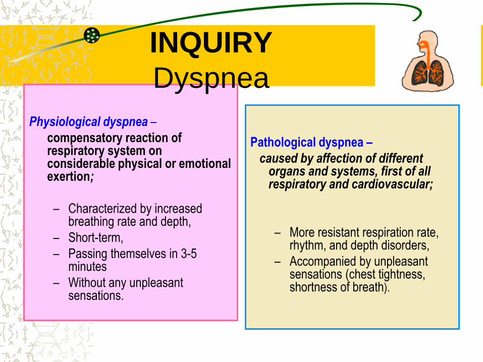
Physiological dyspnea –
compensatory reaction of respiratory system on considerable physical or emotional exertion;
– Characterized by increased breathing rate and depth,
– Short-term,
– Passing themselves in 3-5 minutes
– Without any unpleasant sensations.
Pathological dyspnea –
caused by affection of different organs and systems, first of all respiratory and cardiovascular;
– More resistant respiration rate, rhythm, and depth disorders,
– Accompanied by unpleasant sensations (chest tightness, shortness of breath).
INQUIRY
Dyspnea

INQUIRY
Dyspnea
а) with respect to the patient:
Subjective dyspnea – breathing disorder, manifesting by subjective feeling of tightness in the chest, shortness of breath, difficulty in inspiration or expiration;
Characteristic of hysteria, neurasthenia.
Objective dyspnea – breathing disorder, manifesting by:
Intermittent speech (the patient in conversation gasps)
Tachypnea (respiration rate exceed 30 per min),
Respiration rhythm disorders,
Participation of accessory muscles in the breathing act,
Cyanosis appearance;
Observed in respiratory, cardiovascular, central nervous, and systems diseases.

INQUIRY
Dyspnea
b) according to appearance time:
CONSTANT DYSPNEA remains at rest and increased at insignificant physical exertion (severe forms of respiratory and heart failure, pulmonary emphysema, pneumosclerosis).
PERIODIC (LONG-TERM) DYSPNEA arises in severe diseases and disappears during recovery (pneumonia, pleurisy with effusion, obstructive bronchitis, pneumo- and hydrothorax).
PAROXYSMAL DYSPNEA or ASPHYXIA - severe sudden (paroxysmal) shortness of breath caused by sharp stimulation of the respiratory center.
Are objective signs of acute respiratory failure MAIN CLINICAL SIGNS OF ASPHYXIA:
Sudden onset, intensity;
Shortness of breath;
Rapid growth of respiratory failure objective signs: - Diffuse cyanosis;
– Neck veins swelling,
– Tachypnoe more than 30 per minute,
– Forced posture – orthopnea
.

INQUIRY
Dyspnea
c) according to respiratory cycle:
ISPIRATORY DYSPNEA – respiratory disorder with inhalation difficulties.
Variety of inspiratory dyspnea – stridor breathing - noisy breathing with inspiration difficulties, accompanied by whistles, observed in strong narrowing of the trachea and the upper respiratory tract (foreign body, tumor, scarring or enlarged lymph nodes).
EXPIRATORY DYSPNEA – respiratory disorder with exhalation difficulties, caused by impaired patency of the small bronchi and bronchioles (bronchial asthma, COPD, bronchiolitis)
.
MIXED DYSPNEA – respiratory disorder with simultaneous inspiration and expiration difficulties, more frequently observed in decreased respiratory surface of the lungs, high diaphragm level that restricted lungs excursion, and also in combined affection of the lungs and heart.

INQUIRY
Cough
Cough (tussis) –
is a defensive reflex designed to clear and protect the lower respiratory tract.
The cough reflex can be initiated by stimulation of irritant receptors in the larynx, trachea, and major bronchi. These receptors respond to mechanical irritation by intraluminal material such as mucus, dust, or foreign bodies, and to chemical irritation by fumes and toxic gases.
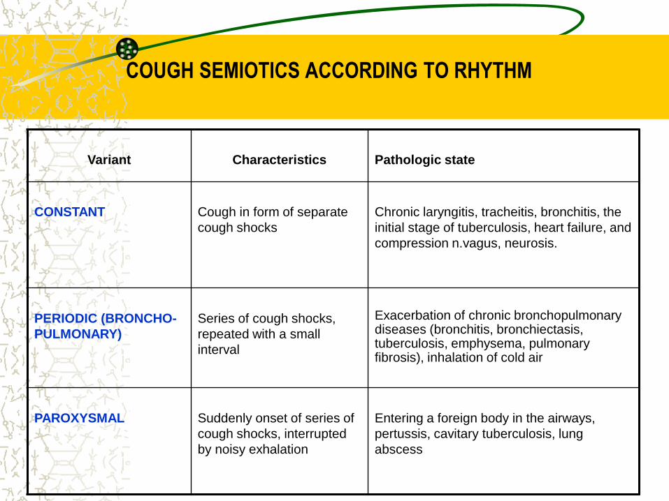
COUGH SEMIOTICS ACCORDING TO RHYTHM
Variant
Characteristics
Pathologic state
CONSTANT
Cough in form of separate
cough shocks
Chronic laryngitis, tracheitis, bronchitis, the
initial stage of tuberculosis, heart failure, and
compression n.vagus, neurosis.
PERIODIC (BRONCHO-
PULMONARY)
Series of cough shocks,
repeated with a small
interval
Exacerbation of chronic bronchopulmonary diseases (bronchitis, bronchiectasis, tuberculosis, emphysema, pulmonary fibrosis), inhalation of cold air
PAROXYSMAL
Suddenly onset of series of
cough shocks, interrupted
by noisy exhalation
Entering a foreign body in the airways,
pertussis, cavitary tuberculosis, lung
abscess
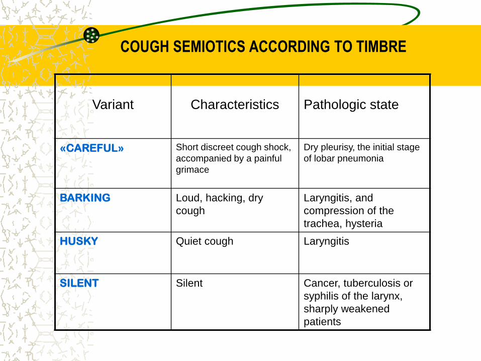
COUGH SEMIOTICS ACCORDING TO TIMBRE
Variant
Characteristics
Pathologic state
«CAREFUL» Short discreet cough shock,
accompanied by a painful
grimace
Dry pleurisy, the initial stage
of lobar pneumonia
BARKING Loud, hacking, dry
cough
Laryngitis, and
compression of the
trachea, hysteria
HUSKY Quiet cough Laryngitis
SILENT Silent Cancer, tuberculosis or
syphilis of the larynx,
sharply weakened
patients
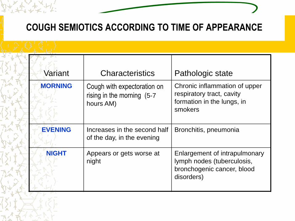
COUGH SEMIOTICS ACCORDING TO TIME OF APPEARANCE
Variant
Characteristics
Pathologic state
MORNING Cough with expectoration on
rising in the morning (5-7
hours AM)
Chronic inflammation of upper
respiratory tract, cavity
formation in the lungs, in
smokers
EVENING Increases in the second half
of the day, in the evening
Bronchitis, pneumonia
NIGHT Appears or gets worse at
night
Enlargement of intrapulmonary
lymph nodes (tuberculosis,
bronchogenic cancer, blood
disorders)

COUGH SEMIOTICS ACCORDING TO CONDITIONS OF APPEARANCE
Variant
Characteristics
Pathologic state
WITHOUT VISIBLE
CAUSES
- COPD, tuberculosis,
bronchogenic tumor, lungs
tumor
CHANGES OF
POSTURE
Arise or increase in definite
posture (frequently on healthy
side)
Cavity formation in the
lungs (bronchiectasis,
abscess, cancer in stages
of decomposition)
FOOD INTAKE Dry cough during a meal or a
cough with mucus and pieces
of food in the sputum
Cancer of the esophagus
with a break in the
respiratory tract;
diaphragmatic hernia
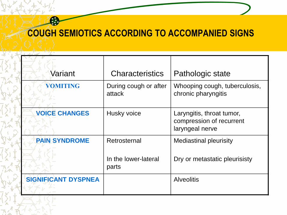
COUGH SEMIOTICS ACCORDING TO ACCOMPANIED SIGNS
Variant
Characteristics
Pathologic state
VOMITING During cough or after
attack
Whooping cough, tuberculosis,
chronic pharyngitis
VOICE CHANGES Husky voice Laryngitis, throat tumor,
compression of recurrent
laryngeal nerve
PAIN SYNDROME Retrosternal
In the lower-lateral
parts
Mediastinal pleurisity
Dry or metastatic pleurisisty
SIGNIFICANT DYSPNEA Alveolitis
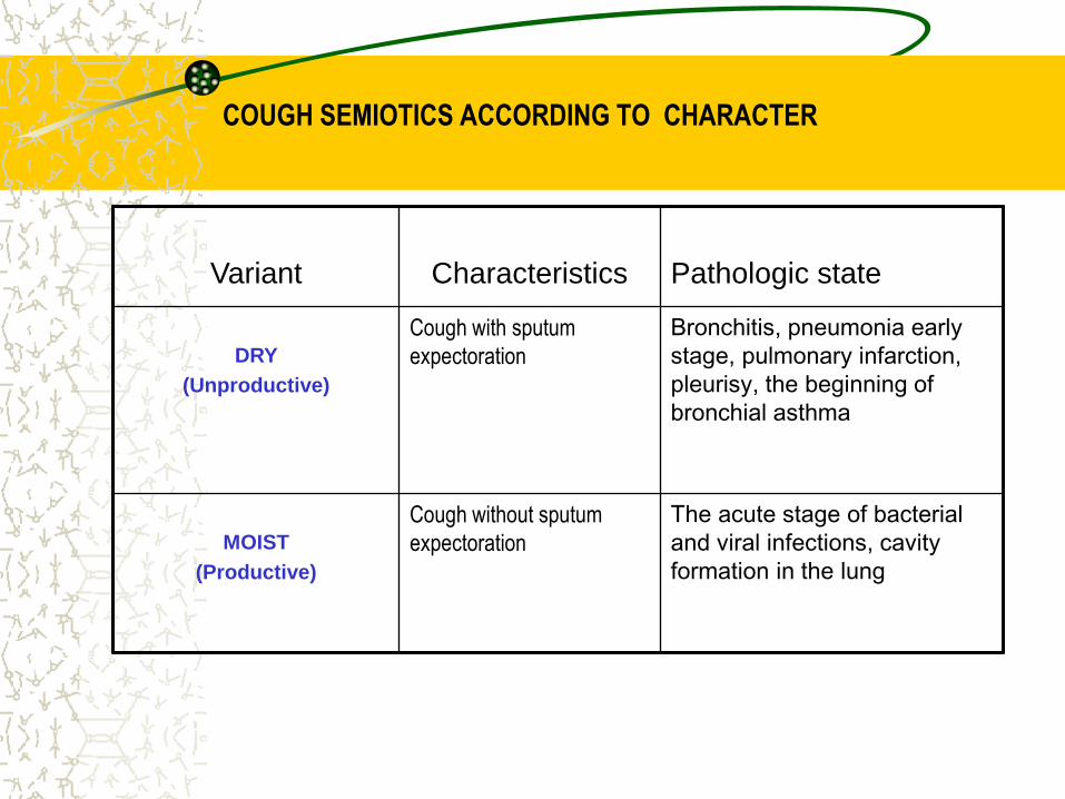
COUGH SEMIOTICS ACCORDING TO CHARACTER
Variant
Characteristics
Pathologic state
DRY
(Unproductive)
Cough with sputum
expectoration
Bronchitis, pneumonia early
stage, pulmonary infarction,
pleurisy, the beginning of
bronchial asthma
MOIST
(Productive)
Cough without sputum
expectoration
The acute stage of bacterial
and viral infections, cavity
formation in the lung
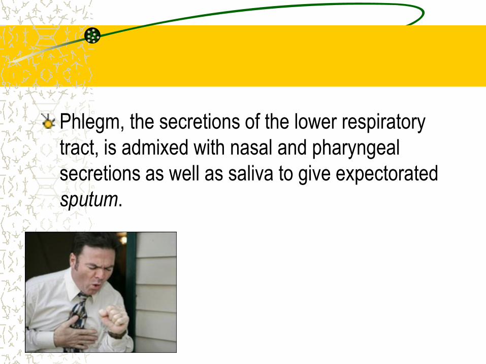
Phlegm, the secretions of the lower respiratory
tract, is admixed with nasal and pharyngeal
secretions as well as saliva to give expectorated
sputum.
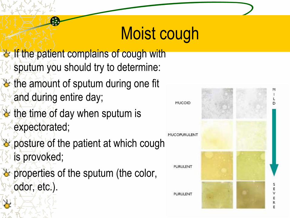
Moist cough If the patient complains of cough with
sputum you should try to determine:
the amount of sputum during one fit
and during entire day;
the time of day when sputum is
expectorated;
posture of the patient at which cough
is provoked;
properties of the sputum (the color,
odor, etc.).
Moist cough
In the patients with chronic bronchitis, bronchiectasis, lung
abscess, and cavernous tuberculosis of the lungs, the sputum
accumulates during the night sleep in the lungs and bronchi.
When the patient gets up in the morning, the sputum moves to
stimulate the reflex zones of the bronchial mucosa and cause
cough and expectoration of sputum. The amount of the morning
sputum is two thirds of the entire daily expectoration. The daily
amount of sputum may vary from 10-15 ml to 2 liters. In unilateral
bronchiectasis, sputum may be better expectorated when the
patient lies on the healthy side.
INQUIRY
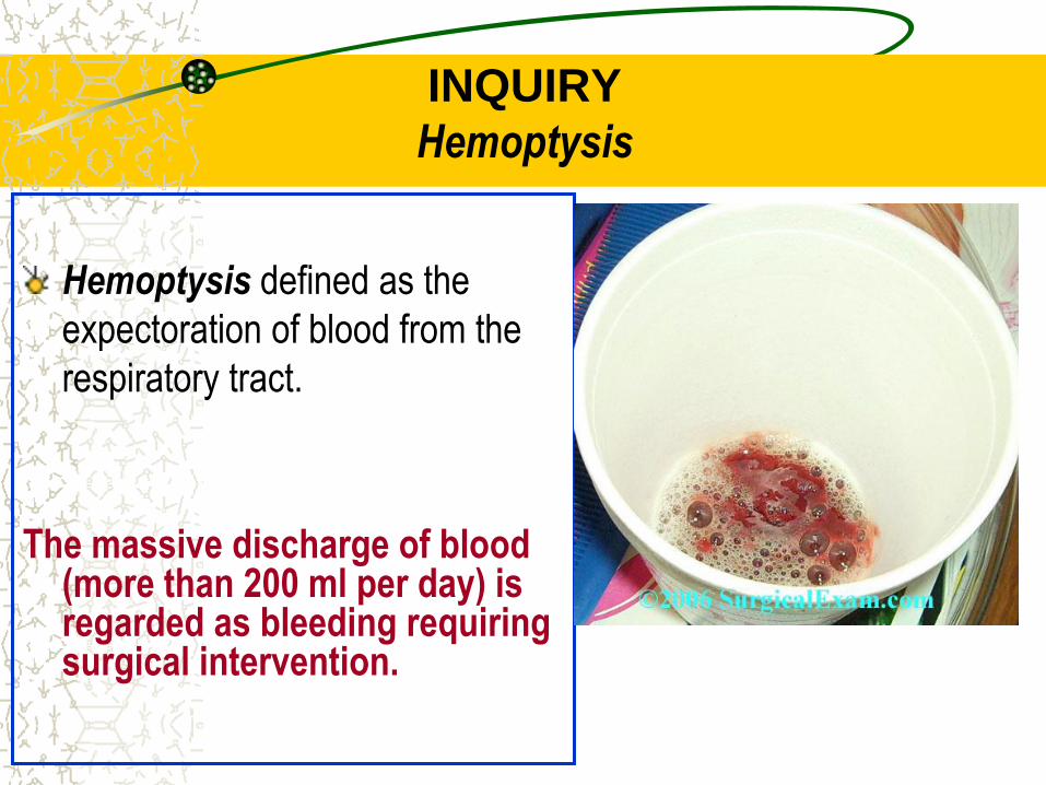
Hemoptysis
Hemoptysis defined as the
expectoration of blood from the
respiratory tract.
The massive discharge of blood (more than 200 ml per day) is regarded as bleeding requiring surgical intervention.
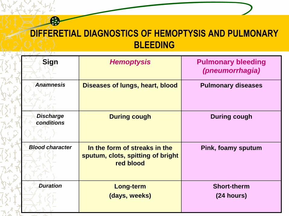
DIFFERETIAL DIAGNOSTICS OF HEMOPTYSIS AND PULMONARY
BLEEDING
Sign Hemoptysis Pulmonary bleeding
(pneumorrhagia)
Anamnesis Diseases of lungs, heart, blood Pulmonary diseases
Discharge
conditions During cough During cough
Blood character In the form of streaks in the
sputum, clots, spitting of bright
red blood
Pink, foamy sputum
Duration Long-term
(days, weeks)
Short-therm
(24 hours)
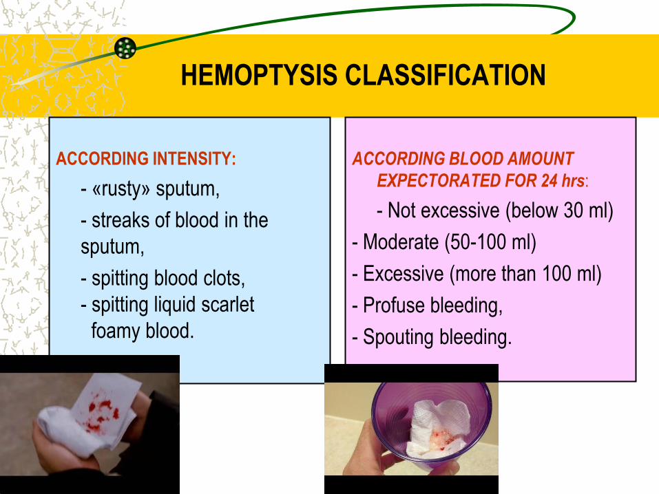
HEMOPTYSIS CLASSIFICATION
ACCORDING INTENSITY:
- «rusty» sputum,
- streaks of blood in the
sputum,
- spitting blood clots,
- spitting liquid scarlet
foamy blood.
ACCORDING BLOOD AMOUNT
EXPECTORATED FOR 24 hrs:
- Not excessive (below 30 ml)
- Moderate (50-100 ml)
- Excessive (more than 100 ml)
- Profuse bleeding,
- Spouting bleeding.
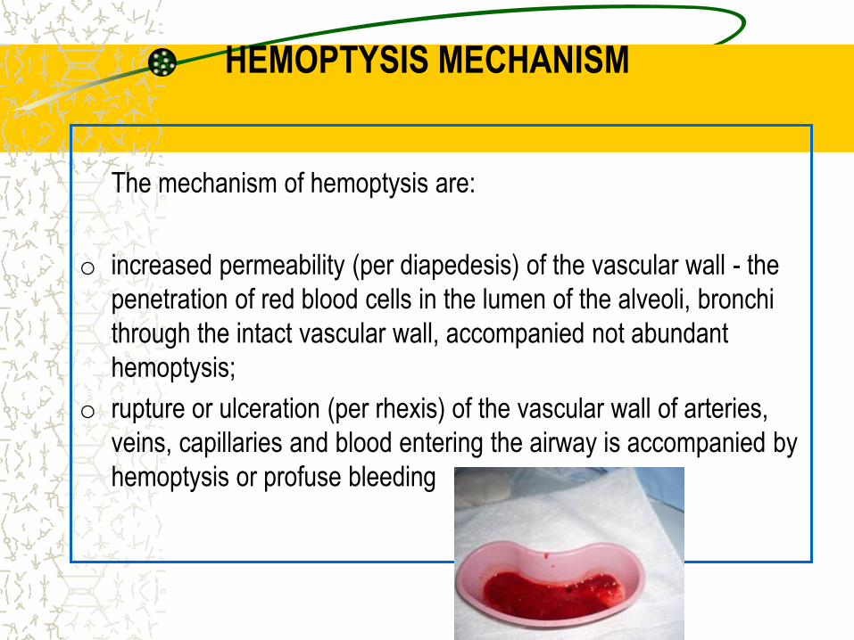
HEMOPTYSIS MECHANISM
The mechanism of hemoptysis are:
o increased permeability (per diapedesis) of the vascular wall - the
penetration of red blood cells in the lumen of the alveoli, bronchi
through the intact vascular wall, accompanied not abundant
hemoptysis;
o rupture or ulceration (per rhexis) of the vascular wall of arteries,
veins, capillaries and blood entering the airway is accompanied by
hemoptysis or profuse bleeding

PULMONARY
– Acute or chronic bronchitis,
bronchiectasis
- Pneumonia,
- BRONCHOGENIC
CANCER,
– TUBERCULOSIS,
– PULMONARY ABSCESS
EXTRA PULMONARY
– MITRAL STENOSIS,
– CONGESTIVE HEART
FAILURE
MOST HEMOPTYSIS
CAUSES
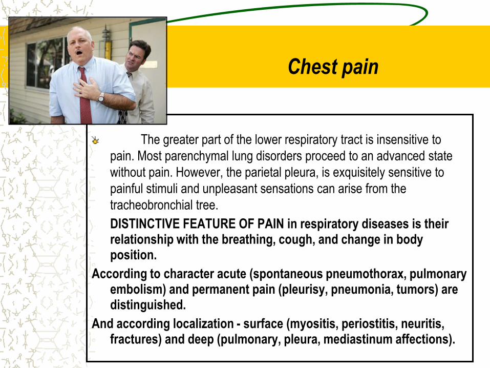
Chest pain
The greater part of the lower respiratory tract is insensitive to
pain. Most parenchymal lung disorders proceed to an advanced state
without pain. However, the parietal pleura, is exquisitely sensitive to
painful stimuli and unpleasant sensations can arise from the
tracheobronchial tree.
DISTINCTIVE FEATURE OF PAIN in respiratory diseases is their relationship with the breathing, cough, and change in body position.
According to character acute (spontaneous pneumothorax, pulmonary embolism) and permanent pain (pleurisy, pneumonia, tumors) are distinguished.
And according localization - surface (myositis, periostitis, neuritis, fractures) and deep (pulmonary, pleura, mediastinum affections).

CHEST PAIN CAUSES
PULMONARY NON PULMONARY
Pleura affection: pleurisy (dry,
metastatic), adhesions (after
purulent pleurisy), spontaneous
pneumothorax
Pulmonary disease with
involvement of the pleura in
pathologic process: lobar
pneumonia, pulmonary infarction,
tuberculosis, cancer, abscess,
gangrene
Trachea and bronchi affection:
bronchogenic carcinoma, thermal
and traumatic injuries of the trachea
(burns, trauma)
Bronchopulmonary lymph nodes
affection
Cardiovascular system: angina pectoris,
myocardial infarction, pulmonary embolism,
dissecting aneurysm of the aorta.
Musculoskeletal system: periostitis,
fractures, myositis, arthritis, metastases, low
back pain
Nervous system: neuritis, intercostal
neuralgia
Blood system: leukemia, anemia Addison-
Birmera, multiple myeloma
Digestive system: cholecystitis, cholelithiasis,
liver abscess, diaphragmatic hernia
Other causes: Shingles
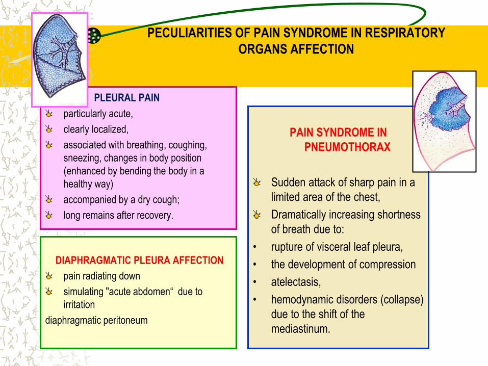
PECULIARITIES OF PAIN SYNDROME IN RESPIRATORY
ORGANS AFFECTION
PLEURAL PAIN
particularly acute,
clearly localized,
associated with breathing, coughing,
sneezing, changes in body position
(enhanced by bending the body in a
healthy way)
accompanied by a dry cough;
long remains after recovery.
DIAPHRAGMATIC PLEURA AFFECTION
pain radiating down
simulating "acute abdomen“ due to
irritation
diaphragmatic peritoneum
PAIN SYNDROME IN
PNEUMOTHORAX
Sudden attack of sharp pain in a
limited area of the chest,
Dramatically increasing shortness
of breath due to:
• rupture of visceral leaf pleura,
• the development of compression
• atelectasis,
• hemodynamic disorders (collapse)
due to the shift of the
mediastinum.
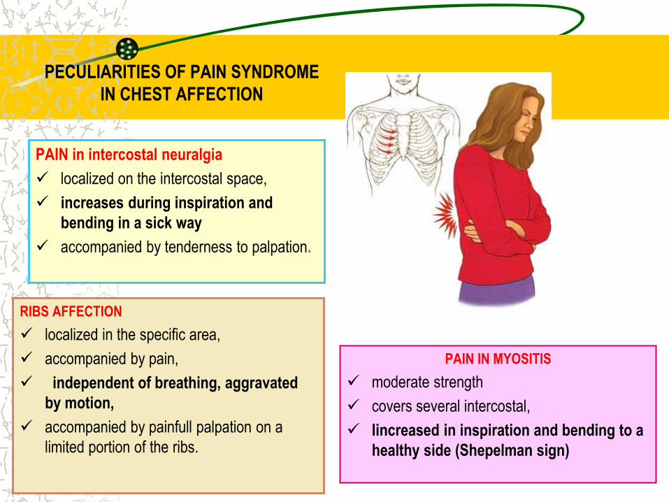
PECULIARITIES OF PAIN SYNDROME
IN CHEST AFFECTION
PAIN in intercostal neuralgia
localized on the intercostal space,
increases during inspiration and
bending in a sick way
accompanied by tenderness to palpation.
PAIN IN MYOSITIS
moderate strength
covers several intercostal,
Iincreased in inspiration and bending to a
healthy side (Shepelman sign)
RIBS AFFECTION
localized in the specific area,
accompanied by pain,
independent of breathing, aggravated
by motion,
accompanied by painfull palpation on a
limited portion of the ribs.
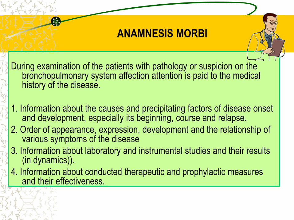
ANAMNESIS MORBI
During examination of the patients with pathology or suspicion on the bronchopulmonary system affection attention is paid to the medical history of the disease.
1. Information about the causes and precipitating factors of disease onset and development, especially its beginning, course and relapse.
2. Order of appearance, expression, development and the relationship of various symptoms of the disease
3. Information about laboratory and instrumental studies and their results (in dynamics)).
4. Information about conducted therapeutic and prophylactic measures and their effectiveness.
OBJECTIVE EXAMINATION
METHODS

GENERAL INSPECTION PLAN
o General condition
o Consciousness
o Posture of the patient
o Facial expression, inspection of
the neck
o Skin and visible mucosa
o Lymph nodes
o Musculoskeletal system

General condition
Consciousness
General condition of patients, depending on the severity and stage of
disease can be:
satisfactory (prodromal period, remission or recovery);
moderate (acute disease or exacerbation of chronic diseases);
severe and very severe (asthma status, spontaneous pneumothorax,
pulmonary embolism, pulmonary infarction, COR-pulmonale).
Consciousness in the majority of patients of respiratory diseases
remains clear. However, in severe hypoxia, resulting in chronic
pulmonary heart failure or intoxication (tuberculosis, cancer), it may
be in violation in a form of a stupor or sopor.
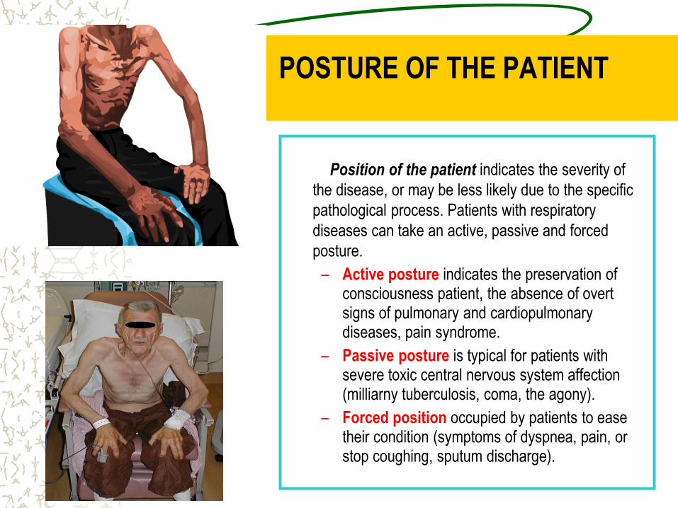
POSTURE OF THE PATIENT
Position of the patient indicates the severity of
the disease, or may be less likely due to the specific
pathological process. Patients with respiratory
diseases can take an active, passive and forced
posture.
– Active posture indicates the preservation of consciousness patient, the absence of overt signs of pulmonary and cardiopulmonary diseases, pain syndrome.
– Passive posture is typical for patients with severe toxic central nervous system affection (milliarny tuberculosis, coma, the agony).
– Forced position occupied by patients to ease their condition (symptoms of dyspnea, pain, or stop coughing, sputum discharge).

Forced posture
Posture Pathophysiologic mechanisms Pathologic condition
Orthopnea
focusing hands
With the palm of hands is fixing the
shoulder girdle and the inclusion in
the process of breathing auxiliary
respiratory muscles, which
facilitates the phase of expiration
and reduces breathlessness
Bronchial asthma
attack
Orthopnea
without the palm
rest
Othopnea facilitates diaphragm
movements, increases the volume
of the chest, improves gas exchange
in system "alveolus, pulmonary
capillary" and cerebral vessels,
which leads to a decrease of
respiratory center excitability and
reduced dyspnea
Foreign body, lung
tumor, lobar
pneumonia, pleural
effusion,
pneumothorax, partial
paralysis of the
respiratory muscles
On affected side Reducing of compression and
compensatory hyperventilation of
healthy lung, a reduction of friction
of inflamed pleura; decrease sputum
discharge and cough reflex
Dry pleurisy, a lung
abscess,
bronchiectasis,
tuberculosis
Prone position
(Face down)
Diaphragm respiration is restricted Diaphragmal pleurisy
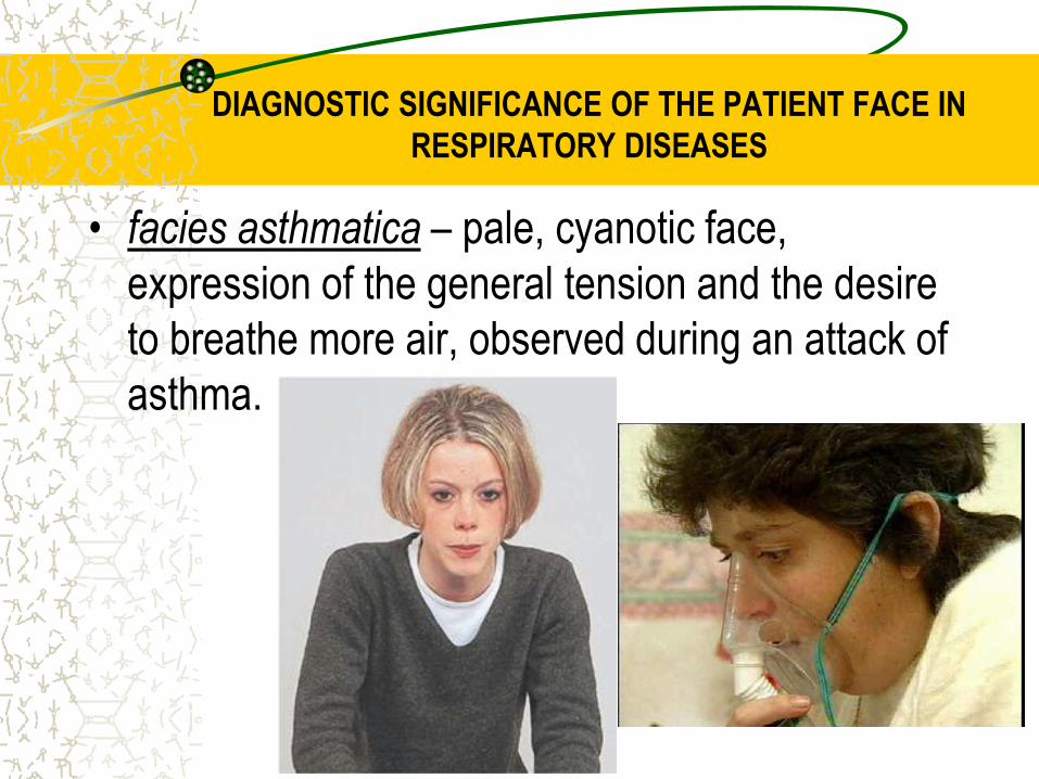
DIAGNOSTIC SIGNIFICANCE OF THE PATIENT FACE IN
RESPIRATORY DISEASES
• facies asthmatica – pale, cyanotic face,
expression of the general tension and the desire
to breathe more air, observed during an attack of
asthma.

DIAGNOSTIC SIGNIFICANCE OF THE PATIENT FACE
IN RESPIRATORY DISEASES
• facies tuberculous – exhausted, pale face
with blush localized on the cheeks
“burning eyes”, dry lips, excited
countenance, half open mouth

DIAGNOSTIC SIGNIFICANCE OF THE PATIENT
FACE IN RESPIRATORY DISEASES
Facies pneumonica – one-sided blush on the same cheek as affected lung, cyanosis, often herpes on the lips and nose;
Patient with chronic pulmonary heart failure
(diffuse cyanosis)
•Stokes collar – means edematous neck with associated with
edematous face due to the compression of lymph ducts and
veins with enlarged mediastinal lymph nodes, tumor of
mediastinum, adhesive mediastinopericarditis, excessive
effusion in the pleural and pericardial cavity.

INSPECTION OF THE NECK
On examination of the neck attention should be paid on filling and increased pulsation of neck veins
due to:
increase in intrathoracic pressure
increased central venous pressure as a result of violations of the outflow of blood from the superior vena cava or the development of pulmonary hypertension
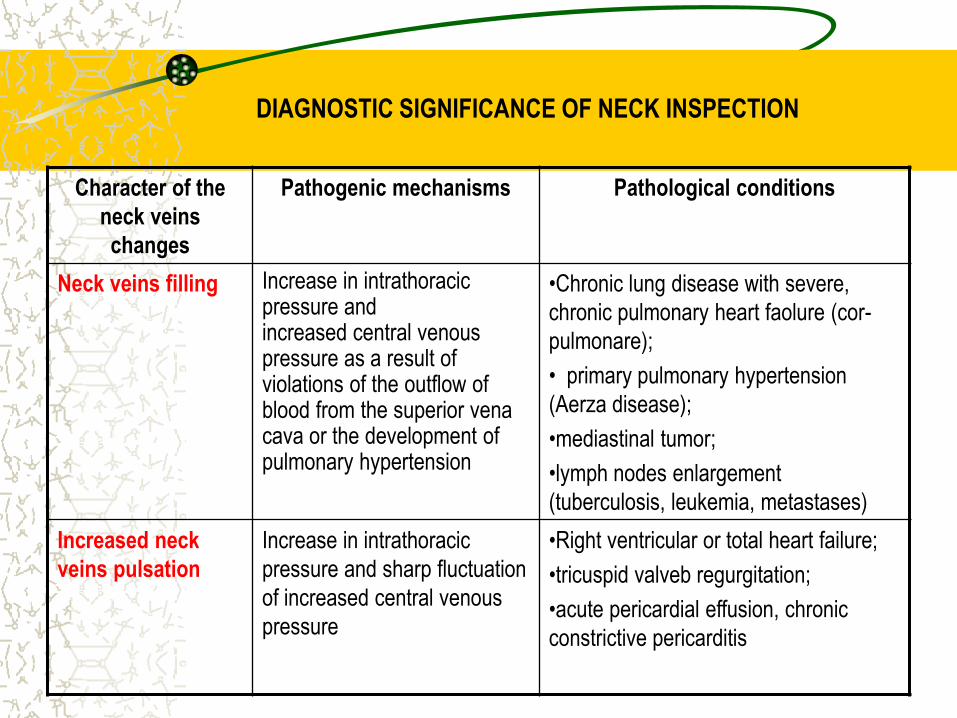
DIAGNOSTIC SIGNIFICANCE OF NECK INSPECTION
Character of the
neck veins
changes
Pathogenic mechanisms Pathological conditions
Neck veins filling Increase in intrathoracic pressure and increased central venous pressure as a result of violations of the outflow of blood from the superior vena cava or the development of pulmonary hypertension
•Chronic lung disease with severe,
chronic pulmonary heart faolure (cor-
pulmonare);
• primary pulmonary hypertension
(Aerza disease);
•mediastinal tumor;
•lymph nodes enlargement
(tuberculosis, leukemia, metastases)
Increased neck
veins pulsation
Increase in intrathoracic
pressure and sharp fluctuation
of increased central venous
pressure
•Right ventricular or total heart failure;
•tricuspid valveb regurgitation;
•acute pericardial effusion, chronic
constrictive pericarditis

DIAGNOSTIC SIGNIFICANCE OF SKIN INSPECTION
SYMPTOM PATHOGENIC MECHANISMS PATHOLOGICAL CONDITIONS
CYANOSIS
а) diffuse
b) peripheral
cyanosis (on face,
neck, upper
extremities region)
c) significant
cyanosis of the face
The imbalance between ventilation and
perfusion, the accumulation of carbon
dioxide in the blood and reduced
hemoglobin, peripheral vasodilatation
The sudden expulsion of breath of lung
tissue
Compression of the superior vena cava,
phlebostasis
Primary and secondary pulmonary artery
sclerosis leads to disruption of gas perfusion
COPD, chronic suppurative disease (abscess,
bronchiectasis), bronchial asthma, pneumonia,
emphysema, pulmonary fibrosis, tuberculosis
Pulmonary embolism, spontaneous
pneumothorax
Bronchogenic lung cancer
Primary (Aerza disease) and secondary
pulmonary hypertension
One-sided face
hyperemia on
background of
cyanosis
Reflex vessels of the face vasodilation on the
affected side
Lobar pneumonia
PALENESS
a) moderate
b) pronounced
pallor
Violation of the ventilation process and
compensatory spasmodermia
hemorrhagic anemia
Pleural effusion
Massive pulmonary hemorrhage, decaying
lung cancer, bronchiectasis,
Erythema nodosum Alveolar hyperventilation Pulmonary sarcoidosis
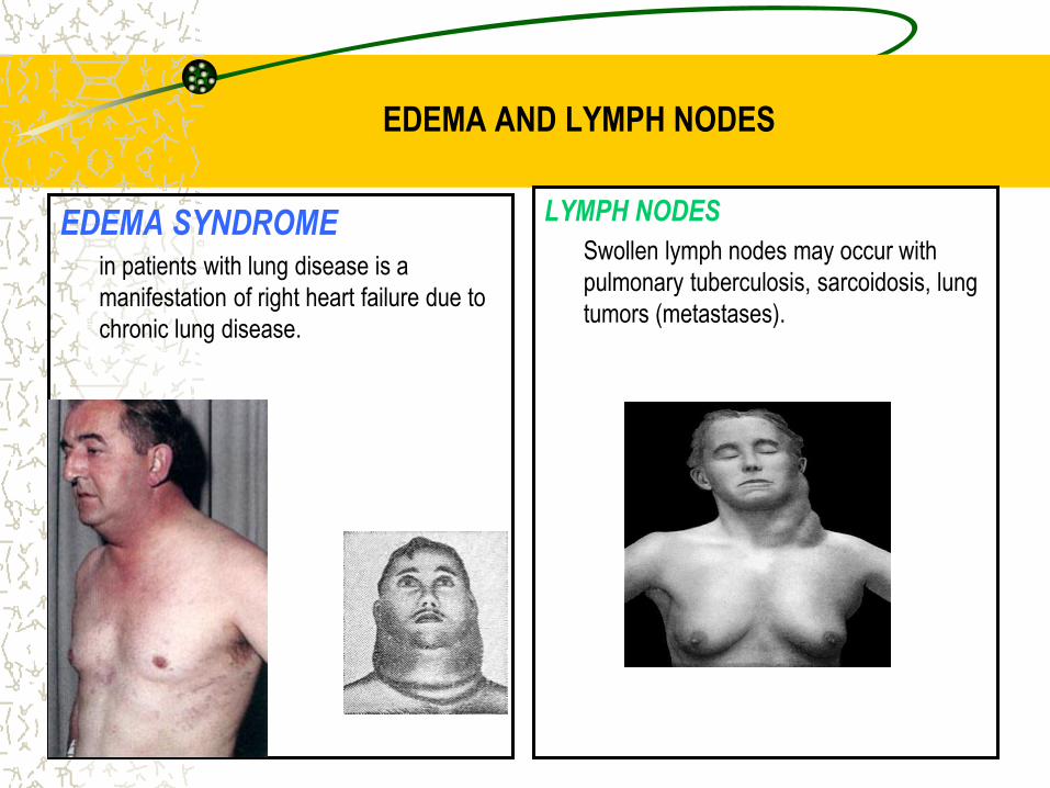
EDEMA AND LYMPH NODES
EDEMA SYNDROME in patients with lung disease is a
manifestation of right heart failure due to
chronic lung disease.
LYMPH NODES
Swollen lymph nodes may occur with
pulmonary tuberculosis, sarcoidosis, lung
tumors (metastases).

Locomotor system
Characteristic changes of:
terminal phalanges in the form of "drumsticks" and
nail shape the type of " watch glass" (Hippocrates nails) -
hypertrophic pulmonary osteoarthropathy
(chronic purullent lung disease, bronchogenic cancer, fibrosing alveolitis).
At the base of symptom is growth of periosteum terminal phalanges in chronic hypoxia. In this case, the terminal phalanges thicken, take the form of "drumsticks", and the nails are raised and hard, like a watch glass.

Clubbing of the Nails
A sign of chronic hypoxia
A sign of chronic hypoxia
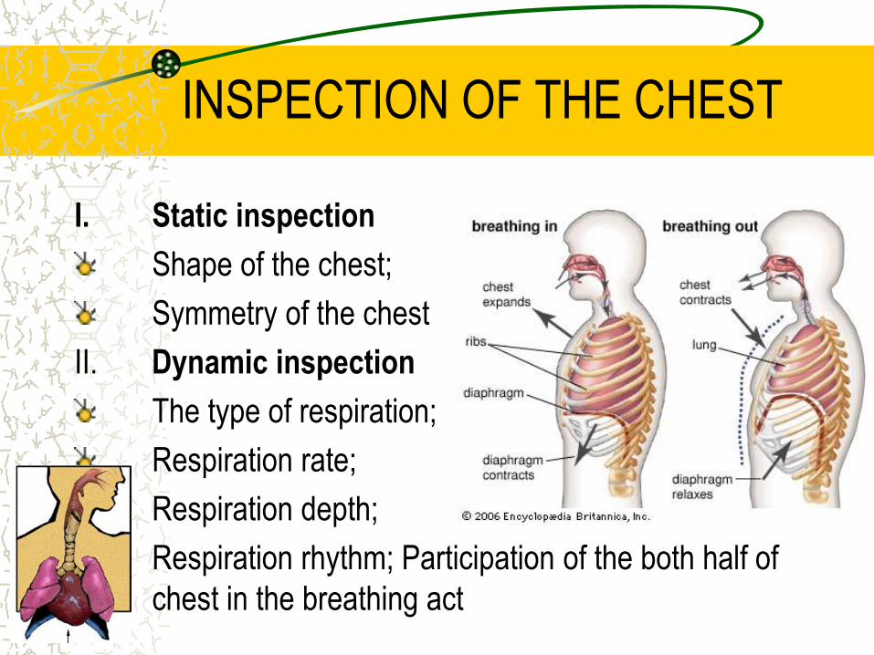
INSPECTION OF THE CHEST
I. Static inspection
Shape of the chest;
Symmetry of the chest
II. Dynamic inspection
The type of respiration;
Respiration rate;
Respiration depth;
Respiration rhythm; Participation of the both half of
chest in the breathing act
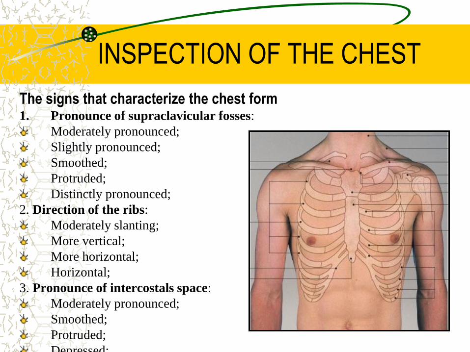
INSPECTION OF THE CHEST
The signs that characterize the chest form 1. Pronounce of supraclavicular fosses:
Moderately pronounced;
Slightly pronounced;
Smoothed;
Protruded;
Distinctly pronounced;
2. Direction of the ribs:
Moderately slanting;
More vertical;
More horizontal;
Horizontal;
3. Pronounce of intercostals space:
Moderately pronounced;
Smoothed;
Protruded;
Depressed;
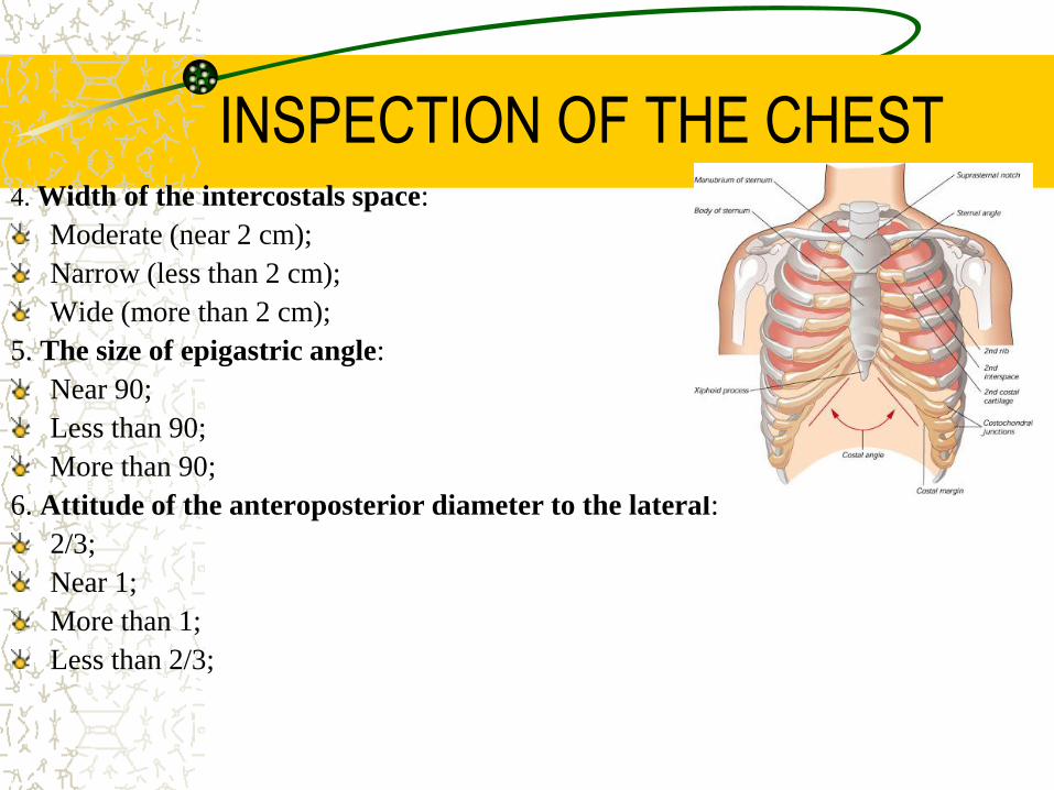
INSPECTION OF THE CHEST 4. Width of the intercostals space:
Moderate (near 2 cm);
Narrow (less than 2 cm);
Wide (more than 2 cm);
5. The size of epigastric angle:
Near 90;
Less than 90;
More than 90;
6. Attitude of the anteroposterior diameter to the lateral:
2/3;
Near 1;
More than 1;
Less than 2/3;
INSPECTION OF THE CHEST
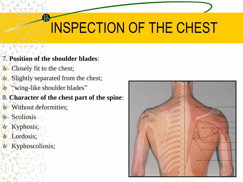
7. Position of the shoulder blades:
Closely fit to the chest;
Slightly separated from the chest;
“wing-like shoulder blades”
8. Character of the chest part of the spine:
Without deformities;
Scoliosis
Kyphosis;
Lordosis;
Kyphoscoliosis;

STATIC INSPECTION OF THE
CHEST Physiological shape of the chest
Normosthenic; Asthenic; Hypersthenic;

STATIC INSPECTION OF THE CHEST
Physiological chest shape
Normosthenic chest: the
shoulders are under the right angle
to the neck, supra- and
infraclavicular fossae feebly
expressed, the ribs are moderately
inclined, the interspaces are visible,
but moderate expressed, epigastric
angle is near 90˚, the lateral
diameter is larger than
anteroposterior, shoulder blades
closely fits to the chest and are on
the same level. The thorax is about
the same height as abdominal part
of the trunk

STATIC INSPECTION OF THE CHEST
Physiological chest shape Asthenic chest: the shoulders are sloping and
are under the dull angle to the neck, clavicles are
well visible, supra- and infraclavicular fossae are
distinctly pronounced, the ribs direct downward
abruptly, more vertical at sides, the 10th ribs are
not attached to the costal arch, the interspaces are
wide and pronounced, epigastric angle is less than
90˚, both lateral and anteroposterior diameter are
smaller than normal, the chest narrow and
elongated, the shoulder blades are separate from
the chest and their angles are well visible. The
muscles of the shoulder girdle are
underdeveloped. The thorax is longer than
abdominal part of the trunk

STATIC INSPECTION OF THE CHEST
Physiological chest shape Hypersthenic chest: the shoulders
are wide and the neck is short, supra- and
infraclavicular fossae are absent (level
with the chest), direction of the ribs are
nearly horizontal, the interspaces are
narrow and slightly expressed, epigastric
angle exceeds 90˚, the lateral diameter is
about the same as anteroposterior, the
chest has form of a cylinder, the shoulder
blades closely fit to the chest. The thorax
is smaller than abdominal part of the trunk
STATIC INSPECTION OF THE CHEST
Pathological chest shape
Pathological shapes of the chest can be
caused either by
chronic diseases of the lungs and pleura
(emphysematous, paralytic chest), or by
pathology of the thorax costal skeleton (rachitic,
funnel, and foveated chest), or by
various deformities of the spine (scoliosis,
kyphosis, lordosis, kyphoscoliosis).
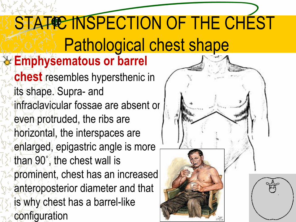
STATIC INSPECTION OF THE CHEST
Pathological chest shape Emphysematous or barrel
chest resembles hypersthenic in
its shape. Supra- and
infraclavicular fossae are absent or
even protruded, the ribs are
horizontal, the interspaces are
enlarged, epigastric angle is more
than 90˚, the chest wall is
prominent, chest has an increased
anteroposterior diameter and that
is why chest has a barrel-like
configuration

STATIC INSPECTION OF THE CHEST
Pathological chest shape
Paralytic chest. The same signs that
peculiar to the asthenic chest but more
pronounced characterize paralytic chest.
The shoulders are sloping, clavicles are
asymmetrical and pronounced, supra-
and infraclavicular fossea depresses, the
ribs are vertical, the interspaces are wide
and depressed, marked atrophy of the
chest muscles, epigastric angle is less
than 90˚, the shoulder blades are not on
the same level, and their movement
during breathing are asynchronous

Barrel Chest: Increased Anterior/Posterior Diameter
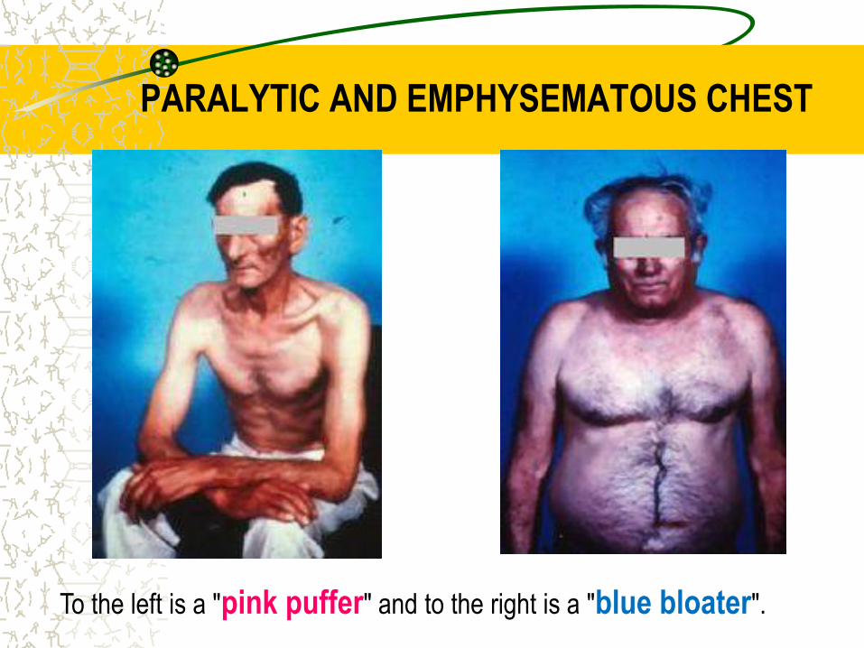
PARALYTIC AND EMPHYSEMATOUS CHEST
To the left is a "pink puffer" and to the right is a "blue bloater".
STATIC INSPECTION OF THE CHEST
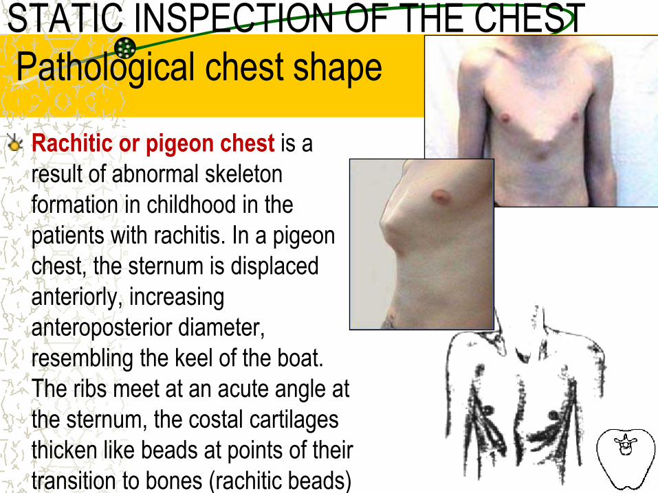
Pathological chest shape
Rachitic or pigeon chest is a
result of abnormal skeleton
formation in childhood in the
patients with rachitis. In a pigeon
chest, the sternum is displaced
anteriorly, increasing
anteroposterior diameter,
resembling the keel of the boat.
The ribs meet at an acute angle at
the sternum, the costal cartilages
thicken like beads at points of their
transition to bones (rachitic beads)
STATIC INSPECTION OF THE CHEST
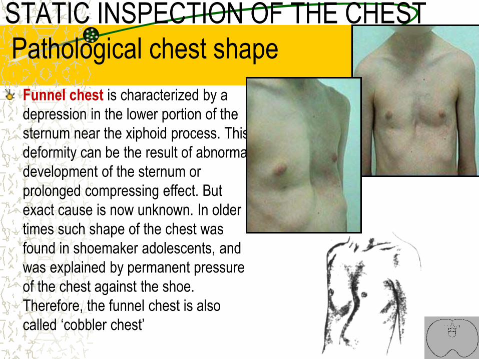
Pathological chest shape
Funnel chest is characterized by a
depression in the lower portion of the
sternum near the xiphoid process. This
deformity can be the result of abnormal
development of the sternum or
prolonged compressing effect. But
exact cause is now unknown. In older
times such shape of the chest was
found in shoemaker adolescents, and
was explained by permanent pressure
of the chest against the shoe.
Therefore, the funnel chest is also
called ‘cobbler chest’

STATIC INSPECTION OF THE CHEST
Pathological chest shape
Foveated chest is
characterized by vertical
depression on the upper
and middle parts of the
anterior surface of the
chest. This deformity arises
in syringomyelia, a rare
disease of the spinal cord.
STATIC INSPECTION OF THE CHEST
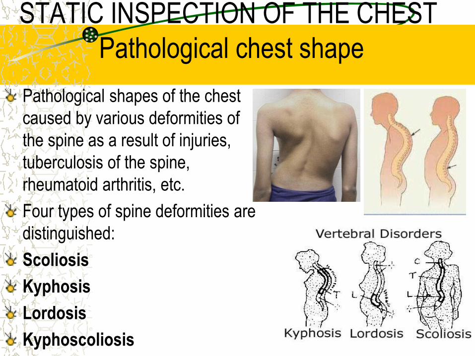
Pathological chest shape
Pathological shapes of the chest
caused by various deformities of
the spine as a result of injuries,
tuberculosis of the spine,
rheumatoid arthritis, etc.
Four types of spine deformities are
distinguished:
Scoliosis
Kyphosis
Lordosis
Kyphoscoliosis

STATIC INSPECTION OF THE CHEST
Pathological chest shape Scoliosis – lateral
curvature of the spine, is
most common. It
develops in
schoolchildren due to
bad habitual posture.
STATIC INSPECTION OF THE CHEST
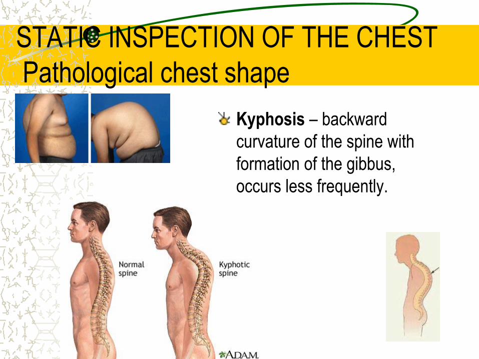
Pathological chest shape
Kyphosis – backward
curvature of the spine with
formation of the gibbus,
occurs less frequently.
STATIC INSPECTION OF THE CHEST
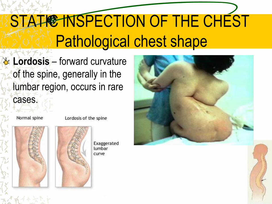
Pathological chest shape Lordosis – forward curvature
of the spine, generally in the
lumbar region, occurs in rare
cases.

STATIC INSPECTION OF THE CHEST
Pathological chest shape
Kyphoscoliosis – combination
of the lateral and backward
curvature of the spine
STATIC INSPECTION OF THE CHEST
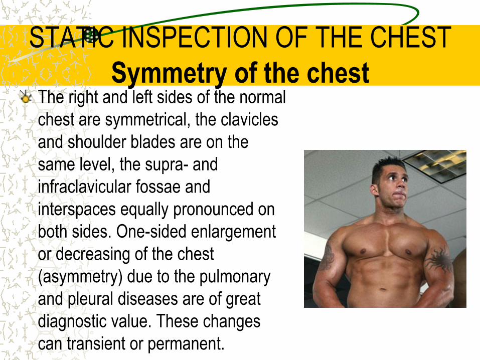
Symmetry of the chest The right and left sides of the normal
chest are symmetrical, the clavicles
and shoulder blades are on the
same level, the supra- and
infraclavicular fossae and
interspaces equally pronounced on
both sides. One-sided enlargement
or decreasing of the chest
(asymmetry) due to the pulmonary
and pleural diseases are of great
diagnostic value. These changes
can transient or permanent.

STATIC INSPECTION OF THE CHEST
Symmetry of the chest Enlarged volume of one half of the chest observes in accumulation
of considerable amount of fluid (exudates, transudate, blood, pus)
in the pleural cavity, or in penetration of air inside the chest in
injuries (pneumothorax).
During examination of the enlarged part of the chest you can see
asymmetry of the clavicles; leveling or protrusion of the
interspaces, and they more wide; the distance between nipple and
median line, and from inside edge of scapula to the spine on
affected side is larger than on healthy one. Enlarged part of the
chest lags in the breathing act. The patient slightly bends to the
affected side of the chest. The chest assumes normal symmetrical
shape after the fluid or air is removed from the pleural cavity.
STATIC INSPECTION OF THE CHEST
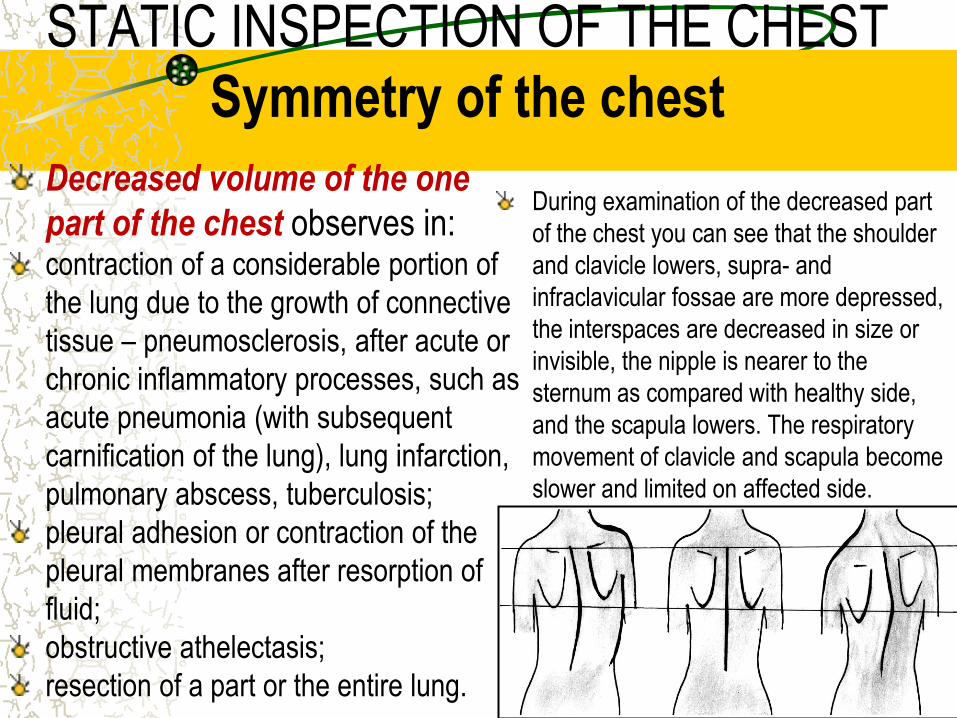
Symmetry of the chest
Decreased volume of the one
part of the chest observes in: contraction of a considerable portion of
the lung due to the growth of connective
tissue – pneumosclerosis, after acute or
chronic inflammatory processes, such as
acute pneumonia (with subsequent
carnification of the lung), lung infarction,
pulmonary abscess, tuberculosis;
pleural adhesion or contraction of the
pleural membranes after resorption of
fluid;
obstructive athelectasis;
resection of a part or the entire lung.
During examination of the decreased part
of the chest you can see that the shoulder
and clavicle lowers, supra- and
infraclavicular fossae are more depressed,
the interspaces are decreased in size or
invisible, the nipple is nearer to the
sternum as compared with healthy side,
and the scapula lowers. The respiratory
movement of clavicle and scapula become
slower and limited on affected side.
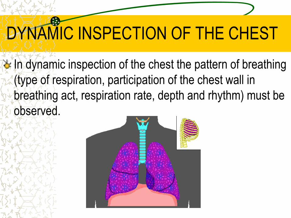
DYNAMIC INSPECTION OF THE CHEST
In dynamic inspection of the chest the pattern of breathing
(type of respiration, participation of the chest wall in
breathing act, respiration rate, depth and rhythm) must be
observed.

DYNAMIC INSPECTION OF THE CHEST
The type of respiration:
Thoracic;
Abdominal;
Mixed;

DYNAMIC INSPECTION OF THE CHEST
Respiration type
Thoracic (costal) respiration. Mainly
the intercostals muscles carry out
respiratory movements. In inspiration
the intercostals muscles contract and
elevate the ribs, these movements
increase the internal capacity of the
lungs. As the thoracic wall expands,
the lungs also expand and draw in air.
In expiration, the thoracic capacity
decreases as the inspiratory muscles
relax – the lungs then shrink by their
own elasticity. This type of breathing is
known as costal and is mostly
characteristic of women
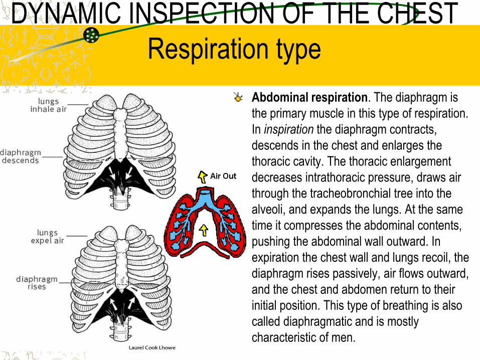
DYNAMIC INSPECTION OF THE CHEST
Respiration type
Abdominal respiration. The diaphragm is
the primary muscle in this type of respiration.
In inspiration the diaphragm contracts,
descends in the chest and enlarges the
thoracic cavity. The thoracic enlargement
decreases intrathoracic pressure, draws air
through the tracheobronchial tree into the
alveoli, and expands the lungs. At the same
time it compresses the abdominal contents,
pushing the abdominal wall outward. In
expiration the chest wall and lungs recoil, the
diaphragm rises passively, air flows outward,
and the chest and abdomen return to their
initial position. This type of breathing is also
called diaphragmatic and is mostly
characteristic of men.
DYNAMIC INSPECTION OF THE CHEST
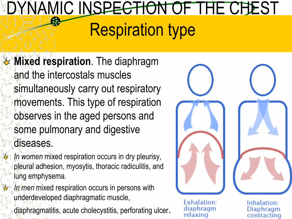
Respiration type
Mixed respiration. The diaphragm
and the intercostals muscles
simultaneously carry out respiratory
movements. This type of respiration
observes in the aged persons and
some pulmonary and digestive
diseases. In women mixed respiration occurs in dry pleurisy,
pleural adhesion, myosytis, thoracic radiculitis, and
lung emphysema.
In men mixed respiration occurs in persons with
underdeveloped diaphragmatic muscle,
diaphragmatitis, acute cholecystitis, perforating ulcer.
DYNAMIC INSPECTION OF THE CHEST
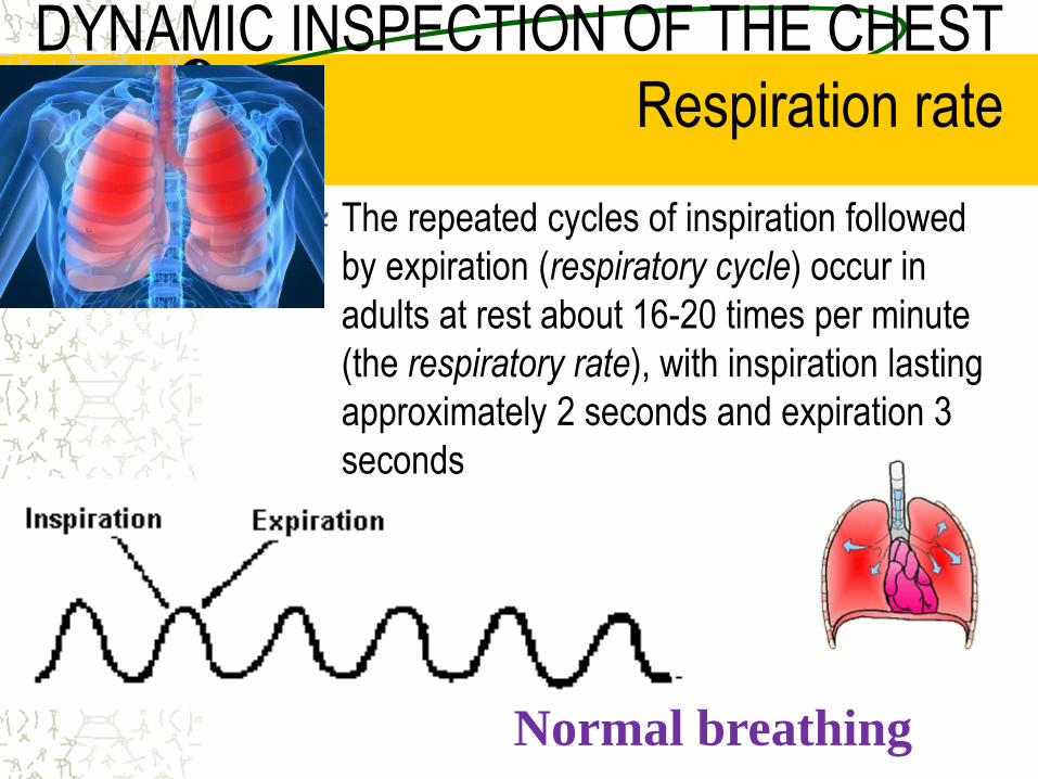
Respiration rate
The repeated cycles of inspiration followed
by expiration (respiratory cycle) occur in
adults at rest about 16-20 times per minute
(the respiratory rate), with inspiration lasting
approximately 2 seconds and expiration 3
seconds
Normal breathing

DYNAMIC INSPECTION OF THE CHEST
Respiration rate
Pathological rapid breathing
above 20 per minute is called
tachypnea
Tachypnea has a number of causes:
conditions associated with decreased
respiratory surface of the lungs: inflammation, tuberculosis, compressive atelectasis
(hydrothorax, pneumothorax, mediastinal tumor),
obstructive atelectesis, pulmonary emphysema, and
pulmonary edema;
narrowing of the small bronchi due to spasm or
diffuse inflammation of their mucosa (bronchiolitis),
which interfere normal air passage into alveoli;
shallow respiration as a result of difficult
contractions of the respiratory muscles in acute
pain (dry pleurisy, acute myositis, intercostals
neuralgia, rib fracture) and in elevated abdominal
pressure and high diaphragm level (ascitis,
meteorism, late pregnancy).

DYNAMIC INSPECTION OF THE CHEST
Respiration rate
Pathological slow
breathing below 16
per minute is called
bradypnea
Bradypnea may be
secondary to such causes
as increased intracranial
pressure (cerebral tumor,
hemorrhage, meningitis,
brain edema) due to
inhibition of the respiratory
center, and also due to the
toxic effect on respiratory
center in uremia, diabetic
or hepatic coma, and
drug-induced respiratory
depression.

DYNAMIC INSPECTION OF THE CHEST
Respiration depth
The volume of the
inhaled and exhaled air
at rest in adults varies
from 300 to 900 ml (500
ml on the average).
Depending on depth,
breathing can be shallow
or deep.

DYNAMIC INSPECTION OF THE CHEST
Respiration depth Shallow respiration is
characterized by short
inspiratory and expiratory
phases. Shallow
breathing is usually
rapid.
In some cases, however,
shallow respiration can
be slow due to inhibition
of the respiratory center,
pronounced pulmonary
emphysema, and sharp
narrowing of the vocal
slit or trachea.
DYNAMIC INSPECTION OF THE CHEST
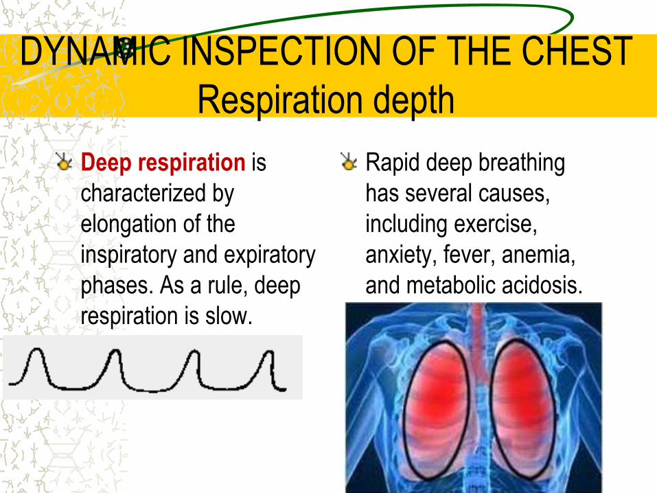
Respiration depth
Deep respiration is
characterized by
elongation of the
inspiratory and expiratory
phases. As a rule, deep
respiration is slow.
Rapid deep breathing
has several causes,
including exercise,
anxiety, fever, anemia,
and metabolic acidosis.
DYNAMIC INSPECTION OF THE CHEST
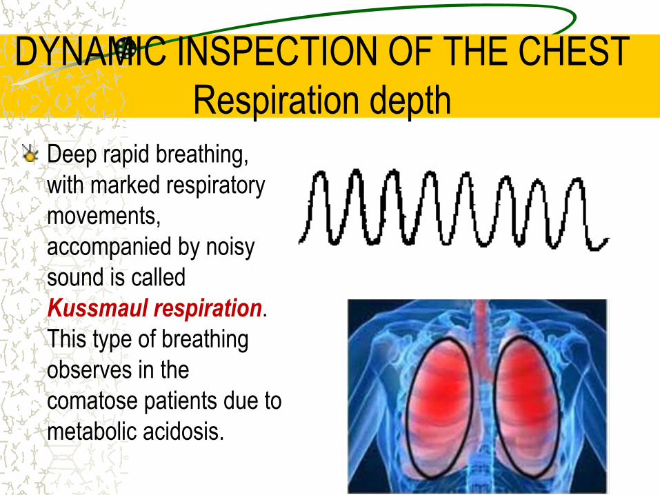
Respiration depth
Deep rapid breathing,
with marked respiratory
movements,
accompanied by noisy
sound is called
Kussmaul respiration.
This type of breathing
observes in the
comatose patients due to
metabolic acidosis.

DYNAMIC INSPECTION OF THE CHEST
Respiration rhytm
A normal rhythm of
breathing is controlled by
groups of nerve cells in
the brainstem, called the
respiratory center. These
nerve cells send impulses
down the spinal cord to
act on the spinal nerve
fibers that supply the
diaphragm and
intercostals muscles.
DYNAMIC INSPECTION OF THE CHEST
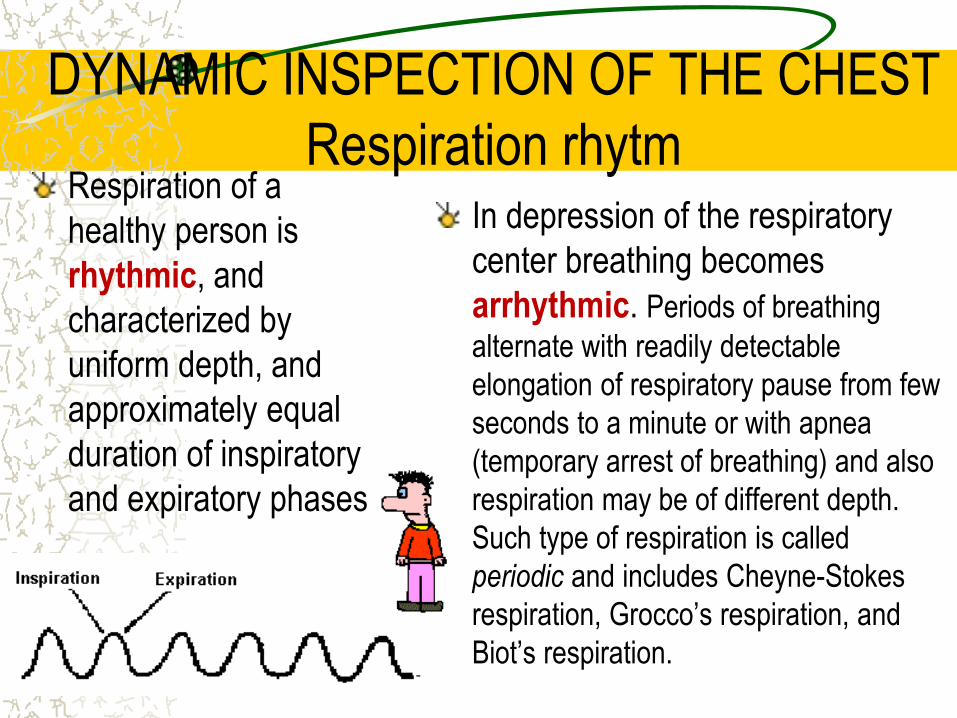
Respiration rhytm Respiration of a
healthy person is
rhythmic, and
characterized by
uniform depth, and
approximately equal
duration of inspiratory
and expiratory phases
In depression of the respiratory
center breathing becomes
arrhythmic. Periods of breathing
alternate with readily detectable
elongation of respiratory pause from few
seconds to a minute or with apnea
(temporary arrest of breathing) and also
respiration may be of different depth.
Such type of respiration is called
periodic and includes Cheyne-Stokes
respiration, Grocco’s respiration, and
Biot’s respiration.

DYNAMIC INSPECTION OF THE CHEST
Respiration rhytm Cheyne-Stokes respiration.
Noiseless shallow respiration
quickly deepens, becomes noisy to
attain its maximum at the 5-7
inspirations and slows down
gradually. Such periods alternate
with periods of apnea (from few
second to a minute), during which
patient can loses orientation in
surroundings or even faints to
recover from unconsciousness
after respiration restores.
Children and aged people
normally may show Cheyne-
Stokes respiration in sleep.
Other causes include heart
failure, uremia, drug-induced
respiratory depression, and
brain damage (acute or chronic
failure of the cerebral
circulation, cerebral hypoxia,
meningitis).
DYNAMIC INSPECTION OF THE CHEST
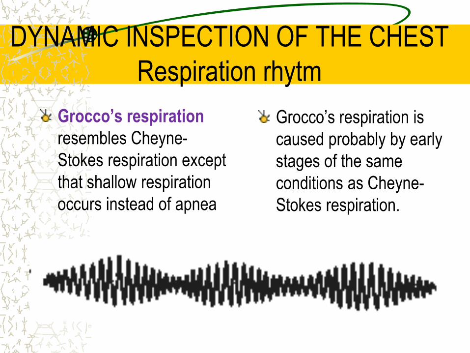
Respiration rhytm
Grocco’s respiration
resembles Cheyne-
Stokes respiration except
that shallow respiration
occurs instead of apnea
Grocco’s respiration is
caused probably by early
stages of the same
conditions as Cheyne-
Stokes respiration.

DYNAMIC INSPECTION OF THE CHEST
Respiration rhytm
Biot’s respiration. In
this type of breathing
deep rhythmic respiration
alternate with apnea
(from few seconds to half
minute).
Causes include
respiratory depression
and brain damage
(meningitis, agony with
disorders of cerebral
circulation).
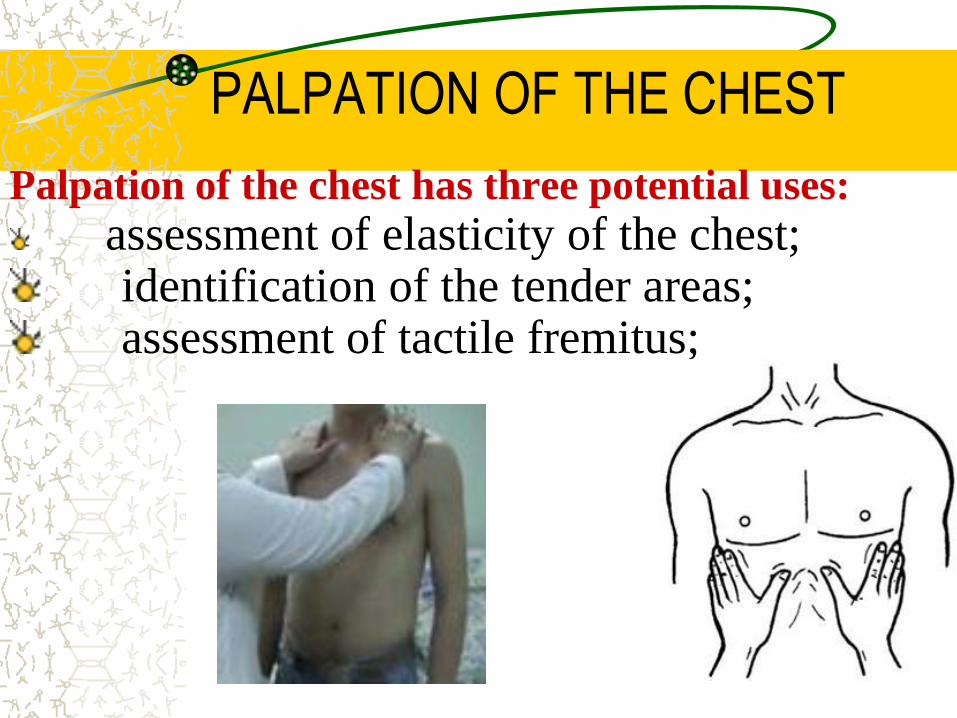
PALPATION OF THE CHEST
Palpation of the chest has three potential uses:
assessment of elasticity of the chest; identification of the tender areas; assessment of tactile fremitus;
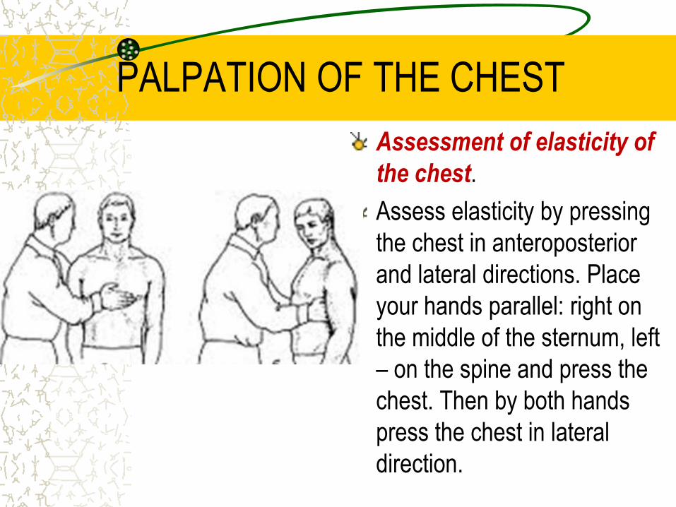
PALPATION OF THE CHEST
Assessment of elasticity of
the chest.
Assess elasticity by pressing
the chest in anteroposterior
and lateral directions. Place
your hands parallel: right on
the middle of the sternum, left
– on the spine and press the
chest. Then by both hands
press the chest in lateral
direction.

PALPATION OF THE CHEST
The chest of a healthy person
is elastic, and yields under the
pressure. Rigidity of the chest
indicates presence of fluid in
the pleural cavity or pleural
tumor, and pulmonary
emphysema. In aged persons
the chest become rigid due to
ossification of the costal
cartilages.
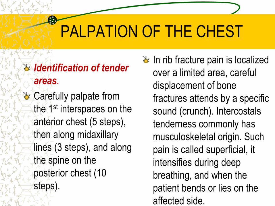
PALPATION OF THE CHEST
Identification of tender
areas.
Carefully palpate from
the 1st interspaces on the
anterior chest (5 steps),
then along midaxillary
lines (3 steps), and along
the spine on the
posterior chest (10
steps).
In rib fracture pain is localized
over a limited area, careful
displacement of bone
fractures attends by a specific
sound (crunch). Intercostals
tenderness commonly has
musculoskeletal origin. Such
pain is called superficial, it
intensifies during deep
breathing, and when the
patient bends or lies on the
affected side.
PALPATION OF THE CHEST
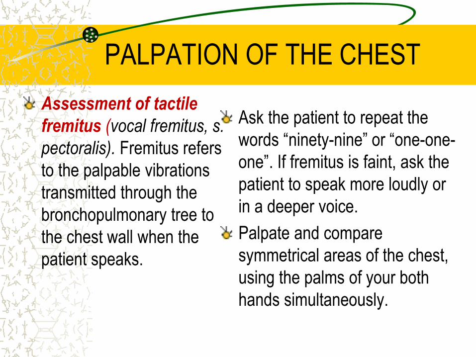
Assessment of tactile
fremitus (vocal fremitus, s.
pectoralis). Fremitus refers
to the palpable vibrations
transmitted through the
bronchopulmonary tree to
the chest wall when the
patient speaks.
Ask the patient to repeat the
words “ninety-nine” or “one-one-
one”. If fremitus is faint, ask the
patient to speak more loudly or
in a deeper voice.
Palpate and compare
symmetrical areas of the chest,
using the palms of your both
hands simultaneously.
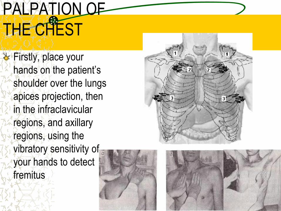
PALPATION OF
THE CHEST
Firstly, place your
hands on the patient’s
shoulder over the lungs
apices projection, then
in the infraclavicular
regions, and axillary
regions, using the
vibratory sensitivity of
your hands to detect
fremitus
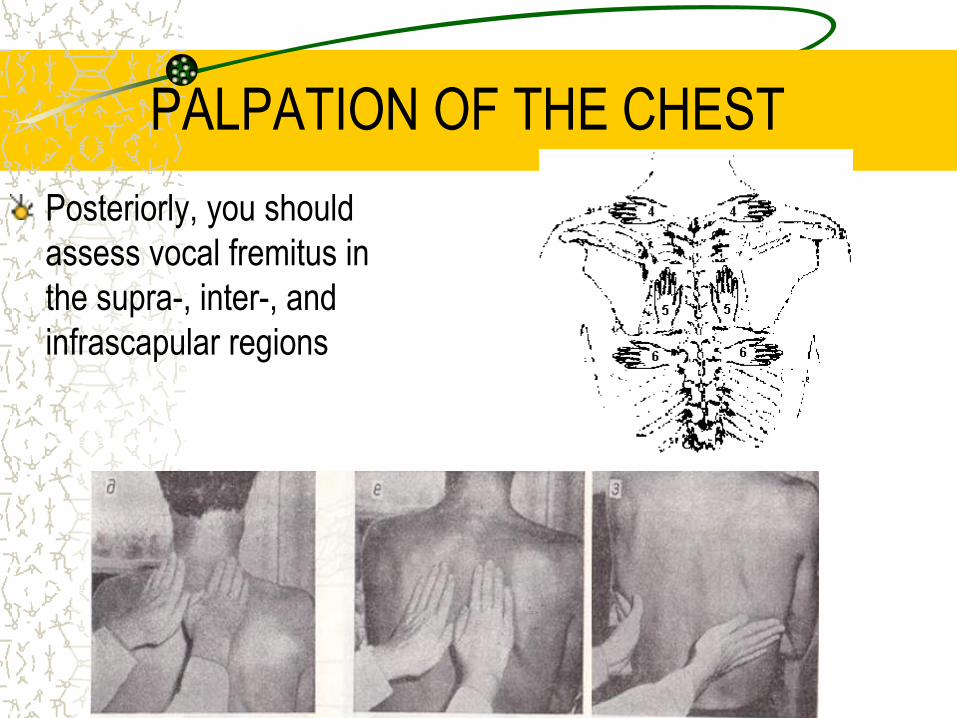
PALPATION OF THE CHEST
Posteriorly, you should
assess vocal fremitus in
the supra-, inter-, and
infrascapular regions

PALPATION OF THE CHEST
Tactile fremitus is typically more
prominent in men than in women
and children; in the upper lung
fields than in the lower one; and
more prominent on the right side
(voice transmission is better
through the shorter right main
bronchus) than on the left.
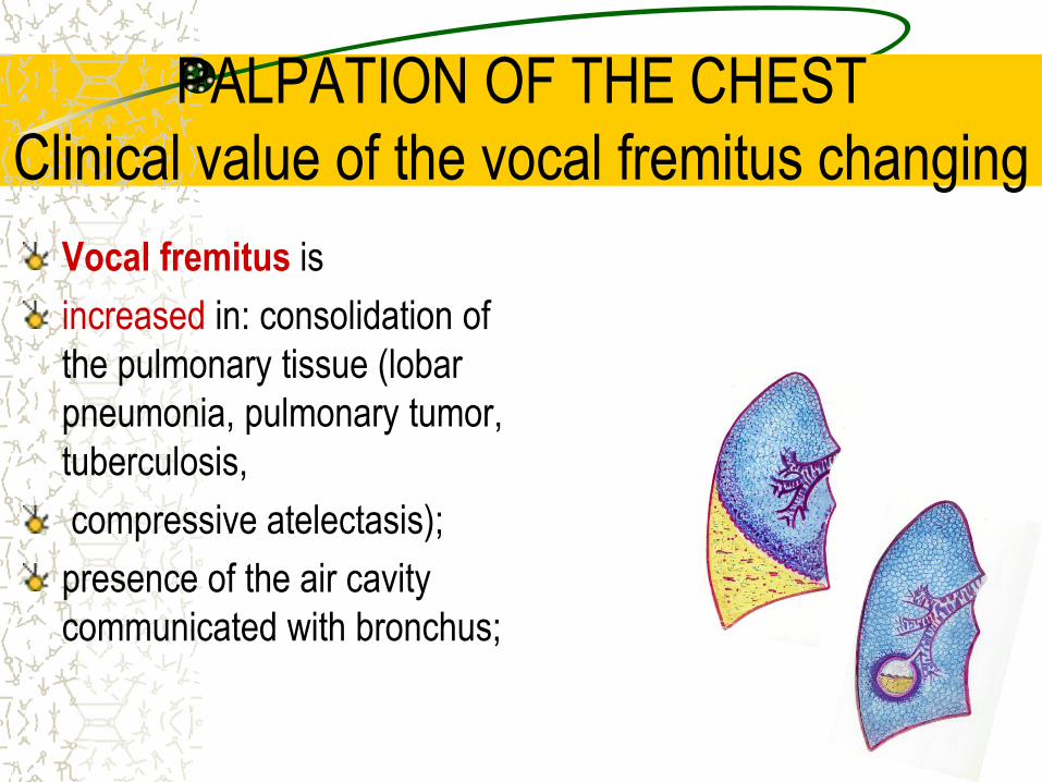
PALPATION OF THE CHEST
Clinical value of the vocal fremitus changing
Vocal fremitus is
increased in: consolidation of
the pulmonary tissue (lobar
pneumonia, pulmonary tumor,
tuberculosis,
compressive atelectasis);
presence of the air cavity
communicated with bronchus;
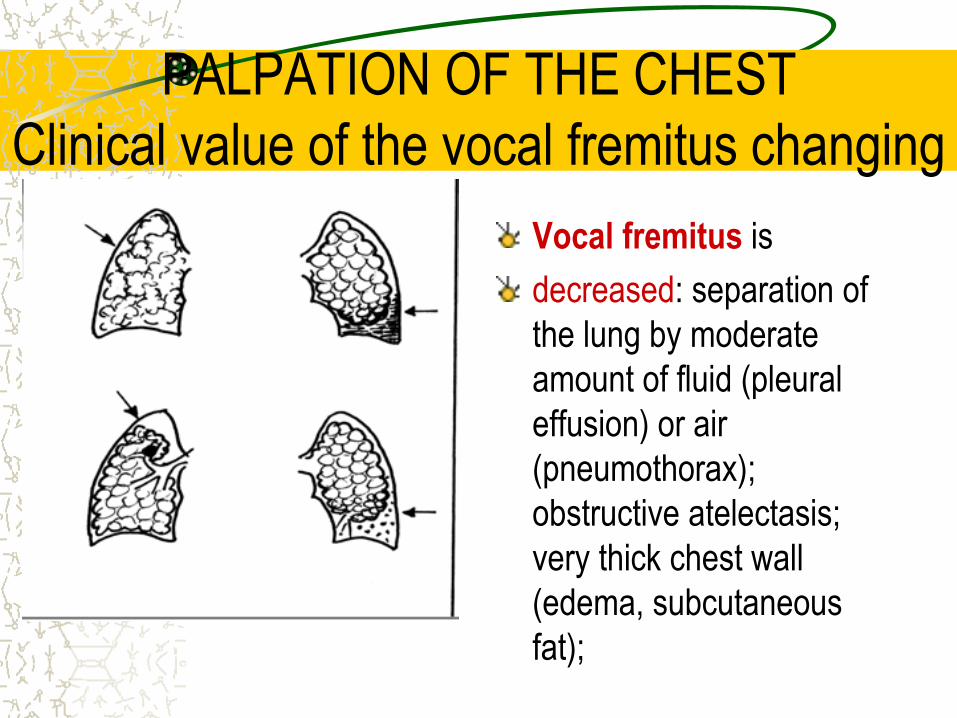
PALPATION OF THE CHEST
Clinical value of the vocal fremitus changing
Vocal fremitus is
decreased: separation of
the lung by moderate
amount of fluid (pleural
effusion) or air
(pneumothorax);
obstructive atelectasis;
very thick chest wall
(edema, subcutaneous
fat);

PALPATION OF THE CHEST
Clinical value of the vocal fremitus changing
Vocal fremitus
can be absent when
significant amount of
fluid or air are
accumulated in the
pleural cavity.
Hydrothorax
Pneumothorax
Obstructive
atelectasis
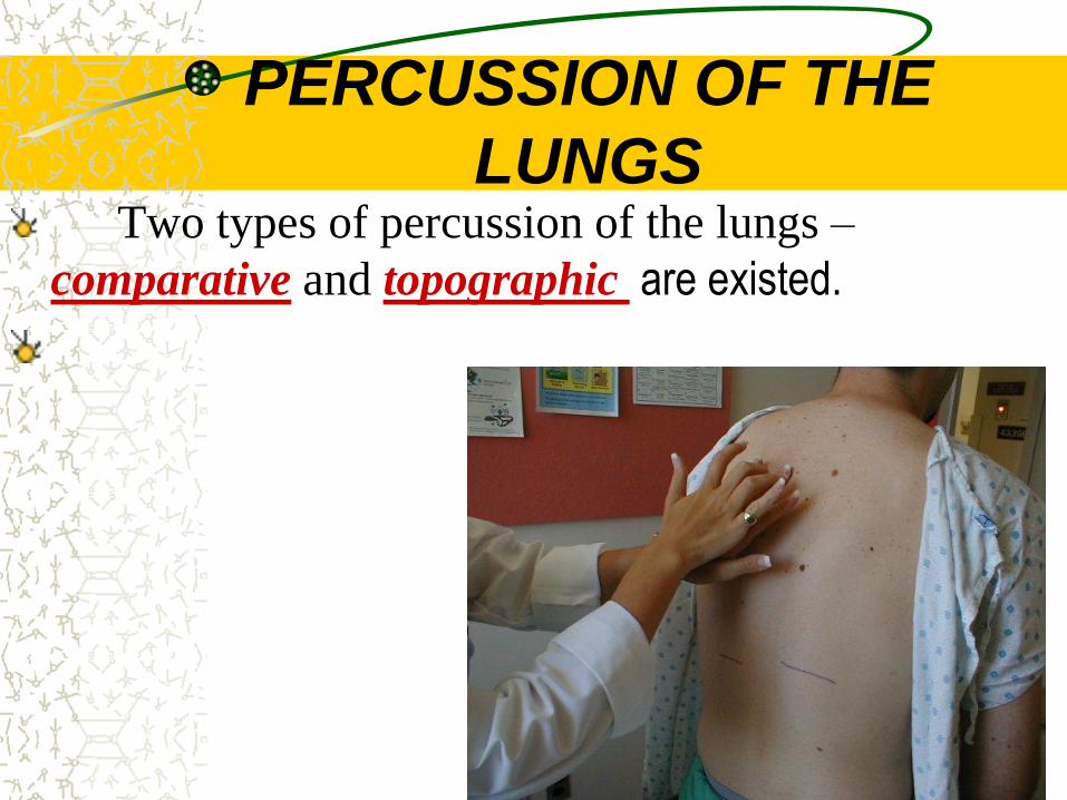
PERCUSSION OF THE
LUNGS Two types of percussion of the lungs –
comparative and topographic are existed.
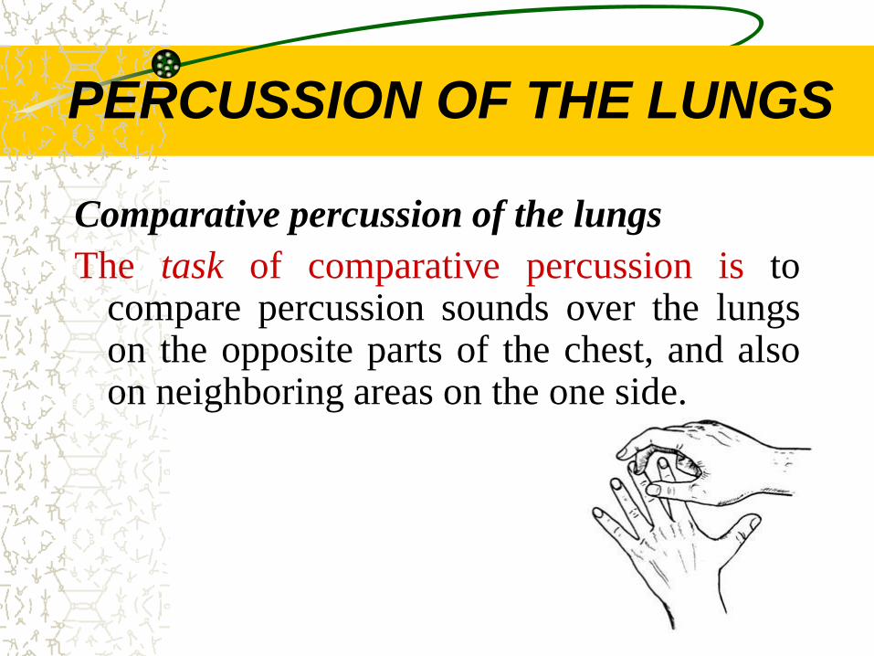
PERCUSSION OF THE LUNGS
Comparative percussion of the lungs
The task of comparative percussion is to compare percussion sounds over the lungs on the opposite parts of the chest, and also on neighboring areas on the one side.
PERCUSSION OF THE LUNGS
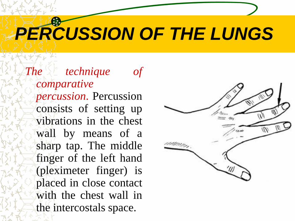
The technique of comparative percussion. Percussion consists of setting up vibrations in the chest wall by means of a sharp tap. The middle finger of the left hand (pleximeter finger) is placed in close contact with the chest wall in the intercostals space.

PERCUSSION OF THE LUNGS
A firm sharp tap is then
made by the middle finger
of the right hand (plexor
finger) kept at right angles
to the pleximeter finger.
Loud percussion (with a normal force of taping) is used.
All areas of the chest are percussed, that is, the front, both axillary regions, and back.

COMPARATIVE PERCUSSION
OF THE LUNGS
In anterior percussion, place
pleximeter finger parallel to the
clavicle in the right, in the left
supraclavicular regions, and
then along midclavicular line. On
the left side percussion is carried
out only to the 3rd interspace,
because underlying heart below
this level changes percussion
sound
COMPARATIVE PERCUSSION
OF THE LUNGS
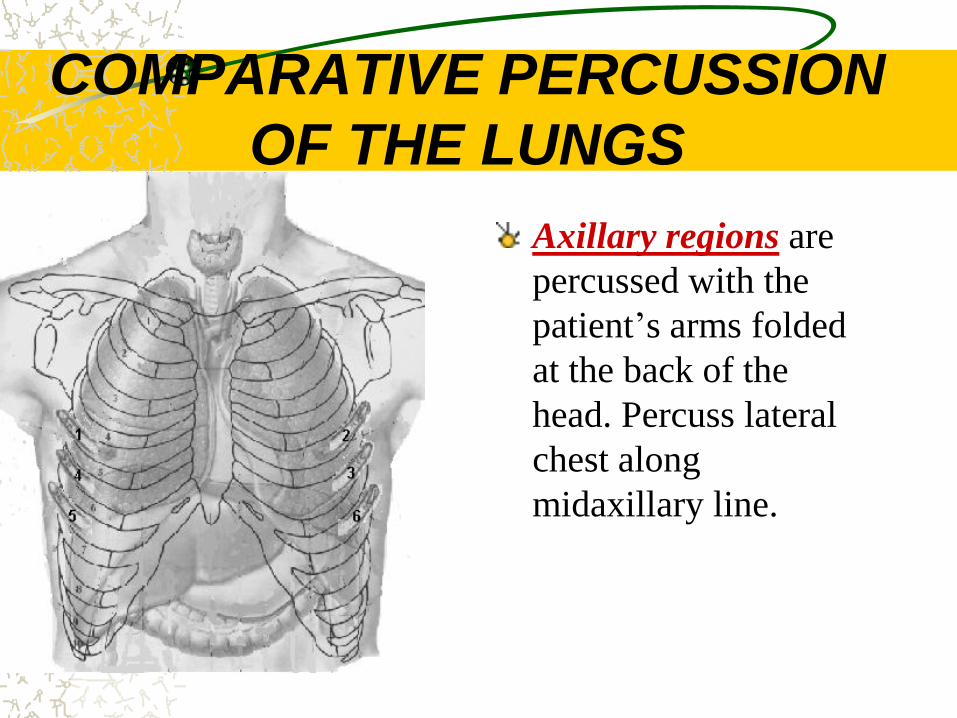
Axillary regions are
percussed with the
patient’s arms folded
at the back of the
head. Percuss lateral
chest along
midaxillary line.
COMPARATIVE PERCUSSION
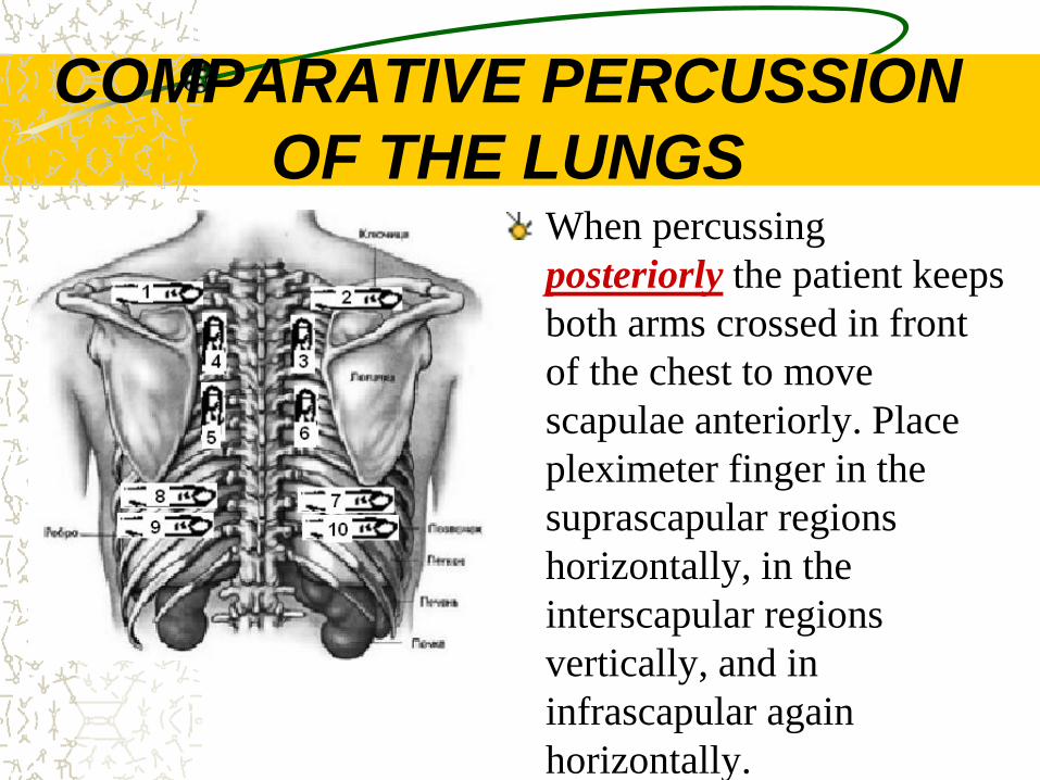
OF THE LUNGS When percussing
posteriorly the patient keeps
both arms crossed in front
of the chest to move
scapulae anteriorly. Place
pleximeter finger in the
suprascapular regions
horizontally, in the
interscapular regions
vertically, and in
infrascapular again
horizontally.
COMPARATIVE PERCUSSION
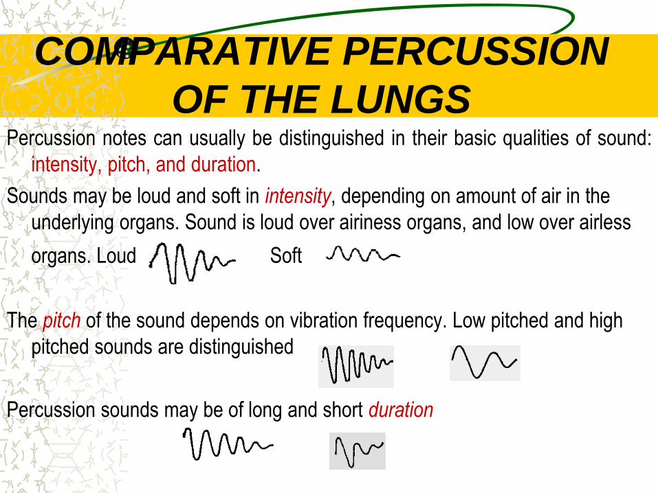
OF THE LUNGS Percussion notes can usually be distinguished in their basic qualities of sound:
intensity, pitch, and duration.
Sounds may be loud and soft in intensity, depending on amount of air in the
underlying organs. Sound is loud over airiness organs, and low over airless
organs. Loud Soft
The pitch of the sound depends on vibration frequency. Low pitched and high
pitched sounds are distinguished
Percussion sounds may be of long and short duration
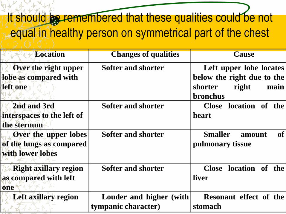
It should be remembered that these qualities could be not
equal in healthy person on symmetrical part of the chest
Location Changes of qualities Cause
Over the right upper
lobe as compared with
left one
Softer and shorter Left upper lobe locates
below the right due to the
shorter right main
bronchus
2nd and 3rd
interspaces to the left of
the sternum
Softer and shorter Close location of the
heart
Over the upper lobes
of the lungs as compared
with lower lobes
Softer and shorter Smaller amount of
pulmonary tissue
Right axillary region
as compared with left
one
Softer and shorter Close location of the
liver
Left axillary region Louder and higher (with
tympanic character)
Resonant effect of the
stomach

COMPARATIVE PERCUSSION
OF THE LUNGS The air-containing lung tissue will give a clear pulmonary sound
(resonance) in percussion.
Comparative percussion helps to determine whether the underlying
tissues are air-filled, fluid-filled, or solid. The common cause of percussion
notes changes include:
The common cause of percussion notes changes include:
decreased airiness of the pulmonary tissue or full absence of air in
a part of the lung;
increased airiness of the pulmonary tissue;
pleural accumulation of fluid;
pleural accumulation of air.
COMPARATIVE PERCUSSION
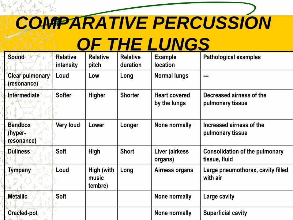
OF THE LUNGS Sound Relative
intensity
Relative
pitch
Relative
duration
Example
location
Pathological examples
Clear pulmonary
(resonance)
Loud Low Long Normal lungs ---
Intermediate Softer Higher Shorter Heart covered
by the lungs
Decreased airness of the
pulmonary tissue
Bandbox
(hyper-
resonance)
Very loud Lower Longer None normally Increased airness of the
pulmonary tissue
Dullness Soft High Short Liver (airkess
organs)
Consolidation of the pulmonary
tissue, fluid
Tympany Loud High (with
music
tembre)
Long Airness organs Large pneumothorax, cavity filled
with air
Metallic Soft None normally Large cavity
Cracled-pot None normally Superficial cavity
COMPARATIVE PERCUSSION
OF THE LUNGS
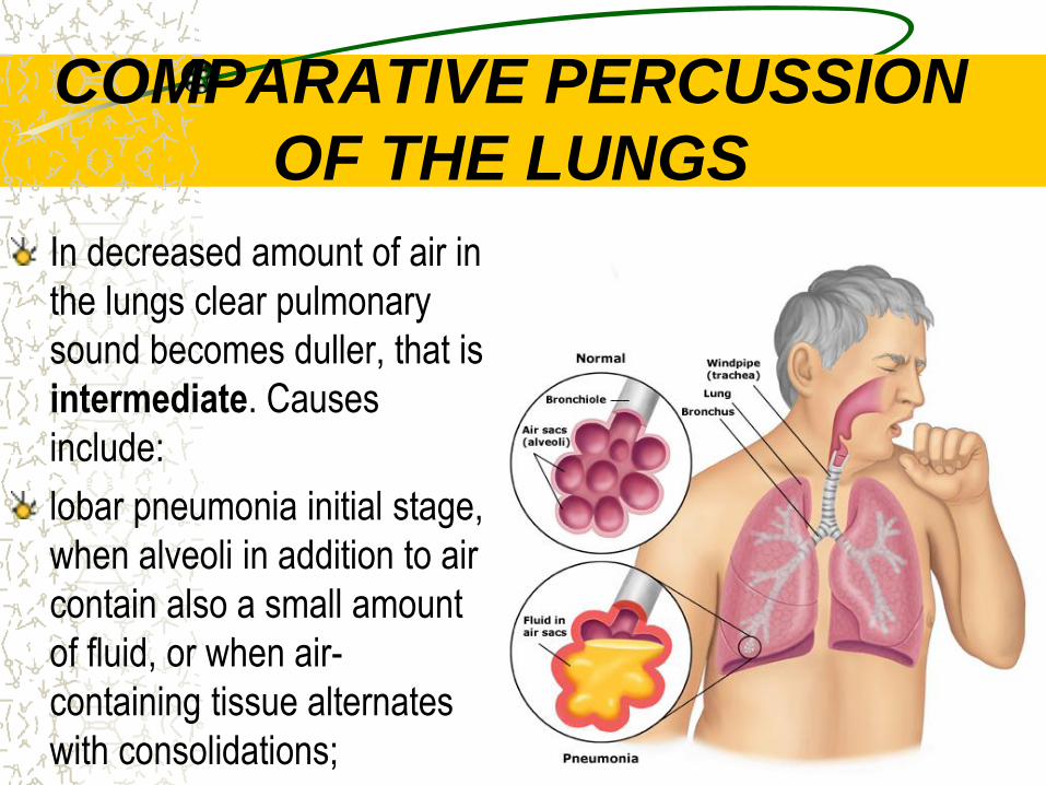
In decreased amount of air in
the lungs clear pulmonary
sound becomes duller, that is
intermediate. Causes
include:
lobar pneumonia initial stage,
when alveoli in addition to air
contain also a small amount
of fluid, or when air-
containing tissue alternates
with consolidations;
COMPARATIVE PERCUSSION
OF THE LUNGS
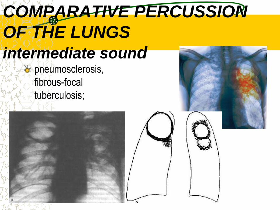
intermediate sound pneumosclerosis,
fibrous-focal
tuberculosis;
COMPARATIVE PERCUSSION
OF THE LUNGS
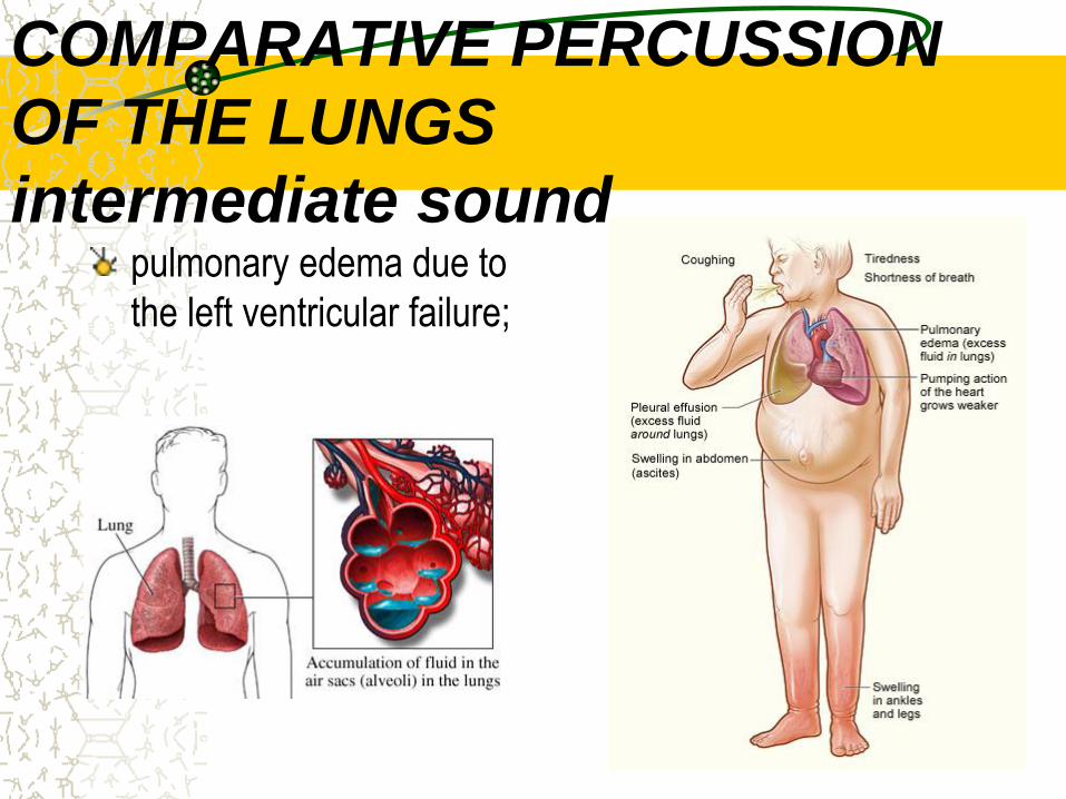
intermediate sound pulmonary edema due to
the left ventricular failure;
COMPARATIVE PERCUSSION
OF THE LUNGS
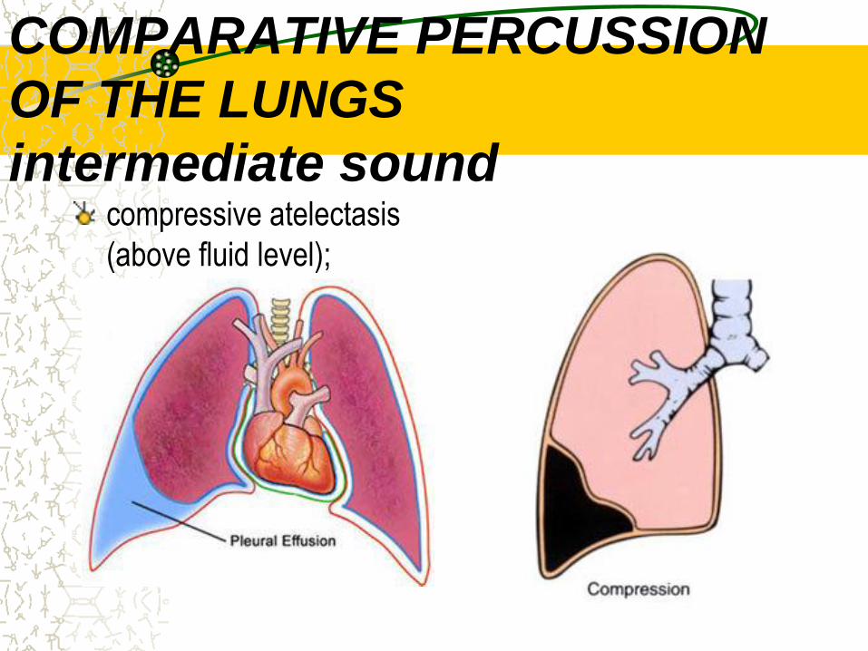
intermediate sound compressive atelectasis
(above fluid level);
COMPARATIVE PERCUSSION
OF THE LUNGS
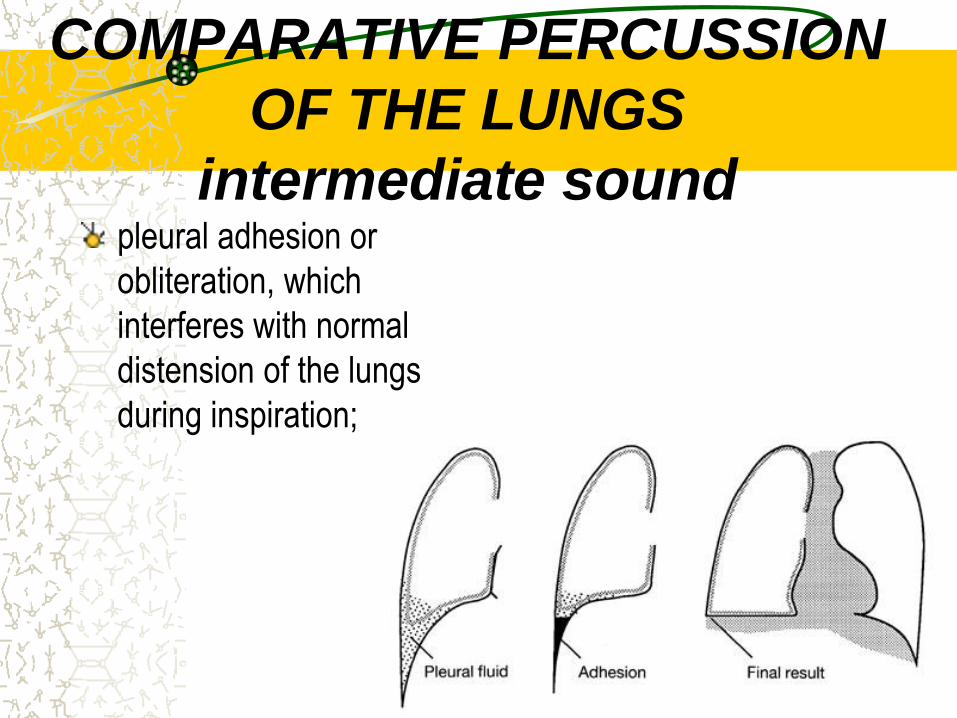
intermediate sound pleural adhesion or
obliteration, which
interferes with normal
distension of the lungs
during inspiration;

COMPARATIVE PERCUSSION OF
THE LUNGS
intermediate sound obstructive atelectasis
due to gradual resorption
of air from the lungs
below obstruction.

COMPARATIVE
PERCUSSION OF THE
LUNGS Dullness replaces
resonance when solid
tissue replaces air-
containing lungs in such
conditions as:
acute lobar pneumonia
(consolidation stage),
when the alveoli are filled
with the exudates;
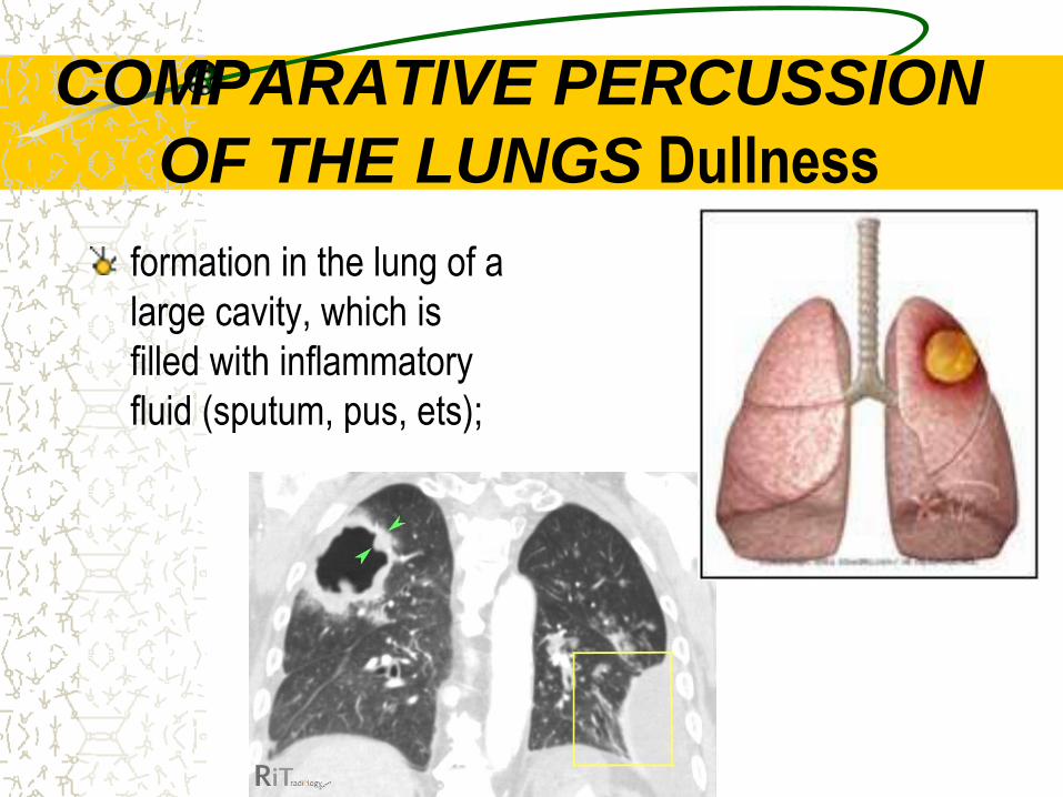
COMPARATIVE PERCUSSION
OF THE LUNGS Dullness
formation in the lung of a
large cavity, which is
filled with inflammatory
fluid (sputum, pus, ets);

COMPARATIVE PERCUSSION
OF THE LUNGS Dullness
pulmonary tumor (airless
tissue);

COMPARATIVE PERCUSSION
OF THE LUNGS Dullness
dullness also heard
when fluid occupies the
pleural space (over fluid):
pleural accumulation of
serous fluid (pleural
effusion), blood
(hemothorax), or pus
(empyema).
COMPARATIVE PERCUSSION
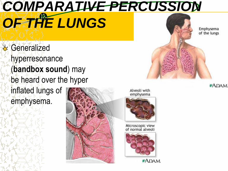
OF THE LUNGS
Generalized
hyperresonance
(bandbox sound) may
be heard over the hyper
inflated lungs of
emphysema.
COMPARATIVE PERCUSSION
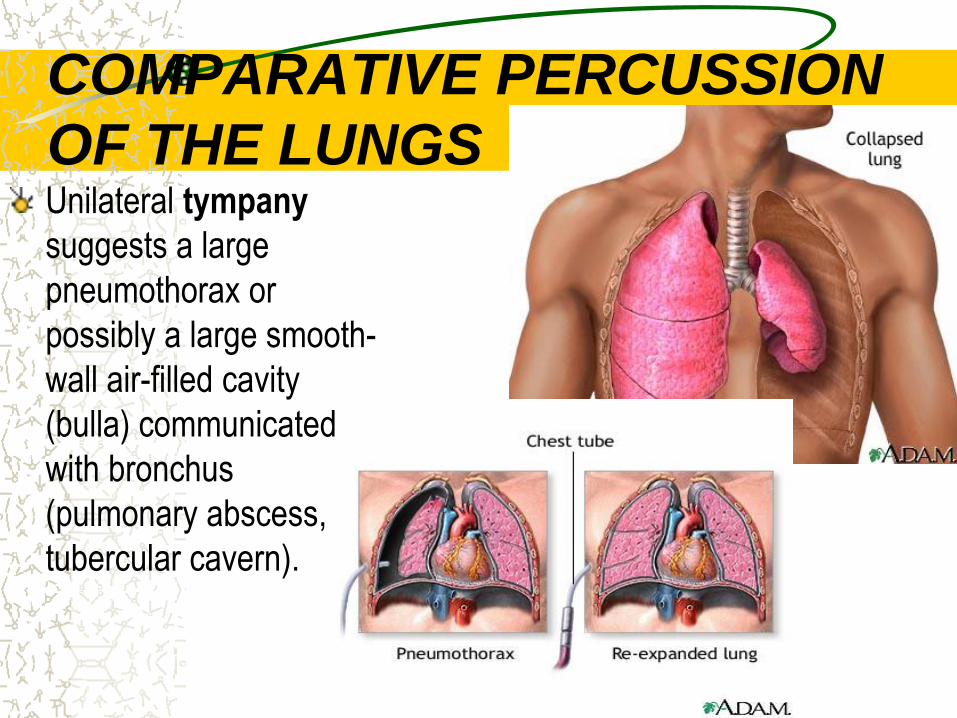
OF THE LUNGS Unilateral tympany
suggests a large
pneumothorax or
possibly a large smooth-
wall air-filled cavity
(bulla) communicated
with bronchus
(pulmonary abscess,
tubercular cavern).

COMPARATIVE PERCUSSION
OF THE LUNGS
Metallic percussion
sound: tympanic sound
resembling a stroke on a
metal may be heard over
a large (6-8 cm in
diameter) air-filled bulla
in the lungs.

COMPARATIVE PERCUSSION
OF THE LUNGS
Cracked-pot percussion
sound (soft, resembles that
of a cracked pot) may be
heard over a large
superficial cavity
communicated with the
bronchus through the narrow
slit.

Topographic percussion
of the lungs
Topographic percussion has following potential uses:
determination of the upper borders (apices) of the lungs;
determination of the lower borders of the lungs;
determination of the excursion of the lower borders of the lungs.
Topographic percussion
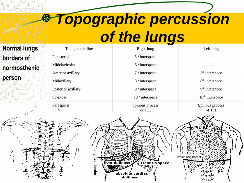
of the lungs Normal lungs
borders of
normosthenic
person
Topographic lines
Right lung
Left lung
Parasternal
5th interspace
---
Midclavicular
6th interspace
---
Anterior axillary
7th interspace
7th interspace
Midaxillary
8th interspace
8th interspace
Posterior axillary
9th interspace
9th interspace
Scapular
10th interspace
10th interspace
Paraspinal
Spinous process
of T11
Spinous process
of T11
Topographic percussion
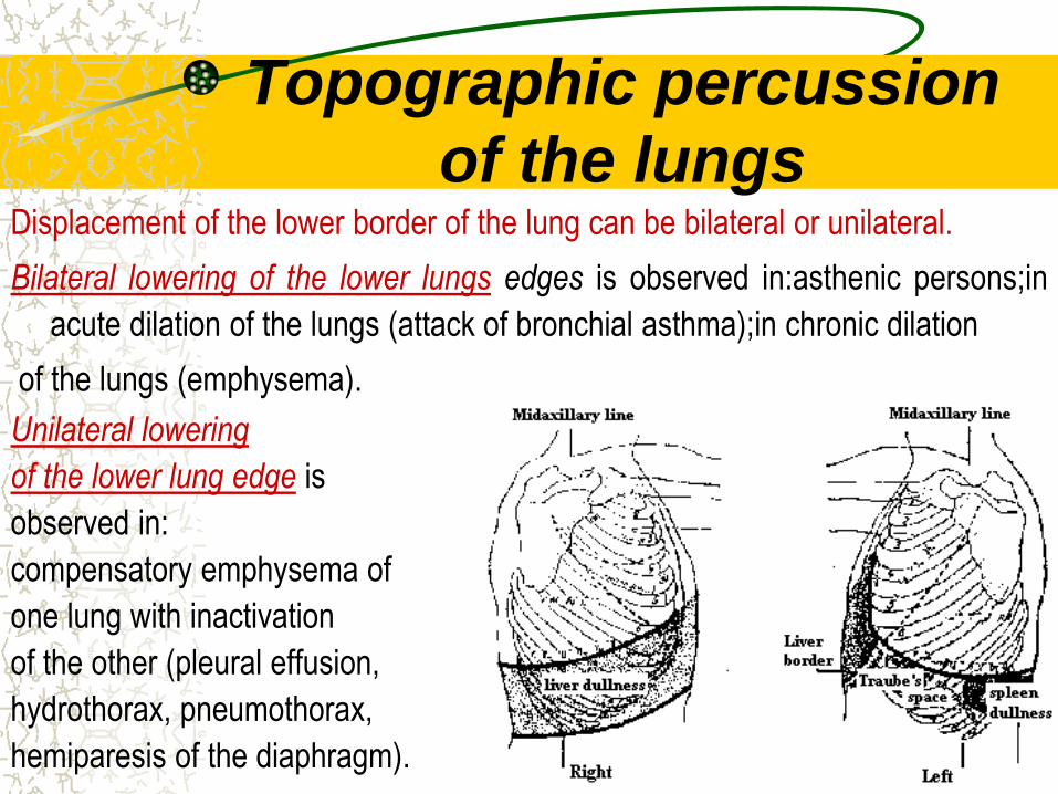
of the lungs Displacement of the lower border of the lung can be bilateral or unilateral.
Bilateral lowering of the lower lungs edges is observed in:asthenic persons;in
acute dilation of the lungs (attack of bronchial asthma);in chronic dilation
of the lungs (emphysema).
Unilateral lowering
of the lower lung edge is
observed in:
compensatory emphysema of
one lung with inactivation
of the other (pleural effusion,
hydrothorax, pneumothorax,
hemiparesis of the diaphragm).
Topographic percussion
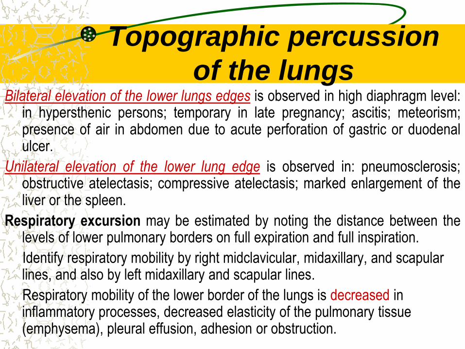
of the lungs Bilateral elevation of the lower lungs edges is observed in high diaphragm level:
in hypersthenic persons; temporary in late pregnancy; ascitis; meteorism; presence of air in abdomen due to acute perforation of gastric or duodenal ulcer.
Unilateral elevation of the lower lung edge is observed in: pneumosclerosis; obstructive atelectasis; compressive atelectasis; marked enlargement of the liver or the spleen.
Respiratory excursion may be estimated by noting the distance between the levels of lower pulmonary borders on full expiration and full inspiration.
Identify respiratory mobility by right midclavicular, midaxillary, and scapular lines, and also by left midaxillary and scapular lines.
Respiratory mobility of the lower border of the lungs is decreased in inflammatory processes, decreased elasticity of the pulmonary tissue (emphysema), pleural effusion, adhesion or obstruction.