organ systems and body cavities
TRANSCRIPT

15
2E X E R C I S E
Organ Systems andBody Cavities
O b j e c t i v e s M a te r i a l s
ACTIVITY 1 Identifying Organ Systems
Identify the organ systems in Figure 2.1 using the organsystem list provided. Refer to Table 2.1.
A. Overview of Organ Systems
The body stays alive due to the interaction of differentorgan systems. An organ system is a group of organs per-forming a common function that helps provide the cellsof the body with an optimal environment. All organ sys-tems cooperate to maintain this optimal environmentthrough a process called homeostasis (homeo- � same;-stasis � standing). Failure to maintain homeostasis resultsin disorders and disease. Organ system functions and majororgans are summarized in Table 2.1.
• human torso models or charts
• male and female human reproductive models orcharts
• articulated skeleton
• one-gallon zippered plastic bags (2 per group)
• masking tape
After completing this exercise,you should be able to:
• Name the organ systems and describe thefunctions of each
• Name the major organs of each organ systemand identify them on models or charts
• Describe the location of the body cavities and namethe organs they contain
• Describe the structure and location of the serousmembranes
• Identify the abdominopelvic quadrants and theorgans found in each
• Identify the abdominopelvic regions and the majororgans found in each
0907T_c02_15-32.qxd 12/1/04 14:52 Page 15 EQA

16 E X E R C I S E 2 O R G A N S Y S T E M S A N D B O D Y C A V I T I E S
TA B L E 2 . 1 Functions and Major Organs of the Organ Systems
O R G A N S Y S T E M F U N C T I O N
Cardiovascular Transports nutrients, chemical messengers, gases, and wastes in blood.Major organs: heart and blood vessels
Respiratory Adds oxygen to blood and removes carbon dioxide from blood; maintenance of carbon dioxidelevels helps regulate pH.
Major organs: nose, pharynx (throat), trachea, bronchi, lungs
Digestive Breaks down food into units that can be absorbed into the body; eliminates wastes andnondigestible fiber in food.
Major organs: mouth, pharynx, esophagus, stomach, intestines, pancreas, liver, gallbladder
Urinary Removes nitrogenous wastes; maintains body fluid volume, pH, and electrolyte levels throughurine production.
Major organs: kidneys, ureters, urinary bladder, urethra
Integumentary Provides a protective barrier for the body and aids in production of vitamin D; contains sensoryreceptors for pain, touch, and temperature; thermoregulation.
Major organs: skin and skin structures (hair, nails, sweat glands, oil glands)
Lymphatic and Immune Returns fluid to cardiovascular system; detects, filters, and eliminates disease-causing organisms,including cancer cells.
Major organs: lymphatic vessels, lymph nodes, spleen, thymus, bone marrow
Skeletal Protects major organs; provides levers and support for body movement.Major organs: bones
Muscular Moves bones and maintains posture.Major organs: skeletal muscles
Nervous Controls cell function with electrical signals; helps control body homeostasis.Major organs: brain, spinal cord, nerves
Endocrine Controls cell function with hormones; helps control body homeostasis.Major organs: hypothalamus, pituitary, thyroid, pancreas, adrenal glands, ovaries, testes
Reproductive Produces gametes; female uterus provides environment for development of fetus.Major organs in the male: testes, epididymis, ductus deferens, prostate penisMajor organs in the female: ovaries, uterine tubes, uterus, vagina
0907T_c02_15-32.qxd 12/1/04 14:52 Page 16 EQA

E X E R C I S E 2 O R G A N S Y S T E M S A N D B O D Y C A V I T I E S 17
FIGURE 2.1 Organ systems and selected organs.
1 2
3 4
65
Hair
Skin and associated glands
Fingernails(and toenails)
Bone
Cartilage
Joint
Skeletalmuscle
Tendon
Blood vessels:
ArteryVein
Heart
Lymphnode
Lymphaticvessel
Spleen
Tonsil
Thymus
Thoracicduct Nerve
Spinalcord
Brain
0907T_c02_15-32.qxd 12/1/04 16:28 Page 17 EQA

18 E X E R C I S E 2 O R G A N S Y S T E M S A N D B O D Y C A V I T I E S
7
9
8
10
11
FIGURE 2.1 Organ systems and selected organs, continued.
Ovary
Pancreas
Thyroidgland
Pinealgland
Thymus
Pituitarygland
Testis
Adrenalgland
LungBronchus
Larynx(voice box)
Pharynx
Trachea(windpipe)
Pancreas(posterior tostomach)
Stomach
Liver
Salivarygland
Esophagus
Mouth
Anus
Gallbladder
Largeintestine
Smallintestine
Pharynx
Ureter
Kidney
Urethra
Urinarybladder
Ovary
Mammarygland
Uterine(fallopian)tube
Uterus
Vagina
Ductus (vas)deferens
Testis
PenisProstate
Seminalvesicle
0907T_c02_15-32.qxd 12/1/04 16:28 Page 18 EQA

B. Identification of MajorOrgans on a Torso Model
You will be identifying organs from anterior to posterior ona torso and answering questions concerning their positionrelative to the organs around them.
ACTIVITY 2 Identif ication of Organs
1 Identify the following organs on the anterior surface ofa torso model. Identify all the organs without removingany organs from the model.• brain• trachea• heart• lungs• liver• stomach (left side of torso)• small intestine• large intestine (colon)
2 Remove the lungs, heart, liver, and stomach. Locate thegallbladder on the inferior surface of the liver.
3 Identify the following organs on the human torso modelor chart.• esophagus• bronchi (right and left) • inferior vena cava• pancreas (posterior to stomach) • spleen
4 Remove the small intestine and large intestine. Locatethe appendix at the inferior right end of the largeintestine.
5 Identify the following organs on the human torso model. • abdominal aorta• adrenal glands (superior surface of kidneys) • kidneys• ureters• urinary bladder
6 Identify the female reproductive organs on a femalereproductive model or chart. Observe the position of theurinary bladder relative to the uterus. • ovaries• uterus• urinary bladder
7 Identify the male reproductive organs on a male repro-ductive model or chart. • penis• scrotum (skin covering testes) • testes
8 Answer the questions about the position of each organ.
1. The stomach is to the small intestine.
a. superior b. inferior c. medial d. lateral
2. The liver is to the lungs.
a. superior b. inferior c. medial d. lateral
3. The lungs are to the heart.
a. superior b. inferior c. medial d. lateral
4. The trachea is to the esophagus.
a. medial b. inferior c. anterior d. posterior
5. The pancreas is to the stomach.
a. superior and medial b. superior and anteriorc. anterior and lateral d. posterior and inferior
6. The gallbladder is on the surface of theliver.
a. superior b. inferior c. posterior d. lateral
7. The stomach is to the spleen.
a. lateral b. medial c. superior d. inferior
8. The abdominal aorta and inferior vena cava areto the kidneys.
a. medial b. lateral c. superior d. inferior
9. The kidneys are to the small intestine.
a. anterior b. posterior c. superior d. inferior
10. The urinary bladder is to the uterus.
a. posterior and superior b. anterior and inferior c. medial and superior d. lateral and posterior
E X E R C I S E 2 O R G A N S Y S T E M S A N D B O D Y C A V I T I E S 19
0907T_c02_15-32.qxd 12/1/04 14:52 Page 19 EQA

C. Body Cavities
The two major cavities of the body are the dorsal andventral body cavities. The dorsal body cavity is locatednear the posterior surface of the body and has two mainsubdivisions: the cranial cavity that contains the brain andthe vertebral (vertebra � back) canal that contains thespinal cord. The cranial cavity and the vertebral canal arecontinuous.
The ventral body cavity is located near the anterior sur-face of the body and has two subdivisions, the thoracic cav-ity and the abdominopelvic cavity, which are separated bythe diaphragm. The thoracic cavity is a space enclosed bythe ribs, sternum, and vertebral column. This cavity con-tains three small cavities: the pericardial cavity ( peri- �around; -cardia � heart) and two pleural cavities ( pleuro-� side or rib). The pericardial cavity surrounds the heart,and each pleural cavity contains a lung. The mediastinum(media- � middle; -stinum � partition), a central areawithin the thoracic cavity, extends from the neck to thediaphragm and from the sternum to the vertebral column.The organs located in the mediastinum are the heart,thymus gland, esophagus, trachea and bronchi. The pleuralcavities are located on either side of the mediastinum.
The abdominopelvic cavity consists of two continuouscavities: the abdominal cavity and the pelvic cavity. Theabdominal cavity is the superior portion located between
the diaphragm superiorly and the brim of the pelvis inferi-orly. This cavity contains the stomach, liver, gallbladder,pancreas, spleen, small intestine, kidneys, appendix, andpart of the large intestine. The pelvic cavity, located be-tween the pelvic brim superiorly and the body wall inferi-orly, is the inferior portion of the abdominopelvic cavity.The pelvic cavity contains part of the large intestine, rec-tum, urinary bladder, female reproductive organs (ovaries,uterine tubes, uterus, vagina), and male reproductive or-gans (prostate, part of ductus deferens). It is important tonote that the testes and penis are not located in the ventralbody cavity but are outside the body wall.
ACTIVITY 3 Body Cavities
1 Label the dorsal and ventral body cavities, their sub-divisions, and the diaphragm on Figure 2.2(a) and (b).
2 Locate the dorsal body cavity and its subdivisions on askeleton and torso model.
3 Locate the ventral body cavity, its subdivisions, and thediaphragm on a torso model.
4 Locate the mediastinum (meed-ee-uh-STINE-um) on atorso model.
5 Using a torso model, complete Table 2.2 by listing themajor organ(s) found in each body cavity.
20 E X E R C I S E 2 O R G A N S Y S T E M S A N D B O D Y C A V I T I E S
0907T_c02_15-32.qxd 12/1/04 14:52 Page 20 EQA

E X E R C I S E 2 O R G A N S Y S T E M S A N D B O D Y C A V I T I E S 21
(a) Lateral view (b) Anterior view
1
2
3
4
5
8
6
7
12
1314
15
10
9
11
(a) Lateral view• abdominal cavity• abdominopelvic cavity• cranial cavity• diaphragm• dorsal cavity• pelvic cavity• thoracic cavity• ventral cavity• vertebral canal
(b) Anterior view• abdominal cavity• abdominopelvic cavity• brim of pelvis• diaphragm• pelvic cavity• thoracic cavity
1
2
3
4
5
6
7
8
9
10
11
12
13
14
15
FIGURE 2.2 Body cavities.
TA B L E 2 . 2 Body Cavities
B O D Y C A V I T Y O R G A N S
Dorsal body cavity
Cranial cavity
Vertebral canal
Ventral body cavity
Thoracic cavity
Pleural cavities (2)
Mediastinum
Abdominopelvic cavity
Abdominal cavity
Pelvic cavity
1.
2.
3.
4. (Within pericardial cavity)
6.
9.
12.
15.
18.
21.
24.
7.
10.
13.
16.
19.
22.
25.
5.
8.
11.
14.
17.
20.
23.
26.
0907T_c02_15-32.qxd 2/11/05 11:36 Page 21 EQA

D. Serous Membranes
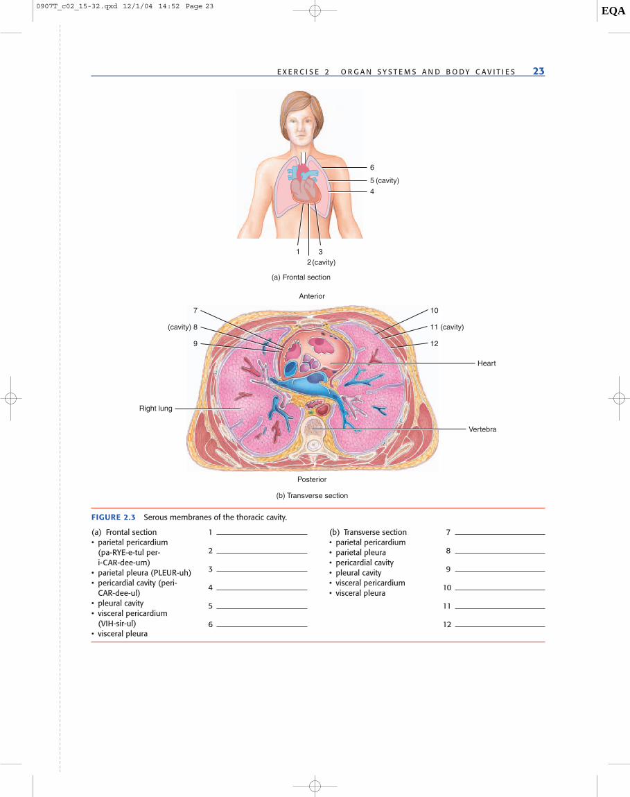
Most of the organs in the ventral body cavity are coveredwith serous (serum � any clear, watery fluid) membranes,which are composed of two layers: a visceral layer and aparietal layer. The visceral (viscera � internal organs)layer covers the organ, whereas the parietal ( paries �wall) layer attaches to and covers the ventral body wall.These two layers comprise one continuous sheet that foldsto form a sac. Between the two layers is a small amount ofserous fluid secreted by the membranes. The clear, wateryserous fluid prevents friction as the organs move withinthe ventral body cavity. For example, the heart has move-ment within the thoracic cavity as it fills with and ejectsblood.
Serous membranes are named for the cavities they sur-round. Thoracic serous membranes include the pleura,which covers the lungs, and the pericardium, which cov-ers the heart. The serous membrane that covers the abdomi-nal organs is the peritoneum ( peri- � around; teinein � tostretch). Although most abdominal organs are positionedwithin the peritoneal cavity, a few organs are retroperit-oneal (retro- � backward), or located posterior to the peri-toneum. These organs are the pancreas, kidneys, adrenalglands, and portions of the large intestine, small intestine,aorta, and inferior vena cava. The peritoneum has three ma-jor double-layered folds called the mesentery, the greateromentum, and the lesser omentum. The mesentery encir-cles and holds the small intestine to the dorsal body wall.The greater omentum is suspended from the inferior por-tion of the stomach and covers the intestines in a similarmanner to an apron. The lesser omentum suspends the su-perior portion of the stomach to the liver.
ACTIVITY 4 Serous Membranes
1 Label the serous membranes and cavities in the thoraciccavity in Figures 2.3(a) and (b).
2 Label the serous membranes and cavities in the abdom-inal cavity in Figures 2.4(a) and (b).
3 Make a replica or model of a serous membrane withyour lab group. (This can be done as a demonstration.)• Obtain 2 one-gallon zippered plastic bags.• Make sure all the air is out of the first bag and then
rezip the bag. • In the second bag, add about 40 to 50 mL of water
and push out the extra air before rezipping.• Have a lab partner place a fist (simulating an organ)
on the bottom edge of the first bag and push up intothe bag.
• Now have the same lab partner place a fist on thebottom edge of the second bag and push up into thebag.
4 Visit www.wiley.com/college/allen to observe humanserous membranes.
5 Answer the discussion questions with your lab partners.
DISCUSSION QUESTIONS:SEROUS MEMBRANES
1 Was it easier to push a fist into the bag with no water orinto the bag with water? Explain.
2 In the bag with water, what is the name of the simulatedserous membrane layer that is touching the fist?
3 In the same bag, what is the name of the simulated outerserous membrane layer?
4 What does the water represent?
22 E X E R C I S E 2 O R G A N S Y S T E M S A N D B O D Y C A V I T I E S
?
0907T_c02_15-32.qxd 12/1/04 16:28 Page 22 EQA
E X E R C I S E 2 O R G A N S Y S T E M S A N D B O D Y C A V I T I E S 23
(a) Frontal section
(cavity)
(cavity)
12
3
4
5
6
Posterior
Anterior
(b) Transverse section
Heart
10
Vertebra
Right lung
11 (cavity)
12
7
(cavity) 8
9
FIGURE 2.3 Serous membranes of the thoracic cavity.
(a) Frontal section• parietal pericardium
(pa-RYE-e-tul per-i-CAR-dee-um)
• parietal pleura (PLEUR-uh)• pericardial cavity (peri-
CAR-dee-ul)• pleural cavity• visceral pericardium
(VIH-sir-ul)• visceral pleura
1
2
3
4
5
6
(b) Transverse section• parietal pericardium• parietal pleura• pericardial cavity• pleural cavity• visceral pericardium• visceral pleura
7
8
9
10
11
12
0907T_c02_15-32.qxd 12/1/04 14:52 Page 23 EQA

FIGURE 2.4 Serous membranes of the abdominal cavity.
1
Transverse Mesocolon
2
3
4
5
6
Diaphragm
Liver
Pancreas
Stomach
Duodenum
Transverse colon
Jejunum
Rectum
Uterus
Sigmoid colon
Ileum
Urinary bladder
Pubic symphysis
(a) Sagittal section through the lower body
ANTERIORPOSTERIOR
Midsagittal plane
(a) Sagittal section• greater omentum• lesser omentum• mesentery• parietal peritoneum
(per-i-toe-NEE-um)• peritoneal cavity
(per-i-toe-NEE-ul) • visceral peritoneum
1
2
3
4
5
6
(b) Transverse section• parietal peritoneum• peritoneal cavity• visceral pertoneum
7
8
9
Stomach
Small intestine
(cavity) 8
7
9
Vena cavaLiver
Kidney
(b) Transverse section through abdomen
Pancreas
Aorta
Spleen
0907T_c02_15-32.qxd 2/11/05 08:37 Page 24 EQA
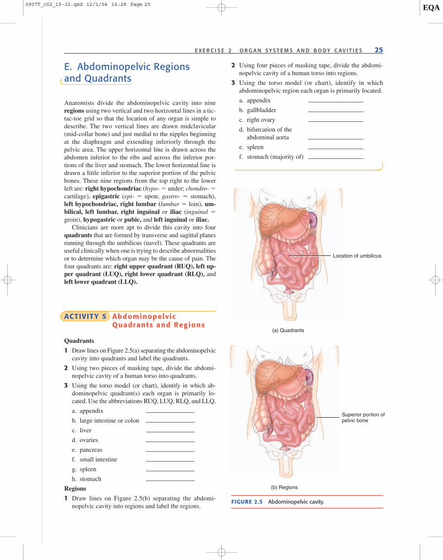
E. Abdominopelvic Regionsand Quadrants
Anatomists divide the abdominopelvic cavity into nineregions using two vertical and two horizontal lines in a tic-tac-toe grid so that the location of any organ is simple todescribe. The two vertical lines are drawn midclavicular(mid-collar bone) and just medial to the nipples beginningat the diaphragm and extending inferiorly through thepelvic area. The upper horizontal line is drawn across theabdomen inferior to the ribs and across the inferior por-tions of the liver and stomach. The lower horizontal line isdrawn a little inferior to the superior portion of the pelvicbones. These nine regions from the top right to the lowerleft are: right hypochondriac (hypo- � under; chondro- �cartilage), epigastric (epi- � upon; gastro- � stomach),left hypochondriac, right lumbar (lumbar � loin), um-bilical, left lumbar, right inguinal or iliac (inguinal �groin), hypogastric or pubic, and left inguinal or iliac.
Clinicians are more apt to divide this cavity into fourquadrants that are formed by transverse and sagittal planesrunning through the umbilicus (navel). These quadrants areuseful clinically when one is trying to describe abnormalitiesor to determine which organ may be the cause of pain. Thefour quadrants are: right upper quadrant (RUQ), left up-per quadrant (LUQ), right lower quadrant (RLQ), andleft lower quadrant (LLQ).
ACTIVITY 5 AbdominopelvicQuadrants and Regions
Quadrants
1 Draw lines on Figure 2.5(a) separating the abdominopelviccavity into quadrants and label the quadrants.
2 Using two pieces of masking tape, divide the abdomi-nopelvic cavity of a human torso into quadrants.
3 Using the torso model (or chart), identify in which ab-dominopelvic quadrant(s) each organ is primarily lo-cated. Use the abbreviations RUQ, LUQ, RLQ, and LLQ.
a. appendix
b. large intestine or colon
c. liver
d. ovaries
e. pancreas
f. small intestine
g. spleen
h. stomach
Regions
1 Draw lines on Figure 2.5(b) separating the abdomi-nopelvic cavity into regions and label the regions.
2 Using four pieces of masking tape, divide the abdomi-nopelvic cavity of a human torso into regions.
3 Using the torso model (or chart), identify in whichabdominopelvic region each organ is primarily located.
a. appendix
b. gallbladder
c. right ovary
d. bifurcation of theabdominal aorta
e. spleen
f. stomach (majority of)
E X E R C I S E 2 O R G A N S Y S T E M S A N D B O D Y C A V I T I E S 25
(a) Quadrants
Location of umbilicus
(b) Regions
Superior portion ofpelvic bone
FIGURE 2.5 Abdominopelvic cavity.
0907T_c02_15-32.qxd 12/1/04 16:28 Page 25 EQA

0907T_c02_15-32.qxd 12/1/04 14:52 Page 26 EQA

27
Reviewing Your Knowledge
A. Functions of Organ Systems
Identify the organ system whose function is described below.
1. Maintains blood oxygen and carbon dioxide levels.
2. Controls muscles and glands by electrical impulses; helps control homeostasis.
3. Causes movement of bones.
4. Waterproof barrier that blocks the entrance of pathogens into the body and the loss ofwater from the body.
5. Transports nutrients, oxygen, and carbon dioxide throughout the body.
6. Changes food into absorbable nutrients; expels wastes.
7. Regulates composition of blood by eliminating nitrogenous wastes, excess water, andminerals.
8. Uses hormones to control cell function; helps control homeostasis.
9. Provides framework for the body and protects body organs.
10. Produces gametes (sperm and egg).
11. Returns fluid to the bloodstream and provides protection against pathogens that haveentered the body.
B. Organ Identification
Identify the correct organ system for the following organs.
Organ System Organ
1. spleen
2. liver
3. trachea
4. blood vessels
5. hair
Name Date Section
2E X E R C I S E
0907T_c02_15-32.qxd 12/1/04 16:28 Page 27 EQA

6. kidney
7. uterus
8. pituitary gland
9. spinal cord
10. testes (2 systems)
11. prostate gland
12. large intestine
13. pancreas (2 systems)
14. adrenal gland
15. thyroid
C. Body Cavities
Identify all the cavities for each organ as follows: dorsal (D), ventral (VN), cranial (C), vertebral (VR), thoracic (T),pleural (PL), pericardial (PC), peritoneal (PT), abdominal (A), or pelvic (P). All structures are present in more thanone cavity, and some are in three cavities.
1. brain
2. small intestine
3. heart
4. lungs
5. bronchi
6. stomach
28 E X E R C I S E 2 O R G A N S Y S T E M S A N D B O D Y C A V I T I E S
7. spinal cord
8. liver
9. kidneys
10. uterus
11. urinary bladder
12. ovaries
D. Serous Membranes
Write the term the phrase describes.
1. Extends from the stomach and drapes over the intestines.
2. Attaches the heart to the body cavity.
3. Covers the surface of the lungs.
4. Covers the surface of abdominal organs.
5. The lubricating liquid in serous cavities.
6. Extends between the superior part of the stomach and the liver.
0907T_c02_15-32.qxd 12/1/04 16:28 Page 28 EQA
7. Attaches the small intestine to the posterior body wall.
8. Circle the organs that are found within the peritoneal cavity: pancreas, liver, kidney, spleen,adrenal glands, abdominal aorta, inferior vena cava, stomach.
E. Abdominopelvic Quadrants and Regions
QuadrantsName the quadrant(s) that the following organs predominantly occupy: RUQ, LUQ, RLQ, and LLQ.
1. liver
2. stomach
3. spleen
4. right adrenal gland
5. gallbladder
E X E R C I S E 2 O R G A N S Y S T E M S A N D B O D Y C A V I T I E S 29
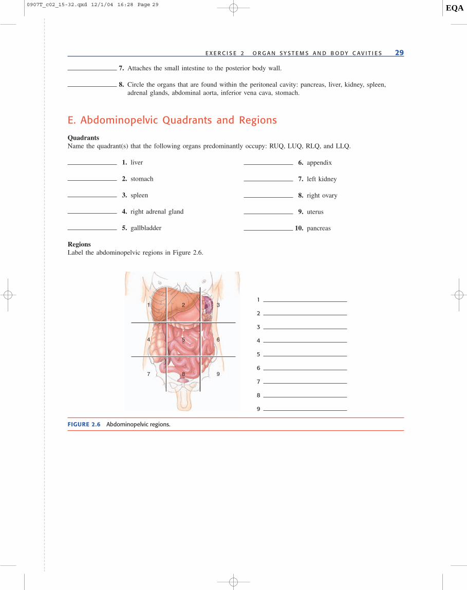
RegionsLabel the abdominopelvic regions in Figure 2.6.
6. appendix
7. left kidney
8. right ovary
9. uterus
10. pancreas
5
2
6
3
98
4
1
7
FIGURE 2.6 Abdominopelvic regions.
1
2
3
4
5
6
7
8
9
0907T_c02_15-32.qxd 12/1/04 16:28 Page 29 EQA

0907T_c02_15-32.qxd 12/1/04 14:52 Page 30 EQA

31
Using Your Knowledge
A. Homeostatic Imbalances of Organ Systems
Using your textbook, identify the organ system that is homeostatically imbalanced in the following diseases ordisorders.
Organ System Disease
1. muscular dystrophy
2. multiple sclerosis
3. myocardial ischemia
4. infectious mononucleosis
B. Body Cavities
Identify all the cavities entered for each procedure, beginning with the largest cavity and ending with the most spe-cific body cavity. Abdominal (A); cranial (C); dorsal (D); pelvic (P); pericardial (PC); pleural (PL); peritoneal (PT);thoracic (T); ventral (VN); and vertebral (VR).
5. coronary bypass surgery
6. cholecystectomy (gallbladder removal)
7. spinal tap
C. Serous Membranes and Abdominopelvic Quadrants
8. A 44-year-old man went to the emergency room complaining of severe pain in his RLQ. The doctor palpated thearea and determined that the pain was originating from an organ in that quadrant. Which organ might be involved?
(a) liver (b) appendix (c) gallbladder (d) spleen (e) stomach
9. A 23-year-old woman went to the doctor with the chief complaint of RLQ pain. Which organ is most likely the cause.
(a) adrenal gland (b) ovary (c) gallbladder (d) pancreas (e) kidney
Name Date Section
2E X E R C I S E
0907T_c02_15-32.qxd 12/1/04 14:52 Page 31 EQA

32 E X E R C I S E 2 O R G A N S Y S T E M S A N D B O D Y C A V I T I E S
D. Organ Identification
Identify the organs in the color enhanced medical images in Figure 2.7.
(a) MRI of head and neck.
10
11
12 13 14
(b) Radiograph of thorax.
1516
17
(c) Radiograph of trunk.
18 19 20
(d) Radiograph of urinary tract organs.
FIGURE 2.7 Identification of organs on medical images.
10
11
12
13
14
15
16
17
18
19
20
0907T_c02_15-32.qxd 12/1/04 14:52 Page 32 EQA