original article activation of integrin-erbb2 signaling in ... · integrin-erbb2 signaling in...
TRANSCRIPT

Am J Cancer Res 2014;4(6):776-788www.ajcr.us /ISSN:2156-6976/ajcr0002615
Original ArticleActivation of integrin-ERBB2 signaling in undifferentiated thyroid cancer
Xuguang Zhu1, Yuelin J Zhu2, Dong Wook Kim1, Paul Meltzer2, Sheue-Yann Cheng1
1Laboratory of Molecular Biology, 2Laboratory Genetics Branch, Center for Cancer Research, National Cancer Institute, National Institutes of Health, Bethesda, MD 20892
Received September 18, 2014; Accepted October 15, 2014; Epub November 19, 2014; Published November 30, 2014
Abstract: Undifferentiated thyroid carcinoma is one of the most aggressive human cancers. Although genetic chang-es underlying this aggressive cancer remain to be elucidated, RAS mutations have been frequently identified in it. Mice harboring a mutant thyroid hormone receptor ThrbPV (ThrbPV/PV) spontaneously develop differentiated follicular thyroid carcinoma similar to human thyroid cancer. We recently demonstrated that targeting a RAS mutation (Kras-G12D) to the thyroid of ThrbPV/PV mice (ThrbPV/PVKrasG12D mice) promotes initiation and progression of undifferentiated thyroid cancer. To uncover genes destined to drive the aggressive cancer phenotype, we used cDNA microarrays to compare the gene expression profiles of thyroid cells of KrasG12D mice and thyroid tumor lesions of ThrbPV/PV and ThrbPV/PVKrasG12D mice. Analyses of microarray data identified 14 upstream regulators that were significantly altered in thyroid tumors of ThrbPV/PV and ThrbPV/PVKrasG12D mice. Most of these genes with altered expression function as key regulators in growth factor-induced signaling. Further analysis identified gene expression profiles of markedly elevated integrin levels, acting as upstream activators to stimulate ERBB2-mediated downstream signaling in thy-roid tumors of ThrbPV/PVKrasG12D mice. The present studies uncovered integrin-activated ERBB2 signaling as one of the mechanisms in synergy between TRβPV and KRASG12D signaling to promote aggressive tumor growth in undif-ferentiated thyroid cancer.
Keywords: Growth regulation, thyroid cancer, ERBB2, integrins, microarrays, gene expression
Introduction
Thyroid cancer is the most common malignancy of the endocrine organs. There are 4 follicular cell-derived thyroid cancers: well-differentiated papillary and follicular carcinomas, poorly dif-ferentiated carcinoma, and undifferentiated carcinoma. Although well-differentiated thyroid cancer often has a favorable outcome, the10-year survival rate of undifferentiated thyroid cancer is less than 10% owing to distant metas-tasis and lack of effective treatment [1].
The point mutations in RAS genes together with the mutations in TP53 and CTNNB1genes are among prevalent genetic alterations identified in undifferentiated thyroid cancer [1]. Previously we demonstrated that mice with a mutant thy-roid hormone receptor ThrbPV (ThrbPV/PV) spon-taneously develop well-differentiated follicular thyroid cancer with pathological progression
and metastasis frequency similar to human thy-roid cancer [2, 3]. The PV mutation was origi-nally identified in a patient with resistance to thyroid hormone (RTH) [4]. The PV mutant has completely lost thyroid hormone (T3) binding activity and transcription capacity. It acts to abnormally regulate the expression of the T3 target genes via dominant negative activity.
Recently, we genetically introduced the KrasG12D mutation to express specifically in the thyroids of the ThrbPV/PV mice. We found that double mutant ThrbPV/PVKrasG12D mice have much worse survival than the mice with a single mutation in either the Kras or the Thrb gene as a result of markedly aggressive thyroid tumors [5]. Capsular invasion, vascular invasion, and dis-tant metastases to the lung occur at an earlier age and at a higher frequency than in ThrbPV/PV mice. We identified the occurrence of anaplas-tic foci with a high frequency in the thyroid of

Integrin-ERBB2 signaling in undifferentiated thyroid cancer
777 Am J Cancer Res 2014;4(6):776-788
ThrbPV/PVKrasG12D mice [5]. These anaplastic foci have lost normal thyroid follicular morphology as well as the expression of the paired box 8 gene (Pax8). Importantly, the protein level of PAX8 is inversely correlated with MYC in the undifferentiated thyroid tumors of ThrbPV/
PVKrasG12D mice. Thus, our recent study estab-lished a mouse model of undifferentiated thy-roid cancer and identified MYC as a potential target for treatment [5].
Moreover, we showed that synergistic signaling of oncogenic actions of TRβPV and KrasG12D in thyroid tumors of ThrbPV/PVKrasG12D mice led to the aggressive thyroid tumor growth resulting from rapid cell proliferation [5]. However, the mechanisms underlying the increased cell pro-liferation remain to be elucidated. In the pres-ent studies, we used cDNA microarray analysis to compare gene expression profiles of thyroid cells of KrasG12D mice and thyroid tumors of ThrbPV/PV and ThrbPV/PVKrasG12D mice. We uncov-ered increased integrins-ERBB2 signaling as a novel pathway resulting from the synergistic signaling of oncogenic actions of TRβPV and KrasG12D, thereby promoting the aggressive tumor growth of undifferentiated thyroid can- cer.
Materials and methods
Experimental animals
All animal experiments were performed accord-ing to the protocols approved by the Animal Care and Use Committee at the National Cancer Institute. The ThrbPV/+, KrasLSL-G12D/+, TPO-Cre (Cre) mice and the mice with four different gen-otypes were previously described [2, 5-7]. Thyroids and other tissues were harvested from the mice and wild-type littermates for weighing, histological analysis, and biochemi-cal studies.
Microarray analysis
Biotinylated-aRNA samples from three individu-al mice of each group were used in hybridiza-tion of the GeneChip Mouse Exon 1.0 ST Array (affymetrix, Santa Clara, CA) and scanned on an Affymetrix GeneChip scanner 3000. Data were collected using Affymetrix GCOS software. Data processing and analysis were done by affy, limma, xps, et al R/Bioconductor packag-es (http://www.bioconductor.org). Briefly, the robust multichip average (RMA) method was
used for computing expression measures, the Benjamini and Hochberg method [8] was used for calculating the adjusted p values. Top dif-ferentially expressed genes were selected by the adjusted p values with minimum 1.5 fold change. The differentially expressed genes were further analyzed for enrichment of path-ways and functions using the DAVID bioinfor-matics database [9], Ingenuity Pathway Ana- lysis (IPA, Ingenuity Systems, Inc., Redwood City, CA) and the Gene Set Enrichment Analysis (GSEA) by the Broad Institute [10]. The GEO array data submission is in progress.
RNA extraction and real time RT-PCR valida-tion of microarray data
Total RNA from thyroids was isolated using TRIzol (Invitrogen, Carlsbad, CA) as indicated by the protocol of the manufacturer. Selected genes from microarray data were chosen for real time RT-PCR validation. A total 50-200 ng of RNA extracted from thyroids of wild-type, KrasG12D, ThrbPV/PV, and ThrbPV/PVKrasG12D mice was used in the real-time RT-PCR. The reac-tions were performed with the QuantiTect SYBR RT-PCR kit (Qiagen, Germantown, MD) on an ABI 7900HT system. In each group, four to six samples with triplicates were tested on the tar-get genes. Data were analyzed using Prism V5 (GraphPad Software, Inc., La Jolla, CA). Primers were as follows: for the endogenous control gene mouse glyceraldehyde-3-phosphate dehy-drogenase (Gapdh), forward, 5’-cgtcccgtaga-caaaatggt-3’; reverse, 5’-gaatttgccgtgagtgga- gt-3’. For Itga6, forward, 5’-CAGGTTGTGGA- ACAGCACAT, reverse, 5’-AAGAACAGCCAGGAG- GATGA-3’. For Itgb4, forward, 5’-GAGGGGCCC- TATAGCTCACT-3’; reverse, 5’-GTTGTCCACGAG- CACCTTCT3’. For Itgb1, forward, 5’-TCGTGCA- TGTTGTGGAGACT-3’; reverse, 5’-CACAGTTGTC- ACGGCACTCT-3’. For Itgb3, forward, 5’-TGACA- TCGAGCAGGTGAAAG-3’; reverse, 5’-GAGTAGC- AAGGCCAATGAGC-3’. For Itgav, forward, 5’-GA- TAGAGGCAAGAGCGCAAT-3’; reverse, 5’-AATGC- CCCAGGTGATGTTAG-3’. For Itga5, forward, 5’- GCCAAGAGAGCCGTAGTCTG-3’; reverse, 5’-CC- TTCTGCCTTGGTCCACT-3’. For Fibronectin, for-ward, 5’-GATCGGCAGGGAGAAAATG-3’; reverse, 5’-CAGGTCTACGGCAGTTGTCA-3’.
Western blot analysis
The Western blot analysis was carried out as described by Furumoto et al. [11]. Primary anti-body for GAPDH (#2118) was purchased from
Integrin-ERBB2 signaling in undifferentiated thyroid cancer
778 Am J Cancer Res 2014;4(6):776-788
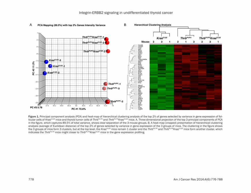
Figure 1. Principal component analysis (PCA) and heat-map of hierarchical clustering analysis of the top 2% of genes selected by variance in gene expression of fol-licular cells of KrasG12D mice and thyroid tumor cells of ThrbPV/PV and ThrbPV/PVKrasG12D mice. A. Three-dimensional projection of the top 3 principal components of PCA in the figure, which captures 89.5% of total variance, shows clear separation of the 3 mouse groups. B. A heat-map (cropped) presentation of hierarchical clustering analysis (average of Euclidean distance) of the top 2% of genes selected by variance in gene expression of the 3 groups of mice. The clustering in the figure shows the 3 groups of mice form 3 clusters, but at the top level, the KrasG12D mice remain 1 cluster and the ThrbPV/PV and ThrbPV/PVKrasG12D mice form another cluster, which indicates the ThrbPV/PV mice might closer to ThrbPV/PVKrasG12D mice in the gene expression profiling.

Integrin-ERBB2 signaling in undifferentiated thyroid cancer
779 Am J Cancer Res 2014;4(6):776-788
Cell Signaling Technology (Danvers, MA). Primary antibodies for Integrin β1 (sc-8978), Integrin β3 (sc-14009), Integrin α5 (sc-10729), Integrin αV (sc-10719), Fibronectin (sc-6952), Integrin β4 (sc-9090), and Integrin α6 (sc-10730) were purchased from Santa Cruz Biotechnology (Santa Cruz, CA). Antibodies were used at the concentration recommended by the manufacturers. For control of protein loading, the blot was probed with the antibody against GAPDH.
Statistical analysis
All data are expressed as means ± standard errors. Statistical analyses were preformed performed and p < 0.05 was considered signifi-cant unless otherwise specified. GraphPad Prism version 5.0 for Mac OS X was used to perform analysis of variances.
Results
Differential gene expression profiles in the thyroid of KrasG12D mice and thyroid tumors of ThrbPV/PV and ThrbPV/PVKrasG12D mice
In our previous studies, we demonstrated that KrasG12D, ThrbPV/PV, and ThrbPV/PVKrasG12D mice had different outcomes of tumorigenesis as the mice aged. KrasG12D mice have normal morphol-ogy up to 10 months old. During the same
observation period, ThrbPV/PV mice develop fol-licular thyroid carcinoma. ThrbPV/PVKrasG12D mice develop aggressive undifferentiated thy-roid carcinoma.
Here we obtained array data from thyroid sam-ples of age-matched KrasG12D, ThrbPV/PV, and ThrbPV/PVKrasG12D mice (n=3 for each type of mice). Figure 1A shows principal component analysis (PCA) of the gene expression profiles from the mice with 3 different genotypes. The 3-dimensional projection of the top 3 principal components of PCA, capturing 89.5% of total variance, shows clear separation of the 3 groups.
A hierarchical clustering analysis shows that the expression of ThrbPV/PVKrasG12D mice was closer to that of ThrbPV/PV mice than that of KrasG12D mice (Figure 1B). However, it is also clear that the gene expression profile in ThrbPV/
PVKrasG12D mice was distinct from that in ThrbPV/
PV mice. Comparison analysis of the array data between ThrbPV/PVKrasG12D and ThrbPV/PV mice showed that 311 genes were differentially expressed (>1.5-fold change, adjusted p < 0.1). Of those 311 genes, 150 were upregulated and 161 were downregulated. Comparison between ThrbPV/PVKrasG12D and KrasG12D mice displayed 2492 genes (>1.5-fold change, adjusted p < 0.1); among them, 1436 genes were upregu-lated and 1056 were downregulated. Com-
Figure 2. A. Differential and common expression genes among KrasG12D, ThrbPV/PV, and ThrbPV/PVKrasG12D mice. B. Top 14 upstream regulators derived from common genes by comparison of ThrbPV/PV vs ThrbPV/PVKrasG12D mice and KrasG12D vs ThrbPV/PVKrasG12D mice.

Integrin-ERBB2 signaling in undifferentiated thyroid cancer
780 Am J Cancer Res 2014;4(6):776-788
parison between ThrbPV/PV and KrasG12D mice identified 1952 differentially expressed genes (>1.5-fold change, adjusted p < 0.1), of which 1143 were upregulated and 809 were downregulated.
Growth factor and growth factor receptor signaling profiles of thyroid tumors in ThrbPV/
PVKrasG12D mice
The distinct clusters of data derived from KrasG12D, ThrbPV/PV, and ThrbPV/PVKrasG12D mice enabled us to compare the changes in gene expression due to either activated TRβPV sig-naling (compare ThrbPV/PVKrasG12D mice with KrasG12D mice), activated KasG12D signaling (compare ThrbPV/PVKrasG12D mice with ThrbPV/PV mice) or synergistic signaling of both TRβPV and KrasG12D signaling in ThrbPV/PVKrasG12D mice. Accordingly, we used a Venn diagram to analyze the relationship of gene expression profiles among the 3 comparisons. Interestingly, only 53 genes were unique to the comparison of ThrbPV/PV vs ThrbPV/PVKrasG12D. Furthermore, 204 genes were common between the comparison of ThrbPV/PV vs ThrbPV/PVKrasG12D and KrasG12D vs ThrbPV/PVKrasG12D. We found 906 genes were specific to the comparisons of KrasG12D vs ThrbPV/PVKrasG12D and 458 were specific to the comparison of KrasG12D vs ThrbPV/PV. In addition, 1440 genes were common between compari-sons of ThrbPV/PVKrasG12D vs ThrbPV/PV and ThrbPV/
PV vs KrasG12D mice. Interestingly, only 58 genes
were common to all in the 3 comparisons (Figure 2A).
To identify the pathways specifically involved in the aggressive thyroid tumor growth of ThrbPV/
PVKrasG12D mice, we examined the upstream regulators present in the differential gene expression profiles. To do this, we uploaded common differentially expressed genes be- tween 2 comparisons—ThrbPV/PV vs ThrbPV/
PVKrasG12D and KrasG12D vs ThrbPV/PVKrasG12D—into Ingenuity Pathway Analysis (IPA, http://www.ingenuity.com). The 14 top potential upstream regulators thus retrieved are listed with p-values in Table 1. These 14 upstream regulators could potentially drive differential expression of genes between ThrbPV/PVKrasG12D and ThrbPV/PV mice (Figure 2B). Among the fac-tors analyzed, 9 of 14 genes were related sig-naling mediated by growth factors and their receptors (Table 1 and Figure 2B). Among these factors, five were in the category of cyto-kines (transforming growth factor beta 1, inter-leukin-1 beta, tumor necrosis factors, interfer-on gamma, and oncostatin M). In our previous studies, we identified transforming growth fac-tor beta 1 (TGFβ) as a critical factor driving thy-roid carcinogenesis [12]. Another 8 factors [epi-dermal growth factor (EGF), epidermal growth factor receptor erbB2 (ERBB2), vascular endo-thelial growth factor A (VEGFA), insulin receptor (INSR), basic fibroblast growth factor 2 (FGF2), hepatogrowth factor/scatter factor (HGF), epi-dermal growth factor receptor (EGFR) and V-Ki-ras2 Kirsten rat sarcoma viral oncogene homo-log (KRAS)] are related to growth factor- and growth factor receptor-promoted cell signaling.
Because of the overwhelming enrichment of growth factors and growth factor receptors, we explored the mechanisms by which these upstream regulators could contribute to the aggressive growth of thyroid tumors in ThrbPV/
PVKrasG12D mice. Among 4 upregulated growth factor receptors (ERBB2, INSR, EGFR, and estrogen receptor), 2 of them (ERBB2 and EGFR) belong to the EGFR family. EGF is a ligand for EGFR family members. As shown in Figure 2B, ERBB2 was the most upregulated growth factor receptor in ThrbPV/PVKrasG12D mice. Therefore, we further explored whether ERBB2 could play a major role in driving aggressive thy-roid tumor growth in ThrbPV/PVKrasG12D mice.
Table 1. Upstream regulators of the common genes
Upstream Regulator Molecule type p-value of overlap
EGF Growth factor 7.48E-12TGFB1 Growth factor 1.90E-10ERBB2 Kinase receptor 2.55E-08IL1B Cytokine 1.73E-07VEGFA Growth factor 1.82E-07INSR Kinase receptor 4.18E-07TNF Cytokine 1.04E-06IFNG Cytokine 1.58E-06FGF2 Growth factor 2.72E-06HGF Growth factor 3.88E-06EGFR Kinase receptor 1.13E-05KRAS Growth factor 1.33E-05OSM Cytokine 1.39E-05ESR Nuclear receptor 1.75E-05
Integrin-ERBB2 signaling in undifferentiated thyroid cancer
781 Am J Cancer Res 2014;4(6):776-788
Integrin-ERBB2 signaling in thyroid tumors of ThrbPV/PVKrasG12D mice
As 1 member of the EGFR family, ERBB2 is a receptor tyrosine kinase whose activity de- pends on dimerization with another ligand-binding ERBB receptor [13]. Binding of ligands such as EGF or neuregulins to the extracellular domain of ERBB receptors induces formation of dimerization and activation of the intrinsic receptor kinase activity, ultimately leading to stimulation of intracellular signaling cascades [14, 15]. The ERBB2 signaling pathway has been shown to increase several other signaling pathways including mitogen-activated protein kinase (MAPK). Overexpression of the ERBB2 gene occurs in 15-30% of breast cancers [16]. It is strongly associated with increased breast cancer recurrence and a poor prognosis [17, 18]. Overexpression is also detected in several cancers including some differentiated and undifferentiated thyroid cancer [19-21].
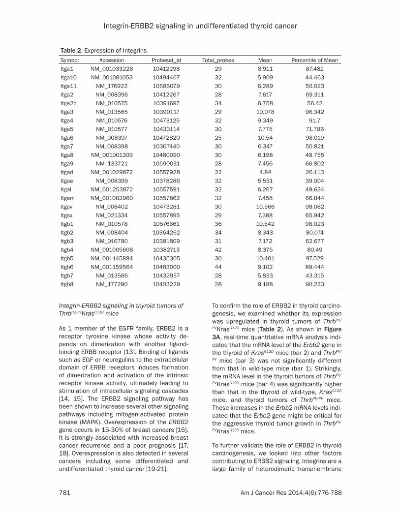
To confirm the role of ERBB2 in thyroid carcino-genesis, we examined whether its expression was upregulated in thyroid tumors of ThrbPV/
PVKrasG12D mice (Table 2). As shown in Figure 3A, real-time quantitative mRNA analysis indi-cated that the mRNA level of the Erbb2 gene in the thyroid of KrasG12D mice (bar 2) and ThrbPV/
PV mice (bar 3) was not significantly different from that in wild-type mice (bar 1). Strikingly, the mRNA level in the thyroid tumors of ThrbPV/
PVKrasG12D mice (bar 4) was significantly higher than that in the thyroid of wild-type, KrasG12D mice, and thyroid tumors of ThrbPV/PV mice. These increases in the Erbb2 mRNA levels indi-cated that the Erbb2 gene might be critical for the aggressive thyroid tumor growth in ThrbPV/
PVKrasG12D mice.
To further validate the role of ERBB2 in thyroid carcinogenesis, we looked into other factors contributing to ERBB2 signaling. Integrins are a large family of heterodimeric transmembrane
Table 2. Expression of IntegrinsSymbol Accession Probeset_id Total_probes Mean Percentile of MeanItga1 NM_001033228 10412298 29 8.911 87.482Itga10 NM_001081053 10494467 32 5.909 44.463Itga11 NM_176922 10586079 30 6.289 50.023Itga2 NM_008396 10412267 28 7.617 69.311Itga2b NM_010575 10391697 34 6.758 56.42Itga3 NM_013565 10390117 29 10.078 96.342Itga4 NM_010576 10473125 32 9.349 91.7Itga5 NM_010577 10433114 30 7.775 71.786Itga6 NM_008397 10472820 25 10.54 98.019Itga7 NM_008398 10367440 30 6.347 50.821Itga8 NM_001001309 10480090 30 6.198 48.755Itga9 NM_133721 10590031 28 7.456 66.802Itgad NM_001029872 10557928 22 4.84 26.113Itgae NM_008399 10378286 32 5.551 39.004Itgal NM_001253872 10557591 32 6.267 49.634Itgam NM_001082960 10557862 32 7.458 66.844Itgav NM_008402 10473281 30 10.566 98.082Itgax NM_021334 10557895 29 7.388 65.942Itgb1 NM_010578 10576661 36 10.542 98.023Itgb2 NM_008404 10364262 34 8.343 80.074Itgb3 NM_016780 10381809 31 7.172 62.677Itgb4 NM_001005608 10382713 42 8.375 80.49Itgb5 NM_001145884 10435305 30 10.401 97.529Itgb6 NM_001159564 10483000 44 9.102 89.444Itgb7 NM_013566 10432957 28 5.833 43.315Itgb8 NM_177290 10403229 28 9.188 90.233

Integrin-ERBB2 signaling in undifferentiated thyroid cancer
782 Am J Cancer Res 2014;4(6):776-788
Figure 3. A. An increased mRNA expression of the Erbb2 gene in the thyroid of ThrbPV/PVKrasG12D mice. Total RNA was extracted from thyroids of wild-type, KrasG12D, ThrbPV/PV, and ThrbPV/PVKrasG12D mice and analyzed by quantitative real-time RT-PCR. B. A heat-map presentation of hierarchical clustering (average of Euclidean distance) analysis of the integrins gene expression in mice with the genotypes indicated.

Integrin-ERBB2 signaling in undifferentiated thyroid cancer
783 Am J Cancer Res 2014;4(6):776-788
Figure 4. Validation of the extent in altered gene expression from arrays by quantitative real-time RT-PCR. Total RNA was extracted from thyroids of wild-type, KrasG12D, ThrbPV/PV, and ThrbPV/PVKrasG12D mice. Eight representative genes: (A) Integrin α6, (B) Integrin β4, (C) Integrin αV, (D) Integrin β1, (E) Integrin β3, (F) Integrin α5, (G) Fibronectin, and (H) Myc.
receptors that mediate cell binding to the extra-cellular matrix and link the extracellular envi-ronment to the intracellular cytoskeleton [22]. The different combination of 18 alpha subunits and 8 beta subunits gives rise to 24 distinct heterodimers [23]. Integrins belonging to the β1, αv, β7, and β4 subfamilies have been shown to potentiate signaling pathways in response to many growth factors [24, 25], cytokines [26], and TGFβ [22, 27]. Overexpression of integrin αvβ3 is associated with the progression and metastasis of several cancers including glio-blastoma, carcinomas of the breast, prostate, pancreas, and lung [28]. Increased expression
of αvβ6 is associated with greater invasive potential in oral squamous carcinoma, pancre-atic, ovarian, breast, and lung cancer [28]. Integrin β4 forms a complex with ERBB2 and enhances activation of the transcription fac-tors. In mice absent an integrin β4 signaling domain, both initiation and metastatic progres-sion of mammary tumors induced by ERRB2 are significantly delayed, suggesting integrins act as cooperating oncogenes [29].
Our analyses of the gene expression profiles showed a global upregulation of integrins in ThrbPV/PVKrasG12D mice (Figure 3B). A hierarchi-

Integrin-ERBB2 signaling in undifferentiated thyroid cancer
784 Am J Cancer Res 2014;4(6):776-788
Figure 5. Protein abundance of integrin-ERBB2 sig-naling pathway in wild-type, KrasG12D, ThrbPV/PV, and ThrbPV/PVKrasG12D mice thyroids. Protein levels were measured using antibodies against (A) ERBB2, (B) Fibronectin, (C) Integrin α6, (D) Integrin β4, (E) Integ-rin αV, (F) Integrin β1, (G) Integrin β3, (H) Integrin α5, and (I) loading control GAPDH. Total protein extracts were prepared from thyroids of wild-type and Kras-G12D mice and thyroid tumors of ThrbPV/PV and ThrbPV/
PVKrasG12D mice, and the Western blot analysis was carried out as described in Materials and Methods.
cal clustering heat map shows that the expres-sion of integrins in ThrbPV/PVKrasG12D mice was closer to that of ThrbPV/PV mice than that of KrasG12D mice (Figure 3B). However, it was also clear that the gene expression profiles in ThrbPV/
PVKrasG12D mice were distinct from those in ThrbPV/PV mice.
We next used qRT–PCR to examine 3 integrin α genes and 3 integrin β genes for the validation of the gene expression profiles obtained by array analysis. Genes selected for validation were integrin α6, integrin β4, integrin αV, integ-rin β1, integrin β3, and integrin α5 as shown in Figure 4A-F. Consistent with array data, qRT–PCR showed that the expression of the 6 integ-rins (Figure 4A-F) was significantly increased in thyroid tumors of ThrbPV/PVKrasG12D mice. The expression patterns of integrins in ThrbPV/PV mice also had an upward trend to be in line with the findings in the array analysis. But the expression of integrins in ThrbPV/PV mice was lower than in ThrbPV/PVKrasG12D mice.
It is known that integrins bind to ligands such as fibronectin to transmit the signaling from the extracellular matrix into cytokeratin inside cells. Fibronectin has been implicated in the
development of multiple types of human can-cer [30, 31], and it has been associated with cell migration and invasion in several metastat-ic models [30, 32]. qRT–PCR determined that fibronectin (Figure 4G) was significantly in- creased with a pattern similar to integrins in ThrbPV/PVKrasG12D mice (Figure 4A-F).
In our previous studies, we found that upregu-lated MYC is crucial for the manifestation of aggressive tumor phenotypes [5]. We exam-ined whether the Myc mRNA had an expression pattern similar to integrins. We found that the expression level of the Myc gene determined by qRT/PCR was significantly increased (Figure 4H). The expression changes of the Myc gene in 4 genotypes of mice were similar to that of integrins. Therefore, we identified increased Myc expression as one of the downstream effector of the integrin-ERBB2 signaling to pro-mote tumor growth.
To confirm that ERBB2 had a crucial role in aggressive thyroid tumor growth, we further compared protein abundance of ERBB2 in thy-roids of wild-type and KrasG12D mice and in thy-roid tumors of ThrbPV/PV and ThrbPV/PVKrasG12D mice. As shown in Figure 5A, there was no sig-nificant difference between ERBB2 protein lev-els in wild-type, KrasG12D, and ThrbPV/PV mice. The level of ERBB2 protein was significantly increased only in ThrbPV/PKrasG12D mice. ERBB2 signaling has been shown to be regulated by integrins [28].
Because fibronectin is one of the ligands for integrins, we also examined its protein level. We found that the abundance of fibronectin was significantly higher in thyroid tumors of ThrbPV/PVKrasG12D mice than in WT, KrasG12D, and ThrbPV/PV mice (compare lanes 7-10 to lanes 1-6, Figure 5B). Co-upregulation of ERBB2 and fibronectin supported the idea that interaction of 2 activated signaling pathways could be important for the tumor phenotypes observed in ThrbPV/PKrasG12D mice.
We also validated the protein abundance of integrins. Consistent with the highly elevated the mRNA expression (Figure 4A-F), Western blot analysis further showed that the protein abundance of integrins αV, β1, β3, and α5 was significantly elevated in thyroid tumors of ThrbPV/PVKrasG12D mice as compared with WT, KrasG12D, and ThrbPV/PV mice (lanes compare

Integrin-ERBB2 signaling in undifferentiated thyroid cancer
785 Am J Cancer Res 2014;4(6):776-788
Figure 6. Proposed molecular pathways for integrin-ERBB2 signaling to stimulate proliferation of thyroid tumor cells in ThrbPV/PVKrasG12D mice. Synergistic on-cogenic actions of TRβPV and KrasG12D activate sig-naling mediated by integrins, ERBB2, and ERK, lead-ing to increased expression of MYC. Elevated MYC stimulates downstream targets such as cyclin D1 to promote aggressive tumor growth in ThrbPV/PVKrasG12D mice.
lanes 7-10 to lanes 1-6, Figure 5C-H). ERBB2 has been shown to promote the formation of a multimeric complex that includes ERBB2 and α6β4 integrins. Cooperative signaling of ERBB2 and α6β4 integrins results in the activation of downstream factors that regulate carcinogene-sis [29]. As shown in Figure 5C and 5D, both integrin α6 and β4 were significantly increased in thyroid tumors of ThrbPV/PVKrasG12D mice. The enrichment of integrins/fibronectin and ERBB2 proteins indicated that their signaling is critical for the aggressive thyroid tumor growth.
Discussion
Recently, we genetically introduced the KrasG12D mutation to express specifically in the thyroids of the ThrbPV/PV mice. Double mutant ThrbPV/
PVKrasG12D mice have much worse survival than the mice with a single mutation in either the Kras or Thrb gene. The more aggressive thyroid tumor phenotypes result from a synergy of the oncogenic actions of 2 mutants in thyroids, leading to increased proliferation for rapid
tumor expansion. However, the molecular events leading to the synergy of 2 mutants were not well characterized. In our current stud-ies, through microarray gene profiling, the top upstream regulators identified are main growth factors or growth factor receptors, which pro-moted the aggressive phenotypes of undiffer-entiated thyroid cancer. We found that ERBB2 was increased only in the aggressive undiffer-entiated thyroid tumors of ThrbPV/PVKrasG12D mice, not in ThrbPV/PV mice with differentiated tumors. We further identified markedly in- creased integrins, such as β1, β3, β4, αV, α5 and α6, as activators to stimulate ERBB2-mediated downstream signaling in thyroid tumors of ThrbPV/PVKrasG12D mice. The present studies have uncovered the collaborative sig-naling by integrins and ERBB2 as one of the mechanisms in synergy mediated by the onco-genic actions of KrasG12D and ThrbPV/PV mutants to promote the aggressive tumor growth.
The critical role of ERBB2 in cancer develop-ment and progression has been well document-ed. Overexpression of the ERBB2 gene occurs in 15-30% of breast cancers [16]. Increased levels of ERBB2 are strongly associated with increased breast cancer recurrence and a poor prognosis [17, 18]. The expression of ERBB2 was detected in differentiated and undifferenti-ated thyroid cancer [19-21]. Binding of ligands, such as EGF or neuroregulins, to the extracel-lular domain of ERBB receptors results in the formation of receptor dimerization and activa-tion of ERBB2. However, integrins such as β4 and α6 are known to amplify ERBB2 signaling [29, 33]. Importantly, it has been demonstrat-ed that ERBB2 makes integrins collaborators in the initiation and progression of carcinogene-sis. In mice without an integrin β4 signaling domain, the initiation and metastatic progres-sion of mammary tumors induced by ERRB2 are significantly delayed [29]. In the presence of integrins, increased ERBB2 induces the intrinsic receptor kinase activity, ultimately leading to stimulation of multiple intracellular signaling cascades including the ERK pathway [14, 15].
In line with these reports, our present studies showed that there was a global upregulation of integrins, including α6 and β4, in thyroid tumors of ThrbPV/PVKrasG12D mice (Figure 3B). Abun- dance of ERBB2 was also markedly increased in the thyroid tumors of ThrbPV/PVKrasG12D mice

Integrin-ERBB2 signaling in undifferentiated thyroid cancer
786 Am J Cancer Res 2014;4(6):776-788
(Figure 5A). Moreover, we have previously shown activated ERK signaling and elevated MYC and cyclin D1 protein abundance in thyroid tumors ThrbPV/PVKrasG12D mice [5]. Taken togeth-er, the present findings support the molecular pathways proposed in Figure 6. The multiple upstream regulators (i.e., integrins and ERBB2) are activated via synergistic oncogenic actions of TRβPV and KRASG12D, converging through ERK to increase MYC and cyclin D1 activation. These 2 critical growth signals downstream of ERK activation drive the aggressive thyroid tumor growth of ThrbPV/PVKrasG12D mice.
The finding uncovered in the present studies that integrins-ERBB2 signaling contributes to aggressive tumor growth of undifferentiated thyroid cancer has important clinical implica-tions. Currently, the modalities for the treat-ment of undifferentiated thyroid cancer are lim-ited. ERBB2 has been shown to express in some differentiated and undifferentiated thy-roid cancers [19-21]. Clinical trials are under-way using anti-ERBB2 agents at various phases in several solid tumors [34]. Moreover, drugs against integrins are being developed to treat cancers [35]. Thus, integrins and ERBB2 could be tested as potential targets for the treatment of thyroid tumors with elevated integrins and ERBB2 expression and/or with resistance to therapy [36].
The prevalent genetic alterations identified in undifferentiated thyroid cancer are point muta-tions of the RAS genes together with the muta-tions of the TP53 and CTNNB1 genes. These observations suggest that mutations of the RAS genes alone might not be sufficient to bring out the aggressive dedifferentiated thy-roid cancer. In our studies, we found mice har-boring the single mutation of the KrasG12D gene did not develop thyroid tumors [5]. ThrbPV/PV mice with the mutation of the 2 alleles of the Thrb gene develop differentiated thyroid can-cer. However, ThrbPV/PVKrasG12D mice exhibit aggressive dedifferentiated thyroid cancer with large and rapid tumor growth [5]. The present studies show that synergistic oncogenic actions of TRβPV and KRASG12D brought out the aggressive tumor phenotypes. Thus findings from the studies using the mouse models sup-port clinical observations that other oncogenic alterations in addition to a single mutation of the RAS gene are necessary to bring out the dedifferentiated thyroid cancer. At present, the
homozygous mutations of the THRB gene have yet to be discovered in human dedifferentiated thyroid cancer. However, it is known that in ThrbPV/PV mice, altered signaling pathways such as the activation of the CTNNB1 gene have been demonstrated. Thus, it is likely that syner-gistic oncogenic actions of TRβPV and KRASG12D could be, at least, the result of cross talk of KRASG12D- and TRβPV-mediated activated β-catenin signaling, which is consis-tent with observations in human patients [37]. Thus, the ThrbPV/PVKrasG12D mouse is an excel-lent thyroid cancer model to further elucidate other altered signaling pathways during devel-opment of undifferentiated thyroid cancer.
Acknowledgements
This research was supported by the Intramural Research Program of the Center for Cancer Research, National Cancer Institute, NIH.
Disclosure of conflict of interest
The authors declare no conflicts of interest.
Address correspondence to: Dr. Sheue-Yann Cheng, Laboratory of Molecular Biology, National Cancer Institute, Room 5128, Bethesda, MD 20892-4264. Tel: 301-496-4280; Fax: 301-402-1344; E-mail: [email protected]
References
[1] Nikiforov YE and Nikiforova MN. Molecular ge-netics and diagnosis of thyroid cancer. Nat Rev Endocrinol 2011; 7: 569-580.
[2] Kaneshige M, Kaneshige K, Zhu X, Dace A, Garrett L, Carter TA, Kazlauskaite R, Pankratz DG, Wynshaw-Boris A, Refetoff S, Weintraub B, Willingham MC, Barlow C and Cheng S. Mice with a targeted mutation in the thyroid hor-mone beta receptor gene exhibit impaired growth and resistance to thyroid hormone. Proc Natl Acad Sci U S A 2000; 97: 13209-13214.
[3] Suzuki H, Willingham MC and Cheng SY. Mice with a mutation in the thyroid hormone recep-tor beta gene spontaneously develop thyroid carcinoma: a mouse model of thyroid carcino-genesis. Thyroid 2002; 12: 963-969.
[4] Parrilla R, Mixson AJ, McPherson JA, McClas-key JH and Weintraub BD. Characterization of seven novel mutations of the c-erbA beta gene in unrelated kindreds with generalized thyroid hormone resistance. Evidence for two “hot spot” regions of the ligand binding domain. J Clin Invest 1991; 88: 2123-2130.
[5] Zhu X, Zhao L, Park JW, Willingham MC and Cheng SY. Synergistic signaling of KRAS and
Integrin-ERBB2 signaling in undifferentiated thyroid cancer
787 Am J Cancer Res 2014;4(6):776-788
thyroid hormone receptor β mutants promotes undifferentiated thyroid cancer through MYC up-regulation. Neoplasia 2014; 16: 757-69.
[6] Jackson EL, Willis N, Mercer K, Bronson RT, Crowley D, Montoya R, Jacks T and Tuveson DA. Analysis of lung tumor initiation and pro-gression using conditional expression of onco-genic K-ras. Genes Dev 2001; 15: 3243-3248.
[7] Kusakabe T, Kawaguchi A, Kawaguchi R, Fei-genbaum L and Kimura S. Thyrocyte-specific expression of Cre recombinase in transgenic mice. Genesis 2004; 39: 212-216.
[8] Klipper-Aurbach Y, Wasserman M, Braunspie-gel-Weintrob N, Borstein D, Peleg S, Assa S, Karp M, Benjamini Y, Hochberg Y and Laron Z. Mathematical formulae for the prediction of the residual beta cell function during the first two years of disease in children and adoles-cents with insulin-dependent diabetes melli-tus. Med Hypotheses 1995; 45: 486-490.
[9] Huang da W, Sherman BT and Lempicki RA. Systematic and integrative analysis of large gene lists using DAVID bioinformatics resourc-es. Nat Protoc 2009; 4: 44-57.
[10] Subramanian A, Tamayo P, Mootha VK, Mukherjee S, Ebert BL, Gillette MA, Paulovich A, Pomeroy SL, Golub TR, Lander ES and Me-sirov JP. Gene set enrichment analysis: a knowledge-based approach for interpreting genome-wide expression profiles. Proc Natl Acad Sci U S A 2005; 102: 15545-15550.
[11] Furumoto H, Ying H, Chandramouli GV, Zhao L, Walker RL, Meltzer PS, Willingham MC and Cheng SY. An unliganded thyroid hormone beta receptor activates the cyclin D1/cyclin-depen-dent kinase/retinoblastoma/E2F pathway and induces pituitary tumorigenesis. Mol Cell Biol 2005; 25: 124-135.
[12] Kim DW, Walker RL, Meltzer PS and Cheng SY. Complex temporal changes in TGFβ oncogenic signaling drive thyroid carcinogenesis in a mouse model. Carcinogenesis 2013; 34: 2389-2400.
[13] Holbro T, Beerli RR, Maurer F, Koziczak M, Bar-bas CF 3rd and Hynes NE. The ErbB2/ErbB3 heterodimer functions as an oncogenic unit: ErbB2 requires ErbB3 to drive breast tumor cell proliferation. Proc Natl Acad Sci U S A 2003; 100: 8933-8938.
[14] Olayioye MA, Neve RM, Lane HA and Hynes NE. The ErbB signaling network: receptor heterodi-merization in development and cancer. EMBO J 2000; 19: 3159-3167.
[15] Yarden Y and Sliwkowski MX. Untangling the ErbB signalling network. Nat Rev Mol Cell Biol 2001; 2: 127-137.
[16] Burstein HJ. The distinctive nature of HER2-positive breast cancers. N Engl J Med 2005; 353: 1652-1654.
[17] Tan M and Yu D. Molecular mechanisms of erbB2-mediated breast cancer chemoresis-tance. Adv Exp Med Biol 2007; 608: 119-129.
[18] Slamon DJ, Clark GM, Wong SG, Levin WJ, Ull-rich A and McGuire WL. Human breast cancer: correlation of relapse and survival with amplifi-cation of the HER-2/neu oncogene. Science 1987; 235: 177-182.
[19] Bieche I, Franc B, Vidaud D, Vidaud M and Li-dereau R. Analyses of MYC, ERBB2 and CCND1 genes in benign and malignant thyroid follicu-lar cell tumors by real-time polymerase chain reaction. Thyroid 2001; 11: 147-152.
[20] Ensinger C, Prommegger R, Kendler D, Gabriel M, Spizzo G, Mikuz G and Kremser R. Her2/neu expression in poorly-differentiated and anaplastic thyroid carcinomas. Anticancer Res 2003; 23: 2349-2353.
[21] Kremser R, Obrist P, Spizzo G, Erler H, Kendler D, Kemmler G, Mikuz G and Ensinger C. Her2/neu overexpression in differentiated thyroid carcinomas predicts metastatic disease. Vir-chows Arch 2003; 442: 322-328.
[22] Brizzi MF, Tarone G and Defilippi P. Extracellu-lar matrix, integrins, and growth factors as tai-lors of the stem cell niche. Curr Opin Cell Biol 2012; 24: 645-651.
[23] Hynes RO. Integrins: bidirectional, allosteric signaling machines. Cell 2002; 110: 673-687.
[24] Giancotti FG and Tarone G. Positional control of cell fate through joint integrin/receptor pro-tein kinase signaling. Annu Rev Cell Dev Biol 2003; 19: 173-206.
[25] Ivaska J and Heino J. Cooperation between in-tegrins and growth factor receptors in signal-ing and endocytosis. Annu Rev Cell Dev Biol 2011; 27: 291-320.
[26] Defilippi P, Rosso A, Dentelli P, Calvi C, Garbar-ino G, Tarone G, Pegoraro L and Brizzi MF. {beta}1 Integrin and IL-3R coordinately regu-late STAT5 activation and anchorage-depen-dent proliferation. J Cell Biol 2005; 168: 1099-1108.
[27] Margadant C and Sonnenberg A. Integrin-TGF-beta crosstalk in fibrosis, cancer and wound healing. EMBO Rep 2010; 11: 97-105.
[28] Sun CC, Qu XJ and Gao ZH. Integrins: players in cancer progression and targets in cancer ther-apy. Anticancer Drugs 2014; 25: 1107-21
[29] Guo W, Pylayeva Y, Pepe A, Yoshioka T, Muller WJ, Inghirami G and Giancotti FG. Beta 4 inte-grin amplifies ErbB2 signaling to promote mammary tumorigenesis. Cell 2006; 126: 489-502.
[30] Waalkes S, Atschekzei F, Kramer MW, Hennen-lotter J, Vetter G, Becker JU, Stenzl A, Merse-burger AS, Schrader AJ, Kuczyk MA and Serth J. Fibronectin 1 mRNA expression correlates with advanced disease in renal cancer. BMC Cancer 2010; 10: 503.

Integrin-ERBB2 signaling in undifferentiated thyroid cancer
788 Am J Cancer Res 2014;4(6):776-788
[31] Hu M, Carles-Kinch KL, Zelinski DP and Kinch MS. EphA2 induction of fibronectin creates a permissive microenvironment for malignant cells. Mol Cancer Res 2004; 2: 533-540.
[32] Jia D, Yan M, Wang X, Hao X, Liang L, Liu L, Kong H, He X, Li J and Yao M. Development of a highly metastatic model that reveals a cru-cial role of fibronectin in lung cancer cell mi-gration and invasion. BMC Cancer 2010; 10: 364.
[33] Yoon SO, Shin S and Lipscomb EA. A novel mechanism for integrin-mediated ras activa-tion in breast carcinoma cells: the alpha6be-ta4 integrin regulates ErbB2 translation and transactivates epidermal growth factor recep-tor/ErbB2 signaling. Cancer Res 2006; 66: 2732-2739.
[34] Tolaney S. New HER2-positive targeting agents in clinical practice. Curr Oncol Rep 2014; 16: 359.
[35] Sun CC, Qu XJ and Gao ZH. Arginine-Glycine-Aspartate-Binding Integrins as Therapeutic and Diagnostic Targets. Am J Ther 2014; [Epub ahead of print].
[36] Moore KM, Thomas GJ, Duffy SW, Warwick J, Gabe R, Chou P, Ellis IO, Green AR, Haider S, Brouilette K, Saha A, Vallath S, Bowen R, Che-lala C, Eccles D, Tapper WJ, Thompson AM, Quinlan P, Jordan L, Gillett C, Brentnall A, Vio-lette S, Weinreb PH, Kendrew J, Barry ST, Hart IR, Jones JL and Marshall JF. Therapeutic tar-geting of integrin alphavbeta6 in breast can-cer. J Natl Cancer Inst 2014; 106.
[37] Castellone MD, De Falco V, Rao DM, Bellelli R, Muthu M, Basolo F, Fusco A, Gutkind JS and Santoro M. The beta-catenin axis integrates multiple signals downstream from RET/papil-lary thyroid carcinoma leading to cell prolifera-tion. Cancer Res 2009; 69: 1867-1876.