original article an in vitro investigation of wear resistance … hardness of composite resins liqun...
TRANSCRIPT

Int J Clin Exp Med 2013;6(6):423-430www.ijcem.com /ISSN:1940-5901/IJCEM1305005
vomiting and regurgitation of gastric juice, for instance, in bulimia and anorexia nervosa (ero-sion) cases [4].
Wear of composites is known to depend on filler particle-related features, particularly on the concentration and size of the filler reinforce-ment [5] and resin formulation [6] et al. Finer particles for a fixed-volume-fraction of filler have been documented to result in decreased interparticle spacing and thereby reduced wear [7, 8]. In terms of filler content, some in vitro wear studies have revealed that increased load-ing may enhance the wear resistance of dental composites [9-11]. As for the resin formulation, the study has shown that increasing resin vis-cosity generally lowers the wear resistance [12].
Ideal dental restorations should have wear resistance similar to that of tooth. Average clini-cal wear rate on occlusal contact areas of teeth
Original ArticleAn in vitro investigation of wear resistance and hardness of composite resins
Liqun Cao1, Xinyi Zhao2, Xu Gong2, Shouliang Zhao1
1School of Stomatology, Tongji University, Shanghai 200072, China; 2School of Stomatology, the Fourth Military Medical University, Xi´an 710032, China
Received May 13, 2013; Accepted June 6, 2013; Epub June 26, 2013; Published July 1, 2013
Abstract: Purpose: The aim of the present study was to investigate the wear resistance and hardness of five kinds of composite resins. Materials and Methods: Sixty-five specimens were fabricated with one nano-hybrid (Charisma Diamond), two micro-hybrid (3MZ250, Clearfil AP-X) and two packable (3MP60, Surefil) composite resins, according to a randomized complete block design (n=13, 8 for wear test; 5 for hardness test). The composites were filled in a rectangular mold, and light polymerization. After storage in 37°C deionized water for 24h, all specimens were tested with a custom-made toothbrush machine with a stainless-steel ball as antagonist (3N loads, 1Hz, 6×10⁵ cycles) im-mersed in calcium fluoride slurry. Wear volume, hardness and surface structure of each tested material was exam-ined by a three-dimensional non-contact optical profilometer, Vickers indentation technique and scanning electron microscope. Results: The volume loss ranked from least to most as follows: Charisma Diamond, P60, Z250, Clearfil AP-X and Surefil. Regarding hardness, the rank from highest to lowest as follows: Clearfil AP-X, P60, Surefil, Z250, Charisma Diamond. The interactions between wear resistance and microhardness were not significant. Conclu-sions: The custom-made machine is considered suitable to simulate sliding of an antagonist cusp on an opposing occlusal composite restoration. Nanofilled composite may have superior wear compared to other composite resins.
Keywords: Composite resins, hardness, wear resistance
Introduction
Composite resins based on dimethacrylates and silane-coated inorganic fillers were intro-duced as dental restoratives in the mid-1960s [1]. Due to their excellent aesthetic properties, composite resins gain steadily importance and popularity for the restoration of all cavity class-es. However, clinically, their relatively poor wear resistance is still considered as a factor that contributes to materials’ failure [2].
Mair [3] defined wear as a consequence of the interaction between surfaces moving in con-tact, causing gradual remove of material. In the oral cavity, a lot of components contribute to the wear of enamel and dentin, such as the occlusal contacts to antagonist teeth (attrition), chewing on food items, toothbrushing with toothpaste or inhalation of dust (abrasion), acid attacks due to the consumption of acidic fruits and beverages, inhalation of industrial acids or
Wear resistance and hardness of composite resins
424 Int J Clin Exp Med 2013;6(6):423-430
is about 29µm per year for molars and about 15µm per year for premolars [6]. Another study showed at the mean pooled occlusal wear of four ultraviolet light-cured posterior compos-ites at 17 years was 264µm, and that most wear (75%) occurred in the first 5 years [13]. Of course, results differ among evaluators due to operator variations, patient variations, evaluat-ed products, and last but not least important, the wear evaluation method [14].
It is worthwhile to note that many factors besides wear can affect the lifespan of resin composites. However, it can be assumed that materials with better wear resistance should do better under cycling loading during normal occlusal and masticatory function. Therefore, the present study was to investigate the wear resistance and microhardness of five resin composites using a device for simulated tooth-brushing, a three-dimension non contact opti-cal profilometer and scanning electron micro-scope. The null hypothesis tested was that there would be no differences in wear resis-tance and microhardness.
Materials and methods
Materials
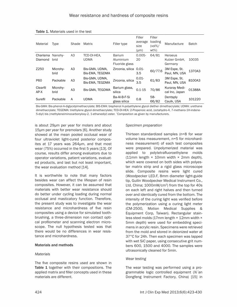
The five composite resins used are shown in Table 1 together with their compositions. The applied matrix and filler concepts used in these materials are different.
Specimen preparation
Thirteen standardized samples (n=8 for wear volume loss measurement, n=5 for microhard-ness measurement) of each test composites were prepared. Unpolymerized material was applied to polytetrafluoroethylene molds (11mm length × 10mm width × 2mm depth), which were covered on both sides with polyes-ter matrix strip and a rigid glass microscope slide. Composite resins were light cured (Woodpecker LED.F, 8mm diameter light-guide tip, Guilin Woodpecker Medical Instrument Co., Ltd, China; 1000mW/cm2) from the top for 40s on each left and right halves and then turned over and identically cured from the bottom. The intensity of the curing light was verified before the polymerization using a curing light meter (CM-2500, Motion Medical Supplies & Equipment Corp, Taiwan). Rectangular stain-less steel molds (17mm length × 12mm width × 5mm depth) were used for embedding speci-mens in acrylic resin. Specimens were retrieved from the mold and stored in deionized water at 37°C for 24h. Then each specimen was lapped with wet SiC paper, using consecutive grit num-bers 600, 1500 and 4000. The samples were ultrasonically cleaned for 5min.
Wear testing
The wear testing was performed using a pro-grammable logic controlled equipment (Xi an Dongfeng Instrument Factory, China) [15] in
Table 1. Materials used in the test
Material Type Shade Matrix Filler type
Filler average size (μm)
Filler loading (vol%/ wt%)
Manufacture Batch
Charisma Diamond
Nanohy-brid
A3 TCD-DI-HEA, UDMA
Barium Aluminium Fluoride glass
0.005-20
64/81 Heraeus Kulzer GmbH, Germany
10035
Z250 Microhy-brid
A3 Bis-GMA, UDMA, Bis-EMA, TEGDMA
Zirconia, silica 0.01-3.5 60/77.6 3M Espe, St.
Paul, MN, USA 1370A3
P60 Packable A3 Bis-GMA, UDMA, Bis-EMA, TEGDMA Zirconia, silica 0.01-
3.5 61/83 3M Espe, St. Paul, MN, USA 8100A3
Clearfil AP-X
Microhy-brid A3 Bis-GMA, TEGDMA Barium glass,
silica 0.1-15 70/86 Kuraray Medi-cal Inc, Japan 01388A
Surefil Packable A UDMA Ba-Al-B-F-Si glass silica 0.8 58-
66/82Dentsply Caulk, USA 101220
Bis-GMA: Bis-phenol-A-diglycidylmethacrylate; BIS-EMA: bisphenol A-polyethylene glycol diether dimethacrylate; UDMA: urethane dimethacrylate; TEGDMA: triethylene glycol dimethacrylate; TCD-DI-HEA: 2-Propenoic acid, (octahydro-4, 7-methano-1H-indene-5-diyl) bis (methyleneiminocarbonyloxy-2, 1-ethanediyl) ester. *Composition as given by manufacturers.

Wear resistance and hardness of composite resins
425 Int J Clin Exp Med 2013;6(6):423-430
10min. The surface of each specimen was recorded using a three-dimensional non con-tact optical profilometer. Table 2 shows the sur-face profile measurement conditions. Volume loss after wear test was measured by comput-ing the volume of the worn area, which was at a level lower than the unworn surface level.
Microhardness measurement (H)
For hardness measurement, a microhardness tester (HXD-1000TM, Shanghai Taiming Optical Instrument Co., Ltd, China) was used. Vickers hardness numbers were determined from indentations made under 50g load for 15s by the arithmetic mean of three indentations ran-domly performed for each specimen and test-ing condition.
Scanning electron microscope examination (SEM)
One random sample of each composite mate-rial after 6×10⁵ abrasive cycles was selected for SEM examination (Type Quanta 200 FEG, FEI Company, Netherlands). The samples were sputter-coated with gold and photographs were taken of representative areas at 1000× magni-fications at 20.0kV acceleration voltage.
Statistical analysis
Data were analyzed using statistical software (SPSS 17.0 for Windows). Means and standard
Figure 1, which allows for the adjustment for the frequency, number of cycles and duration of contact between the specimen and counter-part. The system can produce standardized load application in eight identical specimen compartments. Each compartment has a recess (17mm length × 12mm width × 5mm depth) where a specimen was positioned. Then a spherical antagonist made from stainless-steel (Ø=7.8mm) under a 3N load was applied to the specimens and moved across the sur-face over a 20mm linear path, generating abra-sive wear at a frequency of 1Hz for a total of 6×10⁵ cycles. Tests were carried out in eight individual compartments in the presence of 15ML fresh calcium fluoride slurry in an acrylic chamber [16]. Slurry and antagonists were renewed prior to each wear test.
Wear volume loss measurement (W)
After the test, specimens were cleaned with running water followed by an ultrasonic bath for
Table 2. Surface profile measurement condi-tionsDevice Three-dimensional non
contact optical profilometer (NANOVEA PS50 U.S.A)
Sampling interval X:18µm; Y:50µmMeasuring range 8*7mmResolution of Z-axis 0.1µm
Figure 1. Schematic diagram of wear testing (1. acrylic chamber, 2.1 antagonist, 2.2 sample holder, 2.3 specimen, 3. control box).

Wear resistance and hardness of composite resins
426 Int J Clin Exp Med 2013;6(6):423-430
Figure 2. Two-dimensional and three-dimensional representation of the wear facet.
Significantly highest H was detected for Clearfil AP-X (87.16). P60 (82.12), Surefil (79.37) and Z250 (79.27) showed the inter-mediate values for mean H. Significantly lowest H was found for Charisma Diamond (54.89).
Regression analysis showed no significant correlations between W and H in Figure 3 (P=0.0557).
SEM observation
deviations of W and H were calculated, and analyzed using one-way analysis of variance (ANOVA). Regression analysis was performed to investigate the relation between W and H. The level of significance was set to α=0.05.
Results
Wear facet observation
The wear facet of all the samples is consistent with the shape of the antagonist. Combined with a stereo microscope observation, the wear facet shows wear heavy in the middle and light on both sides, which is consistent with the result of the three-dimensional non contact optical profilometer (Figure 2).
W values and H values
Mean W values and H values are shown in Table 3. One-way ANOVA indicated significant differences in W between the materials (p<0.05). Charisma Diamond (6.0057mm3) had the significantly lowest W, followed by P60 (8.2588mm3), Z250 (8.4432mm3) and Clearfil AP-X (8.5956mm3) which did not differ from each other. Surefil (10.8373mm3) had the sig-nificantly highest W.
One-way ANOVA indicated significant differenc-es in H between the various materials (p<0.05).
Table 3. Mean wear volume loss and microhard-ness of experimental composites (S.D.)
Resin composite Wear volume loss (mm3)
Microhardness (kg/mm2)
Charisma Diamond 6.0057 (1.227) 54.89 (2.04)Z250 8.4432 (2.4983) 79.27 (1.25)P60 8.2588 (1.5561) 82.12 (4.12)Clearfil AP-X 8.5956 (1.6379) 87.16 (2.16)Surefil 10.8373 (1.9858) 79.37 (2.93)
Selected SEM of evaluated groups after wear testing was shown in Figure 4. The SEMs were taken from specimens in wear central. In gen-eral, the specimen surfaces of the five groups revealed observable differences from each other. Charisma Diamonds is characterized by a very smooth and uniformly worn surface. Small voids are seen throughout entire surface. The surfaces of Z250 and P60 show densely packed superficially abraded clusters in the surrounding resin matrix. The surfaces are quite uniformly abraded. The surface of Clearfil AP-X exhibit densely packed fillers with a wide grain size. Surefil presents more accentuated matrix degradation as well as more voids and cracks compared to others.
Discussion
Even though a laboratory study is not able to reproduce all the conditions of the oral environ-ment, it is still relevant for prediction of clinical performance. Within the limitations of this in vitro study, the findings reject the research hypothesis, there doesn’t suggest similar wear resistance and microhardness in the tested materials.
As previously described [17], composite abra-sion occurs mainly in two steps. Initially, there is a selective wear in the organic matrix, which

Wear resistance and hardness of composite resins
427 Int J Clin Exp Med 2013;6(6):423-430
depth and roughness of the worn surface are determined consecutively with the same instru-ment. Since it is a contact detection method, the scratches would be left on the specimen surface, which may affect the final results. With the emergence of the three-dimension non contact optical profilometer, the above-men-tioned trouble could be solved. In agree with a previous study [27], we decided to use a three-dimension non contact optical profilometer to determine the wear volume loss.
In the present study, each composite resin pre-sented a distinct performance, which suggests that results were dependent upon each formu-lation. It has been reported that the filler parti-cles play a particular important role for both hardness and wear resistance. Condon [8] highlighted that the effect of filler volume on wear resistance follows a linear relationship, with high filler volumes decreasing wear rates due to the lower expanse of resin unprotected by filler particles, which was supported by other researcher [28]. For the composite resins investigated in this study, however, regression analysis showed no correlation between the wear resistance and hardness.
According to other literature [29], the filler con-tent of composite material does not influence wear but other mechanical properties, such as diametral tensile strength and Knoop hard-ness. The weight fractions of filler particles of
leads to exposure, protrusion and loss of inor-ganic particles. Afterwards, due to mechanical stress, these particles thus offer protection for the organic matrix, reducing its wear process [18].
Different methods have been employed to eval-uate quantitative and qualitative abrasion resistance of composite resins. Teixeira et al. [19] measured the difference in specimen thickness from their initial thickness using a micrometer caliper. In many published studies on resin composite wear, only surface rough-ness parameter is reported [20, 21]. Such an approach has however serious shortcomings, since assessment of surface texture as a single parameter disregards materials such as Tetric EvoCeram or Grandio that showed low surface roughness and extremely high loss of sub-stance or very little wear and quite high surface roughness in the study [22]. Determination of specimen weight loss after being subjected to toothbrush abrasion is another method used [23, 24]. This method has certain limitations when materials with high abrasion resistance and limited numbers of brushing strokes are investigated. Measurement of depth of wear with a computer-controlled three-dimensional measuring microscope and computing the vol-ume loss from such data is proposed [25]. In addition, many scholars use a profilometric method to determine the depth of wear [22, 26]. This method is advantageous as wear
Figure 3. The relationship between wear volume loss and microhardness of experimental composites.

Wear resistance and hardness of composite resins
428 Int J Clin Exp Med 2013;6(6):423-430
ate W and H. And a comparatively greater num-ber of particles were probably present on the surface. Consequently, a larger contact area may have been established between the fillers and the counterpart, resulting in improved wear resistance.
One suggestion for improving the wear resis-tance of composites is to increase the abrasive resistance of the resin matrix, rather than increasing in the hardness of the filler particles [32, 33]. The predominant base monomer used in commercial dental composites has been bis-GMA, which due to its high viscosity is mixed with other dimethacrylates, such as TEGDMA. UDMA corresponds to another alternative organic matrix composition and it is often pres-ent in current compositions. Söderholm et al. [34] considered the urethane-base composites performed significantly better wear resistance than those which were bisGMA-based over three years clinical observation. It was contrary to this study which UDMA (Surefil) didn’t show the well wear resistance. Furthermore, Kawai et al. [32] suggested that the TMPT-TEGDMA resin showed the most wear resistance, while Bis-GMA- and UDMA-based resins showed increased wear resistance with an increased content of TEGDMA. Different formulations are tried by manufacturers in an attempt to over-come the shortcomings; however, further inves-
the composites tested in this experiment ranged from 72 to 86wt%, and the Charisma Diamond with approximately 80wt% of filler content demonstrated the least W, although the filler content of the Clerafil AP-X was the greatest. The finding is accordance with Hu’s [30] opinion that there is a threshold filler weight near 80% above which wear resistance is decreased.
The Clerafil AP-X showed higher W despite of its highest H and filler weight. As a microhybrid composite, the filler average size is larger than others in this study, which has been attributed to the fact that an increase in fill size causes an increase of the coefficient of friction, the con-tact forces and, thus insufficient wear resis-tance. Furthermore, Bayne et al. [31] suggest-ed that the presence of large particles may theoretically cause greater wear of the restor-ative material. When the restoration is subject-ed to masticatory forces, the stress spreads through the filler particle into the resin matrix. This process results in the easy removal of these particles from the surface, thereby exposing the organic matrix and further accel-erating wear.
Confirming the expectation that as particle size is decreased so is the wear [7], the convention-al hybrid composite Z250 was found intermedi-
Figure 4. Scanning electron micro-photographs at 1000 magnification of resin composites after 6×10⁵ abrasive cycles: Charisma Diamond, Z250, P60, Clearfil AP-X and Surefil.

Wear resistance and hardness of composite resins
429 Int J Clin Exp Med 2013;6(6):423-430
References
[1] Bowen RL. Dental filling material comprising vinyl-silane treated fused silica and a binder consisting of the reaction product of bis-phe-nol and glycidyl acrylate. US patent 3,06,112 1962.
[2] Hickel R, Manhart J. Longevity of restorations in posterior teeth and reasons for failure. J Ad-hes Dent 2001; 3: 45-64.
[3] Mair LH, Stolarski TA, Vowles RW, Lloyd CH. Wear: mechanisms, manifestations and mea-surement. Report of a Workshop. J Dent 1996; 24: 141-148.
[4] Smith BG, Bartlett DW, Robb ND. The preva-lence, etiology and management of tooth wear in the United Kingdom. J Prosthet Dent 1997; 78: 367-372.
[5] Turssi CP, De Moraes Purquerio B, Serra MC. Wear of dental resin composites: insights into underlying processes and assessement meth-ods-a review. J Biomed Mater Res B Appl Bio-mater 2003; 65: 280-285.
[6] Lambrechts P, Braem M, Vuylsteke-Wauters M, Vanherle G. Quantitative in vivo wear of human enamel. J Dent Res 1989; 68: 1752-1754.
[7] Söderholm KJ, Richards ND. Wear resistance of composites: a solved problem? Gen Dent 1998; 46: 256-265.
[8] Turssi CP, Ferracane JL, Vogel K. Filler feature and their effects on wear and degree of con-version of particulate dental resin composites. Biomaterials 2005; 26: 4932-4937.
[9] Condon JR, Ferrancane JL. In vitro wear of composite with varied cure, filler level, and fill-er treatment. J Dent Res 1997; 76: 1404-1411.
[10] Torii Y, Itou K, Itota T, Hama K, Konishi N, Naga-mine M, Inoue K. Influence of filler content and gap dimension on wear resistance of resin composite luting cements around a CAD/CAM ceramic inlay restoration. Dent Mater J 1999; 18: 453-461.
[11] Lim BS, Ferrancane JL, Condon JR, Adey JD. Effect of filler fraction and filler surface treat-ment on wear of microfilled composites. Dent Mater 2002; 8: 1-11
[12] Musanje L, Ferracane JL, Ferracane LL. Effects of resin formulation and nanofiller surface treatment on in vitro wear of experimental hy-brid resin composite. J Biomed Mater Res B Appl Biomater 2006; 77: 120-125.
[13] Wilder AD Jr, May KN Jr, Bayne SC, Taylor DF, Leinfelder KF. Seventeen-year clinical study of ultraviolet-cured posterior composite class I and II restorations. J Esthet Dent 1999; 11: 135-142.
[14] Söderholm KJ, Roberts MJ, Antonson DE, Anusavice KJ, Mauderli AP, Sarrett DC, Warren
tigations need to be performed to evaluate whether these changes promote superior mechanical properties.
Only little information is available about the TCD-DI-HEA monomer. Charisma Diamond is charac-terized by the presence of it, that according to the manufacturer combines low shrinkage with low viscosity and may account for the lower stress values recorded with this material. As for its wear resistance, almost no literature involved. It’s only in Suzuki’s [22] paper that it showed the higher wear resistance than other nanofill and nanohybrid. Of course, it shows the best wear resistance in this study.
Various studies have evaluated material surfac-es by SEM after a wear process [17, 35]. O’Brien and Yee observed five principal wear standards of composite restorations: fracture, loss of par-ticles of filler, wear of the resin matrix, failure of the matrix through cracking, and exposure of air bubbles [17]. These events were noticed in the present study (see Figure 4). SEM illustrations were important to determine the wear patter of the experiment composite resins.
Generally, it is desirable for any dental materials to yield wear behavior similar to the oral environ-ment. So in vivo studies have been used cast replicas for the analysis of clinical composite wear, and found lower vertical substance loss for similar materials than observed in vitro study [36]. The similar results have been reported by Swift et al. [37], who found clinical wear rates lower than 100µm for the microfilled composites EsthetX and Point 4 in class I restorations after 36 months. The differences between the date gathered in vivo and in vitro might be due to the more complex wear in vivo, which may be consid-ered as a result of the contact with different interfaces rather than a sole contact with an antagonist in a two-body wear assay [36]. Thus, a combination of at least two different wear set-tings has been proposed for the evaluation of wear behavior of dental restorative materials [38], which underlines the need for further stud-ies dealing with the wear behavior of novel den-tal composites. Nevertheless, with the limita-tions of this in vitro study, the findings indicate the test resin composites yield the different wear behavior.
Address correspondence to: Shouliang Zhao, School of Stomatology, Tongji University, Shanghai 200072, China. E-mail: [email protected]

Wear resistance and hardness of composite resins
430 Int J Clin Exp Med 2013;6(6):423-430
[27] Bhamra GS, Fleming GJ. Influence of halogen irradiance on short-and long-term wear resis-tance of resin-based composite materials. Dent Mater 2009; 25: 214-220.
[28] Heintze SD, Zellweger G, Zappini G. The rela-tionship between physical parameters and wear of dental composites. Wear 2007; 263: 1138-1146.
[29] Chung KH. The relationship between composi-tion and properties of posterior resin compos-ites. J Dent Res 1990; 69: 852-856.
[30] Hu X, Marquis PM, Shortall AC. Influence of filler loading on the two-body wear of a dental composite. J Oral Rehabil 2003; 30: 729-737.
[31] Bayne SC, Taylor DF, Heymann HO. Protection hypothesis for composite wear. Dent Mater 1992; 8: 305-309.
[32] Kawai K, Iwami Y, Ebisu S. Effect of resin monomer composition on toothbrush wear re-sistance. J Oral Rehabil 1998; 25: 264-268.
[33] Asmussen E, Peutzfeldt A. Influence of UEDMA, BisGMA and TEGDMA on selected mechanical properties of experimental resin composites. Dent Mater 1998; 14: 51-56.
[34] Söderholm KJ, Lambrechts P, Sarrett D, Abe Y, Yang MC, Labella R, Yildiz E, Willems G. Clinical wear performance of eight experimental den-tal composites over three years determined by two measuring methods. Eur J Oral Sci 2001; 109: 273-281.
[35] Xu HC, Tong W, Song SQ. Wear patterns of composite restorative resins in vivo; observa-tions by scanning electron microscopy. J Oral Rehabil 1985; 12: 389-400.
[36] Hahnel S, Schultz S, Trempler C, Ach B, Handel G, Rosentritt M. Two-body wear of dental re-storative materials. J Mech Behav Biomed Ma-ter 2011; 4: 237-244.
[37] Swift EJ Jr, Ritter AV, Heymann HO, Sturdevant JR, Wilder AD Jr. 36-month clinical evaluation of two adhesives and microhybrid resin com-posites in Class I restorations. Am J Dent 2008; 21: 148-152.
[38] Heintze SD, Zappini G, Rousson V. Wear of ten dental restorative materials in five wear simu-lators--results of a round robin test. Dent Mater 2005; 4: 304-317.
JW. Visual and profilometric wear measure-ments. Acta Odontol Scand 1992; 50: 121-127.
[15] Li JC, Bai JC, Lei WZ. Dental materials wear tes-ter. China Utility Model Patent 200610042739.4.
[16] Medical Device Standard of China YY/T0113-93. Wear resistance test method of dental composite resins.
[17] O’Brien WJ, Yee J Jr. Microstructure of posterior restorations of composite resin after clinical wear. Oper Dent 1980; 5: 90-94.
[18] Jørgensen KD. Restorative resins: abrasion vs. mechanical properties. Scand J Dent Res 1980; 88: 557-568.
[19] Teixeira EC, Thompson JL, Piascik JR, Thomp-son JY. In vitro toothbrush-dentifrice abrasion of two restorative composites. J Esthet Restor Dent 2005; 17: 172-182.
[20] Heath JR, Wilson HJ. Abrasion of restorative materials by toothpaste. J Oral Rehabil 1976; 3: 121-138.
[21] De Gee AJ, Harkel-Hagenaar HC, Davidson CL. Structural and physical factors affecting the brush wear of dental composites. J Dent 1985; 13: 60-70.
[22] Suzuki T, Kyoizumi H, Finger WJ, Kanehira M, Endo T, Utterodt A, Hisamitsu H, Komatsu M.Resistance of nanofill and nanohybrid resin composites to toothbrush abrasion with calci-um carbonate slurry. Dent Mater J 2009; 28: 708-716.
[23] Wang L, Garcia FC, Amarante de Araújo P, Fran-co EB, Mondelli RF. Wear resistance of pack-able resin composites after simulated tooth-brushing test. J Esthet Restor Dent 2004; 16: 303-315.
[24] Prakki A, Cilli R, Amarante de Araújo P, Navarro MF, Mondelli J, Mondelli RF. Effect of tooth-brushing abrasion on weight and surface roughness of pH-cycled resin cements and in-direct restorative materials. Quintessence Int 2007; 38: 544-554.
[25] Wonglamsam A, Kakuta K, Ogura H. Effects of occlusal and brushing cycles on wear of com-posite resins in combined wear test. Dent Ma-ter J 2008; 27: 243-250.
[26] Kawai K, Leinfelder KF. In vitro evaluation of OCA wear resistance of posterior composites. Dent Mater 1995; 11: 246-251.