original article comprehensive analysis of pten in primary
TRANSCRIPT

Folia Biologica (Praha) 66, 7-16 (2020)
Original Article
Comprehensive Analysis of PTEN in Primary Cutaneous Melanoma (melanoma / mutation / deletion / loss of expression / PTEN)
K. NĚMEJCOVÁ1, P. DUNDR1, R. JAKŠA1, M. BÁRTŮ1, I. STRužINSKÁ1, J. HOJNÝ1, N. HÁJKOVÁ1, O. KODET2,3
1Institute of Pathology, 2Department of Dermatology and Venereology, First Faculty of Medicine, Charles university and General university Hospital in Prague, Czech Republic.3Institute of Anatomy, First Faculty of Medicine, Charles university, Czech Republic.
Abstract. Phosphatase and tensin homologue (PTEN) is a tumour suppressor gene implicated in tumori-genesis of melanoma, with distinct cytoplasmic and nuclear functions. Cytoplasmic PTEN negatively reg-ulates the PI3K/AKT/mTOR signalling pathway, while nuclear PTEN works as a tumour suppressor. Clinical data suggest that the loss of PTEN function in melanoma is associated with aggressive tumour behaviour. We performed a comprehensive analysis of PTEN in 112 primary cutaneous melanomas in-
Received September 30, 2019. Accepted November 6, 2019.
This work was supported by the Ministry of Health of the Czech Republic (Conceptual development of research organization 64165, General university Hospital in Prague, and project AZV 16-30954A), by Charles university (projects Progress Q28/LF1 and SVV 260367), by the Regional Development Fund (projects BBMRI-CZ No: EF16_013/0001674, and OPPK (Research Lab-oratory of Tumour Diseases, CZ.2.16/3.1.00/24509). The manu-script is an outcome of the project “Centre for Tumour Ecology – Research of the Cancer Microenvironment Supporting Cancer Growth and Spread” (reg. No. CZ.02.1.01/0.0/0.0/16_019/0000785) supported by the Research, Development and Education Opera-tional Programme.
Corresponding author: Kristýna Němejcová, Institute of Pathol-ogy, First Faculty of Medicine, Charles university and General university Hospital in Prague, Studničkova 2, 128 00, Prague 2, Czech Republic. E-mail: [email protected]
Abbreviations: AKT – protein kinase B, ALM – acral lentiginous melanoma, DFS – disease-free survival, FFPE – formalin-fixed paraffin-embedded, FISH – fluorescence in situ hybridization, IHC – immunohistochemical, LFS – local recurrence-free sur-vival, MAPK – mitogen-activated protein kinase, MFS – distant metastasis-free survival, NGS – next-generation sequencing (mas-sive parallel sequencing), NM – nodular melanoma, OS – overall survival, PI3K – phosphatidylinositol-3-kinase, PIP2 – phos-phatidylinositol-bisphosphate, PIP3 – phosphatidylinositol-3,4,5-triphosphate, PTEN – phosphatase and tensin homologue gene, SSM – superficial spreading melanoma, TCGA – The Cancer Genome Atlas, TIL – tumour-infiltrating lymphocytes.
cluding immunohistochemical (IHC), fluorescent in situ hybridization (FISH), next-generation sequenc-ing (NGS), and epigenetic analysis. The goal of our study was to: (a) correlate PTEN expression with se-lected clinico-pathological variables, and assess its prognostic significance; (b) correlate molecular aber-rations with PTEN expression to consider the utility of immunohistochemical analysis of PTEN protein expression for screening PTEN genetic alterations; (c) review the literature and evaluate the PTEN ex-pression level in melanoma with respect to possible therapeutic targeting. Our results showed that PTEN molecular alterations were present in 4/20 (20 %) cases with a loss of expression, 3/11 (27 %) cases with clonal-like expression, and 1/81 (1 %) cases with pos-itive PTEN expression. No PTEN promoter methyla-tion was found in any of the cases. Even though the value of our observation is limited by the low num-ber of cases fully evaluated by IHC (112 cases), FISH (19 cases) and NGS (30 cases), our data suggest that IHC is not an appropriate method for the screening of PTEN genetic alterations. Our survival analysis suggests that patients with positive cytoplasmic PTEN expression show better disease-free survival (P < 0.05).
Introduction Phosphatase and tensin homologue (PTEN; OMIM
#601728) is a tumour suppressor gene located on chro-mosome 10q23.31. It is implicated in the carcinogenesis of a large number of tumours including melanoma (Li and Sun, 1998; Poetsch et al., 2001; Song et al., 2012; Milella et al., 2015; Troyer et al., 2015). PTEN encodes a multifunctional protein, which acts as a lipid and pro-tein phosphatase, and also acts through other non-enzy-matic mechanisms (Milella et al., 2015).
The main function of PTEN lies in negative regula-tion of the anti-apoptotic PI3K/AKT signalling pathway involved in cancer cell growth, survival, angiogenesis, and metabolism. The lipid phosphatase encoded by PTEN

8 Vol. 66
degrades the products of PI3K by dephosphorylating phosphatidylinositol 3,4,5-triphosphate (PIP3) to phos-phatidylinositol 4,5-bisphosphate (PIP2) (Song et al., 2012). Thus, PTEN is a negative regulator of PI3K, and loss of function of PTEN leads to PIP3 accumulation within the cells and subsequent activation of the down-stream AKT signalling (Conde-Perez and Larue, 2012).
PTEN can also function as a tumour suppressor in a PI3K-independent manner by its protein phosphatase activity, which negatively regulates the MAPK pathway and which is also responsible for inhibition of migration and stem cell self-renewal and for inducing cell cycle arrest (Milella et al., 2015). Additionally, PTEN is im-plicated in the regulation of chromosome stability, DNA repair, and apoptosis through a non-enzymatic mecha-nism (Dillon and Miller, 2014; Milella et al., 2015).
Germline PTEN mutations are associated with a rare PTEN hamartoma tumour syndrome, which includes Cowden syndrome and Bannayan-Riley-Ruvalcaba syn-drome, resulting in increased susceptibility to assorted tumours (Song et al., 2012).
Various somatic alterations of the PTEN gene are found in 5–20 % of primary melanomas and in about 30–50 % of melanoma cell lines (Deichmann et al., 2002; Conde-Perez and Larue, 2012; Abbotts et al., 2014; Reddy et al., 2017). Some studies have shown that a loss or de-crease in PTEN expression in melanoma is associated with aggressive tumour behaviour (Mikhail et al., 2005; Bucheit et al., 2014). The most common inactivating so-matic alterations are missense and frameshift mutations, loss of heterozygosity, chromosomal rearrangement, and deletion of PTEN (Guldberg et al., 1997; Leonardi et al., 2018). The loss of PTEN function can also result from epigenetic and transcriptional silencing, aberrant protein localization, and post-translational modifica-tions (Dillon and Miller, 2014; Milella et al., 2015).
The PTEN mutations frequently coexist with activat-ing BRAF mutation, the most common mutation detect-ed in cutaneous melanoma patients (Aguissa-Toure and Li, 2012; Bucheit et al., 2014). Melanomas with concur-rent loss of PTEN and activating BRAF mutations found together show activated PI3K and MAPK pathways, and they are associated with a worse outcome in mela-noma patients (Bucheit et al., 2014; Leonardi et al., 2018). From a clinical point of view, the loss of function is one of the mechanisms responsible for the acquired resistance of BRAF-mutated melanoma treated with BRAF inhibitors (Shi et al., 2014; Leonardi et al., 2018). Several interaction partners of the PTEN protein have been described to date. Most importantly, PTEN is a negative regulator of p53, another major tumour sup-pressor, which regulates cell proliferation and cell death (Freeman et al., 2003; Dillon and Miller, 2014).
In our study we focused on PTEN protein expression and genetic and epigenetic alterations in primary cutane-ous melanoma with respect to (a) immunohistochemical analysis of PTEN, in order to correlate PTEN expres-sion with clinico-pathological variables, and to analyse the prognostic significance of PTEN expression, (b) the
correlation of genetic, cytogenetic, and epigenetic aber-rations with PTEN expression, to consider the utility of immunohistochemical analysis of PTEN protein expres-sion for screening for PTEN genetic alterations, and (c) a review of the literature and evaluation of the PTEN expression level in melanoma with respect to possible therapeutic targeting.
Material and Methods Formalin-fixed paraffin-embedded (FFPE) tissue
blocks were obtained from the archive files of the Institute of Pathology and from the Department of Dermatology and Venereology, First Faculty of Medicine, Charles university and General university Hospital in Prague. A review of the haematoxylin and eosin-stained slides was performed in all cases. In total, 112 FFPE primary cutaneous melanomas were selected for immunohisto-chemical, epigenetic and mutation analysis (Table 1). The mean age of patients was 61.9 years (median 65.5; range 24 to 93 years). Because of the retrospective char-acter of our study, we used the pT classification from the patients charts (7th edition of TNM Classification of Malignant Tumours) as follows: pT1 (pT1 (≤ 1 mm), pT2 (> 1–2 mm), pT3 (> 2–4 mm), pT4 (> 4 mm) (Sobin et al., 2010). In compliance with the Helsinki Decla-ration, the study was approved by the Ethics Committee of the General university Hospital in Prague.
K. Němejcová et al.
Table 1. Characteristics of the 112 patients with primary cutaneous melanoma
Characteristic NGenderMaleFemale
6646
Age≤ 65> 65
5656
Tumour stagepT1 (≤ 1 mm)pT2 (> 1-2 mm)pt3 (> 2-4 mm)pT4 (> 4 mm)
5244736
LocationHeadTrunkUpper extremitiesLower extremities
10671718
Histological subtype*SSMNM
6744
UlcerationYesNo
5260
Sentinel node positivityYesNoNA
195241
* one case of ALM was excluded

Vol. 66 9
Immunohistochemical analysis
Immunohistochemical analysis was performed in 4 µm sections of FFPE tissue manually using the avidin-biotin complex method with antibody against PTEN (clone 6H2.1, 1 : 200; Dako, Glostrup, Denmark) in all samples. Antigen retrieval was performed, including pre-treatment in 0.01 M citrate buffer (pH 9.0) for 40 min in a water bath at 98 °C. The expression of PTEN was double-blindly evaluated by two pathologists. The ambiguous cases were evaluated by a third, independent pathologist. Negativity of PTEN was defined as less than 10 % of tumour cells with PTEN expression when compared to internal positive controls (endothelial cells) (Bucheit et al., 2014; Peng et al., 2016). We used the scoring system suggested by Zhou et al. (2000), based on the comparison of tumour cell staining versus stain-ing of adjacent endothelial cells that were present in all the evaluated samples. The cases with staining intensity stronger than the intensity of endothelial cells were scored as 3+, cases with equivalent staining intensity to endothelial cells were scored 2+, cases with intensity weaker than endothelial cells were scored 1+, and ab-sence of staining was scored as 0. There were also 11 cases with a heterogeneous cytoplasmic staining pat-tern, the so-called clonal-like PTEN expression, which
Comprehensive Analysis of PTEN in Primary Cutaneous Melanoma
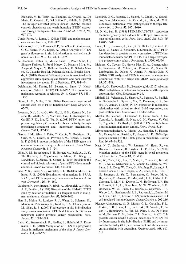
is defined as cases with positive cytoplasmic PTEN ex-pression, showing distinct areas of varying size with a loss of PTEN expression, observed in up to 25 % of the inspected area (Zhou et al., 2000; Goel et al., 2006; Peng et al., 2016). The cytoplasmic IHC staining was categorized as follows: group 0 = negative (scores 3+ and 2+ < 10 %), group 1 = positive (scores 3+ and 2+ ≥ 10 %), group 2 = positive with negative clones. The nu-clear IHC staining was categorized as follows: group 0 = negative (< 10 %), group 1 = positive (≥ 10 %). The representative examples of PTEN staining are shown in Fig. 1 (a, b, c).
Fluorescence in situ hybridization Deletions of the PTEN gene (22q12) were analysed
using the fluorescent in situ hybridization (FISH) meth-od with ZytoLight SPEC PTEN/CEN 10 Dual Color Probe (#Z-2078; ZytoVision, Bremerhaven, Germany). Formalin-fixed paraffin-embedded tissues were sec-tioned at a thickness of 3 μm and processed according to the manufacturer’s instructions. In total, FISH analysis was performed in 31 selected cases comprising 20 cases with negative cytoplasmic, 10 cases with clonal-like, and one case with positive cytoplasmic PTEN expres-sion and detected PTEN mutation. Due to the poor qual-ity of samples (especially in samples older than 10 years),
Fig. 1. Immunohistochemical PTEN expression patterns in primary cutaneous melanoma (A) Positive cytoplasmic and focal nuclear PTEN expres-sion. Note the expression of PTEN in some nuclei of the basal layer of epidermis (200×); (B) Loss of PTEN expres-sion with positive control in endothelial cells; (C) Hetero-geneous cytoplasmic PTEN staining pattern, the so-called clonal-like PTEN expression with cytoplasmic positivity on the left side, and loss of expression on the right side (200×).
a
c
b

10 Vol. 66
we were able to evaluate only 19/31 (61 %) cases. Out of the 19 evaluated samples, only five specimens were not older than three years (those were collected between April and August 2017). The reported invalid results were gained from 11 samples in total, all of which were older than six years. In cases with clonal-like PTEN ex-pression, the evaluation was performed in both negative and positive areas of the tumour tissue for comparison (Zhou et al., 2000; Goel et al., 2006; Bucheit et al., 2014; Peng et al., 2016).
The evaluation of the FISH analysis was performed according to different scoring systems used in five dif-ferent publications (Han et al., 2009; de Campos et al., 2013; Maiques et al., 2014; Picanco-Albuquerque et al., 2016; Lotan et al., 2017), which use different cut-off values (ranging from 24 to 70 %) for evaluation of the normal PTEN copy number (characterized by the pres-ence of two CEN10 and two PTEN signals), PTEN hemizygous detection (LOHe; characterized by the pres-ence of two CEN10 and one PTEN signal), PTEN ho-mozygous deletion (LOHo; characterized by the pres-ence of two CEN10 and no PTEN signal), whole chro-mosome 10 deletion [del(10); characterized by the pres-ence of one CEN10 and one PTEN signal], and polysomy (characterized by the presence of ≥ 3 CEN10 and PTEN signals). The cut-off value of ≥ 50 % tumour cells with the respective event was used to calculate the overall score, which was based on the presence of concordant results confirmed by at least three out of five of the aforementioned published criteria. The FISH signals were scored manually (x100 oil immersion) in morpho-logically intact and non-overlapping nuclei, and a mini-mum of 50 tumour cells was scored. Signals found in the nuclei of tumour-infiltrating lymphocytes were used as an internal control of the normal PTEN copy number.
Isolation of DNASections of formalin-fixed, paraffin-embedded tissue
were used for DNA isolation using standard procedures. HE-stained slides were reviewed by a pathologist, and areas for macrodissection, together with an estimation of the tumour cell percentage, were marked. Sections from each sample were deparaffinized in xylene. The DNA was then extracted using the cobas® DNA Sample Preparation kit (Roche, Mannheim, Germany) or auto-matic isolator MagCore® nucleic Acid Extractor using MagCore Genomic DNA FFPE One-step kit (#MGF-03; RBC Bioscience, New Taipei City, Taiwan).
NGS analysis and biostatistical evaluationSamples for sequence capture NGS (next-generation
sequencing; massive parallel sequencing) were processed using the KAPA HyperPlus kit according to the Seq Cap EZ protocol (NimbleGen, Roche). Target sequences were enriched using a panel of hybridization probes against multiple targets including the whole PTEN cod-ing sequence (219 kbp; NimbleGen, Roche). NimbleGen probes were designed using the online software Nimble-Design (parameters for probe stringency were chosen
as follows: Probe Selection Database: hg19/GRCh37, Homo sapiens Stringent; Preferred Close Matches: 1; Maximum Close Matches: 5). High stringent probes were designed in order to match only one target to avoid enrichment of homologous sequences. The library was pair-end sequenced by the MiSeq instrument (Illumina, San Diego, CA). NextGENe software (Softgenetics, State College, PA) was used for biostatistical analysis of se-quencing data. Nonsynonymous variants in exons and adjacent intronic regions with minimal average cover-age 100× and variant allele frequency (VAF) > 10 % were evaluated and manually inspected using an IGV viewer (Broad Institute, uC San Diego, CA) to avoid false-positive variations. The nomenclature follows the rules of the Human Genome Variation Society, and reference sequences used for PTEN were LRG_311 (NM_000314.4, NP_000305.3).
Methylation analysisBisulphite conversion of DNA was successfully per-
formed in 71 cases of primary melanoma by using EZ DNA Methylation-Lightning Kit (Zymo Research, Irvi-ne, CA) according to the manufacturer’s instructions. The primers used in the methylation-specific assay were designed according to Garcia et al. (2004); amplicon size for methylated DNA is 71 bp and for unmethylated DNA 78 bp (Gacia et al., 2004). Experiments included non-methylated DNA and universally methylated DNA controls (Human HCT116 DKO Non-Methylated DNA and Human HCT116 DKO Methylated DNA; Zymo Research). PCR was carried out with ZymoTaq™ DNA Polymerase (Zymo Research). The PCR products were separated in 2% agarose gel and the intensities of spe-cific PCR products were evaluated. The methylation pattern of selected PCR products was analysed further by employing a more sensitive detection method using the Fragment Analyzer (AATI, Santa Clara, CA) capil-lary electrophoresis system and High Sensitivity NGS Fragment Analysis Kit (#DNF-474; AATI).
Statistical analysesThe Statistica software (StatSoft, Tulsa, OK) was used
to perform all statistical analyses. Pearson χ2 test was used to evaluate the association of PTEN IHC groups with clinico-pathological variables. Association of the occurrence of mutations in PTEN (binary response vari-able: wild-type/mutated) with the clinico-pathological characteristics was also evaluated using Pearson χ2 test (however, our sample set is limited, as only eight cases carried either pathogenic PTEN mutation detected by NGS or showed loss of heterozygosity (LOH) detected by FISH). One case of histological subtype ALM (PTEN mutated) was excluded from all statistical correlations of the PTEN expression/mutation status with the histo-logical subtype. Time-to-event analysis was performed with three outcomes – disease-free survival (DFS: death from melanoma was considered as a failure), local re-currence-free survival (LFS: the period from primary diagnosis until the first local recurrence), and distant me-
K. Němejcová et al.

Vol. 66 11
tastasis-free survival (MFS: the period from primary diagnosis until the first distant metastasis). The initial univariate analysis was performed using the log-rank test to determine the differences between IHC groups, and the Kaplan-Meier method was used to compose the survival curves. All performed tests were two-sided and a P-value < 0.05 was considered as significant.
ResultsThe immunohistochemical evaluation of cytoplasmic
staining revealed 20/112 (18 %) cases with loss of PTEN expression and 92/112 (82.1 %) cases with positive PTEN expression, including 11/112 cases (9.8 %) with heterogeneous clonal-like expression. In all but one of the cases with positive PTEN expression, the 2+ inten-sity staining was dominant (intensity comparable with the intensity of the internal positive control). Surpri-singly, one of the 11 cases with clonal-like loss of ex-pression showed positive nuclear PTEN staining within the “clone” population of cells with negative cytoplas-mic staining. The evaluation of nuclear staining revealed loss of PTEN expression in 83/112 (74 %) and retained PTEN expression in 29/112 (26 %) cases, including 24/29 (83 %) cases with positivity in 10–49 % of tumour
cells and 5/29 (17 %) cases with positivity in ≥ 50 % tu-mour cells.
The correlations of the cytoplasmic and nuclear PTEN expression with the evaluated clinico-pathologi-cal variables are summarized in Table 2. Our data sug-gest that positive PTEN expression is more commonly found in lower stage melanomas compared to pT3 and pT4 stages, and in those located on the head and ex-tremities compared to those on the trunk.
Survival analyses suggested longer DFS for patients with positive cytoplasmic PTEN expression (group 1+2) in primary melanoma when compared with cases with loss of cytoplasmic PTEN expression (group 0; Fig. 2A). The same trend was observed for nuclear PTEN expression; however, the analysis only showed borderline significance in our data set (Fig. 2B). No sig-nificant differences were observed for LFS or MFS (data not shown).
FISH analysis revealed a deletion of PTEN in 3/7 (43 %) analysed cases with clonal-like PTEN expression, including two cases with deletion of PTEN loci [del(10)] and one case with hemizygous deletion of PTEN (LOHe). Interestingly, the case with LOHe showed im-munohistochemical nuclear positivity in the cytoplas-mically negative “clonal” focus. In contrast, del(10) was
Table 2. Association between the IHC expression and clinico-pathological variables
IHC Cytoplasmic expression group (N) P value Nuclear expression group (N) P valueVariables 0 (20) 1 (81) 2 (11) 0 (83) 1 (29)Gender 0.259 0.359Male 13 49 4 51 15Female 7 32 7 32 14Age 0.442 0.281≤ median 8 41 7 39 17> median 12 40 4 44 12Tumour stage 0.013 0.086pT1 + pT2 0 26 3 18 11pT3 + pT4 20 55 8 65 18Location 0.038 0.141Trunk 13 29 3 53 14Head + upper + Lower extremities 7 52 8 30 15Histological subtypea 0.407 0.123NM 12 47 8 36 8SSM 8 34 2 46 21Ulceration 0.729 0.287Yes 8 38 6 41 11No 12 43 5 42 18Sentinel node positivityb 0.750 0.420Yes 5 11 3 15 4No 10 35 7 36 16
IHC groups for cytoplasmic expression: 0 = negative expression, 1 = positive expression, 2 = positive expression with negative clones (clonal-like PTEN expression). P values are based on Pearson’s χ2 test. aOne case of ALM was excluded from the statistics. bData not available for all cases.
Comprehensive Analysis of PTEN in Primary Cutaneous Melanoma

12 Vol. 66
observed in 2/12 (17 %) analysed cases with loss of cy-toplasmic and nuclear PTEN expression. We did not ob-serve any homozygous deletion of the PTEN loci or polysomy.
The methylation analysis of the PTEN promoter was successfully evaluated in 71 cases. However, no case with promoter methylation was found.
The NGS analysis revealed four PTEN variants (clas-sified as pathogenic or splice) in 3/112 (3 %) melanoma cases and one benign variant in one case. However, the frequency of PTEN variants could be underestimated. We may have missed several variants due to unequal coverage of PTEN in the NGS library. Only 27 % (30/112) of cases had a fully sequenced PTEN with suf-ficient average coverage ≥ 100× in all coding exons. The minimal coverage of several exons, mostly exon 5, was too low (< 100×) to interpret the results, which was caused by advanced DNA fragmentation in a majority of the analysed samples and the high stringency design of probes for the target enrichment during the library prep-aration. There were somatic pathogenic mutations in exon 6 c.625G>T, p.(Gly209Ter), VAF 14 % (coverage 394×) and in exon 2 c.86_87insAAA, p.(Tyr29delinsTer), VAF 12 % (coverage 216×), detected in one melanoma case (SSM, pT3, located on the trunk, diagnosed in a male patient at the age of 67). We also detected patho-genic NRAS mutation c.182A>G, p.(Gln61Arg) in the same patient. No PTEN promoter methylation or loss of PTEN expression were found. Another PTEN mutation (c.16A>T, p.(Lys6Ter), VAF 43 % (coverage 386×)) was also detected, this one in an NM, pT3, located on the upper extremity, diagnosed in a female at the age of 77. Methylation could not be tested due to the lack of DNA, FISH was not evaluable due to poor tissue quali-ty, and immunohistochemistry showed a loss of PTEN
expression. A splice mutation was detected in the con-sensus splice site in intron 8 c.1026+2T>G, VAF 40 % (coverage 502×), discovered in an NM, pT3, female, diagnosed at 55 years of age. The melanoma showed non-methylated PTEN promoter region, positive cyto-plasmic expression in 30 % of tumour cells, and a loss of nuclear PTEN expression. FISH was not evaluable. Finally, we also detected a likely benign variant c.883C>G, p.(Leu295Val), VAF 24 % (coverage 309×), and deleted PTEN loci, del(10) in one SSM, pT4, trunk, male, diagnosed at 69 years of age. This sample dis-played a clonal PTEN expression by immunohistochem-istry.
Altogether, seven out of eight cases with some ge-netic aberrations detected by either NGS and/or FISH also showed aberrant PTEN protein expression (four cases with the loss of PTEN expression and three cases with clonal-like PTEN expression). We did not find any statistically significant correlation between the presence of genetic aberrations and any of the observed clinico-pathological variables (data not shown).
Discussion Since PTEN has distinct functions in the cytoplasm
and in the nucleus, we assessed PTEN expression in both of these compartments separately. Cytoplasmic PTEN primarily plays a role in the regulation of PI3K/PIP3 signalling, and influences cancer cell growth, sur-vival, angiogenesis, and metabolism (Song et al., 2012). Nuclear PTEN functions as a tumour suppressor in a PI3K-independent manner through its protein phos-phatase activity, causing inhibition of migration or in-ducing cell cycle arrest (Milella et al., 2015). However, it can also act in a phosphatase-independent way, influ-
Fig. 2. Correlation of PTEN expression with prognosisRepresentative Kaplan-Meier curves for disease specific-free survival (DFS) according to the cytoplasmic (A) and nu-clear (B) expression of PTEN protein evaluated by immunohistochemistry. Group 0 – negative PTEN expression, group 1 – positive PTEN expression, group 2 – clonal-like PTEN expression (only cytoplasmic staining was evaluated). The graphs show marked respective groups with numbers of complete/censored cases in the brackets.
K. Němejcová et al.

Vol. 66 13
encing the regulation of chromosome stability, DNA repair, and apoptosis (Dillon and Miller, 2014; Milella et al., 2015).
We found a loss of cytoplasmic PTEN expression in 20/112 (18 %) cases and clonal-like loss of expression in other 11 (9.8 %) cases, altogether comprising 31 sam-ples (28 %) with aberrant expression. Loss of nuclear expression was found in 83 (74 %) cases (Table 2). Our results are in concordance with most of the previously published studies evaluating primary cutaneous mela-nomas, which used the same antibody clone (Whiteman et al., 2002; Goel et al., 2006; Bucheit et al., 2014). Different results were found in metastatic melanomas, where about 65 % of cases (Whiteman et al., 2002) showed absent or decreased cytoplasmic expression and almost all samples showed none or decreased nuclear expression (Zhou et al., 2000; Whiteman et al., 2002).
Several studies reported somewhat ambiguous results with a higher proportion of positivity in primary or met-astatic melanomas (Deichmann et al., 2002; Mikhail et al., 2005; Giles et al., 2019). The inconsistent results are probably caused by the use of different primary antibod-ies. Another matter is also the subjectivity of the scoring systems, which are based on a comparison of staining intensity with the adjacent normal cells (vascular en-dothelium), and PTEN heterogeneity within tumours (Dillon and Miller, 2014). The PTEN antibody (D4.3) used in the study of Giles et al. (2019) showed a higher sensitivity for the detection of PTEN than the 6H2.1 an-tibody used by us and other aforementioned authors. The discrepant results published in the study by Mikhail et al. (2005) can be explained by the application of a different scoring system and antibody.
Our data showed that the loss of immunohistochemi-cal PTEN expression is more common in higher stages, which is in concordance with others (Goel et al., 2006). Loss of PTEN expression is also more common in mela-nomas located on the trunk when compared to the head and extremities (Whiteman et al., 2002). The absence of cytoplasmic PTEN expression was also associated with a worse DFS (P = 0.045; Fig. 2a). However, the statisti-cal significance of the loss of nuclear staining in our study was only borderline (P = 0.057). We have not found any statistically significant outcome regarding LFS and MFS. Other studies analysing the correlation between PTEN expression and disease outcome are in-consistent and sparse. One study suggests that partial PTEN loss is associated with worse overall survival (OS) (Giles et al., 2019). Another study that focused on metastatic melanomas showed that only a complete loss of PTEN expression is associated with shorter OS (Bucheit et al., 2014). Others did not confirm any sig-nificant association between the PTEN expression and survival (Mikhail et al., 2005). One explanation of this discordance may lie in the divergence between the ana-lysed sample sets and the different used antibodies.
Only a few studies performed a cytogenetic analysis in melanomas to date, and those used centromeric probes for the chromosomes (1, 4, 6-7, 9-12, 15, 17-18, X, and
Y) and a midisatellite probe localized in 1p36, but no study used the PTEN locus-specific FISH probe in cuta-neous melanomas (Poetsch et al., 1998a, b). One study of nine cases of uveal melanoma cell lines did not detect any PTEN mutations or cytogenetic abnormalities in-volving chromosome 10q23, and the authors suggest that PTEN alterations probably do not play a role in the pathogenesis of uveal melanoma when compared to cu-taneous melanoma (Naus et al., 2000).
The loss of PTEN loci was detected in 2 of 12 (17 %) analysed cases with a loss of PTEN expression (cyto-plasmic and nuclear) and in 3/7 (43 %) cases with clon-al-like PTEN expression. Interestingly, one case with a heterozygous PTEN deletion (LOHe) showed immuno-histochemical nuclear positivity in the cytoplasmically negative “clonal” focus. Since this is only an isolated case, it is impossible to find a relevant explanation for this observation. Altogether, the FISH analysis revealed a deletion of PTEN in 5 out of 19 (26 %) analysed cases. Additionally, four pathogenic, likely pathogenic, or splice PTEN variants were detected by NGS in three dif-ferent samples out of the 112 analysed cases. All the cases were associated with a loss of nuclear PTEN ex-pression, and in two cases there was also a loss of cyto-plasmic expression. One case with a retained PTEN cy-toplasmic expression showed staining in 30 % of tumour cells. Altogether, there were eight cases with PTEN ge-netic aberration detected by either FISH or NGS, which included one case with positive expression, four cases with a loss of expression, and three cases with clonal-like PTEN expression. We did not find any statistically significant association between the detected genetic ab-errations (FISH, NGS) and the clinico-pathological var-iables (data not shown). The agreement between the observed aberrant PTEN expression and detected ge-netic aberrations by FISH was described by others in prostatic or endometrial cancer (Han et al., 2009; Maiques et al., 2014; Picanco-Albuquerque et al., 2016; Lotan et al., 2017). Another study described hemizy-gous or homozygous PTEN deletion in 40 % and 6 % renal cell carcinomas, respectively. Nevertheless, no correlations with immunohistochemical PTEN expres-sion were examined in this study (de Campos et al., 2013). Homozygous deletion of PTEN loci or polysomy were also observed in prostatic cancer cases (Han et al., 2009; Picanco-Albuquerque et al., 2016; Lotan et al., 2017). In contrast, no homozygous alterations were de-tected in our study.
Epigenetic changes can explain loss of the PTEN function; however, the precise role of DNA methylation in melanoma is still unclear (Dillon and Miller, 2014). We did not observe any PTEN promoter methylation in our sample set. Data regarding PTEN promoter methyl-ation in melanoma is quite inconsistent in the literature. Our results are in agreement with the data derived from The Cancer Genome Atlas Network (TGCA, http://www.cbioportal.org/), which included a cohort of skin cutaneous melanomas (Cancer Genome Atlas, 2015). The TCGA melanoma DNA methylation data included
Comprehensive Analysis of PTEN in Primary Cutaneous Melanoma

14 Vol. 66
473 melanomas. Methylation of the PTEN promoter was found in only 0.6 % of melanomas (3/473). Our re-sults together with the methylation analysis data derived from TCGA suggest that PTEN promoter methylation is not common in primary cutaneous melanomas. How-ever, the results of some other studies are rather differ-ent. Some studies reported varying rates of methylation in melanomas or melanoma cell lines (23–62 %) (Mir-mohammadsadegh et al., 2006; Lahtz et al., 2010; de unamuno Bustos et al., 2018). Mirmohammadsadegh et al. (2006) have found PTEN promoter methylation in 26 % or 62 % of circulating DNA isolated from the sera of patients with primary or metastatic melanoma, respec-tively. Others found methylation in 24 % of primary cu-taneous melanomas (Mirmohammadsadegh et al., 2006; de unamuno Bustos et al., 2018). The association of PTEN methylation with higher age, Breslow thickness, advanced stage, and acral localization has also been shown (de unamuno Bustos et al., 2018). The PTEN promoter methylation has been found to be an independ-ent prognostic marker in cutaneous melanoma associat-ed with poor survival (Roh et al., 2016; de unamuno Bustos et al., 2018). Micevic et al. (2017) confirmed the correlation between methylation and protein expression and found an association with increased risk of death by Cox regression analysis. In contrast, Giles et al. (2019) did not find any relationship between these variables.
Concurrent loss of PTEN with activating BRAF muta-tion is responsible for the acquired resistance of mela-noma to BRAF inhibitors (Shi et al., 2014; Leonardi et al., 2018). However, the loss of PTEN function by itself does not preclude an antitumor response to BRAF in-hibitors, since that is only one of the mechanisms re-sponsible for the resistance, and it is therefore likely that other co-occurring events cooperate in the development of resistance. Brown et al. (2017) proposed that the ef-fects of β-catenin signalling in melanoma cells differ depending on PTEN expression. The loss or absence of PTEN expression corresponds with increased invasion, and retained PTEN staining is associated with reduced invasion in response to β-catenin signalling (Brown et al., 2017). The authors suggest that the pro-invasive ef-fects of WNT/β-catenin signalling are potentially inde-pendent of metabolic reprogramming. Their results sug-gest that the state of PTEN expression could be a potential biomarker for the implementation of WNT in-hibitors in melanoma. Abbots et al. (2014) provided the evidence that PTEN deficiency is not only a promising biomarker in melanoma, but can also be targeted by a blockage of DNA base excision repair (by APE1 inhibi-tion) and as such could represent a potential target for targeted therapy of PTEN-deficient melanomas. Some recent studies demonstrated that the PTEN expression status also plays an important role in determining the response to MEK inhibition in melanoma, and a loss of PTEN hinders the anti-tumour activity of MEK inhibi-tors in preclinical models (Ciuffreda et al., 2012; Milella et al., 2015).
In conclusion, the results of our study showed that the loss of cytoplasmic PTEN expression was significantly associated with higher stage, location of melanoma on the trunk, and worse DFS. This is in concordance with some of the previously published studies. Concerning molecular findings, we identified eight cases with aber-rations detected by FISH or NGS, but no case with methylation of the PTEN promoter. These findings are in agreement with the results of TCGA study, but not with several other studies, and further research into this topic is needed. Despite the limitation in the number of fully analysed cases, our data showed a poor concord-ance between the aberrant IHC pattern and the presence of PTEN mutation or aberration in the PTEN loci, which suggests that IHC is an inappropriate screening method for the detection of genetic aberrations. Nevertheless, the loss of PTEN expression as detected by immunohis-tochemistry can be clinically significant. However, studies are needed to assess the significance of PTEN analysis, either as a potential biomarker of response to targeted therapy (BRAF inhibitors, MEK inhibitors, WNT inhibitors, blockage of DNA base excision repair, and others), or directly in targeted therapy using PTEN modulators in the future.
AcknowledgementThe authors thank Zachary Harold Kane Kendall,
B.A., for English editing.
ReferencesAbbotts, R., Jewell, R., Nsengimana, J., Maloney, D. J., Sime-
onov, A., Seedhouse, C., Elliott, F., Laye, J., Walker, C., Jadhav, A., Grabowska, A., Ball, G., Patel, P. M., Newton-Bishop, J., Wilson, D. M., 3rd., Madhusudan, S. (2014) Targeting human apurinic/apyrimidinic endonuclease 1 (APE1) in phosphatase and tensin homolog (PTEN) defi-cient melanoma cells for personalized therapy. Oncotarget 5, 3273-3286.
Aguissa-Toure, A. H., Li, G. (2012) Genetic alterations of PTEN in human melanoma. Cell. Mol. Life Sci. 69, 1475-1491.
Brown, K., Yang, P., Salvador, D., Kulikauskas, R., Ruohola-Baker, H., Robitaille, A. M., Chien, A. J., Moon, R. T., Sherwood, V. (2017) WNT/β-catenin signaling regulates mitochondrial activity to alter the oncogenic potential of melanoma in a PTEN-dependent manner. Oncogene 36, 3119-3136.
Bucheit, A. D., Chen, G., Siroy, A., Tetzlaff, M., Broaddus, R., Milton, D., Fox, P., Bassett, R., Hwu, P., Gershenwald, J. E., Lazar, A. J., Davies, M. A. (2014) Complete loss of PTEN protein expression correlates with shorter time to brain metastasis and survival in stage IIIB/C melanoma pa-tients with BRAFV600 mutations. Clin. Cancer Res. 20, 5527-5536.
Cancer Genome Atlas Nerwork (2015) Genomic classification of cutaneous melanoma. Cell 161, 1681-1696.
Ciuffreda, L., Di Sanza, C., Cesta Incani, u., Eramo, A., Desi-deri, M., Biagioni, F., Passeri, D., Falcone, I., Sette, G., Bergamo, P., Anichini, A., Sabapathy, K., McCubrey, J. A.,
K. Němejcová et al.
Vol. 66 15
Ricciardi, M. R., Tafuri, A., Blandino, G., Orlandi, A., De Maria, R., Cognetti, F., Del Bufalo, D., Milella, M. (2012) The mitogen-activated protein kinase (MAPK) cascade controls phosphatase and tensin homolog (PTEN) expres-sion through multiple mechanisms. J. Mol. Med. (Berl.) 90, 667-679.
Conde-Perez, A., Larue, L. (2012) PTEN and melanomagen-esis. Future Oncol. 8, 1109-1120.
de Campos, E. C., da Fonseca, F. P., Zequ Sde, C., Guimaraes, G. C., Soares, F. A., Lopes, A. (2013) Analysis of PTEN gene by fluorescent in situ hybridization in renal cell carci-noma. Rev. Col. Bras. Cir. 40, 471-475.
de unamuno Bustos, B., Murria Estal, R., Perez Simo, G., Simarro Farinos, J., Pujol Marco, C., Navarro Mira, M., Alegre de Miquel, V., Ballester Sanchez, R., Sabater Mar-co, V., Llavador Ros, M., Palanca Suela, S., Botella Estra-da, R. (2018) Aberrant DNA methylation is associated with aggressive clinicopathological features and poor survival in cutaneous melanoma. Br. J. Dermatol. 179, 394-404.
Deichmann, M., Thome, M., Benner, A., Egner, u., Harts-chuh, W., Naher, H. (2002) PTEN/MMAC1 expression in melanoma resection specimens. Br. J. Cancer 87, 1431-1436.
Dillon, L. M., Miller, T. W. (2014) Therapeutic targeting of cancers with loss of PTEN function. Curr. Drug Targets 15, 65-79.
Freeman, D. J., Li, A. G., Wei, G., Li, H. H., Kertesz, N., Le-sche, R., Whale, A. D., Martinez-Diaz, H., Rozengurt, N., Cardiff, R. D., Liu, X., Wu, H. (2003) PTEN tumor sup-pressor regulates p53 protein levels and activity through phosphatase-dependent and -independent mechanisms. Cancer Cell 3, 117-130.
García, J. M., Silva, J., Peña, C., Garcia, V., Rodríguez, R., Cruz, M. A., Cantos, B., Provencio, M., España, P., Bonil-la, F. (2004) Promoter methylation of the PTEN gene is a common molecular change in breast cancer. Genes Chro-mosomes Cancer 41, 117-124.
Giles, K. M., Rosenbaum, B. E., Berger, M., Izsak, A., Li, Y., Illa Bochaca, I., Vega-Saenz de Miera, E., Wang, J., Darvishian, F., Zhong, H., Osman, I. (2019) Revisiting the clinical and biologic relevance of partial PTEN loss in mel-anoma. J. Invest. Dermatol. 139, 430-438.
Goel, V. K., Lazar, A. J., Warneke, C. L., Redston, M. S., Ha-luska, F. G. (2006) Examination of mutations in BRAF, NRAS, and PTEN in primary cutaneous melanoma. J. In-vest. Dermatol. 126, 154-160.
Guldberg, P., thor Straten, P., Birck, A., Ahrenkiel, V., Kirkin, A. F., Zeuthen, J. (1997) Disruption of the MMAC1/PTEN gene by deletion or mutation is a frequent event in malig-nant melanoma. Cancer Res. 57, 3660-3663.
Han, B., Mehra, R., Lonigro, R. J., Wang, L., Suleman, K., Menon, A., Palanisamy, N., Tomlins, S. A., Chinnaiyan, A. M., Shah, R. B. (2009) Fluorescence in situ hybridization study shows association of PTEN deletion with ERG rear-rangement during prostate cancer progression. Mod. Pathol. 22, 1083-1093.
Lahtz, C., Stranzenbach, R., Fiedler, E., Helmbold, P., Dam-mann, R. H. (2010) Methylation of PTEN as a prognostic factor in malignant melanoma of the skin. J. Invest. Der-matol. 130, 620-622.
Leonardi, G. C., Falzone, L., Salemi, R., Zanghi, A., Spandi-dos, D. A., McCubrey, J. A., Candido, S., Libra, M. (2018) Cutaneous melanoma: from pathogenesis to therapy (Re-view). Int. J. Oncol. 52, 1071-1080.
Li, D. M., Sun, H. (1998) PTEN/MMAC1/TEP1 suppresses the tumorigenicity and induces G1 cell cycle arrest in hu-man glioblastoma cells. Proc. Natl. Acad. Sci. USA 95, 15406-15411.
Lotan, T. L., Heumann, A., Rico, S. D., Hicks, J., Lecksell, K., Koop, C., Sauter, G., Schlomm, T., Simon, R. (2017) PTEN loss detection in prostate cancer: comparison of PTEN im-munohistochemistry and PTEN FISH in a large retrospec-tive prostatectomy cohort. Oncotarget 8, 65566-65576.
Maiques, O., Cuevas, D., Garcia Dios, D. A., Coenegrachts, L., Santacana, M., Velasco, A., Romero, M., Sonia, G., Lambrechts, D., Dolcet, X., Amant, F., Matias-Guiu, X. (2014) FISH analysis of PTEN in endometrial carcinoma. Comparison with SNP arrays and MLPA. Histopathology 65, 371-388.
Micevic, G., Theodosakis, N., Bosenberg, M. (2017) Aberrant DNA methylation in melanoma: biomarker and therapeutic opportunities. Clin. Epigenetics 9, 34.
Mikhail, M., Velazquez, E., Shapiro, R., Berman, R., Pavlick, A., Sorhaindo, L., Spira, J., Mir, C., Panageas, K. S., Pol-sky, D., Osman, I. (2005) PTEN expression in melanoma: relationship with patient survival, Bcl-2 expression, and proliferation. Clin. Cancer Res. 11, 5153-5157.
Milella, M., Falcone, I., Conciatori, F., Cesta Incani, u., Del Curatolo, A., Inzerilli, N., Nuzzo, C. M., Vaccaro, V., Vari, S., Cognetti, F., Ciuffreda, L. (2015) PTEN: multiple func-tions in human malignant tumors. Front. Oncol. 5, 24.
Mirmohammadsadegh, A., Marini, A., Nambiar, S., Hassan, M., Tannapfel, A., Ruzicka, T., Hengge, u. R. (2006) Epi-genetic silencing of the PTEN gene in melanoma. Cancer Res. 66, 6546-6552.
Naus, N. C., Zuidervaart, W., Rayman, N., Slater, R., van Drunen, E., Ksander, B., Luyten, . G. P., Klein, A. (2000) Mutation analysis of the PTEN gene in uveal melanoma cell lines. Int. J. Cancer 87, 151-153.
Peng, W., Chen, J. Q., Liu, C., Malu, S., Creasy, C., Tetzlaff, M. T., Xu, C., McKenzie, J. A., Zhang, C., Liang, X., Wil-liams, L. J., Deng, W., Chen, G., Mbofung, R., Lazar, A. J., Torres-Cabala, C. A., Cooper, Z. A., Chen, P. L., Tieu, T. N., Spranger, S., Yu, X., Bernatchez, C., Forget, M. A., Haymaker, C., Amaria, R., McQuade, J. L., Glitza, I. C., Cascone, T., Li, H. S., Kwong, L. N., Heffernan, T. P., Hu, J., Bassett, R. L, Jr.., Bosenberg, M. W., Woodman, S. E., Overwijk, W. W., Lizee, G., Roszik, J., Gajewski, T. F., Wargo, J. A., Gershenwald, J. E., Radvanyi, L., Davies, M. A., Hwu, P. (2016) Loss of PTEN promotes resistance to T cell-mediated immunotherapy. Cancer Discov. 6, 202-216.
Picanco-Albuquerque, C. G., Morais, C. L., Carvalho, F. L., Peskoe, S. B., Hicks, J. L., Ludkovski, O., Vidotto, T., Fe-dor, H., Humphreys, E., Han, M., Platz, E. A., De Marzo, A. M., Berman, D. M., Lotan, T. L., Squire, J. A. (2016) In prostate cancer needle biopsies, detections of PTEN loss by fluorescence in situ hybridization (FISH) and by immu-nohistochemistry (IHC) are concordant and show consist-ent association with upgrading. Virchows Arch. 468, 607-617.
Comprehensive Analysis of PTEN in Primary Cutaneous Melanoma

16 Vol. 66
Poetsch, M., Dittberner, T., Woenckhaus, C. (2001) PTEN/MMAC1 in malignant melanoma and its importance for tumor progression. Cancer Genet. Cytogenet. 125, 21-26.
Poetsch, M., Woenckhaus, C., Dittberner, T., Pambor, M., Lorenz, G., Herrmann, F. H. (1998a) Differences in chro-mosomal aberrations between nodular and superficial spreading malignant melanoma detected by interphase cy-togenetics. Lab. Invest. 78, 883-888.
Poetsch, M., Woenckhaus, C., Dittberner, T., Pambor, M., Lorenz, G., Herrmann, F. H. (1998b) An increased frequen-cy of numerical chromosomal abnormalities and 1p36 de-letions in isolated cells from paraffin sections of malignant melanomas by means of interphase cytogenetics. Cancer Genet. Cytogenet. 104, 146-152.
Reddy, B. Y., Miller, D. M., Tsao, H. (2017) Somatic driver mutations in melanoma. Cancer 123, 2104-2117.
Roh, M. R., Gupta, S., Park, K. H., Chung, K. Y., Lauss, M., Flaherty, K. T., Jonsson, G., Rha, S. Y., Tsao, H. (2016) Promoter methylation of PTEN is a significant prognostic factor in melanoma survival. J. Invest. Dermatol. 136, 1002-1011.
Shi, H., Hugo, W., Kong, X., Hong, A., Koya, R. C., Moric-eau, G., Chodon, T., Guo, R., Johnson, D. B., Dahlman, K. B., Kelley, M. C., Kefford, R. F., Chmielowski, B., Glaspy, J. A., Sosman, J. A., van Baren, N., Long, G. V., Ribas, A., Lo, R. S. (2014) Acquired resistance and clonal evolution in melanoma during BRAF inhibitor therapy. Cancer Dis-cov. 4, 80-93.
K. Němejcová et al.
Sobin, L. H., Gospodarowicz, M. K., Wittekind, C., Interna-tional union against Cancer. (2010) TNM Classification of Malignant Tumours. Chichester, West Sussex, uK ; Hobo-ken, NJ, Wiley-Blackwell.
Song, M. S., Salmena, L., Pandolfi, P. P. (2012) The functions and regulation of the PTEN tumour suppressor. Nat. Rev. Mol. Cell Biol. 13, 283-296.
Troyer, D. A., Jamaspishvili, T., Wei, W., Feng, Z., Good, J., Hawley, S., Fazli, L., McKenney, J. K., Simko, J., Hurtado-Coll, A., Carroll, P. R., Gleave, M., Lance, R., Lin, D. W., Nelson, P. S., Thompson, I. M., True, L. D., Brooks, J. D., Squire, J. A. (2015) A multicenter study shows PTEN dele-tion is strongly associated with seminal vesicle involve-ment and extracapsular extension in localized prostate can-cer. Prostate 75, 1206-1215.
Whiteman, D. C., Zhou, X. P., Cummings, M. C., Pavey, S., Hayward, N. K., Eng, C. (2002) Nuclear PTEN expression and clinicopathologic features in a population-based series of primary cutaneous melanoma. Int. J. Cancer 99, 63-67.
Zhou, X. P., Gimm, O., Hampel, H., Niemann, T., Walker, M. J., Eng, C. (2000) Epigenetic PTEN silencing in malignant melanomas without PTEN mutation. Am. J. Pathol. 157, 1123-1128.