original article diagnostics, monitoring and … function test, six-minute walk test,...
TRANSCRIPT

ORIGINAL ARTICLE
Diagnostics, monitoring and outpatient care inchildren with suspected pulmonary hypertension/paediatric pulmonary hypertensive vascular disease.Expert consensus statement on the diagnosis andtreatment of paediatric pulmonary hypertension.The European Paediatric Pulmonary Vascular DiseaseNetwork, endorsed by ISHLT and DGPKAstrid E Lammers,1 Christian Apitz,2 Peter Zartner,3 Alfred Hager,4
Karl-Otto Dubowy,5 Georg Hansmann6
For numbered affiliations seeend of article.
Correspondence toProfessor Dr Georg Hansmann,Department of PaediatricCardiology and Critical Care,Hannover Medical School,Hannover 30625, Germany;[email protected]
This manuscript is a product ofthe writing group of theEuropean Paediatric PulmonaryVascular Disease (PVD)Network (Writing Group Chair:G. Hansmann, Writing GroupCo-Chair: C. Apitz). ISHLT,International Society of Heartand Lung Transplantation;DGPK, German Society ofPaediatric Cardiology
Received 8 March 2015Revised 29 June 2015Accepted 30 June 2015
To cite: Lammers AE,Apitz C, Zartner P, et al.Heart 2016;102:ii1–ii13.
ABSTRACTPulmonary hypertension (PH) is a condition of multipleaetiologies with underestimated prevalence andincidence. Indeed, despite access to modern therapies,pulmonary hypertensive vascular disease (PHVD) remainsa progressive, usually life-limiting condition, severelyimpacting on the patients’ well-being. We herein providepractical, expert consensus recommendations on theinitial diagnostic work-up, clinical management andfollow-up of children and adolescents with PH/PHVD,including a diagnostic algorithm. The major topics andmethods that need to be tailored and put into context ofthe individual patient include PH classification, clinicalsigns and symptoms, basic diagnostic and advancedimaging measures (ECG, chest X-ray, transthoracicechocardiography, cardiac magnetic resonance, chest CTangiography, cardiac catheterisation, ventilation-perfusion lung scan, abdominal ultrasound), lungfunction tests, 6 min walk and cardiopulmonary exercisetesting, sleep study (polysomnography), laboratory/immunological tests, considerations for elective surgery/general anaesthesia, physical education and exercise,flying on commercial airplanes, vaccinations, care ofcentral intravenous lines and palliative care. Due to thecomplexity of PH/PHVD, the clinical care has to bemultidisciplinary and coordinated by a dedicatedspecialist paediatric PH centre, not only to decreasemortality but to allow children with PH/PHVD to reach areasonable quality of life.
INTRODUCTIONPulmonary hypertension (PH) is a condition ofmultiple aetiologies, sharing a significant increasein pulmonary artery pressure. For the majority ofpatients, PH represents a progressive and life-limiting disease with significant impact on qualityof life, especially in the advanced stages of disease.Diagnostic algorithms and implementation of
careful disease monitoring and follow-up are desir-able to standardise medical care, gain further insightinto disease progression, guide therapy, facilitate mul-ticentre trials and, ultimately, achieve best possible
clinical outcomes. Consensus standardisation is par-ticularly important because PH, especially pulmonaryarterial hypertension (PAH), is a rare condition andsingle-centre experience is often limited.1 2
The aim of this article is to provide an overviewover the condition and existing classifications.Moreover, we introduce a paediatric diagnostic
Box 1: Definitions
Pulmonary Hypertension (PH)mPAP ≥25 mmHg in children >3 months of age atsea levelPulmonary Arterial Hypertension (PAH)mPAP ≥25 mmHgPCWP <15 mmHgPVR index >3 WU × m2
Idiopathic PAH (IPAH)PAH with no underlying disease known to beassociated with PAHHereditary PAH (HPAH)PAH with no underlying disease but with positivefamily history or positive genetic testingPulmonary Hypertensive Vascular Disease(PHVD)For biventricular circulations:mPAP ≥25mmHg and PVR index >3 WU × m2
For circulations with cavopulmonary anastomosis(e.g., Fontan physiology):mean TPG >6 mmHg (calculate mPAP minus mLAPor PCWP) or PVR index >3 WU × m2 Detailedhemodynamic definitions of PH (e.g., precapilaryvs. postcapillary PH, value of the diastolictranspulmonary pressure gardient) are presented inApitz C, Hansmann G, Schranz D. Heart, 2016[1].mPAP, mean pulmonary artery pressure; mLAP,mean left atrial pressure; PAH, pulmonary arteryhypertension; PCWP, pulmonary capillary wedgepressure (syn. PAWP, pulmonary artery wedgepressure); PVRi, pulmonary vascular resistanceindex; TPG, transpulmonary pressure gradient.
Lammers AE, et al. Heart 2016;102:ii1–ii13. doi:10.1136/heartjnl-2015-307792 ii1
Pulmonary vascular disease

algorithm and practical recommendations for the monitoring andoutpatient care of children and adolescents with PH, PAH and/orpulmonary hypertensive vascular disease (PHVD).
METHODSThe recommendations on diagnostics, monitoring and outpatientcare in paediatric PH given in table 1 are based on a gradingsystem currently suggested by the European Society of Cardiology(ESC) and the American Heart Association (AHA), and was basedon paediatric data only (class of recommendation, level of evi-dence). The grading and voting process within the writing group isoutlined in the executive summary69 of this special issue.Computerised searches of the PubMed/MEDLINE bibliographicdatabase from 1990 to June 2015 were conducted. The developersearched using the terms ‘paediatric pulmonary hypertension’,‘pulmonary hypertension and children’, ‘pulmonary hypertensionand children and: diagnosis/diagnostics; follow-up, monitoring,epidemiology, classification, symptoms, functional classification,electrocardiogram, imaging, chest X-ray (CXR), echocardiography,lung function test, six-minute walk test, cardiopulmonary exercisetesting, polysomnography, sleep study, computed tomography(CT), cardiac catheterization, magnetic resonance imaging (MRI),ventilation-perfusion scan, abdominal ultrasound, portal hyperten-sion, chronic thromboembolic pulmonary hypertension, labora-tory testing, serological markers, biomarkers, genetic testing,(lung) transplantation, palliative care, general anaesthesia, risk,surgery, flying, air travelling, contraception’.
DEFINITIONPH has been defined as a mean pulmonary artery pressure(mPAP) ≥25 mm Hg at rest, measured by cardiac catheterisation(according to the most recent 5th World Symposium onPulmonary Hypertension, Nice 2013). The term pulmonaryarterial hypertension (ie, group 1 PH) describes a subpopulationof patients with PH, characterised haemodynamically by thepresence of pre-capillary PH, including an end-expiratory pul-monary artery wedge pressure <15 mm Hg and a pulmonaryvascular resistance >3 Wood units.3 4 While this definition hasbeen widely accepted for paediatric use,5 the PulmonaryVascular Research Institute (PVRI) expanded it and introducedthe term ‘paediatric pulmonary hypertensive vascular disease(PPHVD)’ in 2011 (Panama, 2011). Importantly, the Panamaclassification distinguishes between PH with and without pul-monary vascular disease (PVD), between single and biventricularcirculations, and acknowledges the heterogeneous aetiology inpaediatric PH that can even have prenatal (fetal) origins.Although patients with congenital heart disease (CHD) andsingle-ventricle physiology (without subpulmonary ventricle)often do not meet the criteria as defined above (ie, mPAP<25 mm Hg, but PVD evident), they may benefit from similarpharmacological strategies and share some clinical features ofpatients with PH. For patients with Fontan-type haemodynamics(no subpulmonary ventricle), a PVR index >3 WU×m2 or atranspulmonary gradient of >6 mm Hg has been suggested as adefinition of PPHVD.4 The most common type of PH diag-nosed in childhood is PAH associated with CHD. Frequently,PAH-CHD presents with a pre-tricuspid or post-tricuspid shuntlesion (eg, left-to-right shunt) with or without evident PVD.Children without PVD benefit from shunt closure.70 Childrenwith established PAH and PVD (PPHVD) may not tolerateshunt closure due to the sudden loss of decompression of theRV. The onset and progression of PAH-CHD and consecutivePVD differ in pre-tricuspid versus post-tricuspid lesions sincethe latter (eg, a large ventricular septal defect (VSD)), may
expose the pulmonary vasculature not only to increased flowbut also systemic or near-systemic pressure.
While adults with classical Eisenmenger haemodynamics actu-ally have a better survival than patients with idiopathic pulmon-ary arterial hypertension (IPAH)/hereditary pulmonary arterialhypertension (HPAH), children with PAH-CHD and those withIPAH have a similar mortality with a 5-year survival reported tobe 71% and 75%, respectively.6
PH CLASSIFICATIONNice World Symposium classification (2013) and PanamaPVRI classification (2011)Whereas the first international classification for PH was based onhistological findings,7 more recent classifications (Evian 1998,Venice 2003 and Dana Point 2008) were refined with a focus oncommon clinical features, pathophysiology and to delineate dif-ferences and similarities of the various entities of the disease.
At the last World Symposium of Pulmonary Hypertension inNice (2013, table 2), a new subgroup of ‘multifactorial PH’ wasintroduced to account for the heterogeneity of paediatric PHand special considerations that apply to children.8
The Paediatric Taskforce of the PVRI addressed the need fora more specific paediatric classification, introducing the conceptof hypertensive vascular disease (PPHVD), that is, PH with ele-vated PVR, and focusing on issues and disease entities that areparticularly relevant to children.4 In 2011, a new PaediatricPanama classification was proposed (table 3), suggesting 10 cat-egories of PPHVD. The Panama classification takes perinatalmaladaptation, insufficient development and pulmonary hypo-plasia, as well as genetic findings into account, all of which mayplay a role in the development of PH early in life. It also intro-duces a class of ‘multifactorial PH’ because—especially in chil-dren—often more than one factor may account for thediagnosis of PH. The main aim was to provide a system that isclinically useful, incorporates specific paediatric aspects thatwere previously not adequately addressed and allows categoris-ing complex patients with a multifactorial pathogenesis of theirPH accordingly (table 3).
EPIDEMIOLOGYData on the exact incidence and the prevalence of PH ininfancy and childhood are sparse. PH in childhood is seen witha variety of conditions, however, if at all, limited data aremainly available for IPAH and PH associated with CHD(PH-CHD). Two Dutch nationwide registries of paediatric PHsuggested an annual incidence of 0.7 cases per million childrenfor IPAH and a point prevalence of 4.4 per million children.For PH-CHD, annual incidence and prevalence were 2.2 and15.6 cases per million children, respectively. Compared withadult studies, the incidence of IPAH seems to be lower in chil-dren, whereas PH-CHD may occur more frequently in child-hood.9 Data from the UK Pulmonary Hypertension Servicereported a slightly lower incidence for IPAH, that is, 0.48 casesper million children per year for IPAH and a prevalence of 2.1cases per million children (IPAH).10 A 5-year multicentre pro-spective study including 21 referral centres in France reportedan estimated prevalence of 3.7 paediatric PAH cases per million,and for IPAH, 2.2 cases per million.11
All in all, based on the available data a PH prevalence in2–16 per million children can be estimated, acknowledging thatthere are risk groups in whom occurrence of PH in childhoodand adolescence is substantially higher. This includes, forexample, schistosomiasis,2 and probably other unrecorded,underserved children in developing countries with left heart
ii2 Lammers AE, et al. Heart 2016;102:ii1–ii13. doi:10.1136/heartjnl-2015-307792
Pulmonary vascular disease

Table 1 Recommendations on diagnosis, monitoring and outpatient care in children with suspected or confirmed PH/PPHVD
Recommendations COR LOE
Children with suspected or confirmed PH/PPHVD should be referred to, comprehensively evaluated and treated inmultidisciplinary, specialised paediatric centres.
I C
The initial evaluation should include a comprehensive medical history, thorough physical examination, assignmentto a functional class and formal assessment of cardiac function (ECG and echocardiogram). This should befollowed by further diagnostic testing to delineate the PH aetiology—ideally prior to the initiation of therapy.
I B
A CXR is recommended at baseline with acceptable risk/radiation exposure. I BRegular CXRs at follow-up are not indicated, unless there is a clinical reason. III BSerial echocardiograms and ECGs are recommended every 3–6 months. In unstable or symptomatic patients orthose who undergo therapeutic changes, more frequent echocardiograms may be indicated.
I B
Further imaging is recommended to exclude underlying parenchymal lung disease, pulmonary veno-occlusivedisease, CTEPH and anatomical obstructions, which may be beyond what can be diagnosed by routinetransthoracic echocardiography.
I C
If CTEPH is suspected, and a definite diagnosis is still pending despite exhaustion of other imaging modalities(cardiac catheter, chest CT), a ventilation-perfusion (VQ) scan can be useful for selected patients.
IIa C
If no underlying cause of the PH is evident, an abdominal ultrasound is indicated to rule out liver cirrhosis and/orportal hypertension.
I C
A sleep study (polysomnography) should be performed in patients with PH at risk for sleep-disordered breathing,especially patients with Trisomy 21, other syndromes or patients with significant daytime sleepiness.
I B
In PH patients who are not particularly at risk for sleep-disordered breathing but have an inadequate response toPAH-targeted pharmacotherapy, a sleep study is recommended and can provide additional information.
I B
Serial CPET (treadmill, bicycle) and the 6MWT are recommended to assess and monitor exercise tolerance, gaugeprognosis and for surveillance of therapy in children with PH of an appropriate age.
I B
Pulse oximetry during CPET/6MWT is helpful to allow shunt detection especially in patients with PAH-CHD. IIa CBlood gas analysis in paediatric PH patients can be useful at rest, at ventilatory threshold and at maximal exerciseduring CPET. If available, analysis should include haemoglobin and lactate.
IIa C
A basic lung function test (ie, body plethysmography) should be performed at the time of diagnosis to rule out anycoexisting airway disease/lung disease (obstructive, restrictive and combined), which may need to be addressedindependently to targeted PH therapy.
I C
In children with end-stage disease, timely referral to and discussion with a transplant centre should be sought, iflung transplantation represents a considerable option for the individual patient.
I C
For children with PH/PPHVD undergoing surgery or other interventions requiring sedation or general anaesthesia,consultation with cardiac anaesthesia and the PH service, and appropriate postprocedure monitoring is required.
I C
Good communication between teams, awareness of the severity of the disease, timely preparation (including‘standby’ of inhaled nitric oxide) and conscious anaesthetic strategies maintaining an adequate systemic vascularresistance and adequate preload are recommended to reduce morbidity as well as fatal events.
I C
Female adolescents with PH/PPHVD should undergo timely counselling regarding the significant maternal and fetalpregnancy risks and options for secure contraception.
I B
Paediatric patients with PH in the high-risk category should not participate in competitive sports. I CIt is recommended that children with mild to moderate PH/PPHVD should engage in light to moderate aerobicactivity, but be allowed to self-limit their activities as required.
I C
Children with PH should avoid strenuous and isometric exercise, as well as dehydration. I CIt is recommended that children with PH should only fly on commercial airplanes in a stable and compensatedcondition.
I C
Children with PH should undergo all recommended routine vaccinations to prevent any deteriorations due toavoidable infections.
I C
RSV (<2 years of age), pneumococcal and influenza vaccinations should be administered in paediatric PH patientsif no contraindications exist.
I C
6MWT, 6 min walk test; CHD, congenital heart disease; CPET, cardiopulmonary exercise testing; CTEPH, chronic thromboembolic disease; CXR, chest X-ray; PAH, pulmonary arterialhypertension; PH, pulmonary hypertensive; PPHVD, paediatric pulmonary hypertensive vascular disease; RSV, respiratory syncytial virus.
Lammers AE, et al. Heart 2016;102:ii1–ii13. doi:10.1136/heartjnl-2015-307792 ii3
Pulmonary vascular disease

disease leading to pulmonary venous hypertension, i.e. group 2PH, for example, rheumatic heart disease (with mitral stenosis).
DIAGNOSTIC ALGORITHMIdeally, children with suspected or confirmed PH should bereferred to, comprehensively evaluated and treated in multidis-ciplinary, specialist paediatric PH centres. The full assessment ofinvasive haemodynamics (right and left heart catheterisation),including ventricular performance and advanced non-invasiveimaging, should be performed prior to the initiation of therapyby means of echocardiography,71 cardiac catheterization72 andcardiac MRI.73
However, the clinical status of the patient at the time of pres-entation has to be taken into account and stabilisation of thepatient is paramount, even if this includes initiation of advancedPH-specific therapies before exact diagnosis.
In the process of the initial evaluation of ‘pulmonary hyper-tension’ and work-up, both a comprehensive medical historyand physical examination are mandatory. Particular attention
should be paid to accompanying symptoms and problems thatcould potentially suggest an underlying cause or modifier of PH(eg, prematurity— chronic lung disease; Raynaud’s phenom-enon—systemic sclerosis, systemic lupus erythematodes; Downsyndrome—abnormal sleep study).
The diagnosis of IPAH can only be made after exclusion ofother known factors playing a causative role in the pathobiologyof PH applying a combined diagnostic approach for PHevaluation.
Basic diagnostics should include determination of the func-tional class (FC) of the child or adolescent (table 6). Furtherinvestigations, such as ECG, chest X-ray (CXR), echocardiographyand functional testing (if feasible in that age), that is, lung func-tion test, 6 min walk (6MWT) and cardiopulmonary exercisetesting (CPET), should also be performed at the initial evaluationfor PH.
Symptoms and clinical signsSymptoms in PH are highly variable in individual patients andare often ‘non-specific’.5 12 During infancy and younger child-hood, clinical presentation with overt right heart failure isextremely rare. Symptoms at rest (ie, WHO FC 4) are onlypresent in very advanced disease. Young children may presentwith failure to thrive, school-age children often present withexercise-induced dyspnoea or dyspnoea at rest, syncope or chest
Table 3 Panama classification of Paediatric PulmonaryHypertensive Vascular Disease (PPHVD) (2011): 10 basic categoriesof PPHVD
Category PPHVD category
1 Prenatal or developmental pulmonary hypertensive vascular disease2 Perinatal pulmonary vascular maladaptation3 Paediatric cardiovascular disease4 Bronchopulmonary dysplasia5 Isolated paediatric pulmonary hypertensive vascular disease (isolated
paediatric pulmonary arterial hypertension)6 Multifactorial pulmonary hypertensive vascular disease in congenital
malformation syndromes
7 Paediatric lung disease8 Paediatric thromboembolic disease9 Paediatric hypobaric hypoxic exposure10 Paediatric pulmonary vascular disease associated with other system
disorders
From Cerro et al.4
Table 4 Symptoms and clinical signs of pulmonary hypertension
Clinical signs Symptoms
RV heave SOBActive precordium SOB on exerciseAccentuated P2 Fatigue/tirednessPansystolic murmur (TR) Exercise intoleranceDiastolic murmur (PR) Dizziness/syncopeJV distension Chest pain/anginaAbdominal distension/hepatomegaly Visible neck veinsPeripheral oedema Abdominal distensionAscites OedemaCool peripheries
SOB; shortness of breath.
Table 2 Classification of pulmonary hypertension (5th WorldSymposium on Pulmonary Hypertension, Nice 2013)5
1. Pulmonary arterial hypertension(PAH)1.1. Idiopathic PAH1.2. Heritable PAH 1.2.1. BMPR2
1.2.2. ALK-1, ENG, SMAD9, CAV1, KCNK31.2.3. Unknown
1.3. Drug and toxin induced1.4. Associated with: 1.4.1. Connective tissue disease
1.4.2. HIV infection1.4.3. Portal hypertension1.4.4. Congenital heart diseases1.4.5. Schistosomiasis
10 Pulmonary veno-occlusivedisease and/or pulmonary capillaryhaemangiomatosis100 Persistent pulmonaryhypertension of the newborn2. Pulmonary hypertension due toleft heart disease
2.1. LV systolic dysfunction2.2. LV diastolic dysfunction2.3. Valvular disease2.4. Congenital/acquired left heart inflow/outflow tract obstruction and congenitalcardiomyopathies
3. Pulmonary hypertension due tolung diseases and/or hypoxia
3.1. Chronic obstructive pulmonarydisease3.2. Interstitial lung disease3.3. Other pulmonary diseases with mixedrestrictive and obstructive pattern3.4. Sleep-disordered breathing3.5. Alveolar hypoventilation disorders3.6. Chronic exposure to high altitude3.7. Developmental lung diseases
4. Chronic thromboembolicpulmonary hypertension5. Pulmonary hypertension withunclear multifactorial mechanisms
5.1. Haematological disorders: chronichaemolytic anaemia, myeloproliferativedisorders, splenectomy5.2. Systemic disorders: sarcoidosis,pulmonary histiocytosis,lymphangioleiomyomatosis5.3. Metabolic disorders: glycogen storagedisease, Gaucher disease, thyroiddisorders5.4. Others: tumoral obstruction, fibrosingmediastinitis, chronic renal failure,segmental pulmonary hypertensive
ii4 Lammers AE, et al. Heart 2016;102:ii1–ii13. doi:10.1136/heartjnl-2015-307792
Pulmonary vascular disease

pain.13 The diagnosis of PH may also be the result of coinciden-tal findings (eg, increased cardiothoracic ratio on CXR, ECGabnormalities). Occasionally, children may be misdiagnosed witha bronchial illness or asthma. Possible clinical signs and symp-toms at presentation in a child with PH are listed in table 4.
Physical examination should include the assessment of growth(percentiles on chart) in order to address any nutritional problems.Four limb blood pressure as well as four limb transcutaneousoxygen saturation (SaO2) should be obtained on initial assessmentto detect any aortic arch/obstruction issues (eg, coarctation of the
aorta), hypoxaemia or even differential cyanosis (eg, patent ductusarteriosus with predominant right-to-left shunt).
Functional classificationFunctional classes (FC) are used to categorise patients intogroups with comparable clinical status and disease-related com-promise. FC often correlates with morbidity, quality of life andrisk of death.14 The two most frequently employed functionalclassification schemes are the WHO and the New York HeartAssociation classification (table 5).15 16
Functional classifications are, however, not without problems,particularly in young children and children with handicaps (eg,Trisomy 21).
Therefore, a uniform, comprehensive and applicable classifica-tion with easily to determine variables would be desirable. ThePVRI Paediatric Workforce recently suggested a specific func-tional classification system for different age groups, implement-ing factors such as the ability to thrive, developmental milestonesand attendance of nursery/school (table 6).17 While promising,this newly suggested functional PH classification awaits externalvalidation and correlation with paediatric outcome data. Despitethe pitfalls using any functional classification, WHO FC evolvedas one of few convincing prognostic factors in a recentmeta-analysis.18
ECGRight axis deviation beyond infancy (exceptions may exist incertain patients with PH-CHD), signs of right atrial dilatation,RV hypertrophy or strain, and significant intraventricular
Table 6 Paediatric functional classification of pulmonary hypertension
Agefunctionalclass 0–0.5 years 0.5–1 years 1–2 years 2–5 years 5–16 years
I Asymptomatic, growing along own centiles and developing normally, no limitation of physical activityGains head control and increases body tone from0 to 3 months, then rolls over and has no head lag.Sitting with support
Mobile, sitting, grasping,starting to stand, crawling,playing
Standing, starting towalk/walking, climbing
Regular school/nursery attendance,playing sports with his/her classmates
II Slight limitation of physical activity, unduly dyspnoeic and fatigued. Falling behind physical developmental milestones/delayed physical development.Comfortable at rest. Continues to grow along own centiles
Nursery/school attendance 75% normal.No chest pain
IIIa Marked limitation of physical activity, unduly fatigued. Regression of learned physical activities. Comfortable at rest. Less than ordinary activity causesundue fatigue or syncope and/or pre-syncope (or chest pain)Quiet and needs frequent naps. Growth compromised. Poor appetite. Requiresexcessive medical attention
Nursery/schooling compromised, <50%normal attendance
Hesitant and unadventurous.Stops crawling Reluctant to play Not climbing stairs,
reluctant to play withfriends
No attemptat sports
IIIb Growth severely compromised. Poor appetite. Supplemental feeding. Less than ordinary activity causes undue fatigue or syncope (or chest pain). Plusfeatures of Class IIIa
Unable to attend nursery/school, butmobile at home. Wheelchair neededoutside home
IV Unable to carry out any physical activity without undue dyspnoea, fatigue or syncope (or chest pain), not interacting with family. Syncope and/or rightheart failure. Plus features of class III
Unable to attend nursery/school,wheelchair dependant, not interactingwith friends
Modified from Lammers et al.17
Table 5 WHO functional classification of pulmonary hypertension
Description of functional assessment classification
Class I Patients with PH but without resulting limitation of physical activity.Ordinary physical activity does not cause undue dyspnoea or fatigue,chest pain or near syncope.
Class II Patients with PH resulting in slight limitation of physical activity. Theyare comfortable at rest. Ordinary physical activity causes unduedyspnoea or fatigue, chest pain or near syncope.
Class III Patients with PH resulting in marked limitation of physical activity.They are comfortable at rest. Less than ordinary activity causes unduedyspnoea or fatigue, chest pain or near syncope.
Class IV Patients with PH with inability to carry out any physical activitywithout symptoms. These patients manifest signs of right heartfailure. Dyspnoea and/or fatigue may even be present at rest.Discomfort is increased by any physical activity.
Reprinted from Rubin.68
PH, Pulmonary hypertension.
Lammers AE, et al. Heart 2016;102:ii1–ii13. doi:10.1136/heartjnl-2015-307792 ii5
Pulmonary vascular disease

conduction delay can already provide important clues on thepresence but not the severity of PH.
In patients with IPAH, RV hypertrophy was present in 87%and right axis deviation in 79% of patients on ECG.19 Due tothe physiological right axis in younger children, this parameteris less reliable in infancy and in young children. A normal ECGfor age, however, does not exclude the presence of PH.
Ventricular arrhythmias are rare in PH, but supraventriculartachycardias may occur. Atrial flutter or fibrillation may also beseen in advanced stages of paediatric PH.20 In PH patients withEisenmenger syndrome, the occurrence of arrhythmia is thoughtto be associated with a poor prognosis.21
Taken together, for diagnosis, follow-up, decision-making orguidance of therapy, the ECG can be helpful; however, it cannotserve as a screening tool in isolation due to the limited sensitiv-ity and specificity. However, given the easy access, low costand wide availability, an ECG should be undertaken every3–6 months.
CXR imagingNinety percent of adult patients with IPAH have an abnormalCXR at the time of diagnosis.19 Typical findings on CXR maybe central pulmonary artery dilatation, pruning (suggestive ofloss of peripheral blood vessels), as well as right atrial and RVenlargement in advanced cases.
A CXR usually allows to reasonably exclude moderate-to-severe lung disease or pulmonary venous hypertension due toleft heart disease, leading to pulmonary oedema. However, thedegree of PH does not necessarily correlate with the extent ofradiographic abnormalities. In children with suspected PH,a CXR provides useful baseline information with acceptablerisk/radiation exposure. Regular CXRs at follow-up are not indi-cated, unless there is a specific clinical indication (such as anunderlying parenchymal lung disease).15
EchocardiographyWhile the definition of PH is still based on invasive haemo-dynamic measures, obtained by cardiac catheterisation, the firstdiagnostic tool and modality in suspected PH is generally thetransthoracic echocardiogram.71
Echocardiography does not only allow a comprehensiveinitial assessment of cardiac anatomy, dimensions and functionof both ventricles; it may also confirm the suspicion of elevatedpulmonary pressures and usually allows for the quantificationand estimation of those. Central venous pressure can be roughlyestimated and functional parameters of RV and LV strain, con-tractility and ventriculo–ventricular interactions can beassessed.22
Despite the recognised limitations of echocardiography, todate it remains the imaging modality of choice that currentlyappears to be the most appropriate tool for longitudinal assess-ment of PH in children due to its non-invasiveness, wide avail-ability and relative cost-effectiveness. Further details of astandardised approach including recommended parameters forthe assessment of PH are discussed in greater detail in a dedi-cated article in this issue.71
Serial echocardiograms and ECGs are recommended every 3–6 months. In unstable or symptomatic patients or those whoundergo therapeutic changes, more frequent echocardiogramsmay be indicated.
Lung function testsIn children old and mature enough to reliably cooperate, a basiclung function (or body plethysmography) test should be
performed at the time of diagnosis. This is to rule out anyunderlying or coexisting airway or parenchymal disease(obstructive, restrictive or combined), which may need to beaddressed independently from targeted PH therapy and warrantother treatment modalities. Static and dynamic parametersshould be obtained, assessing presence and degree of restrictiveor obstructive disease.
In patients with significant obstructive airway disease, symp-tomatic relief may be obtained from additional bronchodilatortreatment. Furthermore, it is important to minimise respiratorycomorbidity and avoid air trapping, consolidation or atelectasis,which often results in significant V/Q mismatch. The latter cannegatively affect the underlying pathophysiology of PH aggra-vate PH-associated symptoms and hence contribute to confirmthe severity of PH.
The presence of significant restrictive lung disease warrants amore detailed search for respiratory diseases involving the lungparenchyma and rule out interstitial lung disease, pulmonaryveno-occlusive disease (PVOD) or pulmonary capillary haeman-giomatosis (PCH), treatment of which may differ from advancedPH therapy; pulmonary vasodilator therapy can actually haveadverse effects and may be contraindicated in PCH andPVOD.23 24
In addition, certain drugs (eg, inhaled iloprost) have beenshown to cause bronchospasm in some children, and thus, alung function test should be performed prior to the start of anyinhalative PH therapy.25
6MWT and CPETSerial CPET (treadmill, bicycle) and the 6MWT are recom-mended to assess and monitor exercise tolerance, gauge progno-sis and for surveillance of therapy in children with PH of anappropriate age. Objective measures can be obtained and can becompared against normal values of healthy peers.
Unlike formal CPET, a 6MWT can be reliably performedfrom a young age (school children) onwards. Thus 6MWTrepresents an assessment tool for a wide age spectrum ofpatients. The 6MWToffers technical advantages versus CPET asit is easily applicable, does not require any expensive equipmentand is less time consuming than a standardised CPET.
Box 2 Standardised cardiopulmonary exercise testingramp protocol (sitting ergometer)
▸ 3 min rest on the ergometer▸ 3 min warm-up with unloaded cycling (friction only),
constant pedalling rate 60–70 min−1
▸ Constant increase in work rate to exhaustion– Should be achieved in 8–12 min– Estimated from previous tests, body mass and anamnestic
data:○ Increase 5 W/min in very small children and severely
disabled patients○ Increase 10 W/min in patients with pulmonary
hypertension (PH) and less affected children○ Increase 15 W/min in very fit patients with PH○ Increase 20–50 W/min in healthy adolescents and
adults▸ 3 min cool down with little load (about 1/10 of max. work
load▸ At least another 2 min arrhythmia surveillanceFrom Cooper and Weiler-Ravell.28
ii6 Lammers AE, et al. Heart 2016;102:ii1–ii13. doi:10.1136/heartjnl-2015-307792
Pulmonary vascular disease

Advantages of CPET include the greater variety of physiologicalmeasures, particularly additional information about respiratorycapacity. In children with PH who are less compromised,routine CPET should be considered for an objective evaluationof their exercise capacity.26
During CPET, the following parameters should be obtainedand have proven to be useful monitoring patients with PH:peakVO2, peak blood pressure and the ventilatory equivalentof CO2 (VE/VCO2 measured at ventilatory threshold or as slope)are of predictive value for survival and hospital admission.VE/VCO2- slope, which can be derived from submaximal exer-cise testing, has been shown to correlate with disease severity andmortality in a paediatric cohort with PH.27
In children with PH who still have a relatively preserved exer-cise capacity (eg, walking distance >300 m), there is no goodcorrelation of 6MWT distance with peakVO2, while CPET pro-vides a more accurate estimation of cardiopulmonary limitationsto exercise capacity. However, in children whose 6MWTD is<300 m, peak VO2 correlates well with the distance walked.6MWT may, therefore, actually serve as a test of maximal exer-cise in children with a greater degree of exercise intolerance.26
Box 2 outlines a standardised CPET ramp protocol for a sittingergometer.28 For a comprehensive review of exercise testing inchildren with cardiac disease, see reference.29
Sleep study (polysomnography)Patients with a suspicion or a predisposition for a breathing dis-order (eg, patients with Down syndrome or with relevantdaytime sleepiness, hypertrophic adenoids) and especially thosewith an inadequate response to PH-targeted pharmacotherapyshould undergo a formal sleep study, which could provide add-itional information and may have direct therapeutic implica-tions.30 31 Nocturnal CO2 retention does not only impact onthe subjective well-being of the patient and functional capacityduring daytime but may also aggravate PH/PHVD. If a sleepstudy is positive, appropriate measures such as counselling, theuse of a sleeping mask or adenoidectomy in selected casesshould be considered.32
High-resolution chest CT /CT angiographyAdvanced chest CT imaging is recommended to exclude under-lying parenchymal lung disease, PVOD, chronic thrombo-embolic pulmonary hypertension (CTEPH) and anatomicalobstructions (eg, peripheral pulmonary stenosis or pulmonaryvenous stenosis), which may be beyond the diagnostic scope ofroutine transthoracic echocardiography.
A chest CT angiography is recommended in the initial assess-ment of children with suspected PH. However, if an obviouscause for PH/PHVD exists (eg, a large left-to-right cardiacshunt), these advanced imaging modalities associated with con-siderable costs and radiation may be omitted. A chest CT is theassessment modality of choice if parenchymal lung disease is sus-pected on CXR. In addition, a chest CTangiography is indicatedin every patient being evaluated for lung transplantation.33
Cardiac catheterisation and cardiac MRIA cardiac MRI (cMRI) study should be considered in childrenwith suspected PH/PHVD as part of the initial diagnostic evalu-ation and during follow-up to assess changes in ventricular func-tion, mass and volumes (chamber dimensions) in case it can beperformed without sedation/anaesthesia. If sedation/anaesthesiais needed, the risk of sedation/anaesthesia needs to be balancedagainst the potential gain of information of the cMRI study andits impact on the future therapy of the individual patient with
PH. In our experience, the risk of sedation/general anaesthesiais higher in patients with severe disease (history of pulmonaryvascular crisis, syncope, high PVR, WHO FC 3–4) and thosewithout a RV decompressing shunt e.g., IPAH.
Due to the important therapeutic implications, the complexityand specific risks and benefits of the procedures, separate arti-cles in this issue are dedicated to cMRI73 and cardiac catheter-isation72 including acute vasodilator testing.34–39
Ventilation/perfusion lung scan (V/Q scan)AV/Q lung scan is more sensitive than CT in detecting periph-eral thromboembolic disease, and thus, in adults, it is themethod of choice to rule out potentially treatable CTEPH.40
Because CTEPH has a very low incidence in children comparedwith adults,41 a V/Q scan should only be considered in childrenwhen there is suspicion for the presence of CTEPH rather thanusing this method as a screening tool. However, if a definitediagnosis is still uncertain after exhaustion of other imagingmodalities (cardiac catheterisation, high-resolution chest CT(HRCT)), a V/Q scan can be useful for selected patients.42
Abdominal ultrasoundPorto-pulmonary hypertension in childhood is rare;43 however, ifno underlying cause of the PH/PHVD is evident, an abdominalultrasound is indicated to rule out liver cirrhosis and/or portalhypertension. The use of contrast agents and colour Dopplerhave been reported to improve diagnostic accuracy in adults.43–46
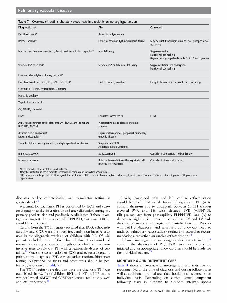
Laboratory and immunological screening testsIt is unclear which laboratory tests are particularly useful in thediagnostic work-up of PH/PHVD in children as data are sparse:table 7 provides an overview of routine blood tests, whichshould be considered for the diagnostic work-up of adolescentand adult patients with PH at first presentation. For children, itappears reasonable to be more selective and to take the ethnicbackground, travel history and to accompanying symptoms intoaccount. For example, although schistosomiasis is a frequentcause of PH worldwide, it is probably not necessary to test forit in most European children (table 7).42 47
Genetic testingGenetic counselling and testing is recommended for familieswith children with IPAH or HPAH. HPAH patients’ familymembers who develop new cardiorespiratory symptoms shouldbe evaluated immediately for PAH.
For further details on comprehensive biomarker and genetictesting, please see the dedicated article in this issue.74
Diagnostic algorithmThe Tracking Outcomes and Practice in Paediatric Pulmonaryhypertension (TOPP) registry investigators reported that to date,in the majority of children with PH, the current diagnosticwork-up recommended for adults are not followed (figure 1).This finding highlights that adult guidelines do not appropri-ately meet the demands of a thorough work-up tailored for chil-dren and/or probably a lack of awareness of existing guidelinesfor adults.2 38
If PH/PHVD is severe and the patient presents severely ill inovert heart failure and/or pulmonary vascular crisis, cardiaccatheterisation may be postponed due to its inherent risks andpharmacotherapy including intravenous prostanoids startedimmediately.72 75 Assessment may be postponed until thepatient is clinically stabilised. A separate article in this issue
Lammers AE, et al. Heart 2016;102:ii1–ii13. doi:10.1136/heartjnl-2015-307792 ii7
Pulmonary vascular disease
discusses cardiac catheterisation and vasodilator testing ingreater detail.72
Screening for paediatric PH is performed by ECG and echo-cardiography at the discretion of and after discussion among theprimary paediatrician and paediatric cardiologist. If these inves-tigations suggest the presence of PH/PHVD, CXR and HRCTshould be considered.
Results from the TOPP registry revealed that ECG, echocardi-ography and CXR were the most frequently non-invasive testsused in the diagnostic work-up of children with PH. Of 456patients included, none of them had all three tests considerednormal, indicating a possible strength of combining these non-invasive tests to rule out PH with a reasonable degree of cer-tainty.38 Once the combination of ECG and echocardiographypoints to the diagnosis ‘PH’, cardiac catheterisation, biomarkertesting (NT-proBNP or BNP) and other tests should be per-formed, as outlined in table 7.
The TOPP registry revealed that once the diagnosis ‘PH’ wasestablished, in <25% of children BNP and NT-proBNP testingwas performed. 6MWT and CPETwere conducted in only 38%and 7%, respectively.38
Finally, (combined right and left) cardiac catheterisationshould be performed in all forms of significant PH (i) toconfirm diagnosis and to distinguish between (ii) PH withoutelevated PVR and PH with elevated PVR (=PPHVD),(iii) pre-capillary from post-capillary PH/PPHVD, and (iv) todetermine right atrial pressure, as well as RV and LV end-diastolic pressures as surrogate for diastolic function. Patientswith PAH at diagnosis (and selectively at follow-up) need toundergo pulmonary vasoreactivity testing (for according recom-mendations, see article on cardiac catheterisation.72
If basic investigations including cardiac catheterisation,72
confirm the diagnosis of PH/PHVD, treatment should beinitiated and an appropriate follow-up plan should be made forthe individual patient.76
MONITORING AND OUTPATIENT CARETable 8 shows an overview of investigations and tests that arerecommended at the time of diagnosis and during follow-up, aswell as additional optional tests that should be considered on anindividual basis. Depending on clinical status, outpatientfollow-up visits in 3-month to 6-month intervals appear
Table 7 Overview of routine laboratory blood tests in paediatric pulmonary hypertension
Diagnostic test Aim Comment
Full blood count* Anaemia, polycytaemia
BNP/NT-proBNP* Detect ventricular dysfunction/heart failure May be useful for longitudinal follow-up/response totreatment
Iron studies (free iron, transferrin, ferritin and iron-binding capacity)* Iron deficiency SupplementationNutritional counsellingRegular testing in patients with PH-CHD and cyanosis
Vitamin B12, folic acid* Vitamin B12 or folic acid deficiency Supplementation, malabsorptionNutritional counselling
Urea and electrolytes including uric acid*
Liver functional enzymes (GOT, GPT, GGT, LDH)* Exclude liver dysfunction Every 4–12 weeks when stable on ERA therapy
Clotting* (PTT, INR, prothrombin, D-dimers)
Hepatitis serology†
Thyroid function test†
CK, CK-MB; troponin†
HIV† Causative factor for PH ELISA
ANAs (anticentromer antibodies, anti-SM, dsDNA, anti-Ro U1-U2RNP, B23, Th/To)†
? connective tissue disease, systemicsclerosis
Anticardiolipin antibodies†Lupus anticoagulant†
Lupus erythematodes, peripheral pulmonaryembolic disease
Thrombophilia screening, including anti-phospholipid antibodies Suspicion of CTEPHAntiphospholipid syndrome
Immunoassay/PCR Schistosomiasis Consider if appropriate medical history
Hb electrophoresis Rule out haemolobinopathy, eg, sickle celldisease/ thalaessaemia
Consider if ethnical risk group
*Recommended at presentation in all patients.†May be useful for selected patients, unmarked decision on an individual patient basis.BNP, brain-natriuretic peptide; CHD, congenital heart disease; CTEPH, chronic thromboembolic pulmonary hypertension; ERA, endothelin receptor antagonists; PH, pulmonaryhypertension.
ii8 Lammers AE, et al. Heart 2016;102:ii1–ii13. doi:10.1136/heartjnl-2015-307792
Pulmonary vascular disease

reasonable. More frequent visits may be indicated in patientswith progressive, advanced or end-stage disease, or after initi-ation or significant changes in therapy.
Patients on intravenous or subcutaneous prostanoid therapyshould be seen at least every three months,48 49 whereas stablepatients on oral (combination) therapy can be seen at 6-monthintervals.
Some advanced therapies (eg, endothelin receptor antago-nists) warrant further visits for laboratory testing.76
During every outpatient visit, a thorough history should besought, and changes or occurrence of new clinical symptomsshould be evaluated. Patients’ functional status and potentialchange of FC should be documented. Depending on children’sage, maturity and cooperation, physical functional testing suchas 6MWT and/or CPET should be performed in 6-month to12-month intervals.
Elective surgery/general anaesthesiaElective procedures under general anaesthesia require carefulconsideration of the underlying indication and judgement of therisk–benefit ratio as any general anaesthesia bares a substantialhigh risk for a child with moderate to severe PH. If a potentialbenefit of the procedure (gain of information) outweighs theestimated risk, or the surgery/intervention is unavoidable, timelyplanning is required. Management by an experienced anaesthe-siologist who is familiar with PH haemodynamics and theinvolved pitfalls should be facilitated.
Good communication between teams, awareness of the sever-ity of the disease and risks, and timely preparation are
recommended. Conscious anaesthetic strategies maintaining anadequate systemic vascular resistance and adequate preload, andappropriate postprocedure monitoring are crucial to reducemorbidity and fatal procedure-related events.50–52
Pregnancy/ContraceptionPregnancy has a very high risk of fatal outcome for a pregnantwoman with PH (30–50%) and for the fetus, and in the majorityof cases, the caring physician will recommend termination of preg-nancy. Female adolescents with PH/PPHVD should undergotimely counselling regarding options for secure contraception.53 54
Physical education and exerciseCPET testing is recommended for children with mild to moder-ate PH/PPHVD prior to engaging in any recreational or athleticactivities.
It is recommended that children with stable mild to moderatePH/PPHVD should engage in light to moderate aerobic activitybut be advised to self-limit their activities as required, avoidstrenuous exertion and dehydration.
Paediatric patients with severe PH (FC III or IV and/or sys-temic or supra-systemic PA pressure) and/or recent history ofsyncope carry a high risk of sudden cardiac death with exerciseand thus should not participate in any (competitive) sports.
Flying on commercial airplanesData on children exposed to cabin pressures and evaluation ofneed for supplemental oxygen are lacking. Generally, it is recom-mended that patients with PH should only fly on commercial
Figure 1 Diagnostic algorithm for a child or adolescent with suspected pulmonary hypertension. CHD, congenital heart disease; CPET,cardiopulmonary exercise testing; CTEPH, chronic thromboembolic pulmonary hypertension; CXR, chest X-ray; HPAH, hereditary pulmonary arterialhypertension; IPAH, idiopathic pulmonary arterial hypertension; PCH, pulmonary capillary haemangiomatosis; PH, pulmonary hypertension; PVOD,pulmonary veno-occlusive disease; BPD, bronchopulmonary dysplasia; CLD, chronic lung disease; LFT, liver function test.
Lammers AE, et al. Heart 2016;102:ii1–ii13. doi:10.1136/heartjnl-2015-307792 ii9
Pulmonary vascular disease

airplanes in a stable and compensated condition. For patientswith advanced disease, systemic pulmonary artery pressures and/or impaired ventricular function, it appears reasonable to usesupplemental oxygen during the flight to minimise hypoxic vaso-constriction. The ESC and European Respiratory Society guide-lines for PH recommend in-flight supplemental oxygen forpatients with PH in FC III or IV and those with arterial partialoxygen pressure consistently <8 kPa (60 mm Hg).42 55
The PH patients’ caregivers should contact the airline inadvance regarding the availability of supplemental oxygen insufficient amounts during airline transport.
Vaccinations/Care of central intravenous linesChildren with PH should be under shared care and should be pri-marily seen and linked to a local paediatrician as first line ofcontact for all regular childhood investigations, routine vaccina-tions and general medical problems. However, good proactive
communication and close cooperation is mandatory between spe-cialised physicians seeing the patient with a PH focus and localcommunity teams. Paediatricians should be actively encouragedto contact PH physicians for advice, for example, seekingsupport for and planning minor procedures, not necessarilyrelated to the PH itself, where the patient could be exposed toany excessive risk (eg, general anaesthetic for dental procedure).
Children with PH should undergo all recommended routinevaccinations to prevent avoidable infections. Respiratory syncytialvirus (<2 years of age), pneumococci and influenza vaccinationsshould be administered in paediatric PH patients if no contraindi-cations exist.
Antibiotic prophylaxis for the prevention of subacute bacterialendocarditis is nowadays reserved for special patient subgroups(eg, cyanotic patients).56
In patients with a permanent central venous access (eg, forthe administration of continuous prostacycline analogues), the
Table 8 Recommended tests/investigations at diagnosis and follow-up of a child or adolescent with pulmonary hypertension/pulmonaryhypertensive vascular disease
Follow-up outpatientvisits
Test/investigation
At firstpresentation/diagnosis
3–6 monthlyintervals
Every12 months
Elective testingwhen indicated Comment
Comprehensive medical history/interim history • •
Assignment to functional class • •
Growth variables (centiles) • •
Physical examination • •
Four limb blood pressure •
Pulse oximetry both arms/one leg • •
Electrocardiogram • •
Transthoracic echocardiogram • •
Chest X-ray • •
Lung function test/body plethysmography • •
6 min walk test • •
Cardiopulmonary exercise test • •
Laboratory testing • • According to drug treatment
NT-proBNP or BNP • • •
Cardiac MRI* • • *MRI at diagnosis or first presentation;indication should be discussed on anindividual patient basis
Comprehensive right and left heartcatheterisation (including full pulmonaryvasoreagibility testing)
• •
(High resolution) chest CT/CT angiogram* • • * Diagnostic value of a pulmonaryangiogram to be discussed on an individualpatient basis
Polysomnography •
Ventilation/perfusion scan •
BNP, brain-natriuretic peptide.
ii10 Lammers AE, et al. Heart 2016;102:ii1–ii13. doi:10.1136/heartjnl-2015-307792
Pulmonary vascular disease

caregivers must be vigilant for possible line infections. If clinic-ally suspected (eg, unexplained fever, erythema or other infec-tious signs around line entry site), the continuous infusion ofthe prostanoid must not be abruptly disconnected. Due to theshort half-life, the prostanoid has to be transitioned to a periph-eral venous access, while the infusion via the central line is care-fully weaned. Transition should ideally take place in a stepwisemanner with monitoring of the clinical status, saturation andblood pressure of the patient, before blood cultures can be with-drawn from the central line. However, if line infection is con-firmed, the chance to permanently clear the existing central lineis often unsuccessful, despite antibiotic treatment. Most patientsrequire removal of the infected line and alternative central lineplacement. Ideally, replacement should take place after a fewdays of antibiotic treatment and control of systemic infection/bacteraemia to avoid instant contamination of the new centralline. The same technical process applies to patients with aleaking line or line dysfunction. They need to be transferred tothe next emergency department immediately for intermittentperipheral intravenous line placement to ensure continuation ofprostanoid therapy (epoprostenol has a very short drughalf-life).
SchoolClose contact to healthcare providers and teachers at schoolshould be sought. The aim is to familiarise them with practical-ities of supporting a chronically ill child, to allow for a (near-)normal school attendance, to maximise pupils’ integration andto address understandable concerns and anxieties of the educa-tors. Arrangements at school have to be made to facilitate theapplication/intake of any medication required during schoolhours (eg, inhaled iloprost, oxygen).
Additional interventional and surgical therapiesAtrial septostomy is often considered as the last opinion beforeactive listing for bilateral lung transplantation. Indeed, atrial sep-tostomy should represent a conscious step of active PH manage-ment particularly in patients with syncope.57 Paediatricexperience is available and describes clinical improvement andabolishment of syncope, albeit an associated procedural risk, hasto be acknowledged particularly for patients with increasedright atrial pressures.58
Lately other surgical measures (Potts shunt) and interventionalprocedures (ductal stenting) have been introduced and offeredto small numbers of patients.76 Paediatric experience is sparseand measures may be discussed as compassionate care. Adetailed discussion of the involved risks and benefits is beyondthe scope of this article; however, further details can be foundin references.59–62 76
Lung transplantationFor some children, bilateral lung transplantation may be anoption. However, despite improved immunosuppression regi-mens, overall survival after lung transplantation remains limitedcompared with other solid organ transplantation.
Median survival following paediatric lung transplantation isreported to be 4.9 years (2013 International Society of Heartand Lung Transplantation guideline).63 64 Acute rejection andbronchiolitis obliterans syndrome remain a major clinicalproblem responsible for both morbidity and mortality.65 Opencounselling is important to balance risks and benefits and gaugewhether this is a desirable and realistic option for each individ-ual patient and their families. In patients with end-stage PHVD,
early referral and evaluation by an interdisciplinary paediatrictransplant centre should be sought.65–67
Palliative careAs disease progresses, it is important to support patients andfamilies beyond providing high-quality medical care. Regardlessof the decision to be actively listed for bilateral lung transplant-ation in end-stage disease, the extent of intensive care supportshould be discussed in a timely manner and alternatives, forexample, palliative care, should be offered. Additional psycho-logical support for families and patients and contact to profes-sional social networks may be helpful.
General supportChildren, siblings and caregivers of paediatric patients with PH/PHVD should be assessed for psychosocial stress and be readilyprovided psychosocial support and referral to appropriate institu-tions as needed. Individualised recommendations should be made,dependent on the underlying cause of the child’s PH and clinicalstatus. Participation in physical education classes, access to educa-tion facilities and supporting aids (eg, wheelchair) to maintainmobility for patients with advanced disease should be discussed onan individual basis to meet the patients’ and families’ needs.
CONCLUSIONDue to the relatively low prevalence of PH/PHVD in child-hood, the complexity of the disease, the heterogeneity ofpresentation and disease progression, and the need of a multi-disciplinary approach, patient care and follow-up should takeplace at dedicated PH centres. In the setting of progressingdisease and over time increasingly compromised patient withPH/PHVD, a balance has to be sought between regular visitsat specialist PH clinics and good competent support fromlocal medical teams, and palliative care services close to thepatient’s home.
Author affiliations1Department of Paediatric Cardiology, University of Münster, Münster, Germany2Division of Paediatric Cardiology, University Children’s Hospital Ulm, Germany3Department of Paediatric Cardiology, German Paediatric Heart Centre, SanktAugustin, Germany4Department of Paediatric Cardiology and Congenital Heart Disease, German HeartCentre Munich and Technical University, Munich, Germany5Department of Paediatric Cardiology and Congenital Heart Disease, Heart andDiabetes Centre NRW, Bad Oeynhausen, Germany6Department of Paediatric Cardiology and Critical Care, Hannover Medical School,Hannover, Germany
Funding AL receives grant support from the dean’s office Medizinische Fakultät,Westfälischen Wilhelms-Universität Münster (15-002). CA currently receives grantfunding from Stiftung Kinderherz (2511-10-13-001) and Behring-Röntgen-Stiftung(59-0018). GH currently receives grant support from the German ResearchFoundation (DFG; HA 4348/2-1), Fördergemeinschaft deutsche Kinderherzzentren(W-H-001-2014) and Stiftung Kinderherz (2511-6-13-011).
Competing interests AH received reimbursements for advisory board meetingsfrom Actelion, and speaker’s reimbursements from Actelion, Pfizer, Encysive, AOPOrphan Pharmaceuticals, OMT, GlaxoSmithKline; he received travel grants fromPfizer, GlaxoSmithKline, AOP Orphan Pharmaceuticals, Lilly, Actelion. His institutioncontributed to clinical trials sponsored by Actelion and Lilly. AH’s institution receivedunrestricted scientific grants from Pfizer, Actelion and GlaxoSmithKline. AL, CA, PZ,KOD and GH declare no conflict of interest.
Provenance and peer review Commissioned; externally peer reviewed.
REFERENCES1 Hansmann G. Interdisciplinary networks for the treatment of childhood pulmonary
vascular disease: what pulmonary hypertension doctors can learn from pediatriconcologists. Pulm Circ 2013;3:792–801.
2 Hansmann G, Hoeper MM. Registries for paediatric pulmonary hypertension. EurRespir J 2013;42:580–3.
Lammers AE, et al. Heart 2016;102:ii1–ii13. doi:10.1136/heartjnl-2015-307792 ii11
Pulmonary vascular disease

3 Hoeper MM, Bogaard HJ, Condliffe R, et al. Definitions and diagnosis of pulmonaryhypertension. J Am Coll Cardiol 2013;62:D42–50.
4 Cerro MJ, Abman S, Diaz G, et al. A consensus approach to the classification ofpediatric pulmonary hypertensive vascular disease: report from the pvri pediatrictaskforce, panama 2011. Pulm Circ 2011;1:286–98.
5 Ivy DD, Abman SH, Barst RJ, et al. Pediatric pulmonary hypertension. J Am CollCardiol 2013;62:D117–126.
6 Barst RJ, McGoon MD, Elliott CG, et al. Survival in childhood pulmonaryarterial hypertension: Insights from the registry to evaluate early and long-termpulmonary arterial hypertension disease management. Circulation2012;125:113–22.
7 Heath D, Edwards JE. The pathology of hypertensive pulmonary vascular disease; adescription of six grades of structural changes in the pulmonary arteries with specialreference to congenital cardiac septal defects. Circulation 1958;18:533–47.
8 Simonneau G, Gatzoulis MA, Adatia I, et al. Updated clinical classification ofpulmonary hypertension. J Am Coll Cardiol 2013;62:D34–41.
9 van Loon RL, Roofthooft MT, Hillege HL, et al. Pediatric pulmonary hypertension inthe netherlands: epidemiology and characterization during the period 1991 to2005. Circulation 2011;124:1755–64.
10 Moledina S, Hislop AA, Foster H, et al. Childhood idiopathic pulmonary arterialhypertension: a national cohort study. Heart 2010;96:1401–6.
11 Fraisse A, Jais X, Schleich JM, et al. Characteristics and prospective 2-year follow-upof children with pulmonary arterial hypertension in France. Arch Cardiovasc Dis2010;103:66–74.
12 Vorhies EE, Ivy DD. Drug treatment of pulmonary hypertension in children. PaediatrDrugs 2014;16:43–65.
13 van Loon RL, Roofthooft MT, van Osch-Gevers M, et al. Clinical characterization ofpediatric pulmonary hypertension: complex presentation and diagnosis. J Pediatr2009;155:176–82.e171.
14 McLaughlin VV, Shillington A, Rich S. Survival in primary pulmonary hypertension:the impact of epoprostenol therapy. Circulation 2002;106:1477–82.
15 Galiè N, Hoeper MM, Humbert M, et al. Guidelines for the diagnosis and treatmentof pulmonary hypertension. Eur Respir J 2009;34:1219–63.
16 [No authors listed]. AHA medical/scientific statement. 1994 revisions toclassification of functional capacity and objective assessment of patients withdiseases of the heart. Circulation 1994;90:644–5.
17 Lammers AE, Adatia I, Cerro MJ, et al. Functional classification of pulmonaryhypertension in children: report from the pvri pediatric taskforce, Panama 2011.Pulm Circ 2011;1:280–5.
18 Ploegstra MJ, Zijlstra WM, Douwes JM, et al. Prognostic factors in pediatricpulmonary arterial hypertension: a systematic review and meta-analysis. Int J Cardiol2015;184C:198–207.
19 Rich S, Dantzker DR, Ayres SM, et al. Primary pulmonary hypertension. A nationalprospective study. Ann Intern Med 1987;107:216–23.
20 Tongers J, Schwerdtfeger B, Klein G, et al. Incidence and clinical relevance ofsupraventricular tachyarrhythmias in pulmonary hypertension. Am Heart J2007;153:127–32.
21 Diller GP, Dimopoulos K, Broberg CS, et al. Presentation, survival prospects, andpredictors of death in Eisenmenger syndrome: a combined retrospective andcase-control study. Eur Heart J 2006;27:1737–42.
22 Jone PN, Ivy DD. Echocardiography in pediatric pulmonary hypertension. FrontPediatr 2014;2:124.
23 Fan LL, Mullen AL, Brugman SM, et al. Clinical spectrum of chronic interstitial lungdisease in children. J Pediatr 1992;121:867–72.
24 Nayyar D, Muthiah K, Kumarasinghe G, et al. Imatinib for the treatment ofpulmonary arterial hypertension and pulmonary capillary hemangiomatosis. PulmCirc 2014;4:342–5.
25 Ivy DD, Doran AK, Smith KJ, et al. Short- and long-term effects of inhaled iloprosttherapy in children with pulmonary arterial hypertension. J Am Coll Cardiol2008;51:161–9.
26 Lammers AE, Diller GP, Odendaal D, et al. Comparison of 6-min walk test distanceand cardiopulmonary exercise test performance in children with pulmonaryhypertension. Arch Dis Child 2011;96:141–7.
27 Rausch CM, Taylor AL, Ross H, et al. Ventilatory efficiency slope correlates withfunctional capacity, outcomes, and disease severity in pediatric patients withpulmonary hypertension. Int J Cardiol 2013;169:445–8.
28 Cooper DM, Weiler-Ravell D. Gas exchange response to exercise in children. AmRev Respir Dis 1984;129:S47–8.
29 Rhodes J, Ubeda Tikkanen A, Jenkins KJ. Exercise testing and training in childrenwith congenital heart disease. Circulation 2010;122:1957–67.
30 O’Driscoll DM, Horne RS, Davey MJ, et al. Cardiac and sympathetic activation arereduced in children with down syndrome and sleep disordered breathing. Sleep2012;35:1269–75.
31 Wise MS, Nichols CD, Grigg-Damberger MM, et al. Executive summary ofrespiratory indications for polysomnography in children: an evidence-based review.Sleep 2011;34:389–398AW.
32 Marcus CL, Moore RH, Rosen CL, et al. A randomized trial of adenotonsillectomyfor childhood sleep apnea. N Engl J Med 2013;368:2366–76.
33 Tsai IC, Tsai WL, Wang KY, et al. Comprehensive mdct evaluation of patients withpulmonary hypertension: diagnosing underlying causes with the updated Dana Point2008 classification. AJR Am J Roentgenol 2011;197:W471–481.
34 Moledina S, Pandya B, Bartsota M, et al. Prognostic significance of cardiacmagnetic resonance imaging in children with pulmonary hypertension. CircCardiovasc Imaging 2013;6:407–14.
35 Pandya B, Quail MA, Steeden JA, et al. Real-time magnetic resonance assessmentof septal curvature accurately tracks acute hemodynamic changes in pediatricpulmonary hypertension. Circ Cardiovasc Imaging 2014;7:706–13.
36 Swift AJ, Wild JM, Nagle SK, et al. Quantitative magnetic resonance imaging ofpulmonary hypertension: a practical approach to the current state of the art. JThorac Imaging 2014;29:68–79.
37 Lopes AA, Barst RJ, Haworth SG, et al. Repair of congenital heart disease withassociated pulmonary hypertension in children: what are the minimal investigativeprocedures? Consensus statement from the congenital heart disease and pediatrictask forces, pulmonary vascular research institute (pvri). Pulm Circ 2014;4:330–41.
38 Beghetti M, Berger RM, Schulze-Neick I, et al. Diagnostic evaluation of paediatricpulmonary hypertension in current clinical practice. Eur Respir J 2013;42:689–700.
39 McLaughlin VV, Archer SL, Badesch DB, et al. ACCF/AHA 2009 expert consensusdocument on pulmonary hypertension: a report of the American College ofCardiology foundation task force on expert consensus documents and the AmericanHeart Association: developed in collaboration with the American College of ChestPhysicians, American Thoracic Society, inc., and the Pulmonary HypertensionAssociation. Circulation 2009;119:2250–94.
40 Tunariu N, Gibbs SJ, Win Z, et al. Ventilation-perfusion scintigraphy is more sensitivethan multidetector ctpa in detecting chronic thromboembolic pulmonary disease as atreatable cause of pulmonary hypertension. J Nucl Med 2007;48:680–4.
41 Berger RM, Beghetti M, Humpl T, et al. Clinical features of paediatric pulmonaryhypertension: a registry study. Lancet 2012;379:537–46.
42 Galiè N, Hoeper MM, Humbert M, et al. Guidelines for the diagnosis and treatmentof pulmonary hypertension: the task force for the diagnosis and treatment ofpulmonary hypertension of the european society of cardiology (ESC) and theeuropean respiratory society (ers), endorsed by the international society of heart andlung transplantation (ISHLT). Eur Heart J 2009;30:2493–537.
43 Condino AA, Ivy DD, O’Connor JA, et al. Portopulmonary hypertension in pediatricpatients. J Pediatr 2005;147:20–6.
44 Ridaura-Sanz C, Mejia-Hernandez C, Lopez-Corella E. Portopulmonary hypertensionin children. A study in pediatric autopsies. Arch Med Res 2009;40:635–9.
45 Whitworth JR, Sokol RJ. Hepato-portopulmonary disorders—not just in adults! JPediatr Gastroenterol Nutr 2005;41:393–5.
46 Weber MA, Ashworth MT, Sebire NJ. Portopulmonary hypertension in childhoodpresenting as sudden death. Pediatr Dev Pathol 2006;9:65–71.
47 Chu JW, Kao PN, Faul JL, et al. High prevalence of autoimmune thyroid disease inpulmonary arterial hypertension. Chest 2002;122:1668–73.
48 McLaughlin VV, Palevsky HI. Parenteral and inhaled prostanoid therapy in thetreatment of pulmonary arterial hypertension. Clin Chest Med 2013;34:825–40.
49 Lammers AE, Hislop AA, Flynn Y, et al. Epoprostenol treatment in children withsevere pulmonary hypertension. Heart 2007;93:739–43.
50 Twite MD, Friesen RH. The anesthetic management of children with pulmonaryhypertension in the cardiac catheterization laboratory. Anesthesiol Clin 2014;32:157–73.
51 Pilkington SA, Taboada D, Martinez G. Pulmonary hypertension and its managementin patients undergoing non-cardiac surgery. Anaesthesia 2015;70:56–70.
52 Fox C, Kalarickal PL, Yarborough MJ, et al. Perioperative management includingnew pharmacological vistas for patients with pulmonary hypertension for noncardiacsurgery. Curr Opin Anaesthesiol 2008;21:467–72.
53 Bedard E, Dimopoulos K, Gatzoulis MA. Has there been any progress made onpregnancy outcomes among women with pulmonary arterial hypertension? EurHeart J 2009;30:256–65.
54 Thorne S, Nelson-Piercy C, MacGregor A, et al. Pregnancy and contraception inheart disease and pulmonary arterial hypertension. J Fam Plann Reprod Health Care2006;32:75–81.
55 Thamm M, Voswinckel R, Tiede H, et al. Air travel can be safe and well tolerated inpatients with clinically stable pulmonary hypertension. Pulm Circ 2011;1:239–43.
56 Habib G, Hoen B, Tornos P, et al. Guidelines on the prevention, diagnosis, andtreatment of infective endocarditis (new version 2009): the task force on theprevention, diagnosis, and treatment of infective endocarditis of the europeansociety of cardiology (esc). Endorsed by the european society of clinical microbiologyand infectious diseases (ESCMID) and the international society of chemotherapy(ISC) for infection and cancer. Eur Heart J 2009;30:2369–413.
57 Lammers AE, Haworth SG, Diller GP. Atrial septostomy in patients with pulmonaryhypertension: should it be recommended? Expert Rev Respir Med 2011;5:363–76.
58 Micheletti A, Hislop AA, Lammers A, et al. Role of atrial septostomy in thetreatment of children with pulmonary arterial hypertension. Heart 2006;92:969–72.
59 Boudjemline Y, Patel M, Malekzadeh-Milani S, et al. Patent ductus arteriosusstenting (transcatheter potts shunt) for palliation of suprasystemic pulmonary arterialhypertension: a case series. Circ Cardiovasc Interv 2013;6:e18–20.
60 Latus H, Delhaas T, Schranz D, et al. Treatment of pulmonary arterial hypertensionin children. Nat Rev Cardiol 2015;12:244–54.
ii12 Lammers AE, et al. Heart 2016;102:ii1–ii13. doi:10.1136/heartjnl-2015-307792
Pulmonary vascular disease

61 Petersen C, Helvind M, Jensen T, et al. Potts shunt in a child with end-stagepulmonary hypertension after late repair of ventricular septal defect. World J PediatrCongenit Heart Surg 2013;4:286–9.
62 Schranz D, Michel-Behnke I, Heyer R, et al. Stent implantation of the arterial duct innewborns with a truly duct-dependent pulmonary circulation: a single-centerexperience with emphasis on aspects of the interventional technique. J IntervCardiol 2010;23:581–8.
63 Benden C, Goldfarb SB, Edwards LB, et al. The registry of the international societyfor heart and lung transplantation: seventeenth official pediatric lung andheart-lung transplantation report—2014; focus theme: retransplantation. J HeartLung Transplant 2014;33:1025–33.
64 Tudorache I, Sommer W, Kuhn C, et al. Lung transplantation for severe pulmonaryhypertension-awake extracorporeal membrane oxygenation for postoperative leftventricular remodelling. Transplantation 2015;99:451–8.
65 Kirkby S, Hayes D Jr. Pediatric lung transplantation: indications and outcomes.J Thorac Dis 2014;6:1024–31.
66 Lammers AE, Burch M, Benden C, et al. Lung transplantation in children withidiopathic pulmonary arterial hypertension. Pediatr Pulmonol 2010;45:263–9.
67 Gorler H, Struber M, Ballmann M, et al. Lung and heart-lung transplantation inchildren and adolescents: a long-term single-center experience. J Heart LungTransplant 2009;28:243–8.
68 Rubin LJ. Diagnosis and management of pulmonary arterial hypertension: ACCPEvidence-Based Clinical Practice Guidelines. Chest 2004;126:7S–10S.
69 Hansmann G (chair), Apitz C (co-chair), Abdul-Khaliq H, et al. Executive Summary.Expert Consensus Statement on the Diagnosis and Treatment of Paediatric PulmonaryHypertension. The European Paediatric Pulmonary Vascular Disease Network,endorsed by ISHLT and DGPK. Heart 2016;102:ii86–100.
70 Kozlik-Feldmann R, Hansmann G, Bonnet D, et al. Pulmonary Hypertension inChildren and Adolescents with Congenital Heart Disease (PAH-CHD, PPHVD-CHD).Expert Consensus Statement on the Diagnosis and Treatment of Paediatric Pulmonary
Hypertension. The European Paediatric Pulmonary Vascular Disease Network,endorsed by ISHLT and DGPK. Heart 2016;102:ii42–8.
71 Koestenberger M, Apitz C, Abdul-Khaliq H, et al. Transthoracic Echocardiography forthe Evaluation of Children and Adolescents with Suspected or Confirmed PulmonaryHypertension. Expert Consensus Statement on the Diagnosis and Treatment ofPaediatric Pulmonary Hypertension. The European Paediatric Pulmonary VascularDisease Network, endorsed by ISHLT and DGPK. Heart 2016;102:ii14–22.
72 Apitz C, Hansmann G, Schranz D. Hemodynamic Assessment and Acute PulmonaryVasoreactivity Testing in the Evaluation of Children with Pulmonary Vascular Disease.Expert Consensus Statement on the Diagnosis and Treatment of Paediatric PulmonaryHypertension. The European Paediatric Pulmonary Vascular Disease Network,endorsed by ISHLT and DGPK. Heart 2016;102:ii23–9.
73 Latus H, Kuehne T, Beerbaum P, et al. Cardiac magnetic resonance and computedtomography imaging in children with suspected or confirmed pulmonaryhypertension/pulmonary hypertensive vascular disease. Expert Consensus Statementon the Diagnosis and Treatment of Paediatric Pulmonary Hypertension. TheEuropean Paediatric Pulmonary Vascular Disease Network, endorsed by ISHLT andDGPK. Heart 2016;102:ii30–6.
74 Pattathu J, Gorenflo M, Hilgendorff M, et al. Genetic Testing and Blood Biomarkers inPaediatric Pulmonary Hypertension. Expert Consensus Statement on the Diagnosis andTreatment of Paediatric Pulmonary Hypertension. The European Paediatric PulmonaryVascular Disease Network, endorsed by ISHLT and DGPK. Heart 2016;102:ii36–41.
75 Kaestner M, Schranz D, Warnecke G, et al. Pulmonary Hypertension in the IntensiveCare Unit. Expert Consensus Statement on the Diagnosis and Treatment ofPaediatric Pulmonary Hypertension. The European Paediatric Pulmonary VascularDisease Network, endorsed by ISHLT and DGPK. Heart 2016;102:ii57–66.
76 Hansmann G, Apitz C. Treatment of Children with Pulmonary Hypertension andCardiac Dysfunction. Expert Consensus Statement on the Diagnosis and Treatmentof Paediatric Pulmonary Hypertension. The European Paediatric Pulmonary VascularDisease Network, endorsed by ISHLT and DGPK. Heart 2016;102:ii67–85.
Lammers AE, et al. Heart 2016;102:ii1–ii13. doi:10.1136/heartjnl-2015-307792 ii13
Pulmonary vascular disease