original article dysbiotic gut microbiota causes...
TRANSCRIPT

ORIGINAL ARTICLE
Dysbiotic gut microbiota causes transmissibleCrohn’s disease-like ileitis independent of failurein antimicrobial defenceMonika Schaubeck,1 Thomas Clavel,2 Jelena Calasan,1 Ilias Lagkouvardos,2
Sven Bastiaan Haange,3 Nico Jehmlich,3 Marijana Basic,6 Aline Dupont,6,7
Mathias Hornef,6,7 Martin von Bergen,3,4,5 André Bleich,8 Dirk Haller1,2
▸ Additional material ispublished online only. To viewplease visit the journal online(http://dx.doi.org/10.1136/gutjnl-2015-309333).
For numbered affiliations seeend of article.
Correspondence toProfessor Dirk Haller, Chair ofNutrition and Immunology,ZIEL-Institute for Food andHealth, Technische UniversitätMünchen, ZIEL—ResearchCenter for Nutrition and FoodSciences, Gregor-Mendel-Str.2,Freising-Weihenstephan85350, Germany,[email protected]
MS and TC contributed equallyto the present work and sharefirst authorship.
Received 6 February 2015Revised 3 March 2015Accepted 21 March 2015
To cite: Schaubeck M,Clavel T, Calasan J, et al.Gut Published Online First:[please include Day MonthYear] doi:10.1136/gutjnl-2015-309333
ABSTRACTObjectives Dysbiosis of the intestinal microbiota isassociated with Crohn’s disease (CD). Functionalevidence for a causal role of bacteria in the developmentof chronic small intestinal inflammation is lacking.Similar to human pathology, TNFdeltaARE mice develop atumour necrosis factor (TNF)-driven CD-like transmuralinflammation with predominant ileal involvement.Design Heterozygous TNFdeltaARE mice and wildtype(WT) littermates were housed under conventional(CONV), specific pathogen-free (SPF) and germ-free (GF)conditions. Microbial communities were analysed byhigh-throughput 16S ribosomal RNA gene sequencing.Metaproteomes were measured using LC-MS. Temporaland spatial resolution of disease development wasfollowed after antibiotic treatment and transfer ofmicrobial communities into GF mice. Granulocyteinfiltration and Paneth cell function was assessed byimmunofluorescence and gene expression analysis.Results GF-TNFdeltaARE mice were free of inflammationin the gut and antibiotic treatment of CONV-TNFdeltaARE
mice attenuated ileitis but not colitis, demonstrating thatdisease severity and location are microbiota-dependent.SPF-TNFdeltaARE mice developed distinct ileitis-phenotypesassociated with gradual loss of antimicrobial defence.16S analysis and metaproteomics revealed specificcompositional and functional alterations of bacterialcommunities in inflamed mice. Transplantation ofdisease-associated but not healthy microbiotatransmitted CD-like ileitis to GF-TNFdeltaARE recipientsand triggered loss of lysozyme and cryptdin-2 expression.Monoassociation of GF-TNFdeltaARE mice with the humanCD-related Escherichia coli LF82 did not induce ileitis.Conclusions We provide clear experimental evidencefor the causal role of gut bacterial dysbiosis in thedevelopment of chronic ileal inflammation withsubsequent failure of Paneth cell function.
INTRODUCTIONCrohn’s disease (CD) and ulcerative colitis (UC) arethe two main forms of inflammatory bowel disease(IBD). While inflammation in patients with CDmainly occurs in the terminal ileum and is found inthe proximal colon only occasionally, inflammationin UC is confined to the colon.1 2 Despite unknownaetiologies, chronic activation of the mucosalimmune system is triggered by gut commensals.3 4
Genome-wide association studies identified linksbetween disease and genes involved in microbial sig-nalling, underlining the role of gut microbes in IBDpathogenesis.5 Genetic predispositions and inflamma-tion of the host were shown to induce altered com-position and metabolic activity of microbialcommunities, defined as dysbiosis,6–8 and, as
Significance of this study
What is already known on this subject?▸ IBD is associated with changes in intestinal
bacterial composition.▸ Germ-free mouse models of IBD-related colitis
are established.▸ Paneth cell function is associated with ileal
disease phenotypes of Crohn’s disease.
What are the new findings?▸ Crohn’s disease-like ileitis development and
location in TNFdeltaARE mice are microbiotadependent, and completely absent in germ-freeconditions.
▸ Antibiotics reduce disease severity, andrecurrence of inflammation follows the relapseof microbiota composition.
▸ Ileitis severity is associated with compositionaland functional dysbiosis of the intestinalcommensal microbiota.
▸ Transfer of dysbiotic microbiota to germ-freerecipients causes Crohn’s disease-likeinflammation in genetically susceptiblerecipients.
▸ Loss of Paneth cell-related antimicrobialdefence is subsequent to the development ofileitis in TNFdeltaARE mice.
How might it impact on clinical practice inthe foreseeable future?▸ This study provides a basal understanding for the
causality of microbe-host interaction in Crohn’sdisease, based on dynamic alterations of themicrobial ecosystem (dysbiosis) in the gut.
▸ This study will help to elucidate the therapeuticrelevance of microbiota transplantation inCrohn’s disease.
Schaubeck M, et al. Gut 2015;0:1–13. doi:10.1136/gutjnl-2015-309333 1
Gut microbiota Gut Online First, published on April 17, 2015 as 10.1136/gutjnl-2015-309333
Copyright Article author (or their employer) 2015. Produced by BMJ Publishing Group Ltd (& BSG) under licence.
on 28 July 2018 by guest. Protected by copyright.
http://gut.bmj.com
/G
ut: first published as 10.1136/gutjnl-2015-309333 on 17 April 2015. D
ownloaded from

expected, patients with IBD are characterised by dysbiotic changesin the gut microbial ecosystem.9–11 Changes in bacterial communi-ties were shown to be shaped by Paneth cell (PC)-derived anti-microbial peptides.12 PCs are located in the crypt base of the smallintestine in close proximity to the epithelial stem cell compartmentand loss of PC function is associated with ileal phenotypes ofCD.12–14 In IBD treatment, the clinical benefit of antibiotics orfaecal-stream diversion supports the pathological role of bac-teria.15 Faecal microbiota transplantation (FMT) is effective inrestoring homoeostasis in Clostridium difficile infections.16–19
Consequently, there is rising interest for a therapeutical implemen-tation of FMT in UC and CD. However, evidence from controlledclinical trials is still limited and characteristics of a successfuldonor microbiota are not yet defined. Moreover, the rationale ofintroducing new antigen pools in a milieu that has been overreact-ing to microbial stimuli is questionable. For colitis, the importanceof bacteria in disease development has been extensively studied inanimal models, for example, by showing reduction of colitis inIL-10−/− mice after antibiotic treatment or the ability of variousbacterial strains to induce inflammation in germ-free (GF) colitismodels.20–23 Due to the lack of GF models for CD-like ileitis,proof for causality of microbes or dysbiosis in the onset of ileitis islacking.
In the present study, we assessed the impact of intestinal bac-terial communities in a spontaneous model of chronic CD-likeileitis. TNFdeltaARE mice carry a deletion in the tumour necrosisfactor (TNF) AU-rich (adenosin-uracil) elements (ARE) leadingto Tnf driven transmural inflammation in the distal ileum.24 Wepreviously showed that iron-induced modulation of the micro-biota is associated with dramatic changes in disease activity ofTNFdeltaARE mice.25 However, mechanistic proof for a causalrole of microbe-host interactions in the pathogenesis of CD-likeileitis is missing. Here, we used antibiotics and differenthygienic conditions (GF, specific pathogen-free (SPF) or conven-tional (CONV) housing) to dissect the relationship betweenmicrobiota changes and ileitis development in TNFdeltaARE mice.To assess the causal role of dysbiotic microbial communities inileal inflammation, we performed microbiota transplant experi-ments with GF-TNFdeltaARE mice and characterised PCfunctions.
METHODSEthics statementAnimal use was approved by the local institution in charge(Regierung von Oberbayern, approval no. 55.2-1-54-2531-75-10 and 55.2-1-54-2531-99-13). All animals were housed inmouse facilities at the Technische Universität München (Schoolof Life Sciences Weihenstephan).
HousingTNFdeltaARE mice were provided by George Kollias (AlexanderFleming BSRC, Greece), bred in our CONV facility and trans-ferred to SPF via embryo transfer. TNFdeltaARE mice were madeGF by hysterectomy (Institute for Laboratory Animal Science;Hannover). Sterility was checked by cultivation of faeces inLuria broth (LB) or wilkins chalgren agar (WCA) broth(OXOID) and by microscopic observation of Gram-stainedfaecal smears every 10–14 days and at sampling. A mould-trapwas used to indicate the presence of mold. No contaminationswere observed during the experiments. HeterozygousTNFdeltaARE and WT littermates (C57BL/6N) were kept inCONV, SPF or GF conditions (12 h light/dark cycles at 24–26°C) until the age of 18 weeks. Mice were fed a standard diet(autoclaved R/M-H for SPF and CONV, or M-Z V1124-300 for
GF-animals, Ssniff, Soest, Germany) ad libitum and were sacri-ficed by CO2.
Antibiotic treatmentCONV-TNFdeltaARE and CONV-WT mice received antibiotics(VM: 0.25 g/L vancomycin and 1.0 g/L metronidazole, Sigma-Aldrich and Fluka) from 8 weeks to 12 weeks of age. Antibioticswere prepared fresh twice a week and administered ad libitumvia drinking water in light-protected bottles. Mice were sacri-ficed 0 weeks, 2 weeks, 4 weeks and 6 weeks after cessation ofVM therapy.
Colonisation of GF miceCaecal content from SPF mice was collected and immediatelysuspended (1:10, weight/volume) in filter-sterilised phosphatebuffered saline (PBS)/glycerol (20%), snap-frozen and stored(−80°C). Aliquots were centrifuged (300 g/3 min/4°C) to pelletdebris. Supernatants were centrifuged (8000 g/10 min/4°C) andpellets were resuspended in equal volumes of PBS. Each mousewas gavaged at 8 weeks of age with 100 mL caecal microbiota-suspensions of one SPF-donor (approximately 1–5×10⁸ cellsper mouse, as determined by THOMA counting-chamber).Mice were housed in microbiota-specific isolators with mixedgenotypes per cage and sacrificed 4 weeks after colonisation. Toinvestigate ileitis development over time, additional mice werecolonised as described above, and samples were collected 1week, 2 weeks and 4 weeks after colonisation. See online sup-plementary methods for association of mice with Escherichiacoli LF-82.
Cultivation of intestinal bacteriaSee online supplementary methods.
HistopathologyH&E stained terminal ileal and proximal colonic tissue sectionswere scored (blindly) by assessing lamina propria mononuclearcell infiltration, crypt hyperplasia, goblet cell depletion and archi-tectural distortion resulting in a score from 0 to 12.26 27 Imageswere acquired using Digital microscope M8 (PreciPoint GmbH).
Immunofluorescence stainingSee online supplementary methods.
Gene expression analysisRNA of total ileal tissue was isolated according to manufac-turer’s instructions (NucleoSpin RNAII kit; Macherey-NagelGmbH, KG). Complementary DNA was synthetised from500 ng RNA using random hexamers and moloney murine leu-kemia virus (M-MLV) reverse transcriptase (RT) Point MutantSynthesis System (Promega). Quantification was performedusing the LightCycler 480 Universal Probe Library System(Roche). For primer sequences and respective probes see supple-mentary methods. Calculations (2-ΔΔCt method28) were donenormalised to gapdh.
16S ribosomal RNA gene sequence analysisSee online supplementary methods.
Mass spectrometry and metaproteome analysisSee online supplementary methods.
StatisticsStatistical analyses were performed with R or Sigma Plot 11.0using analysis of variance (ANOVA) followed by pairwise
2 Schaubeck M, et al. Gut 2015;0:1–13. doi:10.1136/gutjnl-2015-309333
Gut microbiota on 28 July 2018 by guest. P
rotected by copyright.http://gut.bm
j.com/
Gut: first published as 10.1136/gutjnl-2015-309333 on 17 A
pril 2015. Dow
nloaded from
comparison testing (Holm-Sidak test). Graphics were createdusing GraphPad Prism V.5.00. Unless otherwise stated, data arepresented as mean±SD and p values below 0.05 were consideredto be statistically significant. The Benjamini-Hochberg methodwas used for adjustment after multiple testing. For visualisationof the relationships between bacterial profiles, non-parametricalmultiple dimensional scaling plots were computed using thepackages vegan and ade4.
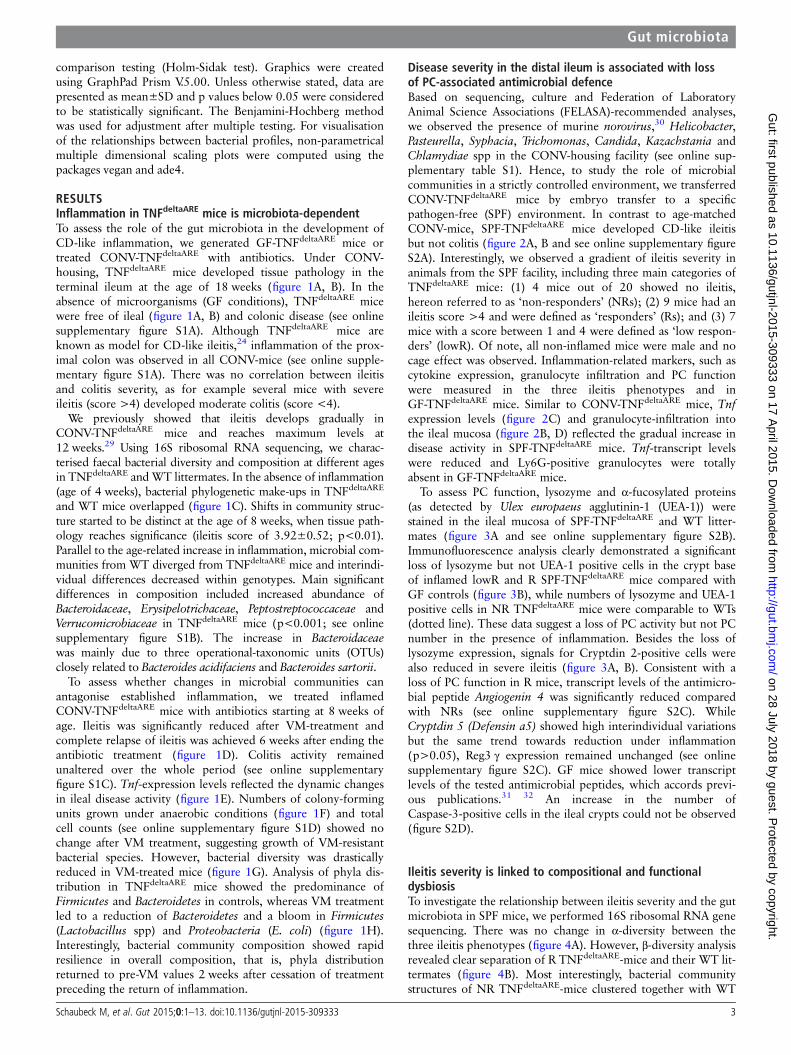
RESULTSInflammation in TNFdeltaARE mice is microbiota-dependentTo assess the role of the gut microbiota in the development ofCD-like inflammation, we generated GF-TNFdeltaARE mice ortreated CONV-TNFdeltaARE with antibiotics. Under CONV-housing, TNFdeltaARE mice developed tissue pathology in theterminal ileum at the age of 18 weeks (figure 1A, B). In theabsence of microorganisms (GF conditions), TNFdeltaARE micewere free of ileal (figure 1A, B) and colonic disease (see onlinesupplementary figure S1A). Although TNFdeltaARE mice areknown as model for CD-like ileitis,24 inflammation of the prox-imal colon was observed in all CONV-mice (see online supple-mentary figure S1A). There was no correlation between ileitisand colitis severity, as for example several mice with severeileitis (score >4) developed moderate colitis (score <4).
We previously showed that ileitis develops gradually inCONV-TNFdeltaARE mice and reaches maximum levels at12 weeks.29 Using 16S ribosomal RNA sequencing, we charac-terised faecal bacterial diversity and composition at different agesin TNFdeltaARE andWT littermates. In the absence of inflammation(age of 4 weeks), bacterial phylogenetic make-ups in TNFdeltaARE
and WT mice overlapped (figure 1C). Shifts in community struc-ture started to be distinct at the age of 8 weeks, when tissue path-ology reaches significance (ileitis score of 3.92±0.52; p<0.01).Parallel to the age-related increase in inflammation, microbial com-munities from WT diverged from TNFdeltaARE mice and interindi-vidual differences decreased within genotypes. Main significantdifferences in composition included increased abundance ofBacteroidaceae, Erysipelotrichaceae, Peptostreptococcaceae andVerrucomicrobiaceae in TNFdeltaARE mice (p<0.001; see onlinesupplementary figure S1B). The increase in Bacteroidaceaewas mainly due to three operational-taxonomic units (OTUs)closely related to Bacteroides acidifaciens and Bacteroides sartorii.
To assess whether changes in microbial communities canantagonise established inflammation, we treated inflamedCONV-TNFdeltaARE mice with antibiotics starting at 8 weeks ofage. Ileitis was significantly reduced after VM-treatment andcomplete relapse of ileitis was achieved 6 weeks after ending theantibiotic treatment (figure 1D). Colitis activity remainedunaltered over the whole period (see online supplementaryfigure S1C). Tnf-expression levels reflected the dynamic changesin ileal disease activity (figure 1E). Numbers of colony-formingunits grown under anaerobic conditions (figure 1F) and totalcell counts (see online supplementary figure S1D) showed nochange after VM treatment, suggesting growth of VM-resistantbacterial species. However, bacterial diversity was drasticallyreduced in VM-treated mice (figure 1G). Analysis of phyla dis-tribution in TNFdeltaARE mice showed the predominance ofFirmicutes and Bacteroidetes in controls, whereas VM treatmentled to a reduction of Bacteroidetes and a bloom in Firmicutes(Lactobacillus spp) and Proteobacteria (E. coli) (figure 1H).Interestingly, bacterial community composition showed rapidresilience in overall composition, that is, phyla distributionreturned to pre-VM values 2 weeks after cessation of treatmentpreceding the return of inflammation.
Disease severity in the distal ileum is associated with lossof PC-associated antimicrobial defenceBased on sequencing, culture and Federation of LaboratoryAnimal Science Associations (FELASA)-recommended analyses,we observed the presence of murine norovirus,30 Helicobacter,Pasteurella, Syphacia, Trichomonas, Candida, Kazachstania andChlamydiae spp in the CONV-housing facility (see online sup-plementary table S1). Hence, to study the role of microbialcommunities in a strictly controlled environment, we transferredCONV-TNFdeltaARE mice by embryo transfer to a specificpathogen-free (SPF) environment. In contrast to age-matchedCONV-mice, SPF-TNFdeltaARE mice developed CD-like ileitisbut not colitis (figure 2A, B and see online supplementary figureS2A). Interestingly, we observed a gradient of ileitis severity inanimals from the SPF facility, including three main categories ofTNFdeltaARE mice: (1) 4 mice out of 20 showed no ileitis,hereon referred to as ‘non-responders’ (NRs); (2) 9 mice had anileitis score >4 and were defined as ‘responders’ (Rs); and (3) 7mice with a score between 1 and 4 were defined as ‘low respon-ders’ (lowR). Of note, all non-inflamed mice were male and nocage effect was observed. Inflammation-related markers, such ascytokine expression, granulocyte infiltration and PC functionwere measured in the three ileitis phenotypes and inGF-TNFdeltaARE mice. Similar to CONV-TNFdeltaARE mice, Tnfexpression levels (figure 2C) and granulocyte-infiltration intothe ileal mucosa (figure 2B, D) reflected the gradual increase indisease activity in SPF-TNFdeltaARE mice. Tnf-transcript levelswere reduced and Ly6G-positive granulocytes were totallyabsent in GF-TNFdeltaARE mice.
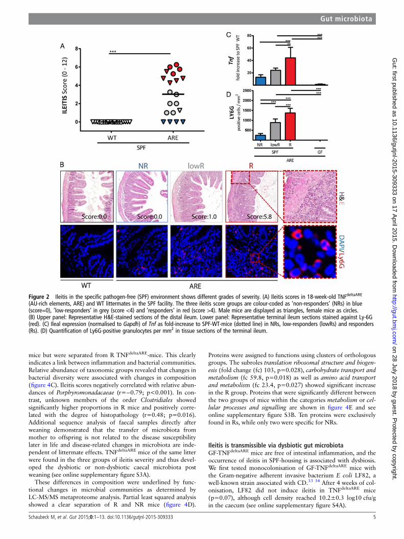
To assess PC function, lysozyme and α-fucosylated proteins(as detected by Ulex europaeus agglutinin-1 (UEA-1)) werestained in the ileal mucosa of SPF-TNFdeltaARE and WT litter-mates (figure 3A and see online supplementary figure S2B).Immunofluorescence analysis clearly demonstrated a significantloss of lysozyme but not UEA-1 positive cells in the crypt baseof inflamed lowR and R SPF-TNFdeltaARE mice compared withGF controls (figure 3B), while numbers of lysozyme and UEA-1positive cells in NR TNFdeltaARE mice were comparable to WTs(dotted line). These data suggest a loss of PC activity but not PCnumber in the presence of inflammation. Besides the loss oflysozyme expression, signals for Cryptdin 2-positive cells werealso reduced in severe ileitis (figure 3A, B). Consistent with aloss of PC function in R mice, transcript levels of the antimicro-bial peptide Angiogenin 4 was significantly reduced comparedwith NRs (see online supplementary figure S2C). WhileCryptdin 5 (Defensin a5) showed high interindividual variationsbut the same trend towards reduction under inflammation(p>0.05), Reg3 γ expression remained unchanged (see onlinesupplementary figure S2C). GF mice showed lower transcriptlevels of the tested antimicrobial peptides, which accords previ-ous publications.31 32 An increase in the number ofCaspase-3-positive cells in the ileal crypts could not be observed(figure S2D).
Ileitis severity is linked to compositional and functionaldysbiosisTo investigate the relationship between ileitis severity and the gutmicrobiota in SPF mice, we performed 16S ribosomal RNA genesequencing. There was no change in α-diversity between thethree ileitis phenotypes (figure 4A). However, β-diversity analysisrevealed clear separation of R TNFdeltaARE-mice and their WT lit-termates (figure 4B). Most interestingly, bacterial communitystructures of NR TNFdeltaARE-mice clustered together with WT
Schaubeck M, et al. Gut 2015;0:1–13. doi:10.1136/gutjnl-2015-309333 3
Gut microbiota on 28 July 2018 by guest. P
rotected by copyright.http://gut.bm
j.com/
Gut: first published as 10.1136/gutjnl-2015-309333 on 17 A
pril 2015. Dow
nloaded from
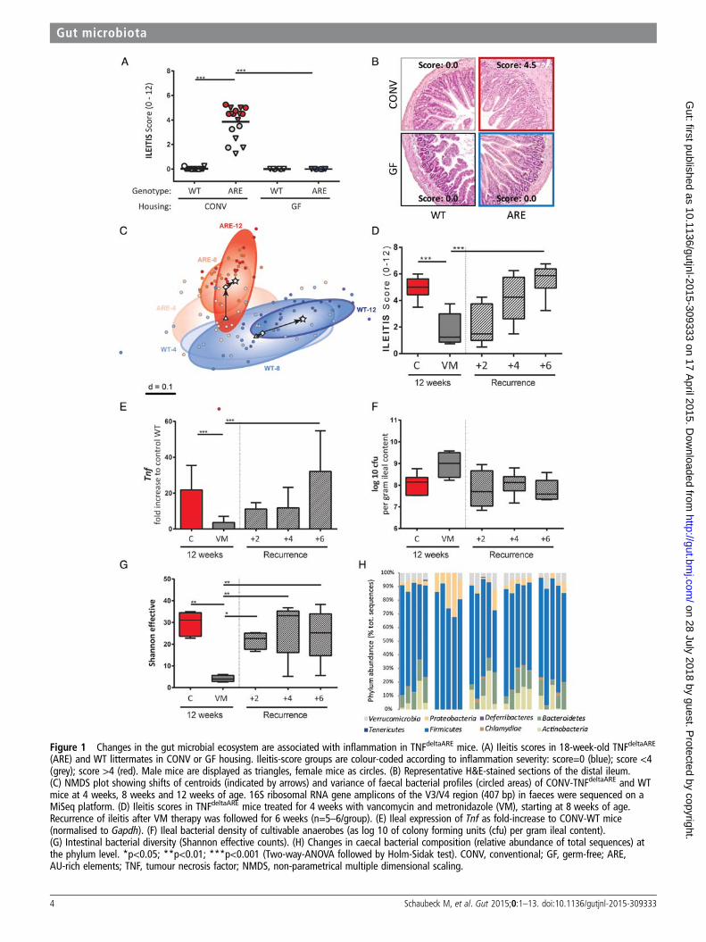
Figure 1 Changes in the gut microbial ecosystem are associated with inflammation in TNFdeltaARE mice. (A) Ileitis scores in 18-week-old TNFdeltaARE
(ARE) and WT littermates in CONV or GF housing. Ileitis-score groups are colour-coded according to inflammation severity: score=0 (blue); score <4(grey); score >4 (red). Male mice are displayed as triangles, female mice as circles. (B) Representative H&E-stained sections of the distal ileum.(C) NMDS plot showing shifts of centroids (indicated by arrows) and variance of faecal bacterial profiles (circled areas) of CONV-TNFdeltaARE and WTmice at 4 weeks, 8 weeks and 12 weeks of age. 16S ribosomal RNA gene amplicons of the V3/V4 region (407 bp) in faeces were sequenced on aMiSeq platform. (D) Ileitis scores in TNFdeltaARE mice treated for 4 weeks with vancomycin and metronidazole (VM), starting at 8 weeks of age.Recurrence of ileitis after VM therapy was followed for 6 weeks (n=5–6/group). (E) Ileal expression of Tnf as fold-increase to CONV-WT mice(normalised to Gapdh). (F) Ileal bacterial density of cultivable anaerobes (as log 10 of colony forming units (cfu) per gram ileal content).(G) Intestinal bacterial diversity (Shannon effective counts). (H) Changes in caecal bacterial composition (relative abundance of total sequences) atthe phylum level. *p<0.05; **p<0.01; ***p<0.001 (Two-way-ANOVA followed by Holm-Sidak test). CONV, conventional; GF, germ-free; ARE,AU-rich elements; TNF, tumour necrosis factor; NMDS, non-parametrical multiple dimensional scaling.
4 Schaubeck M, et al. Gut 2015;0:1–13. doi:10.1136/gutjnl-2015-309333
Gut microbiota on 28 July 2018 by guest. P
rotected by copyright.http://gut.bm
j.com/
Gut: first published as 10.1136/gutjnl-2015-309333 on 17 A
pril 2015. Dow
nloaded from
mice but were separated from R TNFdeltaARE-mice. This clearlyindicates a link between inflammation and bacterial communities.Relative abundance of taxonomic groups revealed that changes inbacterial diversity were associated with changes in composition(figure 4C). Ileitis scores negatively correlated with relative abun-dances of Porphyromonadacaeae (r=−0.79; p<0.001). In con-trast, unknown members of the order Clostridiales showedsignificantly higher proportions in R mice and positively corre-lated with the degree of histopathology (r=0.48; p=0.016).Additional sequence analysis of faecal samples directly afterweaning demonstrated that the transfer of microbiota frommother to offspring is not related to the disease susceptibilitylater in life and disease-related changes in microbiota are inde-pendent of littermate effects. TNFdeltaARE mice of the same litterwere found in the three groups of ileitis severity and thus devel-oped the dysbiotic or non-dysbiotic caecal microbiota postweaning (see online supplementary figure S3A).
These differences in composition were underlined by func-tional changes in microbial communities as determined byLC-MS/MS metaproteome analysis. Partial least squared analysisshowed a clear separation of R and NR mice (figure 4D).
Proteins were assigned to functions using clusters of orthologousgroups. The subroles translation ribosomal structure and biogen-esis (fold change (fc) 103, p=0.028), carbohydrate transport andmetabolism (fc 59.8, p=0.018) as well as amino acid transportand metabolism (fc 23.4, p=0.027) showed significant increasein the R group. Proteins that were significantly different betweenthe two groups of mice within the categories metabolism or cel-lular processes and signalling are shown in figure 4E and seeonline supplementary figure S3B. Ten proteins were exclusivelyfound in Rs, while only two were specific for NRs.
Ileitis is transmissible via dysbiotic gut microbiotaGF-TNFdeltaARE mice are free of intestinal inflammation, and theoccurrence of ileitis in SPF-housing is associated with dysbiosis.We first tested monocolonisation of GF-TNFdeltaARE mice withthe Gram-negative adherent invasive bacterium E coli LF82, awell-known strain associated with CD.33 34 After 4 weeks of col-onisation, LF82 did not induce ileitis in TNFdeltaARE mice(p=0.07), although cell density reached 10.2±0.3 log10 cfu/gin the caecum (see online supplementary figure S4A).
Figure 2 Ileitis in the specific pathogen-free (SPF) environment shows different grades of severity. (A) Ileitis scores in 18-week-old TNFdeltaARE
(AU-rich elements, ARE) and WT littermates in the SPF facility. The three ileitis score groups are colour-coded as ‘non-responders’ (NRs) in blue(score=0), ‘low-responders’ in grey (score <4) and ‘responders’ in red (score >4). Male mice are displayed as triangles, female mice as circles.(B) Upper panel: Representative H&E-stained sections of the distal ileum. Lower panel: Representative terminal ileum sections stained against Ly-6G(red). (C) Ileal expression (normalised to Gapdh) of Tnf as fold-increase to SPF-WT-mice (dotted line) in NRs, low-responders (lowRs) and responders(Rs). (D) Quantification of Ly6G-positive granulocytes per mm2 in tissue sections of the terminal ileum.
Schaubeck M, et al. Gut 2015;0:1–13. doi:10.1136/gutjnl-2015-309333 5
Gut microbiota on 28 July 2018 by guest. P
rotected by copyright.http://gut.bm
j.com/
Gut: first published as 10.1136/gutjnl-2015-309333 on 17 A
pril 2015. Dow
nloaded from

Figure 3 Disease severity is associated with loss of PC function. (A) Paneth cell staining of lysozyme (green; upper panel) combined with UEA-1(red). A representative crypt is shown in the magnification. Lower panel: Paneth cell staining of cryptin 2 (red). A representative crypt is shown inthe magnification. (B) Quantification of lysozyme, UEA-1 and cryptdin-2 positive cells per crypt base in ARE-mice compared with WT mice (dottedline). **p<0.01; ***p<0.001; Two-way ANOVA followed by Holm-Sidak test.
6 Schaubeck M, et al. Gut 2015;0:1–13. doi:10.1136/gutjnl-2015-309333
Gut microbiota on 28 July 2018 by guest. P
rotected by copyright.http://gut.bm
j.com/
Gut: first published as 10.1136/gutjnl-2015-309333 on 17 A
pril 2015. Dow
nloaded from

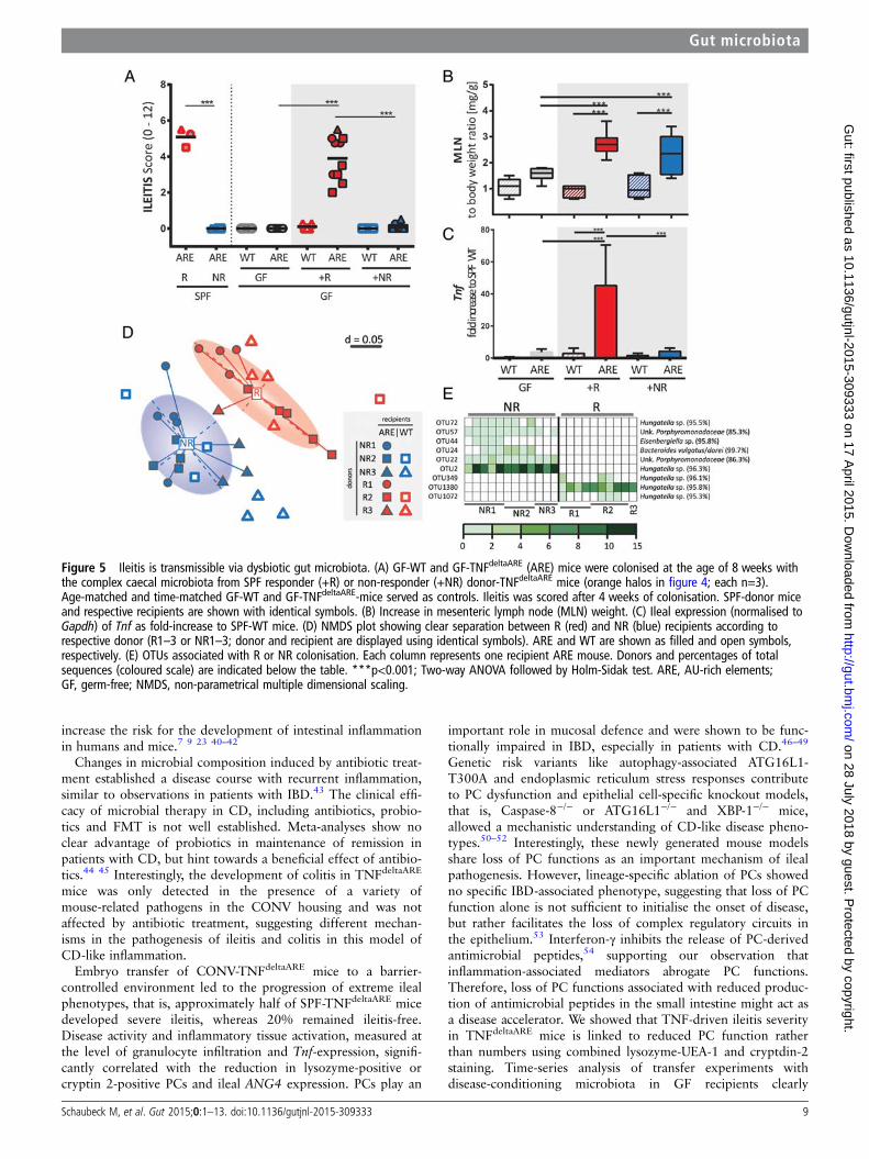
To test the causal relationship of a dysbiotic microbiota asso-ciated with development of CD-like ileitis in TNFdeltaARE mice,we next transferred caecal microbial communities fromSPF-TNFdeltaARE mice into GF-recipients (figure 5A). Therefore,GF-TNFdeltaARE and GF-WT mice were colonised with donor-microbiota from either Rs or NRs. Donor-microbiota is high-lighted with halos in figure 4B. TNFdeltaARE mice colonised withdysbiotic microbiota from Rs (+R; red) developed inflammationafter 4 weeks, mimicking ileitis severity of corresponding donors(recipient mice and respective donors are shown with the samesymbols). WT littermates showed no signs of inflammation, sup-porting the fact that genetic susceptibility was required for thedevelopment of ileitis after transfer. Most remarkably,GF-TNFdeltaARE mice colonised with the microbiota from NRsalso showed no signs of ileitis (+NR; blue). GF-mice were colo-nised by comparable densities of bacteria in both recipient
groups (approximately 109 cfu/g) and tissue maturation was con-firmed by reduced caecum weight (see online supplementaryfigure S4B). Mesenteric lymph node weight to body weight ratioswere significantly increased in both recipient groups when com-pared with GF-TNFdeltaARE mice (figure 5B), suggesting the pres-ence of mucosal immune cell activation independent of tissuepathology. Increased Tnf-transcript levels (p<0.001) correlatedwith the induction of tissue pathology (figure 5C).
To verify the hypothesis that transmissible ileitis was due tothe transfer of dysbiotic bacterial communities, we analysedcaecal content in recipient mice using high-throughput sequen-cing. α-Diversity analysis showed no differences between geno-types and donor groups (see online supplementary figure S4C).β-Diversity analysis displayed again a clear separation betweeninflamed and non-inflamed recipients (figure 5D). Differenceswithin the recipient groups could still be assigned to the
Figure 4 Bacterial profiles and functional dysbiosis mirror ileitis severity. (A) Shannon effective species counts in SPF-WT and SPF-TNFdeltaARE mice.(B) Non-parametrical multiple dimensional scaling analysis showing separation of caecal bacterial communities according to genotype and ileitisseverity. Responders (R; red) were clearly separated from TNFdeltaARE with low ileitis scores (low-responder, lowR; grey) and from non-responders(NR; blue). Microbiota donor-mice used in colonisation experiments are shown with halos. (C) Correlation of bacterial taxa with ileitis scores.(D) Metaproteome analysis of the colonic microbiota. Partial least squared (PLS) analysis differentiated responder (red) and NR (blue)TNFdeltaARE-mice. (E) Significant changes in abundance of protein functions belonging to the clusters of orthologous groups (COGs) main rolemetabolism between R and NR samples. The heatmap represents differences in the abundance of COGs. Protein group data were log10 transformedand normalised by median of bacteria protein data. Protein functions that are unique to either R or NR are shown in the grey squares. COG subrolesare: C, Energy production and conversion; E, amino acid transport and metabolism; F, nucleotide transport and metabolism; G, carbohydratetransport and metabolism; H, coenzyme transport and metabolism; I, lipid transport and metabolism. ***p<0.001; Two-way ANOVA followed byHolm-Sidak test.
Schaubeck M, et al. Gut 2015;0:1–13. doi:10.1136/gutjnl-2015-309333 7
Gut microbiota on 28 July 2018 by guest. P
rotected by copyright.http://gut.bm
j.com/
Gut: first published as 10.1136/gutjnl-2015-309333 on 17 A
pril 2015. Dow
nloaded from

respective donors including WT and TNFdeltaARE mice. Theincrease in Clostridiales spp and decrease inPorphyromonadacaeae observed in inflamed donorSPF-TNFdeltaARE mice also occurred in recipients, but results didnot reach significance (see online supplementary figure S4D).Differential abundance analysis identified two OTUs classified asHungatella sp exclusively present in NRs, with relative abun-dance of up to 15%. Three other OTUs of the same genus wereonly present in Rs (figure 5E), indicating species-specific differ-ences between R and NR mice.
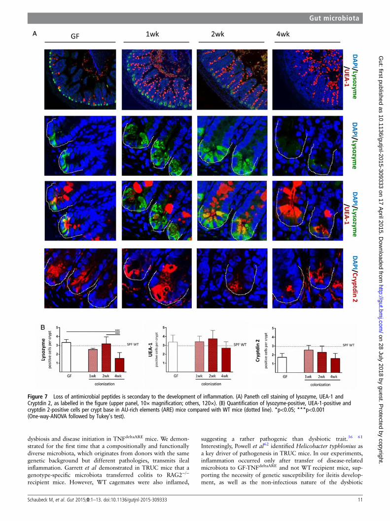
Inflammation precedes loss of PC functionTo better understand the role of PCs in ileitis development, weanalysed time series of tissue pathology and PC function in thetransfer model of CD-like ileitis. GF-TNFdeltaARE and GF-WTrecipients were colonised at the age of 8 weeks with dysbioticmicrobiota from R SPF-TNFdeltaARE mice for 1 week, 2 weeksand 4 weeks. As shown in figure 6A, ileitis was absent after1 week (0.13±0.28), but increased with the duration of colonisa-tion in TNFdeltaARE but not in WT mice. Tnf mRNA expressionlevels were already significantly increased relative to GF-controlsafter 1 week of colonisation (figure 6C; p<0.001), whereas gran-ulocyte infiltration and mesenteric lymph node weight increasedlater, parallel to disease severity in TNFdeltaARE mice (figure 6Dand see online supplementary figure S5A). The influence of col-onisation on caecum-weight and mucus-fucosylation was alreadyvisible 1 week post colonisation showing increased stainingacross the crypt-villus axis (figure 7A; upper panel and see onlinesupplementary figure S5B). Numbers of UEA-1-positive cryptcells remained constant over the entire colonisation period. Mostimportantly, loss of lysozyme-positive crypt cells of recolonised
TNFdeltaARE mice did not precede the onset of tissue pathology(figure 7B), suggesting that loss of PC function is associated butnot causal for the development of CD-like ileitis. Quantificationof Cryptdin 2-positive cells per crypt (figure 7A lower panel and7B right panel) showed no reduction in Cryptdin 2 expression inmice after 4 weeks of colonisation, despite the presence of mod-erate histopathology and inflammatory activation (increased Tnfexpression levels and Ly6G-positive granulocyte infiltration).
DISCUSSIONIn the present work, we provide for the first time evidence fordisease-related causality of bacterial dysbiosis in experimentalCD-like ileitis. We showed that GF-TNFdeltaARE mice are pro-tected from intestinal inflammation, and thereby demonstratedthe essential role of microbial triggers in a model that sharesmain clinical features of CD. In several models of colitis, thedisease-free status under GF conditions has been shown, forexample, in IL-10−/− or T-bet(−/−) ×Rag2(−/−) ulcerativecolitis (TRUC) mouse model (TRUC) mice and HLA/B27-β2mtransgenic rats.23 35 36 However, there are only few IBD-relatedileitis models including TNFdeltaARE and SAMP1/YitFc mice.37
GF housing induced attenuated or no disease activity inSAMP1/YitFc mice, but information about the specific contribu-tion of microbial communities in ileitis development islacking.38 39 Sequential sampling of faecal microbiota demon-strated the gradual influence of inflammation, as illustrated byincreasing divergence of bacterial communities over time inTNFdeltaARE versus WT littermates. Several taxa were signifi-cantly more abundant in TNFdeltaARE mice, including membersof the family Bacteroidaceae, which are frequently reported to
Figure 4 Continued.
8 Schaubeck M, et al. Gut 2015;0:1–13. doi:10.1136/gutjnl-2015-309333
Gut microbiota on 28 July 2018 by guest. P
rotected by copyright.http://gut.bm
j.com/
Gut: first published as 10.1136/gutjnl-2015-309333 on 17 A
pril 2015. Dow
nloaded from
increase the risk for the development of intestinal inflammationin humans and mice.7 9 23 40–42
Changes in microbial composition induced by antibiotic treat-ment established a disease course with recurrent inflammation,similar to observations in patients with IBD.43 The clinical effi-cacy of microbial therapy in CD, including antibiotics, probio-tics and FMT is not well established. Meta-analyses show noclear advantage of probiotics in maintenance of remission inpatients with CD, but hint towards a beneficial effect of antibio-tics.44 45 Interestingly, the development of colitis in TNFdeltaARE
mice was only detected in the presence of a variety ofmouse-related pathogens in the CONV housing and was notaffected by antibiotic treatment, suggesting different mechan-isms in the pathogenesis of ileitis and colitis in this model ofCD-like inflammation.
Embryo transfer of CONV-TNFdeltaARE mice to a barrier-controlled environment led to the progression of extreme ilealphenotypes, that is, approximately half of SPF-TNFdeltaARE micedeveloped severe ileitis, whereas 20% remained ileitis-free.Disease activity and inflammatory tissue activation, measured atthe level of granulocyte infiltration and Tnf-expression, signifi-cantly correlated with the reduction in lysozyme-positive orcryptin 2-positive PCs and ileal ANG4 expression. PCs play an
important role in mucosal defence and were shown to be func-tionally impaired in IBD, especially in patients with CD.46–49
Genetic risk variants like autophagy-associated ATG16L1-T300A and endoplasmic reticulum stress responses contributeto PC dysfunction and epithelial cell-specific knockout models,that is, Caspase-8−/− or ATG16L1−/− and XBP-1−/− mice,allowed a mechanistic understanding of CD-like disease pheno-types.50–52 Interestingly, these newly generated mouse modelsshare loss of PC functions as an important mechanism of ilealpathogenesis. However, lineage-specific ablation of PCs showedno specific IBD-associated phenotype, suggesting that loss of PCfunction alone is not sufficient to initialise the onset of disease,but rather facilitates the loss of complex regulatory circuits inthe epithelium.53 Interferon-γ inhibits the release of PC-derivedantimicrobial peptides,54 supporting our observation thatinflammation-associated mediators abrogate PC functions.Therefore, loss of PC functions associated with reduced produc-tion of antimicrobial peptides in the small intestine might act asa disease accelerator. We showed that TNF-driven ileitis severityin TNFdeltaARE mice is linked to reduced PC function ratherthan numbers using combined lysozyme-UEA-1 and cryptdin-2staining. Time-series analysis of transfer experiments withdisease-conditioning microbiota in GF recipients clearly
Figure 5 Ileitis is transmissible via dysbiotic gut microbiota. (A) GF-WT and GF-TNFdeltaARE (ARE) mice were colonised at the age of 8 weeks withthe complex caecal microbiota from SPF responder (+R) or non-responder (+NR) donor-TNFdeltaARE mice (orange halos in figure 4; each n=3).Age-matched and time-matched GF-WT and GF-TNFdeltaARE-mice served as controls. Ileitis was scored after 4 weeks of colonisation. SPF-donor miceand respective recipients are shown with identical symbols. (B) Increase in mesenteric lymph node (MLN) weight. (C) Ileal expression (normalised toGapdh) of Tnf as fold-increase to SPF-WT mice. (D) NMDS plot showing clear separation between R (red) and NR (blue) recipients according torespective donor (R1–3 or NR1–3; donor and recipient are displayed using identical symbols). ARE and WT are shown as filled and open symbols,respectively. (E) OTUs associated with R or NR colonisation. Each column represents one recipient ARE mouse. Donors and percentages of totalsequences (coloured scale) are indicated below the table. ***p<0.001; Two-way ANOVA followed by Holm-Sidak test. ARE, AU-rich elements;GF, germ-free; NMDS, non-parametrical multiple dimensional scaling.
Schaubeck M, et al. Gut 2015;0:1–13. doi:10.1136/gutjnl-2015-309333 9
Gut microbiota on 28 July 2018 by guest. P
rotected by copyright.http://gut.bm
j.com/
Gut: first published as 10.1136/gutjnl-2015-309333 on 17 A
pril 2015. Dow
nloaded from

demonstrated that loss of PC function was subsequent toTNF-driven development of inflammation, suggesting that lossin PC function might contribute to, but seems not to be causalfor the divergence of disease-related bacterial communities ordevelopment of dysbiosis.
SPF-TNFdeltaARE mice with severe ileitis and loss of antimicro-bial peptides were characterised by a taxonomically and func-tionally dysbiotic gut microbiota. Analysis of intestinalcommunities showed an ileitis-associated increase in the relativeabundance of unknown Clostridiales and reduced abundance ofPorphyromonadacaeae (order of Bacteroidales). While membersof Clostridiales were found in lower abundance in patients withCD, they were increased in TNFdeltaARE ileitis and TM-IEC-C1galt1−/− colitis models, indicating model-specific traits of thisvery broad taxonomic group.55 56 Consistent with changes inour metaproteome analysis, shotgun metagenomic analysis of
microbiota from patients with CD and UC showed functionalshifts especially in ileal CD, driven by pathways of carbohydratetransport and mechanism and amino acid biosynthesis.57
Metaproteomics revealed that, among others, the abundance offucose-utilising enzyme was exclusive for the dysbiotic ecosys-tem in SPF-TNFdeltaARE mice. Inflammation-driven increase inhost-derived fucose was shown to affect metabolic functions offucosidase-expressing commensal bacteria.58 Together with thefact that loss of function in the human Fut2 gene was shown tobe associated with variations in the gut microbiota of patientswith CD, we might speculate a role for the fucose metabolisingenzymes in generating a disease-associated microbial environ-ment in the gut.59 60
We showed that inflammation is associated with the develop-ment of dysbiosis and, most importantly, microbiota transferexperiments confirmed a causal relationship between microbial
Figure 6 Transfer of dysbiotic microbiota induces a gradual disease onset. (A) GF-WT and GF-TNFdeltaARE (ARE) mice were colonised at the age of8 weeks with the complex caecal microbiota from inflamed SPF responder mice (open symbols). After 1 week, 2 weeks and 4 weeks of colonisation,ileitis was assessed. (B) Upper panel: Representative H&E stained terminal ileum sections of ARE-mice colonised with responder microbiota for 1week, 2 weeks and 4 weeks (wk). Age matched GF-AREs (left) served as controls. Lower panel: Representative terminal ileum sections stainedagainst Ly-6G (red). (C) Ileal expression (normalised to Gapdh) of Tnf as fold-increase to SPF-WT mice. Tnf expression of GF-ARE-mice is displayedas dotted line. (D) Quantification of Ly6G-positive granulocytes per mm2 in tissue sections of the terminal ileum after 1 week, 2 weeks and 4 weeksof colonisation with R microbiota. ARE, AU-rich elements; GF, germ-free.
10 Schaubeck M, et al. Gut 2015;0:1–13. doi:10.1136/gutjnl-2015-309333
Gut microbiota on 28 July 2018 by guest. P
rotected by copyright.http://gut.bm
j.com/
Gut: first published as 10.1136/gutjnl-2015-309333 on 17 A
pril 2015. Dow
nloaded from
dysbiosis and disease initiation in TNFdeltaARE mice. We demon-strated for the first time that a compositionally and functionallydiverse microbiota, which originates from donors with the samegenetic background but different pathologies, transmits ilealinflammation. Garrett et al demonstrated in TRUC mice that agenotype-specific microbiota transferred colitis to RAG2−/−
recipient mice. However, WT cagemates were also inflamed,
suggesting a rather pathogenic than dysbiotic trait.36 61
Interestingly, Powell et al62 identified Helicobacter typhlonius asa key driver of pathogenesis in TRUC mice. In our experiments,inflammation occurred only after transfer of disease-relatedmicrobiota to GF-TNFdeltaARE and not WT recipient mice, sup-porting the necessity of genetic susceptibility for ileitis develop-ment, as well as the non-infectious nature of the dysbiotic
Figure 7 Loss of antimicrobial peptides is secondary to the development of inflammation. (A) Paneth cell staining of lysozyme, UEA-1 andCryptdin 2, as labelled in the figure (upper panel, 10× magnification; others, 120×). (B) Quantification of lysozyme-positive, UEA-1-positive andcryptdin 2-positive cells per crypt base in AU-rich elements (ARE) mice compared with WT mice (dotted line). *p<0.05; ***p<0.001(One-way-ANOVA followed by Tukey’s test).
Schaubeck M, et al. Gut 2015;0:1–13. doi:10.1136/gutjnl-2015-309333 11
Gut microbiota on 28 July 2018 by guest. P
rotected by copyright.http://gut.bm
j.com/
Gut: first published as 10.1136/gutjnl-2015-309333 on 17 A
pril 2015. Dow
nloaded from

microbial communities. Vice versa, the microbiota from NRanimals failed to induce CD-like inflammation in GF-TNFdeltaARE
mice, clearly demonstrating the disease-related specificity of thedysbiotic microbiota.
Again, the overall community structure between recipients ofR and NR microbiota were very distinct. OTU-based analysisconfirmed that members of the family Porphyromonadacaeaewere increased in NRs, but also that the relative abundance ofHungatella spp was significantly different. Of note, phylogeny ofthe genus Hungatella is not yet well established and ambiguousdelineation from neighbouring species (eg, Clostridiumclostridioforme, Clostridium aerotolerans, Clostridium bolteaeand Clostridium xylanolyticum) calls for taxonomic amend-ments.63 Hence, different representatives of Hungatella werefound in higher abundances in R and NR TNFdeltaARE mice,which hints at dysbiotic conditions characterised by subtle differ-ences in bacterial profiles down to at least the species level. Thereis rising evidence that protective or deleterious effects of intes-tinal bacteria are strain-specific or species-specific, for example,PrtP expression in Lactobacillus casei, the presence of polysac-charide A in Bacteroides fragilis, or gelatinase E production bycolitogenic Enterococcus faecalis.22 64 65 Some specific patho-bionts, for example, Bilophila wadsworthia in IL-10−/− mice,have been selected from the commensal microbiota with the cap-ability to transfer colitis into susceptible GF hosts.66 However,we failed to induce ileitis in monoassociated TNFdeltaARE miceusing the CD-related pathobiont E. coli LF82.67 68 All our resultspoint towards a community effect of the complex microbiota andloss of aggressive, or gain of protective mechanisms, rather thanthe selection of aggressive phylotypes as single agents causing thedevelopment of CD-like ileitis in TNFdeltaARE mice.
In summary, we report that TNF-driven chronic inflammationwith CD-like ileal pathology depends on microbial triggers andthat inflammation is transmitted via dysbiotic communities ofcommensal bacteria. Consistent with previously published data,maternally inherited factors seem not relevant to the initialdevelopment of dominant dysbiotic bacterial communities anddisease susceptibility later in life.69 Understanding the truenature of a dysbiotic and disease-conditioning microbiota seemsof essential importance to judge the risk of relapse in patientswith IBD after therapeutic intervention or to achieve best pos-sible clinical efficacy in FMT trials. The transfer of IBD-relatedmicrobiota in GF-TNFdeltaARE mice will be an excellent tool togain translational insights into the role of complex microbialcommunities in the progression of CD-like inflammation.
Author affiliations1Chair of Nutrition and Immunology, Technische Universität, Freising-Weihenstephan,Germany2ZIEL-Institute for Food and Health, Technische Universität München, Freising-Weihenstephan, Germany3Department of Proteomics, Helmholtz-Centre for Environmental Research—UFZ,Leipzig, Germany4UFZ, Department of Metabolomics, Helmholtz-Centre for Environmental Research,Leipzig, Germany5Department of Biotechnology, Chemistry and Environmental Engineering, Universityof Aalborg, Aalborg, Denmark6Institut for Medical Microbiology, RWTH University, Aachen, Germany7Institute of Medical Microbiology and Hospital Epidemiology, Hannover MedicalSchool, Hannover, Germany8Institute for Laboratory Animal Science, Hannover Medical School, Hannover,Germany
Acknowledgements The authors thank Arlette Darfeuille-Michaud for providingEscherichia coli LF82, Sigrid Kisling for histopathological scoring, Melanie Klein,Caroline Ziegler and Kathleen Eismann for outstanding technical work, BenjaminScheer for help with maintenance of the mass-spectrometer, and Ludovica Butto andElena Lobner for critical proofreading of the manuscript. The authors are grateful for
using the facilities of the Centre for Chemical Microscopy (ProVIS) at the HelmholtzCentre for Environmental Research (supported by European Regional DevelopmentFunds (EFRE–Europe funds Saxony)) and the Helmholtz Association.
Contributors MS, TC and DH designed experiments and wrote the manuscript.MS, TC and JC performed experiments. MS, TC and IL analysed sequencing data.SBH, NJ and MvB performed metaproteomic studies. MB and AB generatedgerm-free mice. AD and MH performed Cryptdin-2 staining. TC and DH supervisedand coordinated the project.
Funding Generation and housing of germ-free TNFdeltaARE mice was financed bythe Priority Program SPP 16565 of the German Research Foundation (DFG) grantedto DH (HA3148/10-1) and to AB (953/5-1). All experiments with germ-freeTNFdeltaARE were financed by DFG SPP1656 granted to DH (HA3148/8-1).
Competing interests None.
Provenance and peer review Not commissioned; externally peer reviewed.
Data sharing statement We would be happy to make our sequencing data available.
Ethics The animal experiments were performed according to the German guidelinesfor animal care.
Open Access This is an Open Access article distributed in accordance with theCreative Commons Attribution Non Commercial (CC BY-NC 4.0) license, whichpermits others to distribute, remix, adapt, build upon this work non-commercially,and license their derivative works on different terms, provided the original work isproperly cited and the use is non-commercial. See: http://creativecommons.org/licenses/by-nc/4.0/
REFERENCES1 Baumgart DC, Sandborn WJ. Crohn’s disease. Lancet 2012;380:1590–605.2 Ordas I, Eckmann L, Talamini M, et al. Ulcerative colitis. Lancet 2012;380:1606–19.3 Huttenhower C, Kostic AD, Xavier RJ. Inflammatory bowel disease as a model for
translating the microbiome. Immunity 2014;40:843–54.4 Kaser A, Zeissig S, Blumberg RS. Inflammatory bowel disease. Annu Rev Immunol
2010;28:573–621.5 Jostins L, Ripke S, Weersma RK, et al. Host-microbe interactions have shaped the
genetic architecture of inflammatory bowel disease. Nature 2012;491:119–24.6 Mondot S, Barreau F, Al Nabhani Z, et al. Altered gut microbiota composition in
immune-impaired Nod2(−/−) mice. Gut 2012;61:634–5.7 Elinav E, Strowig T, Kau AL, et al. NLRP6 inflammasome regulates colonic microbial
ecology and risk for colitis. Cell 2011;145:745–57.8 Lupp C, Robertson ML, Wickham ME, et al. Host-mediated inflammation disrupts
the intestinal microbiota and promotes the overgrowth of Enterobacteriaceae.Cell Host Microbe 2007;2:204.
9 Frank DN, St Amand AL, Feldman RA, et al. Molecular-phylogenetic characterizationof microbial community imbalances in human inflammatory bowel diseases.Proc Natl Acad Sci USA 2007;104:13780–5.
10 Qin J, Li R, Raes J, et al. A human gut microbial gene catalogue established bymetagenomic sequencing. Nature 2010;464:59–65.
11 Gevers D, Kugathasan S, Denson LA, et al. The treatment-naive microbiome innew-onset Crohn’s disease. Cell Host Microbe 2014;15:382–92.
12 Salzman NH, Hung K, Haribhai D, et al. Enteric defensins are essential regulators ofintestinal microbial ecology. Nat Immunol 2010;11:76–83.
13 Clevers HC, Bevins CL. Paneth cells: maestros of the small intestinal crypts.Annu Rev Physiol 2013;75:289–311.
14 Wehkamp J, Salzman NH, Porter E, et al. Reduced Paneth cell alpha-defensins inileal Crohn’s disease. Proc Natl Acad Sci USA 2005;102:18129–34.
15 Sartor RB. Microbial influences in inflammatory bowel diseases. Gastroenterology2008;134:577–94.
16 van Nood E, Vrieze A, Nieuwdorp M, et al. Duodenal infusion of donor feces forrecurrent Clostridium difficile. N Engl J Med 2013;368:407–15.
17 Angelberger S, Reinisch W, Makristathis A, et al. Temporal bacterial communitydynamics vary among ulcerative colitis patients after fecal microbiotatransplantation. Am J Gastroenterol 2013;108:1620–30.
18 Kump PK, Grochenig HP, Lackner S, et al. Alteration of intestinal dysbiosis by fecalmicrobiota transplantation does not induce remission in patients with chronic activeulcerative colitis. Inflamm Bowel Dis 2013;19:2155–65.
19 Cui B, Feng Q, Wang H, et al. Fecal microbiota transplantation through mid-gut forrefractory Crohn’s disease: Safety, feasibility and efficacy trial results. J GastroenterolHepatol 2015;30:51–8.
20 Madsen KL, Doyle JS, Tavernini MM, et al. Antibiotic therapy attenuates colitis ininterleukin 10 gene-deficient mice. Gastroenterology 2000;118:1094–105.
21 Kim SC, Tonkonogy SL, Albright CA, et al. Variable phenotypes of enterocolitis ininterleukin 10-deficient mice monoassociated with two different commensalbacteria. Gastroenterology 2005;128:891–906.
22 Steck N, Hoffmann M, Sava IG, et al. Enterococcus faecalis metalloproteasecompromises epithelial barrier and contributes to intestinal inflammation.Gastroenterology 2011;141:959–71.
12 Schaubeck M, et al. Gut 2015;0:1–13. doi:10.1136/gutjnl-2015-309333
Gut microbiota on 28 July 2018 by guest. P
rotected by copyright.http://gut.bm
j.com/
Gut: first published as 10.1136/gutjnl-2015-309333 on 17 A
pril 2015. Dow
nloaded from

23 Rath HC, Herfarth HH, Ikeda JS, et al. Normal luminal bacteria, especiallyBacteroides species, mediate chronic colitis, gastritis, and arthritis in HLA-B27/human beta2 microglobulin transgenic rats. J Clin Invest 1996;98:945–53.
24 Kontoyiannis D, Pasparakis M, Pizarro TT, et al. Impaired on/off regulation of TNFbiosynthesis in mice lacking TNF AU-rich elements: implications for joint andgut-associated immunopathologies. Immunity 1999;10:387–98.
25 Werner T, Wagner SJ, Martinez I, et al. Depletion of luminal iron alters the gutmicrobiota and prevents Crohn’s disease-like ileitis. Gut 2011;60:325–33.
26 Erben U, Loddenkemper C, Doerfel K, et al. A guide to histomorphologicalevaluation of intestinal inflammation in mouse models. Int J Clin Exp Pathol2014;7:4557–76.
27 Katakura K, Lee J, Rachmilewitz D, et al. Toll-like receptor 9-induced type I IFNprotects mice from experimental colitis. J Clin Invest 2005;115:695–702.
28 Livak KJ, Schmittgen TD. Analysis of relative gene expression data using real-timequantitative PCR and the 2(-Delta Delta C(T)) Method. Methods 2001;25:402–8.
29 Baur P, Martin FP, Gruber L, et al. Metabolic phenotyping of the Crohn’sdisease-like IBD etiopathology in the TNF(DeltaARE/WT) mouse model. J ProteomeRes 2011;10:5523–35.
30 Mahler Convenor M, Berard M, Feinstein R, et al. FELASA recommendations for thehealth monitoring of mouse, rat, hamster, guinea pig and rabbit colonies inbreeding and experimental units. Lab Anim 2014;48:178–92.
31 Moghadamrad S, McCoy KD, Geuking MB, et al. Attenuated portal hypertension ingerm-free mice: Function of bacterial flora on the development of mesentericlymphatic and blood vessels. Hepatology 2015. doi:10.1002/hep.27698
32 Inoue R, Tsuruta T, Nojima I, et al. Postnatal changes in the expression of genes forcryptdins 1–6 and the role of luminal bacteria in cryptdin gene expression in mousesmall intestine. FEMS Immunol Med Microbiol 2008;52:407–16.
33 Boudeau J, Glasser AL, Masseret E, et al. Invasive ability of an Escherichia colistrain isolated from the ileal mucosa of a patient with Crohn’s disease. InfectImmun 1999;67:4499–509.
34 Conte MP, Longhi C, Marazzato M, et al. Adherent-invasive Escherichia coli (AIEC)in pediatric Crohn’s disease patients: phenotypic and genetic pathogenic features.BMC Res Notes 2014;7:748.
35 Sellon RK, Tonkonogy S, Schultz M, et al. Resident enteric bacteria are necessary fordevelopment of spontaneous colitis and immune system activation ininterleukin-10-deficient mice. Infect Immun 1998;66:5224–31.
36 Garrett WS, Gallini CA, Yatsunenko T, et al. Enterobacteriaceae act in concert withthe gut microbiota to induce spontaneous and maternally transmitted colitis.Cell Host Microbe 2010;8:292–300.
37 Bamias G, Okazawa A, Rivera-Nieves J, et al. Commensal bacteria exacerbateintestinal inflammation but are not essential for the development of murine ileitis.J Immunol 2007;178:1809–18.
38 Matsumoto S, Okabe Y, Setoyama H, et al. Inflammatory bowel disease-like enteritisand caecitis in a senescence accelerated mouse P1/Yit strain. Gut 1998;43:71–8.
39 Bamias G, Dahman MI, Arseneau KO, et al. Intestinal-specific TNFalphaoverexpression induces Crohn’s-like ileitis in mice. PLoS One 2013;8:e72594.
40 Couturier-Maillard A, Secher T, Rehman A, et al. NOD2-mediated dysbiosispredisposes mice to transmissible colitis and colorectal cancer. J Clin Invest2013;123:700–11.
41 Bloom SM, Bijanki VN, Nava GM, et al. Commensal Bacteroides species inducecolitis in host-genotype-specific fashion in a mouse model of inflammatory boweldisease. Cell Host Microbe 2011;9:390–403.
42 Flavell RA. Commensal Bacteroides species induce colitis inhost-genotype-specific fashion in a mouse model of inflammatory bowel disease.Cell 2011;9:390–403.
43 Scribano ML, Prantera C. Antibiotics and inflammatory bowel diseases. Dig Dis2013;31:379–84.
44 Khan KJ, Ullman TA, Ford AC, et al. Antibiotic therapy in inflammatory boweldisease: a systematic review and meta-analysis. Am J Gastroenterol2011;106:661–73.
45 Ghouri YA, Richards DM, Rahimi EF, et al. Systematic review of randomizedcontrolled trials of probiotics, prebiotics, and synbiotics in inflammatory boweldisease. Clin Exp Gastroenterol 2014;7:473–87.
46 Cadwell K, Liu JY, Brown SL, et al. A key role for autophagy and the autophagygene Atg16l1 in mouse and human intestinal Paneth cells. Nature2008;456:259–63.
47 Wehkamp J, Koslowski M, Wang G, et al. Barrier dysfunction due to distinctdefensin deficiencies in small intestinal and colonic Crohn’s disease.Mucosal Immunol 2008;1(Suppl 1):S67–74.
48 Bevins CL, Salzman NH. Paneth cells, antimicrobial peptides and maintenance ofintestinal homeostasis. Nat Rev Microbiol 2011;9:356–68.
49 Cunliffe RN, Rose FR, Keyte J, et al. Human defensin 5 is stored in precursor form innormal Paneth cells and is expressed by some villous epithelial cells and by metaplasticPaneth cells in the colon in inflammatory bowel disease. Gut 2001;48:176–85.
50 Adolph TE, Tomczak MF, Niederreiter L, et al. Paneth cells as a site of origin forintestinal inflammation. Nature 2013;503:272–6.
51 Kaser A, Lee AH, Franke A, et al. XBP1 links ER stress to intestinal inflammationand confers genetic risk for human inflammatory bowel disease. Cell2008;134:743–56.
52 Gunther C, Martini E, Wittkopf N, et al. Caspase-8 regulates TNF-alpha-inducedepithelial necroptosis and terminal ileitis. Nature 2011;477:335–9.
53 Garabedian EM, Roberts LJ, McNevin MS, et al. Examining the role of Paneth cellsin the small intestine by lineage ablation in transgenic mice. J Biol Chem1997;272:23729–40.
54 Farin HF, Karthaus WR, Kujala P, et al. Paneth cell extrusion and release ofantimicrobial products is directly controlled by immune cell-derived IFN-gamma.J Exp Med 2014;211:1393–405.
55 Manichanh C, Rigottier-Gois L, Bonnaud E, et al. Reduced diversity of faecal microbiotain Crohn’s disease revealed by a metagenomic approach. Gut 2006;55:205–11.
56 Perez-Munoz ME, Bergstrom K, Peng V, et al. Discordance between changes in thegut microbiota and pathogenicity in a mouse model of spontaneous colitis.Gut Microbes 2014;5:286–95.
57 Morgan XC, Tickle TL, Sokol H, et al. Dysfunction of the intestinal microbiome ininflammatory bowel disease and treatment. Genome Biol 2012;13:R79.
58 Pickard JM, Maurice CF, Kinnebrew MA, et al. Rapid fucosylation of intestinalepithelium sustains host-commensal symbiosis in sickness. Nature 2014;514:638–41.
59 Rausch P, Rehman A, Kunzel S, et al. Colonic mucosa-associated microbiota isinfluenced by an interaction of Crohn disease and FUT2 (Secretor) genotype.Proc Natl Acad Sci USA 2011;108:19030–5.
60 McGovern DP, Jones MR, Taylor KD, et al. Fucosyltransferase 2 (FUT2) non-secretorstatus is associated with Crohn’s disease. Hum Mol Genet 2010;19:3468–76.
61 Garrett WS, Lord GM, Punit S, et al. Communicable ulcerative colitis induced byT-bet deficiency in the innate immune system. Cell 2007;131:33–45.
62 Powell N, Walker AW, Stolarczyk E, et al. The transcription factor T-bet regulatesintestinal inflammation mediated by interleukin-7 receptor+ innate lymphoid cells.Immunity 2012;37:674–84.
63 Kaur S, Yawar M, Kumar PA, et al. Hungatella effluvii gen. nov., sp. nov., anobligately anaerobic bacterium isolated from an effluent treatment plant, andreclassification of Clostridium hathewayi as Hungatella hathewayi gen. nov., comb.nov. Int J Syst Evol Microbiol 2014;64(Pt 3):710–18.
64 Mazmanian SK, Round JL, Kasper DL. A microbial symbiosis factor preventsintestinal inflammatory disease. Nature 2008;453:620–5.
65 von Schillde MA, Hormannsperger G, Weiher M, et al. Lactocepin secreted byLactobacillus exerts anti-inflammatory effects by selectively degradingproinflammatory chemokines. Cell Host Microbe 2012;11:387–96.
66 Devkota S, Wang Y, Musch MW, et al. Dietary-fat-induced taurocholic acidpromotes pathobiont expansion and colitis in Il10-/- mice. Nature 2012;487:104–8.
67 Carvalho FA, Barnich N, Sivignon A, et al. Crohn’s disease adherent-invasiveEscherichia coli colonize and induce strong gut inflammation in transgenic miceexpressing human CEACAM. J Exp Med 2009;206:2179–89.
68 Darfeuille-Michaud A, Boudeau J, Bulois P, et al. High prevalence ofadherent-invasive Escherichia coli associated with ileal mucosa in Crohn’s disease.Gastroenterology 2004;127:412–21.
69 Hemmerling J, Heller K, Hormannsperger G, et al. Fetal exposure to maternalinflammation does not affect postnatal development of genetically-driven ileitis andcolitis. PLoS One 2014;9:e98237.
Schaubeck M, et al. Gut 2015;0:1–13. doi:10.1136/gutjnl-2015-309333 13
Gut microbiota on 28 July 2018 by guest. P
rotected by copyright.http://gut.bm
j.com/
Gut: first published as 10.1136/gutjnl-2015-309333 on 17 A
pril 2015. Dow
nloaded from