original article dysfunctional subpathways of ...ijcem.com/files/ijcem0059795.pdf · subpathways...
TRANSCRIPT

Int J Clin Exp Med 2018;11(2):1260-1269www.ijcem.com /ISSN:1940-5901/IJCEM0059795
Original ArticleDysfunctional subpathways of osteoarthritis identified through combining lncRNA-mRNA expression profile with pathway topologies
An-Li Zhu, Meng-Po Fan, De-Qiang Liu
Department of Orthopaedic Trauma, The Second People’s Hospital of Liaocheng, Linqing 252600, Shandong, China
Received June 19, 2017; Accepted December 7, 2017; Epub February 15, 2018; Published February 28, 2018
Abstract: Objective: Concept of key local subregion is more meaningful for revealing the pathogenesis of diseas-es, relative to entire pathways. Thus, in our study, we planned to use the subpathway strategy to extract altered subpathways competitively regulated by lncRNAs involved in osteoarthritis (OA) through lncRNA-mRNA expression profile and pathway topologies. Methods: Candidate lncRNA-mRNA interactions were constructed and reweighted using PCC. Using KEGG pathways as backbone, undirected graphs were displayed where genes were as nodes and regulated relations stood for edges. Condition-specific lncRNA competitively regulated signal pathways (LRSP) were inferred. Subsequently, interesting lncRNAs and genes were aligned to LRSP, and then subpahtways were detected using “lenient distance” similarity method. Eventually, we evaluated the significance of candidate subpathways through Wallenius approximation, following by identification of hub lncRNAs in the LRSP. Results: After reweighting using PCC, 54 lncRNAs, 130 mRNAs and 147 co-expressed interactions were detected. Then, 31 seed pathways were identified after these 130 mRNAs were mapped to the reference pathways. we transformed seed pathways into undirected graphs, and the 54 lncRNAs were embedded into pathway graphs to establish the condition-specific LRSP. Overall 35 significant lncRNAs competitively regulating subpathways involved in 24 complete pathways were identified in the LRSP. The top three subpathways included cell cycle, PI3K-Akt signaling pathway, and focal adhe-sion. There were 4 hub lncRNAs, including SLC38A3, LINC00242, DCP1A, and EPB41L4A-AS1. Conclusions: The strategy of subpathways is feasible to predict marker pathways for OA. Subpathways of cell cycle, PI3K-Akt signaling pathway, and focal adhesion might play crucial roles in OA progression.
Keywords: Osteoarthritis, subpathway, long noncoding RNAs
Introduction
Osteoarthritis (OA), as one of the most preva-lent chronic, and age-related degenerative joint disorder, is characterized by degradation of articular cartilage along with pain and disability [1, 2]. More than 50% of patients with symp-tomatic OA are younger than 65 years old [3]. Unfortunately, OA is becoming a primary health problem in China in view of the increasing life expectancy of the population. Therapy strate-gies to slow down the OA progression remain still limited, and the damage is irreversible at the time of diagnosis. Together, this emphasiz-es the importance to increase insight into the disease causes and to optimize therapeutic approaches as well as develop novel therapeu-tic drugs.
OA is considered as a multifactorial disease. In addition to joint instability, aging and obesity, genetic links to OA susceptibility have been found in several studies [4, 5]. Epigenetic effects, especially noncoding RNAs, likely con-tribute to the onset and progression of OA [6]. Noncoding RNAs (ncRNAs) have emerged as a new identified class of RNA molecules without protein-coding capacity [7]. Based on their size, ncRNAs include two main categories: small ncRNAs, and long ncRNAs (lncRNAs) [8]. It has been reported that lncRNA play crucial roles in various key biological processes [9, 10], for example, posttranscriptional regulation [11] and human disease [12]. Significantly, lncRNAs have been indicated to exert important func-tions in the processes of bone and cartilage development [13]. Growing evidence has impli-
Dysfunctional subpathways in OA
1261 Int J Clin Exp Med 2018;11(2):1260-1269
cated that lncRNAs competitively regulate mRNAs expression through sharing common miRNA binding sites with mRNAs [14, 15]. Zhang et al. [16] have suggested that lncRNA UFC1 may play important roles as a endoge-nous miRNA-34a sponge to promote the prolif-eration of OA chondrocyte. Moreover, a former study provided by Song et al. demonstrated that a lncRNA GAS5 contributed to the patho-genesis of OA via serving as a negative regula-tor of miRNA-21 [17]. Further, a previous study has indicated that lncRNA CIR plays a crucial role in the pathogenesis of OA via contributing to the degradation of chondrocyte extracellular matrix [18]. Since the RNA competitive interac-tion could influence biological functions in dis-ease, extracting lncRNA competitively regulat-ed pathways not only shed new insights into the pathogenic processes, but also is beneficial for uncovering the cellular functions of lncRNAs in disease. On the contrary, few methods are able to systematically track dysfunctional pathways which are competitively mediated by lncRNAs in disease states. Additionally, paying more attention on subpathways instead of entire pathways might extract more biologically mean-ingful pathways and dissect the functional roles of lncRNAs. Excitedly, Shi et al. [19] pro-posed the concept of key local subregion which is more meaningful for revealing the molecular mechanisms of diseases relative to the entire pathways.
Consequently, in the current work, we aimed to identify the lncRNAs competitively regulated signal subpathways in OA using the subpath-way strategy based on lncRNA-mRNA expres-sion profile and pathway topologies. We confirm that this novel method can provide a flexible tool to extract lncRNA competitively regulated subpathways in OA, and is beneficial for expounding the functional roles of lncRNAs in OA.
Materials and methods
Proposed protocol
In the present analysis, using KEGG pathways as backbone, undirected graphs were displayed where genes were as nodes and regulated rela-tions stood for edges. Next, condition-specific lncRNA competitively regulated signal path-ways (LRSP) were inferred according to the matched lncRNA-mRNA expression data as well as the common miRNAs. Subsequently, interesting lncRNAs and genes were aligned to
LRSP, and then subpahtways were located in pathways based on the “lenient distance” simi-larity method [19]. Eventually, we evaluated the significance of candidate subpathways by means of Wallenius approximation [20], follow-ing by the identification of hub lncRNAs in the LRSP.
Microarray data about OA
Gene expression profile from OA cartilage were collected from Gene Expression Omnibus (GEO) database (Access ID: GSE57218) [21] bas- ed on the GPL6947 platform of Illumina Hu- manHT-12 V3.0 expression beadchip. In the GSE57218, there were 7 healthy samples, 33 OA samples, and 33 OA preserved cartilage samples. In order to better reveal the molecular mechanisms of OA, we only selected 7 healthy samples and 33 OA samples for further analy-sis. After probes were aligned to the gene sym-bols, a total of 19,293 genes were obtained.
Constructing candidate lncRNA-mRNA interac-tion
LncRNA-related competing triplets (lncRNA-miRNA-mRNA interactions) were firstly collect-ed. Specifically, the lncRNA-miRNA interactions were downloaded from StarBase version 2.0 [22], and mRNA-miRNA interactions were obtained from mirTarBase, TarBase, miRecords (V4.0), and mir2Disease. Next, according to the shared miRNAs, candidate lncRNA-mRNA com-petitively regulated relationships were con-structed. For each lncRNA, we identified its candidate competing mRNAs when they met the following two criteria: first, hypergeometric test of common miRNAs under false discovery rate (FDR) < 0.05; second, Jaccard Coefficient of lncRNA-mRNA intersections ranked at top 20%. With the goal of ensuring the reliability of data, the interactions which fulfilled both condi-tions were reserved. Eventually, candidate lncRNA-mRNA competitively regulated interac-tions covered 7693 lncRNAs-mRNA interac-tions among 1749 genes and 835 lncRNAs. Then, the intersection between 19,293 genes and 7693 lncRNAs-mRNA interactions was taken, and finally, we obtained 1690 mRNAs and 116 lncRNAs.
Reweighting the lncRNAs-mRNA interactions using Pearson correlation coefficient (PCC)
To the best of my knowledge, PCC is a measure about correlation between two variables,

Dysfunctional subpathways in OA
1262 Int J Clin Exp Med 2018;11(2):1260-1269
whose range is from -1 to 1 [23]. In our study, PCC was utilized to measure the co-expression possibility for any pair of relations in the candi-date lncRNA-mRNA interactions on the basis of matched lncRNA and mRNA expression pro-files. Using Fisher’s Z transform [24], those r value had reached a significant positive thresh-old were retained (P < 0.05).
Reconstructing condition-specific LRSP
Identification of seed pathways: Kyoto Ency- clopedia of Genes and Genomes (KEGG) is a database to provide a reference knowledge base for understanding cellular processes
through pathway aligning, which means to map genes to KEGG reference pathways to infer sys-temic behaviors of cell [25]. In the current anal-ysis, we recruited all the KEGG reference path-ways from the database of KEGG. Then, we mapped mRNAs of the reweighted lncRNAs-mRNA interactions to the KEGG reference path-ways to extract the seed pathways. The raw P values were corrected using FDR based on Benjamini-Hochberg method [26]. Seed path-ways were detected when the FDR was set as 0.01.
Linking LncRNAs to regulated-mRNAs within pathway graphs: We transformed seed path-ways into undirected graphs kept original path-way structural information by means of the developed R packages provided by Li et al. [27]. The lncRNAs in the reweighted lncRNAs-mRNA interactions were embedded into pathway graphs as nodes by connecting to their regulat-ed-mRNAs. Finally, we obtained condition-spe-cific LRSP, which covered lncRNA nodes and lncRNA-mRNA competitively regulated edges.
Locate subpathways competing regulated by lncRNAs: LncRNAs participating in the compet-ing regulation and genes of interests were con-sidered as signature nodes. These nodes com-bined with topology feature of LRSP can help us effectively locating lncRNA-regulated subre-gions. To begin with, we aligned signatures nodes to LRSP, then positing subpathways competing regulated by lncRNAs employed “lenient distance” similarity and network topol-ogy feature. Succinctly, the shortest path between any two signature nodes was calcu-lated, if the count of molecules between each pair of signatures was no more than n, then we would merge them into one nodes. At the end, the count of nodes in the molecule sets in path-way no smaller than s were determined as can-didate subpahtways competing regulated by lncRNAs. The n and s indexes respectively con-trolled the intensity of regulated signals and the size of candidate subpathways. In our study, n = 1 and s = 8 were used as default parame- ters.
Assessing the significance of candidate sub-athways: In order to assess whether the candi-date subpathways were competing regulated by lncRNAs comparing random, Wallenius approximation methods was utilized to evalu-ate the significance of candidate subpathways,
Table 1. List of seed pathways based on false discovery rate (FDR) < 0.01Pathways FDRProstate cancer 4.25E-11Cell cycle 5.82E-10Pathways in cancer 3.49E-08PI3K-Akt signaling pathway 5.02E-08Glioma 2.24E-06Small cell lung cancer 3.08E-06Hepatitis B 6.45E-05Focal adhesion 9.17E-05p53 signaling pathway 2.17E-04Endometrial cancer 3.32E-04Adherens junction 3.38E-04Basal cell carcinoma 4.52E-04HIF-1 signaling pathway 5.42E-04ErbB signaling pathway 9.83E-04GnRH signaling pathway 1.37E-03Neurotrophin signaling pathway 1.48E-03Amyotrophic lateral sclerosis (ALS) 2.22E-03Melanogenesis 2.35E-03Non-small cell lung cancer 3.37E-03Epstein-Barr virus infection 3.66E-03DNA replication 3.94E-03Viral carcinogenesis 4.20E-03Hypertrophic cardiomyopathy (HCM) 4.38E-03Oocyte meiosis 4.40E-03Bladder cancer 4.79E-03Transcriptional misregulation in cancers 5.21E-03Dilated cardiomyopathy (DCM) 5.79E-03Herpes simplex infection 6.45E-03Pancreatic cancer 6.82E-03Olfactory transduction 6.82E-03Melanoma 9.24E-03
Dysfunctional subpathways in OA
1263 Int J Clin Exp Med 2018;11(2):1260-1269
which was executed using R package BiasedUrn [28]. The following parameters were used: the count of interesting mRNAs (x); the count of background mRNAs (y); the count of back-ground mRNAs enriched in this subpathways (m1); the count of interesting mRNAs involved in the given subpathway (m2); and the weight value of this subpathway (W). The weight parameter indicated the intensity of competing regulation by lncRNAs annotated into this given subpathway, which was calculated as follows:
In this equation, PG was the number of mRNAs annotated into this subpathways, and GL denot-ed the count of mRNAs competitively regulated by lncRNAs in this subpathway. β represented the parameter of control (herein, β = 1). The original P values were calculated based on the formula of P-values = F (x, m1, m2, y, weighti). Then, the P values were adjusted using FDR. Finally, the significant subpathways with FDR < 0.01 were extracted in our study.
evaluated its performance after randomly dis-turbing LRSP.
Statistical analysis
In our study, we used Feature Extraction soft-ware (version 10.7; Agilent Technologies, Inc., Santa Clara, CA, USA) to analyze the statistical significance of the microarray results. The FDR was computed to correct the original P-values. The threshold value used to designate the com-mon miRNAs was FDR ≤ 0.05, and the cut-off criteria for significant subpathways was FDR < 0.01. P < 0.05 was regarded to indicate a sta-tistically significant difference. In the present study, SPSS version 18.0 (SPSS, Inc., Chicago, IL, USA) was used for statistical analysis.
Results
Identification of co-expressed lncRNA-mRNA interactions and seed pathways
In our study, we used PCC to measure the co-expression possibility for any pair of relations in the candidate lncRNA-mRNA interactions on
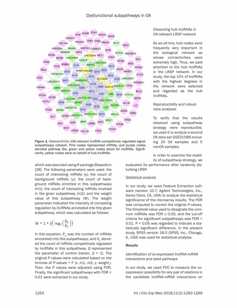
Figure 1. Osteoarthritis (OA)-relevant lncRNA competitively regulated signal subpathways network. Pink nodes represented mRNAs, and purple nodes denoted pathway IDs, green and yellow nodes stood for lncRNAs. Signifi-cantly, yellow nodes were on behalf of hub lncRNAs.
Dissecting hub lncRNAs in OA-relevant LRSP network
As we all kno, hub nodes were frequently very important in the biological network as whose connectivities were extremely high. Thus, we paid attention to the hub lncRNAs in the LRSP network. In our study, the top 10% of lncRNAs with the highest degrees in the network were selected and regarded as the hub lncRNAs.
Reproducibility and robust-ness analyses
To verify that the results obtained using subpathway strategy were reproducible, we used it to analyze a second OA data set GSE51588 includ-ing 20 OA samples and 5 nonOA samples.
In order to examine the stabil-ity of subpathway strategy, we
W 1 log PG
2G
L= + -b `` jj

Dysfunctional subpathways in OA
1264 Int J Clin Exp Med 2018;11(2):1260-1269
the basis of matched lncRNA and mRNA expression profiles. Based on P < 0.05, we identified a total of 54 lncRNAs, 130 mRNAs and 147 co-expressed interactions (Supple- mentary Table 1). Then, we mapped these 130 mRNAs to the reference pathways to further detect the seed pathways. Ultimately, overall 31 seed pathways were identified when the FDR was set as less than 0.01 (Table 1).
Detecting signal subpathways competitively regulated by lncRNAs
After the seed pathways were identified, we transformed seed pathways into undirected graphs, and the 54 lncRNAs in the reweighted lncRNAs-mRNA interactions were embedded into pathway graphs as nodes by connecting to their regulated-mRNAs. Then, condition-specif-ic LRSP was constructed, which covered lncRNA nodes and lncRNA-mRNA competitively regulated edges. Specific LRSP was shown in Figure 1. In this network, we found that overall 35 significant lncRNAs competitively regulating
subpathways involved in 24 complete pathways with FDR < 0.01 (Table 2).
In further analysis, we focused on top three subpathways that competitively regulated by lncRNAs (Figure 2). The first is the most signifi-cant subpathway path: 04110_1, which was a subregion of cell cycle pathway (Figure 2A). Then, we further revealed this subpathway, and observed that this subregion was competitively regulated by 11 lncRNAs. Among these lncRNAs, LINC00482, directly regulated three genes (MCM2, MCM3, and ORC3). Moreover, MCM2 was coordinately regulated by two lncRNAs, LINC00265 and LINC00482.
The second significant subpathways was path: 04151-2, an important sub region in PI3K-Akt signaling pathway (Figure 2B). In this subpath-way, TP53 gene encodes for a nuclear phos-phoprotein of 53 kd (p53) participated in cell cycle control. Notably, this gene TP53 was coor-dinately regulated by two lncRNAs, DCP1A, and SLC38A3.
Table 2. Subpathways extracted based on lncRNA-mRNA expression profile and pathway topologiesPathway ID Pathways Molecule ratio (m2/x) Bg ratio (m1/y) Weight FDR04110_1 Cell cycle 13/135 20/26232 1.621488 4.28E-1704151_2 PI3K-Akt signaling pathway 14/135 19/26232 1.440573 1.06E-1604510_1 Focal adhesion 12/135 17/26232 1.502513 1.91E-1605200_1 Pathways in cancer 21/135 29/26232 1.465664 1.95E-1605214_1 Glioma 8/135 11/26232 1.459432 3.26E-1605215_1 Prostate cancer 14/135 21/26232 1.584963 5.32E-1504115_1 P53 signaling pathway 7/135 9/26232 1.362572 9.90E-1504012_1 ErbB signaling pathway 6/135 8/26232 1.415037 1.01E-1204066_1 HIF-1 signaling pathway 6/135 8/26232 1.415037 1.01E-1204520_1 Adherens junction 6/135 8/26232 1.415037 1.01E-1205414_1 Dilated cardiomyopathy 6/135 8/26232 1.415037 1.01E-1205212_1 Pancreatic cancer 5/135 6/26232 1.263034 3.43E-1105223_1 Non-small cell lung cancer 5/135 6/26232 1.263034 3.43E-1105410_1 Hypertrophic cardiomyopathy (HCM) 5/135 6/26232 1.263034 3.43E-1104151_4 PI3K-Akt signaling pathway 5/135 7/26232 1.485427 1.12E-1004722_1 Neurotrophin signaling pathway 4/135 5/26232 1.321928 4.01E-0905161_2 Hepatitis B 4/135 5/26232 1.321928 4.01E-0905213_1 Endometrial cancer 4/135 5/26232 1.321928 4.01E-0905218_1 Melanoma 4/135 5/26232 1.321928 4.01E-0905219_1 Bladder cancer 4/135 5/26232 1.321928 4.01E-0904114_2 Oocyte meiosis 4/135 6/26232 1.584963 1.04E-0805014_1 Amyotrophic lateral sclerosis (ALS) 4/135 6/26232 1.584963 1.04E-0805222_2 Small cell lung cancer 4/135 6/26232 1.584963 1.04E-0804916_3 Melanogenesis 3/135 4/26232 1.415037 5.31E-07

Dysfunctional subpathways in OA
1265 Int J Clin Exp Med 2018;11(2):1260-1269
The third subpathway, path: 04510_1, was a part of focal adhesion pathway (Figure 2C). Within this subpathway, EGFR was a growth-factor-receptor tyrosine kinase which was com-petitively regulated by four lncRNAs, DCP1A, SLC38A3, ZNF761 and EPB41LA-AS1. In a nut-shell, these results demonstrated that this approach could identified biological meaningful subpathways, and highlighted some critical lncRNAs in OA condition.
Dissecting hub lncRNAs in OA-relevant LRSP network
In order to extract key lncRNAs associated with OA, we performed degree analysis for all nodes within the LRSP. Based on the degree distribu-tion, we extracted 4 hub lncRNAs, including SLC38A3 (degree = 25), LINC00242 (degree = 23), DCP1A (degree = 21), and EPB41L4A-AS1 (degree = 18).
Reproducibility and robustness analyses
Reproducibility analysis: To prove that the results obtained using subpathway strategy were reproducible, we used this method to ana-lyze another OA data set GSE51588 including 20 OA samples and 5 nonOA samples. A total of 19 significant subpathways involved in 19 com-plete pathways were identified from GSE51588 (FDR < 0.01). Among these 19 pathways, 12 pathways were identified by the two OA data sets, and 7 of these pathways undetected in GSE57218. This further suggested that the results obtained using subpathway strategy were reproducible.
Robustness analysis: To test the stability of subpathway strategy, we evaluated the perfor-mance of subpathway strategy through ran-domly removing a percentage of the edges in LRSP from 5% to 30% (N= 5, 10, …, 30). This
Figure 2. Top 3 subpathways identified using this novel method. Pink and green nodes represented mRNAs and lncRNAs, respectively. A. Cell cycle subpathway (path: 04110_1, FDR = 4.28E-17). B. PI3K-Akt signaling pathway subpathway (path: 04151_2, FDR = 1.06E-16). C. Focal adhesion subpathway (path: 04510_1, FDR = 1.91E-16).
Dysfunctional subpathways in OA
1266 Int J Clin Exp Med 2018;11(2):1260-1269
deletion process repeated 100 times to receive 100 random LRSP lists. Subsequently, we con-ducted the subpathway method on OA data for each random LRSP list. When the deletion per-centage increased, the recalled ratio of signifi-cant pathways declined slightly. Subpathway strategy showed that the best performance when the deleted percentage was 15%, recall-ing more than 91% of the pathways. Of note, the recalled ratio was 84%, as disturbing up to 25% edges within the LRSP, In a nutshell, these suggested that the subpathway strategy was robust.
Discussion
In recent years, studies suggests that lncRNAs can interact with miRNAs to further function as the miRNA decoy, thereby competitively affect-ing miRNA regulation of mRNAs as well as maintain normal biological functions [29, 30]. Thus, disruption of the biological functions competitively regulated by lncRNAs may cause the initiation of diseases, and thus, a better understanding of this regulation mechanism might provide new opportunities for developing novel target therapies. Nevertheless, so far, few methods were developed to identify the biological functions competitively regulated by lncRNAs and the functions of these lncRNAs have not be well expounded in disease condi-tions. Moreover, Shi et al. have demonstrated that paying more attention to key local subre-gions instead of complete pathways might be more significant in extraction of disease-asso-ciated pathway and might be more explainable to reveal the functional roles of lncRNAs in dis-ease. In addition, this subregion method com-bining lncRNAmRNA expression profile with pathway topologies had several merits. To begin with, lncRNA representing a new regula-tory layer were included in the pathway analy-sis. Second, it comprehensively analyzed the joint effect of lncRNAs, lncRNAs competitively regulated genes, and pathway topologies. Th- ird, the method of subpathway might screen more biologically meaningful pathways. Thus, in our study, a subregion strategy was proposed to detect lncRNAs competitively regulated sub-pathways for OA, thereby to further explore the molecular mechanism underlying OA. Overall, we identified 35 significant lncRNAs competi-tively regulating subpathways involved in 24 complete pathways. We only focused on the top three different subpathways that competitively mediated by lncRNAs.
The first is the most significant subpathway path: 04110_1, which was a subregion of cell cycle pathway. Cell cycle has been demonstrat-ed to influence the function of chondrocytes, which results in the division and duplication of chondrocytes. Cell proliferation would be blocked in inflammatory synovial tissue to arrest self-renewal and promote inflammation [31]. While, inflammation is a major contribut-ing factor associated with the risk of cartilage loss and symptom progression in OA [32]. In addition, Yang and Wang have implicated that cell cycle as well as the related genes (CDK1 and MAD2L1), might play important roles in the development of OA [33]. Significantly, OA chon-drocytes has a very low proliferative activity, and promoting chondrocyte proliferation through enhancing G2/M transition is a mecha-nism of treating OA [34]. Other studies also have demonstrated that promoting chondro-cyte proliferation might be an efficient treat-ment to cure or delay the progression of OA [35, 36]. Accordingly, we further confirmed that cell cycle indeed contribute the initiation and pro-gression of OA, these also proved that this strategy was an available method to reveal the OA pathogenesis.
The second significant subpathways was path: 04151-2, an important sub region in PI3K-Akt signaling pathway. Intracellular pathway PI3K/Akt has been reported to participate in extra-cellular matrix (ECM) alterations [37]. Fragments of ECM proteins (fibronectin and col-lagen) in turn, stimulate the generation of inflammatory cytokines, and chemokines [38]. ECM protein fragments has also been suggest-ed to stimulate matrix destruction, and then ultimately resulting in cartilage degradation and contributing to the onset of OA [39]. More importantly, PI3K/Akt pathway might play key roles in the OA progression [40]. In this sub-pathway in our study, TP53 gene encodes for a nuclear phosphoprotein of 53 kd (p53) partici-pated in cell cycle control, which exerts impor-tant functions in OA [41, 42]. Thus, we specu-lated that PI3K-Akt signaling subpathway exerted key functions in the occurrence and development of OA.
The third subpathway, path: 04510_1, was a part of focal adhesion pathway. Within this sub-pathway, EGFR was a growth-factor-receptor tyrosine kinase which was competitively regu-lated by four lncRNAs, DCP1A, SLC38A3,

Dysfunctional subpathways in OA
1267 Int J Clin Exp Med 2018;11(2):1260-1269
ZNF761 and EPB41LA-AS1. EGFR is crucially important for cartilage matrix degradation in the period of endochondral ossification [43]. A previous study has reported that activation of EGFR signaling stimulates the expression of matrix metalloproteinases, resulting in incr- eased cartilage matrix degradation by chondro-cytes and osteoclasts at the chondro-osseous junctions in OA [44, 45]. Focal adhesions are specialized structures at the cellular-ECM con-tact points. Zintzaras et al. provided the evi-dence of functions of focal adhesion family genes in OA [46]. In a nutshell, these results demonstrated that this approach could identi-fied biological meaningful subpathways in OA condition.
Notably, in our study, the lncRNA of SLC38A3 had the highest degree in the LRSP. Moreover, this hub lncRNA mediated the subpathways of PI3K-Akt signaling pathway and focal adhesion. SLC38A3 is also known as SNAT3, or SN1 [47]. SLC38A3, one member of System N subfamily, owns a preference for glutamine [48]. Moreover, SLC38A3 has been demonstrated to be involved in glutamate/γ-aminobutyric acid (GABA)-glutamine cycle and regulates the amount of glutamine [49]. Further, GABA levels have been indicated to be related to the chron-ic knee OA pain [50]. As we all know, no studies have reported the relationship between SLC38A3 and OA. Based on these results, we demonstrated that this lncRNA SLC38A3 might play important roles in OA progression.
Conclusion
In conclusion, a total of 35 significant lncRNAs competitively regulating subpathways involved in 24 complete pathways were successfully identified in our study. Based on the results, we indicated that the top 3 subpathways of cell cycle, PI3K-Akt signaling pathway, and focal adhesion were closely related to OA. Never- theless, the confirmation by means of other datasets is needed to prove that these sub-pathways are helpful in distinguishing OA from normal samples.
Acknowledgements
This research received no specific grants from any funding agency in public, commercial, or not-for-profit sectors.
Disclosure of conflict of interest
None.
Address correspondence to: De-Qiang Liu, De- partment of Orthopaedic Trauma, The Second People’s Hospital of Liaocheng, No. 306 Jiankang Street, Linqing 252600, Shandong, China. Fax: +86-0635-2342472; Tel: +86-0635-2342472; E-mail: [email protected]
References
[1] Moulton SG, Bhatia S, Civitarese DM, Frank RM, Dean CS and Laprade RF. Surgical tech-niques and outcomes of repairing meniscal radial tears: a systematic review. Arthroscopy 2016; 32: 1919-1925.
[2] Sacitharan PK and Vincent TL. Cellular ageing mechanisms in osteoarthritis. Mamm Genome 2016; 27: 421-429.
[3] Deshpande BR, Katz JN, Solomon DH, Yelin EH, Hunter DJ, Messier SP, Suter LG and Losi-na E. The number of persons with symptomatic knee osteoarthritis in the united states: impact of race/ethnicity, age, sex, and obesity. Arthri-tis Care Res (Hoboken) 2016; 68: 1743-1750.
[4] Pottie P, Presle N, Terlain B, Netter P, Mainard D and Berenbaum F. Obesity and osteoarthri-tis: more complex than predicted! Ann Rheum Dis 2006; 65: 1403.
[5] Livshits G, Kato BS, Zhai G, Hart DJ, Hunter D, Macgregor AJ, Williams FM and Spector TD. Genomewide linkage scan of hand osteoarthri-tis in female twin pairs showing replication of quantitative trait loci on chromosomes 2 and 19. Ann Rheum Dis 2007; 66: 623-627.
[6] Reynard LN and Loughlin J. Genetics and epi-genetics of osteoarthritis. Maturitas 2012; 71: 200-204.
[7] Riquelme I, Ili C, Roa JC and Brebi P. Long non-coding RNAs in gastric cancer: mechanisms and potential applications. Oncotarget 2016.
[8] Brosnan CA and Voinnet O. The long and the short of noncoding RNAs. Curr Opin Cell Biol 2009; 21: 416-425.
[9] Wilusz JE, Sunwoo H and Spector DL. Long noncoding RNAs: functional surprises from the RNA world. Genes Dev 2009; 23: 1494-1504.
[10] Wang KC and Chang HY. Molecular mecha-nisms of long noncoding RNAs. Mol Cell 2011; 43: 904-914.
[11] Mercer TR, Dinger ME and Mattick JS. Long non-coding RNAs: insights into functions. Nat Rev Genet 2009; 10: 155-159.
[12] Barsytelovejoy D, Lau SK, Boutros PC, Khosravi F, Jurisica I, Andrulis IL, Tsao MS and Penn LZ. The c-Myc oncogene directly induces the H19 noncoding RNA by allele-specific binding to po-

Dysfunctional subpathways in OA
1268 Int J Clin Exp Med 2018;11(2):1260-1269
tentiate tumorigenesis. Cancer Res 2006; 66: 5330-5337.
[13] Ponting CP, Oliver PL, Reik W. Evolution and functions of long noncoding RNAs. Cell 2009; 136: 629-641.
[14] Tay Y, Rinn J and Pandolfi PP. The multilayered complexity of ceRNA crosstalk and competi-tion. Nature 2014; 505: 344.
[15] Ebert MS and Sharp PA. Emerging roles for natural microRNA sponges. Current Biology 2010; 20: R858.
[16] Zhang G, Wu Y, Xu D and Yan X. Long noncod-ing RNA UFC1 promotes proliferation of chon-drocyte in osteoarthritis by acting as a sponge for miR-34a. DNA Cell Biol 2016; 35: 691-695.
[17] Song J, Ahn C, Chun CH and Jin EJ. A long non-coding RNA, GAS5, plays a critical role in the regulation of miR-21 during osteoarthritis. J Orthop Res 2014; 32: 1628-1635.
[18] Liu Q, Zhang X, Dai L, Hu X, Zhu J, Li L, Zhou C and Ao Y. Long noncoding RNA related to carti-lage injury promotes chondrocyte extracellular matrix degradation in osteoarthritis. Arthritis Rheumatol 2014; 66: 969-978.
[19] Shi X, Xu Y, Zhang C, Feng L, Sun Z, Han J, Su F, Zhang Y, Li C, Li X. Subpathway-LNCE: Identify dysfunctional subpathways competitively regu-lated by lncRNAs through integrating lncRNA-mRNA expression profile and pathway topolo-gies. Oncotarget 2016; 7: 69857-69870.
[20] Epstein MP, Duncan R, Jiang Y, Conneely KN, Allen AS and Satten GA. A permutation proce-dure to correct for confounders in case-control studies, including tests of rare variation. Am J Hum Genet 2012; 91: 215.
[21] Ramos YF, den Hollander W, Bovée JV, Bomer N, van der Breggen R, Lakenberg N, Keurentjes JC, Goeman JJ, Slagboom PE, Nelissen RG, Bos SD, Meulenbelt I. Genes involved in the osteoarthritis process identified through ge-nome wide expression analysis in articular car-tilage; the RAAK study. PLoS One 2014; 9: e103056.
[22] Li JH, Liu S, Zhou H, Qu LH and Yang JH. star-base v2.0: decoding miRNA-ceRNA, miRNA-ncRNA and protein–RNA interaction networks from large-scale CLIP-Seq data. Nucleic Acids Res 2014; 42: D92.
[23] Nahler G. Pearson correlation coefficient. Dic-tionary of Pharmaceutical Medicine 2009; 132-132.
[24] Best DJ and Roberts DE. Algorithm AS 89: the upper tail probabilities of spearman’s rho. Ap-plied Statistics 1975; 24: 377-379.
[25] Kanehisa M, Araki M, Goto S, Hattori M, Hi-rakawa M, Itoh M, Katayama T, Kawashima S, Okuda S and Tokimatsu T, Yamanishi Y. KEGG for linking genomes to life and the environ-ment. Nucleic Acids Res 2008; 36: D480-484.
[26] Benjamini Y and Hochberg Y. Controlling the false discovery rate: a practical and powerful approach to multiple testing. Journal of the Royal Statistical Society. Series B (Methodolog-ical) 1995; 289-300.
[27] Li C, Han J, Yao Q, Zou C, Xu Y, Zhang C, Shang D, Zhou L, Zou C and Sun Z, Li J, Zhang Y, Yang H, Gao X, Li X. Subpathway-GM: identification of metabolic subpathways via joint power of interesting genes and metabolites and their topologies within pathways. Nucleic Acids Res 2013; 41: e101.
[28] Epstein MP, Duncan R, Jiang Y, Conneely KN, Allen AS and Satten GA. A permutation proce-dure to correct for confounders in case-control studies, including tests of rare variation. Am J Hum Genet 2012; 91: 215-223.
[29] Liang WC, Fu WM, Wong CW, Wang Y, Wang WM, Hu GX, Zhang L, Xiao LJ, Wan DC and Zhang JF, Waye MM. The lncRNA H19 pro-motes epithelial to mesenchymal transition by functioning as miRNA sponges in colorectal cancer. Oncotarget 2015; 6: 22513-22525.
[30] Bak RO and Mikkelsen JG. miRNA sponges: soaking up miRNAs for regulation of gene ex-pression. Wiley Interdiscip Rev RNA 2014; 5: 317.
[31] Franke S, Sommer M, Rüster C, Bondeva T, Marticke J, Hofmann G, Hein G and Wolf G. Ad-vanced glycation end products induce cell cy-cle arrest and proinflammatory changes in os-teoarthritic fibroblast-like synovial cells. Arthritis Res Ther 2009; 11: R136.
[32] Sellam J, Berenbaum F. The role of synovitis in pathophysiology and clinical symptoms of os-teoarthritis. Nat Rev Rheumatol 2010; 6: 625.
[33] Yang J and Wang N. Genome-wide expression and methylation profiles reveal candidate genes and biological processes underlying sy-novial inflammatory tissue of patients with os-teoarthritis. Int J Rheum Dis 2015; 18: 783-790.
[34] Li X, Ye H, Yu F, Cai L, Li H, Chen J, Wu M, Chen W, Lin R and Li Z, Zheng C, Xu H, Wu G, Liu X. Millimeter wave treatment promotes chondro-cyte proliferation via G1/S cell cycle transition. Int J Mol Med 2012; 29: 823-831.
[35] Kashiwagi A, Schipani E, Fein MJ, Greer PA and Shimada M. Targeted deletion of Capn4 in cells of the chondrocyte lineage impairs chon-drocyte proliferation and differentiation. Mol Cell Biol 2010; 30: 2799-2810.
[36] Huang JG, Xia C, Zheng XP, Yi TT, Wang XY, Song G and Zhang B. 17β-Estradiol promotes cell proliferation in rat osteoarthritis model chondrocytes via PI3K/Akt pathway. Cell Mol Biol Lett 2011; 16: 564-575.
[37] Kang J, Chen W, Xia J, Li Y, Yang B, Chen B, Sun W, Song X, Xiang W, Wang X, Wang F, Bi Z, Wan
Dysfunctional subpathways in OA
1269 Int J Clin Exp Med 2018;11(2):1260-1269
Y. Extracellular matrix secreted by senescent fibroblasts induced by UVB promotes cell prolif-eration in HaCaT cells through PI3K/AKT and ERK signaling pathways. Int J Mol Med 2008; 21: 777.
[38] Loeser RF, Goldring SR, Scanzello CR and Goldring MB. Osteoarthritis: a disease of the joint as an organ. Arthritis and rheumatism 2012; 64: 1697-1707.
[39] Issa RI and Griffin TM. Pathobiology of obesity and osteoarthritis: integrating biomechanics and inflammation. Pathobiol Aging Age Relat Dis 2012; 2.
[40] Chen J, Crawford R and Xiao Y. Vertical inhibi-tion of the PI3K/Akt/mTOR pathway for the treatment of osteoarthritis. J Cell Biochem 2013; 114: 245.
[41] Børresen-Dale AL. TP53 and breast cancer. Hum Mutat 2003; 21: 292.
[42] Chiu TH, Lan KY, Yang MD, Lin JJ, Hsia TC, Wu CT, Yang JS, Chueh FS and Chung JG. Diallyl sulfide promotes cell-cycle arrest through the p53 expression and triggers induction of apop-tosis via caspase- and mitochondria-depen-dent signaling pathways in human cervical cancer Ca Ski cells. Nutr Cancer 2013; 65: 505-514.
[43] Usmani SE, Pest MA, Kim G, Ohora SN, Qin L and Beier F. Transforming growth factor alpha controls the transition from hypertrophic carti-lage to bone during endochondral bone growth. Bone 2012; 51: 131-141.
[44] Hall KC, Hill D, Otero M, Plumb DA, Froemel D, Dragomir CL, Maretzky T, Boskey A, Crawford HC and Selleri L, Goldring MB, Blobel CP. ADAM17 controls endochondral ossification by regulating terminal differentiation of chondro-cytes. Mol Cell Biol 2013; 33: 3077.
[45] Zhang X, Zhu J, Li Y, Lin T, Siclari VA, Chandra A, Candela EM, Koyama E, Enomoto-iwamoto M and Qin L. Epidermal growth factor receptor (EGFR) signaling regulates epiphyseal carti-lage development through β-catenin-depen- dent and -independent pathways. J Biol Chem 2013; 288: 32229-32240.
[46] Zintzaras E, Doxani C, Koufakis T, Kastanis A, Rodopoulou P and Karachalios T. Synopsis and meta-analysis of genetic association studies in osteoporosis for the focal adhesion family genes: the CUMAGAS-OSTEOporosis informa-tion system. BMC Med 2011; 9: 9.
[47] Mackenzie B and Erickson JD. Sodium-coupled neutral amino acid (System N/A) transporters of the SLC38 gene family. Pflugers Arch 2004; 447: 784.
[48] Chaudhry FA, Reimer RJ, Krizaj D, Barber D, Stormmathisen J, Copenhagen DR, Edwards RH. Molecular analysis of system N suggests novel physiological roles in nitrogen metabo-lism and synaptic transmission. Cell 1999; 99: 769-780.
[49] Nissenmeyer LSH and Chaudhry FA. Corrigen-dum: protein kinase C phosphorylates the sys-tem N glutamine transporter SN1 (Slc38a3) and regulates its membrane trafficking and degradation. Front Endocrinol (Lausanne) 2013; 4: 138.
[50] Reckziegel D, Raschke F, Cottam WJ and Auer DP. Cingulate GABA levels inversely correlate with the intensity of ongoing chronic knee os-teoarthritis pain. Mol Pain 2016; 12.

Dysfunctional subpathways in OA
1
Supplementary Table 1. Co-expressed lncRNA-mRNA interactionsLncRNAs mRNAs P valueTAPT1-AS1 C9orf142 0.020796ZNF503-AS2 KCNMA1 0.007918SNHG11 MRPL53 0.001061FOXN3-AS1 PXMP2 0.018733H1FX-AS1 WDR13 0.000287LINC00476 ZFY 0.014779SEMA3B CDC14A 0.018772UHRF1 FZD7 0.005294UHRF1 MMP14 8.37E-05UHRF1 TP53I3 0.008129UBXN8 SHC1 0.022423UBXN8 TCF7L1 0.020685UBXN8 CCNE2 0.006498PPP1R9B CD44 0.014635H19 PTEN 0.001873SPON1 GLI1 0.015996ZNF761 EGFR 0.027012ZNF761 MCL1 0.031725VPS11 CLOCK 0.002545VPS11 FOXO1 0.030394DCP1A EGFR 0.005504DCP1A TP53 0.025784LINC00476 MTOR 0.028292LINC00521 NTRK3 0.006817LINC00116 APP 0.002536LINC00116 HSPB2 5.07E-05CASC2 ATM 0.029099LINC00174 RDX 0.049601LINC00174 IRS1 0.029037PCBP1-AS1 ACSL1 0.000486PCBP1-AS1 SIRT1 0.005737LINC00482 GLTSCR2 0.010638LINC00482 GLTSCR2 0.010638LINC00482 HNRNPU 0.016928LINC00482 CSTF3 0.001814LINC00482 ABCB7 0.003116LINC00482 GNAT2 0.000449LINC00482 ORC3 0.036621LINC00482 NUP160 0.023165LINC00482 UBR5 0.002775LINC00482 PNN 0.014918LINC00482 SYMPK 0.011208LINC00482 POLR2I 0.019806LINC00482 MCM5 0.037922LINC00482 TPM2 0.008203LINC00482 MCM2 1.95E-05
LINC00482 PPP2R5A 0.006401LINC00482 EXOC3 0.005156KTN1-AS1 TAB3 0.030462LINC00265 H3F3B 0.014554LINC00265 H3F3B 0.014554LINC00265 CARD8 3.10E-05LINC00265 MYB 0.001984LINC00265 H3F3B 0.014554LINC00265 PNN 0.023655LINC00265 GTF2H1 0.001533LINC00265 GOLPH3L 0.034189LINC00265 CDK2 0.001312SLC38A3 EP300 0.008672SLC38A3 PKM 0.007572SLC38A3 WASF3 0.005501SLC38A3 TP53 0.033759SLC38A3 SMO 0.000197SLC38A3 EGFR 0.000199LINC00319 ACVR2B 0.026085CRYM-AS1 RBL2 0.004796LINC00173 IL6R 8.89E-05LINC00173 CYLD 0.011211LINC00173 CREB1 1.49E-07RNU12 PTK2B 0.000183SNHG5 CDKN1C 0.043245DGKK IL24 0.010587DGKK TJP2 0.011003MEG3 SRSF1 0.019223MIR17HG LEFTY1 0.000733CROCCP2 CUL5 0.001541CROCCP2 NOTCH2 9.51E-06CROCCP2 SIRT1 0.001238CROCCP2 BMPR2 0.011873EPB41L4A-AS1 E2F3 0.004195EPB41L4A-AS1 EGFR 0.000956MIAT TNFAIP3 0.020953MIAT COL1A1 0.034354MIAT CD276 0.029338ZNF883 NLK 0.020342ZNF883 BCL2 0.010237ZNF883 IL2 0.008702LINC00242 ITGA1 0.023556LINC00242 EMD 0.004885LINC00242 CLTC 0.006018LINC00242 TPM3 0.015882LINC00242 SRSF6 0.000157LINC00242 UGGT1 0.002456LINC00242 GNAS 0.031293LINC00242 RPP40 0.000167

Dysfunctional subpathways in OA
2
LINC00242 PQBP1 0.044638LINC00242 CAMK2G 0.015456LINC00242 CPSF1 1.30E-05LINC00242 CSTF3 0.001131LINC00242 POLA2 0.000137LINC00242 ATP6V1E1 0.004121LINC00242 HNRNPA3 8.01E-05LINC00242 PRKAG1 0.000882LINC00242 XPOT 0.029639LINC00242 UGGT2 0.030781LINC00242 IQGAP3 0.012135LINC00242 CPSF1 1.30E-05LINC00242 CPSF1 1.30E-05LINC00242 HNRNPA1L2 0.001433LINC00242 MCM6 0.002328LINC00242 PARVA 0.022557LINC00242 LNPEP 0.004744LINC00242 AP3B1 0.014676LINC00242 PRKDC 9.34E-06LINC00242 AP1M1 0.001635LINC00242 MAP2K3 0.020865LINC00242 MAP2K3 0.020865DLEU2 ARHGEF6 0.006329SNHG15 YWHAG 0.017051SNHG15 TCF7L1 0.001231LINC00472 LEFTY2 0.001207
TTTY15 NEFM 0.000284MAGI2-AS3 FZD3 0.005842MAGI2-AS3 STAM2 0.028667MAGI2-AS3 LAMB3 0.003474MAGI2-AS3 TIAM1 0.012739JRK IGF1R 0.002306JRK RARA 0.000365JRK RPS6KA1 0.025538MCF2L-AS1 PTK2 0.049984BCYRN1 SRSF2 0.005313BCYRN1 SRSF2 0.005313MIR600HG HES1 0.046238MIR600HG ERBB2 0.044947LINC00312 TP53INP1 0.004705LINC00312 BMPR2 0.015022LINC00312 RBL2 0.003773DNAJC3-AS1 CCNT1 0.000541DNAJC3-AS1 CYP3A4 0.000895PVT1 WEE1 0.020413SCARNA9 P4HB 0.000112C14orf169 ITGA2 0.009380C14orf169 SLC38A1 0.037817RN7SL1 CYP8B1 0.003425ERVK13-1 CYCS 0.015019FBXL19-AS1 TOB2 0.000997FBXL19-AS1 GRB2 0.024463