original article hericium erinaceus synergizing with ... · int j clin exp med 2016;9(2):1447-1457...
TRANSCRIPT
Int J Clin Exp Med 2016;9(2):1447-1457www.ijcem.com /ISSN:1940-5901/IJCEM0015906
Original Article Hericium erinaceus synergizing with doxorubicin induced SGC7901 cell apoptosis
Wenyi Yang1, Dazheng Han2, Liping Wu2, Yinuo Huang2, Jiansheng Li1, Hao Guo2, Yan Liu3
1Department of Digestive, The First Affiliated Hospital of Zhengzhou University, Zhengzhou 450052, Henan Province, China; 2Department of Digestive, The First Affiliated Hospital of He’nan University, Kaifeng 475000, Henan Province, China; 3Department of Dermatology, Guangzhou General Hospital of Guangzhou Military Command (Liuhuaqiao Hospital), Guangzhou, China
Received September 9, 2015; Accepted January 23, 2016; Epub February 15, 2016; Published February 29, 2016
Abstract: Our study was to investigate the impact of Hericium erinaceus on the antitumor effects of doxorubicin (DOX) in gastric cancer cell line SGC7901 and explore the possible mechanism. Hot water extracts of the fruiting body of Hericium erinaceus were used in the study. Hot water extraction was fractionated by DEAE-cellulose and Sepharose CL-6B column chromatography. The purified components (HE) primarily consisted a low-molecular-mass (13 kDa) 1,3-branched-β-1,6-glucan with a triple helix conformation, which we used the concentration of 100 µg/mL in combination with 0-10 µg/mL DOX for the treatment of SGC7901 for 24 h. Cell viability was measured by 3-(4,5-dimethylthiazol-2-yl)-2,5-diphenyltetrazolium bromide (MTT) and trypan blue exclusion. Apoptosis, cell cycle arrest, MMP disruption and ROS production were determined by flow cytometry. We also detected caspase-3 activ-ity by a kit and the expression of caspase-3, pro- and anti-apoptotic proteins, HIF-1α by western blotting (WB). MTT analysis and annexin V-FITC/PI double staining showed that HE alone nearly did not induce SGC7901 cells death under 100 µg/mL while 100 µg/mL HE decreased the IC50 of DOX on SGC7901 cells to 5 µg/mL. Flow cytometry analysis showed that 100 µg/mL HE elevated DOX-induced ROS and the expression of HIF-1α was downregulated by WB detecting. Our results indicate that Hericium erinaceus extract (HE) combined with doxorubicin (DOX) maybe a novel strategy for the treatment of human gastric cancer.
Keywords: Combined treatment, ROS, MMP, human gastric cancer, caspase-3, HIF-1α pathway
Introduction
Gastric cancer, a very common disease and the second most frequent cause of cancer death, causes a significant global health care burden [1, 2]. Globally, about one million new cases occur worldwide annually [3]. There is a marked geographic variation in the incidence of gastric cancer [1]. The highest incidence is in men in northeast Asia (Japan, Korea, and China); inter-mediate incidences occur in Europe and South America; North America, Africa, south Asia, and Oceania (including Australia and New Zealand) are low-incidence regions, with rates of 4-10 cases per 100,000 people [4]. Explanations for these differences in incidence have been sought. High intake of various traditional salt-preserved foods and salt, and low consumption of fresh fruit and vegetables are associated with a raised risk of gastric cancer [5, 6].
The majority of patients newly diagnosed with gastric cancer will present with locally advan- ced or metastatic disease. Of those fortunate enough to undergo a ‘curative-intent’ resection, approximately 40-60% develop recurrence [7]. Thus, the majority of patients with gastric can-cer will have systemic disease at some time during the course of their illness. Surgical resection is the mainstay of curative treatment, but it can be performed in a small subgroup of patients: only 30-50% of patients undergoing surgical exploration can be operated with cura-tive intent, with 5-year survival rates of about 60% and 34% for stage I and stage II disease, respectively [8]. Clinical trials of neoadjuvant and adjuvant therapy have been conducted to improve these results. New cytotoxic agents have been evaluated in large phase III trials in metastatic setting, showing interesting results. In addition, a number of biological agents mod-
Hericium erinaceus enhances apoptosis
1448 Int J Clin Exp Med 2016;9(2):1447-1457
ulating different signal transduction pathways are in clinical development. Advanced gastric cancer is considered to be that diagnosed as non-resectable disease, either because it is locally advanced (30% of the cases at diagno-sis) or that presenting as metastatic disease (another 30%). Also included in this definition are cases of relapse after surgery (60% of the resected). Thus, overall, approximately 84% of patients with gastric cancer will have advanced disease. Even though metastatic, relapsed and locally advanced patients are often enrolled in clinical trials. The prognosis of the locally advanced cancer, although also bad, is slightly better; median survival of 7-10 months in relapsed or metastatic patients treated with chemotherapy and 12-15 months in the locally advanced in this setting. In this subgroup, some type of loco-regional radical treatment can be included, especially if there is a response after a systemic treatment. Thus, it would be best to avoid mixing both types of patients in the same trial or, at least, to analyze them separately.
Mushrooms have been used as edible and medicinal resources for thousands of years and antitumor substances such as polysaccharides have been identified in many mushroom spe-cies [9-12]. Hericium erinaceus is an edible mushroom which has been used as a tradition-al Chinese medicine (TCM) for the treatment of digestive diseases for over 2000 years in China. Present study have shown that for the four Hericium erinaceus extracts evaluated, protein, carbohydrate, and mineral (ash) con-tents were all high, ranging (in % of dry wt) from 35.5 to 38.5, 33.8 to 39.5, and 14.6 to 19.0, respectively [13]. The moisture content was about 8%, and the fiber and fat contents were < 1%. Hericium erinaceus polysaccharides have been widely studied and exhibited anticancer, immune stimulation, lowering cholesterol, and stimulating neurite outgrowth activities [14, 15].
Doxorubicin (DOX), an anthracycline antibiotic, is a potent drug in gastric cancer, yielding a response rate of 17% [16, 17]. Its most critical toxicity is irreversible myocardial damage, after exceeding a dose of 550 mg/m2 [18]. Cytotoxic combinations in gastric cancer have been de- rived from single agents and scheduled to max-imize the antitumor effect while minimizing the toxicity.
Herein our study was to investigate the impact of Hericium erinaceus on the antitumor effects of doxorubicin (DOX) in gastric cancer cell line SGC7901 and explore the possible mechan- ism.
Materials and methods
Cell culture
Human gastric cancer cell line SGC7901 was purchased from ATCC. Cells were cultured in Dulbecco’s modified Eagle’s medium (DMEM) supplemented with 10% Newborn Calf Serum (GIBCO), 4 mM glutamine, 100 U/mL penicillin and 100 mg/mL streptomycin.
Reagents and antibodies
Doxorubicin, rhodamine123 (Rh123), dichloro-dihydrofluorescein diacetate (DCFH-DA),N-ace-tylcysteine (NAC), catalase (CAT), propidium iodide (PI) and rotenone (ROT) were purchased from Sigma Chemical Co. (St. Louis, MO, USA). 3-(4, 5-dimethylthylthiazol-2-yl)-2, 5-diphenyl-te- trazolium bromide (MTT) was purchased Pro- mega (Madison, Wisconsin, USA). Hericium eri-naceus are from Northeast China. Doxorubicin was dissolved in DMSO and kept protected from light. The concentrations of DOX used in the experiments varied between 1-10 µg/mL. In case not specified in the figure legend, val-ues refer to concentration in µg/mL. We pur-chased HIF-1α antibodies from CST (Cell Signal Technology) companies. Antibodies against β-actin, caspase-3 and the secondary HRP-labeled goat-anti-mouse antibodies were ob- tained from Byotime Biotechnology.
Preparation of Hericium erinaceus extracts
The crude water-soluble polysaccharide, which was obtained from the fruiting body of Hericium erinaceus by hot water extraction and ethanol precipitation, was fractionated by DEAE-ce- llulose and Sepharose CL-6B column chroma-tography, as previously reported [19]. The puri-fied components (HE) of Hericium erinaceus primarily consisted of polysaccharide, which was a low-molecular-mass (13 kDa) 1, 3-bran- ched-b-1, 6-glucan with a triple helix conforma-tion. HE contained a level of endotoxin below the detection limits (0.0015 EU/mL) as asse- ssed by an E-TOXATE kit (Sigma, St. Louis, MO, USA) [20].
Hericium erinaceus enhances apoptosis
1449 Int J Clin Exp Med 2016;9(2):1447-1457
Cell viability assay (MTT dye assay)
Cell viability was measured by the MTT method [21]. In brief, cells were collected and seeded in 96-well plates at a density of 5 × 10⁵ cells/cm2. Different seeding densities were optimized at the beginning of the experiments. 20 µL of MTT tetrazolium salt (Sigma) dissolved in Hanks’ balanced solution at a concentration of 5 mg/mL was added to each well with the indicated treatment and incubated in CO2 incubator for 4 h. Finally, the medium was aspirated from each well, and 150 µL of DMSO (Sigma) was added to dissolve formazan crystals, and the absor-bance of each well was obtained using a Dynatech MR5000 plate reader at a test wave-length of 490 nm with a reference wavelength of 630 nm. The following formula was used to calculate cell viability: percentage cell viability = (absorbance of the experiment samples/absorbance of the control) × 100%.
Annexin V-FITC/PI staining
Annexin V-FITC and PI double staining was employed to determine apoptosis. In brief, cells were treated as indicated and then stained with KeyGen Annexin V-FITC apoptosis detec-tion kit according to the manufacturer’s proto-col. Then apoptosis was evaluated using a FACSCalibur flow cytometer (BD Biosciences). Fluorescence was measured with an excitation wavelength of 480 nm through FL-1 filter (530 nm) and FL-2 filter (585 nm).
Cell cycle analysis
Cells were treated with HE, DOX, HE + DOX as indicated. After the treatment cells were har-vested and washed twice with PBS, then fixed in ice-cold 70% (v/v) ethanol for 24 h at 4°C. Before analysis, cells were washed with PBS, suspended in 1 mL of cold PI solution (50 µg/mL PI, 1% (v/v) TritonX-100, 100 µg/mL RN- aseA) and incubated on ice for 30 min in dark-ness. Cytometric analysis was performed using flowcytometer and Cell Quest software. Fluore- scence was measured with an excitation wave-length of 480 nm through FL-2 filter. Apoptotic cells were detected on a PI histogram as a Pro G1 peak.
Measurement of mitochondrial membrane po-tential (MMP)
MMP was measured by flow cytometer using the cationic lipophilic green fluorochrome Rh-
123 [22]. Cells were harvested, washed twice with PBS, incubated with 1 µM Rh123 at 37°C for 30 min, and washed twice with PBS. Fluorescence was determined by flow cytome-ter with an excitation wavelength of 480 nm at FL-1 filter.
Determination of cellular reactive oxygen spe-cies (ROS)
Intracellular ROS was determined by flow cy- tometer and staining with DCFH-DA [23]. DCFH-DA is deacetylated by intracellular esterase and converted to nonfluorescent dichlorodihy- drofluorescein, which is oxidized rapidly to the highly fluorescent compound dichlorofluores-cein (DCF) in the presence of ROS. Cellular ROS content was measured by incubating the cells with 10 µM DCFH-DA at 37°C for 30 min. After incubation with the fluorochrome, cells were washed with PBS and analyzed immediately by flow cytometer through FL-1 filter with an excita-tion wavelength of 480 nm.
Trypan blue exclusion
The cell death rate was determined by trypan blue exclusion method. Cells in the exponential growth phase were plated at 5 × 10⁴ cells/well in 24-wells culture plates. After 12 h growth, the medium was replaced by DMEM supple-mented with 2% FBS containing various doses of DOX plus 100 µg/mL HE. After incubating for the indicated times, the viable cells and dead cells were counted on optical microscope with hemacytometer. Living cells possess intact cell membranes that exclude trypan blue dyes; however dead cells take up dyes and turn to blue. (Cell death%) = (total number of dead cells per mL of aliquot)/(total number of cells per mL of aliquot) × 100%.
Caspase-3 activity assay
Caspase-3 activity was measured using colori-metric assay kits (KeyGen Biotech Co., Ltd., Nanjing, China) according to the manufactur-er’s instructions. Briefly, cells as indicated treatment were harvested and lysed, then the lysate was incubated at 37°C for 0.5 h with 200 µM DEVD-pNA (caspase-3 substrate). Samples were read in Synergy™ 2SL (Bio-Tek Instruments). Protein concentration (total pro-tein of the lysate) was determined by Lowry method [24].
Hericium erinaceus enhances apoptosis
1450 Int J Clin Exp Med 2016;9(2):1447-1457
Hericium erinaceus enhances apoptosis
1451 Int J Clin Exp Med 2016;9(2):1447-1457
Western blot analysis
Cells at 1 × 10⁷ cells/mL were treated with DMSO, HE alone, DOX alone and the combina-tion of HE and DOX and harvested after the indicated times. After the lysis procedure, the lysates were centrifuged at 12000 g for 10 min at 4°C. The determination of the protein con-centration of supernatants using the BCA Pro- tein Assay Reagent (Pierce Chemical Company, Rockford, IL, USA), equal amounts of protein (50 mg) from each sample were separated by electrophoresis through SDS-PAGE gels (4-12% Tris-HCl, Nu, Invitrogen, Merelbeke, Belgium) and transferred to Hybond-C Super membrane (Amersham Pharmacia Biotech, Piscataway, NJ).
Statistical analysis
The data were expressed as means ± S.D. Statistical analysis was performed by using Student’s t-test (two-tailed). The criterion for statistical significance was taken as P < 0.05.
Results
Combination treatment of HE and DOX exhib-its additive effect in SGC7901
Recent studies indicate that Hericium eri-naceus has potential anticancer effects in dif-ferent tumor cell types, however these effects have not been investigated in human gastric cancer. To address the therapeutic effects of Hericium erinaceus in SGC7901, we first inves-tigated the cell death inducing effects of HE with different concentration (10-200 µg/mL).
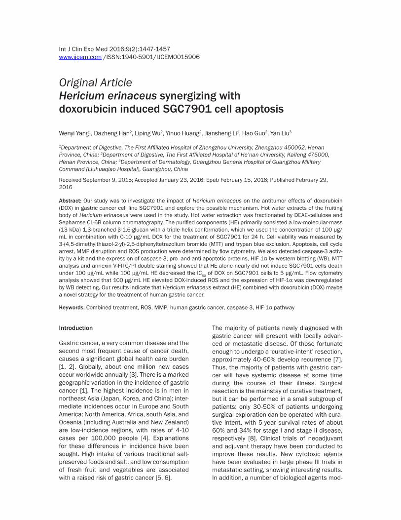
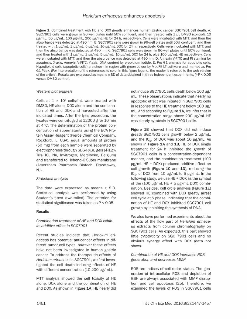
MTT analysis showed the cell toxicity of HE alone, DOX alone and the combination of HE and DOX. As shown in Figure 1A, HE nearly did
not induce SGC7901 cells death below 100 µg/mL. These observations indicate that nearly no apoptotic effect was initiated in SGC7901 cells in response to the HE treatment below 100 µg/mL. And according to Figure 1A, we infer that in the concentration range above 200 µg/mL HE was clearly cytotoxic in SGC7901 cells.
Figure 1B showed that DOX did not induce greatly SGC7901 cells growth below 2 µg/mL, and the IC50 of DOX was about 10 µg/mL. As shown in Figure 1A and 1B, HE or DOX single treatment for 24 h inhibited the growth of SGC7901 cells in a concentration-dependent manner, and the combination treatment (100 µg/mL HE + DOX) produced additive effect on cell growth (Figure 1C and 1D), reducing the IC50 of DOX from 10 µg/mL to 5 µg/mL. In the following study, we use HE + DOX as the symbol of the (100 µg/mL HE + 5 µg/mL DOX) combi-nation. Besides, cell cycle analysis (Figure 1E) showed HE combined with DOX greatly arrest cell cycle at S phase, indicating that the combi-nation of HE and DOX inhibited SGC7901 cell growth by inhibiting the synthesis of DNA.
We also have performed experiments about the effects of the flow part of Hericium erinace- us extracts from column chromatography on SGC7901 cells. As expected, this part showed little cytotoxicity on SGC 7901 cells and no obvious synergy effect with DOX (data not show).
Combination of HE and DOX increases ROS generation and decreases MMP
ROS are indices of cell redox status. The gen-eration of intracellular ROS and depletion of GSH are always associated with MMP disrup-tion and cell apoptosis [25]. Therefore, we examined the levels of ROS in SGC7901 cells
Figure 1. Combined treatment with HE and DOX greatly enhances human gastric cancer SGC7901 cell death. A. SGC7901 cells were grown in 96-well plates until 50% confluent, and then treated with 1 µL DMSO (control), 10 µg/mL, 50 µg/mL, 100 µg/mL, 200 µg/mL HE for 24 h, respectively. Cells were incubated with MTT, and then the absorbance was detected at 490 nm. B. SGC7901 cells were grown in 96-well plates until 50% confluent, and then treated with 1 µg/mL, 2 µg/mL, 5 µg/mL, 10 µg/mL DOX for 24 h, respectively. Cells were incubated with MTT, and then the absorbance was detected at 490 nm. C. SGC7901 cells were grown in 96-well plates until 50% confluent, and then treated with 1 µg/mL, 2 µg/mL, 5 µg/mL, 10 µg/mL DOX for 24 h, plus 100 µg/mL HE respectively. Cells were incubated with MTT, and then the absorbance was detected at 490 nm. D. Annexin V-FITC and PI staining for apoptosis. X-axis, Annexin V-FITC; Y-axis, DNA content by propidium iodide. E. Pro G1 analysis for apoptotic cells. Hypodiploid cells (apoptotic cells) are shown in region with green colour by ModFit LT software and marked as Pro G1 Peak. (For interpretation of the references to color in this figure legend, the reader is referred to the web version of the article). Results are expressed as means ± SD of data obtained in three independent experiments. (*P < 0.05 versus DMSO control).
Hericium erinaceus enhances apoptosis
1452 Int J Clin Exp Med 2016;9(2):1447-1457
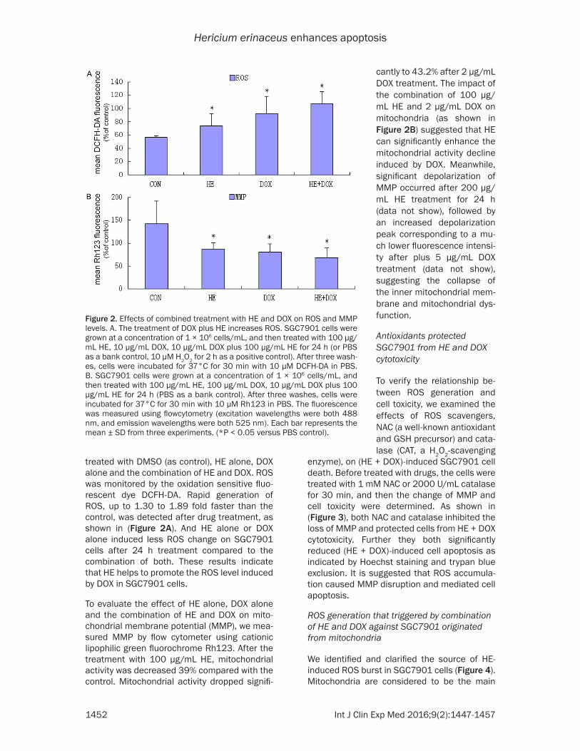
treated with DMSO (as control), HE alone, DOX alone and the combination of HE and DOX. ROS was monitored by the oxidation sensitive fluo-rescent dye DCFH-DA. Rapid generation of ROS, up to 1.30 to 1.89 fold faster than the control, was detected after drug treatment, as shown in (Figure 2A). And HE alone or DOX alone induced less ROS change on SGC7901 cells after 24 h treatment compared to the combination of both. These results indicate that HE helps to promote the ROS level induced by DOX in SGC7901 cells.
To evaluate the effect of HE alone, DOX alone and the combination of HE and DOX on mito-chondrial membrane potential (MMP), we mea-sured MMP by flow cytometer using cationic lipophilic green fluorochrome Rh123. After the treatment with 100 µg/mL HE, mitochondrial activity was decreased 39% compared with the control. Mitochondrial activity dropped signifi-
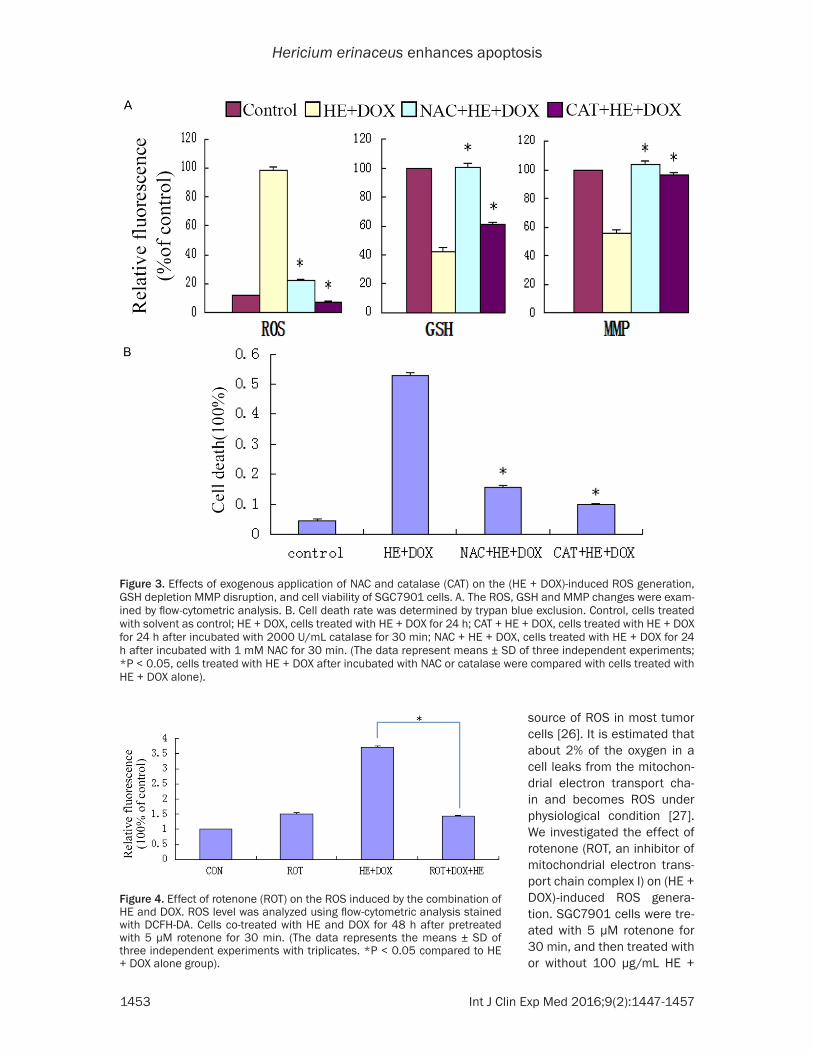
enzyme), on (HE + DOX)-induced SGC7901 cell death. Before treated with drugs, the cells were treated with 1 mM NAC or 2000 U/mL catalase for 30 min, and then the change of MMP and cell toxicity were determined. As shown in (Figure 3), both NAC and catalase inhibited the loss of MMP and protected cells from HE + DOX cytotoxicity. Further they both significantly reduced (HE + DOX)-induced cell apoptosis as indicated by Hoechst staining and trypan blue exclusion. It is suggested that ROS accumula-tion caused MMP disruption and mediated cell apoptosis.
ROS generation that triggered by combination of HE and DOX against SGC7901 originated from mitochondria
We identified and clarified the source of HE- induced ROS burst in SGC7901 cells (Figure 4). Mitochondria are considered to be the main
Figure 2. Effects of combined treatment with HE and DOX on ROS and MMP levels. A. The treatment of DOX plus HE increases ROS. SGC7901 cells were grown at a concentration of 1 × 10⁶ cells/mL, and then treated with 100 µg/mL HE, 10 µg/mL DOX, 10 µg/mL DOX plus 100 µg/mL HE for 24 h (or PBS as a bank control, 10 µM H2O2 for 2 h as a positive control). After three wash-es, cells were incubated for 37°C for 30 min with 10 µM DCFH-DA in PBS. B. SGC7901 cells were grown at a concentration of 1 × 10⁶ cells/mL, and then treated with 100 µg/mL HE, 100 µg/mL DOX, 10 µg/mL DOX plus 100 µg/mL HE for 24 h (PBS as a bank control). After three washes, cells were incubated for 37°C for 30 min with 10 µM Rh123 in PBS. The fluorescence was measured using flowcytometry (excitation wavelengths were both 488 nm, and emission wavelengths were both 525 nm). Each bar represents the mean ± SD from three experiments. (*P < 0.05 versus PBS control).
cantly to 43.2% after 2 µg/mL DOX treatment. The impact of the combination of 100 µg/mL HE and 2 µg/mL DOX on mitochondria (as shown in Figure 2B) suggested that HE can significantly enhance the mitochondrial activity decline induced by DOX. Meanwhile, significant depolarization of MMP occurred after 200 µg/mL HE treatment for 24 h (data not show), followed by an increased depolarization peak corresponding to a mu- ch lower fluorescence intensi-ty after plus 5 µg/mL DOX treatment (data not show), suggesting the collapse of the inner mitochondrial mem-brane and mitochondrial dys- function.
Antioxidants protected SGC7901 from HE and DOX cytotoxicity
To verify the relationship be- tween ROS generation and cell toxicity, we examined the effects of ROS scavengers, NAC (a well-known antioxidant and GSH precursor) and cata-lase (CAT, a H2O2-scavenging
Hericium erinaceus enhances apoptosis
1453 Int J Clin Exp Med 2016;9(2):1447-1457
source of ROS in most tumor cells [26]. It is estimated that about 2% of the oxygen in a cell leaks from the mitochon-drial electron transport cha- in and becomes ROS under physiological condition [27]. We investigated the effect of rotenone (ROT, an inhibitor of mitochondrial electron trans-port chain complex I) on (HE + DOX)-induced ROS genera-tion. SGC7901 cells were tre- ated with 5 µM rotenone for 30 min, and then treated with or without 100 µg/mL HE +
Figure 3. Effects of exogenous application of NAC and catalase (CAT) on the (HE + DOX)-induced ROS generation, GSH depletion MMP disruption, and cell viability of SGC7901 cells. A. The ROS, GSH and MMP changes were exam-ined by flow-cytometric analysis. B. Cell death rate was determined by trypan blue exclusion. Control, cells treated with solvent as control; HE + DOX, cells treated with HE + DOX for 24 h; CAT + HE + DOX, cells treated with HE + DOX for 24 h after incubated with 2000 U/mL catalase for 30 min; NAC + HE + DOX, cells treated with HE + DOX for 24 h after incubated with 1 mM NAC for 30 min. (The data represent means ± SD of three independent experiments; *P < 0.05, cells treated with HE + DOX after incubated with NAC or catalase were compared with cells treated with HE + DOX alone).
Figure 4. Effect of rotenone (ROT) on the ROS induced by the combination of HE and DOX. ROS level was analyzed using flow-cytometric analysis stained with DCFH-DA. Cells co-treated with HE and DOX for 48 h after pretreated with 5 µM rotenone for 30 min. (The data represents the means ± SD of three independent experiments with triplicates. *P < 0.05 compared to HE + DOX alone group).
Hericium erinaceus enhances apoptosis
1454 Int J Clin Exp Med 2016;9(2):1447-1457
DOX for 2 h. Compared with the 2.56-fold in- crease without any pretreatment, the treat-ment with rotenone blocked the (HE + DOX)-induced ROS generation significantly (Figure 4). This result suggested that HE triggered the gen-eration of ROS by the mitochondria.
Combination of HE and DOX induces SGC7901 apoptosis in caspase-dependent pathway and in part through inhibition of HIF-1 pathway
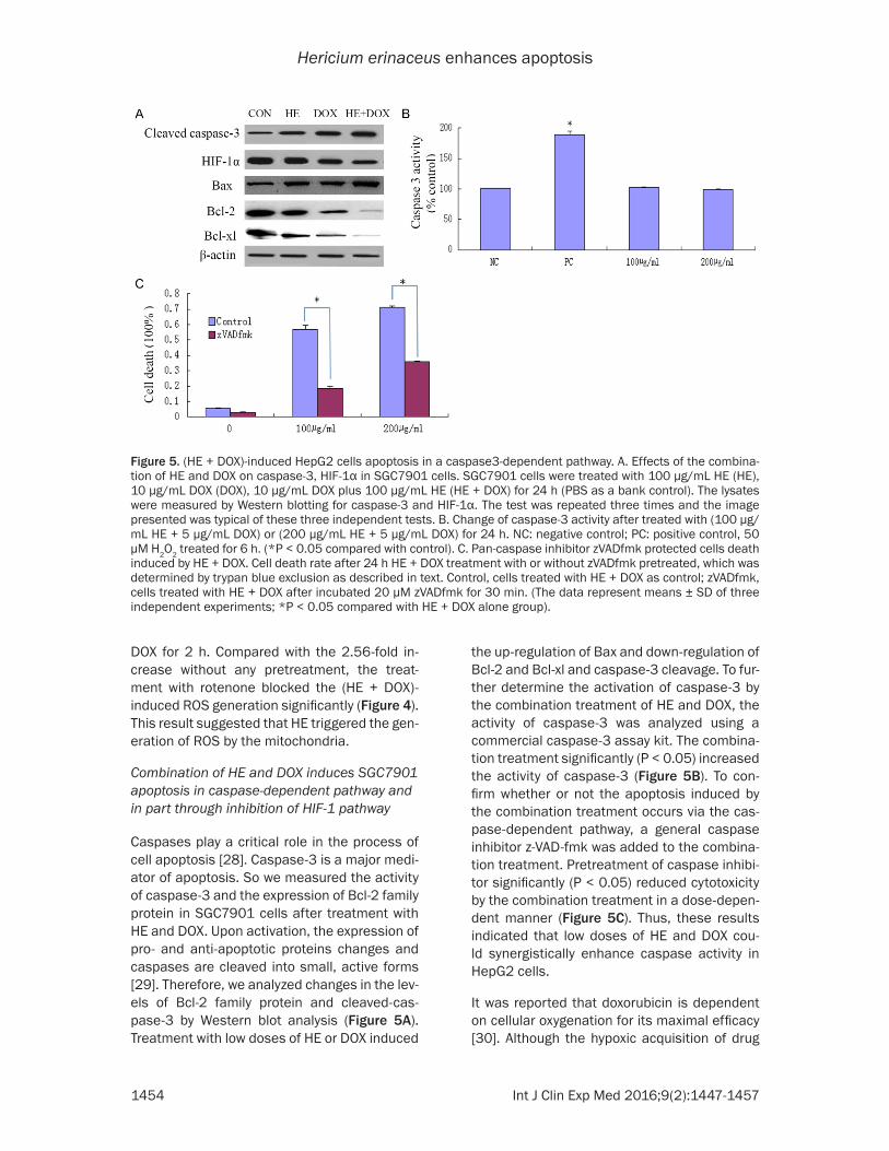
Caspases play a critical role in the process of cell apoptosis [28]. Caspase-3 is a major medi-ator of apoptosis. So we measured the activity of caspase-3 and the expression of Bcl-2 family protein in SGC7901 cells after treatment with HE and DOX. Upon activation, the expression of pro- and anti-apoptotic proteins changes and caspases are cleaved into small, active forms [29]. Therefore, we analyzed changes in the lev-els of Bcl-2 family protein and cleaved-cas-pase-3 by Western blot analysis (Figure 5A). Treatment with low doses of HE or DOX induced
the up-regulation of Bax and down-regulation of Bcl-2 and Bcl-xl and caspase-3 cleavage. To fur-ther determine the activation of caspase-3 by the combination treatment of HE and DOX, the activity of caspase-3 was analyzed using a commercial caspase-3 assay kit. The combina-tion treatment significantly (P < 0.05) increased the activity of caspase-3 (Figure 5B). To con-firm whether or not the apoptosis induced by the combination treatment occurs via the cas-pase-dependent pathway, a general caspase inhibitor z-VAD-fmk was added to the combina-tion treatment. Pretreatment of caspase inhibi-tor significantly (P < 0.05) reduced cytotoxicity by the combination treatment in a dose-depen-dent manner (Figure 5C). Thus, these results indicated that low doses of HE and DOX cou- ld synergistically enhance caspase activity in HepG2 cells.
It was reported that doxorubicin is dependent on cellular oxygenation for its maximal efficacy [30]. Although the hypoxic acquisition of drug
Figure 5. (HE + DOX)-induced HepG2 cells apoptosis in a caspase3-dependent pathway. A. Effects of the combina-tion of HE and DOX on caspase-3, HIF-1α in SGC7901 cells. SGC7901 cells were treated with 100 µg/mL HE (HE), 10 µg/mL DOX (DOX), 10 µg/mL DOX plus 100 µg/mL HE (HE + DOX) for 24 h (PBS as a bank control). The lysates were measured by Western blotting for caspase-3 and HIF-1α. The test was repeated three times and the image presented was typical of these three independent tests. B. Change of caspase-3 activity after treated with (100 µg/mL HE + 5 µg/mL DOX) or (200 µg/mL HE + 5 µg/mL DOX) for 24 h. NC: negative control; PC: positive control, 50 µM H2O2 treated for 6 h. (*P < 0.05 compared with control). C. Pan-caspase inhibitor zVADfmk protected cells death induced by HE + DOX. Cell death rate after 24 h HE + DOX treatment with or without zVADfmk pretreated, which was determined by trypan blue exclusion as described in text. Control, cells treated with HE + DOX as control; zVADfmk, cells treated with HE + DOX after incubated 20 µM zVADfmk for 30 min. (The data represent means ± SD of three independent experiments; *P < 0.05 compared with HE + DOX alone group).
Hericium erinaceus enhances apoptosis
1455 Int J Clin Exp Med 2016;9(2):1447-1457
resistance has been studied with respect to stimulus for some genes, which has been asso-ciated with drug resistance [31, 32]. However, the key transcriptional factors that modulate drug-resistance-associated proteins remain to be identified. One possible protein is HIF-1α. HIF-1α is the major transcriptional factor that is significantly activated by hypoxia, and then transactivates many genes involved in tumor development. Recent experimental studies sh- owed that HIF-1α may be the possible factor involved in drug resistance acquired by hypoxia [33]. Our study tested the expression of HIF-1α in SGC7901 cells after treatment with the com-bination of HE and DOX. Figure 5A shows that HIF-1α decreases significantly, indicating HE synergizes with DOX and HE impairs drug resis-tance of SGC7901 against DOX in part through HIF-1α pathway.
Discussion
Many food have an ability of curing diseases in accordance with an old Chinese saying that medicine and food are from the same source. Hericium erinaceus is a traditional edible food, which has many activities to be elucidated. DOX is a commonly used anticancer drug, but has side effects like cardiac cytotoxicity, limiting its comprehensive use. Cancer progression is par-tially affected by the development of cancer cell resistance to apoptosis induced by chemo-therapeutic drugs like DOX. Here in our study, we combined HE with low doses of DOX in the treatment of SGC7901 gastric cells, presenting an excellent synergistic effect. Besides, our strategy gives us an outstanding idea of the therapy of gastric cancer using edible food like Hericium erinaceus.
In this study, we showed that HE and low doses of DOX synergize to induce apoptosis in human gastric cancer SGC7901 cells. Caspase-3 was activated in the apoptotic process induced by the combination treatment of HE and DOX. The suppression of caspase activity by a general caspase inhibitor z-VAD-fmk confirmed that the promotion of apoptosis by combination treat-ment involved a caspase-dependent pathway.
ROS play a pivotal role in apoptosis. The apop-totic effect of HE combined with DOX on SGC7901 cells was associated with an early elevated level of intracellular ROS. Moreover, in order to confirm that the apoptotic effect of HE
combined with DOX was mediated by ROS, anti-oxidants NAC and catalase were used. Both NAC and GSH inhibited the cytotoxicity of HE and DOX associated with suppressed ROS gen-eration and GSH depletion. And they both can ensure cell survival as shown by trypan blue exclusion and significantly reduced cell apopto-sis as demonstrated by Hoechst staining. Our results also indicated that HE combined with DOX induced a decrease of MMP in SGC7901 cells. In addition, concerning the time course of ROS burst and MMP depolarization, the ROS burst occurred before intracellular MMP depo-larization. This opinion was supported by the results obtained from SGC7901 cells co-incu-bated with HE and DOX and antioxidant NAC or catalase. Both NAC and catalase blocked the MMP depolarization completely. These results demonstrated that the ROS burst was a prereq-uisite for MMP collapse and cell death induced by the combination of HE and DOX.
Importantly, many cancer cells have the ability of resistance against chemotherapy drugs. Here we also found that HE can impair DOX resistance of SGC7901 cells by decreasing the expression of HIF-1α, thus enhancing the utility of DOX in therapy of human gastric cancer.
In conclusion, the apoptotic effect of the combi-nation of HE and DOX on SGC7901 cells is mediated by ROS and relied on caspases-cas-cade. Lower doses of HE or DOX alone had minor cytotoxic effect on SGC7901 cells, while the combination of low doses of both of them has significant apoptotic effect, showing a syn-ergistic effect of Hericium erinaceus.
Disclosure of conflict of interest
None.
Address correspondence to: Dr. Jiansheng Li, De- partment of Digestive, The First Affiliated Hospital of Zhengzhou University, No. 1 of Two Seven District Construction Road, Zhengzhou 450052, Henan Province, China. Tel: 0371-66862052; E-mail: [email protected]
References
[1] Hartgrink HH, Jansen EP, van Grieken NC and van de Velde CJ. Gastric cancer. Lancet 2009; 374: 477-490.
[2] Mocellin S, Verdi D, Pooley KA and Nitti D. Genetic variation and gastric cancer risk: a
Hericium erinaceus enhances apoptosis
1456 Int J Clin Exp Med 2016;9(2):1447-1457
field synopsis and meta-analysis. Gut 2015; 64: 1209-1219.
[3] Power DG, Kelsen DP and Shah MA. Advanced gastric cancer-Slow but steady progress. Can- cer Treat Revi 2010; 36: 384-392.
[4] Yamaoka Y, Kato M and Asaka M. Geographic Differences in Gastric Cancer Incidence Can be Explained by Differences between Helico- bacter pylori Strains. Intern Med 2008; 47: 1077-1083.
[5] Tsugane S and Sasazuki S. Diet and the risk of gastric cancer: review of epidemiological evi-dence. Gastric Cancer 2007; 10: 75-83.
[6] Wada K, Tsuji M, Tamura T, Konishi K, Kawachi T, Hori A, Tanabashi S, Matsushita S, Tokimitsu N and Nagata C. Soy isoflavone intake and stomach cancer risk in Japan: From the Takayama study. Int J Cancer 2015; 137: 885-892.
[7] Jung JJ, Cho JH, Shin S and Shim YM. Surgical treatment of anastomotic recurrence after gastrectomy for gastric cancer. Korean J Thorac Cardiovasc Surg 2014; 47: 269-274.
[8] Kim MG, Lee JH, Ha TK and Kwon SJ. The dis-tance of proximal resection margin dose not significantly influence on the prognosis of gas-tric cancer patients after curative resection. Ann Surg Treat Res 2014; 87: 223-231.
[9] Ikekawa T, Uehara N, Maeda Y, Nakanishi M and Fukuoka F. Antitumor activity of aqueous extracts of edible mushrooms. Cancer Res 1969; 29: 734-735.
[10] Wasser SP. Medicinal mushrooms as a source of antitumor and immunomodulating polysac-charides. Appl Microbiol Biotechnol 2002; 60: 258-274.
[11] Ferreira IC, Vaz JA, Vasconcelos MH and Martins A. Compounds from wild mushrooms with antitumor potential. Anticancer Agents Med Chem 2010; 10: 424-436.
[12] Zou Y, Xiong H, Xiong H, Lu T, Zhu F, Luo Z, Yuan X and Wang Y. A polysaccharide from mush-room Huaier retards human hepatocellular carcinoma growth, angiogenesis, and metasta-sis in nude mice. Tumour Biol 2015; 36: 2929-2936.
[13] Kim SP, Kang MY, Choi YH, Kim JH, Nam SH, Friedman M. Mechanism of Hericium eri-naceus (Yamabushitake) mushroom-induced apoptosis of U937 human monocytic leukemia cells. Food Funct 2011; 2: 348-356.
[14] Geng Y, Zhu S, Lu Z, Xu H, Shi JS and Xu ZH. Anti-inflammatory activity of mycelial extracts from medicinal mushrooms. Int J Med Mush- rooms 2014; 16: 319-325.
[15] Wang M, Gao Y, Xu D, Konishi T and Gao Q. Hericium erinaceus (Yamabushitake): a unique resource for developing functional foods and medicines. Food Funct 2014; 5: 3055-3064.
[16] Kelsen D. The use of chemotherapy in the treatment of advanced gastric and pancreas cancer. Semin Oncol 1994; 21: 58-66.
[17] Konishi T, Teruya M, Kawahara M, Itoh A, Asakura R, Araki S, Hojo K, Nouchi T and Takeda Y. [Chemotherapy of gastric cancer]. Gan To Kagaku Ryoho 1998; 25: 504-515.
[18] Schipper DL and Wagener DJ. Chemotherapy of gastric cancer. Anticancer Drugs 1996; 7: 137-149.
[19] Lee JS and Hong EK. Hericium erinaceus en-hances doxorubicin-induced apoptosis in hu-man hepatocellular carcinoma cells. Cancer Lett 2010; 297: 144-154.
[20] Lee JS, Min KM, Cho JY and Hong EK. Study of macrophage activation and structural charac-teristics of purified polysaccharides from the fruiting body of Hericium erinaceus. J Microbiol Biotechnol 2009; 19: 951-959.
[21] Liang YF, Qing Y, Du QQ, Fan P, Xu YP, Xu HG and Shi N. [Study on mechanism of trimethyl-tin chloride-induced apoptosis in PC12 cells]. Zhonghua Lao Dong Wei Sheng Zhi Ye Bing Za Zhi 2012; 30: 816-819.
[22] Yan H, Wang X, Niu J, Wang Y, Wang P and Liu Q. Anti-cancer effect and the underlying me- chanisms of gypenosides on human colorec- tal cancer SW-480 cells. PLoS One 2014; 9: e95609.
[23] Zhang ZF, Zheng LG, Zhao ZQ, Shi J, Wang X and Huang J. Grape seed proanthocyanidins inhibit H2O2-induced osteoblastic MC3T3-E1 cell apoptosis via ameliorating H2O2-induced mitochondrial dysfunction. J Toxicol Sci 2014; 39: 803-813.
[24] Hartree EF. Determination of protein: a modifi-cation of the Lowry method that gives a linear photometric response. Anal Biochem 1972; 48: 422-427.
[25] Leon IE, Di Virgilio AL, Porro V, Muglia CI, Naso LG, Williams PA, Bollati-Fogolin M and Etcheverry SB. Antitumor properties of a vanadyl(IV) complex with the flavonoid chrysin [VO(chrysin)2EtOH]2 in a human osteosarco-ma model: the role of oxidative stress and apoptosis. Dalton Trans 2013; 42: 11868-11880.
[26] Li J, Xu Z, Tan M, Su W and Gong XG. 3-(4- (Benzo[d]thiazol-2-yl)-1-phenyl-1H-pyrazol-3-yl) phenyl acetate induced Hep G2 cell apoptosis through a ROS-mediated pathway. Chem Biol Interact 2010; 183: 341-348.
[27] Inoue M, Sato EF, Nishikawa M, Park AM, Kira Y, Imada I and Utsumi K. Mitochondrial genera-tion of reactive oxygen species and its role in aerobic life. Curr Med Chem 2003; 10: 2495-2505.
[28] Banerjee P, Chander V and Bandyopadhyay A. Balancing functions of annexin A6 maintain
Hericium erinaceus enhances apoptosis
1457 Int J Clin Exp Med 2016;9(2):1447-1457
equilibrium between hypertrophy and apopto-sis in cardiomyocytes. Cell Death Dis 2015; 6: e1873.
[29] Hamacher-Brady A and Brady NR. Bax/Bak-dependent, Drp1-independent Targeting of X-linked Inhibitor of Apoptosis Protein (XIAP) into Inner Mitochondrial Compartments Co- unteracts Smac/DIABLO-dependent Effector Caspase Activation. J Biol Chem 2015; 290: 22005-22018.
[30] Teicher BA. Hypoxia and drug resistance. Cancer Metastasis Rev 1994; 13: 139-168.
[31] Black JC, Atabakhsh E, Kim J, Biette KM, Van Rechem C, Ladd B, Burrowes PD, Donado C, Mattoo H, Kleinstiver BP, Song B, Andriani G, Joung JK, Iliopoulos O, Montagna C, Pillai S, Getz G and Whetstine JR. Hypoxia drives tran-sient site-specific copy gain and drug-resistant gene expression. Genes Dev 2015; 29: 1018-1031.
[32] Marhold M, Tomasich E, El-Gazzar A, Heller G, Spittler A, Horvat R, Krainer M and Horak P. HIF1 alpha Regulates mTOR Signaling and Viability of Prostate Cancer Stem Cells. Mol Cancer Res 2015; 13: 556-564.
[33] Borsi E, Terragna C, Brioli A, Tacchetti P, Martello M and Cavo M. Therapeutic targeting of hypoxia and hypoxia-inducible factor 1 alpha in multiple myeloma. Transl Res 2015; 165: 641-650.