original article human pluripotent stem cell-derived … · original article human pluripotent stem...
TRANSCRIPT
ORIGINAL ARTICLE
Human pluripotent stem cell-derived acinar/ductalorganoids generate human pancreas uponorthotopic transplantation and allow diseasemodellingMeike Hohwieler,1 Anett Illing,1 Patrick C Hermann,1 Tobias Mayer,1
Marianne Stockmann,1 Lukas Perkhofer,1 Tim Eiseler,1 Justin S Antony,2
Martin Müller,1 Susanne Renz,1 Chao-Chung Kuo,3 Qiong Lin,4 Matthias Sendler,5
Markus Breunig,1 Susanne M Kleiderman,1 André Lechel,1 Martin Zenker,6
Michael Leichsenring,7 Jonas Rosendahl,8 Martin Zenke,4 Bruno Sainz Jr,9
Julia Mayerle,5 Ivan G Costa,3 Thomas Seufferlein,1 Michael Kormann,2
Martin Wagner,1 Stefan Liebau,10 Alexander Kleger1
ABSTRACTObjective The generation of acinar and ductal cellsfrom human pluripotent stem cells (PSCs) is a poorlystudied process, although various diseases arise fromthis compartment.Design We designed a straightforward approach todirect human PSCs towards pancreatic organoidsresembling acinar and ductal progeny.Results Extensive phenotyping of the organoids notonly shows the appropriate marker profile but alsoultrastructural, global gene expression and functionalhallmarks of the human pancreas in the dish. Uponorthotopic transplantation into immunodeficient mice,these organoids form normal pancreatic ducts and acinartissue resembling fetal human pancreas without evidenceof tumour formation or transformation. Finally, weimplemented this unique phenotyping tool as a model tostudy the pancreatic facets of cystic fibrosis (CF). For thefirst time, we provide evidence that in vitro, but also inour xenograft transplantation assay, pancreaticcommitment occurs generally unhindered in CF.Importantly, cystic fibrosis transmembrane conductanceregulator (CFTR) activation in mutated pancreaticorganoids not only mirrors the CF phenotype infunctional assays but also at a global expression level.We also conducted a scalable proof-of-concept screen inCF pancreatic organoids using a set of CFTR correctorsand activators, and established an mRNA-mediated genetherapy approach in CF organoids.Conclusions Taken together, our platform providesnovel opportunities to model pancreatic disease anddevelopment, screen for disease-rescuing agents and totest therapeutic procedures.
INTRODUCTIONGiven their capacity to differentiate into every celltype of the human body, human-induced pluripo-tent stem cells (hiPSCs) provide a unique platformfor developmental studies and regenerative medi-cine.1–4 The generation of pancreatic progenitor
Significance of this study
What is already known on this subject?▸ Human pluripotent stem cells (PSCs) present a
powerful tool for developmental studies andregenerative medicine.
▸ Directed differentiation of PSCs towardspancreatic cell fates requires the formation ofPDX1/NKX6.1-positive progenitor cells.
▸ Mutations in the cystic fibrosis transmembraneconductance regulator (CFTR) perturb fluidtransport causing chronic airway infections orbiliary cirrhosis with variable phenotypes whilethe pancreas is one of the first organs affected.
What are the new findings?▸ Efficient generation of high yields of pancreatic
progenitors from several human pluripotentstem cell lines.
▸ PSC-derived pancreatic progenitors formpancreatic organoids that comprise acinar/ductal-like progeny and resemble humanpancreas upon orthotopic transplantation inmice.
▸ Induced PSCs from cystic fibrosis patientsdisplay normal pancreatic commitment in vitroand in vivo at least until a fetal developmentalstage.
▸ Pancreatic organoids from patients with cysticfibrosis recapitulate defective CFTR function invitro, allowing subsequent drug screening butalso mRNA-mediated gene supplementation.
How might it impact on clinical practice inthe foreseeable future?▸ Our system provides a novel approach to model
human pancreatic development and disease.▸ Humanised platform for (organ-specific and
patient-specific) drug screening and testing oftherapeutic options in vitro and in vivo.
To cite: Hohwieler M, Illing A, Hermann PC, et al. Gut 2017;66:473–486.
► Additional material is published online only. To view please visit the journal online (h t t p : / / d x . d o i . o r g / 1 0 . 1 1 3 6 / g u t j n l - 2 0 1 6 - 3 1 2 4 2 3 )
For numbered affiliations see end of article.
Correspondence toProf Dr Alexander Kleger, Department of Internal Medicine I, University Medical Center Ulm, Albert-Einstein-Allee 23, Ulm 89081, Germany; [email protected]
SL and AK jointly supervised this work and contributed equally.
Received 13 June 2016Accepted 11 August 2016Published Online First 7 October 2016
Pancreas
► http://dx.doi.org/10.1136/gutjnl-2016-312865
473Hohwieler M, et al. Gut 2017;66:473–486. doi:10.1136/gutjnl-2016-312423
on April 21, 2020 by guest. P
rotected by copyright.http://gut.bm
j.com/
Gut: first published as 10.1136/gutjnl-2016-312423 on 15 S
eptember 2016. D
ownloaded from
(PP) cells from PSCs follows the sequential induction of virtu-ally pure definitive endoderm (DE), foregut endoderm (GTE)and pancreatic endoderm (PE, figure 1A).5–7 Over the lastdecade, a series of studies have aimed at improving pancreaticdifferentiation protocols.5–7 While most studies focused on thegeneration of PDX1-positive PE,8 9 true PP cells should coex-press both NKX6.1 and PDX1.10 While the exocrine and endo-crine lineages develop, NKX6.1 is still expressed in β cells, butbecomes mutually exclusive with the expression of Ptf1a drivingthe exocrine lineage.11 Thus, the presence of NKX6.1 is one ofthe key distinguishing features of these two lineages and hencecan be used to monitor the emergence of true progenitors.Despite recent progress in differentiating PSCs towards endo-crine pancreatic progeny,12 13 the generation of ductal andexocrine-like cells has not yet been adequately achieved, apartfrom a recent study modelling human pancreatic cancer.14
Three-dimensional organoid models generated from PSCs canfaithfully model the in vivo situation,13 15 16 and disease-specificiPSCs allow the generation of distinct human diseasemodels.12 13 15 Nevertheless, the generation of human pancreasin mice upon xenotransplantation of non-transformed organoidshas not been achieved to date. However, this would open upentirely new research avenues. In addition, inherited pancreaticdiseases would benefit from in vivo gene supplementation strat-egies as recently shown for a lung disease with specifically modi-fied mRNA, pre-evaluated in organoids.17 18 To tackle thisunmet need in the pancreatic field, we describe herein a newPSC-based organoid system that was used to model pancreaticaspects of cystic fibrosis (CF).
CF is an inherited disease caused by either nonsense or mis-sense mutations in the cystic fibrosis transmembrane conduct-ance regulator (CFTR) gene, resulting in complete absence ofthe protein, a misfolded polypeptide that is degraded by theunfolded protein response, or a dysfunctional protein.19 CFTRencodes a chloride channel gated by cyclic AMP-dependentphosphorylation that is necessary for electrolyte and fluidhomeostasis of epithelia in various organs including the lung,liver, intestine and pancreas. Dysfunction of the CFTR leads tothe production of hyperviscous mucus causing chronic airwayinfections or biliary cirrhosis with variable phenotypes.20
Although the pancreas is one of the first organs affected, knowl-edge about the pathophysiology of the pancreas during CF islimited. Briefly, distinct CFTR genotypes have been shown tonot only increase the probability of developing either pancrea-titis or perinatal exocrine insufficiency21 but also pancreaticcancer.22 Moreover, exocrine insufficiency drives a complex andpoorly understood cascade of events leading to endocrineexhaustion.23 Additionally, the expression of the CFTR geneduring early pancreatic development suggests that CFTR muta-tions could have a developmental impact;24 however, currentCF animal models recapitulate only limited aspects of thehuman disease sparing the pancreas, and in vitro studies havebeen hampered by the lack of primary human pancreaticprogeny. Moreover, with increasing CF patient survival pancre-atic manifestations of CF are becoming progressively more clin-ically relevant but the underlying pathomechanisms remain tobe explored. Thus, innovative model systems for pancreatic CFand other pancreatic disorders are clearly warranted.
MATERIALS AND METHODSDifferentiation of human PSCs into PP cellsFor differentiation, human PSCs were grown to 95% confluenceon growth factor-reduced matrigel (BD, 354 230) and FTDAmedium25 was refreshed 3 hours before initiating
differentiation. The backbone medium for the first 6 days of dif-ferentiation was BE1: MCDB131 (Invitrogen) with 0.8 g/L cellculture tested glucose (Sigma), 1.174 g/L sodium bicarbonate(Sigma), 0.1% fatty acid free (FAF) BSA (A7030, Sigma), 2 mML-glutamine. Later differentiation was performed in BE3 as thebackbone medium: MCDB131 with 0.44 g/L glucose, 1.754 g/Lsodium bicarbonate, 2% FAF-BSA, 2 mM L-glutamine, 44 mg/LL-ascorbic acid, 0.5× insulin-transferrin-selenium-ethanolamine(ITS-X). Cells in differentiation were cultured at 37°C in a 5%CO2 incubator with daily media change. For the first day of dif-ferentiation, cells were washed with phosphate buffered saline(PBS) (Sigma) and incubated with BE1 supplemented with3 mM GSK3β-inhibitor (CHIR99021) (Axon MedChem) and100 ng/mL Activin A. The next day the medium was replacedby BE1 with 100 ng/mL Activin A. After 3 days, media waschanged to BE1 with 50 ng/mL KFG (Peprotech) for 2 days.From day 6 until day 10, cells were cultured in BE3 mediumcontaining 0.25 mM SANT-1 (Sigma), 2 mM retinoic acid(Sigma), 200 nM LDN-193189 (Sigma) and 500 nMPD0325901 (Calbiochem). At days 10–14, the cells receivedBE3 supplemented with 50 ng/mL fibroblast growth factor(FGF10) (Peprotech), 330 nM Indolactam V (Stem CellTechnologies), 10 mM SB431542 (Axon MedChem) and add-itional 16 mM glucose. An outline of the differentiation proto-col can also be found in figure 1A.
3D pancreatic organoid cultureAt the PP stage (day 12 of differentiation), cells were washedwith PBS, incubated with TrypLE at 37°C for 5–6 min and care-fully resuspended in DMEM/F12 resulting in clumps of 3–10cells. After centrifugation at 400 g for 5 min, the pellet waswashed in BE3 medium, centrifuged again and resuspended inprecooled day 12 medium (detailed above) supplemented with10 mM Rock inhibitor. The cell suspension was mixed on icewith growth factor reduced matrigel at a 1:3 ratio and 25 mLwere transferred to a 48-well plate (NunclonΔSurface).Following incubation for 10 min at 37°C, the solidified drop ofmatrigel was overlayed with 200 mL of day 12 medium (seeabove) containing 10 mM Rock inhibitor (always added for thefirst 4 days in three-dimensional (3D) culture), which wasreplaced the next day. On day 14, differentiation was continuedwith 10 ng/mL FGF2 and 10 mM Rock inhibitor in BE3medium. From day 18 on, organoids were cultured in BE3 with10 ng/mL FGF2 and 10 mM nicotinamide (NA) (Sigma)(referred to as ‘FN’). Medium was changed every 2–3 days.Alternatively, organoids were generated by replating PPs in sus-pension in ultra-low-attachment plates (Corning). To preventaggregation, cell clusters were triturated after 1 hour and againafter 2 days. For changing media, half of the fluid was pipettedoff and replaced with fresh differentiation media. Medium con-ditions were identical to the matrigel-based culture. For suspen-sion cultures, another medium (referred to as ‘FEPC’) wastested (based on conditions published for mouse embryonicPPs26) composed of DMEM/F12, 10% knockout serum replace-ment (KOSR) and 0.1 mM β-mercaptoethanol supplemented with50 ng/mL FGF10, 25 ng/mL epidermal growth factor (EGF)(novoprotein), 2 mM CHIR99021 and 16 nM phorbol myristateacetate (Sigma). For passaging, matrigel was scraped off, pipettedin order to mechanically dissociate the organoids into smallclumps, collected in a 15 mL falcon, and further processed asdescribed above. Organoids were passaged every 10–14 days andcultured at 5% CO2 and 37°C.
474 Hohwieler M, et al. Gut 2017;66:473–486. doi:10.1136/gutjnl-2016-312423
Pancreas on A
pril 21, 2020 by guest. Protected by copyright.
http://gut.bmj.com
/G
ut: first published as 10.1136/gutjnl-2016-312423 on 15 Septem
ber 2016. Dow
nloaded from
Functional CFTR assayOrganoids were split 1:4 and 8 mL of the cell/matrigel suspen-sion was transferred to the inner border of each well of a96-well plate (NunclonΔSurface), solidified for 5 min and
100 mL culture medium was added. Six days after seeding, orga-noids were incubated with 20 mM forskolin (FSK) and 100 mM3-isobutyl-1-methylxanthine (IBMX) (or 1:500 dimethyl sulfox-ide (DMSO) (all from Sigma) as negative control) in FN
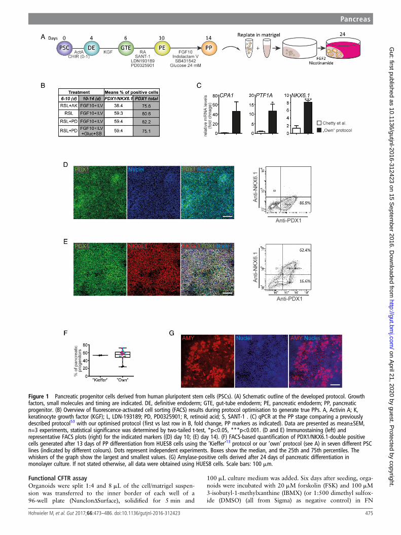
Figure 1 Pancreatic progenitor cells derived from human pluripotent stem cells (PSCs). (A) Schematic outline of the developed protocol. Growthfactors, small molecules and timing are indicated. DE, definitive endoderm; GTE, gut-tube endoderm; PE, pancreatic endoderm; PP, pancreaticprogenitor. (B) Overview of fluorescence-activated cell sorting (FACS) results during protocol optimisation to generate true PPs. A, Activin A; K,keratinocyte growth factor (KGF); L, LDN-193189; PD, PD0325901; R, retinoid acid; S, SANT-1 . (C) qPCR at the PP stage comparing a previouslydescribed protocol53 with our optimised protocol (first vs last row in B, fold change, PP markers as indicated). Data are presented as mean±SEM,n=3 experiments, statistical significance was determined by two-tailed t-test, *p<0.05, ***p<0.001. (D and E) Immunostaining (left) andrepresentative FACS plots (right) for the indicated markers ((D) day 10; (E) day 14). (F) FACS-based quantification of PDX1/NKX6.1-double positivecells generated after 13 days of PP differentiation from HUES8 cells using the ‘Kieffer’13 protocol or our ‘own’ protocol (see A) in seven different PSClines (indicated by different colours). Dots represent independent experiments. Boxes show the median, and the 25th and 75th percentiles. Thewhiskers of the graph show the largest and smallest values. (G) Amylase-positive cells derived after 24 days of pancreatic differentiation inmonolayer culture. If not stated otherwise, all data were obtained using HUES8 cells. Scale bars: 100 mm.
Pancreas
475Hohwieler M, et al. Gut 2017;66:473–486. doi:10.1136/gutjnl-2016-312423
on April 21, 2020 by guest. P
rotected by copyright.http://gut.bm
j.com/
Gut: first published as 10.1136/gutjnl-2016-312423 on 15 S
eptember 2016. D
ownloaded from
medium for 2 hours at 37°C. The forskolin-induced swellingassay was performed as described before in ref 27. Pictures werecaptured before and immediately after treatment using aKeyence Biozero BZ-9000 microscope. Images were manuallyanalysed and spheres were also encircled manually. To quantifythe surface area increase relative to the organoid area beforeCFTR induction (0 hour), we used the area measurement appli-cation of the BZII-analyser software (Keyence). The mean areaincrease (of at least 30 organoids) was calculated per well fol-lowed by summarising the results of three individual wells percondition.
CFTR corrector screenFor screening CFTR modulators, organoids (cultivated in matri-gel for 6 days) were preincubated with the respective com-pound, or combinations thereof, diluted to 10 mM (DMSO,1:500 was used as control) in FN medium for 24 hours (exceptfor P4 and P9, which were applied during forskolin (FSK) treat-ment only), and the functional CFTR assay was subsequentlycarried out as described above. The effect of selected hit com-pounds was quantified by measuring the mean area increaseafter CFTR induction in all organoids (8–12 organoids per con-dition) using ImageJ. All CFTR corrector and potentiator com-pounds, specified in online supplementary table S1, wereprovided by the Cystic Fibrosis Foundation Therapeutics.
RESULTSGenuine PP cells from human PSCsRecent studies have described a complex sequential arrangementof growth factors to generate high numbers of PPs.12 13
Therefore, we implemented, fine-tuned and specifically tailoreda previously described protocol for our requirements: (i) bylimited use of growth factors in exchange with cost-effectivesmall molecules instead, (ii) a PP suitable for acinar/ductallineage commitment and (iii) broad applicability across varioushuman PSC lines has been generated (figure 1A). The growthfactors and small molecules used included the sonic hedgehogantagonist SANT1,28 LDN193189 as an inhibitor of bone mor-phogenetic protein (BMP) signalling, the mitogen-activatedprotein kinase (MEK) inhibitor PD0325901, FGF2 andFGF10,29 SB431542 to inhibit transforming growth factor-β(TGF-β)-signalling,30 NA and Indolactam V to promote PPexpansion and exocrine specification.31 32 Details of our pilottests are outlined in the online supplementary results in supple-mentary figures S1–3 and figure 1B, C. Briefly, high yields ofPDX1-positive PE were achieved by retinoic acid treatment,sonic hedgehog (SHH) and BMP inhibition, and inhibition ofMEK/ERK-signalling (figure 1D). Similarly, we optimised thecommitment towards PDX1/NKX6.1-positive PPs (figure 1B, C,E, F and online supplementary figures S2 and S3) with acombination of Indolactam V,31 FGF10 and SB431542 in high-glucose, serum-free media. This protocol robustly allowed thegeneration of up to 70% PDX1/NKX6.1-positive PPs acrossseveral human PSC lines (figures 1E, F, 4C, F and online supple-mentary figure S8). This combination also resulted in the stron-gest increase in PP-marker gene expression (figure 1C) whilenon-pancreatic lineage marker gene expression was lowestunder these conditions (see online supplementary figure S3C).Cell death was virtually absent at various intermediate stages,while PPs remained proliferative (see online supplementaryfigure S4). To provide an objective rating of the quality of ourprotocol, we applied a protocol published in a recent landmarkstudy,13 which aimed to develop β cells from PDX1/
NKX6.1-positive PPs, to HUES8 cells and found similar, if notsuperior, results with our protocol across seven different humanPSC lines (figure 1F). Upon subsequent monolayer differenti-ation (using FGF2 and then FGF2+NA) to test our novel PPpopulation for its exocrine differentiation capacity, we observedthe generation of some amylase (AMY)-positive cell clusters(figure 1G).
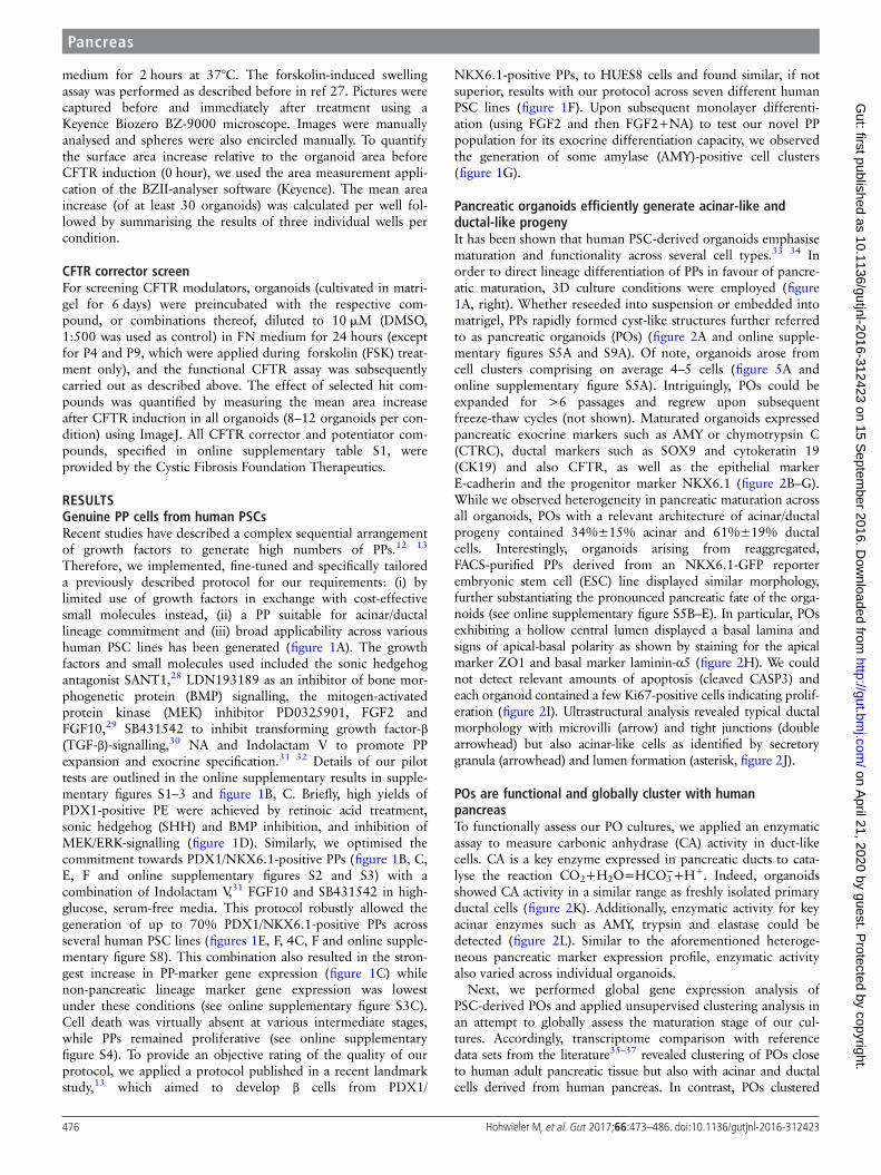
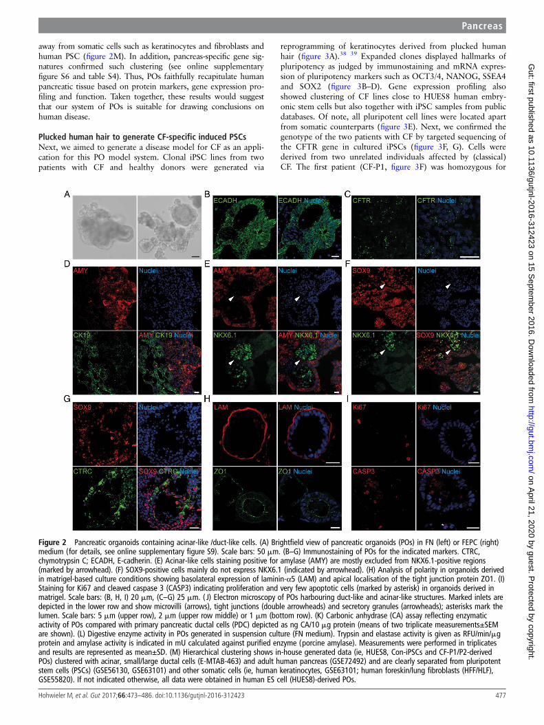
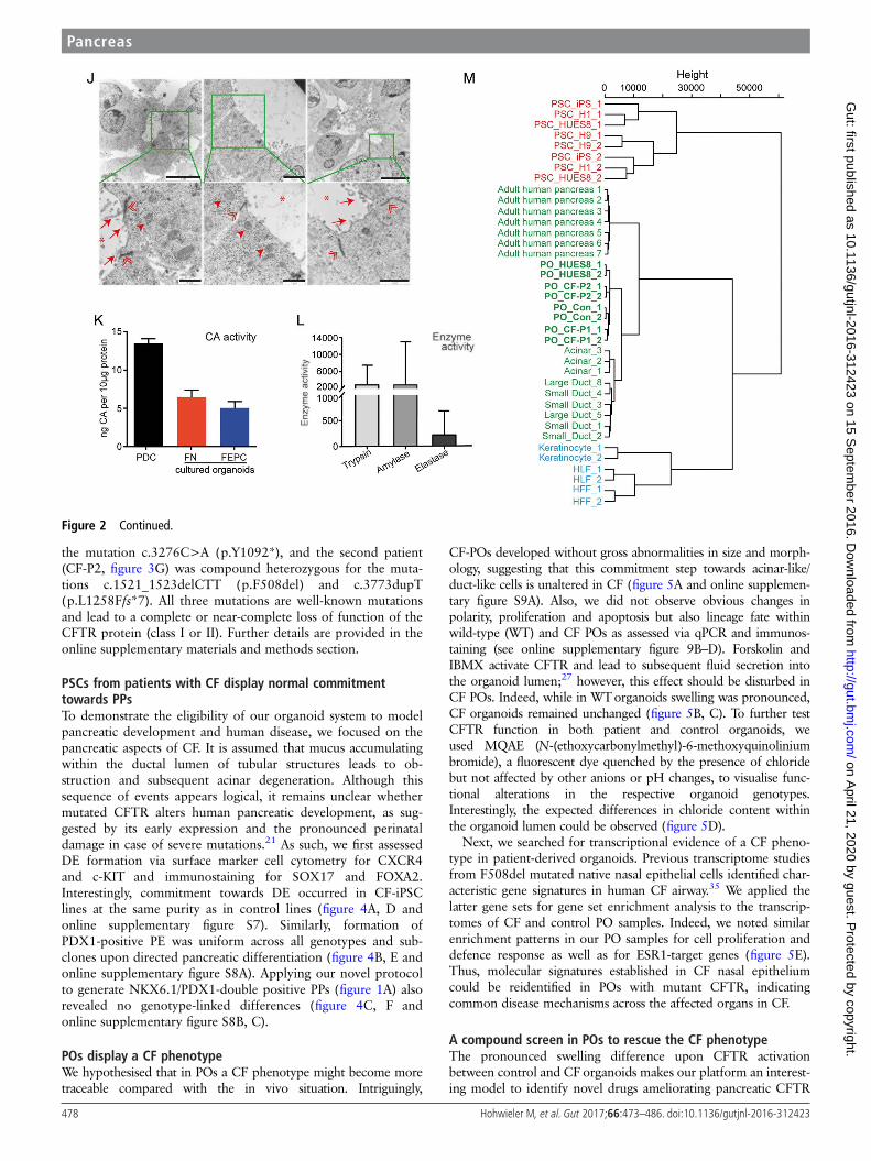
Pancreatic organoids efficiently generate acinar-like andductal-like progenyIt has been shown that human PSC-derived organoids emphasisematuration and functionality across several cell types.33 34 Inorder to direct lineage differentiation of PPs in favour of pancre-atic maturation, 3D culture conditions were employed (figure1A, right). Whether reseeded into suspension or embedded intomatrigel, PPs rapidly formed cyst-like structures further referredto as pancreatic organoids (POs) (figure 2A and online supple-mentary figures S5A and S9A). Of note, organoids arose fromcell clusters comprising on average 4–5 cells (figure 5A andonline supplementary figure S5A). Intriguingly, POs could beexpanded for >6 passages and regrew upon subsequentfreeze-thaw cycles (not shown). Maturated organoids expressedpancreatic exocrine markers such as AMY or chymotrypsin C(CTRC), ductal markers such as SOX9 and cytokeratin 19(CK19) and also CFTR, as well as the epithelial markerE-cadherin and the progenitor marker NKX6.1 (figure 2B–G).While we observed heterogeneity in pancreatic maturation acrossall organoids, POs with a relevant architecture of acinar/ductalprogeny contained 34%±15% acinar and 61%±19% ductalcells. Interestingly, organoids arising from reaggregated,FACS-purified PPs derived from an NKX6.1-GFP reporterembryonic stem cell (ESC) line displayed similar morphology,further substantiating the pronounced pancreatic fate of the orga-noids (see online supplementary figure S5B–E). In particular, POsexhibiting a hollow central lumen displayed a basal lamina andsigns of apical-basal polarity as shown by staining for the apicalmarker ZO1 and basal marker laminin-α5 (figure 2H). We couldnot detect relevant amounts of apoptosis (cleaved CASP3) andeach organoid contained a few Ki67-positive cells indicating prolif-eration (figure 2I). Ultrastructural analysis revealed typical ductalmorphology with microvilli (arrow) and tight junctions (doublearrowhead) but also acinar-like cells as identified by secretorygranula (arrowhead) and lumen formation (asterisk, figure 2J).
POs are functional and globally cluster with humanpancreasTo functionally assess our PO cultures, we applied an enzymaticassay to measure carbonic anhydrase (CA) activity in duct-likecells. CA is a key enzyme expressed in pancreatic ducts to cata-lyse the reaction CO2+H2O=HCO3
−+H+. Indeed, organoidsshowed CA activity in a similar range as freshly isolated primaryductal cells (figure 2K). Additionally, enzymatic activity for keyacinar enzymes such as AMY, trypsin and elastase could bedetected (figure 2L). Similar to the aforementioned heteroge-neous pancreatic marker expression profile, enzymatic activityalso varied across individual organoids.
Next, we performed global gene expression analysis ofPSC-derived POs and applied unsupervised clustering analysis inan attempt to globally assess the maturation stage of our cul-tures. Accordingly, transcriptome comparison with referencedata sets from the literature35–37 revealed clustering of POs closeto human adult pancreatic tissue but also with acinar and ductalcells derived from human pancreas. In contrast, POs clustered
476 Hohwieler M, et al. Gut 2017;66:473–486. doi:10.1136/gutjnl-2016-312423
Pancreas on A
pril 21, 2020 by guest. Protected by copyright.
http://gut.bmj.com
/G
ut: first published as 10.1136/gutjnl-2016-312423 on 15 Septem
ber 2016. Dow
nloaded from
away from somatic cells such as keratinocytes and fibroblasts andhuman PSC (figure 2M). In addition, pancreas-specific gene sig-natures confirmed such clustering (see online supplementaryfigure S6 and table S4). Thus, POs faithfully recapitulate humanpancreatic tissue based on protein markers, gene expression pro-filing and function. Taken together, these results would suggestthat our system of POs is suitable for drawing conclusions onhuman disease.
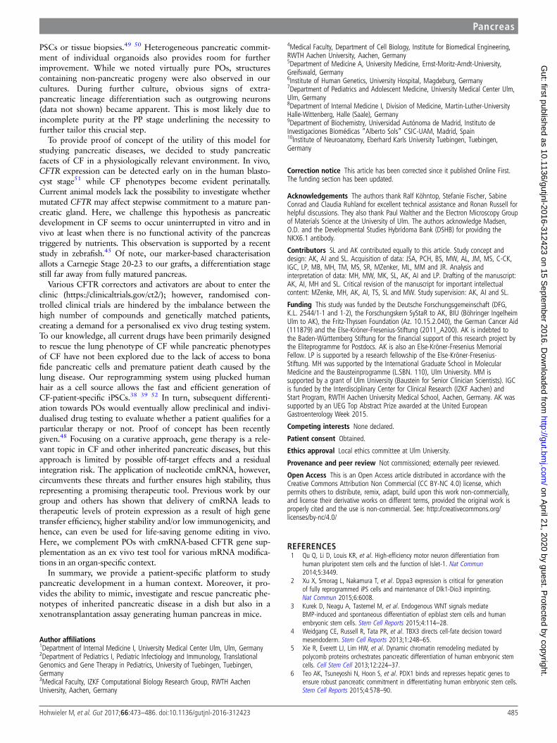
Plucked human hair to generate CF-specific induced PSCsNext, we aimed to generate a disease model for CF as an appli-cation for this PO model system. Clonal iPSC lines from twopatients with CF and healthy donors were generated via
reprogramming of keratinocytes derived from plucked humanhair (figure 3A).38 39 Expanded clones displayed hallmarks ofpluripotency as judged by immunostaining and mRNA expres-sion of pluripotency markers such as OCT3/4, NANOG, SSEA4and SOX2 (figure 3B–D). Gene expression profiling alsoshowed clustering of CF lines close to HUES8 human embry-onic stem cells but also together with iPSC samples from publicdatabases. Of note, all pluripotent cell lines were located apartfrom somatic counterparts (figure 3E). Next, we confirmed thegenotype of the two patients with CF by targeted sequencing ofthe CFTR gene in cultured iPSCs (figure 3F, G). Cells werederived from two unrelated individuals affected by (classical)CF. The first patient (CF-P1, figure 3F) was homozygous for
Figure 2 Pancreatic organoids containing acinar-like /duct-like cells. (A) Brightfield view of pancreatic organoids (POs) in FN (left) or FEPC (right)medium (for details, see online supplementary figure S9). Scale bars: 50 mm. (B–G) Immunostaining of POs for the indicated markers. CTRC,chymotrypsin C; ECADH, E-cadherin. (E) Acinar-like cells staining positive for amylase (AMY) are mostly excluded from NKX6.1-positive regions(marked by arrowhead). (F) SOX9-positive cells mainly do not express NKX6.1 (indicated by arrowhead). (H) Analysis of polarity in organoids derivedin matrigel-based culture conditions showing basolateral expression of laminin-α5 (LAM) and apical localisation of the tight junction protein ZO1. (I)Staining for Ki67 and cleaved caspase 3 (CASP3) indicating proliferation and very few apoptotic cells (marked by asterisk) in organoids derived inmatrigel. Scale bars: (B, H, I) 20 mm, (C–G) 25 mm. ( J) Electron microscopy of POs harbouring duct-like and acinar-like structures. Marked inlets aredepicted in the lower row and show microvilli (arrows), tight junctions (double arrowheads) and secretory granules (arrowheads); asterisks mark thelumen. Scale bars: 5 mm (upper row), 2 mm (upper row middle) or 1 mm (bottom row). (K) Carbonic anhydrase (CA) assay reflecting enzymaticactivity of POs compared with primary pancreatic ductal cells (PDC) depicted as ng CA/10 mg protein (means of two triplicate measurements±SEMare shown). (L) Digestive enzyme activity in POs generated in suspension culture (FN medium). Trypsin and elastase activity is given as RFU/min/mgprotein and amylase activity is indicated in mU calculated against purified enzyme (porcine amylase). Measurements were performed in triplicatesand results are represented as mean±SD. (M) Hierarchical clustering shows in-house generated data (ie, HUES8, Con-iPSCs and CF-P1/P2-derivedPOs) clustered with acinar, small/large ductal cells (E-MTAB-463) and adult human pancreas (GSE72492) and are clearly separated from pluripotentstem cells (PSCs) (GSE56130, GSE63101) and other somatic cells (ie, human keratinocytes, GSE63101; human foreskin/lung fibroblasts (HFF/HLF),GSE55820). If not indicated otherwise, all data were obtained in human ES cell (HUES8)-derived POs.
Pancreas
477Hohwieler M, et al. Gut 2017;66:473–486. doi:10.1136/gutjnl-2016-312423
on April 21, 2020 by guest. P
rotected by copyright.http://gut.bm
j.com/
Gut: first published as 10.1136/gutjnl-2016-312423 on 15 S
eptember 2016. D
ownloaded from
the mutation c.3276C>A (p.Y1092*), and the second patient(CF-P2, figure 3G) was compound heterozygous for the muta-tions c.1521_1523delCTT (p.F508del) and c.3773dupT(p.L1258Ffs*7). All three mutations are well-known mutationsand lead to a complete or near-complete loss of function of theCFTR protein (class I or II). Further details are provided in theonline supplementary materials and methods section.
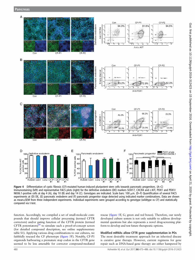
PSCs from patients with CF display normal commitmenttowards PPsTo demonstrate the eligibility of our organoid system to modelpancreatic development and human disease, we focused on thepancreatic aspects of CF. It is assumed that mucus accumulatingwithin the ductal lumen of tubular structures leads to ob-struction and subsequent acinar degeneration. Although thissequence of events appears logical, it remains unclear whethermutated CFTR alters human pancreatic development, as sug-gested by its early expression and the pronounced perinataldamage in case of severe mutations.21 As such, we first assessedDE formation via surface marker cell cytometry for CXCR4and c-KIT and immunostaining for SOX17 and FOXA2.Interestingly, commitment towards DE occurred in CF-iPSClines at the same purity as in control lines (figure 4A, D andonline supplementary figure S7). Similarly, formation ofPDX1-positive PE was uniform across all genotypes and sub-clones upon directed pancreatic differentiation (figure 4B, E andonline supplementary figure S8A). Applying our novel protocolto generate NKX6.1/PDX1-double positive PPs (figure 1A) alsorevealed no genotype-linked differences (figure 4C, F andonline supplementary figure S8B, C).
POs display a CF phenotypeWe hypothesised that in POs a CF phenotype might become moretraceable compared with the in vivo situation. Intriguingly,
CF-POs developed without gross abnormalities in size and morph-ology, suggesting that this commitment step towards acinar-like/duct-like cells is unaltered in CF (figure 5A and online supplemen-tary figure S9A). Also, we did not observe obvious changes inpolarity, proliferation and apoptosis but also lineage fate withinwild-type (WT) and CF POs as assessed via qPCR and immunos-taining (see online supplementary figure 9B–D). Forskolin andIBMX activate CFTR and lead to subsequent fluid secretion intothe organoid lumen;27 however, this effect should be disturbed inCF POs. Indeed, while in WTorganoids swelling was pronounced,CF organoids remained unchanged (figure 5B, C). To further testCFTR function in both patient and control organoids, weused MQAE (N-(ethoxycarbonylmethyl)-6-methoxyquinoliniumbromide), a fluorescent dye quenched by the presence of chloridebut not affected by other anions or pH changes, to visualise func-tional alterations in the respective organoid genotypes.Interestingly, the expected differences in chloride content withinthe organoid lumen could be observed (figure 5D).
Next, we searched for transcriptional evidence of a CF pheno-type in patient-derived organoids. Previous transcriptome studiesfrom F508del mutated native nasal epithelial cells identified char-acteristic gene signatures in human CF airway.35 We applied thelatter gene sets for gene set enrichment analysis to the transcrip-tomes of CF and control PO samples. Indeed, we noted similarenrichment patterns in our PO samples for cell proliferation anddefence response as well as for ESR1-target genes (figure 5E).Thus, molecular signatures established in CF nasal epitheliumcould be reidentified in POs with mutant CFTR, indicatingcommon disease mechanisms across the affected organs in CF.
A compound screen in POs to rescue the CF phenotypeThe pronounced swelling difference upon CFTR activationbetween control and CForganoids makes our platform an interest-ing model to identify novel drugs ameliorating pancreatic CFTR
Figure 2 Continued.
478 Hohwieler M, et al. Gut 2017;66:473–486. doi:10.1136/gutjnl-2016-312423
Pancreas on A
pril 21, 2020 by guest. Protected by copyright.
http://gut.bmj.com
/G
ut: first published as 10.1136/gutjnl-2016-312423 on 15 Septem
ber 2016. Dow
nloaded from
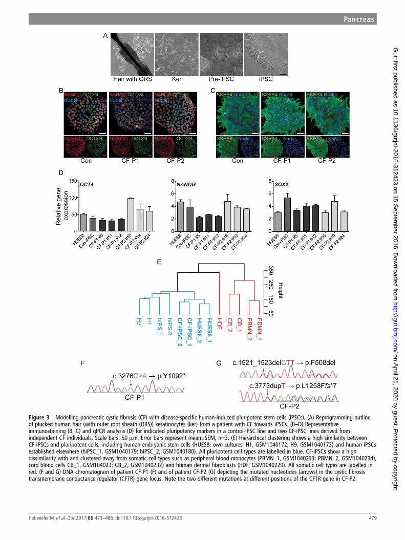
Figure 3 Modelling pancreatic cystic fibrosis (CF) with disease-specific human-induced pluripotent stem cells (iPSCs). (A) Reprogramming outlineof plucked human hair (with outer root sheath (ORS)) keratinocytes (ker) from a patient with CF towards iPSCs. (B–D) Representativeimmunostaining (B, C) and qPCR analysis (D) for indicated pluripotency markers in a control-iPSC line and two CF-iPSC lines derived fromindependent CF individuals. Scale bars: 50 mm. Error bars represent mean±SEM, n=3. (E) Hierarchical clustering shows a high similarity betweenCF-iPSCs and pluripotent cells, including human embryonic stem cells (HUES8, own cultures; H1, GSM1040172; H9, GSM1040173) and human iPSCsestablished elsewhere (hiPSC_1, GSM1040179; hiPSC_2, GSM1040180). All pluripotent cell types are labelled in blue. CF-iPSCs show a highdissimilarity with and clustered away from somatic cell types such as peripheral blood monocytes (PBMN_1, GSM1040233; PBMN_2, GSM1040234),cord blood cells CB_1, GSM104023; CB_2, GSM1040232) and human dermal fibroblasts (HDF, GSM1040229). All somatic cell types are labelled inred. (F and G) DNA chromatogram of patient CF-P1 (F) and of patient CF-P2 (G) depicting the mutated nucleotides (arrows) in the cystic fibrosistransmembrane conductance regulator (CFTR) gene locus. Note the two different mutations at different positions of the CFTR gene in CF-P2.
Pancreas
479Hohwieler M, et al. Gut 2017;66:473–486. doi:10.1136/gutjnl-2016-312423
on April 21, 2020 by guest. P
rotected by copyright.http://gut.bm
j.com/
Gut: first published as 10.1136/gutjnl-2016-312423 on 15 S
eptember 2016. D
ownloaded from
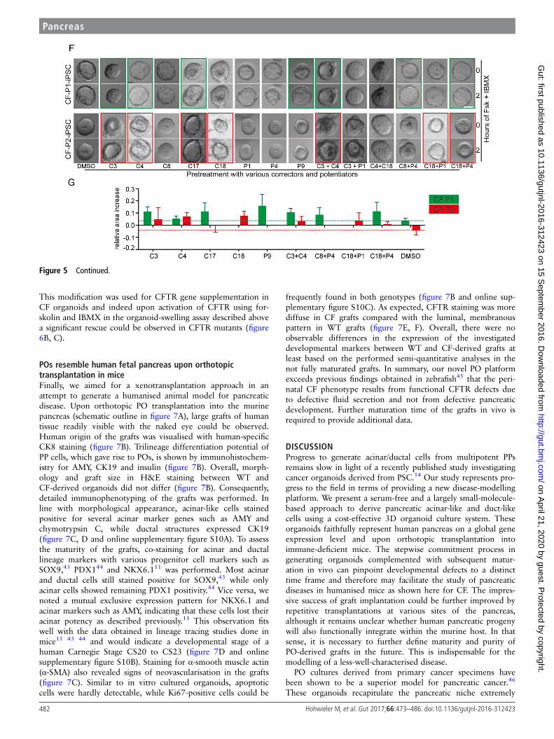
function. Accordingly, we compiled a set of small-molecule com-pounds that should improve cellular processing (termed CFTRcorrectors) and/or gating function of the CFTR protein (termedCFTR potentiators)40 to simulate such a proof-of-concept screen(for detailed compound description, see online supplementarytable S1). Applying various drug combinations to our cultures, wefaithfully rescued the CF phenotype (figure 5F). Notably, CF-P1organoids harbouring a premature stop codon in the CFTR geneseemed to be less amenable for corrector compound-mediated
rescue (figure 5F, G; green and red boxes). Therefore, our newlydeveloped culture system is not only suitable to address develop-mental questions but also represents a novel drug-screening plat-form to develop and test future therapeutic options.
Modified mRNAs allow CFTR gene supplementation in POsThe most desirable treatment approach for an inherited diseaseis curative gene therapy. However, current regimens for generepair such as DNA-based gene therapy are either hampered by
Figure 4 Differentiation of cystic fibrosis (CF)-mutated human-induced pluripotent stem cells towards pancreatic progenitors. (A–C)Immunostaining (left) and representative FACS plots (right) for the definitive endoderm (DE) markers SOX17, CXCR4 and c-KIT, PDX1 and PDX1/NKX6.1-positive cells at day 4 (A), day 10 (B) and day 14 (C). Genotypes are indicated. Scale bars: 100 mm. (D–F) Quantification of several FACSexperiments at (D) DE, (E) pancreatic endoderm and (F) pancreatic progenitor stage detected using indicated marker combinations. Data are shownas mean±SEM from three independent experiments. Individual experiments were grouped according to genotype (wildtype vs CF) and statisticallycompared via t-test.
480 Hohwieler M, et al. Gut 2017;66:473–486. doi:10.1136/gutjnl-2016-312423
Pancreas on A
pril 21, 2020 by guest. Protected by copyright.
http://gut.bmj.com
/G
ut: first published as 10.1136/gutjnl-2016-312423 on 15 Septem
ber 2016. Dow
nloaded from

safety concerns41 and/or low gene transfer efficiency.42 Theapplication of nucleotide chemically modified mRNA (cmRNA),circumvents these caveats and further ensures high stability, thusrepresenting a promising therapeutic tool. Previous work by ourgroup and others has shown that delivery of cmRNA leads totherapeutic levels of protein expression as a result of high genetransfer efficiency, higher stability and/or low immunogenicity,and hence, can even be used for life-saving genome editing invivo.18 Unfortunately, labour and cost-intensive testing isrequired to identify organ-optimised and personalised cmRNAs
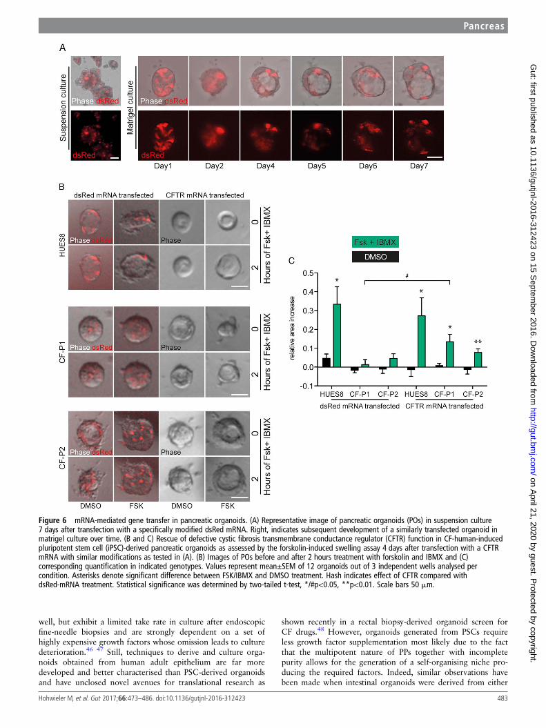
for gene supplementation in vivo. Organoids, however, wouldbe a desirable system to screen and validate cmRNAs in vitrofor subsequent in vivo applications, although the utility of sucha system remains to be validated. To address the latter, we aimedto establish cmRNAs optimised for POs in an attempt to rescueCFTR function in CF POs. We tested a set of different modifica-tions (unpublished data) using mRNAs encoding for dsRed.With one modification, we obtained robust dsRed proteinexpression even 7 days after transfection in subsequently devel-oped organoids independent of the culture regimen (figure 6A).
Figure 5 Generation of pancreatic organoids (POs) from cystic fibrosis (CF)-mutated patients and phenotypic rescue. (A) Time course of growingPOs derived from control (Con) and CF- human-induced pluripotent stem cells (iPSCs) (CF-P1). (B) Images of POs before and after 2 hours treatmentwith forskolin and IBMX. (C) Corresponding quantification from forskolin-induced PO swelling (B) in each indicated genotype. Error bars representmean area increase±SEM of three individual wells, statistical significance was determined by two-tailed t-test, *p<0.05. (D) Luminal(N-(ethoxycarbonylmethyl)-6-methoxyquinolinium bromide) (MQAE) fluorescence (Cl−-sensitive dye) is quenched in wild-type (WT) organoidsindicating an increase in intraluminal chloride secretion after cystic fibrosis transmembrane conductance regulator (CFTR) activation and subsequentchallenge with a chloride ion-rich solution, which is impaired in CF organoids. (E) Gene set enrichment analysis of three gene signatures wereperformed on WT ESC/iPSC (CON) and CF samples. Genes involved in cell proliferation, immune response and oestrogen receptor 1 (ESR1) signallingare negatively correlated in CFTR-mutated pancreatic organoids as previously described for airway cells.54 CON: HUES8 (WT ESC line, n=2) and Con1(WT iPSC) line, n=2), CF: CF-P1 and CF-P2, n=2 each. ES, enrichment score; FDR, false discovery rate; FWER, family-wise error rate; NES, normalisedenrichment score. (F and G) Small-scale rescue screen using indicated compounds in two independent CF patient-derived iPSC-POs. Organoids werepreincubated with compounds before applying FSK/IBMX. Green (CF-P1) and red (CF-P2) boxes mark most pronounced rescue, which is representedas quantification of 8–12 organoids in (G). Dotted lines indicate relative organoid size upon treatment with DMSO as a solvent control. All scalebars: 50 mm.
Pancreas
481Hohwieler M, et al. Gut 2017;66:473–486. doi:10.1136/gutjnl-2016-312423
on April 21, 2020 by guest. P
rotected by copyright.http://gut.bm
j.com/
Gut: first published as 10.1136/gutjnl-2016-312423 on 15 S
eptember 2016. D
ownloaded from
This modification was used for CFTR gene supplementation inCF organoids and indeed upon activation of CFTR using for-skolin and IBMX in the organoid-swelling assay described abovea significant rescue could be observed in CFTR mutants (figure6B, C).
POs resemble human fetal pancreas upon orthotopictransplantation in miceFinally, we aimed for a xenotransplantation approach in anattempt to generate a humanised animal model for pancreaticdisease. Upon orthotopic PO transplantation into the murinepancreas (schematic outline in figure 7A), large grafts of humantissue readily visible with the naked eye could be observed.Human origin of the grafts was visualised with human-specificCK8 staining (figure 7B). Trilineage differentiation potential ofPP cells, which gave rise to POs, is shown by immunohistochem-istry for AMY, CK19 and insulin (figure 7B). Overall, morph-ology and graft size in H&E staining between WT andCF-derived organoids did not differ (figure 7B). Consequently,detailed immunophenotyping of the grafts was performed. Inline with morphological appearance, acinar-like cells stainedpositive for several acinar marker genes such as AMY andchymotrypsin C, while ductal structures expressed CK19(figure 7C, D and online supplementary figure S10A). To assessthe maturity of the grafts, co-staining for acinar and ductallineage markers with various progenitor cell markers such asSOX9,43 PDX144 and NKX6.111 was performed. Most acinarand ductal cells still stained positive for SOX9,43 while onlyacinar cells showed remaining PDX1 positivity.44 Vice versa, wenoted a mutual exclusive expression pattern for NKX6.1 andacinar markers such as AMY, indicating that these cells lost theiracinar potency as described previously.11 This observation fitswell with the data obtained in lineage tracing studies done inmice11 43 44 and would indicate a developmental stage of ahuman Carnegie Stage CS20 to CS23 (figure 7D and onlinesupplementary figure S10B). Staining for α-smooth muscle actin(α-SMA) also revealed signs of neovascularisation in the grafts(figure 7C). Similar to in vitro cultured organoids, apoptoticcells were hardly detectable, while Ki67-positive cells could be
frequently found in both genotypes (figure 7B and online sup-plementary figure S10C). As expected, CFTR staining was morediffuse in CF grafts compared with the luminal, membranouspattern in WT grafts (figure 7E, F). Overall, there were noobservable differences in the expression of the investigateddevelopmental markers between WT and CF-derived grafts atleast based on the performed semi-quantitative analyses in thenot fully maturated grafts. In summary, our novel PO platformexceeds previous findings obtained in zebrafish45 that the peri-natal CF phenotype results from functional CFTR defects dueto defective fluid secretion and not from defective pancreaticdevelopment. Further maturation time of the grafts in vivo isrequired to provide additional data.
DISCUSSIONProgress to generate acinar/ductal cells from multipotent PPsremains slow in light of a recently published study investigatingcancer organoids derived from PSC.14 Our study represents pro-gress to the field in terms of providing a new disease-modellingplatform. We present a serum-free and a largely small-molecule-based approach to derive pancreatic acinar-like and duct-likecells using a cost-effective 3D organoid culture system. Theseorganoids faithfully represent human pancreas on a global geneexpression level and upon orthotopic transplantation intoimmune-deficient mice. The stepwise commitment process ingenerating organoids complemented with subsequent matur-ation in vivo can pinpoint developmental defects to a distincttime frame and therefore may facilitate the study of pancreaticdiseases in humanised mice as shown here for CF. The impres-sive success of graft implantation could be further improved byrepetitive transplantations at various sites of the pancreas,although it remains unclear whether human pancreatic progenywill also functionally integrate within the murine host. In thatsense, it is necessary to further define maturity and purity ofPO-derived grafts in the future. This is indispensable for themodelling of a less-well-characterised disease.
PO cultures derived from primary cancer specimens havebeen shown to be a superior model for pancreatic cancer.46
These organoids recapitulate the pancreatic niche extremely
Figure 5 Continued.
482 Hohwieler M, et al. Gut 2017;66:473–486. doi:10.1136/gutjnl-2016-312423
Pancreas on A
pril 21, 2020 by guest. Protected by copyright.
http://gut.bmj.com
/G
ut: first published as 10.1136/gutjnl-2016-312423 on 15 Septem
ber 2016. Dow
nloaded from
well, but exhibit a limited take rate in culture after endoscopicfine-needle biopsies and are strongly dependent on a set ofhighly expensive growth factors whose omission leads to culturedeterioration.46 47 Still, techniques to derive and culture orga-noids obtained from human adult epithelium are far moredeveloped and better characterised than PSC-derived organoidsand have unclosed novel avenues for translational research as
shown recently in a rectal biopsy-derived organoid screen forCF drugs.48 However, organoids generated from PSCs requireless growth factor supplementation most likely due to the factthat the multipotent nature of PPs together with incompletepurity allows for the generation of a self-organising niche pro-ducing the required factors. Indeed, similar observations havebeen made when intestinal organoids were derived from either
Figure 6 mRNA-mediated gene transfer in pancreatic organoids. (A) Representative image of pancreatic organoids (POs) in suspension culture7 days after transfection with a specifically modified dsRed mRNA. Right, indicates subsequent development of a similarly transfected organoid inmatrigel culture over time. (B and C) Rescue of defective cystic fibrosis transmembrane conductance regulator (CFTR) function in CF-human-inducedpluripotent stem cell (iPSC)-derived pancreatic organoids as assessed by the forskolin-induced swelling assay 4 days after transfection with a CFTRmRNA with similar modifications as tested in (A). (B) Images of POs before and after 2 hours treatment with forskolin and IBMX and (C)corresponding quantification in indicated genotypes. Values represent mean±SEM of 12 organoids out of 3 independent wells analysed percondition. Asterisks denote significant difference between FSK/IBMX and DMSO treatment. Hash indicates effect of CFTR compared withdsRed-mRNA treatment. Statistical significance was determined by two-tailed t-test, */#p<0.05, **p<0.01. Scale bars 50 mm.
Pancreas
483Hohwieler M, et al. Gut 2017;66:473–486. doi:10.1136/gutjnl-2016-312423
on April 21, 2020 by guest. P
rotected by copyright.http://gut.bm
j.com/
Gut: first published as 10.1136/gutjnl-2016-312423 on 15 S
eptember 2016. D
ownloaded from
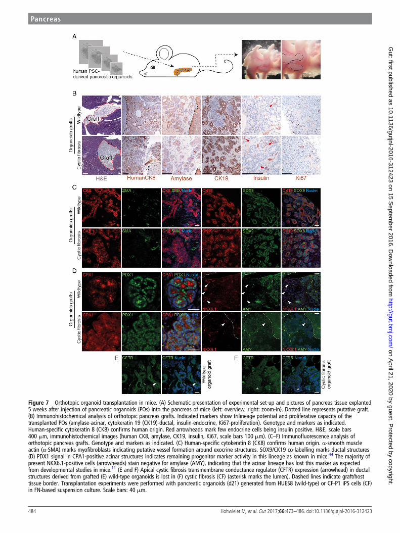
Figure 7 Orthotopic organoid transplantation in mice. (A) Schematic presentation of experimental set-up and pictures of pancreas tissue explanted5 weeks after injection of pancreatic organoids (POs) into the pancreas of mice (left: overview, right: zoom-in). Dotted line represents putative graft.(B) Immunohistochemical analysis of orthotopic pancreas grafts. Indicated markers show trilineage potential and proliferative capacity of thetransplanted POs (amylase-acinar, cytokeratin 19 (CK19)-ductal, insulin-endocrine, Ki67-proliferation). Genotype and markers as indicated.Human-specific cytokeratin 8 (CK8) confirms human origin. Red arrowheads mark few endocrine cells being insulin positive. H&E, scale bars400 mm, immunohistochemical images (human CK8, amylase, CK19, insulin, Ki67, scale bars 100 mm). (C–F) Immunofluorescence analysis oforthotopic pancreas grafts. Genotype and markers as indicated. (C) Human-specific cytokeratin 8 (CK8) confirms human origin. α-smooth muscleactin (α-SMA) marks myofibroblasts indicating putative vessel formation around exocrine structures. SOX9/CK19 co-labelling marks ductal structures(D) PDX1 signal in CPA1-positive acinar structures indicates remaining progenitor marker activity in this lineage as known in mice.44 The majority ofpresent NKX6.1-positive cells (arrowheads) stain negative for amylase (AMY), indicating that the acinar lineage has lost this marker as expectedfrom developmental studies in mice.11 (E and F) Apical cystic fibrosis transmembrane conductance regulator (CFTR) expression (arrowhead) in ductalstructures derived from grafted (E) wild-type organoids is lost in (F) cystic fibrosis (CF) (asterisk marks the lumen). Dashed lines indicate graft/hosttissue border. Transplantation experiments were performed with pancreatic organoids (d21) generated from HUES8 (wild-type) or CF-P1 iPS cells (CF)in FN-based suspension culture. Scale bars: 40 mm.
484 Hohwieler M, et al. Gut 2017;66:473–486. doi:10.1136/gutjnl-2016-312423
Pancreas on A
pril 21, 2020 by guest. Protected by copyright.
http://gut.bmj.com
/G
ut: first published as 10.1136/gutjnl-2016-312423 on 15 Septem
ber 2016. Dow
nloaded from
PSCs or tissue biopsies.49 50 Heterogeneous pancreatic commit-ment of individual organoids also provides room for furtherimprovement. While we noted virtually pure POs, structurescontaining non-pancreatic progeny were also observed in ourcultures. During further culture, obvious signs of extra-pancreatic lineage differentiation such as outgrowing neurons(data not shown) became apparent. This is most likely due toincomplete purity at the PP stage underlining the necessity tofurther tailor this crucial step.
To provide proof of concept of the utility of this model forstudying pancreatic diseases, we decided to study pancreaticfacets of CF in a physiologically relevant environment. In vivo,CFTR expression can be detected early on in the human blasto-cyst stage51 while CF phenotypes become evident perinatally.Current animal models lack the possibility to investigate whethermutated CFTR may affect stepwise commitment to a mature pan-creatic gland. Here, we challenge this hypothesis as pancreaticdevelopment in CF seems to occur uninterrupted in vitro and invivo at least when there is no functional activity of the pancreastriggered by nutrients. This observation is supported by a recentstudy in zebrafish.45 Of note, our marker-based characterisationallots a Carnegie Stage 20-23 to our grafts, a differentiation stagestill far away from fully matured pancreas.
Various CFTR correctors and activators are about to enter theclinic (https://clinicaltrials.gov/ct2/); however, randomised con-trolled clinical trials are hindered by the imbalance between thehigh number of compounds and genetically matched patients,creating a demand for a personalised ex vivo drug testing system.To our knowledge, all current drugs have been primarily designedto rescue the lung phenotype of CF while pancreatic phenotypesof CF have not been explored due to the lack of access to bonafide pancreatic cells and premature patient death caused by thelung disease. Our reprogramming system using plucked humanhair as a cell source allows the fast and efficient generation ofCF-patient-specific iPSCs.38 39 52 In turn, subsequent differenti-ation towards POs would eventually allow preclinical and indivi-dualised drug testing to evaluate whether a patient qualifies for aparticular therapy or not. Proof of concept has been recentlygiven.48 Focusing on a curative approach, gene therapy is a rele-vant topic in CF and other inherited pancreatic diseases, but thisapproach is limited by possible off-target effects and a residualintegration risk. The application of nucleotide cmRNA, however,circumvents these threats and further ensures high stability, thusrepresenting a promising therapeutic tool. Previous work by ourgroup and others has shown that delivery of cmRNA leads totherapeutic levels of protein expression as a result of high genetransfer efficiency, higher stability and/or low immunogenicity, andhence, can even be used for life-saving genome editing in vivo.Here, we complement POs with cmRNA-based CFTR gene sup-plementation as an ex vivo test tool for various mRNA modifica-tions in an organ-specific context.
In summary, we provide a patient-specific platform to studypancreatic development in a human context. Moreover, it pro-vides the ability to mimic, investigate and rescue pancreatic phe-notypes of inherited pancreatic disease in a dish but also in axenotransplantation assay generating human pancreas in mice.
Author affiliations1Department of Internal Medicine I, University Medical Center Ulm, Ulm, Germany2Department of Pediatrics I, Pediatric Infectiology and Immunology, TranslationalGenomics and Gene Therapy in Pediatrics, University of Tuebingen, Tuebingen,Germany3Medical Faculty, IZKF Computational Biology Research Group, RWTH AachenUniversity, Aachen, Germany
4Medical Faculty, Department of Cell Biology, Institute for Biomedical Engineering,RWTH Aachen University, Aachen, Germany5Department of Medicine A, University Medicine, Ernst-Moritz-Arndt-University,Greifswald, Germany6Institute of Human Genetics, University Hospital, Magdeburg, Germany7Department of Pediatrics and Adolescent Medicine, University Medical Center Ulm,Ulm, Germany8Department of Internal Medicine I, Division of Medicine, Martin-Luther-UniversityHalle-Wittenberg, Halle (Saale), Germany9Department of Biochemistry, Universidad Autónoma de Madrid, Instituto deInvestigaciones Biomédicas “Alberto Sols” CSIC-UAM, Madrid, Spain10Institute of Neuroanatomy, Eberhard Karls University Tuebingen, Tuebingen,Germany
Correction notice This article has been corrected since it published Online First.The funding section has been updated.
Acknowledgements The authors thank Ralf Köhntop, Stefanie Fischer, SabineConrad and Claudia Ruhland for excellent technical assistance and Ronan Russell forhelpful discussions. They also thank Paul Walther and the Electron Microscopy Groupof Materials Science at the University of Ulm. The authors acknowledge Madsen,O.D. and the Developmental Studies Hybridoma Bank (DSHB) for providing theNKX6.1 antibody.
Contributors SL and AK contributed equally to this article. Study concept anddesign: AK, AI and SL. Acquisition of data: JSA, PCH, BS, MW, AL, JM, MS, C-CK,IGC, LP, MB, MH, TM, MS, SR, MZenker, ML, MM and JR. Analysis andinterpretation of data: MH, MW, MK, SL, AK, AI and LP. Drafting of the manuscript:AK, AI, MH and SL. Critical revision of the manuscript for important intellectualcontent: MZenke, MH, AK, AI, TS, SL and MW. Study supervision: AK, AI and SL.
Funding This study was funded by the Deutsche Forschungsgemeinschaft (DFG,K.L. 2544/1-1 and 1-2), the Forschungskern SyStaR to AK, BIU (Böhringer IngelheimUlm to AK), the Fritz-Thyssen Foundation (Az. 10.15.2.040), the German Cancer Aid(111879) and the Else-Kröner-Fresenius-Stiftung (2011_A200). AK is indebted tothe Baden-Württemberg Stiftung for the financial support of this research project bythe Eliteprogramme for Postdocs. AK is also an Else-Kröner-Fresenius MemorialFellow. LP is supported by a research fellowship of the Else-Kröner-Fresenius-Stiftung. MH was supported by the International Graduate School in MolecularMedicine and the Bausteinprogramme (L.SBN. 110), Ulm University. MM issupported by a grant of Ulm University (Baustein for Senior Clinician Scientists). IGCis funded by the Interdisciplinary Center for Clinical Research (IZKF Aachen) andStart Program, RWTH Aachen University Medical School, Aachen, Germany. AK wassupported by an UEG Top Abstract Prize awarded at the United EuropeanGastroenterology Week 2015.
Competing interests None declared.
Patient consent Obtained.
Ethics approval Local ethics committee at Ulm University.
Provenance and peer review Not commissioned; externally peer reviewed.
Open Access This is an Open Access article distributed in accordance with theCreative Commons Attribution Non Commercial (CC BY-NC 4.0) license, whichpermits others to distribute, remix, adapt, build upon this work non-commercially,and license their derivative works on different terms, provided the original work isproperly cited and the use is non-commercial. See: http://creativecommons.org/licenses/by-nc/4.0/
REFERENCES1 Qu Q, Li D, Louis KR, et al. High-efficiency motor neuron differentiation from
human pluripotent stem cells and the function of Islet-1. Nat Commun2014;5:3449.
2 Xu X, Smorag L, Nakamura T, et al. Dppa3 expression is critical for generationof fully reprogrammed iPS cells and maintenance of Dlk1-Dio3 imprinting.Nat Commun 2015;6:6008.
3 Kurek D, Neagu A, Tastemel M, et al. Endogenous WNT signals mediateBMP-induced and spontaneous differentiation of epiblast stem cells and humanembryonic stem cells. Stem Cell Reports 2015;4:114–28.
4 Weidgang CE, Russell R, Tata PR, et al. TBX3 directs cell-fate decision towardmesendoderm. Stem Cell Reports 2013;1:248–65.
5 Xie R, Everett LJ, Lim HW, et al. Dynamic chromatin remodeling mediated bypolycomb proteins orchestrates pancreatic differentiation of human embryonic stemcells. Cell Stem Cell 2013;12:224–37.
6 Teo AK, Tsuneyoshi N, Hoon S, et al. PDX1 binds and represses hepatic genes toensure robust pancreatic commitment in differentiating human embryonic stem cells.Stem Cell Reports 2015;4:578–90.
Pancreas
485Hohwieler M, et al. Gut 2017;66:473–486. doi:10.1136/gutjnl-2016-312423
on April 21, 2020 by guest. P
rotected by copyright.http://gut.bm
j.com/
Gut: first published as 10.1136/gutjnl-2016-312423 on 15 S
eptember 2016. D
ownloaded from
7 Kao DI, Lacko LA, Ding BS, et al. Endothelial cells control pancreatic cell fateat defined stages through EGFL7 signaling. Stem Cell Reports 2015;4:181–9.
8 Kroon E, Martinson LA, Kadoya K, et al. Pancreatic endoderm derived from humanembryonic stem cells generates glucose-responsive insulin-secreting cells in vivo.Nat Biotechnol 2008;26:443–52.
9 D’Amour KA, Bang AG, Eliazer S, et al. Production of pancreatichormone-expressing endocrine cells from human embryonic stem cells.Nat Biotechnol 2006;24:1392–401.
10 Nair G, Hebrok M. Islet formation in mice and men: lessons for the generation offunctional insulin-producing beta-cells from human pluripotent stem cells. Curr OpinGenet Dev 2015;32:171–80.
11 Schaffer AE, Freude KK, Nelson SB, et al. Nkx6 transcription factors and Ptf1afunction as antagonistic lineage determinants in multipotent pancreatic progenitors.Dev Cell 2010;18:1022–9.
12 Nostro MC, Sarangi F, Yang C, et al. Efficient generation of NKX6-1+ pancreaticprogenitors from multiple human pluripotent stem cell lines. Stem Cell Reports2015;4:591–604.
13 Rezania A, Bruin JE, Arora P, et al. Reversal of diabetes with insulin-producing cellsderived in vitro from human pluripotent stem cells. Nat Biotechnol2014;32:1121–33.
14 Huang L, Holtzinger A, Jagan I, et al. Ductal pancreatic cancer modeling and drugscreening using human pluripotent stem cell- and patient-derived tumor organoids.Nat Med 2015;21:1364–71.
15 Sampaziotis F, Cardoso de Brito M, Madrigal P, et al. Cholangiocytes derived fromhuman induced pluripotent stem cells for disease modeling and drug validation.Nat Biotechnol 2015;33:845–52.
16 Freedman BS, Brooks CR, Lam AQ, et al. Modelling kidney disease withCRISPR-mutant kidney organoids derived from human pluripotent epiblastspheroids. Nat Commun 2015;6:8715.
17 Zeyer F, Mothes B, Will C, et al. mRNA-mediated gene supplementation of toll-likereceptors as treatment strategy for asthma in vivo. PLoS ONE 2016;11:e0154001.
18 Kormann MS, Hasenpusch G, Aneja MK, et al. Expression of therapeutic proteinsafter delivery of chemically modified mRNA in mice. Nat Biotechnol2011;29:154–7.
19 Cutting GR. Cystic fibrosis genetics: from molecular understanding to clinicalapplication. Nat Rev Genet 2015;16:45–56.
20 Pier GB. The challenges and promises of new therapies for cystic fibrosis. J Exp Med2012;209:1235–9.
21 Ooi CY, Dorfman R, Cipolli M, et al. Type of CFTR mutation determines risk ofpancreatitis in patients with cystic fibrosis. Gastroenterology 2011;140:153–61.
22 Hamoir C, Pepermans X, Piessevaux H, et al. Clinical and morphologicalcharacteristics of sporadic genetically determined pancreatitis as compared toidiopathic pancreatitis: higher risk of pancreatic cancer in CFTR variants. Digestion2013;87:229–39.
23 Constantinescu AA, Gleizes C, Alhosin M, et al. Exocrine cell-derived microparticlesin response to lipopolysaccharide promote endocrine dysfunction in cystic fibrosis.J Cyst Fibros 2014;13:219–26.
24 Zertal-Zidani S, Busiah K, Edelman A, et al. Small-molecule inhibitors of the cysticfibrosis transmembrane conductance regulator increase pancreatic endocrine celldevelopment in rat and mouse. Diabetologia 2013;56:330–9.
25 Frank S, Zhang M, Scholer HR, et al. Small molecule-assisted, line-independentmaintenance of human pluripotent stem cells in defined conditions. PLoS ONE2012;7:e41958.
26 Greggio C, De Franceschi F, Figueiredo-Larsen M, et al. Artificial three-dimensionalniches deconstruct pancreas development in vitro. Development2013;140:4452–62.
27 Dekkers JF, Wiegerinck CL, de Jonge HR, et al. A functional CFTR assay usingprimary cystic fibrosis intestinal organoids. Nat Med 2013;19:939–45.
28 Stanton BZ, Peng LF. Small-molecule modulators of the Sonic Hedgehog signalingpathway. Mol Biosyst 2010;6:44–54.
29 Ye F, Duvillie B, Scharfmann R. Fibroblast growth factors 7 and 10 are expressed inthe human embryonic pancreatic mesenchyme and promote the proliferation ofembryonic pancreatic epithelial cells. Diabetologia 2005;48:277–81.
30 Cleveland MH, Sawyer JM, Afelik S, et al. Exocrine ontogenies: on the developmentof pancreatic acinar, ductal and centroacinar cells. Semin Cell Dev Biol2012;23:711–19.
31 Chen S, Borowiak M, Fox JL, et al. A small molecule that directs differentiation ofhuman ESCs into the pancreatic lineage. Nat Chem Biol 2009;5:258–65.
32 Jiang W, Shi Y, Zhao D, et al. In vitro derivation of functional insulin-producing cellsfrom human embryonic stem cells. Cell Res 2007;17:333–44.
33 Morizane R, Lam AQ, Freedman BS, et al. Nephron organoids derived from humanpluripotent stem cells model kidney development and injury. Nat Biotechnol2015;33:1193–200.
34 Finkbeiner SR, Hill DR, Altheim CH, et al. Transcriptome-wide analysis revealshallmarks of human intestine development and maturation in vitro and in vivo.Stem Cell Reports 2015;4:1140–55.
35 Clarke LA, Sousa L, Barreto C, et al. Changes in transcriptome of native nasalepithelium expressing F508del-CFTR and intersecting data from comparable studies.Respir Res 2013;14:38.
36 Arda HE, Li L, Tsai J, et al. Age-dependent pancreatic gene regulation revealsmechanisms governing human beta cell function. Cell Metab 2016;23:909–20.
37 Dorrell C, Schug J, Lin CF, et al. Transcriptomes of the major human pancreatic celltypes. Diabetologia 2011;54:2832–44.
38 Illing A, Stockmann M, Swamy Telugu N, et al. Definitive endoderm formation fromplucked human hair-derived induced pluripotent stem cells and SK channelregulation. Stem Cells Int 2013;2013:360573.
39 Linta L, Stockmann M, Kleinhans KN, et al. Rat embryonic fibroblasts improvereprogramming of human keratinocytes into induced pluripotent stem cells.Stem Cells Dev 2012;21:965–76.
40 Veit G, Avramescu RG, Chiang AN, et al. From CFTR biology toward combinatorialpharmacotherapy: expanded classification of cystic fibrosis mutations. Mol Biol Cell2016;27:424–33.
41 McCormack MP, Rabbitts TH. Activation of the T-cell oncogene LMO2 after genetherapy for X-linked severe combined immunodeficiency. N Engl J Med2004;350:913–22.
42 Sanders N, Rudolph C, Braeckmans K, et al. Extracellular barriers in respiratory genetherapy. Adv Drug Deliv Rev 2009;61:115–27.
43 Furuyama K, Kawaguchi Y, Akiyama H, et al. Continuous cell supply from aSox9-expressing progenitor zone in adult liver, exocrine pancreas and intestine.Nat Genet 2011;43:34–41.
44 Gu G, Brown JR, Melton DA. Direct lineage tracing reveals the ontogeny ofpancreatic cell fates during mouse embryogenesis. Mech Dev 2003;120:35–43.
45 Navis A, Bagnat M. Loss of CFTR function leads to pancreatic destruction in larvalzebrafish. Dev Biol 2015;399:237–48.
46 Boj SF, Hwang CI, Baker LA, et al. Organoid models of human and mouse ductalpancreatic cancer. Cell 2015;160:324–38.
47 Huch M, Bonfanti P, Boj SF, et al. Unlimited in vitro expansion of adultbi-potent pancreas progenitors through the Lgr5/R-spondin axis. EMBO J2013;32:2708–21.
48 Dekkers JF, Berkers G, Kruisselbrink E, et al. Characterizing responses toCFTR-modulating drugs using rectal organoids derived from subjects with cysticfibrosis. Sci Transl Med 2016;8:344ra84.
49 Watson CL, Mahe MM, Munera J, et al. An in vivo model of human small intestineusing pluripotent stem cells. Nat Med 2014;20:1310–14.
50 Sato T, Vries RG, Snippert HJ, et al. Single Lgr5 stem cells build crypt-villusstructures in vitro without a mesenchymal niche. Nature 2009;459:262–5.
51 Ben-Chetrit A, Antenos M, Jurisicova A, et al. Expression of cystic fibrosistransmembrane conductance regulator during early human embryo development.Mol Hum Reprod 2002;8:758–64.
52 Raab S, Klingenstein M, Liebau S, et al. A comparative view on human somatic cellsources for iPSC generation. Stem Cells Int 2014;2014:768391.
53 Chetty S, Pagliuca FW, Honore C, et al. A simple tool to improve pluripotent stemcell differentiation. Nat Methods 2013;10:553–6.
54 Subramanian A, Tamayo P, Mootha VK, et al. Gene set enrichment analysis:a knowledge-based approach for interpreting genome-wide expression profiles.Proc Natl Acad Sci USA 2005;102:15545–50.
486 Hohwieler M, et al. Gut 2017;66:473–486. doi:10.1136/gutjnl-2016-312423
Pancreas on A
pril 21, 2020 by guest. Protected by copyright.
http://gut.bmj.com
/G
ut: first published as 10.1136/gutjnl-2016-312423 on 15 Septem
ber 2016. Dow
nloaded from