original article leucine deprivation increases hepatic ... · leucine deprivation increases hepatic...
TRANSCRIPT
Leucine Deprivation Increases Hepatic Insulin Sensitivityvia GCN2/mTOR/S6K1 and AMPK PathwaysFei Xiao,
1Zhiying Huang,
1Houkai Li,
1Junjie Yu,
1Chunxia Wang,
1Shanghai Chen,
1Qingshu Meng,
1
Ying Cheng,1Xiang Gao,
2Jia Li,
3Yong Liu,
1and Feifan Guo
1
OBJECTIVE—We have previously shown that serum insulinlevels decrease threefold and blood glucose levels remain normalin mice fed a leucine-deficient diet, suggesting increased insulinsensitivity. The goal of the current study is to investigate thispossibility and elucidate the underlying cellular mechanisms.
RESEARCH DESIGN AND METHODS—Changes in metabolicparameters and expression of genes and proteins involved inregulation of insulin sensitivity were analyzed in mice, humanHepG2 cells, and mouse primary hepatocytes under leucinedeprivation.
RESULTS—We show that leucine deprivation improves hepaticinsulin sensitivity by sequentially activating general control non-derepressible (GCN)2 and decreasing mammalian target ofrapamycin/S6K1 signaling. In addition, we show that activationof AMP-activated protein kinase also contributes to leucinedeprivation–increased hepatic insulin sensitivity. Finally, weshow that leucine deprivation improves insulin sensitivity underinsulin-resistant conditions.
CONCLUSIONS—This study describes mechanisms underlyingincreased hepatic insulin sensitivity under leucine deprivation.Furthermore, we demonstrate a novel function for GCN2 in theregulation of insulin sensitivity. These observations providea rationale for short-term dietary restriction of leucine for thetreatment of insulin resistance and associated metabolic dis-eases. Diabetes 60:746–756, 2011
Insulin resistance is a common feature of manymetabolic diseases, including type 2 diabetes andnonalcoholic fatty liver diseases. The hallmark ofinsulin resistance is reduced glucose uptake in
muscle and adipose tissue, and increased glucose pro-duction in liver (1,2). Various strategies have been proposedto treat insulin resistance, including lifestyle modificationsand pharmacologic interventions (3,4).
Recently, there has been a growing interest in treatinginsulin resistance with dietary manipulation of micronu-trients, including branched-chain amino acids (BCAAs).The BCAAs comprise the three essential amino acidshaving nonlinear aliphatic side chains: leucine, isoleucine,
and valine. These amino acids not only serve as precursorsin protein synthesis but also play regulatory roles in in-tracellular signaling (5).
Several studies have shown that dietary supplementa-tion of leucine influences insulin sensitivity. For example,Zhang and colleagues (6) recently demonstrated that in-creased oral intake of leucine improves whole-body glu-cose metabolism in mice maintained on a high-fat diet(HFD). The effect of increasing dietary leucine, however,is controversial. For example, other studies reported thatincreased serum levels of leucine have no effect (7) orincrease insulin resistance in humans and in animalmodels of obesity (8,9).
By contrast, our research has focused on the effect ofeliminating leucine from the diet. As shown in our previousstudy (10), serum insulin levels decrease threefold in micefed a leucine-deficient [(2) leu] diet. Blood glucose levelsremain normal in these mice, however, suggesting in-creased insulin sensitivity. The goal of our current study isto investigate this possibility and elucidate the underlyingmolecular and cellular mechanisms.
In our current study, we observed that leucine depriva-tion increases whole-body insulin sensitivity and insulinsignaling in liver. Furthermore, we show that leucine dep-rivation improves hepatic insulin sensitivity by sequentiallyactivating general control nonderepressible (GCN)2 and de-creasing mammalian target of rapamycin (mTOR) signaling.In addition, we show that activation of AMP-activated pro-tein kinase (AMPK) also contributes to increased hepaticinsulin sensitivity under leucine deprivation. Finally, weshow that leucine deprivation improves insulin sensitivityunder insulin-resistant conditions.
RESEARCH DESIGN AND METHODS
Chemicals and plasmids. Insulin and rapamycin were from Sigma and TautoBiotech (Shanghai, China), respectively. The vector p-CAGGS-DN-AMPK-IRES-EGFP encoding dominant-negative AMPK protein was a gift from Dr. Jia Li,Shanghai Institute of Materia Medica, China. The HA-tagged constitutivelyactive S6K1 expression plasmid was the Addgene plasmid 8991, originallyfrom John Blenis’s laboratory. Effectene Transfection Reagent was fromQiagen (Hilden, Germany).Animals and treatment. Male C57BL/6J mice were obtained from ShanghaiLaboratory Animal Co., Ltd. (Shanghai, China). GCN2 knockout (Gcn22/2) andleptin receptor–deficient (db/db) mice were provided by Dr. Douglas Cavener,Penn State University, and Dr. Xiang Gao, Nanjing University, China, re-spectively. Eight- to ten-week-old mice were maintained on a 12-h light/darkcycle at 25°C. Control (nutritionally complete amino acid) and (2) leu dietswere obtained from Research Diets, Inc. (New Brunswick, NJ). All diets wereisocaloric and compositionally the same in terms of carbohydrate and lipidcomponent. At the start of the feeding experiments, mice were acclimated toa control diet for 7 days and then randomly divided into control and (2) leudiet groups, with free access to control and (2) leu diets, respectively, for 7days. In addition, a pair-fed group was included to distinguish the possibleinfluence of the minor reduction in food intake, as previously observed inthe (2) leu group (11). A subset of mice underwent glucose tolerance tests(GTTs) and insulin tolerance tests (ITTs) before they were killed by CO2
inhalation. These experiments were conducted in accordance with the
From the 1Key Laboratory of Nutrition and Metabolism, Institute for Nutri-tional Sciences, Shanghai Institute for Biological Sciences, Chinese Acad-emy of Sciences, the Graduate School of the Chinese Academy of Sciences,Shanghai, China; the 2Model Animal Research Center, Nanjing University,Nanjing, China; and the 3National Center for Drug Screening, Shanghai In-stitute of Materia Medica, Chinese Academy of Sciences, Shanghai, China.
Corresponding author: Feifan Guo, [email protected] 7 September 2010 and accepted 19 December 2010.DOI: 10.2337/db10-1246This article contains Supplementary Data online at http://diabetes.
diabetesjournals.org/lookup/suppl/doi:10.2337/db10-1246/-/DC1.� 2011 by the American Diabetes Association. Readers may use this article as
long as the work is properly cited, the use is educational and not for profit,and the work is not altered. See http://creativecommons.org/licenses/by-nc-nd/3.0/ for details.
746 DIABETES, VOL. 60, MARCH 2011 diabetes.diabetesjournals.org
ORIGINAL ARTICLE
guidelines of the Institutional Animal Care and Use Committee of the In-stitute for Nutritional Sciences, Shanghai Institute for Biological Sciences,Chinese Academy of Sciences.Cell culture and treatments. HepG2 cells were maintained in Dulbecco’smodified Eagle’s medium (DMEM) with 25 mmol/L glucose (Gibco, Invitrogen,Carlsbad, CA), 10% FBS, 50 mg/mL penicillin and streptomycin at 37°C, and 5%CO2–95% air. Control and (2) leu medium were prepared from amino acid-freeDMEM (Invitrogen) by adding back all the amino acids contained in regularDMEM and without leucine only, respectively. For the rapamycin treatment,cells were incubated with 50 nmol/L rapamycin for 1 h. To induce insulinresistance, HepG2 cells were incubated with 18 mmol/L glucosamine for 18 hin serum-free medium as previously described (12), followed by incubation inserum-free (2) leu or control medium for 24 h in the presence of glucosamine.The GCN2-knockdown (KO) stable cell lines were obtained by transfectingpSilencer4.1-CMV-puro-GCN2 small interfering (si)RNA vector into HepG2cells and selecting for stable clones in the presence of 0.5 mg/mL puromycin.Isolation of primary hepatocytes.Male C57BL/6J mice were anesthetized byintraperitoneal injection with chloral hydrate. Hepatocytes were prepared bycollagenase perfusion as described previously (13). Isolated hepatocytes wereresuspended in 10% FBS DMEM and incubated for 1 day before switching to(2) leu medium.Generation and administration of recombinant adenoviruses. The recom-binant adenoviruses used for the S6K1 expression were generated using AdEasyVector System (Qbiogene, Montreal, Canada). Briefly, S6K1 gene was clonedinto a transfer vector. The resulting plasmid was then linearized with PmeIand cotransformed into Escherichia coli strain BJ5183 together with pAdEasy-1.After transformation in DH5a for greater yields of DNA production, therecombinant adenoviral construct was cleaved with PacI and transfectedinto QBI-293A cells to produce viral particles. Adenoviruses were purified byultracentrifugation in cesium chloride gradient and quantified. Viruses were di-luted in PBS and administered via tail vein injection, using 5 3 108 pfu/mice.Blood glucose, serum insulin, GTT, ITT, and homeostasis model
assessment–insulin resistance index. Blood glucose levels were mea-sured using a Glucometer Elite monitor. Serum insulin levels were measuredusing the Mercodia Ultrasensitive Rat Insulin ELISA kit (ALPCO Diagnostic,Salem, NH). GTTs and ITTs were performed by i.p. injection of 2 g/kg glucoseafter overnight fasting and 0.75 units/kg insulin after 4-h fasting, respectively.Homeostasis model assessment–insulin resistance (HOMA-IR) index wascalculated according to the formula: [fasting glucose levels (mmol/L)] 3[fasting serum insulin (mU/mL)]/22.5.Glycogen synthesis assay. HepG2 cells were incubated in (2) leu mediumfor the indicated times before adding 100 nmol/L insulin and 1 mCi/mL [3H]glucose. Cells were incubated with control or (2) leu medium for 3 h beforeharvesting for glycogen synthesis assays, as previously described (12).In vivo insulin signaling assay. Mice maintained on different diets werefasted for 6 h before insulin injection as previously described (13). Sections ofliver, soleus muscle, and abdominal fat were excised from anesthetized miceand snap-frozen, as untreated controls. Three to five minutes after injectionwith 2 units/kg of insulin via the portal vein, pieces of tissue section wereexcised and snap-frozen for Western blot analysis.Western blot analysis. Whole-cell lysates were isolated using RIPA lysisbuffer (150 mmol/L Tris-HCl, 50 mmol/L NaCl, 1% NP-40, 0.1% Tween 20).Protease and phosphatase inhibitors were added to all buffers beforeexperiments. Western blot analysis was performed as previously described (11).Protein concentrations were assayed using the BCA Kit (Pierce, Rockford, IL).Primary antibodies (anti–p-insulin receptor, anti-insulin receptor, anti–p-AKT,anti-AKT, anti–p-IRS1, anti-IRS1, anti–p-mTOR, anti-mTOR, anti–p-p70 S6K1,anti-p70 S6K1, anti–p-S6, anti-S6, anti–p-GCN2, anti-GCN2, anti–p-ACC, anti-ACC, anti–p-AMPK, anti–t-AMPK [all from Cell Signaling Technology, Danvers,MA]), anti–p-IRS1, and HA probe (Y-11) were incubated overnight at 4°C, andspecific proteins were visualized by ECL Plus (Amersham Biosciences, Uppsala,Sweden). Band intensities were measured using Quantity One (Bio-Rad Labo-ratories, Hercules, CA) and normalized to total protein or actin.Statistics. All data are expressed as means 6 SEM. Significant differenceswere assessed by two-tailed Student t test or one-way ANOVA followed by theStudent-Newman-Keuls test. P , 0.05 was considered statistically significant.
RESULTS
Leucine deprivation increases whole-body insulinsensitivity. Mice were fed a control, (2) leu, or pair-feddiet for 7 days. As shown previously (14), leucine depri-vation results in a decrease in leucine and increases inisoleucine, valine, and several other amino acids in theserum (Supplementary Fig. 1). Consistent with previous
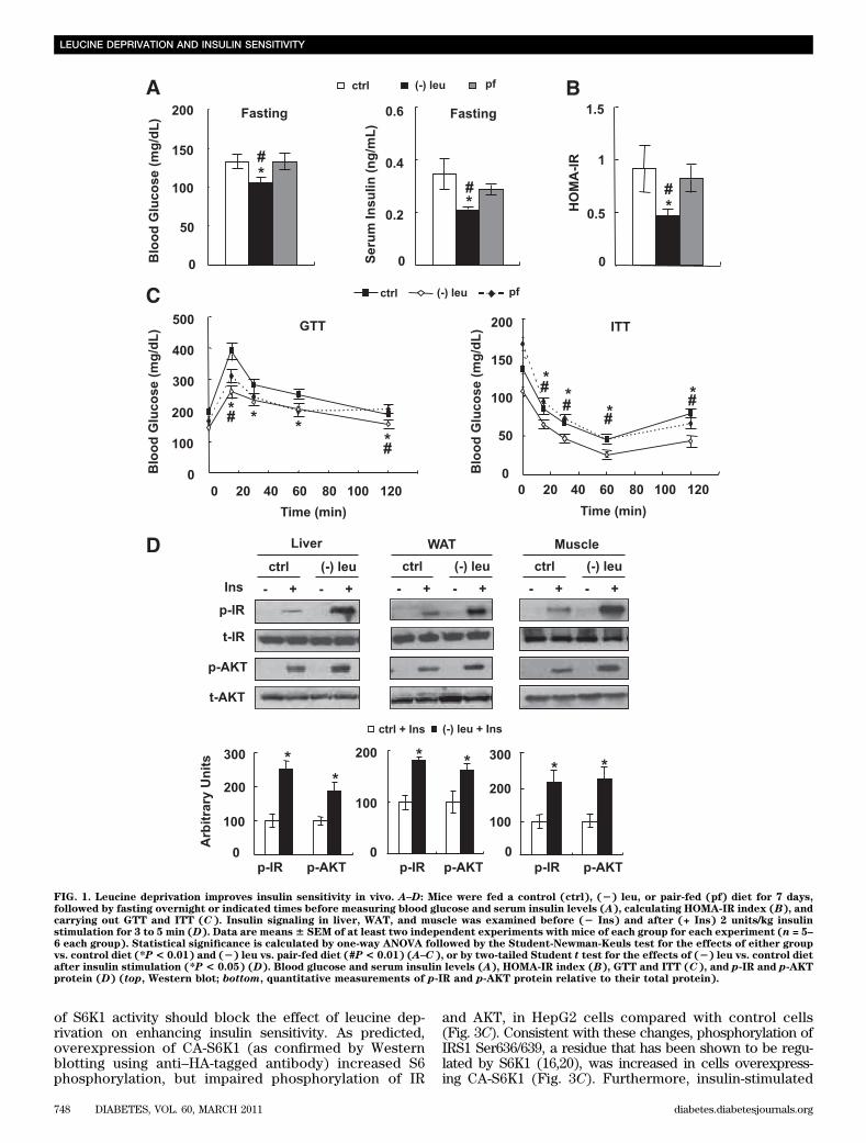
observations (10), levels of serum insulin decreased,but blood glucose levels remained unchanged, in leucine-deprived mice in the fed state (data not shown). By contrast,fasting blood glucose levels were lower in leucine-deprivedmice compared with control or pair-fed mice. Levels ofserum insulin were also decreased 50% in these mice (Fig.1A). Consistent with these changes, the HOMA-IR indexwas decreased in leucine-deprived mice (Fig. 1B). Glucosetolerance and clearance were further examined by GTTsand ITTs, respectively. Fifteen minutes after injection ofglucose, blood glucose levels were significantly lower inpair-fed and leucine-deprived mice compared with con-trols, with lowest levels in leucine-deprived mice. In ad-dition, blood glucose levels were decreased more quicklyafter administration of insulin in leucine-deprived micecompared with control or pair-fed mice (Fig. 1C).
Increased insulin sensitivity in mice under leucine dep-rivation suggests an increase in insulin sensitivity in oneor more peripheral tissues, including liver, white adiposetissue (WAT), and muscle. To test this possibility, weexamined the levels of phosphorylation of two key com-ponents in the insulin signaling pathways (15), insulin re-ceptor (IR) and protein kinase B (AKT), in these tissuesafter infusion of insulin (2 units/kg) into the hepatic portalvein, as described previously (13). As expected, insulin-stimulated phosphorylation of IR and AKT was increased inthese tissues in leucine-deprived mice compared with micemaintained on a control diet (Fig. 1D).Leucine deprivation increases insulin sensitivity invitro. To determine whether leucine deprivation has a di-rect effect on the insulin signaling in liver, we examinedthe effect of leucine deprivation on insulin signaling inboth the human hepatoma–derived cell line HepG2 andprimary cultured mice hepatocytes. Consistent with ourin vivo observations (Fig. 1D), insulin-stimulated phos-phorylation of IR and AKT was significantly elevatedunder leucine deprivation in HepG2 cells (Fig. 2A) andprimary cultured hepatocytes (Fig. 2B). Furthermore,insulin-stimulated incorporation of glucose into glycogenwas significantly increased in cells incubated in (2) leumedium for 36 h. Leucine deprivation, however, also in-creased baseline levels of glucose incorporation intoglycogen (Fig. 2C).Leucine deprivation increases insulin sensitivity bydecreasing mTOR/S6K1 signaling in vitro. DecreasedmTOR/S6K1 signaling has been shown to increase insulinsensitivity (16,17). Because mTOR activity is decreased inthe livers of mice maintained on a (2) leu diet (14), wespeculated that mTOR/S6K1 signaling may regulate insulinsensitivity under leucine deprivation.
To investigate this possibility, we examined levels ofphosphorylation of mTOR and its downstream targets, in-cluding S6K1 and ribosomal protein S6, in HepG2 cellsincubated in (2) leu medium for 24 h. Levels of phos-phorylation of these proteins were also used as indicatorsfor the activation status of mTOR (18). Consistent withprevious results (19), phosphorylation of these proteinswas decreased by leucine deprivation in HepG2 cells (Fig.3A). Similar to the effect of leucine deprivation, pre-treatment with the mTOR inhibitor 50 nmol/L rapamycinfor 1 h significantly decreased phosphorylation of S6 inHepG2 cells compared with pretreatment with vehicle. Bycontrast, insulin-stimulated phosphorylation of IR and AKTwas increased significantly in these cells (Fig. 3B).
If decreased mTOR/S6K1 signaling regulates hepaticinsulin sensitivity under leucine deprivation, upregulation
F. XIAO AND ASSOCIATES
diabetes.diabetesjournals.org DIABETES, VOL. 60, MARCH 2011 747
of S6K1 activity should block the effect of leucine dep-rivation on enhancing insulin sensitivity. As predicted,overexpression of CA-S6K1 (as confirmed by Westernblotting using anti–HA-tagged antibody) increased S6phosphorylation, but impaired phosphorylation of IR
and AKT, in HepG2 cells compared with control cells(Fig. 3C). Consistent with these changes, phosphorylation ofIRS1 Ser636/639, a residue that has been shown to be regu-lated by S6K1 (16,20), was increased in cells overexpress-ing CA-S6K1 (Fig. 3C). Furthermore, insulin-stimulated
FIG. 1. Leucine deprivation improves insulin sensitivity in vivo. A–D: Mice were fed a control (ctrl), (2) leu, or pair-fed (pf) diet for 7 days,followed by fasting overnight or indicated times before measuring blood glucose and serum insulin levels (A), calculating HOMA-IR index (B), andcarrying out GTT and ITT (C). Insulin signaling in liver, WAT, and muscle was examined before (2 Ins) and after (+ Ins) 2 units/kg insulinstimulation for 3 to 5 min (D). Data are means6 SEM of at least two independent experiments with mice of each group for each experiment (n = 5–6 each group). Statistical significance is calculated by one-way ANOVA followed by the Student-Newman-Keuls test for the effects of either groupvs. control diet (*P< 0.01) and (2) leu vs. pair-fed diet (#P< 0.01) (A–C), or by two-tailed Student t test for the effects of (2) leu vs. control dietafter insulin stimulation (*P < 0.05) (D). Blood glucose and serum insulin levels (A), HOMA-IR index (B), GTT and ITT (C), and p-IR and p-AKTprotein (D) (top, Western blot; bottom, quantitative measurements of p-IR and p-AKT protein relative to their total protein).
LEUCINE DEPRIVATION AND INSULIN SENSITIVITY
748 DIABETES, VOL. 60, MARCH 2011 diabetes.diabetesjournals.org
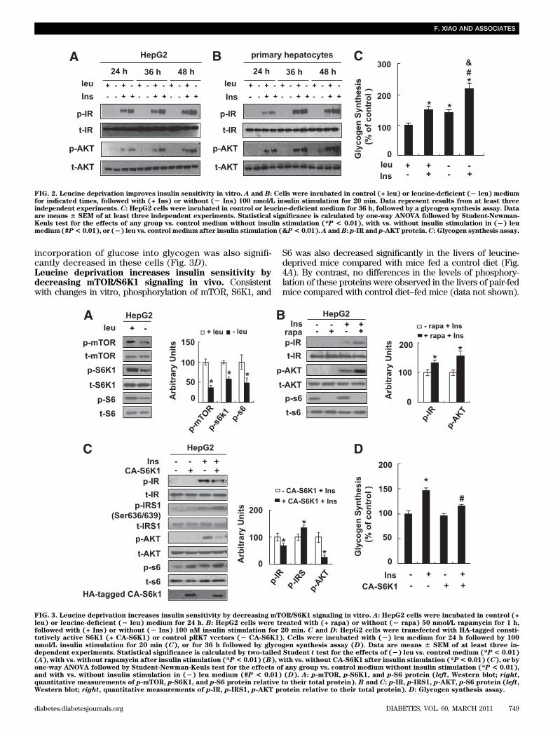
incorporation of glucose into glycogen was also signifi-cantly decreased in these cells (Fig. 3D).Leucine deprivation increases insulin sensitivity bydecreasing mTOR/S6K1 signaling in vivo. Consistentwith changes in vitro, phosphorylation of mTOR, S6K1, and
S6 was also decreased significantly in the livers of leucine-deprived mice compared with mice fed a control diet (Fig.4A). By contrast, no differences in the levels of phosphory-lation of these proteins were observed in the livers of pair-fedmice compared with control diet–fed mice (data not shown).
FIG. 2. Leucine deprivation improves insulin sensitivity in vitro. A and B: Cells were incubated in control (+ leu) or leucine-deficient (2 leu) mediumfor indicated times, followed with (+ Ins) or without (2 Ins) 100 nmol/L insulin stimulation for 20 min. Data represent results from at least threeindependent experiments. C: HepG2 cells were incubated in control or leucine-deficient medium for 36 h, followed by a glycogen synthesis assay. Dataare means 6 SEM of at least three independent experiments. Statistical significance is calculated by one-way ANOVA followed by Student-Newman-Keuls test for the effects of any group vs. control medium without insulin stimulation (*P < 0.01), with vs. without insulin stimulation in (2) leumedium (#P< 0.01), or (2) leu vs. control medium after insulin stimulation (&P< 0.01).A andB: p-IR and p-AKT protein.C: Glycogen synthesis assay.
FIG. 3. Leucine deprivation increases insulin sensitivity by decreasing mTOR/S6K1 signaling in vitro. A: HepG2 cells were incubated in control (+leu) or leucine-deficient (2 leu) medium for 24 h. B: HepG2 cells were treated with (+ rapa) or without (2 rapa) 50 nmol/L rapamycin for 1 h,followed with (+ Ins) or without (2 Ins) 100 nM insulin stimulation for 20 min. C and D: HepG2 cells were transfected with HA-tagged consti-tutively active S6K1 (+ CA-S6K1) or control pRK7 vectors (2 CA-S6K1). Cells were incubated with (2) leu medium for 24 h followed by 100nmol/L insulin stimulation for 20 min (C), or for 36 h followed by glycogen synthesis assay (D). Data are means 6 SEM of at least three in-dependent experiments. Statistical significance is calculated by two-tailed Student t test for the effects of (2) leu vs. control medium (*P < 0.01)(A), with vs. without rapamycin after insulin stimulation (*P< 0.01) (B), with vs. without CA-S6K1 after insulin stimulation (*P< 0.01) (C), or byone-way ANOVA followed by Student-Newman-Keuls test for the effects of any group vs. control medium without insulin stimulation (*P < 0.01),and with vs. without insulin stimulation in (2) leu medium (#P < 0.01) (D). A: p-mTOR, p-S6K1, and p-S6 protein (left, Western blot; right,quantitative measurements of p-mTOR, p-S6K1, and p-S6 protein relative to their total protein). B and C: p-IR, p-IRS1, p-AKT, p-S6 protein (left,Western blot; right, quantitative measurements of p-IR, p-IRS1, p-AKT protein relative to their total protein). D: Glycogen synthesis assay.
F. XIAO AND ASSOCIATES
diabetes.diabetesjournals.org DIABETES, VOL. 60, MARCH 2011 749
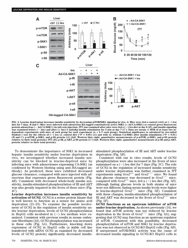
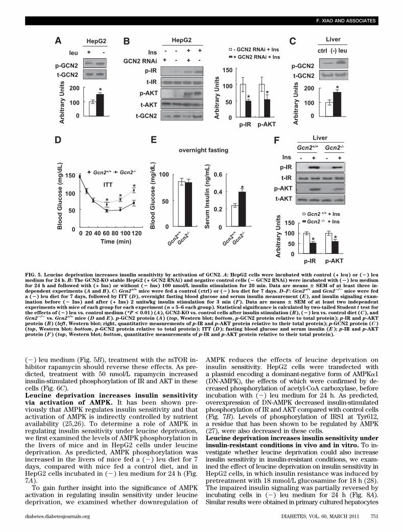
To demonstrate the importance of S6K1 in increasedhepatic insulin sensitivity under leucine deprivation invivo, we investigated whether increased insulin sen-sitivity can be blocked in leucine-deprived mice byinfecting mice with adenoviruses expressing CA-S6K1 (asconfirmed by Western blotting using anti–HA-tagged an-tibody). As predicted, these mice exhibited decreasedglucose clearance, compared with mice injected with ad-enovirus that expresses green fluorescent protein (Fig.4B). Consistent with decreased whole-body insulin sen-sitivity, insulin-stimulated phosphorylation of IR and AKTwas also greatly impaired in the livers of these mice (Fig.4C).Leucine deprivation increases insulin sensitivity byactivation of GCN2. GCN2 is a serine protein kinase thatis well known to function as a sensor for amino aciddeprivation (21–23). To examine the possible involve-ment of GCN2 in the regulation of insulin sensitivity un-der leucine deprivation, levels of GCN2 phosphorylationin HepG2 cells incubated in (2) leu medium were ex-amined. Consistent with previous results in mouse embry-onic fibroblasts (24), GCN2 phosphorylation was increasedin these cells (Fig. 5A). Furthermore, knocking downexpression of GCN2 in HepG2 cells (a stable cell linetransfected with siRNA GCN2 as examined by decreasedlevels of GCN2 protein) significantly decreased insulin-
stimulated phosphorylation of IR and AKT under leucinedeprivation (Fig. 5B).
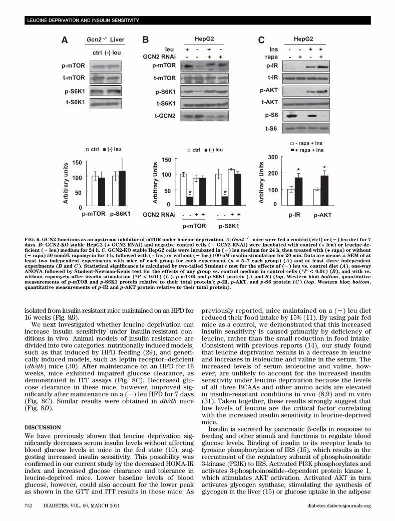
Consistent with our in vitro results, levels of GCN2phosphorylation were also increased in the livers of micemaintained on a (2) leu diet for 7 days (Fig. 5C). The roleof GCN2 in the regulation of increased insulin sensitivityunder leucine deprivation was further examined in ITTexperiments using Gcn2+/+ and Gcn22/2 mice. We foundthat glucose clearance was decreased in Gcn22/2 mice,compared with Gcn2+/+ mice, fed a (2) leu diet (Fig. 5D).Although fasting glucose levels in both strains of micewere not different, fasting serum insulin levels were higherin leucine-deprived Gcn22/2 mice (Fig. 5E). Consistentwith these changes, insulin-stimulated phosphorylation ofIR and AKT was decreased in the livers of Gcn22/2 mice(Fig. 5F).GCN2 functions as an upstream inhibitor of mTORunder leucine deprivation. As shown previously (14), wefound that mTOR activity was not reduced by leucinedeprivation in the livers of Gcn22/2 mice (Fig. 6A), sug-gesting that GCN2 may function as an upstream regulatorof mTOR. Consistent with this possibility, decreasedphosphorylation of mTOR and S6K1 by leucine depriva-tion was not observed in GCN2-KO HepG2 cells (Fig. 6B).If unrepressed mTOR/S6K1 activity was the cause ofdecreased insulin signaling in GCN2-KO HepG2 cells in
FIG. 4. Leucine deprivation increases insulin sensitivity by decreasing mTOR/S6K1 signaling in vivo. A: Mice were fed a control (ctrl) or (2) leudiet for 7 days. B and C: Mice were infected with adenovirus HA-tagged constitutively active S6K1 (+ Ad-CA-S6K1) or control green fluorescentprotein adenovirus (2 Ad-CA-S6K1) via tail-vein injection. ITT was examined after mice were fed a (2) leu diet at day 6 (B), and insulin signalingwas examined before (2 Ins) and after (+ Ins) 2 units/kg insulin stimulation for 3 min at day 7 (C). Data are means 6 SEM of at least two in-dependent experiments with mice of each group for each experiment (n = 5–7 each group). Statistical significance is calculated by two-tailedStudent t test for the effects of (2) leu vs. control diet (*P < 0.05) (A) and with vs. without CA-S6K1 after insulin stimulation (*P < 0.01)(B and C). p-mTOR, p-S6K1, and p-S6 protein (A) (left, Western blot; right, quantitative measurements of p-mTOR, p-S6K1, and p-S6 proteinrelative to their total protein); ITT (B); p-IR, p-AKT, p-S6 protein (C) (left, Western blot; right, quantitative measurements of p-IR and p-AKT,protein relative to their total protein).
LEUCINE DEPRIVATION AND INSULIN SENSITIVITY
750 DIABETES, VOL. 60, MARCH 2011 diabetes.diabetesjournals.org
(2) leu medium (Fig. 5B), treatment with the mTOR in-hibitor rapamycin should reverse these effects. As pre-dicted, treatment with 50 nmol/L rapamycin increasedinsulin-stimulated phosphorylation of IR and AKT in thesecells (Fig. 6C).Leucine deprivation increases insulin sensitivityvia activation of AMPK. It has been shown pre-viously that AMPK regulates insulin sensitivity and thatactivation of AMPK is indirectly controlled by nutrientavailability (25,26). To determine a role of AMPK inregulating insulin sensitivity under leucine deprivation,we first examined the levels of AMPK phosphorylation inthe livers of mice and in HepG2 cells under leucinedeprivation. As predicted, AMPK phosphorylation wasincreased in the livers of mice fed a (2) leu diet for 7days, compared with mice fed a control diet, and inHepG2 cells incubated in (2) leu medium for 24 h (Fig.7A).
To gain further insight into the significance of AMPKactivation in regulating insulin sensitivity under leucinedeprivation, we examined whether downregulation of
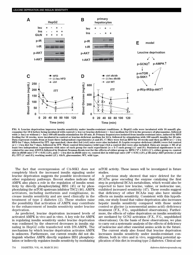
AMPK reduces the effects of leucine deprivation oninsulin sensitivity. HepG2 cells were transfected witha plasmid encoding a dominant-negative form of AMPKa1(DN-AMPK), the effects of which were confirmed by de-creased phosphorylation of acetyl-CoA carboxylase, beforeincubation with (2) leu medium for 24 h. As predicted,overexpression of DN-AMPK decreased insulin-stimulatedphosphorylation of IR and AKT compared with control cells(Fig. 7B). Levels of phosphorylation of IRS1 at Tyr612,a residue that has been shown to be regulated by AMPK(27), were also decreased in these cells.Leucine deprivation increases insulin sensitivity underinsulin-resistant conditions in vivo and in vitro. To in-vestigate whether leucine deprivation could also increaseinsulin sensitivity in insulin-resistant conditions, we exam-ined the effect of leucine deprivation on insulin sensitivity inHepG2 cells, in which insulin resistance was induced bypretreatment with 18 mmol/L glucosamine for 18 h (28).The impaired insulin signaling was partially reversed byincubating cells in (2) leu medium for 24 h (Fig. 8A).Similar results were obtained in primary cultured hepatocytes
FIG. 5. Leucine deprivation increases insulin sensitivity by activation of GCN2. A: HepG2 cells were incubated with control (+ leu) or (2) leumedium for 24 h. B: The GCN2-KO stable HepG2 (+ GCN2 RNAi) and negative control cells (2 GCN2 RNAi) were incubated with (2) leu mediumfor 24 h and followed with (+ Ins) or without (2 Ins) 100 nmol/L insulin stimulation for 20 min. Data are means 6 SEM of at least three in-dependent experiments (A and B). C: Gcn2+/+ mice were fed a control (ctrl) or (2) leu diet for 7 days. D–F: Gcn2+/+ and Gcn22/2
mice were feda (2) leu diet for 7 days, followed by ITT (D), overnight fasting blood glucose and serum insulin measurement (E), and insulin signaling exam-ination before (2 Ins) and after (+ Ins) 2 units/kg insulin stimulation for 3 min (F). Data are means 6 SEM of at least two independentexperiments with mice of each group for each experiment (n = 5–6 each group). Statistical significance is calculated by two-tailed Student t test forthe effects of (2) leu vs. control medium (*P < 0.01) (A), GCN2-KO vs. control cells after insulin stimulation (B), (2) leu vs. control diet (C), andGcn22/2
vs. Gcn2+/+ mice (D and E). p-GCN2 protein (A) (top, Western blot; bottom, p-GCN2 protein relative to total protein); p-IR and p-AKTprotein (B) (left, Western blot; right, quantitative measurements of p-IR and p-AKT protein relative to their total protein); p-GCN2 protein (C)(top, Western blot; bottom, p-GCN2 protein relative to total protein); ITT (D); fasting blood glucose and serum insulin (E); p-IR and p-AKTprotein (F) (top, Western blot; bottom, quantitative measurements of p-IR and p-AKT protein relative to their total protein).
F. XIAO AND ASSOCIATES
diabetes.diabetesjournals.org DIABETES, VOL. 60, MARCH 2011 751
isolated from insulin-resistant mice maintained on an HFD for16 weeks (Fig. 8B).
We next investigated whether leucine deprivation canincrease insulin sensitivity under insulin-resistant con-ditions in vivo. Animal models of insulin resistance aredivided into two categories: nutritionally induced models,such as that induced by HFD feeding (29), and geneti-cally induced models, such as leptin receptor–deficient(db/db) mice (30). After maintenance on an HFD for 16weeks, mice exhibited impaired glucose clearance, asdemonstrated in ITT assays (Fig. 8C). Decreased glu-cose clearance in these mice, however, improved sig-nificantly after maintenance on a (2) leu HFD for 7 days(Fig. 8C). Similar results were obtained in db/db mice(Fig. 8D).
DISCUSSION
We have previously shown that leucine deprivation sig-nificantly decreases serum insulin levels without affectingblood glucose levels in mice in the fed state (10), sug-gesting increased insulin sensitivity. This possibility wasconfirmed in our current study by the decreased HOMA-IRindex and increased glucose clearance and tolerance inleucine-deprived mice. Lower baseline levels of bloodglucose, however, could also account for the lower peakas shown in the GTT and ITT results in these mice. As
previously reported, mice maintained on a (2) leu dietreduced their food intake by 15% (11). By using pair-fedmice as a control, we demonstrated that this increasedinsulin sensitivity is caused primarily by deficiency ofleucine, rather than the small reduction in food intake.Consistent with previous reports (14), our study foundthat leucine deprivation results in a decrease in leucineand increases in isoleucine and valine in the serum. Theincreased levels of serum isoleucine and valine, how-ever, are unlikely to account for the increased insulinsensitivity under leucine deprivation because the levelsof all three BCAAs and other amino acids are elevatedin insulin-resistant conditions in vivo (8,9) and in vitro(31). Taken together, these results strongly suggest thatlow levels of leucine are the critical factor correlatingwith the increased insulin sensitivity in leucine-deprivedmice.
Insulin is secreted by pancreatic b-cells in response tofeeding and other stimuli and functions to regulate bloodglucose levels. Binding of insulin to its receptor leads totyrosine phosphorylation of IRS (15), which results in therecruitment of the regulatory subunit of phosphoinositide3-kinase (PI3K) to IRS. Activated PI3K phosphorylates andactivates 3-phosphoinositide–dependent protein kinase 1,which stimulates AKT activation. Activated AKT in turnactivates glycogen synthase, stimulating the synthesis ofglycogen in the liver (15) or glucose uptake in the adipose
FIG. 6. GCN2 functions as an upstream inhibitor of mTOR under leucine deprivation. A: Gcn22/2mice were fed a control (ctrl) or (2) leu diet for 7
days. B: GCN2-KO stable HepG2 (+ GCN2 RNAi) and negative control cells (2 GCN2 RNAi) were incubated with control (+ leu) or leucine-de-ficient (2 leu) medium for 24 h. C: GCN2-KO stable HepG2 cells were incubated in (2) leu medium for 24 h, then treated with (+ rapa) or without(2 rapa) 50 nmol/L rapamycin for 1 h, followed with (+ Ins) or without (2 Ins) 100 nM insulin stimulation for 20 min. Data are means 6 SEM of atleast two independent experiments with mice of each group for each experiment (n = 5–7 each group) (A) and at least three independentexperiments (B and C). Statistical significance is calculated by two-tailed Student t test for the effects of (2) leu vs. control diet (A), one-wayANOVA followed by Student-Newman-Keuls test for the effects of any group vs. control medium in control cells (*P < 0.01) (B), and with vs.without rapamycin after insulin stimulation (*P < 0.01) (C). p-mTOR and p-S6K1 protein (A and B) (top, Western blot; bottom, quantitativemeasurements of p-mTOR and p-S6K1 protein relative to their total protein); p-IR, p-AKT, and p-S6 protein (C) (top, Western blot; bottom,quantitative measurements of p-IR and p-AKT protein relative to their total protein).
LEUCINE DEPRIVATION AND INSULIN SENSITIVITY
752 DIABETES, VOL. 60, MARCH 2011 diabetes.diabetesjournals.org

tissue and muscle (32). Our current results show thatleucine deprivation increases insulin signaling in all thetissues examined, including liver, WAT, and muscle, con-sistent with increased insulin sensitivity.
mTOR is a serine-threonine protein kinase that has beenshown to be essential for protein synthesis, growth,development, and proliferation (18,33,34). Studies haveshown that mTOR activity is regulated by BCAA availability,especially leucine (35). For example, mTOR activity is in-creased by elevated extracellular levels of leucine (33). Bycontrast, its activity is decreased in the livers of mice underleucine deprivation (14). Activation of mTOR/S6K1 sig-naling has been shown to contribute to the developmentof insulin resistance. Recent studies demonstrate thatthe mTOR downstream target S6K1 directly phosphor-ylates IRS1 serine residues, including Ser-302/307 (36),Ser-307/312 (37), Ser-632/636 (20), and Ser-1097/1101(38). Increased IRS1 serine phosphorylation reduces theactivity of IRS1, thereby impairing PI3K/AKT signaling andincreasing insulin resistance (39,40). This effect is reversedin vitro by rapamycin (37). Consistent with these results,mice deleted for S6K1 have reduced phosphorylation ofIRS1 Ser-636 (16) and do not develop diet-induced insulinresistance.
On the basis of these studies, we hypothesized thatleucine deprivation increases insulin sensitivity by de-creasing mTOR/S6K1 activity. Consistent with our hy-pothesis, decreased mTOR/S6K1 signaling in the livers ofmice and HepG2 cells under leucine deprivation was ob-served. Furthermore, overexpression of CA-S6K1 inhibitedinsulin signaling in HepG2 cells incubated in (2) leumedium. A key role for S6K1 in the regulation of insulinsensitivity was confirmed in mice injected with adeno-viruses expressing CA-S6K1. These mice exhibited de-creased glucose clearance and insulin signaling in liver,compared with control mice, when fed a (2) leu diet. Taken
together, our findings demonstrate an important role formTOR/S6K1 in the regulation of insulin sensitivity underleucine deprivation.
To look for upstream regulators for mTOR, our atten-tion was drawn to the amino acid sensor GCN2, a kinasethat is activated by uncharged tRNAs in response todeprivation of essential amino acids, including leucine, inyeast and mammals (21–23). Activated GCN2 phosphor-ylates eukaryotic initiation factor 2-a, thereby repressinggeneral protein synthesis, but increasing translation ofproteins related to amino acid biosynthesis and transport(41–44). We recently demonstrated that GCN2 also reg-ulates lipid metabolism during leucine deprivation (10).Because deficiency in amino acid is sensed by GCN2,we speculated that GCN2 may also regulate glucose uti-lization by modulating insulin sensitivity during leucinedeprivation.
Consistent with this hypothesis, knocking down ex-pression of GCN2 using siRNA decreased insulin signalingin HepG2 cells incubated in (2) leu medium. The key rolefor GCN2 in the regulation of insulin sensitivity was con-firmed by the observation that Gcn22/2 mice exhibiteddecreased glucose clearance, compared with wild-typemice, when fed a (2) leu diet. Consistent with these results,higher serum insulin levels were required to maintain thesame levels of fasting blood glucose in Gcn22/2 micecompared with Gcn2+/+ mice. In addition, we provide ev-idence that GCN2 regulates insulin sensitivity via in-hibition of mTOR activity during leucine deprivation. Thus,in addition to regulation of amino acid and lipid metabo-lism (10,14), GCN2 also functions in glucose metabolism.Taken together, our results strongly suggest that GCN2functions as a master regulator of metabolic adaptation todeprivation of essential amino acids. The mechanism bywhich leucine deprivation activates GCN2 requires furtherinvestigation.
FIG. 7. Leucine deprivation improves insulin sensitivity via activation of AMPK. A: Mice were fed a control (ctrl) or (2) leu diet for 7 days, andHepG2 cells were incubated with control (+ leu) or leucine-deficient (2 leu) medium for 24 h. B: HepG2 cells were transfected with dominant-negative form of AMPKa1 (+ DN-AMPK) or empty vectors (2 DN-AMPK). After incubation with (2) leu medium for 24 h, cells were stimulatedwith (+ Ins) or without (2 Ins) 100 nmol/L insulin for 20 min. Data are means 6 SEM of at least three independent experiments. Statisticalsignificance is calculated by two-tailed Student t test for the effects of (2) leu vs. control treatment (*P < 0.01) (A) and with vs. without DN-AMPK after insulin stimulation (B). p-AMPK protein (A) (top, Western blot; bottom, quantitative measurements of p-AMPK protein relative totheir total protein); p-IR, p-IRS1, p-AKT, p-ACC protein (B) (left, Western blot; right, quantitative measurements of p-IR, p-IRS1, and p-AKTproteins relative to their total protein).
F. XIAO AND ASSOCIATES
diabetes.diabetesjournals.org DIABETES, VOL. 60, MARCH 2011 753
The fact that overexpression of CA-S6K1 does notcompletely block the increased insulin signaling underleucine deprivation suggests the possible involvement ofother regulatory pathways. Recent studies indicate thatAMPK also plays a role in the regulation of insulin sensi-tivity by directly phosphorylating IRS1 (45) or by phos-phorylating the mTOR upstream inhibitor TSC2 (46). AMPKactivators, including metformin and rosiglitazone, in-crease insulin sensitivity and are used clinically in thetreatment of type 2 diabetes (2). These studies raisethe possibility that activation of AMPK may contributeto the enhancement of insulin sensitivity under leucinedeprivation.
As predicted, leucine deprivation increased levels ofactivated AMPK in vivo and in vitro. A key role for AMPKin regulating insulin sensitivity under leucine deprivationwas confirmed by the observed decreases in insulin sig-naling in HepG2 cells transfected with DN-AMPK. Themechanism by which leucine deprivation activates AMPKis unknown. Furthermore, our current results did not dis-tinguish whether AMPK directly regulates IRS1 phosphory-lation or indirectly regulates insulin sensitivity by modulating
mTOR activity. These issues will be investigated in futurestudies.
A previous study showed that mice deleted for theBCATm gene encoding the enzyme catalyzing the firststep in peripheral BCAA metabolism, which would also beexpected to have low leucine, valine, or isoleucine use,exhibited increased sensitivity (47). These results suggestthat deficiency of other BCAAs may also have similareffects on insulin sensitivity. Consistent with this hypoth-esis, our study found that valine deprivation also increaseshepatic insulin sensitivity compared with those undercontrol or glycine (a nonessential amino acid)–deficiencytreatment (F.X., F.G., unpublished observations). Further-more, the effects of valine deprivation on insulin sensitivityare mediated by GCN2 activation (F.X., F.G., unpublishedobservations). On the basis of these observations, we planto carry out a systematic analysis of the effects of deficiencyof isoleucine and other essential amino acids in the future.
The current study also found that leucine deprivationincreases insulin sensitivity under insulin-resistant con-ditions, both in vitro and in vivo, suggesting potential ap-plication of this diet in treating type 2 diabetes. Clinical use
FIG. 8. Leucine deprivation improves insulin sensitivity under insulin-resistant conditions. A: HepG2 cells were incubated with 18 mmol/L glu-cosamine for 18 h before being incubated with control (+ leu) or leucine-deficient (2 leu) medium for 24 h in the presence of glucosamine, followedwith (+ Ins) or without (2 Ins) 100 nM insulin stimulation for 20 min. B: Primary hepatocytes isolated from insulin-resistant mice, induced by HFDfeeding for 16 weeks, were incubated in control or leucine-deficient medium for 24 h, followed by stimulation with 100 nmol/L insulin for 20 min.Data represent results from at least three independent experiments (A and B). C: Mice were fed an HFD for 16 weeks and then an HFD or (2) leuHFD for 7 days, followed by ITT. Age-matched, chow-diet fed (ctrl) mice were also included. D: Leptin receptor–deficient (db/db) were fed controlor (2) leu diet for 7 days, followed by ITT. Their control littermates (wild type) fed a control diet were also included. Data are means 6 SE of atleast two independent experiments with mice of each group for each experiment (n = 5–7 each group) (C and D). Statistical significance is cal-culated by one-way ANOVA followed by Student-Neuman-Keuls test for the effects of either group vs. HFD (*P< 0.01) (C), either group vs. controldiet in db/dbmice (*P< 0.01) (D), and (2) leu diet in db/dbmice vs. control diet in wild-type mice (#P< 0.01) (D). p-IR and p-AKT protein (A andB); ITT (C and D); working model (E). GlcN, glucosamine; WT, wild type.
LEUCINE DEPRIVATION AND INSULIN SENSITIVITY
754 DIABETES, VOL. 60, MARCH 2011 diabetes.diabetesjournals.org
of this diet, however, is still premature, because the safetyof short- and long-term leucine deprivation in humans hasnot been examined. Determining the optimal concentra-tion of leucine and the duration of therapeutic (2) leudiets will be important in future studies.
In summary, we have shown that leucine deprivationimproves insulin sensitivity under normal and insulin-resistant conditions, both in vitro and in vivo. Further-more, the GCN2/mTOR/S6K1 and AMPK pathways playimportant roles in the regulation of hepatic insulin sen-sitivity by leucine deprivation. The relative contributionof each of these pathways remains to be demonstrated. Italso remains to be demonstrated whether these pathwaysfunction in the regulation of insulin sensitivity in othertissues, including muscle and WAT.
ACKNOWLEDGMENTS
This work was supported by grants from the Ministryof Science and Technology of China (973 Program2009CB919001 and 2010CB912502); National Natural Sci-ence Foundation (30871208 and 30890043); Chief ScientistProgram of Shanghai Institutes for Biological Sciences,Chinese Academy of Sciences (SIBS2008006); Scienceand Technology Commission of Shanghai Municipality(08DJ1400601); 2010 Key Program of Clinical ResearchCenter, Institute for Nutritional Sciences, Shanghai Insti-tutes for Biological Sciences, Chinese Academy of Sci-ences (CRC2010005); Key Program of Shanghai Scientificand Technological Innovation Action Plan (10JC1416900);and CAS-Pfizer Project (Pfizer-SIBS2 01002). F.G. was alsosupported by the One Hundred Talents Program of theChinese Academy of Sciences and the Pujiang Talents Pro-gram of Shanghai Municipality (08PJ1410700).
No potential conflicts of interest relevant to this articlewere reported.
F.X. researched data, contributed to discussion, andwrote, reviewed, and edited the article. Z.H. researcheddata and contributed to discussion. H.L. researched data,contributed to discussion, and reviewed and edited thearticle. J.Y., C.W., S.C., Q.M., and Y.C. researched data andcontributed to discussion. X.G. and J.L. provided researchmaterial and contributed to discussion. Y.L. contributedto discussion and reviewed and edited the article. F.G.contributed to discussion and wrote, reviewed, and editedthe article.
REFERENCES
1. Savage DB, Petersen KF, Shulman GI. Disordered lipid metabolism and thepathogenesis of insulin resistance. Physiol Rev 2007;87:507–520
2. Zhang BB, Zhou G, Li C. AMPK: an emerging drug target for diabetes andthe metabolic syndrome. Cell Metab 2009;9:407–416
3. Monzillo LU, Hamdy O, Horton ES, et al. Effect of lifestyle modification onadipokine levels in obese subjects with insulin resistance. Obes Res 2003;11:1048–1054
4. Buchanan TA, Xiang AH, Peters RK, et al. Preservation of pancreatic beta-cell function and prevention of type 2 diabetes by pharmacological treat-ment of insulin resistance in high-risk Hispanic women. Diabetes 2002;51:2796–2803
5. Nair KS, Short KR. Hormonal and signaling role of branched-chain aminoacids. J Nutr 2005;135(Suppl.):1547S–1552S
6. Zhang Y, Guo K, LeBlanc RE, Loh D, Schwartz GJ, Yu YH. Increasing di-etary leucine intake reduces diet-induced obesity and improves glucoseand cholesterol metabolism in mice via multimechanisms. Diabetes 2007;56:1647–1654
7. Nairizi A, She P, Vary TC, Lynch CJ. Leucine supplementation of drinkingwater does not alter susceptibility to diet-induced obesity in mice. J Nutr2009;139:715–719
8. Newgard CB, An J, Bain JR, et al. A branched-chain amino acid-relatedmetabolic signature that differentiates obese and lean humans and con-tributes to insulin resistance. Cell Metab 2009;9:311–326
9. Tremblay F, Krebs M, Dombrowski L, et al. Overactivation of S6 kinase 1as a cause of human insulin resistance during increased amino acidavailability. Diabetes 2005;54:2674–2684
10. Guo F, Cavener DR. The GCN2 eIF2alpha kinase regulates fatty-acid ho-meostasis in the liver during deprivation of an essential amino acid. CellMetab 2007;5:103–114
11. Cheng Y, Meng Q, Wang C, et al. Leucine deprivation decreases fat massby stimulation of lipolysis in white adipose tissue and upregulation ofuncoupling protein 1 (UCP1) in brown adipose tissue. Diabetes 2010;59:17–25
12. Sun C, Zhang F, Ge X, et al. SIRT1 improves insulin sensitivity underinsulin-resistant conditions by repressing PTP1B. Cell Metab 2007;6:307–319
13. Wang Q, Jiang L, Wang J, et al. Abrogation of hepatic ATP-citrate lyaseprotects against fatty liver and ameliorates hyperglycemia in leptin receptor-deficient mice. Hepatology 2009;49:1166–1175
14. Anthony TG, McDaniel BJ, Byerley RL, et al. Preservation of liver proteinsynthesis during dietary leucine deprivation occurs at the expense ofskeletal muscle mass in mice deleted for eIF2 kinase GCN2. J Biol Chem2004;279:36553–36561
15. Saltiel AR, Kahn CR. Insulin signalling and the regulation of glucose andlipid metabolism. Nature 2001;414:799–806
16. Um SH, Frigerio F, Watanabe M, et al. Absence of S6K1 protects againstage- and diet-induced obesity while enhancing insulin sensitivity. Nature2004;431:200–205
17. Krebs M, Brunmair B, Brehm A, et al. The mammalian target of rapamycinpathway regulates nutrient-sensitive glucose uptake in man. Diabetes2007;56:1600–1607
18. Wullschleger S, Loewith R, Hall MN. TOR signaling in growth and me-tabolism. Cell 2006;124:471–484
19. Sarbassov DD, Sabatini DM. Redox regulation of the nutrient-sensitiveraptor-mTOR pathway and complex. J Biol Chem 2005;280:39505–39509
20. Ozes ON, Akca H, Mayo LD, et al. A phosphatidylinositol 3-kinase/Akt/mTOR pathway mediates and PTEN antagonizes tumor necrosis factorinhibition of insulin signaling through insulin receptor substrate-1. ProcNatl Acad Sci U S A 2001;98:4640–4645
21. Wek RC, Jackson BM, Hinnebusch AG. Juxtaposition of domains homol-ogous to protein kinases and histidyl-tRNA synthetases in GCN2 proteinsuggests a mechanism for coupling GCN4 expression to amino acidavailability. Proc Natl Acad Sci U S A 1989;86:4579–4583
22. Wek SA, Zhu S, Wek RC. The histidyl-tRNA synthetase-related sequence inthe eIF-2 alpha protein kinase GCN2 interacts with tRNA and is requiredfor activation in response to starvation for different amino acids. Mol CellBiol 1995;15:4497–4506
23. Hinnebusch AG. The eIF-2 alpha kinases: regulators of protein synthesis instarvation and stress. Semin Cell Biol 1994;5:417–426
24. Harding HP, Zhang Y, Zeng H, et al. An integrated stress response regulatesamino acid metabolism and resistance to oxidative stress. Mol Cell 2003;11:619–633
25. Ruderman N, Prentki M. AMP kinase and malonyl-CoA: targets for therapyof the metabolic syndrome. Nat Rev Drug Discov 2004;3:340–351
26. Long YC, Zierath JR. AMP-activated protein kinase signaling in metabolicregulation. J Clin Invest 2006;116:1776–1783
27. Wang C, Mao X, Wang L, et al. Adiponectin sensitizes insulin signaling byreducing p70 S6 kinase-mediated serine phosphorylation of IRS-1. J BiolChem 2007;282:7991–7996
28. Sakai K, Clemmons DR. Glucosamine induces resistance to insulin-likegrowth factor I (IGF-I) and insulin in Hep G2 cell cultures: biological sig-nificance of IGF-I/insulin hybrid receptors. Endocrinology 2003;144:2388–2395
29. Winzell MS, Ahrén B. The high-fat diet-fed mouse: a model for studyingmechanisms and treatment of impaired glucose tolerance and type 2 di-abetes. Diabetes 2004;53(Suppl. 3):S215–S219
30. Kodama H, Fujita M, Yamaguchi I. Development of hyperglycaemia andinsulin resistance in conscious genetically diabetic (C57BL/KsJ-db/db)mice. Diabetologia 1994;37:739–744
31. Iwanaka N, Egawa T, Satoubu N, et al. Leucine modulates contraction- andinsulin-stimulated glucose transport and upstream signaling events in ratskeletal muscle. J Appl Physiol 2010;108:274–282
32. Whiteman EL, Cho H, Birnbaum MJ. Role of Akt/protein kinase B in me-tabolism. Trends Endocrinol Metab 2002;13:444–451
33. Cota D, Proulx K, Smith KA, et al. Hypothalamic mTOR signaling regulatesfood intake. Science 2006;312:927–930
34. Inoki K, Corradetti MN, Guan KL. Dysregulation of the TSC-mTOR path-way in human disease. Nat Genet 2005;37:19–24
F. XIAO AND ASSOCIATES
diabetes.diabetesjournals.org DIABETES, VOL. 60, MARCH 2011 755
35. Lynch CJ. Role of leucine in the regulation of mTOR by amino acids:revelations from structure-activity studies. J Nutr 2001;131:861S–865S
36. Harrington LS, Findlay GM, Gray A, et al. The TSC1-2 tumor suppressorcontrols insulin-PI3K signaling via regulation of IRS proteins. J Cell Biol2004;166:213–223
37. Carlson CJ, White MF, Rondinone CM. Mammalian target of rapamycinregulates IRS-1 serine 307 phosphorylation. Biochem Biophys Res Com-mun 2004;316:533–539
38. Tremblay F, Brûlé S, Hee Um S, et al. Identification of IRS-1 Ser-1101 asa target of S6K1 in nutrient- and obesity-induced insulin resistance. ProcNatl Acad Sci U S A 2007;104:14056–14061
39. Draznin B. Molecular mechanisms of insulin resistance: serine phos-phorylation of insulin receptor substrate-1 and increased expression ofp85alpha: the two sides of a coin. Diabetes 2006;55:2392–2397
40. Pederson TM, Kramer DL, Rondinone CM. Serine/threonine phosphory-lation of IRS-1 triggers its degradation: possible regulation by tyrosinephosphorylation. Diabetes 2001;50:24–31
41. Bruhat A, Jousse C, Fafournoux P. Amino acid limitation regulates geneexpression. Proc Nutr Soc 1999;58:625–632
42. Kilberg MS, Pan YX, Chen H, Leung-Pineda V. Nutritional control of geneexpression: how mammalian cells respond to amino acid limitation. AnnuRev Nutr 2005;25:59–85
43. Averous J, Bruhat A, Mordier S, Fafournoux P. Recent advances in theunderstanding of amino acid regulation of gene expression. J Nutr 2003;133(Suppl. 1):2040S–2045S
44. Anthony TG, Reiter AK, Anthony JC, Kimball SR, Jefferson LS. Deficiencyof dietary EAA preferentially inhibits mRNA translation of ribosomalproteins in liver of meal-fed rats. Am J Physiol Endocrinol Metab 2001;281:E430–E439
45. Jakobsen SN, Hardie DG, Morrice N, Tornqvist HE. 59-AMP-activatedprotein kinase phosphorylates IRS-1 on Ser-789 in mouse C2C12 myotubesin response to 5-aminoimidazole-4-carboxamide riboside. J Biol Chem2001;276:46912–46916
46. Inoki K, Zhu T, Guan KL. TSC2 mediates cellular energy response tocontrol cell growth and survival. Cell 2003;115:577–590
47. She P, Reid TM, Bronson SK, et al. Disruption of BCATm in mice leads toincreased energy expenditure associated with the activation of a futileprotein turnover cycle. Cell Metab 2007;6:181–194
LEUCINE DEPRIVATION AND INSULIN SENSITIVITY
756 DIABETES, VOL. 60, MARCH 2011 diabetes.diabetesjournals.org