original article modified tricuspid annular plane systolic
TRANSCRIPT

24https://e-jcvi.org
ABSTRACT
BACKGROUND: Tricuspid annular plane systolic excursion (TAPSE) has become a popular tool for assessing right ventricular (RV) systolic function because of its ease of application. TAPSE using transesophageal echocardiography (TEE) is limited by alignment with the lateral wall of the RV. Modified TAPSE (m-TAPSE) is a novel method for measuring TAPSE. m-TAPSE is the difference in the ‘apical to lateral tricuspid annulus distance’ during diastole and systole. The aim of the present study was to compare prospectively m-TAPSE with the most commonly used parameter TAPSE and near-gold standard 2D echocardiographic parameter RV fractional area change (RV FAC).METHODS: We conducted a prospective observational study of 125 consecutive patients undergoing coronary artery bypass graft surgery in a single tertiary care center. Post-anesthetic induction TAPSE was recorded using transthoracic echocardiography (TTE). m-TAPSE was recorded using TEE in the mid-esophageal four-chamber view. RV FAC was also assessed using TEE. m-TAPSE < 16 mm, TAPSE < 16 mm and RV FAC < 35% were taken as cut-offs for RV systolic dysfunction. Correlations were assessed using the Pearson correlation coefficient. Sensitivity, specificity, positive predictive value (PPV), and negative predictive value (NPV) were calculated using 2 × 2 cross table.RESULTS: m-TAPSE was significantly correlated with TAPSE (r = 0.797, p < 0.001). Similarly, a significant correlation was observed between m-TAPSE and RV FAC (r = 0.602, p < 0.001). The sensitivity, specificity, PPV, NPV, and accuracy of m-TAPSE were 100%, 98.3%, 80%, 100% and 98.4%, respectively.CONCLUSIONS: m-TAPSE correlated well with both RV FAC and TAPSE. Therefore, m-TAPSE can be considered an easily measurable alternative parameter for evaluating RV systolic function in a busy intraoperative setting.
Keywords: Modified tricuspid annular plane systolic excursion; Tricuspid annular plane systolic excursion; Right ventricular fractional area change; Right ventricular function
J Cardiovasc Imaging. 2019 Jan;27(1):24-33https://doi.org/10.4250/jcvi.2019.27.e8pISSN 2586-7210·eISSN 2586-7296
Original Article
Received: Nov 1, 2018Revised: Dec 8, 2018Accepted: Dec 26, 2018
Address for Correspondence:Neeti Makhija, MDDepartment of Cardiac Anesthesia, Room No. 9, 7th Floor, Cardiothoracic Centre, All India Institute of Medical Sciences, Ansari Nagar, New Delhi 110029, India.E-mail: [email protected]
Copyright © 2019 Korean Society of EchocardiographyThis is an Open Access article distributed under the terms of the Creative Commons Attribution Non-Commercial License (https://creativecommons.org/licenses/by-nc/4.0/) which permits unrestricted non-commercial use, distribution, and reproduction in any medium, provided the original work is properly cited.
This manuscript was presented on September 20, 2018 as an oral paper at the 33rd Annual Congress of the European Association of Cardiothoracic Anesthesiology (EACTA 2018), which was held in Manchester from September 19-21, 2018.
Conflict of InterestThe authors have no financial conflicts of interest.
Ira Dhawan, MD, Neeti Makhija, MD, Minati Choudhury, MD, and Arindam Choudhury, MD
Department of Cardiac Anesthesia, Cardiothoracic Centre, CNC, All India Institute of Medical Sciences, Ansari Nagar, New Delhi-110029, India
Modified Tricuspid Annular Plane Systolic Excursion for Assessment of Right Ventricular Systolic Function

INTRODUCTION
Right ventricular (RV) function plays a pivotal role in the outcomes of patients undergoing cardiac surgery. RV function is considered an important prognostic factor1)2)since its dysfunction may lead to difficult weaning from cardiopulmonary bypass and increased postoperative morbidity and mortality.3) The survival rate associated with severe RV dysfunction may be as low as 25% to 30%.2) This emphasizes the importance of early diagnosis leading to early intervention and better outcomes,4) since it is challenging to manage RV dysfunction.
Echocardiographic assessment of RV function is challenging. Commonly used methods are RV fractional area change (RV FAC), tissue Doppler-derived tricuspid lateral annular systolic velocity S′, and M-mode measurement of tricuspid lateral annulus - tricuspid annular plane systolic excursion (TAPSE).5) The proclivity towards using TAPSE for assessing RV systolic function is due to its ease of application and high reproducibility.6) TAPSE is now included in the American Society of Echocardiography (ASE) guidelines for the echocardiographic assessment of the right heart in the adult.5)
Modified TAPSE (m-TAPSE) is a novel method where TAPSE is measured by taking the difference between the apical to lateral tricuspid annulus distance during diastole and systole. It can be measured using transesophageal echocardiography (TEE) in the mid-esophageal four-chamber (ME 4CH) view and it is an easily measurable alternate parameter for intraoperative evaluation of RV systolic function as long as optimal imaging quality is ensured.
There are few options for echocardiographic parameters that can be accurately and easily measured. There is only one retrospective study7) comparing the accuracy of m-TAPSE with RV FAC. To the best of our knowledge, this is the first prospective study comparing RV FAC, TAPSE and m-TAPSE for RV function assessment. Our objective is to prospectively compare these load dependent parameters, m-TAPSE with the near-gold standard echocardiographic parameter of RV function RV FAC and with the most commonly used parameter TAPSE.
METHODS
After institutional ethical committee approval and informed consent from the patients, a prospective observational study was carried out in our tertiary care center. One hundred and twenty-five consecutive patients undergoing elective coronary artery bypass grafting surgery were enrolled after excluding patients with atrial fibrillation, poor transthoracic echocardiographic window and those with contraindication to TEE probe insertion.
All patients were premedicated with 0.1 mg/kg morphine and 0.5 mg/kg promethazine intramuscularly 45 minutes prior to induction of anesthesia. In the operating room, venous access and radial artery cannulation were performed after local infiltration of lignocaine hydrochloride. Anesthesia was induced with etomidate 0.3 mg/kg, fentanyl 3 µg/kg and midazolam 0.01-0.02 mg/kg. Endotracheal intubation was facilitated with rocuronium 1 mg/kg. Anesthesia was maintained with oxygen-air and isoflurane (0.5%-2%), intermittent doses of fentanyl, midazolam and vecuronium. Ventilation was maintained at a tidal volume of 6–8 mL/kg and positive end-expiratory pressure of 5 mmHg. Standard American Society of Anesthesiologists monitoring was used in all patients. Additional monitoring included
25https://e-jcvi.org https://doi.org/10.4250/jcvi.2019.27.e8
m-TAPSE for RV function assessment

invasive arterial blood pressure, right atrial pressure, bispectral index, TEE, hourly urine output, intermittent arterial blood gases, electrolytes, and blood glucose level.
The X7-2t TEE probe and S5-1 TTE probe were connected to the console (iE33, Philips; Bothell, WA, USA). Transthoracic echocardiography (TTE) and TEE were performed by two experienced echocardiographers. Three echocardiographic parameters for RV function were measured.
1. TAPSE by TTE: TAPSE was acquired by placing an M-mode cursor through the lateral tricuspid annulus in the apical 4-chamber view and measuring the maximum longitudinal excursion (Figure 1).
2. m-TAPSE by TEE: m-TAPSE was calculated as the difference in ‘apical to lateral tricuspid annulus distance’ during diastole and systole measured in the ME 4CH view. The standard ME 4CH view was acquired with care to avoid foreshortening. Within this loop, maximum and minimum dimensions were acquired, and the difference was used to estimate the m-TAPSE (Figure 2).
3. RV FAC by TEE: RV FAC, defined as (end-diastolic area - end-systolic area)/end-diastolic area × 100, was obtained by tracing the RV endocardium from the annulus, both in end-diastole and end-systole, along the free wall to the apex, and then back to the annulus, along the interventricular septum using the ME 4CH view (Figure 3) as recommended by the ASE guidelines for the echocardiographic assessment of the right heart in adults.5)
TAPSE < 16 mm,5) m-TAPSE < 16 mm7) and RV FAC < 35% were taken as cut-offs for RV systolic dysfunction.
TAPSE was measured using TTE whereas m-TAPSE and RV FAC were measured on TEE in the same ME 4CH view. The measurements were recorded post induction at stable hemodynamics.
To minimize error, two experienced echocardiographers acquired an apical 4-chamber view on TTE and ME 4CH view on TEE each. The loops were acquired post induction at
26https://e-jcvi.org https://doi.org/10.4250/jcvi.2019.27.e8
m-TAPSE for RV function assessment
Figure 1. Measurement of TAPSE. In this figure TAPSE = 2.33 cm. TAPSE: tricuspid annular plane systolic excursion.

hemodynamic parameters near baseline to avoid errors in measurement due to loading conditions. TAPSE, m-TAPSE and RV FAC were measured by each investigator. The first investigator took two measurements of each parameter to determine intraobserver variability. The measurements were taken in the same acquired loop. The average of the two values from the first investigator were compared with the single value of the second investigator for interobserver variability. The average of all three values (two of the first and one of the second echocardiographer) of m-TAPSE, TAPSE and RV FAC each were used to compare m-TAPSE with both TAPSE and RV FAC. RV FAC was considered the gold standard to define the presence of RV dysfunction in a patient.
Statistical analysisSample size was estimated based on the assumption that the total incidence of RV dysfunction was approximately 9%. To estimate this proportion with a 95% confidence
27https://e-jcvi.org https://doi.org/10.4250/jcvi.2019.27.e8
m-TAPSE for RV function assessment
A B
Figure 3. Mid-esophageal four-chamber view demonstrating RV FAC in diastole (A) and in systole (B). Trabeculation, tricuspid leaflets, and chordae are included in the chamber. In this figure RV FAC = 21.4 - 13.6 / 21.4 × 100 = 36.44%. FAC: fractional area change, RV: right ventricular.
A B
Figure 2. Measurement of m-TAPSE. m-TAPSE was measured as the difference in ‘apical to lateral tricuspid annulus distance’ during systole (A) showing the minimum dimension and diastole (B) showing the maximum dimension on the mid-esophageal four-chamber view. In this figure, m-TAPSE was 9.03-6.97 = 2.06 cm. m-TAPSE: modified tricuspid annular plane systolic excursion.

interval of proportion and margin of error at 5%, the required sample size was 112 subjects using the formula: n0 = z2 p q / d2.
Where: n0 is the sample size, z is the value for the selected alpha level (e.g., 1.96 for (0.05) i.e. at 95 percent confidence level), p is the estimated proportion of an attribute that is present in the population, q is 1-p, and d is the acceptable margin of error for proportion being estimated. To account for possible dropouts, we included 125 patients.
Categorical data are represented as either a number or a percentage (%); continuous data were tested for normal distribution, and are written in the form of mean and standard deviation. McNemar's test was used to see differences between the two modalities. Concordance and discordance were calculated using the Kappa test of agreement. The Correlation was carried out using the Pearson correlation coefficient. Chi-squre test was used to compare the proportions. Intraobserver variability was measured using intraclass correlation coefficient. Bland Altman plot was used for interobserver variability as well as for comparing similar variables namely TAPSE and m-TAPSE. Sensitivity, specificity, positive predictive value (PPV), negative predictive value (NPV) and accuracy were calculated. All statistical tests were two-sided and performed at a significance level of α = 0.05. The analysis was conducted using IBM SPSS Statistics (version 20.0, IBM Corp.; Armonk, NY, USA).
RESULTS
All 125 enrolled patients completed the study and were analyzed. The demographic data are presented in Table 1. Eight of 125 patients (6.4%) had RV dysfunction based on RV FAC. The mean and standard deviation of m-TAPSE, TAPSE and RV FAC were comparable between the two observers (Table 2).
28https://e-jcvi.org https://doi.org/10.4250/jcvi.2019.27.e8
m-TAPSE for RV function assessment
Table 1. Demographic dataCharacteristics No. (%) of patients (N = 125)Age
< 40 years 3 (2.4)41–60 years 65 (52)> 60 years 57 (45.6)
SexMale 105 (84)Female 20 (16)
Ejection fraction> 50% 72 (57.6)30–50% 48 (38.45)< 30% 5 (4)
Table 2. Mean ± SD of m-TAPSE, TAPSE and RV FACParameter Mean value obtained
by the first investigatorMean value obtained
by the second investigatorm-TAPSE 19.9 ± 3.34 20.0 ± 3.17TAPSE 20.61 ± 3.17 20.59 ± 3.21RV FAC 44.122 ± 7.38 43.33 ± 6.25FAC: fractional area change, m-TAPSE: modified tricuspid annular plane systolic excursion, RV: right ventricular, TAPSE: tricuspid annular plane systolic excursion.

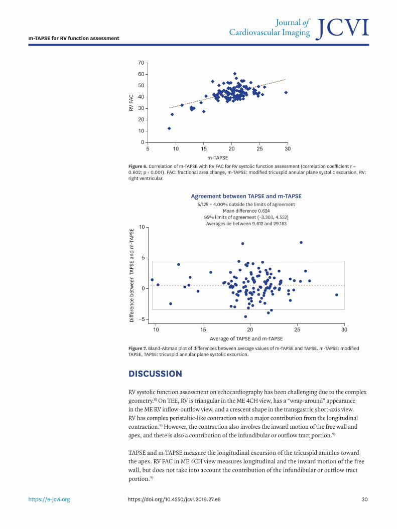
The intraclass correlation coefficient for intra-observer variability of TAPSE, m-TAPSE and RV FAC were 0.95, 0.81 and 0.82, respectively, suggesting good correlation. To assess the inter-observer variability, a Bland-Altman plot was analyzed that showed the differences and average values of two echocardiographers for m-TAPSE (Figure 4). The two investigators tended to agree with the bias of 0.051, which is clinically acceptable. Ninety-five percent of the differences fell within -3.303 and +3.405, which were the limits of agreement.
The averaged values for m-TAPSE correlated significantly with both TAPSE (r = 0.797; p < 0.001) (Figure 5) as well as RV FAC (r = 0.602; p < 0.001) (Figure 6). The Bland-Altman plot for comparing m-TAPSE and TAPSE is shown in Figure 7.
The sensitivity, specificity, PPV, NPV and accuracy of m-TAPSE calculated using 2 × 2 cross table were 100%, 98.3%, 80%, 100%, and 98.4%, respectively.
29https://e-jcvi.org https://doi.org/10.4250/jcvi.2019.27.e8
m-TAPSE for RV function assessment
Agreement (first and second investigator's m-TAPSE readings)7/125 = 5.60% outside the limits of agreement
Mean difference 0.11495% limits of agreement (−3.318, 3.547)Averages lie between 8.800 and 29.650
Average of first and second investigator's readings
−6
15 25 40
Diffe
renc
e (fi
rst a
nd s
econ
d in
vest
igat
or's
m-T
APSE
and
TAP
SE re
adin
gs)
4
2
0
10 20 3530
−4
−2
Figure 4. Bland-Altman plot of differences (y-axis) and average of 2 independent observers (x-axis) for m-TAPSE assessment (inter observer variability). m-TAPSE: modified TAPSE, TAPSE: tricuspid annular plane systolic excursion.
TAPSE
5
15 25
m-T
APSE
35
25
30
10 20 305
10
0
15
20
Figure 5. Correlation of TAPSE with m-TAPSE for RV systolic function assessment (correlation coefficient r = 0.797; p < 0.001). m-TAPSE: modified TAPSE, RV: right ventricular, TAPSE: tricuspid annular plane systolic excursion.
DISCUSSION
RV systolic function assessment on echocardiography has been challenging due to the complex geometry.8) On TEE, RV is triangular in the ME 4CH view, has a “wrap-around” appearance in the ME RV inflow-outflow view, and a crescent shape in the transgastric short-axis view. RV has complex peristaltic-like contraction with a major contribution from the longitudinal contraction.9) However, the contraction also involves the inward motion of the free wall and apex, and there is also a contribution of the infundibular or outflow tract portion.9)
TAPSE and m-TAPSE measure the longitudinal excursion of the tricuspid annulus toward the apex. RV FAC in ME 4CH view measures longitudinal and the inward motion of the free wall, but does not take into account the contribution of the infundibular or outflow tract portion.9)
30https://e-jcvi.org https://doi.org/10.4250/jcvi.2019.27.e8
m-TAPSE for RV function assessment
m-TAPSE
10
15 25
RV F
AC
70
50
60
10 20 305
20
0
30
40
Figure 6. Correlation of m-TAPSE with RV FAC for RV systolic function assessment (correlation coefficient r = 0.602; p < 0.001). FAC: fractional area change, m-TAPSE: modified tricuspid annular plane systolic excursion, RV: right ventricular.
Agreement between TAPSE and m-TAPSE5/125 = 4.00% outside the limits of agreement
Mean difference 0.62495% limits of agreement (−3.303, 4.552)Averages lie between 9.612 and 29.183
Average of TAPSE and m-TAPSE
−5
15 25 30
Diffe
renc
e be
twee
n TA
PSE
and
m-T
APSE
10
5
0
10 20
Figure 7. Bland-Altman plot of differences between average values of m-TAPSE and TAPSE. m-TAPSE: modified TAPSE, TAPSE: tricuspid annular plane systolic excursion.

There is a lack of easily measurable quantitative parameters. RV ejection fraction (EF) by cardiac magnetic resonance (CMR) imaging is the gold standard, but it is not feasible to use in all cases. Only RV EF provides an adequate assessment of true global RV pump function, and 3D echocardiography remains the only echocardiographic technique capable of a reliable calculation of RV EF from end-diastolic and end-systolic volume measurements.10) Others are surrogate parameters like RV FAC, TAPSE, peak systolic wave velocity of lateral tricuspid annulus by tissue Doppler imaging, which gives a rough estimate of the RV systolic function. RV myocardial performance index or Tei index and 3D RV function evaluation are time-consuming in a busy intraoperative setting, leaving 2D parameters as the only ones that can be quickly assessed.
Compared with other measures of RV systolic function, RV FAC correlated best with CMR-derived RV EF (CMR vs. RVFAC, r = 0.80, p < 0.0001).11) Therefore, RV FAC is considered a near-gold standard 2D-echocardiographic parameter for RV assessment, but needs the entire endocardium to be clearly visible. TAPSE is the most commonly employed method, but M-mode cannot always be aligned along the motion of the lateral tricuspid annulus in the ME 4CH view.
Different methods have been described for calculating longitudinal excursion of the lateral tricuspid annulus using TEE; 1) TAPSE, which is taken by aligning M-mode with the lateral tricuspid annulus;10) 2) tricuspid annular motion (TAM), which is defined as the difference of the longitudinal distance between the midpoint of the tricuspid valve to the endocardial border of the RV apex in end-diastole and end-systole;12) 3) 2D-TAPSE, wherein an annotation arrow is placed at the junction of the tricuspid annulus and RV free wall during diastole, and a second arrow at this junction at end-systole, and the 2D-TAPSE is the distance between these arrows;13) and 4) the difference of ‘apical to lateral tricuspid annulus distance’ during diastole and systole in the ME 4CH view.7) We used this last method in our study.
In our study, the average values for m-TAPSE correlated significantly with RV FAC (r = 0.602; p < 0.0001). The correlation coefficient between m-TAPSE and RV FAC was 0.618 (p < 0.001) in a study by Morita et al.7) TAM was significantly correlated with the MRI-derived RV EF (Spearman ρ = 0.57; p = 0.004).12) Since TAM is also a measure of longitudinal excursion, this result can be extrapolated to other methods of RV function assessment that use longitudinal excursion like TAPSE, m-TAPSE and 2D-TAPSE.
m-TAPSE is a useful parameter for assessment of RV systolic function especially in the intraoperative and perioperative setting when TTE cannot be performed or does not provide adequate imaging. In addition, this parameter is not as dependent on image quality. Only the apex and the lateral tricuspid annulus should be clearly visible when calculating m-TAPSE. The whole endocardium does not need to be visualized as in the case of RV FAC. This is especially helpful when acoustic shadowing from mitral valve calcification or mechanical valve replacement covers the RV free wall or the septum. The RV is more trabeculated than the left ventricle, which limits endocardial delineation and hence the accuracy of RV FAC.9) Alignment of M-mode with the lateral tricuspid annulus in the ME 4CH view is difficult using TEE. mTAPSE is not as simple as TAPSE for RV function assessment since it needs to be calculated in two phases of the cardiac cycle. However, it is still a simple parameter compared with other available options. Hence, it can be used as a reliable alternative parameter when other methods are either not feasible or are non-conclusive. m-TAPSE can be used as an easily reproducible simple rescue parameter in these cases. m-TAPSE also shows a moderate correlation to RV FAC in patients with heart transplantation and left ventricular assist device implantation.14)
31https://e-jcvi.org https://doi.org/10.4250/jcvi.2019.27.e8
m-TAPSE for RV function assessment

However, use of m-TAPSE can be limited when the maximum tricuspid annulus is beyond the widest sector in the ME 4CH view during diastole. In these cases, TAPSE or RV FAC can be used. Also, if the point at the apex for calculating m-TAPSE is not clearly defined, measurements may be erroneous.
LimitationsRV function is dependent on both load and heart rate. Although attempts were made to measure echocardiographic parameters at near baseline hemodynamics, some errors may have occurred due to the effects of general anesthesia and volume status of the patient. Secondly, this study has taken RV FAC as the sole diagnostic criteria for RV dysfunction. Studies comparing this parameter with CMR imaging may provide further validation.
Conclusionm-TAPSE is a novel addition to the existing armamentarium of echocardiographic RV function assessment. It is a simple, less time-consuming, reliable and easily reproducible parameter that correlates well with the near-gold-standard echocardiographic parameter, RV FAC, and the most commonly used parameter, TAPSE.
ACKNOWLEDGMENTS
The authors acknowledge the contributions of Dr. Vishwas Malik, MD, DM, Professor, Department of Cardiac Anesthesia, All India Institute of Medical Sciences, New Delhi, India for conducting anesthesia in the study cases. Dr. Milind P. Hote, MCh, Department of Cardiothoracic and Vascular Surgery, All India Institute of Medical Sciences, New Delhi, India for his cooperation, encouragement and patience during the course of the study. Dr. R.M. Pandey, MD, Professor and Head of the Department of Biostatistics, All India Institute of Medical Sciences, New Delhi, India and his team for their valuable time and input in statistical analysis of the data.
REFERENCES
1. Dell'Italia LJ. The right ventricle: anatomy, physiology, and clinical importance. Curr Probl Cardiol 1991;16:653-720. PUBMED | CROSSREF
2. Kaul TK, Fields BL. Postoperative acute refractory right ventricular failure: incidence, pathogenesis, management and prognosis. Cardiovasc Surg 2000;8:1-9. PUBMED | CROSSREF
3. Denault AY, Haddad F, Jacobsohn E, Deschamps A. Perioperative right ventricular dysfunction. Curr Opin Anaesthesiol 2013;26:71-81. PUBMED | CROSSREF
4. Haddad F, Couture P, Tousignant C, Denault AY. The right ventricle in cardiac surgery, a perioperative perspective: I. Anatomy, physiology, and assessment. Anesth Analg 2009;108:407-21. PUBMED | CROSSREF
5. Rudski LG, Lai WW, Afilalo J, et al. Guidelines for the echocardiographic assessment of the right heart in adults: a report from the American Society of Echocardiography endorsed by the European Association of Echocardiography, a registered branch of the European Society of Cardiology, and the Canadian Society of Echocardiography. J Am Soc Echocardiogr 2010;23:685-713. PUBMED | CROSSREF
6. Hu R, Mazer CD, Tousignant C. Relationship between tricuspid annular excursion and velocity in cardiac surgical patients. J Cardiothorac Vasc Anesth 2014;28:1198-202. PUBMED | CROSSREF
32https://e-jcvi.org https://doi.org/10.4250/jcvi.2019.27.e8
m-TAPSE for RV function assessment

7. Morita Y, Nomoto K, Fischer GW. Modified tricuspid annular plane systolic excursion using transesophageal echocardiography for assessment of right ventricular function. J Cardiothorac Vasc Anesth 2016;30:122-6. PUBMED | CROSSREF
8. Miller D, Farah MG, Liner A, Fox K, Schluchter M, Hoit BD. The relation between quantitative right ventricular ejection fraction and indices of tricuspid annular motion and myocardial performance. J Am Soc Echocardiogr 2004;17:443-7. PUBMED | CROSSREF
9. Maus TM. TAPSE: a red herring after cardiac surgery. J Cardiothorac Vasc Anesth 2018;32:779-81. PUBMED | CROSSREF
10. Surkova E, Peluso D, Kasprzak JD, Badano LP. Use of novel echocardiographic techniques to assess right ventricular geometry and function. Kardiol Pol 2016;74:507-22.PUBMED
11. Horton KD, Meece RW, Hill JC. Assessment of the right ventricle by echocardiography: a primer for cardiac sonographers. J Am Soc Echocardiogr 2009;22:776-92. PUBMED | CROSSREF
12. Srinivasan C, Sachdeva R, Morrow WR, Greenberg SB, Vyas HV. Limitations of standard echocardiographic methods for quantification of right ventricular size and function in children and young adults. J Ultrasound Med 2011;30:487-93. PUBMED | CROSSREF
13. Skinner H, Kamaruddin H, Mathew T. Tricuspid annular plane systolic excursion: comparing transthoracic to transesophageal echocardiography. J Cardiothorac Vasc Anesth 2017;31:590-4. PUBMED | CROSSREF
14. Morita Y, Lencho T, Gunasekaran S, Modak R. Modified tricuspid annular plane systolic excursion using transesophageal echocardiography and its utility to predict postoperative course in heart transplantation and left ventricular assist device implantation. J Cardiothorac Vasc Anesth 2018;32:1316-24. PUBMED | CROSSREF
33https://e-jcvi.org https://doi.org/10.4250/jcvi.2019.27.e8
m-TAPSE for RV function assessment