origins of serum alkaline phosphatase
TRANSCRIPT

J. clin. Path. (1967), 20, 647
Origins of serum alkaline phosphataseJ. M. YONG
From the Department of Chemical Pathology, St. George's Hospital Medical School, London
SYNOPSIS A very rapid method of agar gel electrophoresis on glass slides, together with a superiorvisualization technique employing simultaneous coupling of a hydrolysed naphthol substrate,have been developed for the identification of the tissues of origin of serum alkaline phosphatase.Combined with L-phenylalanine inhibition, specific for the intestinal enzyme, and heat inactivation,specific for the placental enzyme, the heterogeneity of serum alkaline phosphatase has been demon-strated. Normal adult serum contains predominantly liver-type alkaline phosphatase with a smallbut variable quantity of intestinal enzyme, and little or no bone enzyme.
In childhood and in infancy there is in addition a bone isoenzyme present, the amount graduallyfalling to adult levels with age. In pregnancy, the rise in serum alkaline phosphatase is due to theplacental enzyme.A study of nearly 2,000 sera has been undertaken and it is found that the bone enzyme is increased
in osteoblastic bone diseases while in hepato-biliary disorders there is an increase in liver typeenzyme. The main theories explaining the rise in serum alkaline phosphatase are examined.
The origin of serum alkaline phosphatase hasinterested many workers ever since Kay (1929) andRoberts (1930) first demonstrated that increasedserum activity was found in certain bone and hepato-biliary disorders. The problem of the origin of theenzyme is intricately bound up with that of themechanisms by which the serum activity is increasedor decreased in health and in disease. It has longbeen realized that serum alkaline phosphatase arisesfrom tissues rich in the enzyme (Kay, 1932). Previousattempts at identification of the tissues of origin werehandicapped not only by lack of direct methods, butalso by the influence of the work of Robison andhis associates linking alkaline phosphatase withcalcification (Robison, 1923; Robison and Soames,1924,1925,1930; Robison, MacLeod, and Rosenheim,1930; Robison and Rosenheim, 1934; Martlandand Robison, 1924; Fell and Robison, 1929).The former led to theories based on indirect obser-vations. Nearly all these theories assumed that theserum enzyme was of osseous origin.
In recent years, the realization that enzymesmay exist in multiple structural forms has providedan impetus to development of techniques of separa-ting these 'isoenzymes'. Of the large number oftechniques attempted, few were found to haveadequate resolving power to differentiate the varioustissue-specific alkalinephosphatases (Moss, Campbell,Received for publication,12 January 1967.
Anagnostou-Kakaras, and King, 1961; Boyer, 1963;Peacock, Reed, and Highsmith, 1963; Neale, Clubb,Hotchkis, and Posen, 1965).A technique of agar gel electrophoresis has been
developed in this laboratory which enables alkalinephosphatase isoenzymes to be rapidly and reliablydifferentiated. Extensive studies have been made onnormal and pathological sera, and, in conjunctionwith other techniques described below, a clearerpicture of the origins of serum alkaline phosphataseand of the mechanisms involved in alterations ofenzyme activity in health and disease has emerged.
METHODS
AGAR GEL ELECTOPHORESIS A technique modified afterWieme (1959) using 1 1 % Noble agar in pH 8-6 barbit-urate buffer on glass microscope slides was used. Veryrapid electrophoresis was achieved by using a voltagegradient of 70 volts per centimetre, and cooling by pump-ing petrol ether (BP 40-60°C.) into the tank and re-cycling through a coil immersed in dry-ice water mixture.A 40 mm. separation between serum albumin and
y-globulin could be thus achieved in 10 minutes.
PROTEIN STAINING Sera or tissue extracts were electro-phoresed on pairs of slides, one of these being stained forproteins and the other for alkaline phosphatase.
Ponceau S, 0 5 %, in 3 % trichloroacetic acid was usedfor protein staining. Afterwards the gels were elutedfor two hours in three changes of distilled water to
647
on Decem
ber 7, 2021 by guest. Protected by copyright.
http://jcp.bmj.com
/J C
lin Pathol: first published as 10.1136/jcp.20.4.647 on 1 July 1967. D
ownloaded from
on D
ecember 7, 2021 by guest. P
rotected by copyright.http://jcp.bm
j.com/
J Clin P
athol: first published as 10.1136/jcp.20.4.647 on 1 July 1967. Dow
nloaded from
on Decem
ber 7, 2021 by guest. Protected by copyright.
http://jcp.bmj.com
/J C
lin Pathol: first published as 10.1136/jcp.20.4.647 on 1 July 1967. D
ownloaded from
648 J. M. Yong
remove excess dye, and then dried overnight in a 37°C. Isoenzymesincubator.
ENZYME STAINING Alkaline phosphatase isoenzymeswere visualized by a simultaneous coupling diazo reaction.The gel was immersed for 15 to 30 minutes inthe following substrate at room temperature (20-22°C):
Disodium phosphate ester of 2-hydroxy naphthelene3-carboxylic acid 41-chloro, 61-methyl anilide ......0-002MDiazotized 5-chloro-o-toluidine .....................0-0016MMagnesium chloride ...............................O 0S005MSodium carbonate-bicarbonate bufferpH 10 ...... 0.05M
The alkaline phosphatase isoenzymes appeared asred bands while serum proteins were stained a very faintyellow. The positions of these isoenzymes relative tothe protein fractions could thus be easily seen. Aftereluting as for the proteins, incubations overnight pro-duced transparent slides.
PHENYL-ALANINE INHIBITOR 0-005M L-phenyl alanine,which specifically inhibits intestinal alkaline phosphatase(Fishman, Green, and Inglis, 1963), was incorporatedinto the substrates in some instances. The D-isomerwas then used as a control.
HEATING Electrophoresis was performed on serabefore and after heating to 56°C. for 30 minutes in thepresence of OO1M magnesium chloride (Neale et al.,1965).
TISSUE EXTRACTS Tissue for the extraction of alkalinephosphatase were obtained at necropsies on youngadults dying from road traffic accidents. They wereused within 24 hours or stored at - 20°C. Adult bonewas too difficult to homogenize and femurs from still-born infants were used instead. After cutting the tissuesinto small pieces homogenization was performed usinga Potter-Elvehjem homogenizer and the enzyme extractedby the butanol method of Morton (1954). The finalaqueous extract was adjusted to about 30 to 40 King-Armstrong units of activity per 100 ml. before electro-phoresis.
NEURAMINDASE The procedure described by Robinsonand Pierce (1964) was used. Electrophoretic mobilitiesbefore and after 20 hours incubation at 37°C. withneuraminidase was compared.
RESULTS
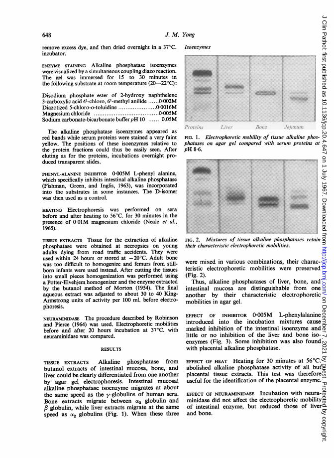
TISSUE EXTRACTS Alkaline phosphatase frombutanol extracts of intestinal mucosa, bone, andliver could be clearly differentiated from one anotherby agar gel electrophoresis. Intestinal mucosalalkaline phosphatase isoenzyme migrates at aboutthe same speed as the y-globulins of human sera.Bone extracts migrate between a2 globulin andc globulin, while liver extracts migrate at the samespeed as CX2 globulins (Fig. 1). When these three
FIG. 1. Electrophoretic mobility of tissue alkaline phos-phatases on agar gel compared with serum proteins atpH 8-6.
FIG. 2. Mixtures of tissue alkaline phosphatases retaintheir clharacteristic electrophoretic mobilities.
were mixed in various combinations, their charac-teristic electrophoretic mobilities were preserved(Fig. 2).
Thus, alkaline phosphatases of liver, bone, andintestinal mucosa are distinguishable from oneanother by their characteristic electrophoreticmobilities in agar gel.
EFFECT OF INHIBITOR 0-005M L-phenylalanineintroduced into the incubation mixtures causemarked inhibition of the intestinal isoenzyme andlittle or no inhibition of the liver and bone iso-enzymes (Fig. 3). Some inhibition was also foundwith placental alkaline phosphatase.
EFFECT OF HEAT Heating for 30 minutes at 560C.abolished alkaline phosphatase activity of all butplacental tissue extracts. This test was thereforeuseful for the identification of the placental enzyme.
EFFECT OF NEURAMINIDASE Incubation with neura-minidase did not affect the electrophoretic mobilityof intestinal enzyme, but reduced those of liverand bone.
.-oP4
I
on Decem
ber 7, 2021 by guest. Protected by copyright.
http://jcp.bmj.com
/J C
lin Pathol: first published as 10.1136/jcp.20.4.647 on 1 July 1967. D
ownloaded from

Origins ofserum alkaline phosphataseY V~~~~~~~~~W
:.: .... ... .... ......
FIG. 3. Effect of0,005M L-phenylalanine on tissue
alkaline phosphatases. The intestinal isoenzyme is strongly
inhibited. 0O005M D-phenylalanine is used as control in
each pair of slides.
This technique thus provided further evidence of
the validity of the electrophoretic technique in
separating intestinal from bone and liver phos-
phatases. It could not, however, be used for dis-
tinguishing liver from bone alkaline phosphatase.
Tissueof Origin
TABLE IIDENTIFICATION OF TISSUE OF ORIGIN
Electrophoretic L-Ph AI HeatMobility Inhibition StabilityCompared withSerum Proteins
LiverAortic endotheliumLungBonePlacentaKidneyIntestineBrain
a,
at
as
a, and
Rl/li/
0
0
0
0
++
LabileLabileLabileLabileStableLabileLabileLabile
Heating at 56°C. for 30 minutes completelyabolished all enzyme activity.These findings favour an origin in liver (or possibly
lung or blood vessel) for alkaline phosphatase foundin normal adult sera. In addition there may besmaller amounts of intestinal alkaline phosphatasepresent.
TABLE IICOMPARISON WITH OTHER TISSUES Alkaline phos-phatase from stomach, duodenum, jejunum, ileum,and colon all have the same electrophoretic mobili-ties, migrating between and y globulins. Thiselectrophoretic mobility is found only with oneother tissue studied, the brain.The electrophoretic mobilities of alkaline phos-
phatase extracted from lung tissue and the endo-thelium of the aorta are identical. They all migrateat the same speed as (x2-globulins, are heat labile,and are not inhibited by L-phenylalanine to anydegree.
Placental alkaline phosphatase migrates at aspeed intermediate between (X2 and P globulins, andis thus impossible to distinguish electrophoreticallyfrom bone alkaline phosphatase. However, heatingdestroys the bone but not the placental enzyme.
IDENTIFICATION OF TISSUE ORIGIN By use of electro-phoresis, L-phenylalanine inhibition, and heatstability, it can be seen that the tissue of origin ofserum alkaline phosphatase may be inferred (Table I).
NORMAL ADULT SERA Sera from 50 healthy blooddonors were subjected to electrophoresis. In allinstances a single alkaline phosphatase isoenzyme,moving at the 062 globulins or at the same speed asliver isoenzyme, was found (Table II). No inhibitionby L-phenylalanine was found.Ten sera after electrophoresis were allowed pro-
longed staining (over one hour) and in three a secondisoenzyme was found. This isoenzyme was inhibitedby L-phenylalanine.
ORIGIN OF ALKALINE PHOSPHATASE IN NORMAL SERA
Properties of Alkaline PhosphataseSera(Number) Percentage Electro-
phoreticMobility
Normal 100'adults(50)Normal 70children 100(30) 3-3Infants 45(20) 100Pregnancy 100at term 100(25)
Heat L-Pheny- Likely OriginStability lalanine
Inhibition
as Labile 0 Liver
a,/,a,liis/a,laI2a2a,Ilis
Labile
Labile
LabileLabileLabileStable
0
0
0
0
0
LiverBoneIntestineLiverBoneLiverPlacenta
"Note that intestinal isoenzyme was found in three of 10 normal serawhen prolonged incubation times were used for isoenzyme visualization
SERA FROM CHILDREN Sera from 30 normal healthychildren on electrophoresis showed, in addition tothe adult type of alkaline phosphatase, an isoenzymemigrating between the cX2 and , globulins. Thestaining intensity of this isoenzyme decreased withage, being very faint at 15 years of age. One child,aged 8, had a third isoenzyme migrating slightlyfaster than y globulin.
Heating for 30 minutes at 56°C. destroyedvirtually all alkaline phosphatase activity.
Sera obtained at birth from cord blood of 10infants also showed two isoenzymes as for olderchildren. The slow moving isoenzyme was, however,much more intense.These findings favour more than one tissue of
origin. In addition to the normal adult type there
I649
on Decem
ber 7, 2021 by guest. Protected by copyright.
http://jcp.bmj.com
/J C
lin Pathol: first published as 10.1136/jcp.20.4.647 on 1 July 1967. D
ownloaded from

J. Al. Yong
TABLE IIISERUM ALKALINE PHOSPHATASE ISOENZYMES IN BONE
AND HEPATO-BILIARY DISORDERSDisorder No. of Sera Alkaline Phosphatase Isoenzymes
Cases AnalysedLiver Bone Intestinal
FIG. 4. Changing serum isoenzyme pattern in infancyand childhood. The high serum alkaline phosphataseactivity in childhood is due to an extra isoenzyme derivedfrom bone. The sera have been concentrated about threeto four-fold by dialysis before electrophoresis.
Bone disease 48 70 45(93-8 %)
Biliary tract 68 130 68disease (100%)Hepatitis 66 90 59
(89-4%)Cirrhosis and 85 129 85hepatic (100%)infiltrations
Disorder
TABLE IVDETAILED ANALYSIS OF SERA OFPATIENTS WITH BONE DISORDERS
No. of Alkaline Phosphatase IsoenzymtiesCases
Liver Bone Intestinal
FIG. 5. Serum alkaline phosphatase isoenzymes inpregnancy. At 28 weeks a faint heat-stable placentalisoenzyme is just visible. At term this isoenzyme is veryprominent. At two weeks post partlum this has virtuiallydisappeared.
Paget's disease 12Bony metastases with 9raised serum
alkaline phosphataseMultiple fractures 4Rickets and 8osteomalaciaOsteogenic sarcomaHypertrophic )osteoarthropathyOsteosclerosis 9Osteitis fibrosacystica J
Total (osteoblastic 42disorders)
Osteoporosis 4Ankylosing spondylitis 2(inactive)Total (bone disorders) 48
11 10 08 8 0
4 4 07 8 2
9 9 0
39 39 2
4 0 02 0 0
45 39 2
appears to be bone alkaline phosphatase present.In one example there was intestinal enzyme as well.
SERUM ALKALINE PHOSPHATASE IN PREGNANCY To
study variations in total activity as well as qualitativechanges during pregnancy, 10 patients were followedat roughly fortnightly intervals until term. Themean values have been plotted in Fig. 6 showingthe total as well as the heat-stable (placental) enzymeactivity.
Electrophoresis of such sera provided additionalevidence of the placental origin of the enzyme (Fig. 5).
BONE DISEASE An extra isoenzyme, migratingat the same speed as bone extracts, was found inthe sera of the majority (93 %) of 42 patients withosteoblastic lesions. In contrast six patients withosteoporous or burnt-out ankylosing spondylitis(Table III) showed no bone isoenzyme.
Thus, osteoblastic bone lesions give rise to a
detectable bone alkaline phosphatase isoenzymein the serum, even when the total activity is withinnormal limits.
HEPATO-BILIARY DISORDERS Two hundred andnineteen patients with obstructive disease of theextrahepatic bile ducts, infiltrations of the liver,and hepatitis from virus infections or drugs, suchas chlorpromazine, were studied by electrophoresisof the serum alkaline phosphatase levels on 349occasions.
All of 85 patients with hepatic infiltrations andof 68 patients with obstructive bile duct disordersshowed increased liver-type alkaline phosphatase. Asmall number also had bone and intestinal typeisoenzymes. More careful analysis of the lattershowed various associated bone disorders ormultiorgan disorders such as disseminated cancer.
39(81-3%)16(23-5 %)18(24 2%)12(14 1 %)
2(4 2%)9(13 2%)12(18 2%)6(7 1%)
i
650
on Decem
ber 7, 2021 by guest. Protected by copyright.
http://jcp.bmj.com
/J C
lin Pathol: first published as 10.1136/jcp.20.4.647 on 1 July 1967. D
ownloaded from

Origins ofserum alkaline phosphatase
DISCUSSION
Numerous attempts have been made in the past toelucidate the problem of the origin of serum alkalinephosphatase. Most of these attempts have met withlimited success.Bodansky (1937) found that bile salts inhibited
bone and kidney phosphates but not the intestinalenzyme. He was, however, unable to come to anyconclusion about the origin of human serum alkalinephosphatase. Schlamowitz prepared antibodiesagainst various human alkaline phosphatases butcross-reactions made interpretation difficult(Schlamowitz, 1958; Schlamowitz and Bodansky,1959). Landau and Schlamowitz (1961) utilizedKm measurements without success.
Significant advances were made when L-pheny-lalanine, zone electrophoresis, and heat stabilitywere utilized. Fishman and his associates (Fishmanet al., 1962, 1963) showed that L-phenylalaninespecifically inhibited intestinal alkaline phosphatase.Paper electrophoresis of alkaline phosphatase
was introduced by Baker and Pellegrino (1954)and by Eisfeld and Koch (1954). Other workershave since utilized starch block (Rosenberg, 1959;Keiding, 1959), agar gel (Stevenson, 1961), celluloseacetate (Korner, 1962), starch gel (Estbom, 1959),and polyacrylamide gel (Allen and Hyncik, 1963).However, although the intestinal isoenzyme could
be separated from the liver or bone isoenzymes bysuch techniques, the latter two could not be differen-tiated from one another. Further, starch gel electro-phoresis, the most popular of these techniques,usually separates each tissue alkaline phosphataseinto two or more isoenzymes, so that when there ismore than one tissue of origin, interpretation be-comes difficult.
In this laboratory, a very rapid and relativelysimple technique of agar gel electrophoresis hasbeen developed in conjunction with a superiorvisualization technique to enable liver, bone,intestinal, and other types of alkaline phosphataseto be clearly separated. When used in conjunctionwith L-phenylalanine inhibition and heat stability,the tissues of origin can be reliably made on everyspecimen studied.
Studies on nearly 2,000 sera have shown thatserum alkaline phosphatase originates from severalorgans. In infants and children, two isoenzymesare found. One appears to be derived from the liverand the other from bone. The activity of the boneisoenzyme appears to be highest at birth and de-clines with age. This is not surprising in view of theclose relationship of total serum alkaline phos-phatase activity in children to the measured rateof bone growth (Clark and Beck, 1950).
In adult life there is no bone isoenzyme detectedin the serum by the technique used. The liver typeisoenzyme is present in the vast majority of people.Prolonged incubation with the substrate producedan intestinal isoenzyme in three of 10 sera. It isinteresting to compare this with the result reportedby other workers using starch gel electrophoresisand rather long incubation times. Arfors and hisassociates (Arfors, Beckman, and Lundin, 1963a,1963b) have found 28% having a second slowmoving isoenzyme. Beckman (1964) found it innearly 32% of donors of blood group 0 and B butnone in blood group A.
Later studies (Bamford, Harris, Luffman, Robson,and Cleghorn, 1965; Hope, 1966) have found it inbetween 16-1 and 69-2%, depending on the bloodgroup. Thus, it appears that as longer incuba-tion times are used, the small amount of intestinalphosphatase activity present can be shown up.Significantly, while bone isoenzyme was readilydetected in osteoblastic bone disorders in the presentstudy, even prolonged incubation times failed toshow it in normal adult sera. It may thus be inferredthat the predominant serum alkaline phosphataseis of the liver type, with variable but small amountsof intestinal isoenzyme, and very little, if any, boneisoenzyme.
In pregnancy, the electrophoretic properties andthe heat stability of the serum enzyme imply thatthe increase is of placental origin. These findingsconfirm those of McMaster, Tennant, Clubb,
E00
U)
C11
:3
Tota[
15 20 25 30 35 40Gestation (weeks)
FIG. 6. Mean serum alkaline phosphatase in 10 womenduring the course of pregnancy. The heat-stable portionrepresents the placental contribution.
651
on Decem
ber 7, 2021 by guest. Protected by copyright.
http://jcp.bmj.com
/J C
lin Pathol: first published as 10.1136/jcp.20.4.647 on 1 July 1967. D
ownloaded from

652 J. M
Neale, and Posen (1964), who were the first toutilize the heat-stable property of placental alkalinephosphatase to study this problem, and those ofZuckerman and Sadovsky (1965) who used thesame technique. The latter have also shown thatplacental alkaline phosphatase has a greater affinityfor ,B-glycerophosphate than p-nitrophene phosphate.During pregnancy, serum alkaline phosphataseshows a progressively increasing preference for/-glycerophosphate while this is not seen in patientswith bone or hepatobiliary disorders (Sadovsky andZuckerman, 1965).The overall evidence thus leaves little doubt that
the increase in serum alkaline phosphatase inpregnancy is of placental origin.
In bone disorders associated with increasedosteoblastic activity there is an increase in serumalkaline phosphatase activity (Gutman, 1959). Suchpatients show an additional isoenzyme having theelectrophoretic mobility characteristic of bone orplacental isoenzyme. However, as none of thepatients studied were pregnant, and the isoenzymewas found regardless of the sex of the patients, itmay be safely concluded that the increase is due tothe bone isoenzyme.
In hepatobiliary disorders, there is no detectablebone isoenzyme in the serum but instead the hepatictype of alkaline phosphatase is increased. Thissuggests that in these disorders the raised serumactivity is due to increased output of the enzymeby the liver into the blood stream.
Electrophoresis of bile reveals an isoenzymehaving the same electrophoretic mobility as livertype alkaline phosphatase but not bone type. Thus,the widely held theory of impaired excretion of thebone enzyme (Roberts, 1930, 1933; Armstrong,King, and Harris, 1934; Armstrong and Banting,1935; Gutman, Olson, Gutman, and Flood, 1940;Gutman, 1959) is not supported by this evidence.
Indeed, other workers have previously foundevidence which conflicted with the theory. Thereis often a dissociation between serum bilirubin andserum alkaline phosphatase in various liver disorders(Burke, 1950). Metastatic cancer in the liver haslong been known to produce hyperphosphatasiawithout hyperbilirubinaemia (Meranze, Meranze,and Rothman, 1938; Ross, Iber, and Harvey, 1956;Gibbons, 1957), but the level of alkaline phosphatasedoes not correlate with the extent of the infiltration.Similarly a proportion of patients with hepatitishave very high serum alkaline phosphatase levelswhile a proportion with common bile duct obstruc-tion have appeared normal. Gutman et al. (1940)have drawn attention to the anomaly that infantswith biliary atresia usually do not have a serumalkaline phosphatase activity greater than expected
Yong
for their age. This finding is, however, compatiblewith the hypothesis that the enzyme originates fromthe liver. It is known that the livers of infants havea poorer metabolic ability than adult livers, and itis interesting to note that liver type isoenzyme wasabsent in 30% of the sera of children studied inthe present work.Experiments on animals produce even stronger
evidence against the retention theory. Ligation ofone hepatic (bile) duct produces gross elevation ofserum alkaline phosphatase but little jaundice(Freeman, Chen, and Ivy, 1938; Polin, Spellberg,Teitelman, and Okumura, 1962). The bile alkalinephosphatase activity was increased, as was theenzyme content of the liver lobe drained by theligated duct (Polin et al., 1962).
Sebesta, Bradshaw, and Prockop (1964), usingisolated perfused cat livers, found no increase inalkaline phosphatase activity in the perfusate whenthe bile ducts were patent, but large increases wereobtained when these were occluded. This providesstrong evidence that the liver is the source of theenzyme. Further, transfusion of high alkalinephosphatase blood from dogs with common bileduct stricture to normal animals resulted in highserum levels for many days, the enzyme activityonly declining slowly (Freeman and Chen, 1938;Cantarow and Miller, 1948; Dalgaard, 1951).
Clubb, Neale, and Posen (1965) have infusedplacental and bone alkaline phosphatase into humansand shown that the half-line in plasma was aboutseven days. No significant difference was foundbetween subjects with bile duct obstruction andthose without. Indeed, the disappearance rate ofinfused placental alkaline phosphatase from thecirculation is similar to that of infused 1311 albumin(Clubb et al., 1965). This behaviour is to be ex-pected, for the molecular weight of purified alkalinephosphatases from various tissues has been esti-mated to be between 120,000 and 130,000 (Boyer,1963). Indeed, excretion would require a highlyspecialized transport system for molecules of thissize.
Finally one must consider the origin of the hepatictype alkaline phosphatase found in normal adultsera in the present study.
Identical isoenzyme patterns are found withalkaline phosphatase from the endothelium, lung,and choroid plexus. It is also known that in theliver, endothelial cells of the portal and centralveins are richer in this enzyme than bile ducts orliver parenchymal cells (Morrison, Karl, Schwartz,and Shank, 1965). It is therefore probable that liveralkaline phosphatase is derived from the endothelialcells, and the enzyme is indistinguishable fromother blood vessel endothelial alkaline phosphatase.
on Decem
ber 7, 2021 by guest. Protected by copyright.
http://jcp.bmj.com
/J C
lin Pathol: first published as 10.1136/jcp.20.4.647 on 1 July 1967. D
ownloaded from

Origins of serum alkaline phosphatase
There is probably a second type of alkaline phos-phatase found in the biliary system. This enzymecannot be separated from the other liver typealkaline phosphatase by the agar gel electrophoresistechnique, but differences have been shown byPope and Cooperband (1966) using starch gelelectrophoresis.
Raised serum alkaline phosphatase levels inhepatobiliary disease thus would arise by increasedliver synthesis of either enzyme: in conditions inwhich bile duct pressure is increasing the amount ofbile type enzyme produced is increased, some ofwhich finds its way into the blood plasma, while inhepatocellular disorders, such as certain cases ofcirrhosis and hepatitis, or in metastatic liver disease,there is an increase in the blood vessel type ofenzyme produced. In infantile livers this ability tosynthesize either enzyme is rudimentary, so thatbile duct atresia hardly promotes any increase inalkaline phosphatase.
My thanks are due to the British Empire CancerCampaign for financial support and to the clinicianswho allowed me to study their patients. I also wish tothank Professor N. H. Martin for his continual and in-spiring help.
REFERENCES
Allen, J. M., and Hyncik, G. (1963). J. Histochem. Cytochem., 11, 169.Arfors, K. E., Beckman, L., and Lundin, L. G. (1963a). Acta genet.
(Basel), 13, 89.____ - (1963b). Ibid., 13, 366.
Armstrong, A. R., and Banting F. G. (1935). Canad. med. Ass. J.,33, 243.King, E. J., and Harris, R. I. (1934). Ibid., 31, 14.
Baker, R. W. R., and Pellegrino, C. (1954). Scand. J. clin. Lab. Invest.,6, 94.
Bamford, K. F., Harris, H., Luffman, J. E., Robson, E. B., andCleghorn, T. E. (1965). Lancet, 1, 530.
Beckman, L. (1964). Acta genet. (Basel), 14, 286.Bodansky, 0. (1937). J. biol. Chem., 118, 341Boyer, S. H. (1963). Ann. N. Y. Acad. Sci., 103, 938.Burke, J. 0. (1950). Gastroenterology, 16, 660.Cantarow, A., and Miller, L. L. (1948). Amer. J. Physiol., 153, 444.Clark, L. C., Jr., and Beck, E. (1950). J. Pediat., 36, 335.
Clubb, J. S., Neale, F. C., and Posen, S. (1965). J. Lab. clin. Med.,66, 493.
Dalgaard, J. B. (1951). Acta physiol. scand., 22, 193.Eisfeld, G., and Koch, E. (1954) Z. ges. inn. Med., 9, 514.Estborn, B. (1959). Nature (Lond.), 184, 1636.Fell, H. B., and Robison, R. (1929). Biochem. J., 23, 767.Freeman, S., and Chen, Y. P. (1938). J. biol. Chem., 123, 239.- - , and Ivy, A. C. (1938). Ibid., 124, 79.Fishman, W. H., Green, S., and Inglis, N. I. (1962). Biochem. biophys.
Acta, 62, 363.- , (1963). Nature (Lond.,) 198, 685.
Gibbons, T. B. (1957): J. Amer. med. Ass., 164, 22.Gutman, A. B. (1959). Amer. J. Med., 27, 875.-, Olson, K. B., Gutman, E. B., and Flood, C. A. (1940). J. clin.
Invest., 19, 129.Hope, R. M. (1966). Aust. J. exp. Biol. med. Sci., 44, 323.Kay, H. D. (1929). Brit. J. exp. Path., 10, 253.- (1932). Physiol. Rev., 12, 384.Keiding, N. R. (1959). Scand J. clin. Lab. Invest., 11, 106.Korner, N. H. (1962). J. clin. Path., 15, 195.Landau, W., and Schlamowitz, M. (1961). Arch. Biochem., 95, 474.Martland, M., and Robison, R. (1924). Biochem. J., 18, 1354.McMaster, Y., Tennant, R., Clubb, J. S., Neale, F. C., and Posen, S.
(1964). J. Obstet. Gynaec. Brit. Cwlth, 71, 735.Meranze, D. R., Meranze, T., and Rothman, M. M. (1938). Penn.
med. J., 41, 1160.Morrison, G. R., Karl, I. E., Schwartz, R., and Shank, R. E. (1965).
J. Lab. clin. Med., 65, 248.Morton, R. K. (1954). Biochem. J., 57, 595.Moss, D. W., Campbell, D. M., Anagnostou-Kakaras, E., and King,
E. J. (1961). Ibid., 81, 441.Neale, F. C., Clubb, J. S., Hotchkis, D., and Posen, S. (1965). J. clin.
Path., 18, 359.Peacock, A. C., Read, R. A., and Highsmith, E. M. (1963). Clin. chim.
Acta, 8, 914.Polin, S. G., Spellberg, M. A., Teitelman, L., and Okumura. M.
(1962). Gastroenterology, 42, 431.Pope, C. E., and Cooperband, S. R. (1966). Ibid., 50, 631.Roberts. W. M. (1930). Brit. J. exp. Path., 11, 90.
(1933). Brit. med. J., 1, 734.Robinson, J. C., and Pierce, J. E. (1964). Nature (Lond.), 204, 472.Robison, R. (1923). Biochem., J. 17, 286.
- and Soames, K. M. (1924). Ibid., 18, 740.(1925). Ibid., 19, 153.
, (1930). Ibid., 24, 1922.-, MacLeod, M., and Rosenheim, A. H. (1930). Ibid., 24,1927.
and Rosenheim, A. H. (1934). Ibid., 28, 684.Rosenberg, I. N. (1959). J. clin. Invest., 38, 630.Ross, R. S., Iber, F. L., and Harvey, A. McG. (1956). Amer. J. Med.,
21, 850.Sadovsky, E., and Zuckerman, H. (1965). Obstet. and Gynec., 26, 211.Schlamowitz, M. (1958). Ann. N.Y. Acad. Sci., 75, 373.-, and Bodansky, 0. (1959). J. biol. Chem., 234, 1433.Sebesta, D. G., Bradshaw, F. J., and Prockop, D. J. (1964). Gastro-
enterology, 47, 166.Stevenson, D. E. (1961). Clin. chim. Acta, 6, 142.Wieme, R. J. (1959). Ibid., 4, 317.Zuckerman, H., and Sadovsky, E. (1965). Israel J. med. Sci., 1, 230.
653
on Decem
ber 7, 2021 by guest. Protected by copyright.
http://jcp.bmj.com
/J C
lin Pathol: first published as 10.1136/jcp.20.4.647 on 1 July 1967. D
ownloaded from

J. F. Heggie
300 post-mortem examinations are carried out annu-ally, a much smaller number of hospitals will be ableto provide for the more advanced training requisitefor the Diploma examination. The Board considersthat the consultant morbid anatomist or consultantpathologist should be responsible for the training,with the assistance of his senior post-mortem roomtechnician, and the Board will grant recognition tohospitals which are suitable for either or both formsof training.The following is the five-year scheme of training
drawn up and recommended by the Board of Educa-tion and Examination for Post-Mortem RoomTechnicians.
FIRST YEAR On appointment a trainee would beseconded to an area or district hospital pathologist(or morbid anatomist) for three months' introductorytraining and assessment of suitability at the end of theprobationary period.
(a) In the preliminary training school for formalinstruction in anatomy and physiology with the nursesin the block, commencing January, May, and Sept-ember (eight weeks). Daily lectures to includeelementary bacteriology, hygiene, and public health(two to three to four hours weekly).
(b) In the post-mortem room instruction in appliedanatomy or practical anatomy, including openingand closing the body cavities and reconstruction.Instruments, use and maintenance; cleanliness, etc.,in the post-mortem room; precautions against in-fection, injury, etc.; the taking ofspecimens-bacterio-logical, biochemical, histological, and museum.
(c) In the laboratory: instruction as for studenttechnicians. Elementary instruction in bacteriology,biochemistry, histology, and mounting specimens.
(d) Tutorial sessions in the post-mortem room,twice monthly (half-day release). Remainder of yearat own hospital.
SECOND YEAR With the area or district hospitalpathologist or morbid anatomist for four to six weeks'
instruction in more advanced post-mortem proce-dures, including work in children and in forensic andneurological work (at special hospitals if necessary).Further experience and training in the laboratory.Tutorial sessions twice monthly. Remainder of yearat own hospital.
THIRD YEAR Course oflecture-demonstrations cover-ing the syllabus for the Certificate in MortuaryHygiene and Technology of the Royal Institute ofPublic Health and Hygiene, the course to be held at atechnical college or university centre or regionaltraining centre. The course to cover winter andspring terms, once weekly for 20 weeks (minimum)with day release as necessary. Weekly tutorial sessionsat a district hospital, leading to Certificate examina-tion in April.
FOURTH OR SUBSEQUENT YEAR At selected and recog-nized hospital with consultant morbid anatomist forthree months' training in advanced procedures, in-cluding those in children and in forensic and neuro-logical work, either in special departments or hospi-tals in which all of this experience can be given.Tutorial sessions, twice monthly on half-day release,the remainder of year at own hospital.
FIFTH OR SUBSEQUENT YEAR Course of lecture-demonstrations covering the syllabus for the Diplomaexamination. Course held as above and also for thewinter and spring terms, 20 weeks (minimum), andat more advanced level. Tutorial sessions, twicemonthly (half-day release). Remainder of year atown hospital. Diploma examination in April.From our experience of all the examinations and of
the courses preparing candidates for these examina-tions we can point to the great advantage to all con-cerned of arrangements being made with a suitabletechnical college or university department. Patho-logists can then concern themselves solely withteaching.
It is regretted that in the paper by J. M. Yong (J.Clin. Path.,1967, 20, 647) no reference was made to the work of Pro-fessor A. L. Latner and his colleagues (Hodson, A. W.,Latner, A. L., and Kaine, L. (1962). Clin. chim. Acta, 7, 255)on serumalkaline phosphatase usingstarch gel electrophoresis.
EDITOR, Journal of Clinical Pathology
794