origins of strabismus and loss of binocular vision
TRANSCRIPT
HYPOTHESIS AND THEORY ARTICLEpublished: 25 September 2014doi: 10.3389/fnint.2014.00071
Origins of strabismus and loss of binocular visionEmmanuel Bui Quoc1*† and Chantal Milleret2 †
1 Ophthalmology Department, Hopital Robert Debre/Assistance Publique Hopitaux de Paris, Paris, France2 Collège de France, Center for Interdisciplinary Research in Biology (CIRB), Spatial Navigation and Memory Team, Paris, France
Edited by:
Olivier A. Coubard, CNS-Fed, France
Reviewed by:
Catherine Elizabeth Stewart, CityUniversity London, UKRobert Hess, McGill University,Canada
*Correspondence:
Emmanuel Bui Quoc,Ophthalmology Department, HopitalRobert Debre/Assistance PubliqueHopitaux de Paris, 48 BoulevardSérurier, 75019 Paris, Ile de France,Francee-mail: [email protected]
†These authors have contributedequally to this work.
Strabismus is a frequent ocular disorder that develops early in life in humans. As ageneral rule, it is characterized by a misalignment of the visual axes which most oftenappears during the critical period of visual development. However other characteristics ofstrabismus may vary greatly among subjects, for example, being convergent or divergent,horizontal or vertical, with variable angles of deviation. Binocular vision may also varygreatly. Our main goal here is to develop the idea that such “polymorphy” reflects awide variety in the possible origins of strabismus. We propose that strabismus mustbe considered as possibly resulting from abnormal genetic and/or acquired factors,anatomical and/or functional abnormalities, in the sensory and/or the motor systems,both peripherally and/or in the brain itself. We shall particularly develop the possible“central” origins of strabismus. Indeed, we are convinced that it is time now to open this“black box” in order to move forward. All of this will be developed on the basis of bothpresently available data in literature (including most recent data) and our own experience.Both data in biology and medicine will be referred to. Our conclusions will hopefullyhelp ophthalmologists to better understand strabismus and to develop new therapeuticstrategies in the future. Presently, physicians eliminate or limit the negative effects of suchpathology both on the development of the visual system and visual perception throughthe use of optical correction and, in some cases, extraocular muscle surgery. To bettercircumscribe the problem of the origins of strabismus, including at a cerebral level, mayimprove its management, in particular with respect to binocular vision, through innovatingtools by treating the pathology at the source.
Keywords: children, early strabismus, binocular vision, brain development, critical period
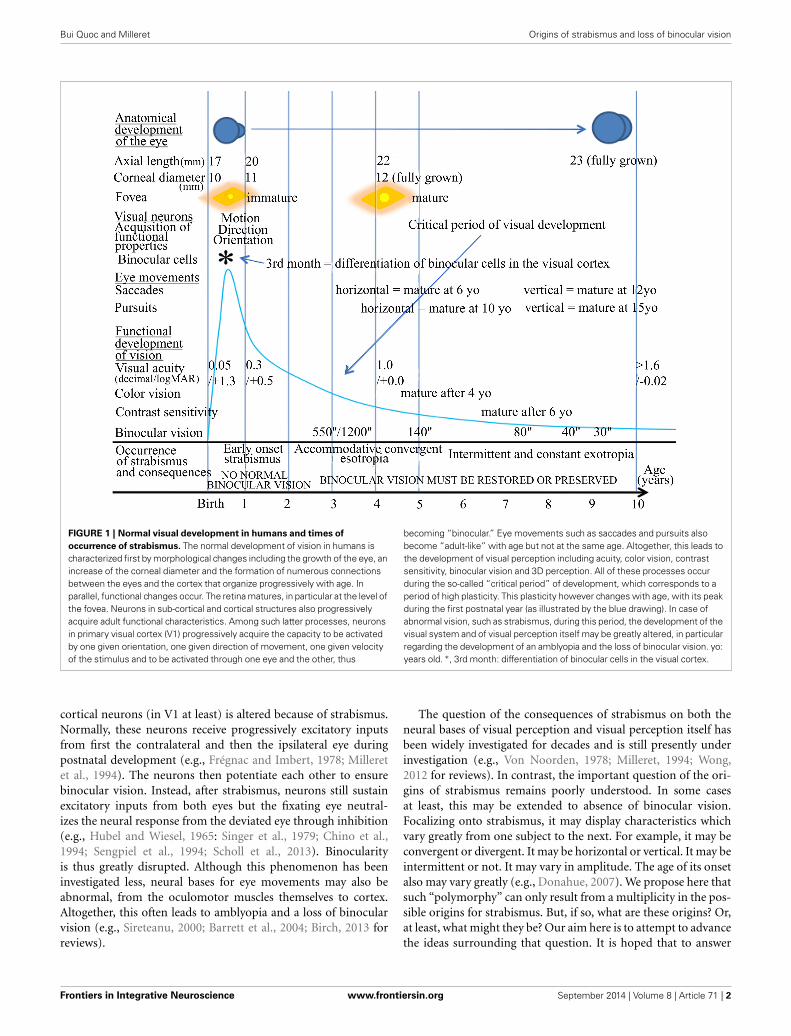
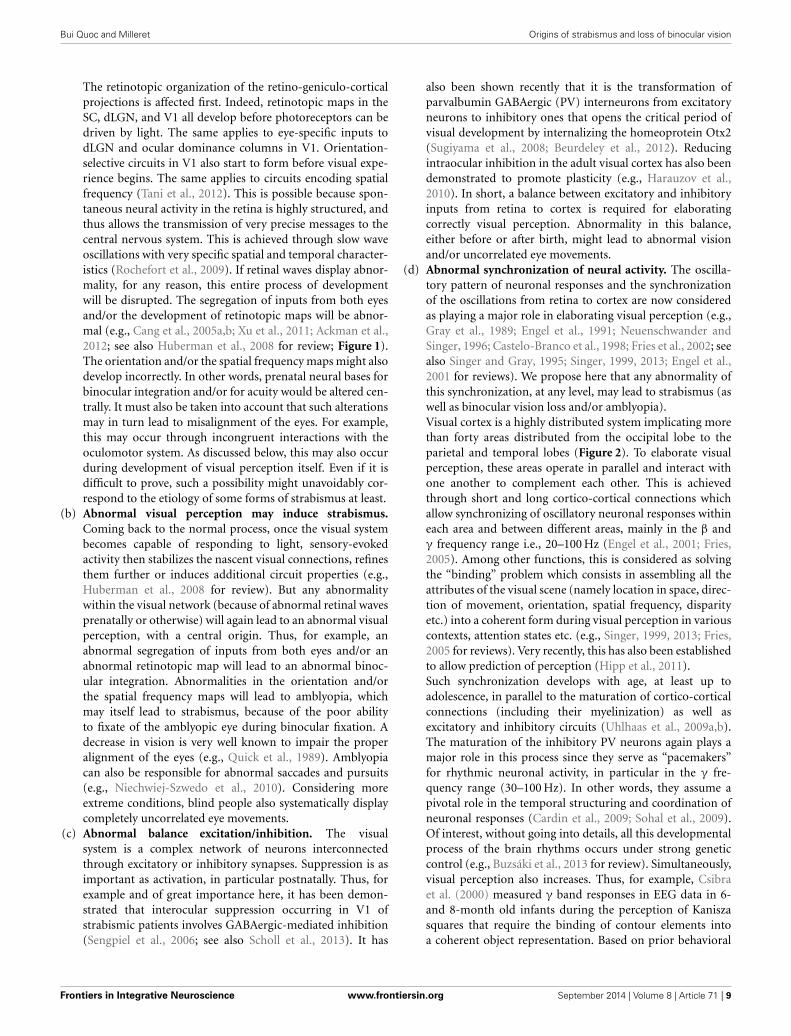
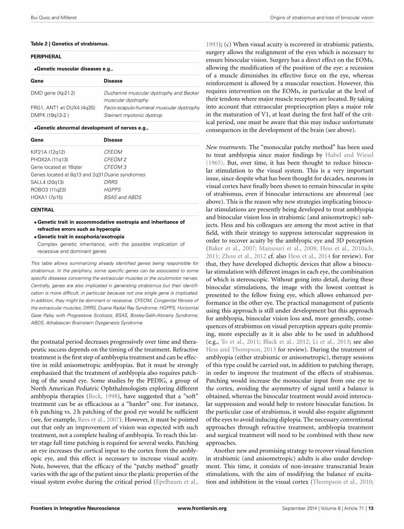
INTRODUCTIONVisual perception is optimal in humans at adulthood, provid-ing that all the developmental processes in relation to it haveoccurred properly both before and after birth, including anatom-ical and functional processes. As illustrated in Figure 1, thisincludes not only the correct development of the eyes them-selves, but also that of eye movements through the extraocularmuscles (EOMs). In parallel, all of the central structures in thebrain that are related to visual perception (including those con-cerned with eye movements) must also develop appropriately. Asa result, for example, each neuron in primary visual cortex (V1)becomes progressively able to encode the different attributes ofthe visual scene, such as orientation and direction of movement.Progressively, most of them also become “binocular,” i.e., able tobe activated through both eyes while they are initially monocular(e.g., Frégnac and Imbert, 1978; Milleret et al., 1988). In par-allel, cortical maps corresponding to each of these attributes ofthe visual scene, including the retinotopic map encoding space,develop (e.g., Chapman et al., 1996; Crair et al., 1998; Smith andTrachtenberg, 2007; White and Fitzpatrick, 2007; Tani et al., 2012for review). The different types of eye movements (saccades, pur-suits) also mature with age (e.g., Ingster-Moati et al., 2009; Bucciand Seassau, 2012, 2014; cf. Figure 1). The “quality” of both thepostnatal visual experience and that of the eye movements play
a major role in this, in particular during the so called “criticalperiod” (e.g., Hubel and Wiesel, 1970; Buisseret, 1995 for review).At the end of all these processes, if they have occurred prop-erly, an optimal visual perception in terms of acuity, color vision,perception of contrasts and binocular vision (which ensures 3Dperception) is acquired progressively with age; cf. Figure 1.
Any alteration of postnatal visual experience and/or of eyemovements during the critical period (which corresponds tothe period of maximum plasticity) leads to the abnormal devel-opment of various structures in the brain, both anatomicallyand functionally. Strabismus is among these alterations. It hasbeen identified for centuries and is characterized by a misalign-ment of the eyes. It presently affects approximately 2% of thehuman population worldwide. When occurring early in life, stra-bismus induces, for example, an abnormal development of boththe geniculo-cortical pathway and interhemispheric connectionsthrough the corpus callosum (CC; e.g., Innocenti and Frost,1979; Schmidt et al., 1997; Löwel et al., 1998; Bui Quoc et al.,2012). In parallel, neurons and neuronal maps in V1, as well asthose in visual areas from the dorsal and the ventral streams,develop abnormal functional properties (e.g., Chino et al., 1983;Milleret and Houzel, 2001; Schmidt et al., 2004; Bui Quoc et al.,2012; see also Von Noorden, 1978; Milleret, 1994; Wong, 2012for reviews). Importantly here, the binocular activation of visual
Frontiers in Integrative Neuroscience www.frontiersin.org September 2014 | Volume 8 | Article 71 | 1
INTEGRATIVE NEUROSCIENCE
Bui Quoc and Milleret Origins of strabismus and loss of binocular vision
FIGURE 1 | Normal visual development in humans and times of
occurrence of strabismus. The normal development of vision in humans ischaracterized first by morphological changes including the growth of the eye, anincrease of the corneal diameter and the formation of numerous connectionsbetween the eyes and the cortex that organize progressively with age. Inparallel, functional changes occur. The retina matures, in particular at the level ofthe fovea. Neurons in sub-cortical and cortical structures also progressivelyacquire adult functional characteristics. Among such latter processes, neuronsin primary visual cortex (V1) progressively acquire the capacity to be activatedby one given orientation, one given direction of movement, one given velocityof the stimulus and to be activated through one eye and the other, thus
becoming “binocular.” Eye movements such as saccades and pursuits alsobecome “adult-like” with age but not at the same age. Altogether, this leads tothe development of visual perception including acuity, color vision, contrastsensitivity, binocular vision and 3D perception. All of these processes occurduring the so-called “critical period” of development, which corresponds to aperiod of high plasticity. This plasticity however changes with age, with its peakduring the first postnatal year (as illustrated by the blue drawing). In case ofabnormal vision, such as strabismus, during this period, the development of thevisual system and of visual perception itself may be greatly altered, in particularregarding the development of an amblyopia and the loss of binocular vision. yo:years old. ∗, 3rd month: differentiation of binocular cells in the visual cortex.
cortical neurons (in V1 at least) is altered because of strabismus.Normally, these neurons receive progressively excitatory inputsfrom first the contralateral and then the ipsilateral eye duringpostnatal development (e.g., Frégnac and Imbert, 1978; Milleretet al., 1994). The neurons then potentiate each other to ensurebinocular vision. Instead, after strabismus, neurons still sustainexcitatory inputs from both eyes but the fixating eye neutral-izes the neural response from the deviated eye through inhibition(e.g., Hubel and Wiesel, 1965: Singer et al., 1979; Chino et al.,1994; Sengpiel et al., 1994; Scholl et al., 2013). Binocularityis thus greatly disrupted. Although this phenomenon has beeninvestigated less, neural bases for eye movements may also beabnormal, from the oculomotor muscles themselves to cortex.Altogether, this often leads to amblyopia and a loss of binocularvision (e.g., Sireteanu, 2000; Barrett et al., 2004; Birch, 2013 forreviews).
The question of the consequences of strabismus on both theneural bases of visual perception and visual perception itself hasbeen widely investigated for decades and is still presently underinvestigation (e.g., Von Noorden, 1978; Milleret, 1994; Wong,2012 for reviews). In contrast, the important question of the ori-gins of strabismus remains poorly understood. In some casesat least, this may be extended to absence of binocular vision.Focalizing onto strabismus, it may display characteristics whichvary greatly from one subject to the next. For example, it may beconvergent or divergent. It may be horizontal or vertical. It may beintermittent or not. It may vary in amplitude. The age of its onsetalso may vary greatly (e.g., Donahue, 2007). We propose here thatsuch “polymorphy” can only result from a multiplicity in the pos-sible origins for strabismus. But, if so, what are these origins? Or,at least, what might they be? Our aim here is to attempt to advancethe ideas surrounding that question. It is hoped that to answer
Frontiers in Integrative Neuroscience www.frontiersin.org September 2014 | Volume 8 | Article 71 | 2
Bui Quoc and Milleret Origins of strabismus and loss of binocular vision
such question will help in the improvement of the treatment ofthese pathologies in the future.
To our knowledge at least, the question of the origin ofstrabismus was first approached “scientifically” in the nine-teenth century. Several theories were suggested. For example, VonGraefe (1854) insisted on mechanistic factors creating strabismus.Donders (1863) pointed out that refraction errors may have a rolein the origin of strabismus through their links with accommo-dation. Duane (1869) proposed that it was an excess in vergenceinnervation that led to strabismus. Worth (1915) suggested thatit was the absence of fusion of the images of both eyes that cre-ated strabismus, and that a “center of fusion” in the brain wasimplicated in this. Chavasse (1939) explained strabismus as a con-sequence of an excess in reflexogenic action. However, none ofthese mechanisms has even been proven. The same holds trueregarding loss of binocular vision which may be either the conse-quence of strabismus or its cause. The absence of alignment itselfprevents the development of normal binocular vision whereas,without binocular vision, the alignment of the eyes becomesunnecessary.
Nowadays, hypotheses on the etiology of strabismus haveevolved and two major theories have thus emerged: a “sensoryvs. motor” theory and a “peripheral vs. central” theory. The for-mer theory proposes that strabismus may have a “sensory” or a“motor” origin, while the latter theory rather suggests that stra-bismus may have a “peripheral” or a “central” origin. The variousforms of strabismus are therefore classified depending on those“sensory vs. motor” or “peripheral vs. central” oppositions.
The “Classification of Eye Movement Abnormalities andStrabismus” (CEMAS; http://www.nei.nih.gov/news/statements/cemas.pdf) is mainly based on the “sensory-motor” opposition.It directly and clearly differentiates strabismus from “other” eyemovement abnormalities that would be not considered as “pure”strabismus. In the CEMAS, first, eye movement abnormalitiesor strabismus are defined as resulting from an abnormal motorsystem, thus having: (1) abnormal full versions and ductions,abnormal fusional vergence amplitudes; (2) non-accurate andabnormal speed saccades, abnormal gain pursuit and vestibularmovements; (3) pathologic oscillations or intrusions. Second, abinocular sensory system is defined as abnormal if there is nobi-fixation with normal visual acuity in each eye, strabismus,diplopia, abnormal retinal correspondence, abnormal fusionalvergence amplitudes, and abnormal stereopsis. It is also sub-normal if there is one or more of the following characteristics:anomalous retinal correspondence, suppression, deficient to nostereopsis, amblyopia, and decreased fusional vergence ampli-tudes. Finally, the system is also considered as abnormal if there isno binocular vision. The complete classification of CEMAS is thendescribed after those statements on motor and sensory aspects ofthe visual system. We shall not detail it here but only recall the fol-lowing divisions: (1) horizontal heterotropias, either concomitantor non-concomitant, either divergent or convergent. In this sec-tion, the early onset esotropia, the nerve palsies, the accommoda-tive esotropia and the constant or intermittent exotropias arealso included; (2) horizontal heterophorias; (3) cyclovertical het-erotropias and special forms of strabismus, i.e., oblique musclespalsy or dysfunction, restrictive strabismus and neuro-myogenic
strabismus. In this latter case, myasthenia gravis, chronic pro-gressive external ophthalmoplegia, internuclear ophthalmoplegia,and skew deviation are also classified. Special forms of strabismusare also mentioned in this section, such as co-contractive retrac-tion syndromes, Restrictive Hypotropia in Adduction (RHA) andCongenital Fibrosis of the EOMs (CFEOM); (4) cycloverticalheterophorias; (5) accommodative disorders (Paralysis, Infacility,Insufficiency, Excess); (6) nystagmus and other ocular motoroscillations.
Contrasting with the former classification, the other way toclassify rationally the different types of strabismus consists ofsegregating strabismus according to its “peripheral” vs. “central”origins. This is the classification which is presented in a recentsynthesis achieved by Péchereau (2013). In his book, the authorclassified strabismus with a “peripheral” origin as those result-ing from abnormalities at the level of the oculomotor musclesthemselves or their innervation. These include, for example, themuscular dystrophies and the palsy of the 3rd, 4th, or 6th cra-nial nerves. Also included are the retraction syndromes (nowcalled CCDD disorders for “Congenital Cranial DysinnervationDisorders”), the Basedow disease (called “Graves disease” in theUnited States), and, finally, oculomotor abnormalities secondaryto orbital fractures. In comparison, strabismus considered as hav-ing a “central” origin have also been classified. But the preciseorigins of those strabismus were not specified since that ques-tion has never been approached in the literature (at least fromour knowledge). Among strabismus with a central origin, theauthor has included different types of strabismus depending on:(a) the type of deviation (vertical or horizontal strabismus, con-vergent or divergent, with or without eye cyclo-torsion); (b) theage of occurrence of strabismus (early onset until 6 or 8 months,late onset after 2.5 years, intermediate); (c) whether the devi-ation is constant or intermittent. He has additionally pointedout the importance of the analysis of binocular status whenclassifying strabismus, which depends on normal (or potentiallynormal) or abnormal binocularity. As indicated above, strabis-mus is most often associated with abnormal binocularity. Thisoccurs in case of: (a) early onset constant divergent or conver-gent strabismus; (b) micro-strabismus caused by a hereditaryabsence of fusion; (c) evolution of a micro-strabismus in stra-bismus; (d) secondary strabismus (i.e., caused by an anatomicalabnormality that decreases the vision of an eye). However, binoc-ularity may be present in spite of strabismus in the followingcases: (a) intermittent early onset strabismus; (b) late onset stra-bismus, convergent or divergent, intermittent or permanent; (c)accommodative strabismus, with or without excess or conver-gence; (d) latent strabismus (heterophorias). The author has alsopointed out that oculomotor abnormalities may exist with orwithout deviation of the eyes (i.e., without strabismus), such asin the case of nystagmus without strabismus, or in the case oftorticollis.
The “sensory vs. motor” and the “peripheral vs. central” the-ories in fact complement each other. This will be obvious fromour further discussions below. Nevertheless, the precise “primummovens” of strabismus remains vague in both theories, and thevery mechanisms and pathophysiology are rarely expressed. Itis our main goal to open here the “black box” dealing with the
Frontiers in Integrative Neuroscience www.frontiersin.org September 2014 | Volume 8 | Article 71 | 3
Bui Quoc and Milleret Origins of strabismus and loss of binocular vision
origins of strabismus, in particular its central origins, and there-fore we shall use the latter classification in the present article.In that regard, we shall tentatively take into account the mostrelevant present knowledge about the organization of the brainand its development. This knowledge will come from both biol-ogy and medicine. Some knowledge from anatomy, physiology,as well as genetics and molecular biology, will be thus consid-ered. “Innate” and “acquired” factors which may potentially leadto strabismus and/or the absence of binocular vision will be alsoexamined, in addition to peripheral vs. central factors and sensoryvs. motor factors. Tychsen has stated already that it is the brainthat must be repaired if ophthalmologists want to treat strabis-mus (Tychsen, 2005). We evidently agree with that idea but muchstill remains to be done prior to the complete treatment of strabis-mus. To treat consequences of strabismus on visual perception isalready relatively effective, with conventional treatments includ-ing optical treatment with glasses, monocular occlusion, andalignment of the eyes through surgery. Newly developed strategiessuch as binocular training and transcranial magnetic stimulations(TMS) could improve in the future the efficacy of conventionalstrabismus treatment since it has been shown that such strate-gies permit the recovery of visual acuity and binocular vision inamblyopia, even at adulthood (after alignment of the eyes); (e.g.,Nyffeler et al., 2006; Hess et al., 2010a,b; Hess and Thompson,2013). Furthermore, considering strabismus at source and deal-ing in particular with its central origins is currently far fromeffective. The same applies to loss of binocular vision with acentral origin. However, as a general rule in medicine, it isalways better to treat pathology at source (provided its originand pathophysiology are precisely defined) rather than dealingwith its dilatory consequences. Our article aims at assisting inthis regard by treating the question of the origins of strabis-mus, even if practical therapeutical consequences will not beimmediate.
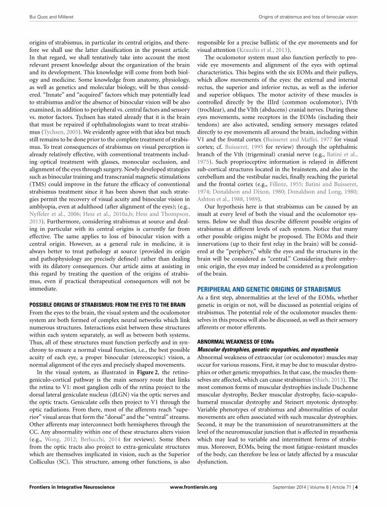
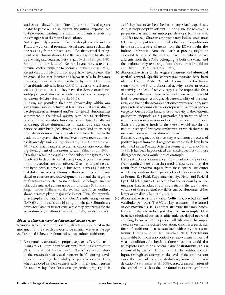
POSSIBLE ORIGINS OF STRABISMUS: FROM THE EYES TO THE BRAINFrom the eyes to the brain, the visual system and the oculomotorsystem are both formed of complex neural networks which linknumerous structures. Interactions exist between these structureswithin each system separately, as well as between both systems.Thus, all of these structures must function perfectly and in syn-chrony to ensure a normal visual function, i.e., the best possibleacuity of each eye, a proper binocular (stereoscopic) vision, anormal alignment of the eyes and precisely shaped movements.
In the visual system, as illustrated in Figure 2, the retino-geniculo-cortical pathway is the main sensory route that linksthe retina to V1: most ganglion cells of the retina project to thedorsal lateral geniculate nucleus (dLGN) via the optic nerves andthe optic tracts. Geniculate cells then project to V1 through theoptic radiations. From there, most of the afferents reach “supe-rior” visual areas that form the “dorsal” and the “ventral” streams.Other afferents may interconnect both hemispheres through theCC. Any abnormality within one of these structures alters vision(e.g., Wong, 2012; Berlucchi, 2014 for reviews). Some fibersfrom the optic tracts also project to extra-geniculate structureswhich are themselves implicated in vision, such as the SuperiorColliculus (SC). This structure, among other functions, is also
responsible for a precise ballistic of the eye movements and forvisual attention (Krauzlis et al., 2013).
The oculomotor system must also function perfectly to pro-vide eye movements and alignment of the eyes with optimalcharacteristics. This begins with the six EOMs and their pulleys,which allow movements of the eyes: the external and internalrectus, the superior and inferior rectus, as well as the inferiorand superior obliques. The motor activity of these muscles iscontrolled directly by the IIIrd (common oculomotor), IVth(trochlear), and the VIth (abducens) cranial nerves. During theseeyes movements, some receptors in the EOMs (including theirtendons) are also activated, sending sensory messages relateddirectly to eye movements all around the brain, including withinV1 and the frontal cortex (Buisseret and Maffei, 1977 for visualcortex; cf. Buisseret, 1995 for review) through the ophthalmicbranch of the Vth (trigeminal) cranial nerve (e.g., Batini et al.,1975). Such proprioceptive information is relayed in differentsub-cortical structures located in the brainstem, and also in thecerebellum and the vestibular nuclei, finally reaching the parietaland the frontal cortex (e.g., Fillenz, 1955; Batini and Buisseret,1974; Donaldson and Dixon, 1980; Donaldson and Long, 1980;Ashton et al., 1988, 1989).
Our hypothesis here is that strabismus can be caused by aninsult at every level of both the visual and the oculomotor sys-tems. Below we shall thus describe different possible origins ofstrabismus at different levels of each system. Notice that manyother possible origins might be proposed. The EOMs and theirinnervations (up to their first relay in the brain) will be consid-ered at the “periphery,” while the eyes and the structures in thebrain will be considered as “central.” Considering their embry-onic origin, the eyes may indeed be considered as a prolongationof the brain.
PERIPHERAL AND GENETIC ORIGINS OF STRABISMUSAs a first step, abnormalities at the level of the EOMs, whethergenetic in origin or not, will be discussed as potential origins ofstrabismus. The potential role of the oculomotor muscles them-selves in this process will also be discussed, as well as their sensoryafferents or motor efferents.
ABNORMAL WEAKNESS OF EOMsMuscular dystrophies, genetic myopathies, and myastheniaAbnormal weakness of extraocular (or oculomotor) muscles mayoccur for various reasons. First, it may be due to muscular dystro-phies or other genetic myopathies. In that case, the muscles them-selves are affected, which can cause strabismus (Shieh, 2013). Themost common forms of muscular dystrophies include Duchennemuscular dystrophy, Becker muscular dystrophy, facio-scapulo-humeral muscular dystrophy and Steinert myotonic dystrophy.Variable phenotypes of strabismus and abnormalities of ocularmovements are often associated with such muscular dystrophies.Second, it may be the transmission of neurotransmitters at thelevel of the neuromuscular junction that is affected in myastheniawhich may lead to variable and intermittent forms of strabis-mus. Moreover, EOMs, being the most fatigue-resistant musclesof the body, can therefore be less or lately affected by a musculardysfunction.
Frontiers in Integrative Neuroscience www.frontiersin.org September 2014 | Volume 8 | Article 71 | 4
Bui Quoc and Milleret Origins of strabismus and loss of binocular vision
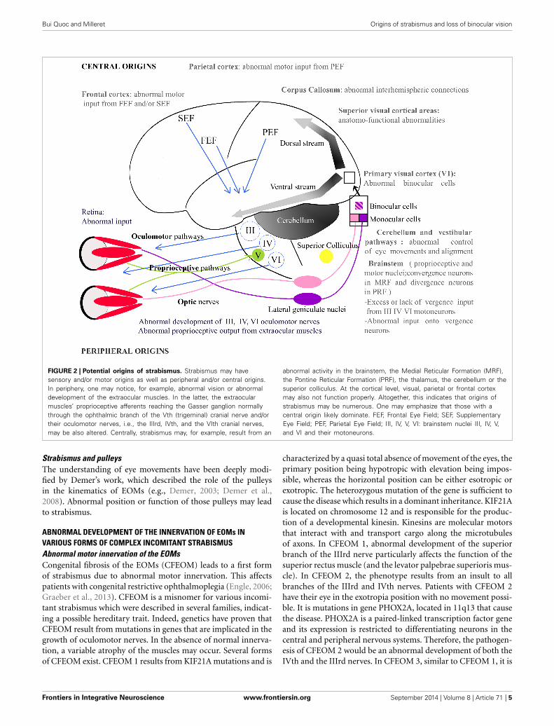
FIGURE 2 | Potential origins of strabismus. Strabismus may havesensory and/or motor origins as well as peripheral and/or central origins.In periphery, one may notice, for example, abnormal vision or abnormaldevelopment of the extraocular muscles. In the latter, the extraocularmuscles’ proprioceptive afferents reaching the Gasser ganglion normallythrough the ophthalmic branch of the Vth (trigeminal) cranial nerve and/ortheir oculomotor nerves, i.e., the IIIrd, IVth, and the VIth cranial nerves,may be also altered. Centrally, strabismus may, for example, result from an
abnormal activity in the brainstem, the Medial Reticular Formation (MRF),the Pontine Reticular Formation (PRF), the thalamus, the cerebellum or thesuperior colliculus. At the cortical level, visual, parietal or frontal cortexmay also not function properly. Altogether, this indicates that origins ofstrabismus may be numerous. One may emphasize that those with acentral origin likely dominate. FEF, Frontal Eye Field; SEF, SupplementaryEye Field; PEF, Parietal Eye Field; III, IV, V, VI: brainstem nuclei III, IV, V,and VI and their motoneurons.
Strabismus and pulleysThe understanding of eye movements have been deeply modi-fied by Demer’s work, which described the role of the pulleysin the kinematics of EOMs (e.g., Demer, 2003; Demer et al.,2008). Abnormal position or function of those pulleys may leadto strabismus.
ABNORMAL DEVELOPMENT OF THE INNERVATION OF EOMs INVARIOUS FORMS OF COMPLEX INCOMITANT STRABISMUSAbnormal motor innervation of the EOMsCongenital fibrosis of the EOMs (CFEOM) leads to a first formof strabismus due to abnormal motor innervation. This affectspatients with congenital restrictive ophthalmoplegia (Engle, 2006;Graeber et al., 2013). CFEOM is a misnomer for various incomi-tant strabismus which were described in several families, indicat-ing a possible hereditary trait. Indeed, genetics have proven thatCFEOM result from mutations in genes that are implicated in thegrowth of oculomotor nerves. In the absence of normal innerva-tion, a variable atrophy of the muscles may occur. Several formsof CFEOM exist. CFEOM 1 results from KIF21A mutations and is
characterized by a quasi total absence of movement of the eyes, theprimary position being hypotropic with elevation being impos-sible, whereas the horizontal position can be either esotropic orexotropic. The heterozygous mutation of the gene is sufficient tocause the disease which results in a dominant inheritance. KIF21Ais located on chromosome 12 and is responsible for the produc-tion of a developmental kinesin. Kinesins are molecular motorsthat interact with and transport cargo along the microtubulesof axons. In CFEOM 1, abnormal development of the superiorbranch of the IIIrd nerve particularly affects the function of thesuperior rectus muscle (and the levator palpebrae superioris mus-cle). In CFEOM 2, the phenotype results from an insult to allbranches of the IIIrd and IVth nerves. Patients with CFEOM 2have their eye in the exotropia position with no movement possi-ble. It is mutations in gene PHOX2A, located in 11q13 that causethe disease. PHOX2A is a paired-linked transcription factor geneand its expression is restricted to differentiating neurons in thecentral and peripheral nervous systems. Therefore, the pathogen-esis of CFEOM 2 would be an abnormal development of both theIVth and the IIIrd nerves. In CFEOM 3, similar to CFEOM 1, it is
Frontiers in Integrative Neuroscience www.frontiersin.org September 2014 | Volume 8 | Article 71 | 5
Bui Quoc and Milleret Origins of strabismus and loss of binocular vision
only the development of the IIIrd nerve that is affected, althoughin a more severe way since all of its branches are affected in thissituation and not only those branches which innervate the supe-rior rectus muscle and the levator palpebrae superioris muscle.The gene responsible for the disease is located at 16qter.
Contrarily to CFEOM syndromes, in which development ofthe IIIrd nerve is affected, it is the development of the VIth nervewhich is affected in the Duane syndromes. The primum movens ofthese latter syndromes is an absence of development of the VIthnerve, either unilaterally or bilaterally. Variable forms of abnor-mal innervation of the lateral rectus muscle by branches of theIIIrd nerve have also been observed, resulting either in esotropiaor exotropia. Again, it is an abnormal development of a nerve thatunderlies the pathogenesis of this condition, with genes located at8q13 and at 2q31. Duane Radial Ray Syndrome (DRRS) is a par-ticular form of Duane Syndrome in which the ocular movementabnormality is associated with bone abnormalities in the hand,such as an absence or a malformation of the thumb which canlook like a finger. Again, it is a mutation in a transcription factorgene SALL4, located at 20q13, which causes the disease by alteringthe normal neural development.
Other insults to the development of the VIth nerve includeHGPPS (Horizontal Gaze Palsy with Progressive Scoliosis), inwhich it is mutations of the gene ROBO3, located at 11q23, whichare responsible for the resulting phenotype. Such phenotypecombines a total impossibility of horizontal gaze movements,along with a scoliosis that occurs during the first decade of life.ROBO3 is a developmental gene and is expressed in the hindbrainof the human fetus. Human ROBO3 is similar to roundaboutgenes that are responsible for axon guidance in other speciessuch as mice, zebrafish or drosophile. Indeed, brainstem neu-rons of ROBO3−/− mice completely fail in crossing the midlineduring development (Marillat et al., 2004). In zebrafish, and indrosophile, the loss of function of ROBO3 results in aberrantmidline crossing by axons (Seeger et al., 1993).
Finally, abnormal development of the VIth nerve occurs intwo conditions in which mutations of the gene HOXA1 (locatedat 7p15 and implicated in hindbrain segmentation during fetaldevelopment) are responsible: the Bosley-Salih-Alorainy syn-drome (BSAS) and the Athabascan Brainstem Dysgenesis syn-drome (ABDS). In BSAS, which is a recessive condition, a bilateralDuane trait is associated with other cranial nerve dysfunctions,such as deafness due to a bilateral absence of the cochlea and mis-development of the VIIIth (vestibulo-cochlear) nerve. In ABDS,horizontal gaze restriction is associated with central deafness andmental retardation.
The various abnormalities of the development of the IIIrd,IVth, or VIth cranial nerves have been regrouped in a genericterm: CCDD (see above). In CCDDs, the insult can cause ocu-lar movement disorders but other conditions without strabismuscan occur, such as isolated congenital ptosis which can resultfrom a mutation in a gene located at 1p34-p32. As a general rule,genetics of CCDDs teach us that an abnormal development ingeneral and an abnormal early routing of neurons in particular,may cause strabismus. In CCDDs, the insult occurs at the fron-tier between peripheral and central locations. It is the same whenconsidering the cranial nerves that emerge from the brainstem
and lead toward abnormal EOMs. The model of the CCDDstherefore emphasizes that an abnormal neural network can causestrabismus.
Palsy of the IIIrd, IVth, or VIth nerve leads to a second formof strabismus. Similar to CCDDs, it is a neural disorder althoughthe cause is often acquired and not innate. The nerve palsy causesan atrophy of the innervated muscle. Acute palsy of the IIIrdnerve is an emergency since it can be caused by a direct com-pression of the nerve by a cavernous sinus thrombophlebitis, orby an aneurysm of the posterior communicant artery. A palsy ofthe IVth nerve is either congenital or acquired. When acquired,it is a peripheral cause that is the primum movens. It may how-ever result from a direct insult after a severe cranial traumatism.A palsy of the VIth nerve may finally result from a direct compres-sion of the nerve’s fine and fragile branches. This can arise from ahypertrophic brainstem due to intracranial hypertension. It mayalso result from numerous other central causes such as tumors,infections, etc. In all cases however, this will induce strabismus.
Strabismus and extraocular proprioceptionThe outflow theory supports the idea that it is an efferent copyof the oculomotor signal from the motor centers that givesinformation about the position of the eyes to the brain (VonHelmholtz, 1866). By contrast, the inflow theory claims that itis the proprioceptive signals from eye muscle receptors that givesuch information (Sherrington, 1918). More recent experimentssupport one theory or the other. Thus, authors now considerthat both theories are right and that efferent copy co-exists withextraocular proprioception. Proprioceptive receptors do exist inthe EOMs, in particular at the level of the tendons (e.g., Cooperand Daniel, 1949; Richmond et al., 1984 for receptors in humans).These receptors are active and send sensory messages to numer-ous regions in the brain implicated both in visual perception andeye movements (e.g., Donaldson, 1979; Donaldson and Dixon,1980; Milleret et al., 1987). Furthermore, they have been demon-strated by our group to strongly contribute to the maturation ofvisual neurons in V1 during development (cf. Buisseret, 1995 forreview; see also Buisseret et al., 1978, 1988). Thus, an abnormalproprioception at the level of the EOMs is also a potential causefor strabismus, since abnormal information about the position ofthe eyes leads to an abnormal central and neural motor commandin return.
Nevertheless, a deafferentation of EOMs has never beendemonstrated to affect ocular motor control and to induce stra-bismus. To our knowledge at least, neither experimental researchnor any medical cases have demonstrated this (the problem is adifficult one to approach). Thus, some authors have concludedthat proprioceptive signals only play a role during developmentin calibrating the efferent copy signal, which is sufficient toprovide information about eye movements and position (Lewiset al., 2001). Other authors, however, have claimed that an insultto proprioceptive receptors of the EOMs could be the causeof strabismus (e.g., Donaldson, 2000). Similarly to the CCDD(see above), abnormal development of the proprioceptive axonswithin the Vth cranial nerve to the Gasser ganglion may occur. Itcould also be hypothesized that abnormalities of the extraocularreceptors could be responsible for strabismus. This is supported
Frontiers in Integrative Neuroscience www.frontiersin.org September 2014 | Volume 8 | Article 71 | 6
Bui Quoc and Milleret Origins of strabismus and loss of binocular vision
by investigations of some strabismic patients whose extraocularmuscle receptors display abnormal morphological characteristics(Li and Shen, 2001). These changes were analyzed using trans-mission electron microscopy and revealed both a decrease in thenumber of mitochondria in axons, and the disappearance of thenerve component of the receptor. Of course, in such a study,whether the abnormalities in the proprioceptors are the causeof strabismus or its consequences cannot be distinguished. Thisrecalls the controversy regarding whether the subtle changes atthe cellular level of the muscles (especially the singly innervatedorbital fibers) of strabismic patients can be the primum movens ofstrabismus, or are simply an adaptative phenomenon to the devi-ation (Lennerstrand, 2007). Finally, the implication of extraocularproprioception during ocular movement disorders can be empha-sized by the fact that a tenotomy of all of the EOMs and theirreattachment, which suppresses the proprioceptive output sig-nals, is an effective therapy in the treatment of some forms ofinfantile nystagmus (Dell’Osso and Wang, 2008).
GENETICS OF CONCOMITANT STRABISMUSContrary to CCDDs or to nerve palsies, most strabismus suchas congenital strabismus, accommodative strabismus or divergentstrabismus are concomitant, meaning that the deviation is alwaysthe same whatever the gaze direction; they also likely display acentral origin (see below). Also contrary to CCDDs, no singlegene has been identified as the direct origin of concomitant stra-bismus. Nevertheless, inheritance and genetics are obvious in thedevelopment of most forms of strabismus, either incomitant orconcomitant (Engle, 2007). However, additional factors, in par-ticular those related to developmental processes, also need to betaken into account.
Hippocrates himself would have stated that: “squinters engen-der squinters.” Physicians are evidently also aware of the influenceof genetics on strabismus and usually advise strabismic par-ents that their children must be screened for strabismus. Indeedapproximately 15% of children of strabismic parents are strabis-mic, compared to the 2% prevalence of strabismus in the generalpopulation (Donnelly, 2012). Ziakas showed however that thisproportion may vary depending on the type of strabismus andthe degree of relationship (Ziakas et al., 2002). In his study of96 index cases with strabismus with either early onset strabismus(26 cases), accommodative esotropia (49 cases), anisometropicesotropia (15 cases), or exotropia (56 cases), he showed that therisk of having strabismus for a first degree relative is 4% forexotropia but 26.1% for accommodative esotropia. In accom-modative esotropia, the risk decreases to 7.5% for second degreerelatives and to 4.8% for third degree relatives. In twin studies, ithas been shown that there is a specific genetic influence for eso-deviation which is independent of the refractive error (Sanfilippoet al., 2012). The heritability of eso-deviation is estimated as64% in a cohort of 1462 twin pairs with a prevalence of 8.6%of eso-deviation, the correlation being significantly greater inmonozygotic twins (r = 0.65) than in dizygotic twins (r = 0.33).But, as indicated above, the genetic contribution to concomitantstrabismus is not easy to circumscribe. Thus, even in the case of“simple” strabismus such as early onset esotropia, accommoda-tive esotropia or exotropia, genetic inheritance is complex, with
the possible implication of recessive genes as well as dominantgenes.
The influence of the degree of development of both the eyesand the brain at birth must also be taken into account. Recallingthe higher proportion of strabismus in premature infants com-pared to full term infants illustrates this (Torp-Pedersen et al.,2010). This emphasizes the relationship between the developmentof the brain (including the eyes) and the potential developmentof strabismus after birth. More generally, this indicates that anabnormal (or an immature) development of the neural networks,resulting from innate (i.e., genetic) or acquired factors, might, inturn, lead to strabismus.
POSSIBLE CENTRAL ORIGINS OF STRABISMUSAs indicated above, it is the anatomo-functional maturationbefore and after birth of multiple neural networks from the eyesto the brain that subtend the normal development of visual per-ception. This occurs by implicating both genetic and epigeneticfactors such as postnatal visual experience. Our driving hypoth-esis here is that any insult to this normal process of maturationmay, in turn, generate strabismus. This applies evidently to anylevel of the sensory and/or motor networks that are involved inthe elaboration of visual perception (cf. Figure 2). Some examplesare provided below to illustrate this. How an abnormal develop-ment of any visual path or any neural activity somewhere withinthe visual system may lead to strabismus are considered in succes-sion. How an abnormal neural activity in the oculomotor systemmay lead to strabismus is also discussed.
ABNORMAL DEVELOPMENT OF THE VISUAL PATHSFirst, we hypothesize that any insult during the normal processesof neurogenesis, axonal growth, migration of neurons, synapto-genesis, myelination, apoptosis or even elimination of juvenileexuberant axons, may potentially lead to strabismus. For exam-ple, strabismus may be the consequence of the misrouting of somepaths within the visual and/or the oculomotor networks.
Abnormal routing of ganglion cell axonsInterestingly, Siamese cats spontaneously display a convergentstrabismus. They also have an abnormal predominance of thecrossed retino-geniculo-cortical pathway compared to normalcats (Montero and Guillery, 1978; Shatz and Levay, 1979). Thisresults from stagnation at an early stage of development, whichitself recalls the development of visual pathways during phylo-genesis. We propose that such abnormal predominance of thecrossed retino-geniculo-cortical pathway may also be the causeof the early onset convergent strabismus in humans, in whichthe early asymmetry of the optokinetic nystagmus also persistswith age.
Paradoxically, in case of divergent strabismus, it could also behypothesized that a predominance of the crossed pathways couldbe the primum movens of strabismus. During evolution, the visualsystem is first an “only crossed fibers” network with lateral eyesand panoramic vision. It then evolves to a balanced system withequal importance between the direct pathway and the crossedpathway, and frontal eyes allowing binocular vision. We proposehere that an abnormal routing of the retinal ganglion cell axons
Frontiers in Integrative Neuroscience www.frontiersin.org September 2014 | Volume 8 | Article 71 | 7
Bui Quoc and Milleret Origins of strabismus and loss of binocular vision
at the level of the optic chiasm might lead to a loss of balancebetween crossed and direct fibers and thus lead to strabismus.This might occur by an abnormal expression of ephrins, i.e., sur-face molecules which are specifically implicated in guiding theretinal ganglion cell axons at the level of the optic chiasm dur-ing the developmental process (Petros et al., 2009). The axons ofipsilateral projections from temporal retina (direct fibers) expressthe guidance receptor ephrin B1 (but not the axons of contralat-eral projections from the nasal retina, i.e., crossed fibers). At theoptic chiasm, radial glia cells express ephrin B2, which repulsesthe ephrin B1 axons from crossing the midline, unlike the con-tralateral fibers from the nasal retina. Expressions of ephrins andof ephrin receptors are specific and precise timing is necessaryto ensure the normal and balanced development of visual path-ways. If this system was altered through an abnormal expressionof ephrins and/or ephrin receptors during development, an asym-metrical neural network of crossed and uncrossed fibers woulddevelop and could result in the development of strabismus.
Misrouting and abnormal retinotopyDuring development, ganglion cell axons reach progressively cen-tral visual structures by respecting “retinotopy.” The visual fieldis thus encoded by neurons with precision from the retina up tothe cortex (e.g., Tootell et al., 1998 for review), a necessary con-dition to ensure normal visual perception, including binocularvisual perception. Guidance of axons creating retinotopy is alsopermitted by ephrins. Gradients of ephrins A and ephrins B, bothin the retina and in the visual cortex, allow the creation of x and ycoordinates (Cang et al., 2005a,b). This leads to the establishmentof neuronal “retinotopic maps,” which are refined with age andvisual experience. Again we propose here that abnormal guidancethrough abnormal levels of ephrins A or B and/or their receptorsduring development would alter retinotopy and would cause, inturn, strabismus. In some cases at least, distortions within retino-topic maps, which may lead to abnormal retinal correspondencein early onset strabismus (e.g., Wong, 2000; Popple and Levi,2005; Mansouri et al., 2009; Wang et al., 2012), would thereforebe a cause of strabismus rather than a consequence.
The subplate, i.e., mostly temporary cells located below layerVI of Area V1, plays a major role for growing axons to reachthe visual cortical plate during development (Ghosh et al., 1980;McConnell et al., 1989). Any abnormality during such processwould interfere with normal development of geniculo-corticalconnections, thus with normal development of retinotopic mapsin V1. Again, this could potentially induce strabismus.
Abnormal cortico-cortical connectionsStrabismus is now well known to disrupt the development ofnumerous cortico-cortical connections implicated in visual per-ception. These cortico-cortical connections may be “short” andlocated within one given area or “long,” thus linking variousareas which may be located very far from one to the other.Thus, for example, strabismus is known to stabilize normallytransient intra-hemispheric cortico-cortical connections in V1,leading to interconnect larger cell groups driven through thesame eye than in the normal case (e.g., Löwel and Singer, 1992;Schmidt and Löwel, 2008). It is also known to lead to drastic
anatomo-functional changes in the organization of the inter-hemispheric callosal connections, which normally link recipro-cally and homotopically various visual areas to “glue” both visualhemifields into a single scene (Payne, 1990, 1991; Payne andSiwek, 1991a,b; Bui Quoc et al., 2012). In particular, in the caseof strabismus, it leads to the development of asymmetrical inter-hemispheric connections which prevent the fusion of both visualhemifields along the vertical midline (Lund and Mitchell, 1979;Milleret and Houzel, 2001; Bui Quoc et al., 2012).
Our idea here is that abnormal anatomical cortico-corticalconnections within or between visual cortical areas (whatevertheir origin) may conversely lead in turn to strabismus. Ourhypothesis may be supported first by experiments which haveconsisted in cutting the CC of adult cats, who rapidly displayeda misalignment of their eyes and even strabismus (Elberger, 1979;Payne et al., 1981; Elberger and Hirsch, 1982). This is further sup-ported by studies showing the implication of the CC during eyemovements (e.g., Pasik et al., 1971; Tusa and Ungerleider, 1988;Zernicki et al., 1997). Our hypothesis is also strengthened by ana-lyzing the deficits in visual and visuo-spatial developments thatare present in young children with Williams syndrome. They areinterpreted as the result of a split between the ventral and dor-sal stream processing of visual information (see Figure 2), witha generalized deficit in dorsal stream processing (Atkinson et al.,2001). Of great interest here, the authors underlined that patientswith such syndrome also display a much higher incidence of stra-bismus, visual acuity loss, amblyopia and reduced stereopsis thanthe general population.
ABNORMAL DEVELOPMENT OF NEURONAL ACTIVITYIn addition to genes, axonal guidance cues and molecules, spon-taneous and early visually evoked neural activity are necessaryfor anatomical and functional refinement of developing visualcircuits (e.g., Huberman et al., 2008 for review). Appropriate syn-chronizations within the visual network then need to develop inorder to elaborate visual perception optimally (e.g., Singer, 1999,2013; Uhlhaas et al., 2009a,b; Menon, 2013). Any abnormality insuch neural activities from the retina to the visual cortex may alsolead to strabismus. Some data in the literature strengthens thisidea already. The same applies to neural circuits subtending eyemovements.
Effects of an abnormal neuronal activity on visual systemLet us evaluate, in succession, the potential impacts on the align-ment of the eyes of: (a) abnormal prenatal retinal waves; (b)abnormalities during postnatal visual experience; (c) abnormalexcitation/inhibition balance; and (d) pathological asynchrony ofneural activity.
(a) Abnormal retinal waves. First, prenatal spontaneous neu-ral activity in retina, discovered by Galli and Maffei (1988),must be absolutely normal to allow the visual system to orga-nize with precision. It plays both permissive and instructiveroles. Indeed, even if this activity is generated very early inlife, before vision begins, it is a necessity for the proper devel-opment of functional properties of visual neurons and thatof the various functional maps all along the visual system.
Frontiers in Integrative Neuroscience www.frontiersin.org September 2014 | Volume 8 | Article 71 | 8
Bui Quoc and Milleret Origins of strabismus and loss of binocular vision
The retinotopic organization of the retino-geniculo-corticalprojections is affected first. Indeed, retinotopic maps in theSC, dLGN, and V1 all develop before photoreceptors can bedriven by light. The same applies to eye-specific inputs todLGN and ocular dominance columns in V1. Orientation-selective circuits in V1 also start to form before visual expe-rience begins. The same applies to circuits encoding spatialfrequency (Tani et al., 2012). This is possible because spon-taneous neural activity in the retina is highly structured, andthus allows the transmission of very precise messages to thecentral nervous system. This is achieved through slow waveoscillations with very specific spatial and temporal character-istics (Rochefort et al., 2009). If retinal waves display abnor-mality, for any reason, this entire process of developmentwill be disrupted. The segregation of inputs from both eyesand/or the development of retinotopic maps will be abnor-mal (e.g., Cang et al., 2005a,b; Xu et al., 2011; Ackman et al.,2012; see also Huberman et al., 2008 for review; Figure 1).The orientation and/or the spatial frequency maps might alsodevelop incorrectly. In other words, prenatal neural bases forbinocular integration and/or for acuity would be altered cen-trally. It must also be taken into account that such alterationsmay in turn lead to misalignment of the eyes. For example,this may occur through incongruent interactions with theoculomotor system. As discussed below, this may also occurduring development of visual perception itself. Even if it isdifficult to prove, such a possibility might unavoidably cor-respond to the etiology of some forms of strabismus at least.
(b) Abnormal visual perception may induce strabismus.Coming back to the normal process, once the visual systembecomes capable of responding to light, sensory-evokedactivity then stabilizes the nascent visual connections, refinesthem further or induces additional circuit properties (e.g.,Huberman et al., 2008 for review). But any abnormalitywithin the visual network (because of abnormal retinal wavesprenatally or otherwise) will again lead to an abnormal visualperception, with a central origin. Thus, for example, anabnormal segregation of inputs from both eyes and/or anabnormal retinotopic map will lead to an abnormal binoc-ular integration. Abnormalities in the orientation and/orthe spatial frequency maps will lead to amblyopia, whichmay itself lead to strabismus, because of the poor abilityto fixate of the amblyopic eye during binocular fixation. Adecrease in vision is very well known to impair the properalignment of the eyes (e.g., Quick et al., 1989). Amblyopiacan also be responsible for abnormal saccades and pursuits(e.g., Niechwiej-Szwedo et al., 2010). Considering moreextreme conditions, blind people also systematically displaycompletely uncorrelated eye movements.
(c) Abnormal balance excitation/inhibition. The visualsystem is a complex network of neurons interconnectedthrough excitatory or inhibitory synapses. Suppression is asimportant as activation, in particular postnatally. Thus, forexample and of great importance here, it has been demon-strated that interocular suppression occurring in V1 ofstrabismic patients involves GABAergic-mediated inhibition(Sengpiel et al., 2006; see also Scholl et al., 2013). It has
also been shown recently that it is the transformation ofparvalbumin GABAergic (PV) interneurons from excitatoryneurons to inhibitory ones that opens the critical period ofvisual development by internalizing the homeoprotein Otx2(Sugiyama et al., 2008; Beurdeley et al., 2012). Reducingintraocular inhibition in the adult visual cortex has also beendemonstrated to promote plasticity (e.g., Harauzov et al.,2010). In short, a balance between excitatory and inhibitoryinputs from retina to cortex is required for elaboratingcorrectly visual perception. Abnormality in this balance,either before or after birth, might lead to abnormal visionand/or uncorrelated eye movements.
(d) Abnormal synchronization of neural activity. The oscilla-tory pattern of neuronal responses and the synchronizationof the oscillations from retina to cortex are now consideredas playing a major role in elaborating visual perception (e.g.,Gray et al., 1989; Engel et al., 1991; Neuenschwander andSinger, 1996; Castelo-Branco et al., 1998; Fries et al., 2002; seealso Singer and Gray, 1995; Singer, 1999, 2013; Engel et al.,2001 for reviews). We propose here that any abnormality ofthis synchronization, at any level, may lead to strabismus (aswell as binocular vision loss and/or amblyopia).Visual cortex is a highly distributed system implicating morethan forty areas distributed from the occipital lobe to theparietal and temporal lobes (Figure 2). To elaborate visualperception, these areas operate in parallel and interact withone another to complement each other. This is achievedthrough short and long cortico-cortical connections whichallow synchronizing of oscillatory neuronal responses withineach area and between different areas, mainly in the β andγ frequency range i.e., 20–100 Hz (Engel et al., 2001; Fries,2005). Among other functions, this is considered as solvingthe “binding” problem which consists in assembling all theattributes of the visual scene (namely location in space, direc-tion of movement, orientation, spatial frequency, disparityetc.) into a coherent form during visual perception in variouscontexts, attention states etc. (e.g., Singer, 1999, 2013; Fries,2005 for reviews). Very recently, this has also been establishedto allow prediction of perception (Hipp et al., 2011).Such synchronization develops with age, at least up toadolescence, in parallel to the maturation of cortico-corticalconnections (including their myelinization) as well asexcitatory and inhibitory circuits (Uhlhaas et al., 2009a,b).The maturation of the inhibitory PV neurons again plays amajor role in this process since they serve as “pacemakers”for rhythmic neuronal activity, in particular in the γ fre-quency range (30–100 Hz). In other words, they assume apivotal role in the temporal structuring and coordination ofneuronal responses (Cardin et al., 2009; Sohal et al., 2009).Of interest, without going into details, all this developmentalprocess of the brain rhythms occurs under strong geneticcontrol (e.g., Buzsáki et al., 2013 for review). Simultaneously,visual perception also increases. Thus, for example, Csibraet al. (2000) measured γ band responses in EEG data in 6-and 8-month old infants during the perception of Kaniszasquares that require the binding of contour elements intoa coherent object representation. Based on prior behavioral
Frontiers in Integrative Neuroscience www.frontiersin.org September 2014 | Volume 8 | Article 71 | 9
Bui Quoc and Milleret Origins of strabismus and loss of binocular vision
studies that showed that infants up to 6 months of age areunable to perceive Kanisza figures, the authors hypothesizedthat perceptual binding in 8-month-old infants is related tothe emergence of the γ band oscillations.Not surprisingly, epigenetic factors also play a role in this.Thus, any abnormal postnatal visual experience such as theone resulting from strabismus modifies the normal develop-ment of synchronization within the visual system by alteringboth wiring and neural activity (e.g., Löwel and Singer, 1992;Schmidt and Löwel, 2008). Neuronal synchrony is reducedin visual cortex compared to normal (Roelfsema et al., 1994).Recent data from Hess and his group have strengthened thisby establishing that interactions between cells in disparatebrain regions are reduced when driven by the amblyopic eyeof strabismic subjects, from dLGN to superior visual areas,via V1 (Li et al., 2011). They have also demonstrated thatamblyopia (in strabismic patients) is associated to temporalsynchrony deficits (Huang et al., 2012).In turn, we postulate that any abnormality within onegiven visual area or between at least two visual areas, due todevelopmental anatomical and/or functional abnormalitiessomewhere in the visual system, may lead to strabismus(and amblyopia and/or binocular vision loss) by alteringsynchrony. Since abnormalities in synchrony may occurbefore or after birth (see above), this may lead to an earlyor a late strabismus. The same idea may be extended to theoculomotor system since it has been shown recently that ithas its own dynamics (Gregoriou et al., 2012; Cordones et al.,2013) and that changes in neural synchrony also occur dur-ing development of the motor system (Kilner et al., 2000).Situations when the visual and the oculomotor systems needto interact to elaborate visual perception, i.e., during sensori-motor processing, are also affected. One may underline thatour hypothesis is directly in line with increasing evidencethat disturbances of synchrony in the developing brain, asso-ciated to aberrant neurodevelopment, subtend the cognitivedysfunctions associated to major brain pathologies such asschizophrenia and autism spectrum disorders (Uhlhaas andSinger, 2006; Uhlhaas et al., 2009a,b, 2011). As outlinedabove, genetics play a major role in that. Thus, for example,in schizophrenic patients, the GABA synthesizing enzymeGAD 65 and the calcium-binding protein parvalbumin aredown-regulated in basket cells, while they are crucial for thegeneration of γ rhythms (Lewis et al., 2005; see also above).
Effects of abnormal neural activity on oculomotor systemNeuronal activity within the various structures implicated in themovement of the eyes also needs to be normal whatever the age.As illustrated below, any abnormality may induce strabismus.
(a) Abnormal extraocular proprioceptive afferents fromEOMs to V1. Proprioceptive afferents from EOMs project toV1 (Buisseret and Maffei, 1977). They strongly contributeto the maturation of visual neurons in V1 during devel-opment, including their ability to perceive details. Thus,when removed in their entirety early in life, visual neuronsdo not develop their functional properties properly. It is
as if they had never benefited from any visual experience.Also, if proprioceptive afferents in one plane are removed, aperpendicular meridian amblyopia develops (cf. Buisseret,1995 for review). Since an amblyopia may induce strabismus(cf. above), we put forward the idea that any disequilibriumin the proprioceptive afferents from the EOMs might alsoinduce strabismus. Note that such a process might beextended to any of the central structures which receiveafferents from the EOMs, belonging to both the visual andthe oculomotor systems (e.g., Donaldson, 1979; Donaldsonand Dixon, 1980; Milleret et al., 1987).
(b) Abnormal activity of the vergence neurons and abnormalcortical control. Specific convergence neurons have beenidentified in the Medial Reticular Formation of the brain-stem (Mays, 1984) and abnormal activity, either an excessof activity or a loss of activity, may also be responsible for adeviation of the eyes. Hyperactivity of these neurons couldlead to convergent esotropia. Hyperexcitability of the neu-rons, enhancing the accommodation/convergence loop, mayplay a role in accommodative esotropia with an excess of con-vergence. On the other hand, a loss of activity of the neurons,premature apoptosis or a progressive degeneration of theneurons or axons may also induce exophoria and exotropia.Such a progressive insult to the system would explain thenatural history of divergent strabismus, in which there is anincrease in divergent deviation with time.Similarly, divergent strabismus may result from an excess ofpositive inputs from the divergence neurons which have beenidentified in the Pontine Reticular Formation (cf. also Mays,1984). It has been hypothesized that a lack of activity of thosedivergence neurons would induce esotropia.Higher structures command eye movement and eye position.Our hypothesis here is that the genesis of strabismus may alsoresult from abnormal inputs from those cortical structureswhich play a role in the triggering of ocular movements suchas Frontal Eye Field, Supplementary Eye Field, and ParietalEye Field (cf. Figure 2). Indeed, it has been shown by neuro-imaging that, in adult strabismic patients, the gray mattervolume of those cortical eye fields can be abnormal, eitherlarger or smaller (Chan et al., 2004).
(c) Abnormal activity in Superior Colliculus, cerebellum andvestibular pathways. The SC is a key structure in the controlof eye movements. It is another structure that may poten-tially contribute to inducing strabismus. For example, it hasbeen hypothesized that an insufficiently developed neuronalcoupling between both superior colliculi would be impli-cated in vertical dissociated deviation, which is a particularform of strabismus that is associated with early onset stra-bismus (Brodsky, 2011; Ten Tusscher, 2011). Cerebellumand vestibular nuclei also control eye movements in normalvisual conditions. An insult to those structures could alsobe hypothesized to be a central cause of strabismus. This issupported by the fact that an insult to the vestibulo-ocularinput, through an attempt at the level of the otoliths, cancause this particular vertical strabismus, known as a “skewdeviation” (Schlenker et al., 2009). Also, a malformation ofthe cerebellum, such as the one found in Joubert syndrome
Frontiers in Integrative Neuroscience www.frontiersin.org September 2014 | Volume 8 | Article 71 | 10
Bui Quoc and Milleret Origins of strabismus and loss of binocular vision
or in rhombencephalosynapsis, is associated with strabismus(Canturk et al., 2008; Keskinbora, 2008).
CONCLUSIONFor the first time, at least in our knowledge, instead of treatingthe question of the consequences of strabismus in humans, ourarticle highlights the question of the origins of strabismus. The“polymorphy” of strabismus indeed suggests multiple origins butmost of them are presently unknown. Those strabismus with aperipheral origin are rather well characterized but they are onlyfew. In contrast, the other forms of strabismus, which are con-sidered as having a central origin, are poorly understood despitebeing the most frequent. At present it is as if these latter forms ofstrabismus are included in a “black box” that has never really beenopened by anyone. To move forward, we have decided here to ten-tatively open this box. We have proposed mechanisms which showthat a central abnormality may lead to the development of strabis-mus. Our hypothesis on the mechanisms of strabismus are basedon both classical and the most recent knowledge about the devel-opment and the organization of the visual system in mammalsboth before and after birth. Research in that field has indeed beenvery active all around the world for decades and is still very activetoday, providing numerous “keys” which might open our blackbox. Some other mechanisms are based on knowledge regardingthe oculo-motor system from EOMs to cortex. In that context,we have also revisited the question of the origins of binocularvision loss, with tentative new ideas on the question. Interestingly,whether the origins of strabismus or those of binocular visionloss are considered, some of the new mechanisms we proposeare already supported by published data. Altogether, our findingsclearly emphasize the necessity to develop and to apply as soonas possible new strategies to treat strabismus and binocular visionloss, in particular through “central” therapies, in addition to theperipheral ones.
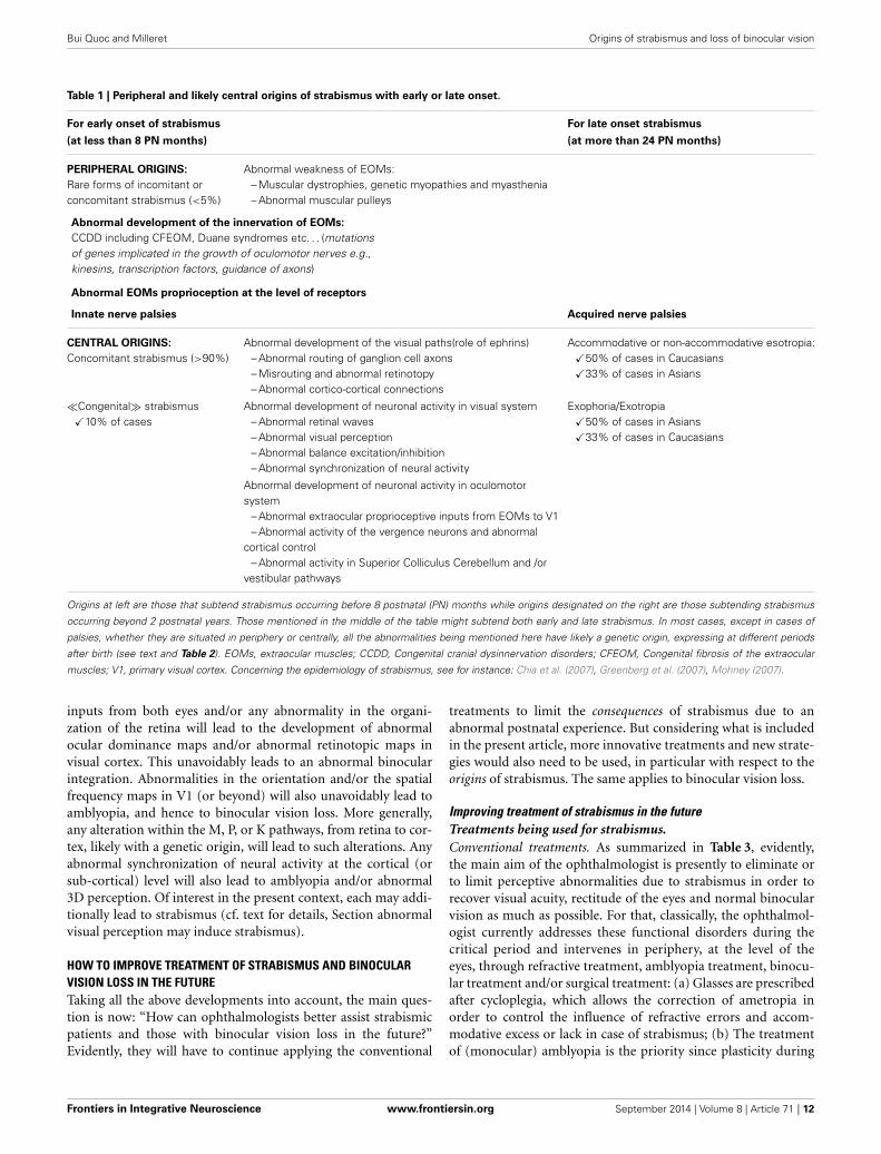
ABOUT THE ORIGINS OF STRABISMUSOrigins of strabismus with a peripheral origin are rather wellknown but they represent less than 5% of strabismic patients. Asdiscussed above, they display either rare forms of incomitant orconcomitant strabismus. As illustrated in Table 1, it is possible todistinguish which origins may induce early strabismus (up to 8PN months) and/or late strabismus (from 24 PN months). Thus,an abnormal weakness of EOMs, such as the ones due to mus-cular dystrophies, myopathies, myasthenia or abnormal muscularpulleys, may induce both forms of strabismus. In contrast, abnor-mal development of the innervation of the EOMs, either motor orproprioceptive, may only induce an early strabismus. Not surpris-ingly, innate nerve palsies and acquired ones may induce early andlate strabismus respectively. Of interest, it is also already estab-lished that most forms of strabismus with a peripheral origin havea genetic origin. As summarized in Table 2, most genes associatedto such forms of strabismus have been successfully identified. Thishas been facilitated by the fact that the alteration of only a few spe-cific genes is generally associated to each specific disease leadingto strabismus. Thus, for example, Duchenne muscular dystrophyleading to the weakness of EOMs implicates only the DMD genelocated on Xp21.2. The abnormal innervation of the EOMs in
the context of HGPPS syndrome only implicates the ROBO3 genelocated on 11q23.
By contrast, other forms of strabismus (mostly concomitant)are presently poorly understood while they are the most numer-ous (>90%). About 10% of them are “congenital” strabismus(thus occurring before 8 PN months), while the remaining occurat a later stage. In this latter situation, they are characterizedby either an accommodative or non-accommodative esotropiaor an exophoria or an exotropia (cf. Table 1). They are alsoknown to have a genetic origin (see Table 2). However, by con-trast to strabismus with an identified (peripheral) origin, thoseforms of strabismus are likely related to both recessive and dom-inant genes, thus resulting from a complex genetic inheritance.To move forward, we have proposed different possibilities tojustify the emergence of such “uncharacterized” forms of stra-bismus, by evoking the occurrence of abnormal development of“central” paths and abnormal development of “central” neuralactivity. This evidently concerns both the visual and the oculo-motor systems, up to the cortex, since they are closely related.Indeed, these forms of strabismus, with unknown origins, aregenerally “supposed” to have a central origin. Of interest, some ofthe anatomo-functional abnormalities we have proposed as beingresponsible for strabismus may take place before birth or post-natally, with consequences that the different forms of strabismuswith a central origin may have early or late onset. Thus we didnot dissociate them in Table 1. This may help to justify, however,the occurrence of early and late strabismus. It is not necessary tounderline that many other mechanisms could have been proposedas the possibilities are vast. To explain each form of strabismussupposes that a lot of mechanisms might be at source of strabis-mus with a central origin. Most of the mechanisms we propose areclearly hypothetical and remain to be proven. But, as indicatedabove, some mechanisms are already supported by precise data.For example, disrupting callosal connections alters the alignmentof the eyes (Elberger, 1979; Payne et al., 1981; Elberger and Hirsch,1982). A split between the ventral and the dorsal streams suchas the one occurring in Williams syndrome most often leads tostrabismus (Atkinson et al., 2001). An abnormal visual perceptionfrom one eye may also lead to strabismus (e.g., Quick et al., 1989;Niechwiej-Szwedo et al., 2010). One may also consider that dis-turbances of synchrony in the developing brain, associated withaberrant neurodevelopment, may also be a source of strabismus,at least in some cases, because of the growing evidence that theygenerally subtend cognitive dysfunction (e.g., Uhlhaas and Singer,2006; Uhlhaas et al., 2009a,b, 2011).
ABOUT THE ORIGINS OF BINOCULAR VISION LOSSOur article also aimed at reconsidering the question of the ori-gins of binocular vision loss, including 3D perception loss andacuity loss. Such deficits are evidently classic consequences of stra-bismus, because of the abnormal visual experience they generatepostnatally. But we are also convinced that their respective ori-gins are central, at least in some cases. Logical deductions leadto such a hypothesis. As outlined above, any abnormality withinthe visual network (because of abnormal retinal waves prena-tally or otherwise) may lead to abnormal visual perception, witha central origin. Thus, for example, an abnormal segregation of
Frontiers in Integrative Neuroscience www.frontiersin.org September 2014 | Volume 8 | Article 71 | 11
Bui Quoc and Milleret Origins of strabismus and loss of binocular vision
Table 1 | Peripheral and likely central origins of strabismus with early or late onset.
For early onset of strabismus For late onset strabismus
(at less than 8 PN months) (at more than 24 PN months)
PERIPHERAL ORIGINS:
Rare forms of incomitant orconcomitant strabismus (<5%)
Abnormal weakness of EOMs:– Muscular dystrophies, genetic myopathies and myasthenia– Abnormal muscular pulleys
Abnormal development of the innervation of EOMs:
CCDD including CFEOM, Duane syndromes etc. . . (mutationsof genes implicated in the growth of oculomotor nerves e.g.,kinesins, transcription factors, guidance of axons)
Abnormal EOMs proprioception at the level of receptors
Innate nerve palsies Acquired nerve palsies
CENTRAL ORIGINS:
Concomitant strabismus (>90%)Abnormal development of the visual paths(role of ephrins)
– Abnormal routing of ganglion cell axons– Misrouting and abnormal retinotopy– Abnormal cortico-cortical connections
Accommodative or non-accommodative esotropia:�50% of cases in Caucasians�33% of cases in Asians
�Congenital� strabismus�10% of cases
Abnormal development of neuronal activity in visual system– Abnormal retinal waves– Abnormal visual perception– Abnormal balance excitation/inhibition– Abnormal synchronization of neural activity
Exophoria/Exotropia�50% of cases in Asians�33% of cases in Caucasians
Abnormal development of neuronal activity in oculomotorsystem
– Abnormal extraocular proprioceptive inputs from EOMs to V1– Abnormal activity of the vergence neurons and abnormal
cortical control– Abnormal activity in Superior Colliculus Cerebellum and /or
vestibular pathways
Origins at left are those that subtend strabismus occurring before 8 postnatal (PN) months while origins designated on the right are those subtending strabismus
occurring beyond 2 postnatal years. Those mentioned in the middle of the table might subtend both early and late strabismus. In most cases, except in cases of
palsies, whether they are situated in periphery or centrally, all the abnormalities being mentioned here have likely a genetic origin, expressing at different periods
after birth (see text and Table 2). EOMs, extraocular muscles; CCDD, Congenital cranial dysinnervation disorders; CFEOM, Congenital fibrosis of the extraocular
muscles; V1, primary visual cortex. Concerning the epidemiology of strabismus, see for instance: Chia et al. (2007), Greenberg et al. (2007), Mohney (2007).
inputs from both eyes and/or any abnormality in the organi-zation of the retina will lead to the development of abnormalocular dominance maps and/or abnormal retinotopic maps invisual cortex. This unavoidably leads to an abnormal binocularintegration. Abnormalities in the orientation and/or the spatialfrequency maps in V1 (or beyond) will also unavoidably lead toamblyopia, and hence to binocular vision loss. More generally,any alteration within the M, P, or K pathways, from retina to cor-tex, likely with a genetic origin, will lead to such alterations. Anyabnormal synchronization of neural activity at the cortical (orsub-cortical) level will also lead to amblyopia and/or abnormal3D perception. Of interest in the present context, each may addi-tionally lead to strabismus (cf. text for details, Section abnormalvisual perception may induce strabismus).
HOW TO IMPROVE TREATMENT OF STRABISMUS AND BINOCULARVISION LOSS IN THE FUTURETaking all the above developments into account, the main ques-tion is now: “How can ophthalmologists better assist strabismicpatients and those with binocular vision loss in the future?”Evidently, they will have to continue applying the conventional
treatments to limit the consequences of strabismus due to anabnormal postnatal experience. But considering what is includedin the present article, more innovative treatments and new strate-gies would also need to be used, in particular with respect to theorigins of strabismus. The same applies to binocular vision loss.
Improving treatment of strabismus in the futureTreatments being used for strabismus.Conventional treatments. As summarized in Table 3, evidently,the main aim of the ophthalmologist is presently to eliminate orto limit perceptive abnormalities due to strabismus in order torecover visual acuity, rectitude of the eyes and normal binocularvision as much as possible. For that, classically, the ophthalmol-ogist currently addresses these functional disorders during thecritical period and intervenes in periphery, at the level of theeyes, through refractive treatment, amblyopia treatment, binocu-lar treatment and/or surgical treatment: (a) Glasses are prescribedafter cycloplegia, which allows the correction of ametropia inorder to control the influence of refractive errors and accom-modative excess or lack in case of strabismus; (b) The treatmentof (monocular) amblyopia is the priority since plasticity during
Frontiers in Integrative Neuroscience www.frontiersin.org September 2014 | Volume 8 | Article 71 | 12
Bui Quoc and Milleret Origins of strabismus and loss of binocular vision
Table 2 | Genetics of strabismus.
PERIPHERAL
•Genetic muscular diseases e.g.,
Gene Disease
DMD gene (Xp21.2) Duchenne muscular dystrophy and Beckermuscular dystrophy
FRG1, ANT1 et DUX4 (4q35) Facio-scapulo-humeral muscular dystrophyDMPK (19q13-2 ) Steinert myotonic dystrop
•Genetic abnormal development of nerves e.g.,
Gene Disease
KIF21A (12q12) CFEOMPHOX2A (11q13) CFEOM 2Gene located at 16qter CFEOM 3Genes located at 8q13 and 2q31Duane syndromesSALL4 (20q13) DRRSROBO3 (11q23) HGPPSHOXA1 (7p15) BSAS and ABDS
CENTRAL
• Genetic trait in accommodative esotropia and inheritance of
refractive errors such as hyperopia
• Genetic trait in exophoria/exotropia
Complex genetic inheritance, with the possible implication ofrecessive and dominant genes
This table allows summarizing already identified genes being responsible for
strabismus. In the periphery, some specific genes can be associated to some
specific diseases concerning the extraocular muscles or the oculomotor nerves.
Centrally, genes are also implicated in generating strabismus but their identifi-
cation is more difficult, in particular because not one single gene is implicated.
In addition, they might be dominant or recessive. CFEOM, Congenital fibrosis of
the extraocular muscles; DRRS, Duane Radial Ray Syndrome; HGPPS, Horizontal
Gaze Palsy with Progressive Scoliosis; BSAS, Bosley-Salih-Alorainy Syndrome;
ABDS, Athabascan Brainstem Dysgenesis Syndrome.
the postnatal period decreases progressively over time and thera-peutic success depends on the timing of the treatment. Refractivetreatment is the first step of amblyopia treatment and can be effec-tive in mild anisometropic amblyopias. But it must be stronglyemphasized that the treatment of amblyopia also requires patch-ing of the sound eye. Some studies by the PEDIG, a group ofNorth American Pediatric Ophthalmologists exploring differentamblyopia therapies (Beck, 1998), have suggested that a “soft”treatment can be as efficacious as a “harder” one. For instance,6 h patching vs. 2 h patching of the good eye would be sufficient(see, for example, Rees et al., 2007). However, it must be pointedout that only an improvement of vision was expected with suchtreatment, not a complete healing of amblyopia. To reach this lat-ter stage full time patching is required for several weeks. Patchingan eye increases the cortical input to the cortex from the ambly-opic eye, and this effect is necessary to increase visual acuity.Note, however, that the efficacy of the “patchy method” greatlyvaries with the age of the patient since the plastic properties of thevisual system evolve during the critical period (Epelbaum et al.,
1993); (c) When visual acuity is recovered in strabismic patients,surgery allows the realignment of the eyes which is necessary toensure binocular vision. Surgery has a direct effect on the EOMs,allowing the modification of the position of the eye: a recessionof a muscle diminishes its effective force on the eye, whereasreinforcement is allowed by a muscular resection. However, thisrequires intervention on the EOMs, in particular at the level oftheir tendons where major muscle receptors are located. By takinginto account that extraocular proprioception plays a major rolein the maturation of V1, at least during the first half of the crit-ical period, one must be aware that this may induce unfortunateconsequences in the development of the brain (see above).
New treatments. The “monocular patchy method” has been usedto treat amblyopia since major findings by Hubel and Wiesel(1965). But, over time, it has been thought to reduce binocu-lar stimulation to the visual system. This is a very importantissue, since despite what has been thought for decades, neurons invisual cortex have finally been shown to remain binocular in spiteof strabismus, even if binocular interactions are abnormal (seeabove). This is the reason why new strategies implicating binocu-lar stimulations are presently being developed to treat amblyopiaand binocular vision loss in strabismic (and anisometropic) sub-jects. Hess and his colleagues are among the most active in thatfield, with their strategy to suppress interocular suppression inorder to recover acuity by the amblyopic eye and 3D perception(Baker et al., 2007; Mansouri et al., 2008; Hess et al., 2010a,b,2011; Zhou et al., 2012 cf. also Hess et al., 2014 for review). Forthat, they have developed dichoptic devices that allow a binocu-lar stimulation with different images in each eye, the combinationof which is stereoscopic. Without going into detail, during thesebinocular stimulations, the image with the lowest contrast ispresented to the fellow fixing eye, which allows enhanced per-formance in the other eye. The practical management of patientsusing this approach is still under development but this approachfor amblyopia, binocular vision loss and, more generally, conse-quences of strabismus on visual perception appears quite promis-ing, more especially as it is also able to be used in adulthood(e.g., To et al., 2011; Black et al., 2012; Li et al., 2013; see alsoHess and Thompson, 2013 for review). During the treatment ofamblyopia (either strabismic or anisometropic), therapy sessionsof this type could be carried out, in addition to patching therapy,in order to improve the treatment of the effects of strabismus.Patching would increase the monocular input from one eye tothe cortex, avoiding the asymmetry of signal until a balance isobtained, whereas the binocular treatment would avoid interocu-lar suppression and would help to restore binocular function. Inthe particular case of strabismus, it would also require alignmentof the eyes to avoid inducing diplopia. The necessary conventionalapproaches through refractive treatment, amblyopia treatmentand surgical treatment will need to be combined with these newapproaches.
Another new and promising strategy to recover visual functionin strabismic (and anisometropic) adults is also under develop-ment. This time, it consists of non-invasive transcranial brainstimulations, with the aim of modifying the balance of excita-tion and inhibition in the visual cortex (Thompson et al., 2010;
Frontiers in Integrative Neuroscience www.frontiersin.org September 2014 | Volume 8 | Article 71 | 13
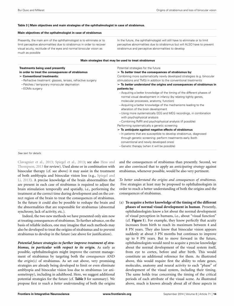
Bui Quoc and Milleret Origins of strabismus and loss of binocular vision
Table 3 | Main objectives and main strategies of the ophthalmologist in case of strabismus.
Main objectives of the opthalmologist in case of strabismus
Presently, the main aim of the ophthalmologist is to eliminate or tolimit perceptive abnormalities due to strabismus in order to recovervisual acuity, rectitude of the eyes and normal binocular vision asmuch as possible
In the future, the ophthalmologist will still have to eliminate or to limitperceptive abnormalities due to strabismus but will ALSO have to preventstrabismus and perceptive abnormalities to develop
Main strategies that may be used to treat strabismus
Treatments being used presently
in order to treat the consequences of strabismus
➢ Conventional treatments:
– Refractive treatment: glasses, lenses, refractive surgery– Patches / temporary monocular deprivation– EOMs surgery
Potential strategies for the future➢ To better treat the consequences of strabismus by:
Combining more systematically newly developed strategies (e.g. binocularstimulations and TMS) in addition to the conventional treatments➢ To better understand the origins and consequences of strabismus in
patients by:
– Acquiring a better knowledge of the timing of the different phases ofnormal visual development in infancy (by relating tightly genes,molecular processes, anatomy, function)
– Acquiring a better knowledge of the mechanisms leading to thealteration of the brain development
– Using more systematically EEG and MEG recordings, in combinationwith psychophysical analysis
– Combining fMRI and psychophysical analysis (if possible)Performing systematically a genetic screening➢ To anticipate against negative effects of strabismus
– In patients that are susceptible to develop strabismus, diagnosedthrough genetic screening: perform treatments early (bothconventional and newly developed ones)
– Genetic therapy (when it will be possible)
See text for details.
Clavagnier et al., 2013; Spiegel et al., 2013; see also Hess andThompson, 2013 for review). Used alone or in combination withbinocular therapy (cf. see above) it may assist in the treatmentof both amblyopia and binocular vision loss (e.g., Spiegel andLi, 2013). A precise knowledge of the brain abnormalities thatare present in each case of strabismus is required to adjust thebrain stimulation temporally and spatially, i.e., performing thetreatment at the correct time during development and on the cor-rect region of the brain to treat the consequences of strabismus.In the future it could also be possible to reshape the brain andthe abnormalities that are responsible for strabismus (abnormalsynchrony, lack of activity, etc.).
Indeed, the two new methods we have presented only aim nowat treating consequences of strabismus. To further advance, on thebasis of reliable indices, one may imagine that such methods mayalso be developed to treat the origins of strabismus and to preventstrabismus to develop in the future (see above for justification).
Potential future strategies to further improve treatment of stra-bismus, in particular with respect to its origin. As early aspossible, ophthalmologists might improve the current manage-ment of strabismus by targeting both the consequences ANDthe origin(s) of strabismus. As set out above, very promisingstrategies are already being developed to limit or even eliminateamblyopia and binocular vision loss due to strabismus (or ani-sometropy), including in adulthood. Here, we suggest additionalpotential strategies for the future (cf. Table 3 for summary). Wepropose first to reach a better understanding of both the origins
and the consequences of strabismus than presently. Second, weare also convinced that to apply an anticipating strategy againststrabismus, whenever possible, would be also very pertinent.
To better understand the origins and consequences of strabismus.Five strategies at least may be proposed to ophthalmologists in
order to reach a better understanding of both the origins and theconsequences of strabismus.
(a) To acquire a better knowledge of the timing of the differentphases of normal visual development in human. Presently,ophthalmologists know a lot about the normal developmentof visual perception in humans, i.e., about “visual function”(cf. Figure 1). For example, they know perfectly that acuityincreases from birth to reach its maximum between 4 and8 PN years. They also know that binocular vision appearssuddenly at about 3 PN months but continues to improveup to 9 PN years. But to move forward in the future,ophthalmologists would need to acquire a precise knowledgeabout the normal development of the visual system itself,from eye to cortex, before and after birth. This wouldconstitute an additional reference for them. As illustratedabove, this would require first the ability to relate genes,molecules, anatomy and neural activity to each “phase” ofdevelopment of the visual system, including their timing.The same holds true concerning the timing of the criticalperiods of each attribute of the visual scene. As illustratedabove, much is known already about all of these aspects in
Frontiers in Integrative Neuroscience www.frontiersin.org September 2014 | Volume 8 | Article 71 | 14
Bui Quoc and Milleret Origins of strabismus and loss of binocular vision
various mammal species, including monkeys, cats, ferrets,rats and mice. The general phases of development we areinterested in here are basically the same in all mammals,except with respect to the timings (e.g., Huberman et al.,2008; Espinosa and Stryker, 2012 for review). Thus, finally,in relation to such an issue, what is missing principally forhumans is the timing of the different phases of development.Second, ophthalmologists would have to understand howinteractions between distributed brain regions maturewith age to produce sophisticated cognitive functions suchas visual perception (see Section Systematic Use of EEGand/or MEG and/or fMRI Recordings, in Combinationwith Psychological Analysis below for details). Interestingly,some recent reviews have started to treat these aspects bytaking into account the time courses of neural proliferation,neural migration, apoptosis, synaptogenesis, establishmentof neural circuits and myelination in the different regionsof the human brain, including the visual cortex, before andafter birth (e.g., Tau and Peterson, 2010; Menon, 2013). Forthat, neuroimaging, EEG and MEG recordings, together withtraditional investigational approaches such as histologicalstudies and cellular and molecular biology, have beenused. Improving our understanding of these developmentalprocesses in humans is likely a major key to the successfultreatment of strabismus and binocular visual loss. To havethis type of information with respect to the oculomotorsystem would be also very interesting.
(b) To acquire a better knowledge of the mechanisms leading tothe alteration of brain development in case of strabismus.As illustrated in the present article, few data are finallyavailable regarding the origins of early or late strabismus, inparticular when these origins are central. We have proposeddifferent mechanisms but most still need to be confirmedas real sources of strabismus. In contrast, much is alreadyknown about both the anatomical and functional conse-quences of strabismus, from retina to cortex (e.g., Wong,2012 for review). But again, it would be interesting forophthalmologists to know more about the mechanisms thatare implicated during these plastic changes in the brain. Suchinformation is necessary to develop new treatment strategies.As an example, if remaining binocularity of cortical neuronshad not been shown to be sustained after strabismus (Chinoet al., 1994; Sengpiel et al., 1994), it is possible that Hess andhis colleagues would have not developed their promisingbinocular therapy (see above).
(c) Systematic use of EEG and/or MEG and/or fMRI record-ings, in combination with psychological analysis. Amongother possibilities, we have proposed here that any anatomo-functional abnormality within the visual cortex may leadto strabismus (and amblyopia and/or binocular visionloss) by altering synchrony. This is in line with increasingevidence that disturbances of synchrony in the developingbrain are associated with aberrant neurodevelopment andsubtend cognitive dysfunctions (Uhlhaas and Singer, 2006;Uhlhaas et al., 2009a,b, 2011). Interestingly, in order tohelp physicians and neurologists to establish a diagnostic inthe future, various authors have now started to address the
question of the relationship between neural synchrony andthe underlying anatomical and physiological changes thatoccur during normal brain development (e.g., Menon, 2013for review). They also aim at associating a specific disruptionof dynamic processes to abnormal connectivity and specificdisturbances of cognitive or executive functions (see above;see also Menon, 2013 for review). Their idea is that recordingdynamics of brain activity using electroencephalography(EEG) and/or magnetoencephalography (MEG) with orwithout new psychophysical measurements of visual per-ception must somehow reflect the functional architecture ofcortical networks. “Because this architecture is determinedby genetic factors and modified by experience, spontaneousor evoked activity patterns should contain informationabout evolutionary and epigenetically acquired knowledgeregarding the world and serve as a convert internal model forperception and action” (e.g., Singer, 2013 for review). Thisindicates that recording resting-state or evoked activity fromthe brain would be sufficient, at least in principle, to identifywhether something is wrong in the cortical networks.As suggested by Buzsáki et al. (2013), “oscillopathies ordysrhythmias could reflect malfunctioning network and, asendophenotypes, could assist in specifying diagnostics.” Thishas not been applied as yet but it is a promising possibilityfor the future, in particular in the context in which we areinterested here. One might speculate that to establish specificrelations between “oscillopathies” or “dysrhythmias” in thebrain and various types of strabismus might greatly help,both to prevent strabismus to develop and to cure dilatoryconsequences due to strabismus, at least in some cases. Suchtechniques are particularly interesting here in that they arenon-invasive techniques and can be used in infants at theearliest ages (e.g., Csibra et al., 2000). The fMRI could alsohelp in assessing abnormal connectivity, although it is noteasy to perform this in young infants (e.g., Li et al., 2011).
To anticipate against negative effects of strabismus. The EEG and/orMEG and/or fMRI recordings, with or without psychologicalanalysis, might be used for the diagnosis of brain “abnormali-ties” that are present in the case of strabismus, and that shouldbe treated, for example by using TMS (see above). Also, it shouldbe necessary to anticipate the negative effects of strabismus onthe visual system, and one may propose to perform a geneticscreening for that purpose. In the future, one may even considerrepairing altered genes.
(a) To perform systematically a genetic screening. Geneticknowledge, whatever its complexity, may be used (see above).Gene profiles can now be established relatively easily inhumans, using non-invasive methods. Some genes at leastare already known to be associated with strabismus (seeabove). To identify more would certainly assist, whether theyare implicated in strabismus or absence of binocular vision.Establishing, as early as possible after birth, that a child isat risk of developing strabismus by showing that he or shehas one or more affected gene(s) would at least allow oph-thalmologists to develop a treatment plan to limit dilatory
Frontiers in Integrative Neuroscience www.frontiersin.org September 2014 | Volume 8 | Article 71 | 15
Bui Quoc and Milleret Origins of strabismus and loss of binocular vision
consequences of strabismus. Indeed, as outlined above, con-sequences of strabismus and/or absence of binocular visionmight be rather difficult to treat in particular when strabis-mus has occurred very early postnatally. Thus, to diagnosestrabismus as early as possible would only be beneficial.
(b) To use genetic therapy. In the future, even if it is currentlynot a reality, one may also expect to use innovative tech-nologies such as genetic therapy. It would allow the repairof altered genes susceptible to induce strabismus and/or lossof binocular vision, at least in some cases. Is it a utopia to beable to repair the brain itself in the present context? Whateverthe answer, intervening as early as possible is the best strategy.
How to improve treatment of binocular vision loss in the future?As mentioned above, some newly developed strategies such asbinocular therapy and TMS stimulations are improving the treat-ment of binocular vision loss caused by strabismus (e.g., Hess andThompson, 2013; Hess et al., 2014 for reviews). But it is importantto emphasize that, at least presently, binocular vision cannot berestored whatever the form of strabismus. In particular, it cannotbe obtained after an early onset strabismus. Congenital binocu-lar vision loss, without strabismus, is also presently impossible totreat. In both cases, this is likely due to a major insult of the matu-rational process of the visual system with respect to binocularity,occurring either before or soon after birth, due to innate/geneticfactors. Evidently, in these cases, neural networks in the brainwould have to be “repaired” through either reshaping (if earlyenough after birth) or by activating functional sleepy synapses orotherwise. However, to reach such goals, the origin and the exactcharacteristics of the disease would need to be identified in greatdetail. Appropriate strategies to remove or even to prevent binoc-ular vision loss should then be developed. Our suggestion forophthalmologists is that they increase their knowledge concern-ing the different phases that characterize the normal developmentof the visual system.
Combining better knowledge of the origins of strabismus andloss of binocular vision with new therapies will no doubt allowthe more efficient management of these pathologies.
ACKNOWLEDGMENTSWe would like to thank the LUZ Group for their financial support,Ms. Nicole Quenech’Du for the conception of the figures and Ms.Chloe Ann Barker for revising the manuscript.
REFERENCESAckman, J. B., Burbridge, T. J., and Crair, M. C. (2012). Retinal waves coordi-
nate patterned activity throughout the developing visual system. Nature 490,219–225. doi: 10.1038/nature11529
Ashton, J. A., Boddy, A., Dean, S. R., Milleret, C., and Donaldson, I. M.(1988). Afferent signals from cat extraocular muscles in the medial vestibularnucleus, the nucleus praepositus hypoglossi and adjacent brainstem structures.Neuroscience 26, 131–145. doi: 10.1016/0306-4522(88)90132-7
Ashton, J. A., Milleret, C., and Donaldson, I. M. (1989). Effects of afferent signalsfrom the extraocular muscles upon units in the cerebellum, vestibular nuclearcomplex and oculomotor nucleus of the trout. Neuroscience 31, 529–541. doi:10.1016/0306-4522(89)90395-3
Atkinson, J., Anker, S., Braddick, O., Nokes, L., Mason, A., and Braddick, F. (2001).Visual and visuospatial development in young children with Williams syn-drome. Dev. Med. Child Neurol. 43, 330–337. doi: 10.1017/S0012162201000615
Baker, D. H., Meese, T. S., Mansouri, B., and Hess, R. F. (2007). Binocular summa-tion of contrast remains intact in strabismic amblyopia. Invest. Ophthalmol. Vis.Sci. 48, 5332–5338. doi: 10.1167/iovs.07-0194
Barrett, B. T., Bradley, A., and McGraw, P. V. (2004). Understanding the neural basisof amblyopia. Neuroscientist 10, 106–117. doi: 10.1177/1073858403262153
Batini, C., and Buisseret, P. (1974). Sensory peripheral pathway from extrinsic eyemuscles. Arch. Ital. Biol. 112, 18–32.
Batini, C., Buisseret, P., and Buisseret-Delmas, C. (1975). Trigeminal pathway ofthe extrinsic eye muscle afferents in cat. Brain Res. 85, 74–78. doi: 10.1016/0006-8993(75)91008-2
Beck, R. W. (1998). The pediatric eye disease investigator group. J. AAPOS 2,255–256. doi: 10.1016/S1091-8531(98)90079-9
Berlucchi, G. (2014). Visual interhemispheric communication and callosal con-nections of the occipital lobes. Cortex 56, 1–13. doi: 10.1016/j.cortex.2013.02.001
Beurdeley, M., Spatazza, J., Lee, H. H., Sugiyama, S., Bernard, C., Di Nardo,A. A., et al. (2012). Otx2 binding to perineuronal nets persistently regu-lates plasticity in the mature visual cortex. J. Neurosci. 32, 9429–9437. doi:10.1523/JNEUROSCI.0394-12.2012
Birch, E. E. (2013). Amblyopia and binocular vision. Prog. Retin. Eye Res. 33, 67–84.doi: 10.1016/j.preteyeres.2012.11.001
Black, J. M., Hess, R. F., Cooperstock, J. R., To, L., and Thompson, B. (2012). Themeasurement and treatment of suppression in amblyopia. J. Vis. Exp. 70:e3927.doi: 10.3791/3927
Brodsky, M. C. (2011). Dissociated vertical divergence: cortical or subcortical inorigin? Strabismus 19, 67–68. doi: 10.3109/09273972.2011.575434
Bucci, M. P., and Seassau, M. (2012). Saccadic eye movements in children: a devel-opmental study. Exp. Brain Res. 222, 21–30. doi: 10.1007/s00221-012-3192-7
Bucci, M. P., and Seassau, M. (2014). Vertical saccades in children: a developmentalstudy. Exp. Brain Res. 232, 927–934. doi: 10.1007/s00221-013-3805-9
Bui Quoc, E., Ribot, J., Quenech’du, N., Doutremer, S., Lebas, N., Grantyn, A.,et al. (2012). Asymmetrical interhemispheric connections develop in cat visualcortex after early unilateral convergent strabismus: anatomy, physiology, andmechanisms. Front. Neuroanat. 5:68. doi: 10.3389/fnana.2011.00068
Buisseret, P., and Maffei, L. (1977). Extraocular proprioceptive projections to thevisual cortex. Exp. Brain Res. 28, 421–425.
Buisseret, P., Gary-Bobo, E., and Imbert, M. (1978). Ocular motility and recovery oforientational properties of visual cortical neurons in dark-reared kittens. Nature272, 816–817. doi: 10.1038/272816a0
Buisseret, P., Gary-Bobo, E., and Milleret, C. (1988). Development of the kittenvisual cortex depends on the relationship between the plane of eye movementsand visual inputs. Exp. Brain Res. 72, 83–94. doi: 10.1007/BF00248503
Buisseret, P. (1995). Influence of extraocular muscle proprioception on vision.Physiol. Rev. 75, 323–338.
Buzsáki, G., Logothetis, N., and Singer, W. (2013). Scaling brain size, keepingtiming: evolutionary preservation of brain rhythms. Neuron 80, 751–764. doi:10.1016/j.neuron.2013.10.002
Cang, J., Kaneko, M., Yamada, J., Woods, G., Stryker, M. P., and Feldheim, D. A.(2005a). Ephrin-as guide the formation of functional maps in the visual cortex.neuron. Neuron 48, 577–589. doi: 10.1016/j.neuron.2005.10.026
Cang, J., Rentería, R. C., Kaneko, M., Liu, X., Copenhagen, D. R., andStryker, M. P. (2005b). Development of precise maps in visual cortexrequires patterned spontaneous activity in the retin. Neuron 48, 797–809. doi:10.1016/j.neuron.2005.09.015
Canturk, S., Oto, S., Kizilkilic, O., Karaca, S., and Akova, Y. A. (2008).Rhombencephalosynapsis associated with infantile strabismus. Strabismus 16,23–27. doi: 10.1080/09273970701863610
Cardin, J. A., Carlen, M., Meletis, K., Knoblich, U., Zhang, F., Deisseroth, K., et al.(2009). Driving fast-spiking cells induces gamma rhythm and controls sensoryresponses. Nature 459, 663–667. doi: 10.1038/nature08002
Castelo-Branco, M., Neuenschwander, S., and Singer, W. (1998). Synchronizationof visual responses between the cortex, lateral geniculate nucleaus, and retina inthe anesthetized cat. J. Neurosci. 18, 6395–6410.
Chan, S. T., Tang, K. W., Lam, K. C., Chan, L. K., Mendola, J. D., and Kwong,K. K. (2004). Neuroanatomy of adult strabismus: a voxel-based morphometricanalysis of magnetic resonance structural scans. Neuroimage 22, 986–994. doi:10.1016/j.neuroimage.2004.02.021
Chapman, B., Stryker, M. P., and Bonhoeffer, T. (1996). Development of orientation
preference maps in ferret primary visual cortex. J. Neurosci. 16, 6443–6453.
Frontiers in Integrative Neuroscience www.frontiersin.org September 2014 | Volume 8 | Article 71 | 16
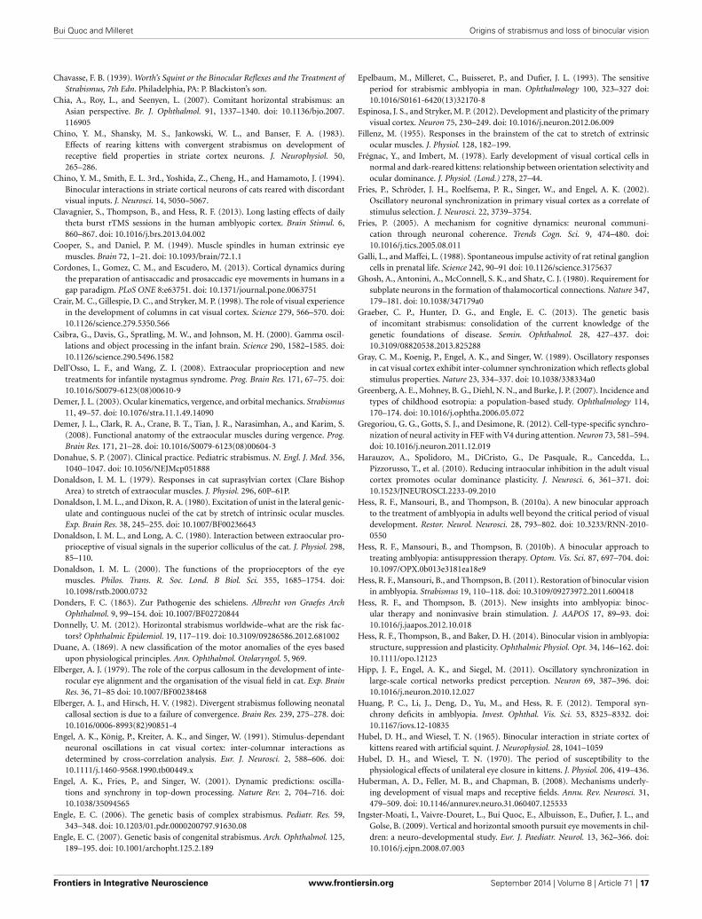
Bui Quoc and Milleret Origins of strabismus and loss of binocular vision
Chavasse, F. B. (1939). Worth’s Squint or the Binocular Reflexes and the Treatment ofStrabismus, 7th Edn. Philadelphia, PA: P. Blackiston’s son.
Chia, A., Roy, L., and Seenyen, L. (2007). Comitant horizontal strabismus: anAsian perspective. Br. J. Ophthalmol. 91, 1337–1340. doi: 10.1136/bjo.2007.116905
Chino, Y. M., Shansky, M. S., Jankowski, W. L., and Banser, F. A. (1983).Effects of rearing kittens with convergent strabismus on development ofreceptive field properties in striate cortex neurons. J. Neurophysiol. 50,265–286.
Chino, Y. M., Smith, E. L. 3rd., Yoshida, Z., Cheng, H., and Hamamoto, J. (1994).Binocular interactions in striate cortical neurons of cats reared with discordantvisual inputs. J. Neurosci. 14, 5050–5067.
Clavagnier, S., Thompson, B., and Hess, R. F. (2013). Long lasting effects of dailytheta burst rTMS sessions in the human amblyopic cortex. Brain Stimul. 6,860–867. doi: 10.1016/j.brs.2013.04.002
Cooper, S., and Daniel, P. M. (1949). Muscle spindles in human extrinsic eyemuscles. Brain 72, 1–21. doi: 10.1093/brain/72.1.1
Cordones, I., Gomez, C. M., and Escudero, M. (2013). Cortical dynamics duringthe preparation of antisaccadic and prosaccadic eye movements in humans in agap paradigm. PLoS ONE 8:e63751. doi: 10.1371/journal.pone.0063751
Crair, M. C., Gillespie, D. C., and Stryker, M. P. (1998). The role of visual experiencein the development of columns in cat visual cortex. Science 279, 566–570. doi:10.1126/science.279.5350.566
Csibra, G., Davis, G., Spratling, M. W., and Johnson, M. H. (2000). Gamma oscil-lations and object processing in the infant brain. Science 290, 1582–1585. doi:10.1126/science.290.5496.1582
Dell’Osso, L. F., and Wang, Z. I. (2008). Extraocular proprioception and newtreatments for infantile nystagmus syndrome. Prog. Brain Res. 171, 67–75. doi:10.1016/S0079-6123(08)00610-9
Demer, J. L. (2003). Ocular kinematics, vergence, and orbital mechanics. Strabismus11, 49–57. doi: 10.1076/stra.11.1.49.14090
Demer, J. L., Clark, R. A., Crane, B. T., Tian, J. R., Narasimhan, A., and Karim, S.(2008). Functional anatomy of the extraocular muscles during vergence. Prog.Brain Res. 171, 21–28. doi: 10.1016/S0079-6123(08)00604-3
Donahue, S. P. (2007). Clinical practice. Pediatric strabismus. N. Engl. J. Med. 356,1040–1047. doi: 10.1056/NEJMcp051888
Donaldson, I. M. L. (1979). Responses in cat suprasylvian cortex (Clare BishopArea) to stretch of extraocular muscles. J. Physiol. 296, 60P–61P.
Donaldson, I. M. L., and Dixon, R. A. (1980). Excitation of unist in the lateral genic-ulate and continguous nuclei of the cat by stretch of intrinsic ocular muscles.Exp. Brain Res. 38, 245–255. doi: 10.1007/BF00236643
Donaldson, I. M. L., and Long, A. C. (1980). Interaction between extraocular pro-prioceptive of visual signals in the superior colliculus of the cat. J. Physiol. 298,85–110.
Donaldson, I. M. L. (2000). The functions of the proprioceptors of the eyemuscles. Philos. Trans. R. Soc. Lond. B Biol. Sci. 355, 1685–1754. doi:10.1098/rstb.2000.0732
Donders, F. C. (1863). Zur Pathogenie des schielens. Albrecht von Graefes ArchOphthalmol. 9, 99–154. doi: 10.1007/BF02720844
Donnelly, U. M. (2012). Horizontal strabismus worldwide–what are the risk fac-tors? Ophthalmic Epidemiol. 19, 117–119. doi: 10.3109/09286586.2012.681002
Duane, A. (1869). A new classification of the motor anomalies of the eyes basedupon physiological principles. Ann. Ophthalmol. Otolaryngol. 5, 969.
Elberger, A. J. (1979). The role of the corpus callosum in the development of inte-rocular eye alignment and the organisation of the visual field in cat. Exp. BrainRes. 36, 71–85 doi: 10.1007/BF00238468
Elberger, A. J., and Hirsch, H. V. (1982). Divergent strabismus following neonatalcallosal section is due to a failure of convergence. Brain Res. 239, 275–278. doi:10.1016/0006-8993(82)90851-4
Engel, A. K., König, P., Kreiter, A. K., and Singer, W. (1991). Stimulus-dependantneuronal oscillations in cat visual cortex: inter-columnar interactions asdetermined by cross-correlation analysis. Eur. J. Neurosci. 2, 588–606. doi:10.1111/j.1460-9568.1990.tb00449.x
Engel, A. K., Fries, P., and Singer, W. (2001). Dynamic predictions: oscilla-tions and synchrony in top-down processing. Nature Rev. 2, 704–716. doi:10.1038/35094565
Engle, E. C. (2006). The genetic basis of complex strabismus. Pediatr. Res. 59,343–348. doi: 10.1203/01.pdr.0000200797.91630.08
Engle, E. C. (2007). Genetic basis of congenital strabismus. Arch. Ophthalmol. 125,189–195. doi: 10.1001/archopht.125.2.189
Epelbaum, M., Milleret, C., Buisseret, P., and Dufier, J. L. (1993). The sensitiveperiod for strabismic amblyopia in man. Ophthalmology 100, 323–327 doi:10.1016/S0161-6420(13)32170-8
Espinosa, J. S., and Stryker, M. P. (2012). Development and plasticity of the primaryvisual cortex. Neuron 75, 230–249. doi: 10.1016/j.neuron.2012.06.009
Fillenz, M. (1955). Responses in the brainstem of the cat to stretch of extrinsicocular muscles. J. Physiol. 128, 182–199.
Frégnac, Y., and Imbert, M. (1978). Early development of visual cortical cells innormal and dark-reared kittens: relationship between orientation selectivity andocular dominance. J. Physiol. (Lond.) 278, 27–44.
Fries, P., Schröder, J. H., Roelfsema, P. R., Singer, W., and Engel, A. K. (2002).Oscillatory neuronal synchronization in primary visual cortex as a correlate ofstimulus selection. J. Neurosci. 22, 3739–3754.
Fries, P. (2005). A mechanism for cognitive dynamics: neuronal communi-cation through neuronal coherence. Trends Cogn. Sci. 9, 474–480. doi:10.1016/j.tics.2005.08.011
Galli, L., and Maffei, L. (1988). Spontaneous impulse activity of rat retinal ganglioncells in prenatal life. Science 242, 90–91 doi: 10.1126/science.3175637
Ghosh, A., Antonini, A., McConnell, S. K., and Shatz, C. J. (1980). Requirement forsubplate neurons in the formation of thalamocortical connections. Nature 347,179–181. doi: 10.1038/347179a0
Graeber, C. P., Hunter, D. G., and Engle, E. C. (2013). The genetic basisof incomitant strabismus: consolidation of the current knowledge of thegenetic foundations of disease. Semin. Ophthalmol. 28, 427–437. doi:10.3109/08820538.2013.825288
Gray, C. M., Koenig, P., Engel, A. K., and Singer, W. (1989). Oscillatory responsesin cat visual cortex exhibit inter-columner synchronization which reflects globalstimulus properties. Nature 23, 334–337. doi: 10.1038/338334a0
Greenberg, A. E., Mohney, B. G., Diehl, N. N., and Burke, J. P. (2007). Incidence andtypes of childhood esotropia: a population-based study. Ophthalmology 114,170–174. doi: 10.1016/j.ophtha.2006.05.072
Gregoriou, G. G., Gotts, S. J., and Desimone, R. (2012). Cell-type-specific synchro-nization of neural activity in FEF with V4 during attention. Neuron 73, 581–594.doi: 10.1016/j.neuron.2011.12.019
Harauzov, A., Spolidoro, M., DiCristo, G., De Pasquale, R., Cancedda, L.,Pizzorusso, T., et al. (2010). Reducing intraocular inhibition in the adult visualcortex promotes ocular dominance plasticity. J. Neurosci. 6, 361–371. doi:10.1523/JNEUROSCI.2233-09.2010
Hess, R. F., Mansouri, B., and Thompson, B. (2010a). A new binocular approachto the treatment of amblyopia in adults well beyond the critical period of visualdevelopment. Restor. Neurol. Neurosci. 28, 793–802. doi: 10.3233/RNN-2010-0550
Hess, R. F., Mansouri, B., and Thompson, B. (2010b). A binocular approach totreating amblyopia: antisuppression therapy. Optom. Vis. Sci. 87, 697–704. doi:10.1097/OPX.0b013e3181ea18e9
Hess, R. F., Mansouri, B., and Thompson, B. (2011). Restoration of binocular visionin amblyopia. Strabismus 19, 110–118. doi: 10.3109/09273972.2011.600418
Hess, R. F., and Thompson, B. (2013). New insights into amblyopia: binoc-ular therapy and noninvasive brain stimulation. J. AAPOS 17, 89–93. doi:10.1016/j.jaapos.2012.10.018
Hess, R. F., Thompson, B., and Baker, D. H. (2014). Binocular vision in amblyopia:structure, suppression and plasticity. Ophthalmic Physiol. Opt. 34, 146–162. doi:10.1111/opo.12123
Hipp, J. F., Engel, A. K., and Siegel, M. (2011). Oscillatory synchronization inlarge-scale cortical networks predicst perception. Neuron 69, 387–396. doi:10.1016/j.neuron.2010.12.027
Huang, P. C., Li, J., Deng, D., Yu, M., and Hess, R. F. (2012). Temporal syn-chrony deficits in amblyopia. Invest. Ophthal. Vis. Sci. 53, 8325–8332. doi:10.1167/iovs.12-10835
Hubel, D. H., and Wiesel, T. N. (1965). Binocular interaction in striate cortex ofkittens reared with artificial squint. J. Neurophysiol. 28, 1041–1059
Hubel, D. H., and Wiesel, T. N. (1970). The period of susceptibility to thephysiological effects of unilateral eye closure in kittens. J. Physiol. 206, 419–436.
Huberman, A. D., Feller, M. B., and Chapman, B. (2008). Mechanisms underly-ing development of visual maps and receptive fields. Annu. Rev. Neurosci. 31,479–509. doi: 10.1146/annurev.neuro.31.060407.125533
Ingster-Moati, I., Vaivre-Douret, L., Bui Quoc, E., Albuisson, E., Dufier, J. L., andGolse, B. (2009). Vertical and horizontal smooth pursuit eye movements in chil-dren: a neuro-developmental study. Eur. J. Paediatr. Neurol. 13, 362–366. doi:10.1016/j.ejpn.2008.07.003
Frontiers in Integrative Neuroscience www.frontiersin.org September 2014 | Volume 8 | Article 71 | 17
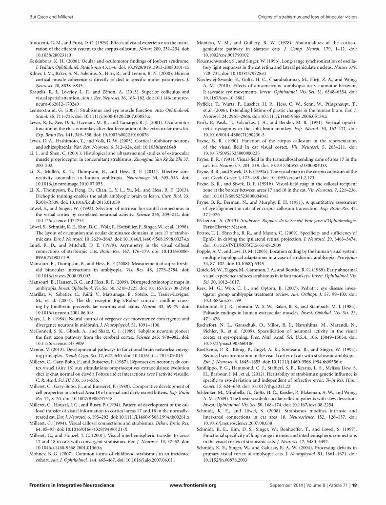
Bui Quoc and Milleret Origins of strabismus and loss of binocular vision
Innocenti, G. M., and Frost, D. O. (1979). Effects of visual experience on the matu-ration of the efferent system to the corpus callosum. Nature 280, 231–234. doi:10.1038/280231a0
Keskinbora, K. H. (2008). Ocular and oculomotor findings of Joubert syndrome.J. Pediatr. Ophthalmol. Strabismus 45, 5–6. doi: 10.3928/01913913-20080101-15
Kilner, J. M., Baker, S. N., Salenius, S., Hari, R., and Lemon, R. N. (2000). Humancortical muscle coherence is directly related to specific motor parameters. J.Neurosci. 20, 8838–8845.
Krauzlis, R. J., Lovejoy, L. P., and Zénon, A. (2013). Superior colliculus andvisual spatial attention. Annu. Rev. Neurosci. 36, 165–182. doi: 10.1146/annurev-neuro-062012-170249
Lennerstrand, G. (2007). Strabismus and eye muscle function. Acta Ophthalmol.Scand. 85, 711–723. doi: 10.1111/j.1600-0420.2007.00853.x
Lewis, R. F., Zee, D. S., Hayman, M. R., and Tamargo, R. J. (2001). Oculomotorfunction in the rhesus monkey after deafferentation of the extraocular muscles.Exp. Brain Res. 141, 349–358. doi: 10.1007/s002210100876
Lewis, D. A., Hashimoto, T., and Volk, D. W. (2005). Cortical inhibitory neuronsand schizophrenia. Nat. Rev. Neurosci. 6, 312–324. doi: 10.1038/nrn1648
Li, J., and Shen, C. (2001). Histological and ultrastructural studies of extraocularmuscle proprioceptor in concomitant strabismus. Zhonghua Yan Ke Za Zhi 37,200–202.
Li, X., Mullen, K. T., Thompson, B., and Hess, R. F. (2011). Effective con-nectivity anomalies in human amblyopia. Neuroimage 54, 505–516. doi:10.1016/j.neuroimage.2010.07.053
Li, X., Thompson, B., Deng, D., Chan, L. Y. L., Yu, M., and Hess, R. F. (2013).Dichoptic training enables the adult amblyopic brain to learn. Curr. Biol. 23,R308–R309. doi: 10.1016/j.cub.2013.01.059
Löwel, S., and Singer, W. (1992). Selection of intrinsic horizontal connections inthe visual cortex by correlated neuronal activity. Science 255, 209–212. doi:10.1126/science.1372754
Löwel, S., Schmidt, K. E., Kim, D. C., Wolf, F., Hoffsüller, F., Singer, W., et al. (1998).The layout of orientation and ocular dominance domains in area 17 of strabis-mic cats. Eur. J. Neurosci. 10, 2629–2643. doi: 10.1046/j.1460-9568.1998.00274.x
Lund, R. D., and Mitchell, D. E. (1979). Asymmetry in the visual callosalconnections of strabismic cats. Brain Res. 167, 176–179. doi: 10.1016/0006-8993(79)90274-9
Mansouri, B., Thompson, B., and Hess, R. F. (2008). Measurement of suprathresh-old binocular interactions in amblyopia. Vis. Res. 48, 2775–2784. doi:10.1016/j.visres.2008.09.002
Mansouri, B., Hansen, B. C., and Hess, R. F. (2009). Disrupted retinotopic maps inamblyopia. Invest. Ophtalmol. Vis. Sci. 50, 3218–3225. doi: 10.1167/iovs.08-2914
Marillat, V., Sabatier, C., Failli, V., Matsunaga, E., Sotelo, C., Tessier-Lavigne,M., et al. (2004). The slit receptor Rig-1/Robo3 controls midline cross-ing by hindbrain precerebellar neurons and axons. Neuron 43, 69–79. doi:10.1016/j.neuron.2004.06.018
Mays, L. E. (1984). Neural control of vergence eye movements: convergence anddivergence neurons in midbrain. J. Neurophysiol. 51, 1091–1108.
McConnell, S. K., Ghosh, A., and Shatz, C. J. (1989). Subplate neurons pioneerthe first axon pathway from the cerebral cortex. Science 245: 978–982. doi:10.1126/science.2475909
Menon, V. (2013). Developmental pathways to functional brain networks: emerg-ing principles. Trends Cogn. Sci. 17, 627–640. doi: 10.1016/j.tics.2013.09.015
Milleret, C., Gary-Bobo, E., and Buisseret, P. (1987). Réponses des neurones du cor-tex visuel (Aire 18) aux stimulations proprioceptives extraoculaires: évolutionchez le chat normal ou élevé à l’obscurité et interactions avec l’activité visuelle.C. R. Acad. Sci. III 305, 531–536.
Milleret, C., Gary-Bobo, E., and Buisseret, P. (1988). Comparative development ofcell properties in cortical Area 18 of normal and dark-reared kittens. Exp. BrainRes. 71, 8–20. doi: 10.1007/BF00247518
Milleret, C., Houzel, J. C., and Buser, P. (1994). Pattern of development of the cal-losal transfer of visual information to cortical areas 17 and 18 in the normally-reared cat. Eur. J. Neurosci. 6, 193–202. doi: 10.1111/j.1460-9568.1994.tb00261.x
Milleret, C. (1994). Visual callosal connections and strabismus. Behav. Brain Res.64, 85–95. doi: 10.1016/0166-4328(94)90121-X
Milleret, C., and Houzel, J. C. (2001). Visual interhemispheric transfer to areas17 and 18 in cats with convergent strabismus. Eur. J. Neurosci. 13, 37–52. doi:10.1046/j.1460-9568.2001.01360.x
Mohney, B. G. (2007). Common forms of childhood strabismus in an incidencecohort. Am. J. Ophthalmol. 144, 465–467. doi: 10.1016/j.ajo.2007.06.011
Montero, V. M., and Guillery, R. W. (1978). Abnormalities of the cortico-geniculate pathway in Siamese cats. J. Comp. Neurol. 179, 1–12. doi:10.1002/cne.901790102
Neuenschwander, S., and Singer, W. (1996). Long-range synchronization of oscilla-tory light responses in the cat retina and lateral geniculate nucleus. Nature 379,728–732. doi: 10.1038/379728a0
Niechwiej-Szwedo, E., Goltz, H. C., Chandrakumar, M., Hirji, Z. A., and Wong,A. M. (2010). Effects of anisometropic amblyopia on visuomotor behavior,I: saccadic eye movements. Invest. Ophthalmol. Vis. Sci. 51, 6348–6354. doi:10.1167/iovs.10-5882
Nyffeler, T., Wurtz, P., Lüscher, H. R., Hess, C. W., Senn, W., Pflugshaupt, T.,et al. (2006). Extending lifetime of plastic changes in the human brain. Eur. J.Neurosci. 24, 2961–2966. doi: 10.1111/j.1460-9568.2006.05154.x
Pasik, P., Pasik, T., Valciukas, J. A., and Bender, M. B. (1971). Vertical optoki-netic nystagmus in the split-brain monkey. Exp. Neurol. 30, 162–171. doi:10.1016/0014-4886(71)90230-5
Payne, B. R. (1990). Function of the corpus callosum in the representationof the visual field in cat visual cortex. Vis. Neurosci. 5, 205–211. doi:10.1017/S0952523800000225
Payne, B. R. (1991). Visual-field in the transcallosal sending zone of area 17 in thecat. Vis. Neurosci. 7, 201–219. doi: 10.1017/S095252380000403X
Payne, B. R., and Siwek, D. F. (1991a). The visual map in the corpus callosum of thecat. Cereb. Cortex 1, 173–188. doi: 10.1093/cercor/1.2.173
Payne, B. R., and Siwek, D. F. (1991b). Visual-field map in the callosal recipientzone at the border between areas 17 and 18 in the cat. Vis. Neurosci. 7, 221–236.doi: 10.1017/S0952523800004041
Payne, B. R., Berman, N., and Murphy, E. H. (1981). A quantitative assessmentof eye alignment in cats after corpus callosum transection. Exp. Brain Res. 43,371–376
Péchereau, A. (2013). Strabisme. Rapport de la Société Française d’Ophtalmologie.Paris: Elsevier Masson.
Petros, T. J., Shrestha, B. R., and Mason, C. (2009). Specificity and sufficiency ofEphB1 in driving the ipsilateral retinal projection. J. Neurosci. 29, 3463–3474.doi: 10.1523/JNEUROSCI.5655-08.2009
Popple, A. V., and Levi, D. M. (2005). Location coding by the human visual system:multiple topological adaptations in a case of strabismic amblyopia. Perception34, 87–107. doi: 10.1068/p5345
Quick, M. W., Tigges, M., Gammon, J. A., and Boothe, R. G. (1989). Early abnormalvisual experience induces strabismus in infant monkeys. Invest. Ophthalmol. Vis.Sci. 30, 1012–1017.
Rees, M. G., Woo, C. L., and Optom, B. (2007). Pediatric eye disease inves-tigator group amblyopia treatment review. Am. Orthopt. J. 57, 99–103. doi:10.3368/aoj.57.1.99
Richmond, F. J. R., Johnson, W. S. W., Baker, R. S., and Steinbach, M. J. (1984).Palisade endings in human extraocular muscles. Invest. Ophthal. Vis. Sci. 25,471–476.
Rochefort, N. L., Garaschuk, O., Milos, R. I., Narushima, M., Marandi, N.,Pichler, B., et al. (2009). Sparsification of neuronal activity in the visualcortex at eye-opening. Proc. Natl. Acad. Sci. U.S.A. 106, 15049–15054. doi:10.1073/pnas.0907660106
Roelfsema, P. R., König, P., Engel, A. K., Sireteanu, R., and Singer, W. (1994).Reduced synchronization in the visual cortex of cats with strabismic amblyopia.Eur. J. Neurosci. 6, 1645–1655. doi: 10.1111/j.1460-9568.1994.tb00556.x
Sanfilippo, P. G., Hammond, C. J., Staffieri, S. E., Kearns, L. S., Melissa Liew, S.H., Barbour, J. M., et al. (2012). Heritability of strabismus: genetic influence isspecific to eso-deviation and independent of refractive error. Twin Res. Hum.Genet. 15, 624–630. doi: 10.1017/thg.2012.22
Schlenker, M., Mirabella, G., Goltz, H. C., Kessler, P., Blakeman, A. W., and Wong,A. M. (2009). The linear vestibulo-ocular reflex in patients with skew deviation.Invest. Ophthalmol. Vis. Sci. 50, 168–174. doi: 10.1167/iovs.08-2254
Schmidt, K. E., and Löwel, S. (2008). Strabismus modifies intrinsic andinter-areal connections in cat area 18. Neuroscience 152, 128–137. doi:10.1016/j.neuroscience.2007.08.038
Schmidt, K. E., Kim, D. S., Singer, W., Bonhoeffer, T., and Löwel, S. (1997).Functional specificity of long-range intrinsic and interhemispheric connectionsin the visual cortex of strabismic cats. J. Neurosci. 17, 5480–5492.
Schmidt, K. E., Singer, W., and Galuske, R. A. W. (2004). Processing deficits inprimary visual cortex of amblyopic cats. J. Neurophysiol. 91, 1661–1671. doi:10.1152/jn.00878.2003
Frontiers in Integrative Neuroscience www.frontiersin.org September 2014 | Volume 8 | Article 71 | 18
Bui Quoc and Milleret Origins of strabismus and loss of binocular vision
Scholl, B., Tan, A. Y., and Priebe, N. J. (2013). Strabismus disrupts binocularsynaptic integration in primary visual cortex. J. Neurosci. 33, 17108–17122. doi:10.1523/JNEUROSCI.1831-13.2013
Seeger, M., Tear, G., Ferres-Marco, D., and Goodman, C. S. (1993). Mutationsaffecting growth cone guidance in Drosophila: genes necessary for guidancetoward or away from the midline. Neuron 10, 409–426. doi: 10.1016/0896-6273(93)90330-T
Sengpiel, F., Blakemore, C., Kind, P. C., and Harrad, R. (1994). Interocular suppres-sion in the visual cortex of strabismic cats. J. Neurosci. 14, 6855–6871.
Sengpiel, F., Jirmann, K. U., Vorobyov, V., and Eysel, U. T. (2006). Strabismic sup-pression is mediated by inhibitory interactions in the primary visual cortex.Cereb. Cortex 16, 1750–1758. doi: 10.1093/cercor/bhj110
Shatz, C. J., and Levay, S. (1979). Siamese cat: altered connections of visual cortex.Science 204, 328–330. doi: 10.1126/science.432647
Sherrington, C. S. (1918). Observations on the sensual role of the propriocep-tive nerve supply of the extrinsic ocular muscles. Brain 41, 332–343. doi:10.1093/brain/41.3-4.332
Shieh, P. B. (2013). Muscular dystrophies and other genetic myopathies. Neurol.Clin. 31, 1009–1029 doi: 10.1016/j.ncl.2013.04.004
Singer, W., Rauschecker, J., and Von Gruenau, M. (1979). Squint effects striate cor-tex cells encoding horizontal image movements. Brain Res. 170, 182–186. doi:10.1016/0006-8993(79)90951-X
Singer, W., and Gray, C. M. (1995). Visual feature integration and thetemporal correlation hypothesis. Annu. Rev. Neurosci. 18, 555–586. doi:10.1146/annurev.ne.18.030195.003011
Singer, W. (1999). Neuronal synchrony: a versatile code of the definition ofrelations. Neuron 24, 49–65. doi: 10.1016/S0896-6273(00)80821-1
Singer, W. (2013). Cortical dynamics revisited. Trends Congn. Sci. 17, 616–626. doi:10.1016/j.tics.2013.09.006
Sireteanu, R. (2000). The binocular visual system in amblyopia. Strabismus 8,39–51. doi: 10.1076/0927-3972(200003)811-6FT039
Smith, S. L., and Trachtenberg, J. T. (2007). Experience-dependent binocular com-petition in the visual cortex begins at eye opening. Nat. Neurosci. 10, 370–375.doi: 10.1038/nn1844
Sohal, V. S., Zhang, F., Yishar, O., and Deisseroth, K. (2009). Parvalbumin neuronsand gamma rhythms enhace cortical circuit performance. Nature 459, 698–702.doi: 10.1038/nature07991
Spiegel, D. P., and Li, J. (2013). Transcranial direct current stimulation enhancesrecovery of stereopsis in adults with amblyopia. Neurotherapeutics 10, 831–839.doi: 10.1007/s13311-013-0200-y
Spiegel, D. P., Byblow, W. D., Hess, R. F., and Thompson, B. (2013). Anodal tran-scranial direct current stimulation transiently improves contrast sensitivity andnormalizes visual cortex activation in individuals with amblyopia. Neurorehabil.Neural Repair 27, 760–769. doi: 10.1177/1545968313491006
Sugiyama, S., Di Nardo, A. A., Aizawa, S., Matsuo, I., Volovitch, M., Prochiantz,A., et al. (2008). Experience-dependent transfer of Otx2 homeoproteininto the visual cortex activates postnatal plasticity. Cell 134, 508–520. doi:10.1016/j.cell.2008.05.054
Tani, T., Ribot, J., O’Hashi, K., and Tanaka, S. (2012). Parallel development of orien-tation maps and spatial frequency selectivity in cat visual cortex. Eur. J. Neurosci.35, 44–55. doi: 10.1111/j.1460-9568.2011.07954.x
Tau, G. Z., and Peterson, B. S. (2010). Normal development of brain circuits.Neuropsychopharmacology 35, 147–168. doi: 10.1038/npp.2009.115
Ten Tusscher, M. P. (2011). A neural model for cyclovertical eye movements andtheir disorders. Strabismus 19, 162–165. doi: 10.3109/09273972.2011.626833
Thompson, B., Mansouri, B., Koski, L., and Hess, R. F. (2010). From motor cortexto visual cortex: application of noninvasive brain stimulation to amblyopia. Dev.Psychobiol. 54, 263–273. doi: 10.1002/dev.20509
To, L., Thompson, B., Blum, J. R., Maehara, G., Hess, R. F., and Cooperstock, J. R.(2011). A game platform for treatment of amblyopia. IEEE Trans. Neural Syst.Rehabil. Eng. 19, 280–289. doi: 10.1109/TNSRE.2011.2115255
Tootell, R. B., Hadjikhani, N. K., Mendola, J. D., Marrett, S., and Dale, A. M. (1998).From retinotopy to recognition: fMRI in human visual cortex. Trends Cogn. Sci.2, 174–183. doi: 10.1016/S1364-6613(98)01171-1
Torp-Pedersen, T., Boyd, H. A., Poulsen, G., Haargaard, B., Wohlfahrt, J., Holmes,J. M., et al. (2010). Perinatal risk factors for strabismus. Int. J. Epidemiol. 39,1229–1239. doi: 10.1093/ije/dyq092
Tusa, R. J., and Ungerleider, L. G. (1988). Fiber pathways of cortical areas mediat-ing smooth pursuit eye movements in monkeys. Ann. Neurol. 23, 174–183. doi:10.1002/ana.410230211
Tychsen, L. (2005). Can ophthalmologists repair the brain in infantile esotropia?Early surgery, stereopsis, monofixation syndrome, and the legacy of MarshallParks. J. AAPOS 9, 510–521. doi: 10.1016/j.jaapos.2005.06.007
Uhlhaas, P. J., and Singer, W. (2006). Neural synchrony in brain disorders: rele-vance for cognitive dysfunctions and pathophysiology. Neuron 52, 155–168. doi:10.1016/j.neuron.2006.09.020
Uhlhaas, P. J., Roux, F., Singer, W., Haenschel, C., and Sireteanu, R. (2009a). Thedevelopment of neural synchrony reflects late maturation and restructuring offunctional networks in human. Proc. Natl. Acad. Sci. U.S.A. 106, 9866–9871. doi:10.1073/pnas.0900390106
Uhlhaas, P. J., Roux, F., Rodriguez, E., Rotarska-Jagiela, A., and Singer, W. (2009b).Neural synchrony and the development of cortical networks. Trends Cogn. Sci.14, 72–80. doi: 10.1016/j.tics.2009.12.002
Uhlhaas, P. J., Pipa, G., Neuenschwander, S., Wibral, M., and Singer, W. (2011).A new look at gamma? (>60 Hz) γ-band activity in cortical networks: func-tion, mechanisms and impairment. Prog. Biophys. Mol. Biol. 105, 14–28. doi:10.1016/j.pbiomolbio.2010.10.004
Von Noorden, G. K. (1978). Application of basic research data to clinical amblyopia.Ophthalmology 85: 496–504 doi: 10.1016/S0161-6420(78)35652-9
Von Graefe, A. (1854). Beitrage zur Physiologie und Pathologie der schiefenAugenmuskeln. Albrecht Von Graefes Arch. Ophthalmol. 1, 1–82. doi:10.1007/BF02720619
Von Helmholtz, H. (1866). “Handbuch der physiologischen Optik. Voss, Leipzig,1866; english translation: ‘Helmoltz’s Treatise on Physiological Optics’,”in Treatise on Physiologoical Optics, 3rd Edn. 1925, Vol. 3, eds J. P. C.Southall (Trans. J. P. C. Southall) (New York, NY: Dover; Optical Society ofAmerica).
Wang, X., Cui, D., Zheng, L., Yang, X., Yang, H., and Zeng, J. (2012). Combinationof blood oxygen level-dependent functional magnetic resonance imaging andvisual evoked potential recordings for abnormal visual cortex in two types ofamblyopia. Mol. Vis. 18, 909–919.
White, L. E., and Fitzpatrick, D. (2007). Vision and cortical map development.Neuron 56, 327–338 doi: 10.1016/j.neuron.2007.10.011
Wong, A. M. (2000). Anomalous retinal correspondence: neuroanatomic mech-anism in strabismic monkeys and clinical findings in strabismic children.J. AAPOS 4, 168–174. doi: 10.1016/S1091-8531(00)70008-5
Wong, A. M. (2012). New concepts concerning the neural mechanisms of ambly-opia and their clinical implications. Can. J. Ophthalmol. 47, 399–409. doi:10.1016/j.jcjo.2012.05.002
Worth, C. (1915). Squint: its Causes, Pathology and Treatment, 4th Edn. London:John Bale and Danielson.
Xu, H. P., Furman, M., Mineur, Y. S., Chen, H., King, S. L., Zenisek, D., et al.(2011). An instructive role of patterned spontaneous retinal activity in mousevisual maps development. Neuron 70, 1115–1127. doi: 10.1016/j.neuron.2011.04.028
Zernicki, B., Stasiak, M., and Doty, R. W. (1997). Habituation of ocular followingreflex requires corpus callosum for interhemispheric transfer. Behav. Brain Res.84: 269–274. doi: 10.1016/S0166-4328(97)83334-7
Ziakas, N. G., Woodruff, G., Smith, L. K., and Thompson, J. R. (2002). A studyof heredity as a risk factor in strabismus. Eye (Lond.) 16, 519–521. doi:10.1038/sj.eye.6700138
Zhou, J., Jia, W., Huang, C. B., and Hess, R. F. (2012). The effects of unilateralmean luminance on binocular combination in normal and amblyopic vision.Sci. Rep. 3, 1–7. doi: 10.1038/srep02012
Conflict of Interest Statement: The authors declare that the research was con-ducted in the absence of any commercial or financial relationships that could beconstrued as a potential conflict of interest.
Received: 05 March 2014; accepted: 27 August 2014; published online: 25 September2014.Citation: Bui Quoc E and Milleret C (2014) Origins of strabismus and loss of binocularvision. Front. Integr. Neurosci. 8:71. doi: 10.3389/fnint.2014.00071This article was submitted to the journal Frontiers in Integrative Neuroscience.Copyright © 2014 Bui Quoc and Milleret. This is an open-access article distributedunder the terms of the Creative Commons Attribution License (CC BY). The use, dis-tribution or reproduction in other forums is permitted, provided the original author(s)or licensor are credited and that the original publication in this journal is cited, inaccordance with accepted academic practice. No use, distribution or reproduction ispermitted which does not comply with these terms.
Frontiers in Integrative Neuroscience www.frontiersin.org September 2014 | Volume 8 | Article 71 | 19