orthodontic examination
DESCRIPTION
summaryTRANSCRIPT
Orthodontic examination of patients “Examination and the findings should be recorded in
systematic manner”
CLINICAL EXAMINATION 1. General Examination- The clinician should observe the gait, posture and physique of the patient.- Height and weight are recorded to assess for the physical growth and development of the
patient.- Abnormal gait may be present due to an underlying neuromuscular disorder. Abnormal posture
also may lead to malocclusions. Body Build Sheldon classified body build into: a. Ectomorphic: Tall and thin physique b. Mesomorphic: Average physique c. Endomorphic: Short and obese physique.
Cephalic and Facial Examination A. The shape of the head can be evaluated based on the “cephalic index of the head” which was
formulated by Martin and Saller (1957) as:
Index values • Mesocephalic (average) 76.0-80.9 • Brachycephalic (short, broad skull) 81.0- 85.4 • Dolichocephalic (long, narrow skull) < – 75.9 • Hyper brachy cephalic –> 85.5
B. The shape of the face is assessed by the “morphologic facial index” which was given by Martin and Saller (1957) as:
Index values • Hypereuryprosopic (low facial skeleton) × – 78.9 • Euryprosopic 79.0 – 83. • Mesoprosopic (average facial skeleton) 84.0 – 87.9 • Leptoprosopic (high facial skeleton) 88.0 – 92.9 • Hyperleptoprosopic 93.0 – ×
Diagnostic Aids—Case History and Clinical Examination 67POSTNATAL HISTORYThe postnatal history should concentrate on the typeof feeding, presence of habits especially digit/thumbsucking and the milestones of normal development.
Tongue thrust and digit sucking habits are asso-ciated with malocclusions. These will be discussedlater in detail.
FAMILY HISTORYSkeletal malocclusions especially skeletal Class IIImalocclusions and congenital conditions such as cleftlip and palate are inherited. Detailed records of suchmalocclusions might aid in any future studies on thesubject.
CLINICAL EXAMINATIONGENERAL EXAMINATIONGeneral examination should begin as soon as thepatient first comes to the clinic. A general appraisal ofthe patient is done. The clinician should observe thegait, posture and physique of the patient. Height andweight are recorded to assess for the physical growthand development of the patient. Abnormal gait maybe present due to an underlying neuromusculardisorder. Abnormal posture also may lead tomalocclusions.
Body BuildSheldon classified body build into:a. Ectomorphic: Tall and thin physiqueb. Mesomorphic: Average physiquec. Endomorphic: Short and obese physique.
Cephalic and Facial ExaminationThe shape of the head can be evaluated based on thecephalic index of the head which was formulated byMartin and Saller (1957) as:
Maximum skull widthI = ________________________________
Maximum skull length
Index values• Mesocephalic (Fig. 7.1A) (average) 76.0-80.9• Brachycephalic (Fig. 7.1B) (short, broad skull) 81.0-
85.4• Dolicocephalic (Fig. 7.1C) (long, narrow skull)
< – 75.9• Hyperbrachycephalic –> 85.5
Figs 7.1A to C: Classification of head types: (A) Mesocephalichead, (B) Brachycephalic head, and (C) Dolicocephalic head
A
B
CTextbook of Orthodontics68The index is based on the anthropometrics deter-
mination of the maximum width of the head and themaximum length.
The shape of the face is assessed by the morpho-logic facial index which was given by Martin and Saller(1957) as:
Morphologic facial height (distance between nasionand gnathion)
I = ______________________________________________________
Bizygomatic width (distance between the zygomapoints)
Index values• Hypereuryprosopic} low facial × – 78.9• Euryprosopic (Fig. 7.2A)} skeleton 79.0 – 83.• Mesoprosopic (Fig. 7.2B)} average facial skeleton
84.0 – 87.9• Leptoprosopic (Fig. 7.2C)} high facial
88.0 – 92.9• Hyperleptoprosopic } skeleton 93.0 – ×
The type of facial morphology has a certainrelationship to the shape of the dental arch, e.g.euryprosopic face types have broad, square arches;border line crowding in such cases should be treatedby expansion. On the other hand, leptoprosopic facetypes often have narrow apical base/arches. Therefore,extraction is preferred over expansion.
Assessment of Facial SymmetryA certain degree of asymmetry between the right andleft sides of the face is seen in most individuals. Theface should be examined in the transverse and verticalplanes to determine a greater degree of asymmetrythan is considered normal. Gross facial asymmetries(Fig. 7.3) may be seen in patients with:
Fig. 7.2B: Mesoprosopic face
Fig. 7.3: Facial asymmetry
i. Hemifacial hypertrophy/atrophyii. Congenital defects.
iii. Unilateral condylar hyperplasiaiv. Unilateral Ankylosis, etc.
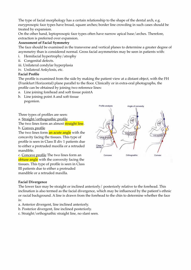
Facial ProfileThe profile is examined from the side by making thepatient view at a distant object, with the FH planeparallel to the floor. Clinically or in extraoral photo-graphs, the profile can be obtained by joining tworeference lines:a. Line joining forehead and soft tissue point Ab. Line joining point A and soft tissue pogonion.
Three types of profiles are seen:a. Straight/ orthognathic profile The two lines form an
almost straight line (Fig. 7.4A).Fig. 7.2A: Euryprosopic face
Fig. 7.2C: Leptoprosopic face
The type of facial morphology has a certain relationship to the shape of the dental arch, e.g. euryprosopic face types have broad, square arches; border line crowding in such cases should be treated by expansion. On the other hand, leptoprosopic face types often have narrow apical base/arches. Therefore, extraction is preferred over expansion. Assessment of Facial Symmetry The face should be examined in the transverse and vertical planes to determine a greater degree of asymmetry than is considered normal. Gross facial asymmetries may be seen in patients with: i. Hemifacial hypertrophy/atrophy ii. Congenital defects. iii. Unilateral condylar hyperplasiaiv. Unilateral Ankylosis, etc. Facial Profile The profile is examined from the side by making the patient view at a distant object, with the FH (Frankfurt Horizontal) plane parallel to the floor. Clinically or in extra-oral photographs, the profile can be obtained by joining two reference lines: a. Line joining forehead and soft tissue pointA b. Line joining point A and soft tissue
pogonion.
Three types of profiles are seen: a. Straight/orthognathic profileThe two lines form an almost straight line. b. Convex profile The two lines form an acute angle with the concavity facing the tissues. This type of profile is seen in Class II div 1 patients due to either a protruded maxilla or a retruded mandible. c. Concave profile The two lines form an obtuse angle with the convexity facing the tissues. This type of profile is seen in Class III patients due to either a protruded mandible or a retruded maxilla.
Facial Divergence The lower face may be straight or inclined anteriorly/ posteriorly relative to the forehead. This inclination is also termed as the facial divergence, which may be influenced by the patient’s ethnic or racial background. A line is drawn from the forehead to the chin to determine whether the face is: a. Anterior divergent, line inclined anteriorly. b. Posterior divergent, line inclined posteriorly. c. Straight/orthognathic straight line, no slant seen.
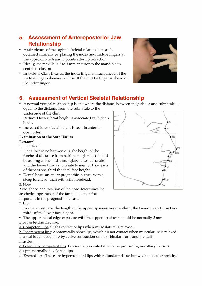
5. Assessment of Anteroposterior Jaw Relationship
- A fair picture of the sagittal skeletal relationship can be obtained clinically by placing the index and middle fingers at the approximate A and B points after lip retraction.
- Ideally, the maxilla is 2 to 3 mm anterior to the mandible in centric occlusion.
- In skeletal Class II cases, the index finger is much ahead of the middle finger whereas in Class III the middle finger is ahead of the index finger.
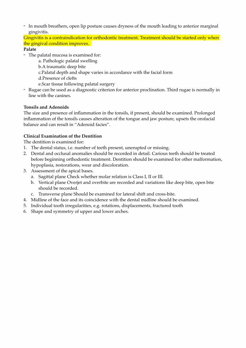
6. Assessment of Vertical Skeletal Relationship- A normal vertical relationship is one where the distance between the glabella and subnasale is
equal to the distance from the subnasale to the under side of the chin.
- Reduced lower facial height is associated with deep bites .
- Increased lower facial height is seen in anterior open bites.
Examination of the Soft Tissues Extraoral 1. Forehead- For a face to be harmonious, the height of the
forehead (distance from hairline to glabella) should be as long as the mid-third (glabella to subnasale) and the lower third (subnasale to menton), i.e. each of these is one-third the total face height.
- Dental bases are more prognathic in cases with a steep forehead, than with a flat forehead.
2. Nose Size, shape and position of the nose determines the aesthetic appearance of the face and is therefore important in the prognosis of a case. 3. Lips - In a balanced face, the length of the upper lip measures one-third, the lower lip and chin two-
thirds of the lower face height.- The upper incisal edge exposure with the upper lip at rest should be normally 2 mm. Lips can be classified into:a. Competent lips: Slight contact of lips when musculature is relaxed. b. Incompetent lips: Anatomically short lips, which do not contact when musculature is relaxed. Lip seal is achieved only by active contraction of the orbicularis oris and mentalis muscles. c. Potentially competent lips: Lip seal is prevented due to the protruding maxillary incisors despite normally developed lips. d. Everted lips: These are hypertrophied lips with redundant tissue but weak muscular tonicity.

2. Nasolabial angle- This is the angle formed between a tangent to the lower
border of the nose and a line joining the subnasale with the tip of the upper lip (labrale superius).
- Normal value is 110 degrees. - In patients with maxillary prognathism and proclined
upper anteriors this angle reduces whereas it becomes more obtuse in cases with a retrognathic maxilla or retroclined maxillary anteriors.
Chin The configuration of the chin is determined not only by the bone structure, but also by the thickness and tone of the mentalis muscle. • Mentalis activity A normal mentalis muscle becomes hyperactive in certain malocclusions like Class II div 1 cases, wherein puckering of the chin may be seen. • Mentolabial sulcusIt is the concavity present below the lower lip. Deep sulcus is seen in Class II cases whereas a shallow sulcus is seen usually in bimaxillary protrusion cases. Along with the chin width, development of chin height is important. Chin height is the distance from the • Mentolabial sulcus to mentonOver development of chin height alters the lower lip position and interferes with lip closure. • Chin position and prominenceProminent chin is usually associated with Class III malocclusions whereas recessive chin is seen in Class II malocclusion.
3. Intraoral ExaminationTongue - Tongue is examined for shape, color and configuration.- Tongue size can be roughly estimated with the help of a lateral cephalogram.- An excessively large tongue (macroglossia) usually shows imprints on its lateral margins, which
gives the tongue a scalloped appearance. However diagnosis of macroglossia requires a detailed diagnostic investigation (e.g. cineradiography).
- The lingual frenum should be examined for tongue tie.Lip and Cheek Frena - Among the different frena, the maxillary labial frenum is most commonly the cause of a
malocclusion. A thick, fibrous, low labial frenum prevents upper central incisors from approximating each other leading to a midline diastema.
- An IOPA of the area may show a bony fissure between the roots of the upper central incisors. - Blanch test can be done to confirm diagnosis wherein the upper lip is stretched upward and
outwards. Presence of blanching in the papilla region indicates an abnormal attachment.- The mandibular labial frenum is less often associated with a diastema.Gingiva - The gingiva should be examined for the type (thick fibrous or thin fragile), inflammation and
muco- gingival lesions.- In children, most commonly generalised marginal gingivitis occurs due to plaque accumulation
and can be resolved by improving the oral hygiene. In adults, scaling followed by curettage and sometimes mucogingival surgery is usually required.
- Local gingival lesions may occur due to occlusal trauma, abnormal functional loadings or medication (e.g. Dilantin).
- In mouth breathers, open lip posture causes dryness of the mouth leading to anterior marginal gingivitis.
Gingivitis is a contraindication for orthodontic treatment. Treatment should be started only when the gingival condition improves. Palate - The palatal mucosa is examined for:
a. Pathologic palatal swellingb.A traumatic deep bite c.Palatal depth and shape varies in accordance with the facial form d.Presence of cleftse.Scar tissue following palatal surgery
- Rugae can be used as a diagnostic criterion for anterior proclination. Third rugae is normally in line with the canines.
Tonsils and Adenoids The size and presence of inflammation in the tonsils, if present, should be examined. Prolonged inflammation of the tonsils causes alteration of the tongue and jaw posture, upsets the orofacial balance and can result in “Adenoid facies”.
Clinical Examination of the Dentition The dentition is examined for: 1. The dental status, i.e. number of teeth present, unerupted or missing.2. Dental and occlusal anomalies should be recorded in detail. Carious teeth should be treated
before beginning orthodontic treatment. Dentition should be examined for other malformation, hypoplasia, restorations, wear and discoloration.
3. Assessment of the apical bases. a. Sagittal plane Check whether molar relation is Class I, II or III. b. Vertical plane Overjet and overbite are recorded and variations like deep bite, open bite
should be recorded. c. Transverse plane Should be examined for lateral shift and cross-bite.
4. Midline of the face and its coincidence with the dental midline should be examined. 5. Individual tooth irregularities, e.g. rotations, displacements, fractured tooth 6. Shape and symmetry of upper and lower arches.

FUNCTIONAL EXAMINATION 1. Assessment of postural rest position and maximum
intercuspationA-Determination of postural rest position:“The postural rest position is the position of the mandible at which the synergists and antagonists of the orofacial system are in their basic tonus and balanced dynamically.”The space which exists between the upper and lower jaws at the postural rest position is the interocclusal clearance or freeway space which is normally 3 mm in the canine region.
The rest position should be determined with the patient relaxed and seated upright with the back unsupported. The head is oriented by making the patient look straight ahead. The head can also be positioned with the Frankfurt horizontal parallel to the floor. Various methods to record the postural rest position:
a. Phonetic method The patient is told to pronounce some consonants like “M” or words like “Mississippi” repeatedly. The mandible returns to the postural rest position 1-2 seconds after the exercise.
b. Command methodThe patient is asked to perform selected functions like swallowing, at the end of which the mandible returns spontaneously to the rest position. Phonetic exercise is also a type of command method.
c. Non-command methodThe clinician talks to the patient on unrelated topics and observes the patient as he speaks and swallows while he remains distracted. Patient is not aware that any examination is being carried out. While talking, the patients musculature is relaxed and the mandible reverts to the postural rest position.
d. Combined methodsA combination of the above methods is most suitable for functional analysis in children. The patient is observed during swallowing and speaking.The “Tapping test” can also be carried out to relax the musculature. Here, the clinician holds the chin with his index finger and thumb and then opens and closes the mandible passively with constantly increasing frequency until the musculature is relaxed. This can be confirmed by palpating the submental muscles. The rest position can then be determined.
Regardless of the method, mandible position is checked extraorally and the patient is told not to change the jaw, lip or tongue position. The lips are then parted and the maxillomandibular relation as well as the freeway space is determined. Registration of the Rest Position 1. Intra oral methodsa. Direct method:Vernier calipers can be used directly to measure the interocclusal clearance in the canine region.
b. Indirect method:Impression material is used to register the freeway space.
2. Extra oral methods a. Direct method Reference points are made on the skin with plaster, one on the nose and the other on the chin in the mid sagittal plane. The distance between these two points is measured at rest position and centric occlusion. The difference between the two is the freeway space.
b. Indirect method Includes • Cephalometric registration: Two cephalogram, one at postural rest position and other in centric occlusion are taken to determine the freeway space. • Kinesiographic registration: A magnet is fixed on the lower anterior teeth and the mandibular movements are recorded by sensors which is then processed in the Kinesiograph.
B-Evaluation of the Path of Closure The path of closure is the movement of the mandible from rest position to full articulation which should be analysed in all 3 planes of space, i.e. sagittal, vertical and frontal planes. The amount of rotation and sliding during mandibular closure is analysed.
Sagittal Plane In Class II malocclusions, three types of movements can be seen. a. Pure rotational movement without a sliding c o m p o n e n t — s e e n i n functional true Class II malocclusion. b.Forward path of closure—i.e. rotational movement with anterior sliding movement. The mandible slides into a more forward position, therefore, Class II malocclusion is more pronounced than can be seen in habitual occlusion. c.Backward path of closure, i.e. rotational movement with posterior sliding movement.
In Class II div 2 cases, the mandible slides backward into a posterior occlusal position because of premature contact with retroclined maxil lary incisors. Vertical Plane It is important to differentiate between two types of overbites. The true deep overbite is caused by infra occlusion of the molars and can be diagnosed by the presence of a large freeway space. The prognosis with functional therapy is favourable. Pseudo-deep bite is caused due to over-eruption of the incisors and is characterised by a small freeway space.
Prognosis with functional therapy is unfavourable.
Transverse Plane During mandibular closure, the midline of the mandible is observed. In case of unilateral cross bite, this analysis is relevant to differentiate between l a t e r o g n a t h y a n d laterocclusion. Laterognathy-or true cross bite, the centre of the mandible and the facial midline do not coincide in rest and in occlusion. Laterocclusion—the centre of the mandible and facial midline coincide in rest position but in occlusion the mandible deviates due to tooth interference leading to non-coinciding midlines.
2. Examination of the temporomandibular jointExamination:The clinical examination of the TMJ should include- Auscultation - Palpation of the temporomandibular joint and the musculature associated with mandibular
movements - Functional analysis of the mandibular movements. Objective:The main objective of this examination is to look for symptoms of TMJ dysfunction such as crepitus, clicking, pain, hyper mobility, deviation, dislocation, limitation of jaw movements and other morphological abnormalities. Clinical Aids:- TMJ radiographs- Tomograms of the TMJ in habitual occlusion and maximum mouth opening.
3. Examination of orofacial dysfunctions Includes evaluation of: • Swallowing • Tongue• Speech • Lips• Respiration
Swallowing At birth the tongue protrudes anteriorly between the gum pads to establish lip seal. Therefore the infant swallows viscerally for the first 1 1⁄2 to 2 years of age. This infantile swallow is gradually replaced by the mature swallow as the deciduous dentition is completed. If infantile swallow persists beyond the fourth year, it is considered as an orofacial dysfunction.
Tongue Tongue thrust is one of the most common dysfunction of the tongue. Tongue dysfunction can be assessed clinically by - Electromyographic examination - Cephalometric analysis - Cine radiographic examination- Palatographic examination- Neurophysiologic examinations.
Cephalograms can help to evaluate the position and size of the tongue in relation to the available space. However, in orthodontics diagnostic registration of tongue position is usually more important than its size. Palatography involved applying a thin layer of contrasting impression material to the patients tongue.
