osmotic stress affects functional properties of human · osmotic stress affects functional...
TRANSCRIPT
EPJ manuscript No.(will be inserted by the editor)
Osmotic stress affects functional properties of humanmelanoma cell linesCaterina A. M. La Porta1, Anna Ghilardi, Maria Pasini1, Lasse Laurson2,3, Mikko J. Alava2,3, Stefano Zapperi3,4,5,6, andMartine Ben Amar7,8
1 Department of Biosciences, University of Milano, via Celoria 26, 20133 Milano, Italy2 COMP Centre of Excellence, Aalto University, P.O. Box 14100, FIN-00076 Aalto, Espoo, Finland3 Department of Applied Physics, Aalto University, P.O. Box 14100, FIN-00076 Aalto, Espoo, Finland4 Physics Department, University of Milano, via Celoria 16, 20133 Milano, Italy5 CNR-IENI, Via R. Cozzi 53, 20125 Milano, Italy6 ISI Foundation, Via Alassio 11C, 10126 Torino, Italy7 Laboratoire de Physique Statistique, Ecole Normale Superieure, UPMC Univ Paris 06, Universite Paris Diderot, CNRS, 24 rue Lhomond,
75005 Paris, France8 Institut Universitaire de Cancerologie, Faculte de medecine, Universite Pierre et Marie Curie-Paris 6, 91 boulevard de l’hopital, 75013 Paris,
France
Received: date / Revised version: date
Abstract. Understanding the role of microenvironment in cancer growth and metastasis is a key issue for cancer re-search. Here, we study the effect of osmotic pressure on the functional properties of primary and metastatic melanomacell lines. In particular, we experimentally quantify individual cell motility and transmigration capability. We then per-form a circular scratch assay to study how a cancer cell front invades an empty space. Our results show that primarymelanoma cells are sensitive to a low osmotic pressure, while metastatic cells are less. To better understand the exper-imental results, we introduce and study a continuous model for the dynamics of a cell layer and a stochastic discretemodel for cell proliferation and diffusion. The two models capture essential features of the experimental results andallow to make predictions for a wide range of experimentally measurable parameters.
PACS. PACS-key discribing text of that key – PACS-key discribing text of that key
1 Introduction
The microenvironment is a key factor in tumour developmentand progression. Its influence on the behaviour of cancer cellsis to a great extent mediated by the composition, structure, anddimensionality of the extracellular matrix, the polymeric scaf-fold that surrounds cells within tissues. Research shows thatthe mechanical properties of tumour microenvironment can fa-cilitate or oppose tumour growth and dynamics, although thiseffect is poorly understood. Mechanical stresses such as thoseexperienced by cancer cells during the expansion of the tumouragainst the stromal tissue have been shown to release and ac-tivate growth factors involved in the progression of cancer [1].Moreover, the stiffness of the matrix surrounding a tumour de-termines how cancer cells polarize, adhere, contract, and mi-grate, thus regulating their invasiveness [1]. Another possibil-ity is that mechanical stresses directly regulate the growth anddeath rates of cancer cells. Recently, Montel et al. have ex-plored this possibility, investigating the effect of a constantstress applied on cellular spheroids over long time scales byinducing osmotic pressure by a solution of dextran, a biocom-patible polymer which is neutral and can not be metabolisedby mammalian cells [2, 3]. Using a similar method, some of us
recently reported that a constant low osmotic pressure (1kPa)affects more strongly the proliferative capability of primary hu-man melanoma cells (IgR39) in comparison to the correspond-ing metastatic ones (IgR37) [4]. Furthermore, a computer sim-ulation analysis of the growth of melanocytic nevi inside epi-dermal or dermal tissues shows that the shape of the nevi is cor-related with the elastic properties of the surrounding tissue[4].
Several studies in the literature reported important changesin cellular functioning due to osmotic pressure [5, 6], but thestresses involved (in the MPa range) were orders of magnitudelarger than those (in the kPa range) studied in Refs. [2, 3, 4].It is interesting to notice that compressive stresses of slightlyless than 1 kPa applied through a piston were recently foundto induce a metastatic phenotype in cancer cells [7]. In a re-cent paper, Simonsen et al. showed that interstitial fluid pres-sure (IFP) [8] was associated with high geometric resistanceto blood flow caused by tumour-line specific vascular abnor-malities in xenografted tumours from two human melanomalines with different angiogenic profiles [9]. In another recentpaper, Wu et al. investigated how nonlinear interactions amongthe vascular and lymphatic networks and proliferating tumourcells might influence IFP transport of oxygen, and tumour pro-gression [10]. They also investigated the possible consequences
arX
iv:1
504.
0088
8v1
[ph
ysic
s.bi
o-ph
] 3
Apr
201
5
2 Caterina A. M. La Porta et al.: Osmotic stress affects functional properties of human melanoma cell lines
of tumour-associated pathologies such as elevated vascular hy-draulic conductivities and decreased osmotic pressure differ-ences. All these parameters might affect microenvironmentaltransport barriers, and the tumour invasive and metastatic po-tential, opening interesting new therapeutic perspectives. Therole of IFP for drug delivery was also analyzed computationallyin Ref. [11]. In general, understanding the influence of mechan-ical stress on cancer growth could shed new light on tumourdevelopment and progression. Studies present in the literaturealso use hypertonic conditions, but the effects are always ob-served at high osmotic pressure [12, 13].
In this paper, we show that low osmotic pressure (1kPa)besides proliferation, which was the focus of previous works[2, 3, 4], also modulates key functional biological aspects ofcancer cells such as their motility and transmigration, possiblycontributing to the acquisition of a more aggressive phenotype.The choice of an osmotic pressure of 1kPa was suggested byprevious experiments [4] showing that this is the smallest valuethat has an appreciable effect on the growth of melanoma cells.Melanoma is an aggressive, radio- and chemo-resistant tumourwhich becomes impossible to cure when metastasised. There-fore, a pressing clinical problem in the treatment of melanomais to understand how to stop or prevent the capability of thetumour to give rise to metastasis. Using an interdisciplinaryapproach combining cellular biology and theoretical physics,we show that a low osmotic pressure (1kPa) leads to significantchanges in cell F-actin organization compromising the capabil-ity of the cells to move and transmigrate. Interestingly, osmoticpressure is more effective on the primary human melanoma cellline with respect to the metastatic one belonging to the samepatient. We perform quantitative analysis on the experimentaldata and extract parameters for cell motilities that we can thenuse in a continuum theory and a discrete model for cancer cellfront dynamics. Overall, our results suggest that osmotic pres-sure could contribute to the selection of a subpopulation in theprimary human melanoma cells contributing to the acquisitionof a more aggressive phenotype.
2 Experimental results
2.1 Effect of osmotic pressure on F-actinorganization
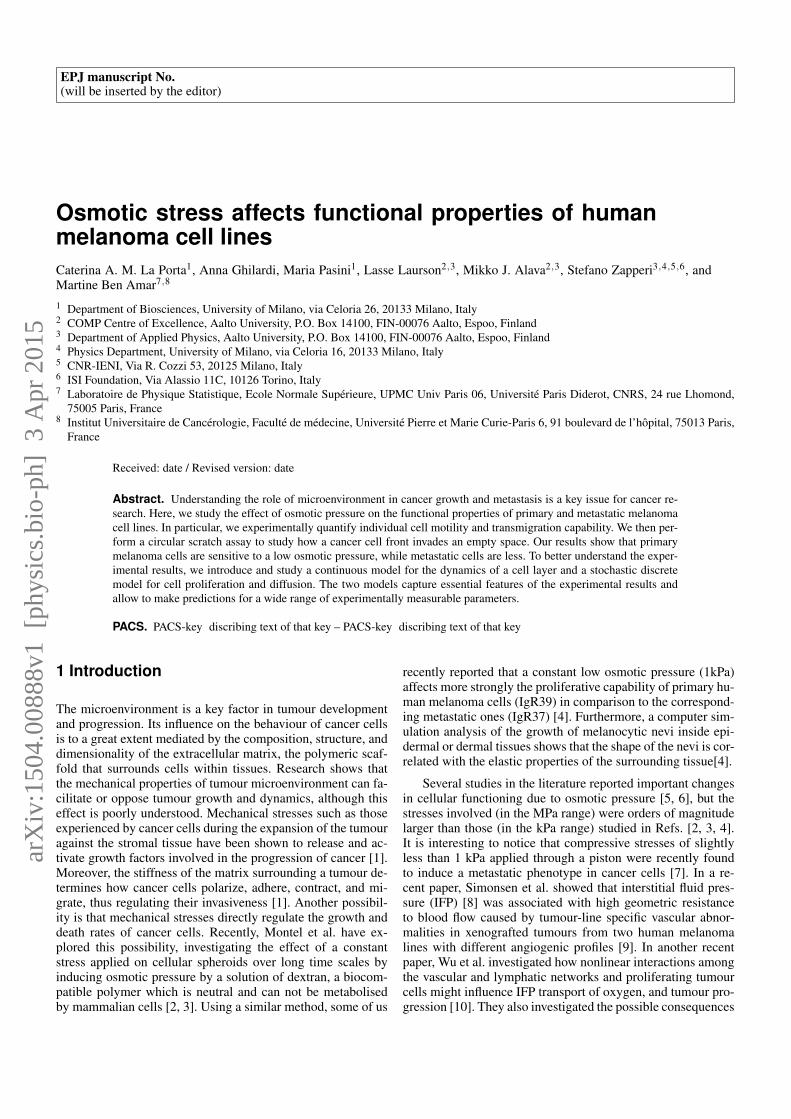
The organization of F-actin in the two human melanoma celllines is shown in Figure 1 under normal growth condition or1kPa osmotic pressure without or with collagen as physiologi-cal substrate. In both cell lines, 1kPa induces a rearrangementin F-actin organization (Fig. 1). In particular in IgR39, 1KPaosmotic pressure induces the appearance of filopodia and stressfibers (Fig. 1a-b) Similar changes have been shown plating thecells on a collagen pre-coated Petri dish (Fig. 1c-d). Indeed col-lagen should create a resistance against cell deformations thatis similar to a confining pressure. The metastatic cell line IgR37shows, however, is only slightly affected by osmotic pressuredisplaying a more elongated shape (Fig.1e-f).
0 kPa
1kPa
IgR39
IgR39 IgR39
IgR39
1KPaIgR37
IgR37 WTa)
b)
c)
d)
e)
f)
Collagen Type I
Collagen Type I +1kPa
Fig. 1 Effect of osmotic pressure on F-actin. Subconfluent cellswere plated on 33cm2 Petri dish untreated or pre-coated with type Icollagen. After 6 days the cells were fixed with 3.7% paraformaldeideand incubated with Red-Falloidin for 30min at room temperature.The nuclei were counterstained with DAPI and the slides weremounted with Pro-long anti fade reagent (Invitrogen). The imageswere acquired using a Leica TCS NT confocal microscope. Panel a-drepresent IgR39 cells and panel e-f IgR37 cells. Scale bar is 50µm
2.2 Effect of osmotic pressure on transmigrationcapability of IgR39 and IgR37 human melanoma cells
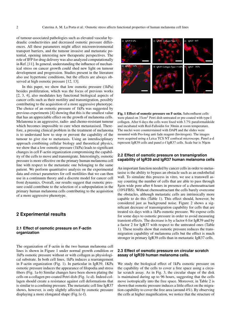
An important function needed by cancer cells in order to metas-tasise is the ability to bypass an obstacle such as an endothelialwall. To simulate this process in vitro, we use a transwell as-say counting the number of cells that are able to pass through8µm wide pore after 6 hours in presence of a chemoattractant(10%FBS). Without chemoattractant the cells barely overcomethe obstacles, although metastatic cells are intrinsically morecapable to do this (Table 1). This effect should, however, beconsidered just as background noise. Figure 2 shows a sig-nificant decrease of transmigration capability for cells that aretreated six days with a 1kPa osmotic pressure. We expose cellsfor some days to osmotic pressure in order to avoid measuringtransient effects. The decrease is by a factor 6 for IgR39 and bya factor 2 for IgR37 with respect to the untreated ones (Table1). These results show that osmotic pressure reduces the trans-migration capability of melanoma cells but the effect is muchstronger in primary IgR39 cells than in metastatic IgR37 cells.
2.3 Effect of osmotic pressure on circular scratchassay of IgR39 human melanoma cells.
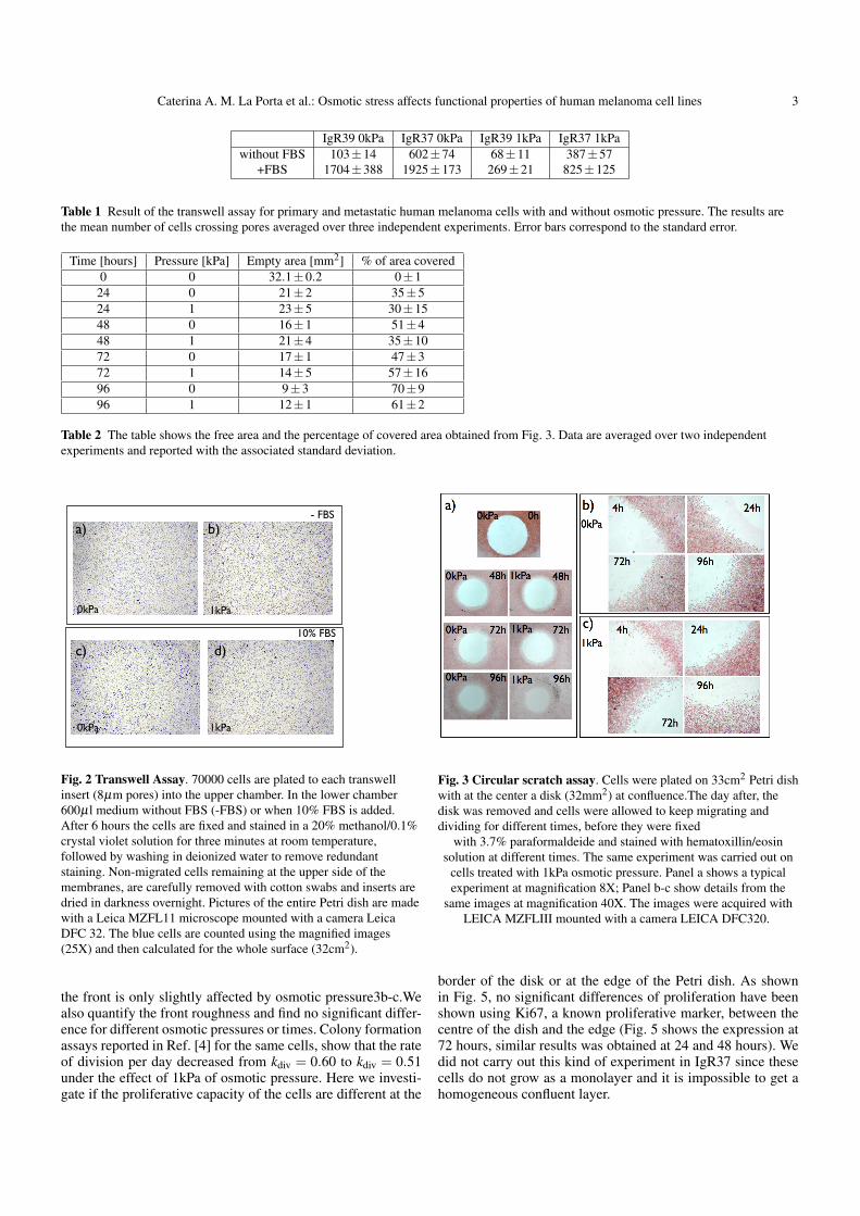
We study the biological effect of 1kPa osmotic pressure onthe capability of the cells to cover a free space using a circu-lar scratch assay. As in Fig. 3, the circular shape of the diskis maintained during up to 96 hours, suggesting that the cellsmove isotropically into the free space. Moreover, in Table 2 isshown that osmotic pressure induces a little effect on the migra-tion capability to cover the free area (around 4%). By observingthe cells at higher magnification, we notice that the structure of
Caterina A. M. La Porta et al.: Osmotic stress affects functional properties of human melanoma cell lines 3
IgR39 0kPa IgR37 0kPa IgR39 1kPa IgR37 1kPawithout FBS 103±14 602±74 68±11 387±57
+FBS 1704±388 1925±173 269±21 825±125
Table 1 Result of the transwell assay for primary and metastatic human melanoma cells with and without osmotic pressure. The results arethe mean number of cells crossing pores averaged over three independent experiments. Error bars correspond to the standard error.
Time [hours] Pressure [kPa] Empty area [mm2] % of area covered0 0 32.1±0.2 0±124 0 21±2 35±524 1 23±5 30±1548 0 16±1 51±448 1 21±4 35±1072 0 17±1 47±372 1 14±5 57±1696 0 9±3 70±996 1 12±1 61±2
Table 2 The table shows the free area and the percentage of covered area obtained from Fig. 3. Data are averaged over two independentexperiments and reported with the associated standard deviation.
- FBS
1kPa
1kPa
a)
10% FBS
0kPa
0kPa
b)
c) d)
Fig. 2 Transwell Assay. 70000 cells are plated to each transwellinsert (8µm pores) into the upper chamber. In the lower chamber600µl medium without FBS (-FBS) or when 10% FBS is added.After 6 hours the cells are fixed and stained in a 20% methanol/0.1%crystal violet solution for three minutes at room temperature,followed by washing in deionized water to remove redundantstaining. Non-migrated cells remaining at the upper side of themembranes, are carefully removed with cotton swabs and inserts aredried in darkness overnight. Pictures of the entire Petri dish are madewith a Leica MZFL11 microscope mounted with a camera LeicaDFC 32. The blue cells are counted using the magnified images(25X) and then calculated for the whole surface (32cm2).
the front is only slightly affected by osmotic pressure3b-c.Wealso quantify the front roughness and find no significant differ-ence for different osmotic pressures or times. Colony formationassays reported in Ref. [4] for the same cells, show that the rateof division per day decreased from kdiv = 0.60 to kdiv = 0.51under the effect of 1kPa of osmotic pressure. Here we investi-gate if the proliferative capacity of the cells are different at the
Fig. 3 Circular scratch assay. Cells were plated on 33cm2 Petri dishwith at the center a disk (32mm2) at confluence.The day after, thedisk was removed and cells were allowed to keep migrating anddividing for different times, before they were fixed
with 3.7% paraformaldeide and stained with hematoxillin/eosinsolution at different times. The same experiment was carried out on
cells treated with 1kPa osmotic pressure. Panel a shows a typicalexperiment at magnification 8X; Panel b-c show details from the
same images at magnification 40X. The images were acquired withLEICA MZFLIII mounted with a camera LEICA DFC320.
border of the disk or at the edge of the Petri dish. As shownin Fig. 5, no significant differences of proliferation have beenshown using Ki67, a known proliferative marker, between thecentre of the dish and the edge (Fig. 5 shows the expression at72 hours, similar results was obtained at 24 and 48 hours). Wedid not carry out this kind of experiment in IgR37 since thesecells do not grow as a monolayer and it is impossible to get ahomogeneous confluent layer.
4 Caterina A. M. La Porta et al.: Osmotic stress affects functional properties of human melanoma cell lines
0 1 2 3 4 5Time (days)
0
0.05
0.1
0.15
0.2
Roug
hnes
s (m
m)
Experiment 0kPAExperiment 1kPaSimulations 0kPaSimulations 1kPa
a) b)
c) d)
Fig. 4 Estimate of the front roughness. a) Plates resulting from thecircular scratch assay are digitized and b) thresholded using the sameexposure and the same threshold for all the images. c) The cell frontis obtained by a percolation cluster algorithm and d) the roughness ofthe front is computed for each experimental condition (results of twoindependent experiments). For comparison, we also report theroughness obtained from the simulations of the computational model.
2.4 Osmotic pressure affects cell motility
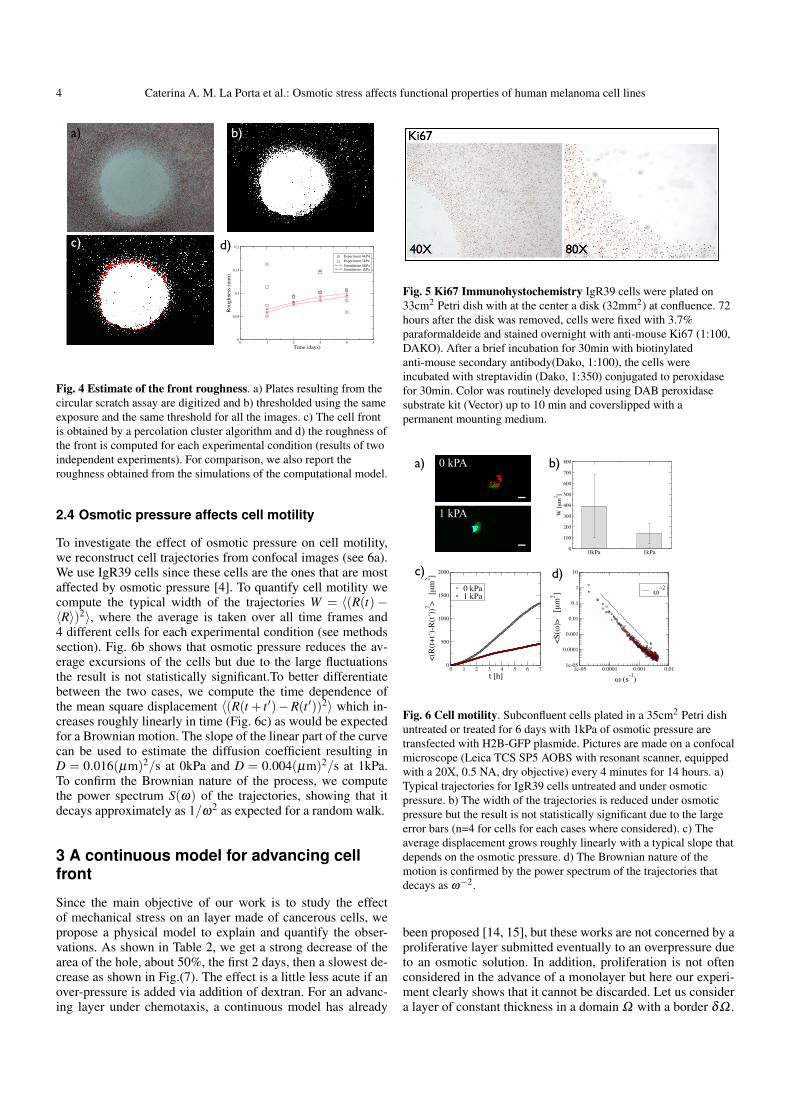
To investigate the effect of osmotic pressure on cell motility,we reconstruct cell trajectories from confocal images (see 6a).We use IgR39 cells since these cells are the ones that are mostaffected by osmotic pressure [4]. To quantify cell motility wecompute the typical width of the trajectories W = 〈(R(t)−〈R〉)2〉, where the average is taken over all time frames and4 different cells for each experimental condition (see methodssection). Fig. 6b shows that osmotic pressure reduces the av-erage excursions of the cells but due to the large fluctuationsthe result is not statistically significant.To better differentiatebetween the two cases, we compute the time dependence ofthe mean square displacement 〈(R(t + t ′)−R(t ′))2〉 which in-creases roughly linearly in time (Fig. 6c) as would be expectedfor a Brownian motion. The slope of the linear part of the curvecan be used to estimate the diffusion coefficient resulting inD = 0.016(µm)2/s at 0kPa and D = 0.004(µm)2/s at 1kPa.To confirm the Brownian nature of the process, we computethe power spectrum S(ω) of the trajectories, showing that itdecays approximately as 1/ω2 as expected for a random walk.
3 A continuous model for advancing cellfront
Since the main objective of our work is to study the effectof mechanical stress on an layer made of cancerous cells, wepropose a physical model to explain and quantify the obser-vations. As shown in Table 2, we get a strong decrease of thearea of the hole, about 50%, the first 2 days, then a slowest de-crease as shown in Fig.(7). The effect is a little less acute if anover-pressure is added via addition of dextran. For an advanc-ing layer under chemotaxis, a continuous model has already
Fig. 5 Ki67 Immunohystochemistry IgR39 cells were plated on33cm2 Petri dish with at the center a disk (32mm2) at confluence. 72hours after the disk was removed, cells were fixed with 3.7%paraformaldeide and stained overnight with anti-mouse Ki67 (1:100,DAKO). After a brief incubation for 30min with biotinylatedanti-mouse secondary antibody(Dako, 1:100), the cells wereincubated with streptavidin (Dako, 1:350) conjugated to peroxidasefor 30min. Color was routinely developed using DAB peroxidasesubstrate kit (Vector) up to 10 min and coverslipped with apermanent mounting medium.
0 1 2 3 4 5 6 7t [h]
0
500
1000
1500
2000
<(R(
t+t’)
-R(t’
))2 > [
µm
2 ]
0 kPa1 kPa
1e-05 0.0001 0.001 0.01
t (s-1)
1e-05
0.0001
0.001
0.01
0.1
1
10
<S(t
)>
[µm
2 ] t<2
0kPa 1kPa0
100
200
300
400
500
600
700
800
W [µ
m2 ]
a) b)
c) d)
0 kPA
1 kPA
Fig. 6 Cell motility. Subconfluent cells plated in a 35cm2 Petri dishuntreated or treated for 6 days with 1kPa of osmotic pressure aretransfected with H2B-GFP plasmide. Pictures are made on a confocalmicroscope (Leica TCS SP5 AOBS with resonant scanner, equippedwith a 20X, 0.5 NA, dry objective) every 4 minutes for 14 hours. a)Typical trajectories for IgR39 cells untreated and under osmoticpressure. b) The width of the trajectories is reduced under osmoticpressure but the result is not statistically significant due to the largeerror bars (n=4 for cells for each cases where considered). c) Theaverage displacement grows roughly linearly with a typical slope thatdepends on the osmotic pressure. d) The Brownian nature of themotion is confirmed by the power spectrum of the trajectories thatdecays as ω−2.
been proposed [14, 15], but these works are not concerned by aproliferative layer submitted eventually to an overpressure dueto an osmotic solution. In addition, proliferation is not oftenconsidered in the advance of a monolayer but here our experi-ment clearly shows that it cannot be discarded. Let us considera layer of constant thickness in a domain Ω with a border δΩ .
Caterina A. M. La Porta et al.: Osmotic stress affects functional properties of human melanoma cell lines 5
The cell displacement in Ω is mostly driven by the cell pro-liferation, but at the border the cells may be more proliferativeeventually and even detach and move freely. Due to prolifera-tion, the mass conservation equation is then transformed into:
∂ρ0
∂ t+∇(ρ0V ) =−kv(P−Ph), (1)
where Ph is the homeostatic pressure for which cell prolifera-tion just compensates apoptosis, ρ0 is the constant mass den-sity, V is the velocity at an arbitrary point of Ω . This sim-ple idea about regulation in living systems is due to ClaudeBernard, a french physiologist, in 1865 [16]. Mathematically,such a continuous model is valid at a scale larger than the cellsize and the velocity is an averaged quantity on a sample ofintermediate scale between the single cell and the full layer.Adding dextran to the solution increases the pressure P, slightlyinhibiting the proliferation rate. For a layer crawling on a sub-strate in strong adhesion, the second Newton’s law of dynamicsimplifies into a Darcy law ([17, 14])
V =− 1M
∇P, (2)
where the mobility coefficient M is related to the friction coef-ficient. Combining both equations gives:
∆P−α2P = 0 with P = P−Ph (3)
and α2 = (kvMR2i )/ρ0, with Ri the initial radius of the hole of
order the millimeter, chosen as our length unit in the follow-ing. At the free interface, boundary conditions include the me-chanical equilibrium and continuity of normal velocities. Thepressure at the layer border Pi is equal to the pressure of thesolution (if capillary effect is neglected). It is smaller at the in-terface δΩ than in the layer bulk Ω for a proliferative layer.The interface velocity δΩt is given by the velocity of the cellsat the interface which may be different from the cells in thebulk Ω . Indeed, these peripheral cells have more room both fordisplacement and proliferation. Then the normal front velocityδΩt is given by
N ·δΩt =−1M
N ·∇P+N · vs (4)
where vs may represent a specific proliferation rate of the bor-der cells. Other boundary conditions may exist due to the ge-ometry of the experiment, that is why we consider now 3 dif-ferent growth geometries.
3.1 Growing circular disc
We imagine a disc of a cell monolayer and assume that thegrowth process preserves the circular geometry. Eq. (3) for thepressure P = P−Ph is then transformed into an ordinary differ-ential equation of second order:
1R
ddR
(R
dPdR
)= α
2P (5)
The regular solution at the origin is the modified Bessel func-tion of zero order I0 so
P(R) = PiI0(αR)
I0(αRi(t))(6)
and Pi = Pi−Ph is negative, being fixed by the nutrient medium,whatever the hole radius Ri(t). From Darcy’s law (Eq. (2)), wederive the growing velocity at the interface:
N ·δΩt = Ri(t) =−Piα
MI1(αRi(t))I0(αRi(t))
(7)
This equation means that the interface velocity has the same ve-locity as the moving cells of the interface. As for the Saffman-Taylor viscous fingering in radial geometry [18], our set ofequations is time independent but the time dependence reap-pears via the condition of continuity of velocities given by Eq. (7).It is an implicit equation for the time variation of the radiuswhich can be solved numerically. However one can make anasymptotic analysis of Eq.(7) and gets a constant growth rate atlong times :
Ri(t)|t→∞→−Piα
Mand a radius growing linearly in time. One can notice that a con-stant proliferation rate into Eq.(1) will lead to an exponentialtime behavior for the radius so to follow the growth expansionof the layer at long times may be a good test for the validationof the model.
3.2 A hole in an infinite layer
In this case, we can accept the irregular Bessel function at thecenter K0 as the solution of Eq.(5), but not I0, which growsexponentially. The solution reads
P(R) = PiK0(αR)
K0(αRi(t)). (8)
The pressure P vanishes at infinity where there is no cell pro-liferation. The velocity given by Darcy’s law is then:
R =α
MPi
K1(αR)K0(αRi(t))
(9)
being negative as Pi and vanishing as R→ ∞ as expected. Hereagain the radius of the hole is given numerically if we takeEq.(4) on the interface. In addition, the radius velocity divergeswhen the total cover of the hole is reached, and the continu-ous model stops to be valid for Ri(t)→ 0. Indeed, Eq.(9) isa nonlinear first order differential equation for the hole radiusthat we cannot integrate explicitly. However, for a large enoughhole, assuming α of order 1, the decrease of the radius is linearat initial time, but the radius will finally vanish like a squareroot, (t − tc)1/2, near the complete closure arising at time tc.Before presenting more elaborated model for our experiment,we would like to comment about the stability analysis of theborder and the assumption of circular symmetry for our set ofequations. A careful observation of our experiments presented
6 Caterina A. M. La Porta et al.: Osmotic stress affects functional properties of human melanoma cell lines
in Fig. (3) shows that the inner border is somewhat noisy butdoes not present really instabilities similar to patterns observedin viscous flow [19] or bacteria colonies [14]. It turns out that,even for a planar front, this model is stable against transverseperturbations, even in absence of capillary effect. Although as-tonishing, it is in agreement with observation. In order to con-vince the reader, we simply present the stability treatment forthis geometry in the Appendix section and analytically provethat our model predicts that the migration induced by growthis stable in the circular geometry. So the border irregularitiesobserved in Fig. (3) are not fingering but can be interpreted asthe stochastic behavior of tumor cells.
0 1 2 3 4 5Time (days)
0.2
0.4
0.6
0.8
1
Rela
tive
hole
are
a
Experiment 0kPAExperiment 1kPaTheory 0kPaTheory 1kPaSimulations 0kPaSimulations 1kPa
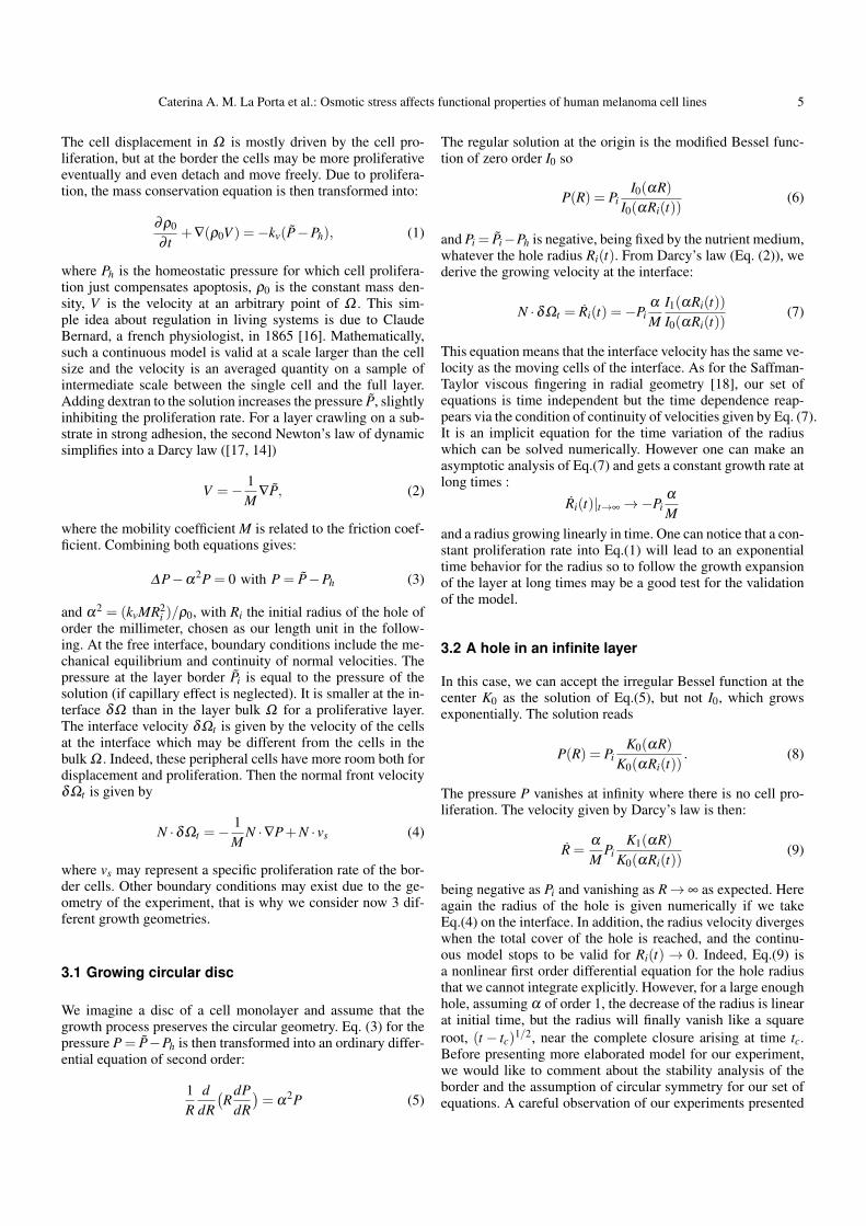
Fig. 7 Front propagation. Relative area of the hole as a function oftime for experiments (see Table 2), theory and simulations.Parameters for the theory are Pi =−1 for 0kPa and Pi =−0.98 for1kPa, αi = 0.71,αe = 0.53, δ = 0.3 (with initial radius 1), τ = 1.These numerical values correspond to the best estimated fitting forour experimental data. Parameters for the simulations are kdiv = 32division/day while keeping kdiff/kdiv = 0.9 or 1.7, for 0kPa and 1kPa,respectively.
3.3 The circular scratch assay
Here we focus on the geometry of the circular scratch assay:it is made of a planar annulus of cells limited by the rigid cir-cular wall border of the Petri dish at R = Re and , at R = Ri,is free to move into the empty space. The interior interface atR = Ri is rather diffuse but an average circular border can bedefined. In addition a thin circular ring (of thickness δ ) withsmaller cell density can be observed appearing less contrastedthan the inner core, located at R = Ri + δ . We will adapt theprevious model to this inhomogeneous ring which has a den-sity ρi for R between Ri(t) and Ri(t)+ δ while the density inthe bulk (Ri(t)+ δ < R < Re) is called ρe . It is important toremember that ρ is indeed a density per unit surface, the volu-metric density being the same for all cells. As a consequence, ifwe maintain the hypothesis of a monolayer, the surface densitymay be larger at the inner border where the cells are more free
to advance than in the annulus where they are more confined.It explains the optical difference in Fig. (3). We take constantboth ρ values. Moreover, in vitro, it has been shown that inconfluent layers, live cell extrusion is induced to recover thehomeostatic state in case of over-crowding and it is clear thatthe border allows more easily such extrusion [20]. So we pre-sume that ρi < ρe. Because the layer geometry excludes R = 0and R→ ∞, possible solutions for Eq.(5) are superpositions ofI0 and K0 and we get
Pi(R) =Pi
1+b
I0(αiR)I0(αiRi(t))
+bK0(αiR)
K0(αiRi(t))
(10)
with α2i = kvM/ρi. The velocity of the cells at the outer border
being :
Ri =−αi
MPi
1+b
I1(αiR)I0(αiRi(t))
−bK1(αiR)
K0(αiRi(t))
(11)
Pi keeps the same definition as before : the pressure in the so-lution minus the homeostatic pressure. For Ri(t)+δ < R < Rewe have:
Pe(R) = Pe
I0(αeR)I1(αeRe)
+K0(αeR)K1(αeRe)
− τ (12)
τ being an evaporation rate of cells as mentioned by [20] and
Re =−αe
MPe
I1(αeR)I1(αeRe)
− K1(αeR)K1(αeRe)
(13)
with α2e = (kvMR2
i )/ρe. In Eqs.(12,13), we have applied thecancellation of cell velocity at the exterior border Re. We have2 unknowns: the coefficient b and Pe which are fixed by themechanical equilibrium Pi(Ri(t)+ δ ) = Pe(Ri(t)+ δ ) and thecontinuity of the flux: ρidPi/dR = ρedPe/dR for R = Ri(t)+δ . The principle of such analysis is simple but requires alsoa numerical solution that we present in Fig.(7) in compari-son with experimental data. We have four unknown parame-ters αi,αe, τ and Pi. Varying parameters, the comparison withthe experiment is mostly controlled by the quantity αPi/M =√
kv/(ρiM)Ri which is the inverse of a typical time. In the ex-periment the time scale is the day. Taking into account the ex-perimental results shown in Fig.(7), the relative area variationof the hole after 2 hours is 0.1 for an osmotic pressure varia-tion of 1 kPa. It corresponds to a variation of the front veloc-ity in international unit of 710−9 m/s (evaluated from the ex-perimental data of Fig.(7)) which gives the unknown value ofM of 1014 Pas/m2, using Eq(13). Once we have this value wecan derived easily the proliferation rate kv/ρ0 of order 510−9
SI. The friction coefficient M for an advancing epithelium hasbeen evaluated in [14] for MDCK cells giving a value an orderof magnitude larger 1015. Of course, the value of M depends onthe cells involved in the experimental set-up, and are not uni-versal. However this estimation is consistent with usual orderof magnitude for biophysical values. Taking again the order ofmagnitude of the velocity as 10−8m/s, knowing that forces oneneeds or can apply to a cell are typically of order pico-Newtonso 10−12 N gives a friction coefficient of 1014 Ns/m4 (being theratio of the force value by cell volume and by cell velocity). Fi-nally sensitive variation by osmotic pressure being 0.02 in our
Caterina A. M. La Porta et al.: Osmotic stress affects functional properties of human melanoma cell lines 7
model which corresponds to 1 kPa, gives a naive estimation of50 kPa for the departure from the homeostatic pressure. A bet-ter calculation can be done from the model using Eq.(11) andgives 25 kPa.
4 Computational model of cell duplicationand movement
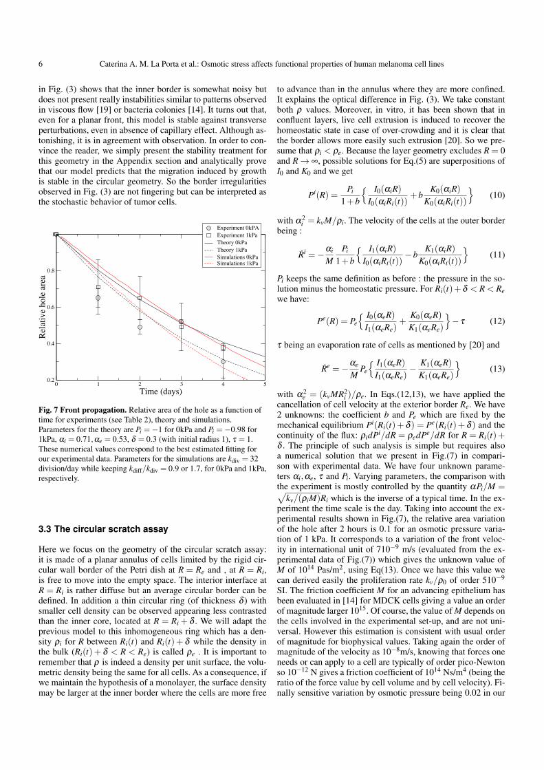
To understand how the morphology of the front in the circu-lar scratch assay depends on the interplay between cell divi-sion and mobility, we simulate a simple lattice model includ-ing mechanisms for cell division and motility, with the rela-tive rates of the two as the tunable parameter. The model is de-fined on a square lattice of linear size L = 700, in which all thesites are occupied by cells except those inside a circle of radiusr = 320. Cells that have at least one unoccupied nearest neigh-bour site can either divide with rate kdiv or diffuse with ratekdiff. The kinetics of the model follows the Gillespie algorithm[21]. At each time step we count how many cells have 1, 2, 3, or4 neighbours (i.e. n1, n2, n3 and n4). Then the total rate is takento be Rtot = (n4+0.75∗n3+0.5∗n2+0.25∗n1)∗(kdiv+kdiff),where the reaction rate is taken to be proportional to the num-ber of neighbours a cell has, with the bare rates given by thoseof a cell with all its four nearest neighbours vacant. At eachtime step, the reaction to take place is chosen randomly, with aprobability (rate of the reaction)/Rtot. There are 8 possible reac-tions with different rates, i.e. 4 possible division reactions and4 possible diffusion reactions, each corresponding to a differentnumber of nearest neighbours a cell has (1,2,3 or 4). First, thereaction to take place is chosen, and then a random cell with thenumber of nearest neighbours corresponding to the selected re-action is found, and the reaction is carried out. The correspond-ing time step is generated from an exponential distribution withRtot as the rate parameter. Then the simulation proceeds to thenext time step, and the process continues until cells invade thecenter of the hole. Results are averaged over 10 realizationsfor each case. In Fig. 8, we report typical configurations of thecircular scratch assay obtained by numerical simulations fordifferent values of kdiff/kdiv. When this ratio is large the in-terface is very diffuse, with several cells moving ahead of thefront. As the ratio decreases, the front is more sharp, but stillremains rough due to the randomness in the division process.In order to compare our simulations with experiments we firstestimate the ratio kdiff/kdiv from other experiments. In partic-ular, we can estimate kdiff ' D/a2, where a is the typical celldiameter. From Fig. 1, we obtain that a ' 20µm at 0kPa anda ' 50µm at 1kPa. Using our measurements for D and kdiv[4], we obtain kdiff/kdiv ' 0.9 at 0kPa and kdiff/kdiv ' 1.7 at1kPa. If we use these value of kdiv and kdiff, the simulated frontadvances too slowly with respect to the experiments. This isexpected since in the circular scratch assay cells are induced torapidly fill the hole. A good agreement between model and ex-periments is obtained setting kdiv = 32 division/day while keep-ing kdiff/kdiv = 0.9 or 1.7, for 0kPa and 1kPa, respectively. Theresult is reported in Fig. 7 for the front evolution and in Fig.4 for the roughness. The parameters employed in the model toobtain the best fit to the experiments look, however, unrealisticwhen compared with estimates obtained from experiments on
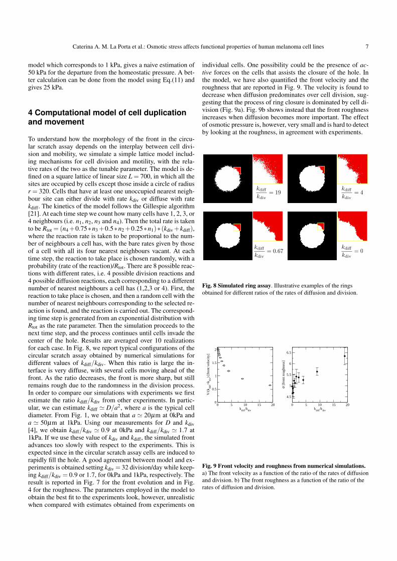
individual cells. One possibility could be the presence of ac-tive forces on the cells that assists the closure of the hole. Inthe model, we have also quantified the front velocity and theroughness that are reported in Fig. 9. The velocity is found todecrease when diffusion predominates over cell division, sug-gesting that the process of ring closure is dominated by cell di-vision (Fig. 9a). Fig. 9b shows instead that the front roughnessincreases when diffusion becomes more important. The effectof osmotic pressure is, however, very small and is hard to detectby looking at the roughness, in agreement with experiments.
kdiff
kdiv= 19
kdiff
kdiv= 4
kdiff
kdiv= 0
kdiff
kdiv= 0.67
Fig. 8 Simulated ring assay. Illustrative examples of the ringsobtained for different ratios of the rates of diffusion and division.
0 5 10 15 20kdiff/kdiv
0
0.5
1
1.5
2
V/(k
diff+k
div) [
fron
t vel
ocity
]
0 5 10 15 20kdiff/kdiv
4.5
5
5.5
6
6.5
σ [f
ront
roug
hnes
s]
Fig. 9 Front velocity and roughness from numerical simulations.a) The front velocity as a function of the ratio of the rates of diffusionand division. b) The front roughness as a function of the ratio of therates of diffusion and division.
8 Caterina A. M. La Porta et al.: Osmotic stress affects functional properties of human melanoma cell lines
5 Discussion
Melanoma is one of the most aggressive tumours and practi-cally impossible to cure once it becomes metastatic. Hence,one of the main goals of research is to understand the processesinvolved in metastasis. The classical approach is to considerthe biological properties of the cell trying to identify biologi-cal markers. Here, we investigated this problem from a differ-ent point of view, focusing on the possible role of mechani-cal stress in the selection of a more aggressive cell subpopula-tion. Recent research has shown the involvement of mechani-cal stresses, such as the osmotic pressure, in the modulation ofcritical biological properties [1, 2, 3, 4, 7]. Therefore, we haveinvestigated the effect of low osmotic pressure on two cell linesbelonging to the same patient, IgR39 and IgR37 a primary andmetastatic cell line, respectively. Our results show that a lowosmotic pressure which gives little effect on cell proliferationon the primary human melanoma cell line [4], induces signif-icant changes on the F-actin organization,with the appearanceof filopodia and stress fibres. These changes are more evidentin primary human melanoma cell lines than in metastatic ones.Earlier studies on endothelial cells have shown that an osmoticpressure around 1.6 kPa induces an elongation of the cells dueto a rearrangement of the cytoskeleton [22, 23], in agreementwith our results. In human breast cancer, mechanical compres-sion has an effect on the contractility of the cells suggesting thepresence of a mechano-regulation which is able to integrate ex-ternal physics stimulation into cytoskeletal changes and there-fore cell phenotypes [7]. A critical question is whether F-actinrearrangement has a functional effect on biological propertiesof cancer cells such as their capability to move or to invade.Metastasis is indeed an integrated process composed by sev-eral steps. At least two key aspects play a critical role in thisprocess: first the capability of the cell to move and then thecapability of the cell to squeeze through the endothelium. Toreproduce in vitro these two processes we have carried out thecircular scratch assay which gives the possibility to study themovement of the cell into an open space and the transwell assaywhich investigates the capability of the cell to squeeze througha pore of a fixed size. Primary human melanoma cell are lessable to move through fixed size pores using transwell assayin comparison to metastatic cells. Moreover, interestingly, lowosmotic pressure leads to a reduced capability to transmigratein both cell lines, however, the primary human melanoma cellline is more affected. The circular scratch assay confirms aninhibitory effect of osmotic pressure on the capability to moveof the cells. This is also confirmed by quantitative measure-ments of cell motility from series of confocal images. In or-der to better understand the biophysical aspects of the circularscratch experiments, we have approached the problem theoreti-cally and numerically. We have derived a continuum theory forthe growth of a single layer of cells with different boundaryconditions such those used in the circular scratch assay. Thisallows to obtain quantitative predictions for the dynamics ofthe cell front. In particular, we calculate the dependence of thefront velocity on a few experimentally measurable parameterssuch as the cell division rate and motility. The theoretical re-sults are in good agreement with the experiments. The mainlimitation of the theoretical model is that it does not includenoise and fluctuations which are obviously present in our ex-
periments. To deal with this issue in a simple way, we introducea discrete lattice model of cell proliferation and diffusion whichwe simulate in similar conditions. The discrete model allows toreproduce the experimentally observed roughening of the cellfront. This combination of theoretical and numerical models al-lows to understand better the role of motility and cell divisionunder osmotic pressure and provide a useful tool to explore theeffect of experimentally relevant parameter. Both cell prolifer-ation and motility rates are reduced by osmotic pressure, whichalso changes the shape of cells making them thinner and moreelongated. The combination of this factor leads to an slight in-crease of kdiff/kdiv under osmotic pressure. All together our re-sults show how low osmotic pressure changes functional prop-erties of tumour cells but have less effect on more aggressivemetastatic cell populations. These results might be in agree-ment with previous papers showing a role of osmotic pressurein the selection of more resistant subpopulation [24, 1, 25].
6 Material and Methods
6.1 Dextran solution
A master solution of dextran at 10% (w/v) was formed (Dextranfrom Leuconostoc spp, Fluka) and diluted to the desired con-centration with complete medium. Transformation from dex-tran concentration to osmotic pressure was performed accord-ing to the calibration curve measured in Ref. [26].
6.2 Cell lines
Human IGR39 and IGR37 cells were obtained from DeutscheSammlung von Mikroorganismen und Zellkulturen GmbH andcultured as previously described [27]. IGR39 was derived froma primary amelanotic cutaneous tumour and IGR37 from aninguinal lymph node metastasis in the same patient. The cellswere plated on pre-coated collagen type I plates according tothe manufacturer’s instructions for further experiments whenspecified in the results section.
6.3 Cell transfection
Cells were transfected with 1µg H2B-GFP plasmide (plasmide116980, Addgene) according to the manufactories instructionof SuperFect Transfection reagent by Qiagen. The cells wereanalyzed by time lapse using Leica TCS SP5 AOBS with reso-nant scanner, equipped with a 20X, 0.5 NA, dry objective (Le-ica Microsystems GmbH, Wetzlar Germany) with fluorescenceunder 5% CO2 and 37C temperature.
6.4 Immunofluorescence-Phalloidin
Subconfluent cells grown on glass coverslip were fixed with3.7% parafolmaldeide in PBS for 10min, permealized with 0.5%Triton X-100 in PBS for 5min at room temperature and. stainedwith 200µl of 100nM Acti-stain
TM555 Phalloidin (TebuBio,
Cat. # PHDH1) for 30min. DNA was counterstained for 30s
Caterina A. M. La Porta et al.: Osmotic stress affects functional properties of human melanoma cell lines 9
with 200µl of 100nM DAPI and the slides were mounted withPro-long anti fade reagent (Invitrogen). The images were ac-quired using a Leica TCS NT confocal microscope.The sameexperiments were carried out using pre-treated collagen type Icoverslips (Sigma).
6.5 Immunohystochemistry-Ki67
Cells plated for the circular scratch assay were fixed after 72hours with 3.7%paraformaldeide for 10min at room tempera-ture, incubated for 1hr in 1%BSA, 10NGS, 0.3M glycin, 0.1%Tweenin PBS and overnight with anti-Ki67 (clone MIB-1, Dako, 1:100cod.) at 4C. All the sections were incubated for 30min with bi-otinylated anti-mouse secondary antibody in PBS (Dako, 1:100).This was followed by incubation with streptavidin (Dako, 1:350)coniugated to peroxidase in PBS for 30min, and by a brief rinsein PBS. Color was routinely developed using DAB peroxidasesubstrate kit (Vector) up to 10 min, rinsed in distilled water, andcoverslipped with a permanent mounting medium. The imageswere acquired using a microscope Leica MZ FLIII mountedwith a camera Leica DFC320.
6.6 Circular scratch assay
Cells were plated on 33cm2 Petri dish with a disk at the cen-ter (Sigma cod. z370789, diameter: 6.4mm, area 32mm2) toreach 70-80% confluence as a monolayer. When they wereat confluence, the disk was removed. At different times afterremoval (from 4 to 96 hours), the cells were fixed with 3.7%paraformaldeide a different times (from 4 to 96hrs) and stainedwith hematoxillin-eosin. The images were acquired using a Le-ica MZFL11 microscope mounted with a Camera Leica DFC32 at different magnification (8, 16 and 25X). To quantify thefree area and the correspondent percentage of covered area, weused Gimp (GNU Image Manipulation Program) to thresholdthe images. The front profile is obtained by using a simple clus-ter algorithm, as illustrated in Fig. 4. We then compute the areaenclosed by the front and average the result over two indepen-dent realizations of the experiment under the same experimen-tal conditions. We also use the same algorithm to obtain thefront roughness σ , defined as the standard deviation of the frontradii.
6.7 Transwell migration assay
Migration experiments were conducted using a conventional24-well Transwell system (6.5 mm TranswellH (# 3422), Corn-ingH, NY, USA) with each well separated by a microporouspolycarbonate membrane (10 µm thickness, 8 µm pores) intoan upper (insert) and a lower chamber (well) according to [16].After 24 hours of serum deprivation, cells were detached, countedand resuspended in growth media without FBS to obtain equalcell densities (296000 cells/cm2). A volume of 250 µl contain-ing 70000 cells was plated to each insert and 600 µl mediumwas added to the wells. As chemotactic factors, a medium con-taining 10% FBS was used. After 6 hours the cells were fixedand stained in a 20% methanol/0.1% crystal violet solution
for three minutes at room temperature, followed by washingin deionized water to remove redundant staining [16]. Non-migrated cells remaining at the upper side of the membraneswere carefully removed with cotton swabs and inserts weredried in darkness overnight. The following day at stained mem-branes were made pictures using a transmitted-light microscope(Leica MZFL11 microscope mounted with a camera Leica DFC32) at different magnification (8, 16 and 25X). The blue cellswere counted using the magnified images (25X) and then cal-culated for the whole surface (32cm2).
6.8 Cell tracking
Cell tracking has been done in ImageJ by first projecting theconfocal stacks (maximum intensity) and then finding the max-ima for different frames. In each frame i = 1, ...,N, we iden-tify the cell center of mass (Xi,Yi) and then compute the widthof the trajectory as W = ∑i((Xi− X)2 +(Yi− Y )2)/N, whereX = ∑i Xi/N and Y = ∑i Yi/N. The mean-square displacementis calculated as
C(i) =N/2
∑j=1
2((Xi+ j−X j)2 +(Yi+ j−Yj)
2)/N, (14)
and the power spectrum by taking the discrete Fourier trans-form of (Xi,Yi) and then squaring the amplitudes.
6.9 Statistical analysis
Statistical significance was evaluated using the Kolomogorov-Smirnov non parametric test (see http://www.physics.csbsju.edu/stats/KS-test.html).
6.10 Appendix
Let us consider the stability analysis of the model of section3.2. For that we expand the pressure field and the inner borderas
P(R,θ , t) = P(R)+ ε p(R)cos(mθ)eΩmt (15)
Ri(θ , t) = Ri(t)+ ε cos(mθ)eΩmt (16)
where P(R), the pressure calculated in the circular geometryand the inner hole radius Ri(t) are given respectively by Eq.(8,9).Hereafter we simplify the notations into Ri. Replacing P by Pinto Eq.(3) gives for p(R) the following equation:
R2 p”+Rp′− (α2R2 +m2)p = 0 (17)
whose regular solution at infinity is the modified Bessel func-tion of m order Km(αR):
p(R) = p0Km(αR)Km(αRi)
(18)
Because P = Pi at Ri, the linear variation of the pressure van-ishes at R = Ri giving p0 +P′(Ri) = 0. So we deduce the value
10 Caterina A. M. La Porta et al.: Osmotic stress affects functional properties of human melanoma cell lines
of p0 = αPiK1(αRi)/K0(αRi). The continuity equation modi-fied by the border distortion is then
Ωm =− 1M(P”+ p′) (19)
After some elementary calculation involving the modified Besselfunctions we get
Ωm =−α2Pi
M
1+
K1(αRi)
K0(αRi)
(1−mαRi
− Km−1(αRi)
Km(αRi)
)(20)
Taking the 2 extreme limiting values Ri→ 0 or Ri→∞, we findΩm always negative. Indeed for Ri going to zero, the secondterm in the bracket is dominant and negative (Pi < 0)
Ωm ∼−Pi(1−m2)
2M(21)
which shows that Ωm is always negative when the hole ra-dius decreases. This stability linear analysis proves that, for thecircular geometry of the hole closing, induced by cell prolif-eration, our modeling predicts a stable process. Although theequations have a strong similarities with Laplacian growth [19]this model predicts that there is no fingering as time goes on.
7 Acknowledgments
This work is supported in part by AAP Physique and Cancer2012, project DERMA (MBA). SZ acknowledges the visitingprofessor programm of UPMC, the European Research Coun-cil Advanced grant SIZEFFECTS and the Academy of Fin-land FiDiPro progam, project 13282993. CAMLP acknowl-edges the visiting professor programme of Aalto Universityand support from PRIN 2010. LL and MJA are supported bythe Academy of Finland through an Academy Research Fel-lowship (LL., Project No. 268302) and through the Centres ofExcellence Program (MJA and L. L., Project No. 251748).
References
1. M.J. Paszek, N. Zahir, K.R. Johnson, J.N. Lakins, G.I.Rozenberg, A. Gefen, C.A. Reinhart-King, S.S. Margulies,M. Dembo, D. Boettiger et al., Cancer Cell 8(3), 241(2005)
2. F. Montel, M. Delarue, J. Elgeti, L. Malaquin, M. Basan,T. Risler, B. Cabane, D. Vignjevic, J. Prost, G. Cappelloet al., Phys. Rev. Lett. 107, 188102 (2011)
3. F. Montel, M. Delarue, J. Elgeti, D. Vignjevic, G. Cap-pello, J. Prost, New Journal of Physics 14(5), 055008(2012)
4. A. Taloni, A.A. Alemi, E. Ciusani, J.P. Sethna, S. Zapperi,C.A.M. La Porta, PLoS One 9(4), e94229 (2014)
5. B. Racz, D. Reglodi, B. Fodor, B. Gasz, A. Lubics,F. Gallyas, Jr, E. Roth, B. Borsiczky, Bone 40(6), 1536(2007)
6. M.B. Nielsen, S.T. Christensen, E.K. Hoffmann, Am JPhysiol Cell Physiol 294(4), C1046 (2008)
7. J.M. Tse, G. Cheng, J.A. Tyrrell, S.A. Wilcox-Adelman,Y. Boucher, R.K. Jain, L.L. Munn, Proc Natl Acad Sci U SA 109(3), 911 (2012)
8. D. Fukumura, R.K. Jain, J. Cell. Biochem. 101, 937 (2007)9. T.G. Simonsen, J.V. Gaustad, M.N. Leinaas, E.K. Rofstad,
PLoS One 7(6), e40006 (2012)10. M. Wu, H.B. Frieboes, S.R. McDougall, M.A.J. Chaplain,
V. Cristini, J. Lowengrub, J Theor Biol 320, 131 (2013)11. M. Welter and H. Rieger PLoS ONE 8, e70395 (2013)12. O. Bounedjah, L. Hamon, P. Savarin, B. Desforges, P.A.
Curmi, D. Pastre, J Biol Chem 287(4), 2446 (2012)13. Z. Ignatova, L.M. Gierasch, Proc Natl Acad Sci U S A
103(36), 13357 (2006)14. M. Ben Amar, Eur Phys J E Soft Matter 36(6), 64 (2013)15. M. Ben Amar, M. Wu, J R Soc Interface 11(93), 20131038
(2014)16. C. Bernard, Introduction a l’ etude de la medecine
experimentale (Paris, 1865)17. A.C. Callan-Jones, J.F. Joanny, J. Prost, Phys Rev Lett
100(25), 258106 (2008)18. L. Patterson, Journ. of Fluid Mech. 113, 513 (1981)19. M.B. Amar, J. Phys.I France 2, 353 (1993)20. G.T. Eisenhoffer, P.D. Loftus, M. Yoshigi, H. Otsuna, C.B.
Chien, P.A. Morcos, J. Rosenblatt, Nature 484(7395), 546(2012)
21. D.T. Gillespie, Journal of Computational Physics 22(4),403 (1976), I
22. A.D. Acevedo, S.S. Bowser, M.E. Gerritsen, R. Bizios, JCell Physiol 157(3), 603 (1993)
23. S.A. Salwen, D.H. Szarowski, J.N. Turner, R. Bizios, MedBiol Eng Comput 36(4), 520 (1998)
24. Y. Boucher, J.M. Kirkwood, D. Opacic, M. Desantis, R.K.Jain, Cancer Res 51(24), 6691 (1991)
25. E.K. Rofstad, S.H. Tunheim, B. Mathiesen, B.A. Graff,E.F. Halsør, K. Nilsen, K. Galappathi, Cancer Res 62(3),661 (2002)
26. C. Bonnet-Gonnet, L. Belloni, B. Cabane, Langmuir10(11), 4012 (1994),
27. R. Taghizadeh, M. Noh, Y.H. Huh, E. Ciusani, L. Sigalotti,M. Maio, B. Arosio, M.R. Nicotra, P. Natali, J.L. Sherleyet al., PLoS One 5(12), e15183 (2010)