osteochondral lesions of the talus - sports med · pdf fileortho-biologics • biologics:...
TRANSCRIPT

DR. CONNIE LEBRUN, MPE, CCFP(SEM), DIP SPORT MED, FACSMPROFESSOR, DEPARTMENT OF FAMILY PRACTICE FACULTY OF MEDICINE & DENTISTRY UNIVERSITY OF ALBERTAEDMONTON, ALBERTA, CANADA
Osteochondral Lesionsof the Talus

Definitions• Osteochondritis
dissecans (OCD) of talus• “Osteochondrosis”??• Accounts for only 4% of
reported cases of OCD– Vascular etiology?– Localized ischemia & AVN– Not an inflammatory
process so the term“osteochondritis”is a misnomer
– Younger patient– No history of trauma– More on medial side

Definitions• Osteochondral lesion of
talus (OLT):• Only 0.09% of all fractures and
1% of all talus fractures– Traumatic etiology– Commonly: ankle sprain
• Inversion injury• 6.5 per 100 ankle sprains
– Bosien et al. 1955– Older age group:
• 2nd through 4th decade• Average age 25 years
– More on lateral side– More prone to develop OA

Classification of OLT
Berndt and Hardy (1959):I. Compression of subchondral
bone – non surgical II. Partial detached flap -
non surgical vs arthroscopyIII. Detached but reduced -
lateral – surgery medial – casting
IV. Detached and loose - surgery – debridement - microfracture

Traumatic Etiology
• Lateral lesion:– Inversion injury of
dorsi-flexed ankle– Shallow and wafer-like
• Medial lesion:– Inversion injury of
plantar-flexed ankle– Deep and cup-shaped

Symptoms
• Activity-related pain• Stiffness• Swelling• Decreased ROM• Locking• Crepitus• Weakness• Instability• Palpable loose body

Diagnosis
• Xrays– AP, lateral, mortise view– AP in plantar-flexion
for medial lesions– Can diagnose 70-100%
of lesions
• CT scan– High-resolution with
2 mm cuts– Best for bone detail

Diagnosis and Prognosis?• Use of bone scan as
predictor is controversial– “Limited correlation of
bone scan findings, stability of lesion and ultimate need for surgery”
• Cahill, Am J Sports Med, 1989 (level IV)
– “4/4 patients with increased activity healed while 2/2 with decreased activity did not heal.”Therefore consider sx if decreased activity.”
• Paletta, Am J Sports Med, 1998 (level III)

Diagnosis
• MRI– Can show loosening
of fragment– Helpful for determining
stage of lesion – T2-weighted images with
high signal intensity beneath chondral surface
– 85% correlation with arthroscopic findings
– Improved with arthrography or IV Gadolinium contrast
• Dipaola, Arthroscopy, 1991

MRI Staging of Joints with Osteochondritis Dissecans
• Stage I--Thickening of articular cartilage and low signal changes (stable)
• Stage II--Articular cartilage breached, low-signal rim behind fragment indicating fibrous attachment (stable) (maybe surgery if chronic)
• Stage III--Articular cartilage breached, high-signal changes behind fragment and underlying subchondral bone (unstable…surgery!!!)
• Stage IV--Loose body (unstable….surgery!!!)
DiPaola et al. 1991

MRI Staging of OCD Lesions of the Talus

Snowboarders’ fracture
• Lateral process of talus– Can present like acute
ankle sprain
• Valderrabano 2005• von Knoch 2007

Snowboarders’ fracture
• Lateral process of talus

Snowboarders’ fracture

Management of Stage 1 & 2 OCL
• Teenagers:– Immobilization– Casting– Restricted
weightbearing? – Rehabilitative exercises?
• Systematic review –non-operative treatment leads to poor outcome!– Tol et al. 2000

Juvenile OCD?
• Retrospective chart review 2007-2011
• 85 patients:– Females 1.5X risk– Teenagers 7x risk of
children aged 6-11 years– Non-Hispanic whites
at greater risk? – African Americans –
lowest• Kessler et al. 2014

Juvenile OCD?
• Fixation of fragment?– Only if non-displaced– Viability of fragment?– Pin– Lag or compression
screw– Cortical bone plugs
• Buda 2016

Management of Osteochondral Lesions

Surgical Debridement• Best results with less
than 12-month delay– Excision of necrotic
sequestrum– Abrading crater– Drilling subchondral
bone or micro-fracture: stimulate formation of fibro-cartilage to fill gap
– Arthroscopy ±arthrotomy
– May require osteotomy depending on location

Management of Osteochondral Lesions
• Marrow stimulation:– Debridement +– Subchondral drilling– Microfracture
– 8-10 good results with arthroscopic debridement of posterior medial
– (level IV)• Baker, Arthroscopy,1986

Retrograde Drilling
• Used for primary OCDs– Subchondral bone
lesions with intact overlying cartilage
– Protects integrity of articular cartilage
– When defect is difficult to reach by arthroscopy
– May require osteotomy

Osteochondral Lesions of the TalusIndications & Outcomes
• Comparison of 39 studies treating ankle OCD’s with excision, excision and curettage, osteochondraltransplantation, fixation, retrograde drilling)– (level 2)
• Best outcome (86% good to excellent results) with:excision, curettage and drilling!
• Verhagen et al. Foot And Ankle Clin, 2003

Outcomes of Osteochondral Grafting
• 94% good to excellent results at 7 yrs of talus
• Hangody, 2003
• Allograft• Autograft• Mosaicplasty
– Morbidity of donor site– Need for osteotomies– NWB protocol after
• De L’Escalopier 2015• Emre 2012

Outcomes of Osteochondral Grafting
Talusan 2014

Management of Cystic Lesion
• Cancellous bone graft• Bone graft substitute• Osteoarticular
allografts• Alternative to
arthrodesis• NWB 6-8 weeks• CT to check healing

Osteochondral Grafting
• Allogenicosteochondraltransplantation (i.e. DeNovo NT®)– “off the shelf”– Arthroscopic
implantation– Secured with fibrin
sealant


Newer Techniques: ACI in AnkleAutologous chondrocyte implantation (ACI)

Newer Techniques: ACI in Ankle
• Autologous chondrocyte implantation (ACI)
• 14 patients with 32 month follow-up
• 79% good, 21% poor results– Mandelbaum et al. 2003 – (level IV)

Ortho-Biologics
• Biologics:– PRP– Stem cell therapy
• Platelet-derived growth factor (PDGF)– Arthroscopic
debridement followed by placement of recombinant human DPGF in matrix of tricalcium phosphate
– Younger 2016– DiGiovanni 2013

Bone Marrow Aspirate Concentration (BMAC) Transplantation
• Juvenile OCD results– Pagliazzi 2016
• Platelet-rich fibrin (PRF) harvested day before surgery
• 1-step technique• Bone marrow from iliac
crest- concentrated to remove RBC’s, plasma
• Collagen membrane

On the horizon???
• Focussed ESWT?– Early stages of adult OCD– Some success with
avascular femoral head necrosis
– Need electrohydraulic high-energy ESWT
– More difficult to focus in the ankle than in knee
• Thiele 2015
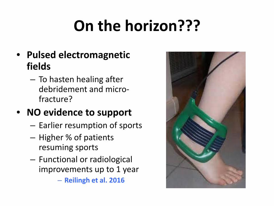
On the horizon???
• Pulsed electromagnetic fields– To hasten healing after
debridement and micro-fracture?
• NO evidence to support– Earlier resumption of sports– Higher % of patients
resuming sports– Functional or radiological
improvements up to 1 year– Reilingh et al. 2016

Osteochondral Lesions of the Talus
Case presentation

References• Baker CL et al. Arthroscopic treatment of transchondral talar dome
fractures. Arthroscopy 1986; 2(2): 82-87.• Berndt AL & Harty M. Transchondral fractures (osteochondritis
dissecans) of the talus. J Bone Joint Surg 1959; 41A:988-1020.• Bosien WR, Staples OS, Russell SW. Residual disability following acute
ankle sprains. J Bone Joint Surg 1955; 37A;1237-1243.• Buda R Treatment of osteochondritis dissecans of the talus in skeletally
immature population: a critical analysis of the available evidence. Foot and Ankle Specialist 2016; 9(3):265.
• Cahill et al. The results of conservative management of juvenile osteochondritis dissecans using joint scintigraphy. A prospective study. Am J Sports Med 1989; 17 (5):601-5.
• Corominas L et al. Retrograde percutaneous drilling for osteochondritisdissecans of the head of the talus: case report and review of the literature. J Foot Ankle Surg 2016; 55 (2):328-32.

References• De L’Escalopier et al. Outcomes of talar dome osteochondral defect
repair using osteocartilaginous autografts: 37 cases of Mosaicplasty®. Ortho Traumatol: Surg & Research 2015; 101:97-102.
• DiGiovanni et al. A review of the clinical experience with recombinant human platelet-derived growth factor-BB (rhPDGF-BB) in orthopaedicbone repair and regeneration. Current Ortho Practice 2013; 24(5):476-481.
• Dipaola, JD et al. Characterizing osteochondral lesions by magnetic resonance imaging. Arthroscopy. 1991;7:101–104.
• Emre et al. Open mosaicplasty in osteochondral lesions of the talus: a prospective study. Foot Ankle Surg 2012;51:556-60.
• Goh GSH et al. Outcomes are favorable after arthroscopic treatment of osteochondritis dissecans of the talus. J Foot Ankle Surg 2015; 54(1): 57-60.
• Hangody et al. Mosaicplasty for the treatment of articular defects of the knee and ankle. Clin Orthop Relat Re 2001; 391:5328-5336.

References• Heyse TJ et al. Juvenile osteochondritis dissecans of the talus: predictors
of conservative treatment failure. Acta Orthop Trauma Surg 2015; 135:1337-1341.
• Kessler et al. Osteochondritis dissecans of the ankle in children and adolescents: demographics and epidemiology. Am J Sports Med 2014; 42:2165-2171.
• Kruse et al. Arthoscopic De Novo NT® juvenile allograft cartilage implantation in the talus: a case presentation. J Foot Ankle Surg 2012; 51:218-221.
• Kumai T et al. Fixation of osteochondral lesions of the talus using cortical bone pegs. J Bone Joint Surg Br 2002;84:369–74.
• Mandelbaum BR et al. Autologous chondrocyte implantation of the talus. Arthroscopy 2003; 19(Suppl 1):129-37.
• Ogilvie-Harris DJ & Sarrosa EA. Arthroscopic treatment of osteochondritis dissecans of the talus. Arthroscopy 1999; 15:805-808.

References• Pagliazzi G et al. Tissue bioengineering in the treatment of
osteochondritis dissecans of the talus in children with open physis: preliminary results. J Pediatr Orthop 2016 Jul 2.
• Paletta GA et al. The prognostic value of quantitative bone scan in knee osteochondritis dissecans. A preliminary experience. Am J Sports Med 1998; 26(1):7-14.
• Reilingh ML et al. Effects of pulsed electromagnetic fields on return to sports after arthroscopic debridement and microfracture of osteochondral talar defects: a randomized, double-blind, placebo-controlled multicenter trial. Am J Sports Med 2016;44;1292-1300.
• Taluson PG et al. Osteochondritis dissecans of the talus: diagnosis and treatment in athletes. Clin Sports Med 2014; 33(2):267-84.
• Thiele S, Thiele R, Gerdesmeyer. Adult osteochondritis dissecans and focussed ESWT: a successful treatment option. Int J Surg 2015;24:191-194.
• Tol JL et al. Treatment strategies in osteochondral defects of the talardome: a systematic review. Foot Ankle Int 2000; 21: 119-126.

References• Valderrabano V et al. Snowboarder’s talus fracture: treatment outcome
of 20 cases after 3.5 years. Am J Sports Med 2005; 33(6): 871-880.• Verhagen et al. Systematic review of treatment strategies for
osteochondral defects of the talar dome. Foot Ankle Clin 2003;8:233-242.• von Knoch F et al. Fracture of the lateral process of the talus in
snowboarders. J Bone Joint Surg (Br) 2007; 89(6):772-777. • Woelfle JV et al. Clinical outcome and magnetic resonance imaging after
osteochondral autologous transplantation in osteochondritis dissecansof the talus. Foot Ankle Int 2013; 34 (2): 173-9.
• Younger A et al. A study to evaluate the safety of platelet-derived growth factor for treatment of osteochondral defects of the talus. Knee SurgSports Traumatol Arthrosc 2016; 24:1250-1258.
• Zengerink M et al. Treatment of osteochondral lesions of the talus: a systematic review. Knee Surg Sports Traumatol Arthosc 2010; 18:238-246.