osteopontin in obesity-associated adipose tissue ... in obesity-associated adipose tissue...
TRANSCRIPT

Doctoral Thesis at the Medical University of Vienna for obtaining the
Degree ‘Doctor of Philosophy - PhD’
Osteopontin in obesity-associated adipose tissue inflammation and insulin resistance and its interaction with monocyte chemoattractant
protein-1
Author
Florian Kiefer, MD
Supervisor
Ao. Univ. Prof. Thomas Stulnig, MD
Clinical Division of Endocrinology and Metabolism
Department of Medicine III
Medical University of Vienna
Waehringer Guertel 18-20
A-1090 Vienna, Austria
Vienna, January 2010
1

TABLE OF CONTENTS
1. ABSTRACT.............................................................................................................5 1.1 Abstract – English .............................................................................................5
1.2 Kurzfassung – Deutsch .....................................................................................6
2. INTRODUCTION .....................................................................................................7 2.1 Obesity and Epidemiology.................................................................................7
2.2 Obesity and Inflammation: Evolutionary Considerations ...................................7
2.3 Obesity and Adipose Tissue Inflammation ........................................................8
2.4 Endoplasmic Reticulum Stress – Initiator of Adipose Tissue Inflammation? .....9
2.5 Adipose Tissue Macrophages .........................................................................10
2.5.1 The Role of Macrophages in Adipose Tissue Inflammation .....................10 2.5.2 Macrophage Recruitment to Adipose Tissue............................................11
2.5.3 Phenotype and Activation of Adipose Tissue Macrophages.....................11
2.6 Adipokines.......................................................................................................13
2.6.1 Osteopontin (OPN)...................................................................................13
2.6.2. Monocyte Chemoattractant Protein-1 (MCP-1) .......................................14 2.7 Insulin Resistance ...........................................................................................15
3. AIM OF THE STUDY.............................................................................................17
4. RESEARCH DESIGN AND METHODS ................................................................18
4.1 Animals and diets ............................................................................................18
4.2 Experimental Setting .......................................................................................19
4.3 Generation of DoKo mice ................................................................................20
4.4 Genotyping ......................................................................................................20
4.5 Antibody treatment ..........................................................................................20
4.6 Metabolic measurements ................................................................................20
4.7 Indirect calorimetry ..........................................................................................21
4.8 Immunoflourescence, immunohistochemistry, tunel staining, and flow
cytometry ........................................................................................................21
4.9 Immunoblotting................................................................................................22
2
4.10 Reverse transcription and gene expression ..................................................23
4.11 Statistics ........................................................................................................23
5. RESULTS..............................................................................................................24 5.1 Studies in OPN-/- mice....................................................................................24
5.1.1 OPN deficiency in diet-induced obesity ....................................................24
5.1.2 Improved insulin sensitivity in OPN deficient mice ...................................25
5.1.3 Genetic OPN deficiency moderately reduces adipose tissue inflammation
but does not affect macrophage infiltration ...............................................27
5.2. OPN neutralization studies .............................................................................30
5.2.1 OPN neutralization in diet-induced obesity ..............................................30
5.2.2 Diet-induced insulin resistance is reversed by antibody-mediated OPN
neutralization ...........................................................................................31
5.2.3 OPN neutralization inhibits macrophage accumulation in obese adipose
tissue ........................................................................................................33
5.2.4 OPN neutralization promotes adipose macrophage apoptosis.................34
5.2.5 OPN neutralization attenuates obesity-induced adipose tissue
inflammation ............................................................................................36
5.3 Studies in DoKo mice ..........................................................................................38
5.3.1 OPN and MCP-1 double deficiency in diet-induced obesity .....................38
5.3.2 Obesity-induced insulin resistance is enhanced in DoKo mice ................39
5.3.3 OPN and MCP-1 double deficiency does not affect adipose tissue
inflammation .............................................................................................41
6. DISCUSSION ........................................................................................................44
7. REFERENCES ......................................................................................................49 8. LIST OF ABBREVIATONS ...................................................................................56 9. PUBLICATIONS BASED ON THE THESIS..........................................................58
10. ACKNOWLEDGEMENTS ...................................................................................59
3

11. FUNDING ............................................................................................................60 12. AUTHORS CURRICULIM VITAE........................................................................61
4

1. ABSTRACT
1.1 Abstract – English Obesity is associated with a state of chronic low-grade inflammation mediated
by immune cells that are primarily located to the adipose tissue. The chronic
inflammatory response appears to underlie obesity-induced metabolic deterioration
including insulin resistance and type 2 diabetes. Osteopontin (OPN) and monocyte
chemoattractant protein-1 (MCP-1) are inflammatory cytokines; the expression of
both is strongly upregulated in adipose tissue upon obesity.
Here I studied OPN effects on obesity-induced adipose tissue inflammation
and insulin resistance in genetic OPN deficiency and by antibody-mediated OPN
neutralization. Next I generated OPN and MCP-1 double knockout (DoKo) mice to
evaluate possible interaction of both cytokines with respect to obesity-associated
metabolic and inflammatory alterations.
After feeding high-fat diet to induce obesity, mice lacking OPN gene displayed
markedly improved insulin sensitivity compared with their wild-type littermates.
Genetic OPN deficiency only moderately reduced obesity-induced adipose tissue
inflammation and did not significantly affect macrophage accumulation. Targeting
OPN action by a neutralizing antibody for five days significantly improved insulin
sensitivity in diet-induced obese mice. Anti-OPN treatment attenuated adipose tissue
macrophage infiltration and inflammatory gene expression and significantly reduced
deleterious signal transduction related to insulin resistance. Notably, combined
deletion of OPN and MCP-1 in DoKo mice led to augmented diet-induced obesity and
insulin resistance compared to control mice.
These findings demonstrate that OPN is critically involved in obesity-
associated adipose tissue inflammation and insulin resistance. Unexpectedly, OPN
and MCP-1 double deficiency does not improve but even worsen the obesity-related
metabolic perturbation indicating antagonistic effects. Taken together, targeting OPN
action could provide a novel approach for the treatment of obesity-associated
metabolic disorders.
5

1.2 Kurfassung – Deutsch Adipositas geht mit einer chronischen subklinischen Entzündung einher, die
ihren Ursprung im Fettgewebe nimmt. Diese chronisch entzündlichen Prozesse
scheinen die Grundlage für Adipositas-assoziierte metabolische Veränderungen wie
Insulinresistenz und somit auch für Diabetes mellitus Typ 2 zu sein. Osteopontin
(OPN) und Monocyte Chemoattractant Protein-1 (MCP-1) sind inflammatorische
Zytokine, deren Expression im Fettgewebe durch Adipositas deutlich hochreguliert
wird.
In dieser Studie untersuchte ich Effekte von OPN auf die Adipositas-
assoziierte Fettgewebsentzündung und Insulinresistenz bei genetischer OPN
Defizienz und anhand von Antikörper-mediierter OPN Neutralisation. Weiteres
generierte ich OPN und MCP-1 Doppel Knockout (DoKo) Mäuse, um mögliche
Wechselwirkungen zwischen den beiden Entzündungsmediatoren hinsichtlich
Adipositas-induzierter metabolischer und inflammatorischer Veränderungen zu
studieren.
OPN Knockout Mäuse, in denen mit einer fettreichen Diät Adipositas induziert
wurde, zeigten gegenüber den Wildtyp-Mäusen eine deutlich verbesserte
Insulinsensitivität, wobei die Fettgewebsentzündung nur moderat und die
Makrophagenanzahl im Fettgewebe nicht signifikant vermindert waren. Die
Neutralisation der OPN Aktivität mittels eines Antikörpers verbesserte innerhalb von
fünf Tagen signifikant die Insulinsensitivität in adipösen Mäusen. Die Anti-OPN
Behandlung verringerte die Makrophageninfiltration sowie die inflammatorische
Genexpression und Signaltransduktion im Fettgewebe. Bemerkenswerterweise
führte die kombinierte Defizienz von OPN und MCP-1 in den DoKo Mäusen zu einer
Verstärkung der Diät-induzierten Adipositas und Insulinresistenz im Vergleich zu
Kontrollmäusen.
Diese Daten zeigen, dass OPN ein kritischer Mediator der Adipositas-
assoziierten Fettgewebsentzündung und Insulinresistenz ist. Wider Erwarten
verbessert das gemeinsame Fehlen von OPN und MCP-1 die Stoffwechsellage bei
Adipositas nicht, sondern führt sogar zu einer Aggravierung der Adipositas-
induzierten metabolischen Veränderungen. Zusammenfassend könnte die Blockade
von OPN Effekten eine neue therapeutische Strategie zur Behandlung der
Insulinresistenz und des Diabetes mellitus Typ 2 darstellen
6

2. INTRODUCTION
2.1 Obesity and Epidemiology Obesity is increasing at an alarming rate worldwide and is a fundamental risk
factor for type 2 diabetes, cardiovascular disease and the metabolic syndrome (1).
According to WHO estimations, globally more the one billion adults are overweight,
300 million of whom are obese.
Mammals have evolutionary evolved mechanisms to store energy in order to
guarantee survival in periods of drought and famine. However, the long-term storage
of excessive amounts of nutrients can have negative impact on health. Weight gain
and obesity are in favor to occur when food is energy dense because of high
proportions of simple carbohydrates and saturated fats, such as is common in
developed Western societies (2). In parallel with nutrient overload physical activity in
industrialized countries has been declining over decades due to passive leisure
activities, less physically demanding work and technological advances.
The metabolic syndrome and type 2 diabetes confer a high risk for
cardiovascular morbidity and mortality. Many studies have reported an association
between obesity and cardiovascular risk factors such as hypertension and
dyslipidemia. The Multi-Ethnic Study of Atherosclerosis (MESA) revealed that obesity
is also linked to subclinical cardiovascular disease independent of other classical
cardiovascular risk factors. E.g. coronary artery calcium deposition and carotid artery
intima-media-thickness are higher in obese populations than in lean controls
irrespective of the ethnic origin (3).
The fiscal importance of the obesity epidemic is enormous since most patients
need medical care throughout their lives. Hence, elucidating underlying
pathophysiologic mechanisms and the development of novel therapeutic approaches
targeting obesity and associated metabolic disorders are main issues for medical
research and pharmaceutical industry.
2.2 Obesity and Inflammation: Evolutionary Considerations Inflammation is described as the principal response of the body invoked to
deal with injuries, the hallmarks of which include swelling, redness, pain and fever. A
short-term adaptive inflammatory response is a crucial component of tissue repair
and involves integration of many complex signals in distinct cells and organs.
7

However, the long-term consequences of prolonged inflammation are almost never
beneficial. This certainly seems to be the case in metabolic diseases (4). But why are
metabolic disorders often linked to inflammation? The functional units that control key
metabolic and immune reactions in mammals have evolved from common ancestral
structures. One such structure is the Drosophila fat body, which contains the
mammalian homologues of the liver, the haematopoietic and immune system (5).
Interestingly, this site is also recognized as the equivalent of mammalian adipose
tissue (6). The Drosophila’s fat body functions in sensing energy and nutrient
availability, and coordinates the appropriate metabolic and survival responses (5).
Although in mammals distinct functional units and higher organs have formed they
carry with them a phylogenetic heritage. Hence, a scenario is plausible where
overlapping pathways regulate both metabolic and immune functions through
common key regulatory molecules (4).
2.3 Obesity and Adipose Tissue Inflammation The adipose tissue in mammals consists of 2 types: white and brown adipose
tissue. Both share many metabolic characteristics but, whereas white adipose tissue
mainly stores excess energy for subsequent needs, brown adipose tissue functions
as an energy-dissipating organ. The white adipose tissue consists of subcutaneous
and visceral depots. The visceral depot has been receiving most attention because it
is considered to be more metabolically active, and because its released factors can
be delivered to the portal venous system and, thus, can directly impact liver
metabolism (7).
Epidemiologic evidence for a relationship between obesity and inflammation
has existed for years, although these findings were not appreciated in terms of the
pathophysiologic conditions associated with obesity. Obesity has been known to be
associated with signs of chronic low-grade inflammation such as elevated serum
concentrations of C-reactive protein (CRP) and inflammatory cytokines (8; 9).
Though subclinical, this inflammatory state represents a crucial link between obesity
and insulin resistance (2) and predisposes to cardiovascular disease in obese
patients (10). The systemic inflammatory response primarily originates from white
adipose tissue (6) which produces a variety of inflammatory proteins such as
interleukin (IL)-1β, IL-6, tumor necrosis factor (TNF)-α, monocyte chemoattractant
protein-1 (MCP-1), and CRP (11; 12). Within obese adipose tissue, inflammatory
8
molecules are predominantly produced by nonfat cells such as macrophages (13;
14). Only very recently it has been discovered that genetic and diet-induced obesity
causes an infiltration of adipose tissue with macrophages that most probably
underlies the systemic inflammatory state (13; 15). Adipose tissue infiltrating
macrophages are bone marrow-derived and an important source of inflammatory
cytokines that interfere with adipocyte function. Since adipocyte function is crucial for
whole body insulin sensitivity, adipose tissue macrophages are considered as a
critical factor for the development of obesity-induced insulin resistance and type 2
diabetes (4; 16). The number of macrophages present in white adipose tissue
strongly correlates with obesity and adipocyte size in both human subjects and mice.
However, the molecular mechanisms underlying the obesity-induced adipose tissue
inflammation are still enigmatic. Several adipokines, i.e. cytokines secreted from cells
located in adipose tissue including macrophages, were proposed to be crucially
involved, but a causal mechanism of specific molecules mediating adipose tissue
infiltration with macrophages and subsequent deterioration of insulin sensitivity in
obesity has not yet been unequivocally defined.
2.4 Endoplasmic Reticulum Stress – Initiator of Adipose Tissue Inflammation? The primary cause for obesity-induced inflammation is not yet fully
understood. A potential source for the initiation of inflammation in obesity is
endoplasmic reticulum (ER) stress. Overnutrition and obesity cause ER stress in liver
and adipose tissue due to excess lipid accumulation and disturbed energy
metabolism (17). ER stress activates a stress response signaling network called the
unfolded protein response (UPR) that drives protective but also apoptotic and
inflammatory reactions. Transmembrane proteins including the protein
kinase/endoribonuclease IRE1 get activated upon ER stress. IRE1 initiates non-
spliceosomal splicing of the mRNA for the transcription factor X-box binding protein 1
(XBP-1) that controls protective responses to ER stress. IRE1 also induces an
inflammatory signaling cascade by activating I-kappa-B kinase (IKK), the mitogen-
activated protein kinase (MAPK) p38 and c-Jun N-terminale kinase (JNK), and finally
the major inflammatory transcription factor nuclear factor kappa B (NF-κB).
Consequently, obesity-induced ER stress leads to insulin receptor substrate-1 (IRS-
1) serine phosphorylation and thereby inhibits insulin signaling (18). Reports that
protective responses to ER stress are partially blunted in mice lacking one allele of
9

the XBP-1 gene support the proposed concept of ER stress regulation. Interestingly,
heterozygous XBP-1+/- mice develop severe insulin resistance even when on a
genetic background that is less susceptible to obesity (17). Vice versa, administration
of chemical chaperones that are known to reduce ER stress can restore insulin
sensitivity in obese mice (19). Furthermore, ER stress has been shown to
downregulate expression of the insulin sensitivity marker glucose transporter type 4
(GLUT4) in adipocytes (20). Taken together, ER stress directly affects insulin signal
transduction in insulin target cells, e.g., by inducing inflammatory signaling and
thereby contributes to insulin resistance. ER might be a site for the sensing of
metabolic stress and the translation of that stress into inflammatory signaling and
responses. In fact, the ER can be considered as an essential site of integration
between nutrient and pathogen responses since it is very sensitive to glucose and
energy availability, lipids and pathogens. Increased obesity leads to an environment
that further challenges ER function and capacity owing to architectural constraints
that limit ER expansion. Hence, obesity provides many conditions that could lead to
ER stress (6).
2.5 Adipose Tissue Macrophages 2.5.1 The Role of Macrophages in Adipose Tissue Inflammation
The adipose tissue is nowadays regarded as an endocrine organ that
produces large amounts of inflammatory cytokines and chemokines amongst others.
Mediators derived from the adipose tissue are called adipokines including Leptin,
TNF-α, IL-1β, IL-6, and MCP-1. Notably, the main source of inflammatory mediators
within murine and human adipose tissue are macrophages (14), although other cell
types, including adipocytes, preadipocytes, vascular endothelial cells, T-lymphocytes,
and the mesothelium may contribute. A major advance in the understanding of
obesity-induced inflammation was the finding that the augmented inflammatory
adipokine production in obesity is associated with an increased abundance of
macrophages in adipose tissue of obese mice (13). Numerous clinical studies have
confirmed a correlation between body mass index (BMI) and adipose tissue
macrophage numbers in humans. In particular, the visceral adipose tissue that is
metabolically most relevant is enriched with macrophages under obese conditions
(21). Adipose tissue macrophages probably interfere with adipocyte function by
secreting inflammatory cytokines. Since adipocyte function is strongly associated
10

with systemic insulin sensitivity the local action of macrophages and their secreted
molecules could enhance systemic insulin resistance (18). Hence, adipose tissue
macrophages are assumed to critically contribute to obesity-induced inflammation
and to the pathogenesis of type 2 diabetes.
2.5.2 Macrophage Recruitment to Adipose Tissue One of the most important questions in the understanding of obesity-induced
adipose tissue inflammation is what triggers macrophage recruitment into the
adipose tissue. Obesity promotes necrosis-like adipocyte cell death most probably
due to detrimental effects of adipocyte hypertrophy as it occurs during the
enlargement of the fat depots. Dead adipocytes are frequently found to be
surrounded by macrophages building so-called “crown-like structures”. Those
macrophages accumulating in immediate vicinity of dead adipocytes are supposed to
scavenge cell debris and free lipid droplets (22). Crown-like structures can be
observed in subcutaneous as well as in visceral adipose tissue of obese patients,
although less frequently as in rodent models of obesity (21; 22). However, the signal
by which adipocyte death leads to increased adipose tissue macrophage recruitment
is not known. The presence of necrotic-like adipocytes and crown-like structures in
non-obese hormone-sensitive lipase mutant mice (22) suggests that metabolic
alterations probably involving endoplasmic reticulum stress provoke chemokine
production by stressed adipocytes. Chemokines such as MCP-1 released by adipose
tissue resident cells are capable to navigate immune cells into the adipose tissue at
sites of disturbed homeostasis. However, the particular chemotatic signals by which
adipocyte death leads to increased adipose tissue macrophage recruitment warrant
further investigations and alternative mechanisms are still elusive.
2.5.3 Phenotype and Activation of Adipose Tissue Macrophages Similar to the Th1/Th2 concept of T-cell activation, a concept of M1/M2
polarization has recently been suggested for macrophages. Depending on the stimuli
such as cytokines and microbial products, macrophages polarize into specialized cell
types and exert unique functional properties (23). “Classical” macrophage activation
occurs upon stimulation with interferon-gamma (IFN-γ) alone or in combination with
lipopolysaccharide (LPS). Classically activated macrophages produce inflammatory
cytokines (e.g., IL-1, IL-6, TNF-α), reactive oxygen species such as nitric oxide (NO)
11

upon inducible nitric oxide synthase (iNOS) activity, and are capable of inducing Th1-
polarized T-cell responses. These “classical” macrophages are referred to as M1
type in contrast to “alternatively activated” M2 macrophages. M2 have been
described to be primarily induced by IL-4 and IL-13 (23). M2 macrophages appear to
play a role in tissue repair and parasite defense but exert poor anti-bacterial killing
activity. M2 are of a predominantly anti-inflammatory phenotype as they
counterbalance inflammatory properties of M1 macrophages by release of IL-10, IL-1
receptor antagonist (IL-1Ra) and transforming growth factor beta (TGF-β) (24-26).
Characterization of human adipose tissue macrophages revealed that their
phenotype predominantly resembles the alternatively activated M2 type since they
express typical M2 surface markers like the mannose receptor CD206, scavenger
receptors, and distinct integrins (27). Also murine adipose tissue macrophages
express M2-associated genes such as Ym1, arginase and Il10 (28). The factors
driving alternative macrophage differentiation in adipose tissue are still elusive. The
marked expression of scavenger receptors and the mannose receptor, together with
high endocytic activity, implicates a role of adipose tissue macrophages in uptake of
lipids and lipoproteins, apoptotic cells, and glycoproteins and hence supports the
concept of obesity-induced recruitment of macrophages into adipose tissue due to an
increased abundance of necrotic-like adipocytes that need to be removed (18). In
accordance with their M2 phenotype adipose tissue macrophages produce anti-
inflammatory cytokines such as IL-10 and the IL-1Ra. On the other hand, adipose
tissue macrophages are capable of secreting large amounts of pro-inflammatory
cytokines such as TNF-α, IL-6, and IL-1β underlining the role of macrophages in
obesity-induced adipocyte dysfunction and metabolic disorders (21). Accordingly,
high-fat diet feeding in mice causes a shift in cytokine expression of adipose tissue
macrophages from M2- to M1-like patterns. Gene Expression of IL-10, Ym1, and
arginase appeared to be downregualted in adipose tissue macrophages of obese
mice while TNF-α and Nos2 (iNOS) were upregulated (28). In contrast, weight
reduction induces an increase of M2-like macrophages in adipose tissue of obese
patients (29). Adipose tissue macrophages may be activated by factors released by
adipocytes. However, the particular mechanisms how high-fat diet and/or weight gain
influence the M1/M2 polarization and cytokine expression in adipose tissue
macrophages is still unclear. Hence, identification of factors that attract macrophages
12

to the adipose tissue and determine their pro- or anti-inflammatory phenotype is one
of the most intriguing current issues of obesity research.
2.6 Adipokines
Adipose tissue is no longer considered to be an inert tissue serving solely as
an energy store, but is emerging as an important organ in the regulation of many
pathological processes. The adipose tissue is releasing various soluble factors
termed as adipokines or adipocytokines. Some adipokines such as adiponectin or
lepitn are exclusively produced by the adipose tissue others like TNF-α or IL-6 can be
secreted by diverse cell types in different organs. Adipokines were found to influence
lipid and glucose metabolism, not only in adipose tissue, but also in liver and skeletal
muscle. They are involved in appetite regulation and in local as well as systemic
inflammatory processes. Hence, the adipose tissue is now recognized as an
endocrine organ with autocrine regulation mediated by adipokines.
2.6.1 Osteopontin (OPN) Osteopontin (gene Spp1), also named secreted phosphoprotein-1 and
sialoprotein-1, is a multifunctional protein expressed in activated macrophages and T
cells, osteoclasts, hepatocytes, smooth muscle, endothelial, and epithelial cells (30;
31). OPN was originally classified as a T helper type 1 (Th1) cytokine that is involved
in physiological and pathological mineralization in bone and kidney, cell survival,
inflammation, and tumor biology (30; 32). OPN induces the expression of a variety of
proinflammatory cytokines and chemokines in peripheral blood mononuclear cells
(33). Moreover, it functions in cell migration, particularly of monocytes/macrophages
(31), and stimulates expression of matrix metalloproteases to induce matrix
degradation and facilitate cell motility (34). Notably, OPN plays a role in various
inflammatory disorders, such as rheumatoid arthritis (35) and atherosclerosis (36), in
diabetic macro- and microvascular diseases (37) and hepatic inflammation (38).
Hepatic OPN expression is upregulated in obesity (39) and in various models of liver
injury where OPN is localized to macrophages and Kupffer cells (40; 41).
Furthermore, OPN is involved in the pathogenesis of non-alcoholic fatty liver disease
(NAFLD), which is strongly associated with visceral obesity (39; 42; 43).
As reported recently, OPN gene expression is extensively upregulated upon
obesity in human and murine adipose tissue (44-46). While OPN plasma
13

concentrations are elevated in morbidly obese patients, data are inconsistent in
different murine models of obesity (44-47).
2.6.2. Monocyte Chemoattractant Protein-1 (MCP-1)
MCP-1 (gene: Ccl2) is a chemokine and a member of the small inducible
cytokine family, which plays a role in the recruitment of monocytes and T
lymphocytes to sites of injury and infection (48). It is expressed by a number of cell
types including skeletal muscle, smooth muscle and endothelial cells, macrophage
but also adipocytes (49; 50). Via binding to its main receptor chemokine CC motif
receptor 2 (CCR2) MCP-1 directs monocytes and macrophage precursors to sites of
inflammatory lesions such as atherosclerotic plaques (51; 52). MCP-1 has been
shown to trigger firm adhesion of monocytes to vascular endothelium under flow
conditions (53). Indeed, MCP-1 expression is crucially implicated in monocyte
extravasation through vascular endothelium (54). Hence, it is strongly involved in the
pathogenesis of atherosclerosis by promoting directed migration of immune cells.
Augmented MCP-1 concentrations are found in synovial fluids of patients
suffering from rheumatoid arthritis as well as in the bronchial epithelium of idiopathic
pulmonary fibrosis and in tuberculosis effusions. MCP-1 is also significantly elevated
during inflammatory skin diseases, in acute hepatitis or fulminant hepatic failure, and
during experimental autoimmune encephalomyelitis (55). Accumulating evidence
links MCP-1 to the pathogenesis of obesity-induced inflammation.
Gene expression of MCP-1 as well as of the chemokine receptor CCR2 is
increased in the adipose tissue of obese humans and mice. Notably, MCP-1
expression has been found to be higher in visceral adipose tissue than in
subcutaneous tissue and is closely related to the number of residing macrophages.
Plasma concentrations of MCP-1 are significantly elevated in obese and diabetic
patients suggesting a pathophysiologic implication in systemic insulin resistance (56;
57). In vitro data showing that treatment with MCP-1 impairs glucose uptake in 3T3-
L1 adipocytes support this concept (58). However, recent studies in diet-induced
obese MCP-1 deficient mice emerged conflicting results in terms of adipose tissue
macrophage accumulation and insulin resistance (56; 59). Taken together, some
data point towards a pathophysiologic role of MCP-1 in obesity-associated
inflammatory alterations including macrophage recruitment while other reports do not
14
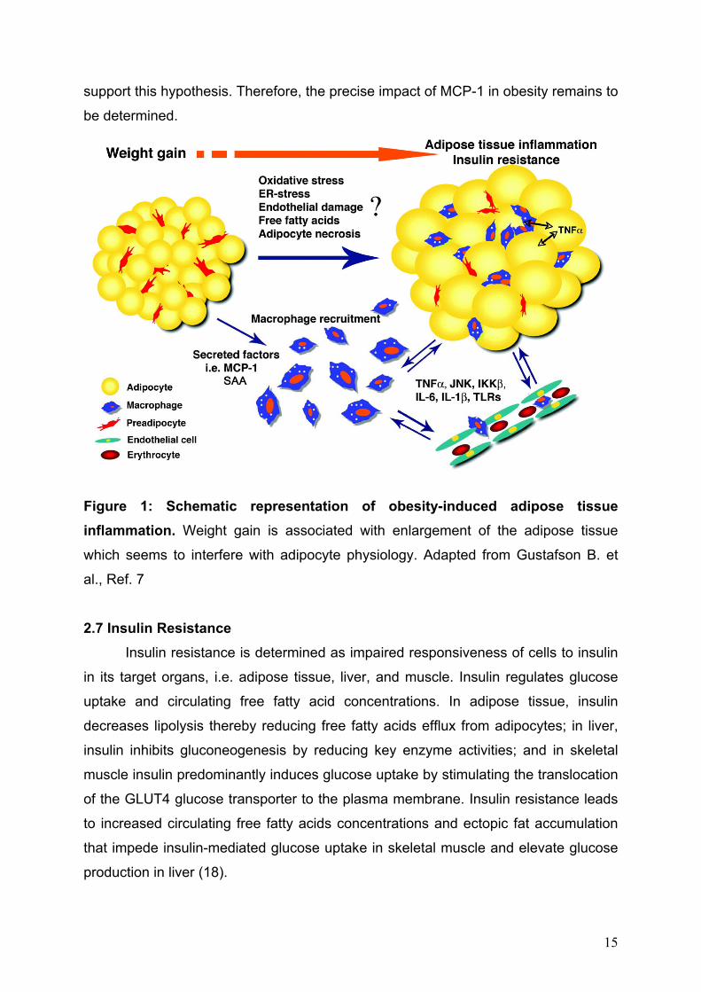
support this hypothesis. Therefore, the precise impact of MCP-1 in obesity remains to
be determined.
Figure 1: Schematic representation of obesity-induced adipose tissue inflammation. Weight gain is associated with enlargement of the adipose tissue
which seems to interfere with adipocyte physiology. Adapted from Gustafson B. et
al., Ref. 7 2.7 Insulin Resistance
Insulin resistance is determined as impaired responsiveness of cells to insulin
in its target organs, i.e. adipose tissue, liver, and muscle. Insulin regulates glucose
uptake and circulating free fatty acid concentrations. In adipose tissue, insulin
decreases lipolysis thereby reducing free fatty acids efflux from adipocytes; in liver,
insulin inhibits gluconeogenesis by reducing key enzyme activities; and in skeletal
muscle insulin predominantly induces glucose uptake by stimulating the translocation
of the GLUT4 glucose transporter to the plasma membrane. Insulin resistance leads
to increased circulating free fatty acids concentrations and ectopic fat accumulation
that impede insulin-mediated glucose uptake in skeletal muscle and elevate glucose
production in liver (18).
15

By binding to its receptor insulin induces complex signaling cascades. In brief,
insulin receptor-mediated tyrosine phsophorylation of insulin receptor substrates
(IRS) leads to activation of two major pathways. The phosphatidylinositol 3-kinase
(PI3K)-AKT pathway is largely responsible for insulin action on glucose uptake and
suppression of gluconeogenesis. It results in GLUT4 translocation from its
intracellular pool to the plasma membrane and glucose transport into the cell. The
second pathway is the MAPK pathway that regulates gene expression and
additionally interacts with the PI3K-AKT pathway to control cell growth and
differentiation (60).
In the case of insulin resistance insulin signaling is negatively regulated as it
occurs during phosphorylation of certain serine residues on IRS. This is mainly
triggered by free fatty acid and inflammatory cytokine action. Serine kinases that
phosphorylate IRS and thus hinder proper insulin signal transduction are IKK, JNK
and other MAP kinases. The named serine kinases of IRS are also typical mediators
of inflammatory signaling pathways, thus, providing an inhibitory crosstalk between
inflammatory and insulin signaling. Other important molecular mediators that link
inflammatory pathways to inhibition of insulin signaling are suppressor of cytokine
signaling (SOCS) 1 and 3 as well as nitric oxide (NO). SOCS proteins are
upregulated during inflammation, e.g. by interleukin-6, and induce ubiquitinylation
and degradation of IRS proteins and thereby impede insulin signal transduction (18).
As mentioned above accumulating evidence has emerged that obesity is
strongly associated with inflammation and is thereby involved in the development of
insulin resistance. The chronic low-grade inflammation occurring in obese patients is
determined by increased plasma levels of C-reactive protein, inflammatory cytokines
such as TNF-α, IL-6, IL-1β, IL-8, as well as the multifunctional protein leptin and free
fatty acids. Almost all of those inflammatory markers were proven to alter insulin
signal transduction in vitro and in vivo (2).
Taken together, insulin resistance in obesity is a reflection of long-term
nutrient excess and is manifested through complex, heterogeneous mechanisms that
can involve increased fatty acid flux, nutrient overload, ER stress, secretion of
adipocyte-derived cytokines, and chronic tissue inflammation.
16

3. AIM OF THE STUDY Obesity is associated with a state of chronic low-grade inflammation mediated
by accumulating macrophages in the adipose tissue. The chronic inflammatory
activity in adipose tissue appears to underlie obesity-induced metabolic deterioration
including insulin resistance and type 2 diabetes. OPN and MCP-1 are inflammatory
cytokines; the expression of both is strongly upregulated in the adipose tissue upon
obesity. Determining the role of OPN and its interaction with MCP-1 in adipose tissue
inflammation could contribute to the development of innovative strategies for
treatment of obesity-induced complications including metabolic disorders. It is the
aim of this thesis to elucidate the role of OPN in obesity-associated adipose tissue
inflammation and insulin resistance and its interference with MCP-1.
17

4. RESEARCH DESIGN AND METHODS
4.1 Animals and diets C57BL/6J wild type (WT), B6.Cg-Spp1tm1Blh/J (OPN knockout; Spp1-/- here
referred to as OPN-/-) and B6.129S4-Ccl2tm1Rol/J (MCP-1 knockout, Ccl2-/- here
referred to as MCP-1-/-) mice were purchased from Charles River Laboratories
(Sulzfeld, Germany). At 7 weeks of age male littermates were placed for 24 weeks on
high-fat diet (HF, 60kcal% fat, D12492, Research Diets Inc., New Brunswick, NJ,
USA) and low-fat diet (10kcal% fat, D12450B, Research Diets Inc.) or normal chow
(both referred to as LF) to induce obesity and to serve as lean controls, respectively.
All mice were housed in specific pathogen-free facility that maintained 12-hour
light/dark cycle. Mice had free access to food and water and food intake was
monitored. Blood was drawn after 3 hr fasting immediately before mice were
sacrificed. Gonadal white adipose tissue (GWAT) pads were collected. The protocol
was approved by the local ethics committee for animal studies and the Federal
Ministry for Science and Research and followed the guidelines on accommodation
and care of animals formulated by the European Convention for the Protection of
Vertebrate Animals Used for Experimental and Other Scientific Purposes.
4.2 Experimental Setting We studied the role of OPN in obesity-induced adipose tissue inflammation and
insulin resistance in OPN knockout (OPN-/-) mice and in wild-type (WT) mice that
were treated with an OPN neutralizing antibody. Possible interference between OPN
and MCP-1 action in obesity-associated adipose tissue inflammation and insulin
resistance was investigated in OPN and MCP-1 double knockout (DoKo) mice.
Studies in OPN-/- mice:
genotype diet sample size
function
WT LF 10 Lean control lacking adipose tissue inflammation
WT HF 10 Control for obesity-induced adipose tissue
inflammation and insulin resistance
18
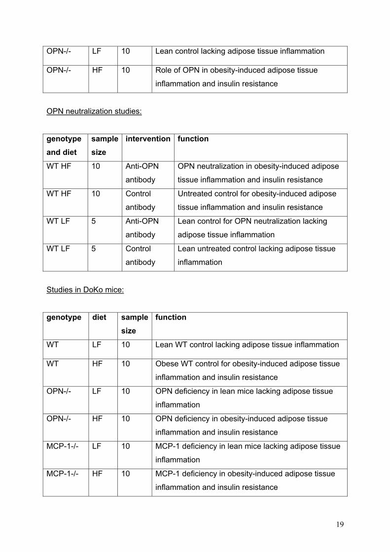
OPN-/- LF 10 Lean control lacking adipose tissue inflammation
OPN-/- HF 10 Role of OPN in obesity-induced adipose tissue
inflammation and insulin resistance
OPN neutralization studies:
genotype and diet
sample size
intervention function
WT HF 10 Anti-OPN
antibody
OPN neutralization in obesity-induced adipose
tissue inflammation and insulin resistance
WT HF 10 Control
antibody
Untreated control for obesity-induced adipose
tissue inflammation and insulin resistance
WT LF 5 Anti-OPN
antibody
Lean control for OPN neutralization lacking
adipose tissue inflammation
WT LF 5 Control
antibody
Lean untreated control lacking adipose tissue
inflammation
Studies in DoKo mice:
genotype diet sample size
function
WT LF 10 Lean WT control lacking adipose tissue inflammation
WT HF 10 Obese WT control for obesity-induced adipose tissue
inflammation and insulin resistance
OPN-/- LF 10 OPN deficiency in lean mice lacking adipose tissue
inflammation
OPN-/- HF 10 OPN deficiency in obesity-induced adipose tissue
inflammation and insulin resistance
MCP-1-/- LF 10 MCP-1 deficiency in lean mice lacking adipose tissue
inflammation
MCP-1-/- HF 10 MCP-1 deficiency in obesity-induced adipose tissue
inflammation and insulin resistance
19

DOKO LF 10 Combined OPN and MCP-1 deficiency in lean mice
lacking adipose tissue inflammation
DOKO HF 10 Interference of OPN and MCP-1 in obesity-induced
adipose tissue inflammation and insulin resistance
4.3 Generation of DoKo mice To generate DoKo mice, OPN-/- and MCP-1-/- single knockouts were
intercrossed to receive double heterozygous offspring in the F1 generation. Double
heterozygous littermates from F1 were crossed to generate DoKo mice in the next
generation (F2). Animals obtained from F2 were genotyped by genomic PCR to
identify DoKo mice.
4.4 Genotyping At the age of two weeks tail tips (approx. 3mm) of mice obtained in the F2
generation were cut and immediately frozen on dry ice. Following proteinase K
digestion total DNA was purified using commercially available DNeasy Blood &
Tissue Kit® (Qiagen, Hilden, Germany). DNA concentrations were measured by
NanoDrop® spectrophotometer (Peqlab Biotechnologie GmBH, Erlangen, Germany).
Genomic PCR was performed using equal amounts of DNA, Taq polymerase
(Roche, Basel, Switzerland) and the following set of primers (provided by Charles
River). For OPN gene: wild-type forward 3’-CCATACAGGAAAGAGAGACC-5’;
mutant forward 3’-AACTGTTTTGCTTGCATGCG-5’; common reverse 3’-
CGTCCTGTAAGTCTGCAGAA-5’. For MCP-1 gene: wild-type forward 3’-
ACAGCTTCTTTGGGACACC-5’; mutant forward 3’-
CCTTCTATCGCCTTCTTGACG-5’; common reverse 3’-
GGAGCATCCACGTGTTGGC-5’. Size of PCR products was assessed by agarose
gel electrophoresis and ethidium bromide staining.
4.5 Antibody treatment Mice were treated with a neutralizing anti-mouse OPN IgG (50 µg/mouse in
phosphate buffered saline) or preimmune goat IgG for three times during five days by
tail-vein injection. Osteopontin specific IgG (R&D Systems, Minneapolis, MN, USA)
was produced in goats by immunizing with NSO-derived, recombinant mouse
osteopontin. Mice were killed two days after last antibody application.
20

4.6 Metabolic measurements
Plasma glucose, cholesterol, triglyceride, and free fatty acid concentrations
were measured in EDTA plasma by an automated analyzer (Falcor 350, A.Menarini
Diagnostics, Florence, Italy). We used commercially available ELISA kits to
determine plasma insulin (Mercodia AB, Uppsala, Sweden), IL-6, TNF-α, leptin,
adiponectin, osteopontin (all R&D Systems), serum amyloid P (SAP) and high
sensitivity C-reactive protein concentrations (hsCRP; both Alpco Diagnostics,
Windham, NH, USA). We calculated homeostasis model assessment of insulin
resistance (HOMA-IR) as an index for insulin resistance (61). Insulin sensitivity was
assessed by insulin tolerance test (ITT) after a three hour fasting period. Blood
glucose concentrations were measured before and 30, 60, 90 and 120 minutes after
an intraperitoneal injection of recombinant human insulin (Actrapid®, Novo Nordisk
A/S, Bagsværd, Denmark; 0.75 U/kg body weight for HF and 0.25 U/kg for LF,
respectively).
4.7 Indirect calorimetry Indirect calorimetry was performed for 72h using an open-circuit, indirect calorimetry
system including spontaneous activity by beam breaking (Oxylet, Panlab-Bioseb,
Chaville, France). WT, OPN-/-, MCP-1-/- and DoKo mice on HF were analyzed for
oxygen consumption (VO2), carbon dioxide production (VCO2), energy expenditure
[calculated according to the following formula: 1.44 x VO2 x (3.815 x 1.232 x
respiratory quotient (RQ)] and spontaneous activity. Activities of the mice were
monitored by an infrared photocell beam interruption method. Mice were allowed to
adapt for 24hrs to the new environment before indirect calorimetric measurements
were performed. Food and water intake were continuously monitored. Data were
analyzed by Metabolism 2.0 software (Panleb-Bioseb).
4.8 Immunoflourescence, immunohistochemistry, tunel staining, and flow cytometry
Frozen sections were prepared from murine GWAT. Sections were stained
with rat anti-mouse F4/80 and Mac-2 IgG antibodies (Serotec, Oxford, UK and
Cedarlane, Burlington, Ontario, Canada, respectively). Primary antibodies were
detected with AlexaFluor 488 or AlexaFluor 594 goat anti-rat IgG antibodies
21

(Molecular Probes, Eugene, OR, USA). Nuclei were visualized by DAPI staining.
Slides were mounted in Vectashield® (Vector Laboratories Inc., Burlingame, CA,
USA) and examined under a fluorescence microscope (Leica, Wetzlar, Germany).
Macrophage infiltration in adipose tissue was quantified by calculating the ratio of
F4/80 and Mac-2 positive cells to total nuclei as described previously (62). Apoptotic
cells were stained on frozen sections using the Fluorescin In Situ Cell Dection Kit
from Roche, according to manufacturer’s instruction, in parallel with double-staining
for F4/80 and Mac-2, respectively, as described above.
For paraffin sections, GWAT were fixed with neutral buffered 4%
paraformaldehyde and were paraffin-embedded. After dewaxation and rehydration
immunohistochemical staining for Mac-2 (Serotec) was performed on adipose tissue
sections using the ABC kit (Vector Laboratories) according to the manufacturer´s
recommendations. As a negative control, staining was performed on selected
sections with isotype control. Samples were analyzed with standard light microscopy.
Stromal vascular cells (SVC) of GWAT were isolated by collagenase digestion and
centrifugation to remove adipocytes as described (44). Briefly, murine GWAT was cut
into small pieces washed in PBS, and 0.5 g tissue/ml was digested with 0.03 mg/ml
Liberase Blendzyme 3 (Roche) and 50 U/ml DNase I (Sigma, St. Louis, MO, USA) in
RPMI-1640 (Invitrogen) for 60 minutes at 37°C. Digested tissues were passed
through 200 μm mesh filters. After centrifugation at 1000 x g for 10 min at 4°C
floating cells were removed. The pellets comprised the SVC fraction. Red blood cells
were lysed in haemolysis buffer and remaining cells passed through a 70 µm mesh
filter. SVC were subjected to flow-cytometry using directly fluorochrome-labeled
antibodies against F4/80 (Serotec), and CD11c (BD Biosciences, San Jose, CA,
USA).
4.9 Immunoblotting
Phosphorylation of c-Jun NH2-terminal kinase (JNK) was determined
essentially as described (62). Briefly, GWAT was homogenized and lysed on ice for
30min in tris-buffered saline, pH 7.4, containing 1% Triton X-100 (Pierce) and
phosphatase and protease inhibitors. The tissue extract was cleared from fat, nuclei
and debris by centrifugation. Identical amounts of protein were separated by SDS-
PAGE and blotted onto nitrocellulose membranes (Hybond ECL, Little Chalfont,
Amersham, UK). Phosphorylated JNK (Thr-183, Tyr-185) and total JNK were
22

analyzed using respective mouse polyclonal antibodies (Cell Signaling) followed by a
horseradish peroxidase-labelled secondary antibody (Accurate, Westbury, NY, USA).
Chemiluminescence was generated by BM chemiluminescence substrate (Roche)
and quantified on a Lumi-Imager (Roche).
4.10 Reverse transcription and gene expression Parts of GWAT were immediately snap-frozen in liquid nitrogen for RNA
isolation. Adipose tissue was homogenized in TRIzol reagent (Invitrogen, Carlsbad,
CA, USA) and RNA was isolated according to manufacturer’s protocol. One
microgram of total RNA was treated with DNase I and reverse transcribed into cDNA
using Superscript II and random hexamer primers (all Invitrogen). Gene expression
normalized to 18S rRNA and Ubiquitin C, respectively, was analyzed by quantitative
real-time RT-PCR on an ABI Prism 7000 cylcer using commercial Assays-on-
Demand kits (all Applied Biosystems, Foster City, CA). Alternatively, the expression
of following murine genes were quantified by use of self-designed primer pairs and
iTaq SYBR Green Supermix (Bio-Rad Laboratories, Hercules, CA): Tnf
(5’-CCAGACCCTCACACTCAGATCA-3’) forward,
(5’-TGGTATGAGATAGCAAATCGGCT-3’) reverse; Ccl2 (5’-
AGGTCCCTGTCATGCTTCTGG-3’) forward, (5’-CTGCTGCTGGTGATCCTCTTG-3’)
reverse; Il10 (5’-CTGCTCTTACTGACTGGCATGAG-3’) forward, (5’-
CGCAGCTCTAGGAGCATGTG-3’) reverse; AdipoQ (5’-
GTCATGCCGAAGATGACGTTACT-3’) forward, (5’-
TCACCCTTAGGACCAAGAAGAC -3’) reverse; Il6 (5’-
CTGCAAGAGACTTCCATCCAGTT-3’) forward, (5’-GAAGTAGGGAAGGCCGTGG-
3’) reverse.
4.11 Statistics All data are given as means ± SE. Comparisons were assessed by unpaired
2-tail Student’s t-test. For comparisons between DoKo mice and respective controls
univariate ANOVA was calculated followed by Post Hoc Dunnett’s test. A P-value of
0.05 or less was considered statistically significant.
23
5. RESULTS 5.1 Studies in OPN-/- mice 5.1.1 OPN deficiency in diet-induced obesity
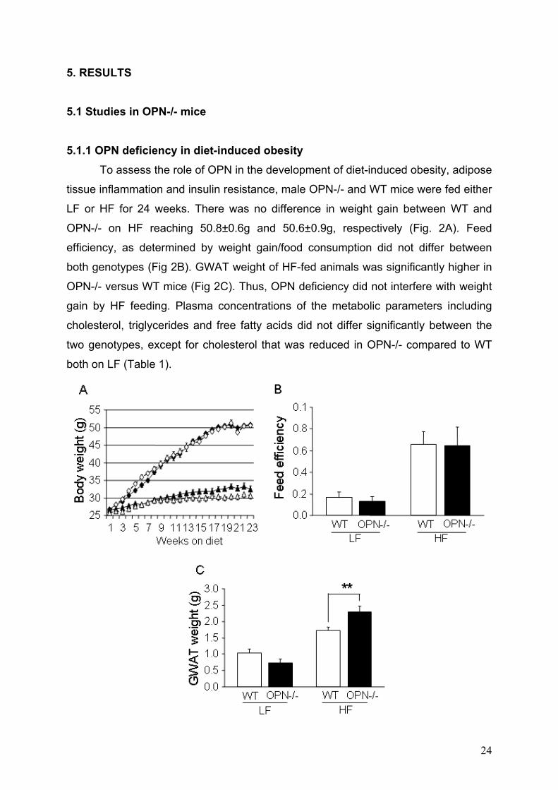
To assess the role of OPN in the development of diet-induced obesity, adipose
tissue inflammation and insulin resistance, male OPN-/- and WT mice were fed either
LF or HF for 24 weeks. There was no difference in weight gain between WT and
OPN-/- on HF reaching 50.8±0.6g and 50.6±0.9g, respectively (Fig. 2A). Feed
efficiency, as determined by weight gain/food consumption did not differ between
both genotypes (Fig 2B). GWAT weight of HF-fed animals was significantly higher in
OPN-/- versus WT mice (Fig 2C). Thus, OPN deficiency did not interfere with weight
gain by HF feeding. Plasma concentrations of the metabolic parameters including
cholesterol, triglycerides and free fatty acids did not differ significantly between the
two genotypes, except for cholesterol that was reduced in OPN-/- compared to WT
both on LF (Table 1).
24

Figure 2: Body weight, feed efficiency and GWAT weight of WT and OPN-/- mice. WT and OPN-/- mice (n = 10 per group) were fed a LF or HF for 24 weeks. (A) Weight gain was monitored in WT (black symbols) and OPN-/- (white symbols) mice
on LF (triangles) or HF (squares) and did not differ between the two genotypes when
on the same diet. (B) Feed efficiency (grams weight gain/grams food consumption) of
WT and OPN-/- mice. (C) GWAT weight of WT and OPN-/- mice. Data are expressed
as mean ± SEM. **P < 0.01.
Plasma measurements
Parameter
WT LF OPN-/- LF WT HF OPN-/- HF
Cholesterol (mg/dl) 104.9 ± 3.8 70.4 ± 5.3*** 142.2 ± 7.6 133.7 ± 8.0
Triglycerides (mg/dl) 39.5 ± 3.9 42.7 ± 3.2 42.6 ± 3.0 39.0 ± 1.6
Free fatty acids (µmol/l) 694.0 ± 20.7 626.9 ± 79.4 647.8 ± 34.6 688.8 ± 50.4
IL-6 (pg/ml) n.d. n.d. 5.8 ± 1.1 8.2 ± 1.3
MCP-1 (pg/ml) n.d. n.d. 93.2 ± 6.4 84.6 ± 5.8
Table 1: WT and OPN-/- were fed a LF or HF for 24 weeks. Blood samples were
obtained after a three hour fasting period and analyzed for indicated plasma
parameters (n = 10 per group). Data are expressed as mean ± SEM. ***P < 0.001.
n.d. = not determined.
5.1.2 Improved insulin sensitivity in OPN deficient mice
To determine whether OPN deficiency affects insulin resistance in obesity,
parameters of glucose metabolism were analyzed and insulin tolerance test (ITT)
was performed in OPN-/- and WT mice with HF-induced obesity. Whereas fasting
plasma glucose concentrations were comparable in OPN-/- and WT mice on HF,
OPN-/- mice on LF showed reduced plasma glucose compared to WT animals (Fig.
3A). Fasting plasma insulin concentrations were significantly decreased in HF-fed
OPN-/- versus WT mice, while plasma insulin was not different between the two
genotypes on LF (Fig. 3B). Accordingly, HOMA-IR was significantly lower in OPN-/-
25

compared to WT mice (Fig. 3C) indicating enhanced insulin sensitivity. This
observation was further confirmed by ITT that revealed significant improvement in
obese OPN-/- compared to WT mice on HF (Fig. 3D). Taken together, these data
show that genetic OPN deficiency ameliorates insulin sensitivity in obese mice.
Figure 3: Insulin sensitivity in lean and obese WT and OPN-/- mice. WT and
OPN-/- mice (n = 10 per group) were fed LF or HF for 24 weeks prior to metabolic
characterisation. (A) Fasting plasma glucose concentrations; (B) fasting plasma
insulin concentrations; (C) HOMA-IR. (D) An insulin tolerance test was performed in
WT (solid lines) and OPN-/- (dashed lines) after feeding a LF (triangles) or HF
(squares). Blood glucose was measured following an intraperitoneal injection of
insulin (0.75 U/kg body weight for HF and 0.3 U/kg body weight for LF. *P < 0.05, **P
< 0.01
26
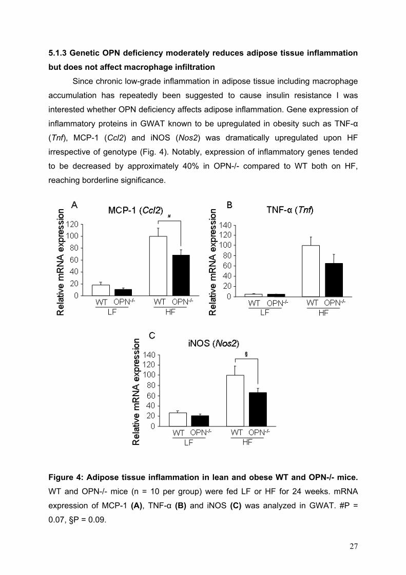
5.1.3 Genetic OPN deficiency moderately reduces adipose tissue inflammation but does not affect macrophage infiltration
Since chronic low-grade inflammation in adipose tissue including macrophage
accumulation has repeatedly been suggested to cause insulin resistance I was
interested whether OPN deficiency affects adipose inflammation. Gene expression of
inflammatory proteins in GWAT known to be upregulated in obesity such as TNF-α
(Tnf), MCP-1 (Ccl2) and iNOS (Nos2) was dramatically upregulated upon HF
irrespective of genotype (Fig. 4). Notably, expression of inflammatory genes tended
to be decreased by approximately 40% in OPN-/- compared to WT both on HF,
reaching borderline significance.
Figure 4: Adipose tissue inflammation in lean and obese WT and OPN-/- mice. WT and OPN-/- mice (n = 10 per group) were fed LF or HF for 24 weeks. mRNA
expression of MCP-1 (A), TNF-α (B) and iNOS (C) was analyzed in GWAT. #P =
0.07, §P = 0.09.
27

Obesity-induced macrophage infiltration in GWAT was analyzed by gene
expression, immunoflourescence and flow cytometry. Gene expression of the
macrophage marker F4/80 (Emr-1) was considerably higher in HF compared to LF in
both genotypes, but differences between WT and OPN-/- mice on HF were not
observed (Fig 5A). Accordingly, macrophage accumulation as assessed by F4/80
staining of adipose sections was unaltered between both genotypes on the
respective diet (Fig. 5B,C). In addition, FACS analysis of GWAT stromal vascular
cells (SVC) revealed similar counts for the suggested high-fat diet-activated type of
macrophages (F4/80+CD11c+) (28) irrespective of OPN deficiency (Fig. 5D). Hence,
adipose tissue infiltration by macrophages was not affected by OPN deficiency.
Figure 5: Adipose macrophage accumulation in lean and obese WT and OPN-/- mice. WT and OPN-/- mice (n = 10 per group) were fed LF or HF for 24 weeks. (A) mRNA expression of the macrophage marker F4/80 was analyzed in GWAT. (B-C)
28
Adipose tissue macrophage accumulation was determined by immunoflourescence
analysis of GWAT isolated from WT and OPN-/- mice. (B) Representative pictures
are given. (C) Macrophages were counted as F4/80+ cells relative to total number of
cells. (D) Flow cytometric quantitation of SVC isolated from GWAT of WT and OPN-/-
mice. SVC of WT and OPN-/- were stained for F4/80 and the macrophage activation
maker CD11c and analyzed by flow cytometry.
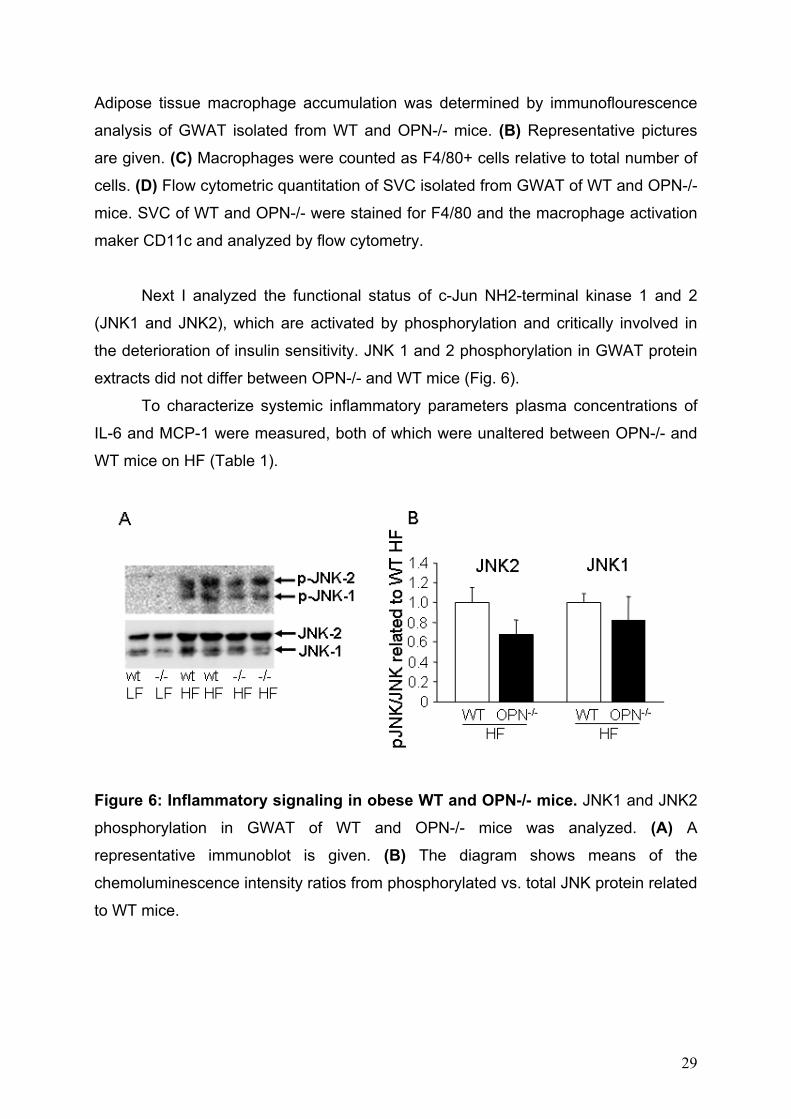
Next I analyzed the functional status of c-Jun NH2-terminal kinase 1 and 2
(JNK1 and JNK2), which are activated by phosphorylation and critically involved in
the deterioration of insulin sensitivity. JNK 1 and 2 phosphorylation in GWAT protein
extracts did not differ between OPN-/- and WT mice (Fig. 6).
To characterize systemic inflammatory parameters plasma concentrations of
IL-6 and MCP-1 were measured, both of which were unaltered between OPN-/- and
WT mice on HF (Table 1).
Figure 6: Inflammatory signaling in obese WT and OPN-/- mice. JNK1 and JNK2
phosphorylation in GWAT of WT and OPN-/- mice was analyzed. (A) A
representative immunoblot is given. (B) The diagram shows means of the
chemoluminescence intensity ratios from phosphorylated vs. total JNK protein related
to WT mice.
29
5.2. OPN neutralization studies 5.2.1 OPN neutralization in diet-induced obesity
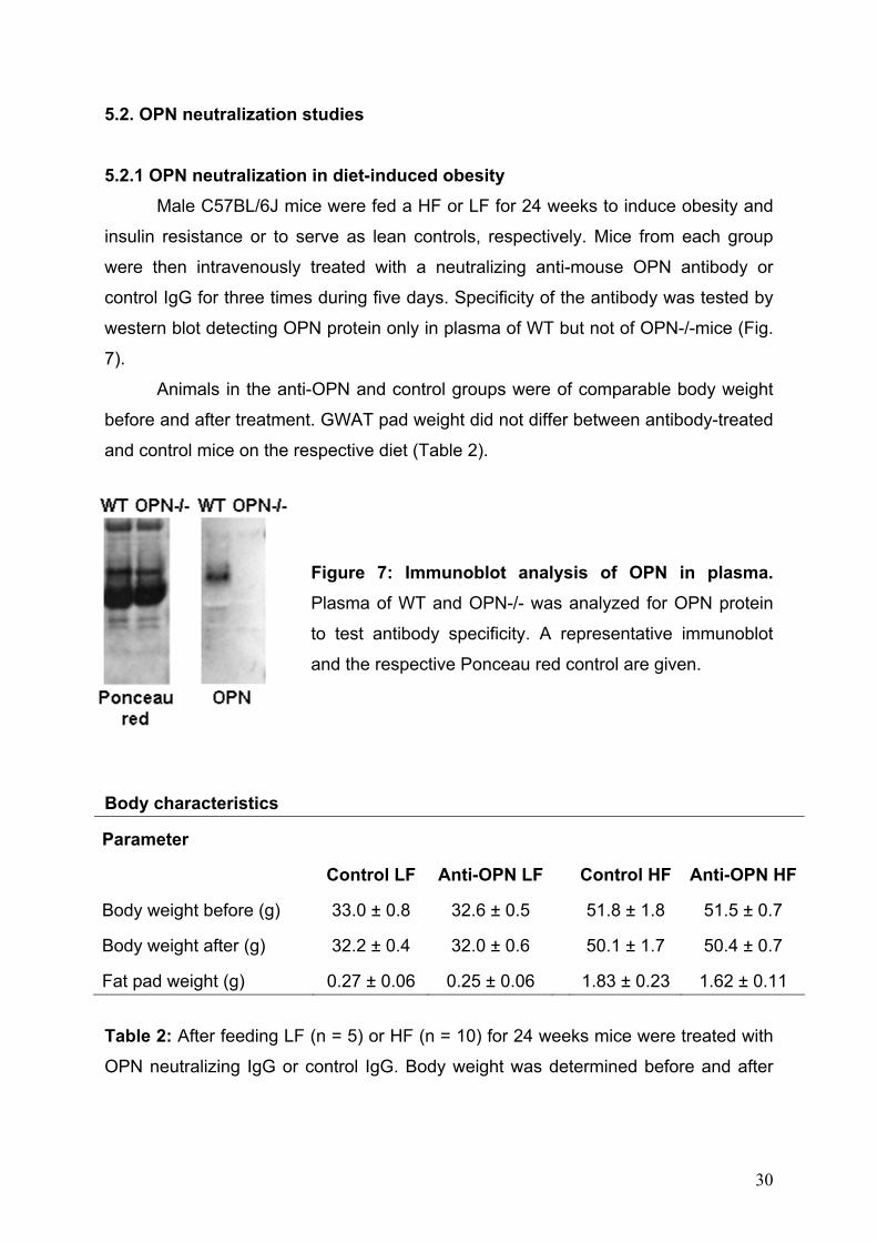
Male C57BL/6J mice were fed a HF or LF for 24 weeks to induce obesity and
insulin resistance or to serve as lean controls, respectively. Mice from each group
were then intravenously treated with a neutralizing anti-mouse OPN antibody or
control IgG for three times during five days. Specificity of the antibody was tested by
western blot detecting OPN protein only in plasma of WT but not of OPN-/-mice (Fig.
7).
Animals in the anti-OPN and control groups were of comparable body weight
before and after treatment. GWAT pad weight did not differ between antibody-treated
and control mice on the respective diet (Table 2).
Figure 7: Immunoblot analysis of OPN in plasma. Plasma of WT and OPN-/- was analyzed for OPN protein
to test antibody specificity. A representative immunoblot
and the respective Ponceau red control are given.
Body characteristics
Parameter
Control LF Anti-OPN LF Control HF Anti-OPN HF
Body weight before (g) 33.0 ± 0.8 32.6 ± 0.5 51.8 ± 1.8 51.5 ± 0.7
Body weight after (g) 32.2 ± 0.4 32.0 ± 0.6 50.1 ± 1.7 50.4 ± 0.7
Fat pad weight (g) 0.27 ± 0.06 0.25 ± 0.06 1.83 ± 0.23 1.62 ± 0.11
Table 2: After feeding LF (n = 5) or HF (n = 10) for 24 weeks mice were treated with
OPN neutralizing IgG or control IgG. Body weight was determined before and after
30

treatment. GWAT pad weight was measured immediately after sacrifice. Data are
expressed as mean ± SEM.
5.2.2 Diet-induced insulin resistance is reversed by antibody-mediated OPN neutralization
I first investigated whether insulin resistance in obese mice is ameliorated by
systemic neutralization of OPN action. Strikingly, treatment with OPN neutralizing
antibody markedly improved insulin sensitivity in obese mice as shown by
significantly reduced blood glucose concentrations at 60, 90 and 120 minutes of an
insulin tolerance test (Fig. 8A) and a declined area under the curve (Fig. 8C). In
addition, insulin resistance as estimated by HOMA-IR was significantly lower after
anti-OPN treatment (Fig. 8D). Insulin sensitivity was unaltered in mice on LF
irrespective of anti-OPN treatment (Fig. 8B). Taken together these data strongly
indicate enhanced insulin sensitivity in obese mice upon OPN neutralization. Plasma
concentrations of glucose, cholesterol, triglycerides, free fatty acids, adiponectin,
leptin, TNF-α and IL-6 did not significantly differ between groups (Table 3).
31

Figure 8: Insulin sensitivity is improved by OPN neutralization. Mice were fed
HF to induce obesity or LF, respectively, for 24 weeks and were treated intravenously
with an OPN neutralizing (Anti-OPN) or control antibody three times during five days
at the end of the feeding period. An ITT was performed in lean and obese OPN
antibody (dashed lines) and control antibody-treated mice (solid lines) one day after
the last antibody application (n = 5 per group for LF and n = 8 per group for HF). (A-B) Percent of basal glucose during ITT in mice on HF (A) and LF (B). (C) Area under
the curve. (D) HOMA-IR was calculated. *P ≤ 0.05, **P ≤ 0.01, #P = 0.06
Plasma measurements
Parameter
Control LF Anti-OPN LF Control HF Anti-OPN HF
Glucose (mg/dl) 175 ± 5.3 170.7 ± 11.7 332.9 ± 18.5 304.0 ± 23.7
Insulin (µU/ml) 2.46 ± 1.8 1.7 ± 0.7 48.4 ± 9.6 30.5 ± 5.3
Cholesterol (mg/dl) 1.1 ± 0.8 0.7 ± 0.3 98.1 ± 5.5 96.0 ± 7.8
Triglycerides (mg/dl) 60.2 ± 4.4 50.2 ± 3.0 62.6 ± 11.5 42.4 ± 1.9#
Free fatty acids (µmol/l) 162.6 ± 12.5 131.6 ± 15.4 309.7 ± 39.7 282.3 ± 17.9
Adiponectin (µg/ml) n.d. n.d. 38.8 ± 9.1 41.9 ± 10.6
Leptin (ng/ml) n.d. n.d. 43.3 ± 4.8 38.0 ± 3.9
IL-6 (pg/ml) n.d. n.d. 17.2 ± 6.2 12.2 ± 3.2
TNF-α (pg/ml) n.d. n.d. 3.1 ± 0.5 4.1 ± 2.8
Serum amyloid P (ng/ml) 36.5 ± 6.4 29.6 ± 5.8 86.0 ± 24.4 30.9 ± 8.1*
OPN (ng/ml) 180.5 ± 23.1 156.7 ± 13.0 180.5 ± 22.7 171.3 ± 11.9
Table 3: After feeding a LF or HF for 24 weeks mice were treated with OPN
neutralizing IgG or control IgG. Blood samples were obtained after a three hour
fasting period and analyzed for depicted plasma parameters (n = 5 – 10 per group).
Data are expressed as mean ± SEM. *P ≤ 0.05, #P = 0.09 compared to Control HF.
32
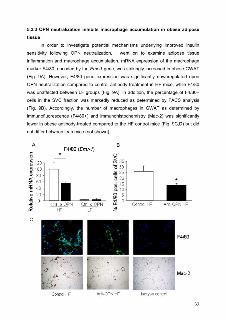
5.2.3 OPN neutralization inhibits macrophage accumulation in obese adipose tissue
In order to investigate potential mechanisms underlying improved insulin
sensitivity following OPN neutralization, I went on to examine adipose tissue
inflammation and macrophage accumulation. mRNA expression of the macrophage
marker F4/80, encoded by the Emr-1 gene, was strikingly increased in obese GWAT
(Fig. 9A). However, F4/80 gene expression was significantly downregulated upon
OPN neutralization compared to control antibody treatment in HF mice, while F4/80
was unaffected between LF groups (Fig. 9A). In addition, the percentage of F4/80+
cells in the SVC fraction was markedly reduced as determined by FACS analysis
(Fig. 9B). Accordingly, the number of macrophages in GWAT as determined by
immunofluorescence (F4/80+) and immunohistochemistry (Mac-2) was significantly
lower in obese antibody-treated compared to the HF control mice (Fig. 9C,D) but did
not differ between lean mice (not shown).
33

Figure 9: Adipose tissue macrophage accumulation is reduced by OPN neutralization. Adipose tissue macrophage accumulation is reduced by OPN
neutralization. Obese HF- and lean LF-fed mice were treated with OPN neutralizing
(Anti-OPN) or control antibody (n = 10 per group for HF and n = 5 per group for LF).
(A) mRNA expression of the macrophage marker F4/80 (encoded by Emr1 gene)
was analyzed in GWAT by real-time RT-PCR. (B) Percentage of macrophages
(F4/80-positive cells) in the SVC fraction of GWAT as determined by flow cytometry.
(C) Adipose tissue macrophage accumulation was determined by
immunofluorescence of F4/80+ cells (upper row) and immunohistochemical staining
of Mac-2+ cells (bottom row) in GWAT isolated from HF-fed mice after anti-OPN or
control antibody treatment. Representative pictures are given in 40-fold
magnification. (D) Adipose tissue macrophages as detected by F4/80 positivity in
tissue sections were counted as F4/80+ cells relative to total number of cells. (α-OPN
= Anti-OPN, Ctrl = Control).
5.2.4 OPN neutralization promotes adipose macrophage apoptosis Given the rapid reduction of adipose tissue macrophage numbers after OPN
neutralization and a potential anti-apoptotic role of OPN in macrophages (63), I
hypothesized that enhanced apoptosis in anti-OPN-treated mice could contribute to
the disappearance of adipose tissue macrophages. TUNEL staining of GWAT
sections revealed that the proportion of apoptotic F4/80+ cells was significantly
increased by 2.16±0.30–fold in obese OPN antibody-treated compared to control-
treated mice (Fig. 10). The abundance of apoptotic non-macrophages was generally
low (< 15% of apoptotic cells) and did not differ between the antibody-treated and the
control group on HF (data not shown). Hence, OPN neutralization in obese mice
34
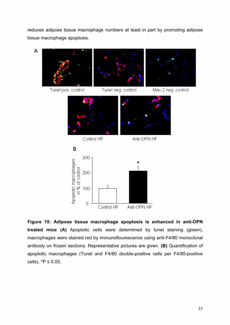
reduces adipose tissue macrophage numbers at least in part by promoting adipose
tissue macrophage apoptosis.
Figure 10: Adipose tissue macrophage apoptosis is enhanced in anti-OPN treated mice (A) Apoptotic cells were determined by tunel staining (green),
macrophages were stained red by immunoflourescence using anti-F4/80 monoclonal
antibody on frozen sections. Representative pictures are given. (B) Quantification of
apoptotic macrophages (Tunel and F4/80 double-positive cells per F4/80-positive
cells). *P ≤ 0.05.
35

5.2.5 OPN neutralization attenuates obesity-induced adipose tissue inflammation
Immunoblot quantification of OPN protein revealed decreased OPN content in
obese GWAT after OPN neutralization, even though not statistically significant (Fig.
11). However, OPN plasma concentrations were similar in antibody-treated and
control mice and did not differ between lean and obese two days after the last
antibody application (Table 3). Systemic concentrations of the inflammation marker
serum amyloid P (SAP) were markedly elevated in obese control mice but returned to
lean levels upon OPN neutralization (Table 3). In order to investigate potential effects
of OPN neutralization on inflammatory signaling related to impaired insulin sensitivity
in obese mice, I analyzed activation of JNK by determining phosphorylation of JNK1
and JNK2 in GWAT (64). Notably, anti-OPN treatment abolished JNK
phosphorylation in obese (Fig. 12A,B) but not in lean mice (Fig. 12C,D). To further
determine effects of OPN neutralization on adipose tissue inflammation, gene
expression of the adipokines IL-6, TNF-α, MCP-1 was analyzed in GWAT. Notably,
IL-6 gene expression in obese mice was markedly decreased upon anti-OPN
treatment while TNF-α, MCP-1 were not significantly reduced (Fig. 12E-G). However,
adiponectin mRNA expression was similar in all groups irrespective of diet and
antibody treatment (Fig. 12H). Taken together these data strongly suggest that OPN
neutralization effectively decreases deleterious inflammatory alterations in adipose
tissue of obese mice.
Figure 11: OPN protein expression in adipose tissue after anti-OPN treatment. GWAT of HF-fed anti-OPN (Ab) and control antibody-treated mice (Ctr) was analyzed
for OPN protein. (A) A representative immunoblot is given together with a loading
control (tubulin). (B) Quantification of OPN protein in GWAT. The diagram shows
means of the chemiluminescence intensity. n.s. = not statistically significant.
36

37

Figure 12: Adipose tissue inflammatory signaling and cytokine expression is attenuated by OPN neutralization in obese mice. Obese HF- and lean LF-fed mice
were treated with OPN neutralizing (Anti-OPN) or control antibody (n = 10 per group
for HF and n = 5 per group for LF). (A-D) Immunoblot analysis and quantification of
JNK1 and JNK2 phosphorylation in GWAT. Representative blots are given for obese
(A) and lean (C) adipose tissue. The diagrams show means of the
chemiluminescence intensity ratios from phosphorylated vs. total JNK protein for
obese (B) and lean (D) anti-OPN and control-treated mice. (E-H) mRNA expression
of the inflammatory genes for IL-6 (Il6; E), TNF-α (Tnf; F), MCP-1 (Ccl2; G) and of
adiponectin (Adipoq, H) was analyzed in GWAT. *P ≤ 0.05, **P ≤ 0.01
5.3 Studies in DoKo mice 5.3.1 OPN and MCP-1 double deficiency in diet-induced obesity To assess the role of combined OPN and MCP-1 deletion in diet-induced obesity I
generated OPN-/-MCP-1-/- double knockout (DoKo) mice. OPN-/- and MCP-1-/-
single knockouts were intercrossed to receive double heterozygous offspring in the
F1 generation. Heterozygous littermates from F1 were crossed to generate DoKo
mice in the next generation (F2). Approximately 300 pups were obtained in F2, all of
which were genotyped by genomic PCR. One mouse out of fifteen was double
deficient for OPN and MCP-1 which closely reflects Mendelian inheritance
distribution of 1:16. Hence, I could identify 20 DoKo mice, males and females equally
distributed.
DoKo, OPN-/-, MCP-1-/- and WT mice were fed either LF or HF for 20 weeks.
There was no difference in body weight between genotypes on LF (DoKo: 30.1±0.8g,
OPN-/-: 31.1±0.6g, MCP-1-/-: 31.5±0.9g and WT: 30.9±1.0g). Unexpectedly, DoKo
mice on HF gained significantly more weight than all other genotypes (Fig. 13A).
Feed efficiency, as determined by weight gain/food consumption did not differ
between obese mice (Fig. 13B). In order to test whether altered metabolic rate
accounted for the differences in body weight indirect calorimetric measurements
were performed. After 18 weeks of HF-feeding mice of each genotype were
monitored in metabolic cages for 72h. Assessment of energy expenditure as well as
spontaneous activity did not reveal any alteration in metabolic rates (Fig 13 C,D).
38
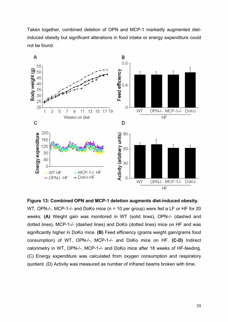
Taken together, combined deletion of OPN and MCP-1 markedly augmented diet-
induced obesity but significant alterations in food intake or energy expenditure could
not be found.
Figure 13: Combined OPN and MCP-1 deletion augments diet-induced obesity. WT, OPN-/-, MCP-1-/- and DoKo mice (n = 10 per group) were fed a LF or HF for 20
weeks. (A) Weight gain was monitored in WT (solid lines), OPN-/- (dashed and
dotted lines), MCP-1-/- (dashed lines) and DoKo (dotted lines) mice on HF and was
significantly higher in DoKo mice. (B) Feed efficiency (grams weight gain/grams food
consumption) of WT, OPN-/-, MCP-1-/- and DoKo mice on HF. (C-D) Indirect
calorimetry in WT, OPN-/-, MCP-1-/- and DoKo mice after 18 weeks of HF-feeding.
(C) Energy expenditure was calculated from oxygen consumption and respiratory
quotient. (D) Activity was measured as number of infrared beams broken with time.
39
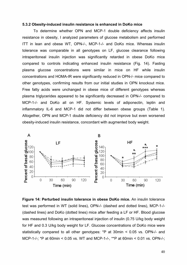
5.3.2 Obesity-induced insulin resistance is enhanced in DoKo mice To determine whether OPN and MCP-1 double deficiency affects insulin
resistance in obesity, I analyzed parameters of glucose metabolism and performed
ITT in lean and obese WT, OPN-/-, MCP-1-/- and DoKo mice. Whereas insulin
tolerance was comparable in all genotypes on LF, glucose clearance following
intraperitoneal insulin injection was significantly retarded in obese DoKo mice
compared to controls indicating enhanced insulin resistance (Fig. 14). Fasting
plasma glucose concentrations were similar in mice on HF while insulin
concentrations and HOMA-IR were significantly reduced in OPN-/- mice compared to
other genotypes, confirming results from our initial studies in OPN knockout mice.
Free fatty acids were unchanged in obese mice of different genotypes whereas
plasma triglycerides appeared to be significantly decreased in OPN-/- compared to
MCP-1-/- and DoKo all on HF. Systemic levels of adiponectin, leptin and
inflammatory IL-6 and MCP-1 did not differ between obese groups (Table 1).
Altogether, OPN and MCP-1 double deficiency did not improve but even worsened
obesity-induced insulin resistance, concordant with augmented body weight.
Figure 14: Perturbed insulin tolerance in obese DoKo mice. An insulin tolerance
test was performed in WT (solid lines), OPN-/- (dashed and dotted lines), MCP-1-/-
(dashed lines) and DoKo (dotted lines) mice after feeding a LF or HF. Blood glucose
was measured following an intraperitoneal injection of insulin (0.75 U/kg body weight
for HF and 0.3 U/kg body weight for LF. Glucose concentrations of DoKo mice were
statistically compared to all other genotypes: *P at 30min < 0.05 vs. OPN-/- and
MCP-1-/-; *P at 60min < 0.05 vs. WT and MCP-1-/-, **P at 60min < 0.01 vs. OPN-/-;
40

*P at 90min < 0.05 vs MCP-1-/-, ***P at 90min < 0.001 vs. OPN-/-; *P at 120min <
0.05 vs. OPN-/-.
Plasma measurements
Parameter
WT HF OPN-/- HF MCP-1-/-HF DoKo HF
Glucose (mg/dl) 245.3 ± 17.3 278.9 ± 14.1 260.8 ± 11.5 240.7 ± 18.2
Insulin (µU/ml) 61.9 ± 9.1 18.6 ± 3.3** 52.7 ± 11.6 41.6 ± 5.7
HOMA-IR 39.5 ± 6.9 13.3 ± 2.3** 29.5 ± 10.2 41.6 ± 5.7
Triglycerides (mg/dl) 36.2 ± 3.1 33.3 ± 1.0* 37.8 ± 2.1 39.5 ± 2.4
Free fatty acids (µmol/l) 224.5 ± 13.2 271.3 ± 27.0 223.3 ± 16.9 296.9 ± 36.7
Adiponectin (µg/ml) 56.2 ± 4.0 49.3 ± 4.8 48.3 ± 6.8 54.2 ± 5.0
Leptin (ng/ml) 86.8 ± 8.9 95.4 ± 13.6 79.4 ± 7.9 78.2 ± 6.9
IL-6 (pg/ml) 14.2 ± 4.7 20.6 ± 9.1 19.0 ± 8.6 16.5 ± 5.0
MCP-1 (pg/ml) 82.8 ± 17.3 67.6 ± 8.1
Table 4: WT, OPN-/-, MCP-1-/- and DoKo mice (n = 10 per group) were fed HF for
20 weeks. Blood samples were obtained after a three hour fasting period and
analyzed for indicated plasma parameters (n = 10 per group). Data are expressed as
mean ± SEM. **P (Insulin) < 0.01 vs. WT, MCP-1-/- and DoKo; **P (HOMA-IR) < 0.01
vs. WT, MCP-1-/- and DoKo; *P (Triglycerides) < 0.05 vs. MCP-1-/- and DoKo.
5.3.3 OPN and MCP-1 double deficiency does not affect adipose tissue inflammation
Adipose tissue inflammation was assessed by gene expression analyses of
inflammatory mediators known to be upregulated upon obesity. I studied mRNA
expression of the macrophage markers CD68 and CD11c in adipose tissue of lean
and obese WT, OPN-/-, MCP-1-/- and DoKo mice. I did not detect any differences
between genotypes on the respective diet. Next, gene expression of inflammatory IL-
6 and TNF-α was determined without observing any alterations between different
groups of mice (Fig. 15). Hence, combined deletion of OPN and MCP-1 does not
affect obesity-induced adipose tissue inflammation.
41

42

Figure 15: Adipose tissue inflammation in DoKo mice. WT, OPN-/-, MCP-1-/- and
DoKo mice (n = 10 per group) were fed a LF or HF for 20 weeks. mRNA expression
of the given inflammatory genes was analyzed in GWAT of (A) lean and (B) obese
mice.
43

6. DISCUSSION
The increasing prevalence of obesity demands novel preventive and
therapeutic approaches to treat obesity-associated complications particularly insulin
resistance that leads to type 2 diabetes and promotes cardiovascular disease. We
and others have recently reported that OPN and MCP-1 expression is considerably
upregulated in human obesity as well as mouse models of genetic and diet-induced
obesity (12; 44; 45; 56). MCP-1 was initially shown to critically contribute to obesity-
induced adipose tissue inflammation and insulin resistance (56). However, during the
experimental work of this thesis conflicting results regarding the role of MCP-1 in
obesity-associated adipose tissue macrophage accumulation and metabolic
deterioration were published. Here I show that not only genetic OPN deficiency but
also antibody targeting of OPN markedly improves insulin sensitivity in murine diet-
induced obesity. Unexpectedly, combined deletion of OPN and MCP-1 leads to an
increase in diet-induced obesity and subsequently reverses beneficial effects of
single OPN deletion resulting in impaired insulin sensitivity.
Obesity-associated insulin resistance was significantly reduced in obese mice
irrespective of whether OPN was eliminated by genetic knockout or antibody-
mediated neutralization (Figs. 2 and 8). The genetic approach shows that obesity-
induced metabolic alterations are improved if OPN is already absent during
development of fat depots. Short-term neutralization of OPN demonstrates that
inflammation and insulin resistance is attenuated when obesity has already been
established. In addition, these results indicate that negative OPN effects on glucose
metabolism are obesity-dependent because neither genetic OPN deficiency nor OPN
neutralization did alter insulin sensitivity and adipose tissue inflammation in lean
mice. However, not all of the inflammatory parameters tested concurred in both
experimental models. Whereas macrophage accumulation in adipose tissue differed
significantly between antibody-treated and control mice, it was comparable in obese
OPN-/- mice and the respective WT controls (Figs. 5 and 9). Inflammatory gene
expression in GWAT was only moderately decreased in HF-fed OPN-/- compared to
WT (Fig. 4), and the activation of the inflammatory proteins JNK 1 and 2 was similar
in both genotypes (Fig. 6), other than in anti-OPN treated WT mice where
inflammatory gene expression and JNK activation in GWAT were considerably
downregulated (Fig. 12). Apparently, macrophage accumulation and inflammatory
44
processes occurring in adipose tissue upon diet-induced obesity are reversed by
short-term neutralization of OPN but not by genetic deficiency. Therefore, it could be
speculated that unidentified escape mechanisms emerge in OPN-/- mice during the
development of diet-induced obesity that restore macrophage migration and adipose
tissue inflammation despite lack of OPN. However, during the preparation of this
thesis an article was published, demonstrating that genetic OPN deficiency is able to
attenuate adipose tissue macrophage infiltration and insulin resistance in a model of
murine diet-induced obesity (46). One noticeable difference between the present
study and the study by Nomiyama et al. was the genetic background of the mice.
While we used OPN-/- mice on a C57BL/6J background, mice used in Ref. 46 were
on a Black Swiss background. Whether and how the differences in the genetic
background in these particular studies can impact obesity-induced adipose tissue
alterations remains unsolved.
Obesity-induced insulin resistance is associated with macrophage
accumulation in adipose tissue (13). OPN is involved in macrophage migration and
macrophage-driven inflammatory disorders (31; 35; 36). Treatment with anti-OPN
antibody for only 5 days significantly decreased adipose tissue macrophage numbers
in obese mice (Fig. 9). Similarly, blocking of the chemokine receptor CCR2 for 9 days
resulted in a significant reduction of adipose tissue macrophage accumulation (12).
Thus, macrophage turnover appears to be rather high in obese murine adipose
tissue.
OPN has previously been shown to be a survival factor for macrophages in
HIV-induced brain disease (63) and for T-cells in an arthritis model (65). Here I show
that the rapid reduction of adipose tissue macrophages by OPN neutralization was
accompanied by a significantly increased number of apoptotic macrophages (Fig.
10), indicating that macrophage apoptosis contributes to reduced macrophage
abundance in anti-OPN treated mice. Aside from enhanced macrophage apoptosis,
disturbed cell migration could contribute to the reduction of macrophages in adipose
tissue of anti-OPN treated mice, since OPN is well-known to be critically involved in
migration of monocytes and macrophages (31; 46). Hence, the marked obesity-
induced upregulation of OPN expression in adipose tissue (44) could promote
macrophage accumulation not only by stimulating cell migration (66) but also by
preventing macrophage apoptosis.
45

Since reduced OPN plasma concentrations after anti-OPN treatment were not
observed (Table 3) I assume functional neutralization of OPN to underlie the
decrease in adipose tissue macrophage numbers (67; 68). Since macrophages are
the major source of OPN in adipose tissue (44), the somewhat reduced tissue OPN
content following OPN neutralization (Fig. 11) is probably due to diminished local
production.
HF causes activation of the JNK pathway that leads to insulin resistance by
serine phosphorylation of IRS proteins (6; 64; 69). OPN neutralization inhibited
inflammatory signaling by negatively regulating JNK 1 and JNK 2 phosphorylation in
obese adipose tissue (Fig. 12). In contrast to decreased GWAT expression of IL-6, a
potent mediator between obesity-induced adipose tissue inflammation and insulin
resistance (70), TNF-α and MCP-1expression remained unaffected (Fig. 12). These
data are in accordance with a recent publication showing that adipose tissue specific
deletion of JNK1 significantly reduced obesity-induced insulin resistance and IL-6
expression in adipose tissue while TNF-α remained unaltered (71). Taken together,
these results emphasize abolished JNK activation to be a crucial mechanism for anti-
inflammatory effects of OPN neutralization in adipose tissue.
MCP-1 is important for recruiting monocytes that represent macrophage
precursors into atherosclerotic tissue and other chronic inflammatory lesions (55; 72).
A central role in inducing insulin resistance in adipocytes and skeletal muscle cells
has been proposed. MCP-1 interacts with its receptor, CCR2 on the surface of
monocytes to initiate signaling, which ultimately leads to cell adhesion and tissue
infiltration. In mice and humans, MCP-1 production increases in plasma and adipose
tissue in both diet-induced and genetic forms of obesity. These data suggest that
monocytes are recruited to and infiltrate expanding adipose tissue, where they
differentiate into macrophages (73). Mice deficient for the MCP-1 receptor CCR2
showed reduced macrophage content and improved insulin sensitivity after HF-
feeding compared to wild-type animals (12). Studies in MCP-1 knockout mice and
mice overexpressing MCP-1 support a role of this chemokine in attracting
macrophages to adipose tissue and reduced insulin sensitivity in HF-induced obesity
(56), but these data have recently been combated by others (59; 73). Those
contradicting studies have emerged during the preparation of this thesis and showed
that macrophages accumulate in adipose tissue in response to diet-induced obesity
even in the absence of MCP-1. Inouye et al. as well as Kirk et al. detected similar
46

numbers of macrophages in adipose tissue of MCP-1-/- and control mice fed a HF.
They also found that MCP-1-/- mice on that diet were insulin resistant, suggesting
that MCP-1 does not account for obesity-induced macrophage recruitment in adipose
tissue and related metabolic effects (59; 73). These data are in accordance with
results of this thesis that did neither reveal a difference in adipose tissue
inflammation and macrophage accumulation nor in metabolic alterations between
HF-fed MCP-1-/- and WT mice (Fig. 14 and 15, and Table 1). Therefore, a
pathogenic role of MCP-1 in obesity-induced inflammation and metabolic
deterioration remains speculative. However, in addition to MCP-1 numerous other
chemokines such as MCP-2, -3, and -4 can bind to and subsequently activate CCR2
and, hence, could promote macrophage migration into the adipose tissue. Indeed,
gene expression of almost all of the named chemotactic factors is upregulated in the
adipose tissue of obese subjects. In addition to CCR2, adipose tissue expression of
some other chemokine receptors including CCR1, CCR3 and CCR5 is increased in
obesity (74). Their precise role in obesity-associated macrophage recruitment to
adipose tissue has not been studied yet.
Since previous reports (12; 56) and own data (Figs. 3 – 10) strongly suggested
a pathogenic role for OPN and MCP-1 in obesity-associated adipose tissue
macrophage infiltration and insulin resistance I sought to investigate a possible
interference between both cytokines in adipose tissue inflammation and metabolic
alterations. Unexpectedly, studies in DoKo mice revealed that combined deletion of
OPN and MCP-1 augments rather than reduces diet-induced obesity compared to
single knockout and WT controls (Fig.13). Neither OPN nor MCP-1 is known to
impact food intake or energy expenditure. Nevertheless I tested whether OPN and
MCP-1 double deficiency did affect feed efficiency or metabolic rate and could not
detect any difference by indirect calorimetry (Fig. 13). Hence, altered feed efficiency
and disturbed substrate utilization are unlikely to account for increased body weight
in DoKo mice on HF.
Adipose tissue macrophage accumulation as assessed by gene expression of
highly specific macrophage markers CD68 and CD11c did not differ between
genotypes (Fig. 15), suggesting that OPN and MCP-1 double deficiency does not
impact obesity-associated macrophage recruitment to adipose tissue.
Characterization of glucose metabolism showed markedly enhanced insulin
resistance in obese DoKo mice compared to controls (Fig. 14), indicating that
47
combined deletion of OPN and MCP-1 does not improve but even worsen obesity-
related metabolic perturbation. The reason why beneficial effects of OPN deficiency
(Fig. 3 and Ref. 46) are reversed by additional MCP-1 deficiency is elusive but it is
most probable that increased body weight in DoKo mice could account for enhanced
insulin resistance. Moreover, it could be speculated that genetic knockout of two
inflammatory cytokines that are involved in the regulation of many immune processes
initiates unknown antagonistic mechanisms that disturb metabolic balance. Notably,
tissues and organs other than adipose could contribute to the antagonistic effect
such as the central nervous system.
The proposed thesis revealed a pathogenic role of OPN in obesity-associated
insulin resistance by showing that genetic OPN deficiency as well as systemic OPN
neutralization improved insulin sensitivity in obesity. OPN neutralization interferes
with obesity-associated macrophage accumulation and inflammatory signaling in
adipose tissue. Surprisingly, combined deletion of OPN and MCP-1 increased diet-
induced obesity and insulin resistance suggesting that concurrent loss of both
inflammatory cytokines perturbs metabolic balance. In conclusion, targeting OPN
action in vivo could improve metabolic regulation and cardiovascular risk in obese
patients.
48

7. REFERENCES 1. Mensah GA, Mokdad AH, Ford E, Narayan KM, Giles WH, Vinicor F, Deedwania
PC: Obesity, metabolic syndrome, and type 2 diabetes: emerging epidemics and
their cardiovascular implications. Cardiol Clin 22:485-504, 2004
2. Shoelson SE, Herrero L, Naaz A: Obesity, inflammation, and insulin resistance.
Gastroenterology 132:2169-2180, 2007
3. Burke GL, Bertoni AG, Shea S, Tracy R, Watson KE, Blumenthal RS, Chung H,
Carnethon MR: The impact of obesity on cardiovascular disease risk factors and
subclinical vascular disease: the Multi-Ethnic Study of Atherosclerosis. Arch Intern
Med 168:928-935, 2008
4. Wellen KE, Hotamisligil GS: Obesity-induced inflammatory changes in adipose
tissue. J Clin Invest 112:1785-1788, 2003
5. Sondergaard L: Homology between the mammalian liver and the Drosophila fat
body. Trends Genet 9:193, 1993
6. Hotamisligil GS: Inflammation and metabolic disorders. Nature 444:860-867, 2006
7. Gustafson B, Hammarstedt A, Andersson CX, Smith U: Inflamed adipose tissue: a
culprit underlying the metabolic syndrome and atherosclerosis. Arterioscler Thromb
Vasc Biol 27:2276-2283, 2007
8. Spiegelman BM, Hotamisligil GS: Through thick and thin: wasting, obesity, and
TNF alpha. Cell 73:625-627, 1993
9. Bastard JP, Jardel C, Delattre J, Hainque B, Bruckert E, Oberlin F: Evidence for a
link between adipose tissue interleukin-6 content and serum C-reactive protein
concentrations in obese subjects. Circulation 99:2221-2222, 1999
10. Berg AH, Scherer PE: Adipose tissue, inflammation, and cardiovascular disease.
Circ Res 96:939-949, 2005
11. Bastard JP, Maachi M, Lagathu C, Kim MJ, Caron M, Vidal H, Capeau J, Feve B:
Recent advances in the relationship between obesity, inflammation, and insulin
resistance. Eur Cytokine Netw 17:4-12, 2006
12. Weisberg SP, Hunter D, Huber R, Lemieux J, Slaymaker S, Vaddi K, Charo I,
Leibel RL, Ferrante AW, Jr.: CCR2 modulates inflammatory and metabolic effects of
high-fat feeding. J Clin Invest 116:115-124, 2006
49
13. Weisberg SP, McCann D, Desai M, Rosenbaum M, Leibel RL, Ferrante AW, Jr.:
Obesity is associated with macrophage accumulation in adipose tissue. J Clin Invest
112:1796-1808, 2003
14. Fain JN: Release of interleukins and other inflammatory cytokines by human
adipose tissue is enhanced in obesity and primarily due to the nonfat cells. Vitam
Horm 74:443-477, 2006
15. Xu H, Barnes GT, Yang Q, Tan G, Yang D, Chou CJ, Sole J, Nichols A, Ross JS,
Tartaglia LA, Chen H: Chronic inflammation in fat plays a crucial role in the
development of obesity-related insulin resistance. J Clin Invest 112:1821-1830, 2003
16. Arkan MC, Hevener AL, Greten FR, Maeda S, Li ZW, Long JM, Wynshaw-Boris
A, Poli G, Olefsky J, Karin M: IKK-beta links inflammation to obesity-induced insulin
resistance. Nat Med 11:191-198, 2005
17. Ozcan U, Cao Q, Yilmaz E, Lee AH, Iwakoshi NN, Ozdelen E, Tuncman G,
Gorgun C, Glimcher LH, Hotamisligil GS: Endoplasmic reticulum stress links obesity,
insulin action, and type 2 diabetes. Science 306:457-461, 2004
18. Zeyda M, Stulnig TM: Obesity, inflammation, and insulin resistance--a mini-
review. Gerontology 55:379-386, 2009
19. Ozcan U, Yilmaz E, Ozcan L, Furuhashi M, Vaillancourt E, Smith RO, Gorgun
CZ, Hotamisligil GS: Chemical chaperones reduce ER stress and restore glucose
homeostasis in a mouse model of type 2 diabetes. Science 313:1137-1140, 2006
20. Miller RS, Diaczok D, Cooke DW: Repression of GLUT4 expression by the
endoplasmic reticulum stress response in 3T3-L1 adipocytes. Biochem Biophys Res
Commun 362:188-192, 2007
21. Zeyda M, Farmer D, Todoric J, Aszmann O, Speiser M, Gyori G, Zlabinger GJ,
Stulnig TM: Human adipose tissue macrophages are of an anti-inflammatory
phenotype but capable of excessive pro-inflammatory mediator production. Int J
Obes (Lond) 31:1420-1428, 2007
22. Cinti S, Mitchell G, Barbatelli G, Murano I, Ceresi E, Faloia E, Wang S, Fortier M,
Greenberg AS, Obin MS: Adipocyte death defines macrophage localization and
function in adipose tissue of obese mice and humans. J Lipid Res 46:2347-2355,
2005
23. Gordon S: Alternative activation of macrophages. Nat Rev Immunol 3:23-35,
2003
50

24. Herbert DR, Holscher C, Mohrs M, Arendse B, Schwegmann A, Radwanska M,
Leeto M, Kirsch R, Hall P, Mossmann H, Claussen B, Forster I, Brombacher F:
Alternative macrophage activation is essential for survival during schistosomiasis and
downmodulates T helper 1 responses and immunopathology. Immunity 20:623-635,
2004
25. Noel W, Raes G, Hassanzadeh Ghassabeh G, De Baetselier P, Beschin A:
Alternatively activated macrophages during parasite infections. Trends Parasitol
20:126-133, 2004
26. Takahashi H, Tsuda Y, Takeuchi D, Kobayashi M, Herndon DN, Suzuki F:
Influence of systemic inflammatory response syndrome on host resistance against
bacterial infections. Crit Care Med 32:1879-1885, 2004
27. Zeyda M, Stulnig TM: Adipose tissue macrophages. Immunol Lett 112:61-67,
2007
28. Lumeng CN, Bodzin JL, Saltiel AR: Obesity induces a phenotypic switch in
adipose tissue macrophage polarization. J Clin Invest 117:175-184, 2007
29. Clement K, Viguerie N, Poitou C, Carette C, Pelloux V, Curat CA, Sicard A,
Rome S, Benis A, Zucker JD, Vidal H, Laville M, Barsh GS, Basdevant A, Stich V,
Cancello R, Langin D: Weight loss regulates inflammation-related genes in white
adipose tissue of obese subjects. Faseb J 18:1657-1669, 2004
30. Mazzali M, Kipari T, Ophascharoensuk V, Wesson JA, Johnson R, Hughes J:
Osteopontin--a molecule for all seasons. Qjm 95:3-13, 2002
31. Standal T, Borset M, Sundan A: Role of osteopontin in adhesion, migration, cell
survival and bone remodeling. Exp Oncol 26:179-184, 2004
32. Rangaswami H, Bulbule A, Kundu GC: Osteopontin: role in cell signaling and
cancer progression. Trends Cell Biol 16:79-87, 2006
33. Xu G, Nie H, Li N, Zheng W, Zhang D, Feng G, Ni L, Xu R, Hong J, Zhang JZ:
Role of osteopontin in amplification and perpetuation of rheumatoid synovitis. J Clin
Invest 115:1060-1067, 2005
34. Teti A, Farina AR, Villanova I, Tiberio A, Tacconelli A, Sciortino G, Chambers AF,
Gulino A, Mackay AR: Activation of MMP-2 by human GCT23 giant cell tumour cells
induced by osteopontin, bone sialoprotein and GRGDSP peptides is RGD and cell
shape change dependent. Int J Cancer 77:82-93, 1998
35. Xu G, Sun W, He D, Wang L, Zheng W, Nie H, Ni L, Zhang D, Li N, Zhang J:
Overexpression of osteopontin in rheumatoid synovial mononuclear cells is
51

associated with joint inflammation, not with genetic polymorphism. J Rheumatol
32:410-416, 2005
36. Isoda K, Kamezawa Y, Ayaori M, Kusuhara M, Tada N, Ohsuzu F: Osteopontin
transgenic mice fed a high-cholesterol diet develop early fatty-streak lesions.
Circulation 107:679-681, 2003
37. Takemoto M, Yokote K, Nishimura M, Shigematsu T, Hasegawa T, Kon S, Uede
T, Matsumoto T, Saito Y, Mori S: Enhanced expression of osteopontin in human
diabetic artery and analysis of its functional role in accelerated atherogenesis.
Arterioscler Thromb Vasc Biol 20:624-628, 2000
38. Ramaiah SK, Rittling S: Pathophysiological role of osteopontin in hepatic
inflammation, toxicity, and cancer. Toxicol Sci 103:4-13, 2008
39. Bertola A, Deveaux V, Bonnafous S, Rousseau D, Anty R, Wakkach A, Dahman
M, Tordjman J, Clement K, McQuaid SE, Frayn KN, Huet PM, Gugenheim J,
Lotersztajn S, Le Marchand-Brustel Y, Tran A, Gual P: Elevated expression of
osteopontin may be related to adipose tissue macrophage accumulation and liver
steatosis in morbid obesity. Diabetes 58:125-133, 2009
40. Wang Y, Mochida S, Kawashima R, Inao M, Matsui A, YouLuTu ZY, Nagoshi S,
Uede T, Fujiwara K: Increased expression of osteopontin in activated Kupffer cells
and hepatic macrophages during macrophage migration in Propionibacterium acnes-
treated rat liver. J Gastroenterol 35:696-701, 2000
41. Kawashima R, Mochida S, Matsui A, YouLuTu ZY, Ishikawa K, Toshima K,
Yamanobe F, Inao M, Ikeda H, Ohno A, Nagoshi S, Uede T, Fujiwara K: Expression
of osteopontin in Kupffer cells and hepatic macrophages and Stellate cells in rat liver
after carbon tetrachloride intoxication: a possible factor for macrophage migration
into hepatic necrotic areas. Biochem Biophys Res Commun 256:527-531, 1999
42. Sahai A, Malladi P, Melin-Aldana H, Green RM, Whitington PF: Upregulation of
osteopontin expression is involved in the development of nonalcoholic steatohepatitis
in a dietary murine model. Am J Physiol Gastrointest Liver Physiol 287:G264-273,
2004
43. Sahai A, Malladi P, Pan X, Paul R, Melin-Aldana H, Green RM, Whitington PF:
Obese and diabetic db/db mice develop marked liver fibrosis in a model of
nonalcoholic steatohepatitis: role of short-form leptin receptors and osteopontin. Am
J Physiol Gastrointest Liver Physiol 287:G1035-1043, 2004
52
44. Kiefer FW, Zeyda M, Todoric J, Huber J, Geyeregger R, Weichhart T, Aszmann
O, Ludvik B, Silberhumer GR, Prager G, Stulnig TM: Osteopontin expression in
human and murine obesity: extensive local up-regulation in adipose tissue but
minimal systemic alterations. Endocrinology 149:1350-1357, 2008
45. Gomez-Ambrosi J, Catalan V, Ramirez B, Rodriguez A, Colina I, Silva C, Rotellar
F, Mugueta C, Gil MJ, Cienfuegos JA, Salvador J, Fruhbeck G: Plasma osteopontin
levels and expression in adipose tissue are increased in obesity. J Clin Endocrinol
Metab 92:3719-3727, 2007
46. Nomiyama T, Perez-Tilve D, Ogawa D, Gizard F, Zhao Y, Heywood EB, Jones
KL, Kawamori R, Cassis LA, Tschop MH, Bruemmer D: Osteopontin mediates
obesity-induced adipose tissue macrophage infiltration and insulin resistance in mice.
J Clin Invest 117:2877-2888, 2007
47. Riedl M, Vila G, Maier C, Handisurya A, Shakeri-Manesch S, Prager G, Wagner
O, Kautzky-Willer A, Ludvik B, Clodi M, Luger A: Plasma osteopontin increases after
bariatric surgery and correlates with markers of bone turnover but not with insulin
resistance. J Clin Endocrinol Metab 93:2307-2312, 2008
48. Baggiolini M: Chemokines and leukocyte traffic. Nature 392:565-568, 1998
49. Rollins BJ: Chemokines. Blood 90:909-928, 1997
50. Gerhardt CC, Romero IA, Cancello R, Camoin L, Strosberg AD: Chemokines
control fat accumulation and leptin secretion by cultured human adipocytes. Mol Cell
Endocrinol 175:81-92, 2001
51. Aiello RJ, Bourassa PA, Lindsey S, Weng W, Natoli E, Rollins BJ, Milos PM:
Monocyte chemoattractant protein-1 accelerates atherosclerosis in apolipoprotein E-
deficient mice. Arterioscler Thromb Vasc Biol 19:1518-1525, 1999
52. Gu L, Okada Y, Clinton SK, Gerard C, Sukhova GK, Libby P, Rollins BJ: Absence
of monocyte chemoattractant protein-1 reduces atherosclerosis in low density
lipoprotein receptor-deficient mice. Mol Cell 2:275-281, 1998
53. Gerszten RE, Garcia-Zepeda EA, Lim YC, Yoshida M, Ding HA, Gimbrone MA,
Jr., Luster AD, Luscinskas FW, Rosenzweig A: MCP-1 and IL-8 trigger firm adhesion
of monocytes to vascular endothelium under flow conditions. Nature 398:718-723,
1999
54. Weber KS, von Hundelshausen P, Clark-Lewis I, Weber PC, Weber C:
Differential immobilization and hierarchical involvement of chemokines in monocyte
53

arrest and transmigration on inflamed endothelium in shear flow. Eur J Immunol
29:700-712, 1999
55. Melgarejo E, Medina MA, Sanchez-Jimenez F, Urdiales JL: Monocyte
chemoattractant protein-1: a key mediator in inflammatory processes. Int J Biochem
Cell Biol 41:998-1001, 2009
56. Kanda H, Tateya S, Tamori Y, Kotani K, Hiasa K, Kitazawa R, Kitazawa S,
Miyachi H, Maeda S, Egashira K, Kasuga M: MCP-1 contributes to macrophage
infiltration into adipose tissue, insulin resistance, and hepatic steatosis in obesity. J
Clin Invest 116:1494-1505, 2006
57. Sell H, Eckel J: Monocyte chemotactic protein-1 and its role in insulin resistance.
Curr Opin Lipidol 18:258-262, 2007
58. Sartipy P, Loskutoff DJ: Monocyte chemoattractant protein 1 in obesity and
insulin resistance. Proc Natl Acad Sci U S A 100:7265-7270, 2003
59. Inouye KE, Shi H, Howard JK, Daly CH, Lord GM, Rollins BJ, Flier JS: Absence
of CC chemokine ligand 2 does not limit obesity-associated infiltration of
macrophages into adipose tissue. Diabetes 56:2242-2250, 2007
60. de Luca C, Olefsky JM: Inflammation and insulin resistance. FEBS Lett 582:97-
105, 2008
61. Ferrannini E, Mari A: How to measure insulin sensitivity. J Hypertens 16:895-906,
1998
62. Todoric J, Loffler M, Huber J, Bilban M, Reimers M, Kadl A, Zeyda M, Waldhausl
W, Stulnig TM: Adipose tissue inflammation induced by high-fat diet in obese diabetic
mice is prevented by n-3 polyunsaturated fatty acids. Diabetologia 49:2109-2119,
2006
63. Burdo TH, Wood MR, Fox HS: Osteopontin prevents monocyte recirculation and
apoptosis. J Leukoc Biol 81:1504-1511, 2007
64. Hirosumi J, Tuncman G, Chang L, Gorgun CZ, Uysal KT, Maeda K, Karin M,
Hotamisligil GS: A central role for JNK in obesity and insulin resistance. Nature
420:333-336, 2002
65. Fan K, Dai J, Wang H, Wei H, Cao Z, Hou S, Qian W, Wang H, Li B, Zhao J, Xu
H, Yang C, Guo Y: Treatment of collagen-induced arthritis with an anti-osteopontin
monoclonal antibody through promotion of apoptosis of both murine and human
activated T cells. Arthritis Rheum 58:2041-2052, 2008
54

66. Zhu B, Suzuki K, Goldberg HA, Rittling SR, Denhardt DT, McCulloch CA, Sodek
J: Osteopontin modulates CD44-dependent chemotaxis of peritoneal macrophages
through G-protein-coupled receptors: evidence of a role for an intracellular form of
osteopontin. J Cell Physiol 198:155-167, 2004
67. Banerjee A, Apte UM, Smith R, Ramaiah SK: Higher neutrophil infiltration
mediated by osteopontin is a likely contributing factor to the increased susceptibility
of females to alcoholic liver disease. J Pathol 208:473-485, 2006
68. Koguchi Y, Kawakami K, Uezu K, Fukushima K, Kon S, Maeda M, Nakamoto A,
Owan I, Kuba M, Kudeken N, Azuma M, Yara S, Shinzato T, Higa F, Tateyama M,
Kadota J, Mukae H, Kohno S, Uede T, Saito A: High plasma osteopontin level and its
relationship with interleukin-12-mediated type 1 T helper cell response in
tuberculosis. Am J Respir Crit Care Med 167:1355-1359, 2003
69. Aguirre V, Uchida T, Yenush L, Davis R, White MF: The c-Jun NH(2)-terminal
kinase promotes insulin resistance during association with insulin receptor substrate-
1 and phosphorylation of Ser(307). J Biol Chem 275:9047-9054, 2000
70. Kern PA, Ranganathan S, Li C, Wood L, Ranganathan G: Adipose tissue tumor
necrosis factor and interleukin-6 expression in human obesity and insulin resistance.
Am J Physiol Endocrinol Metab 280:E745-751, 2001
71. Sabio G, Das M, Mora A, Zhang Z, Jun JY, Ko HJ, Barrett T, Kim JK, Davis RJ: A
stress signaling pathway in adipose tissue regulates hepatic insulin resistance.
Science 322:1539-1543, 2008
72. Gosling J, Slaymaker S, Gu L, Tseng S, Zlot CH, Young SG, Rollins BJ, Charo
IF: MCP-1 deficiency reduces susceptibility to atherosclerosis in mice that
overexpress human apolipoprotein B. J Clin Invest 103:773-778, 1999
73. Kirk EA, Sagawa ZK, McDonald TO, O'Brien KD, Heinecke JW: Monocyte
chemoattractant protein deficiency fails to restrain macrophage infiltration into
adipose tissue [corrected]. Diabetes 57:1254-1261, 2008
74. Huber J, Kiefer FW, Zeyda M, Ludvik B, Silberhumer GR, Prager G, Zlabinger
GJ, Stulnig TM: CC chemokine and CC chemokine receptor profiles in visceral and
subcutaneous adipose tissue are altered in human obesity. J Clin Endocrinol Metab
93:3215-3221, 2008
55

8. LIST OF ABBREVIATONS BMI body mass index
Ccl2 chemokine (C–C motif) ligand 2
CCR2 C–C chemokine receptor type 2
DoKo OPN and MCP-1 double knockout
ER endoplasmic reticulum
GLUT4 glucose transporter type 4
GWAT gonadal white adipose tissue
HE hematoxylin and eosin
HF high-fat diet
HOMA-IR homeostasis model assessment of insulin resistance
hsCRP high sensitivity C-reactive protein
IKK I-kappa-B kinase
IL- interleukin-
IFN-γ interferon-gamma
iNOS inducible nitric oxide synthase
IRE1 protein kinase/endoribonuclease 1
IRS insulin receptor substrate
ITT insulin tolerance test
JNK c-Jun N-terminale kinase
LF low-fat diet;
LPS lipopolysaccharide
MAPK mitogen-activated protein kinase
MCP-1 monocyte chemoattractant protein-1
NAFLD non-alcoholic fatty liver disease
NF-κB nuclear factor kappa B
NO nitric oxide
OPN osteopontin
PI3K phosphatidylinositol 3-kinase
SAP serum amyloid P
SEM standard error of the mean
SOCS suppressor of cytokine signaling
Spp1 secreted phosphoprotein 1
56

STAT3 signal transducer and activator of transcription 3
SVC stromal vascular cells
Th1 T helper type 1
TNF-α tumor necrosis factor alpha
UPR unfolded protein response
WT wild-type
WHO World Health Organization
XBP-1 X-box binding protein 1
57

9. PUBLICATIONS BASED ON THE THESIS Kiefer FW, Zeyda M, Todoric J, Huber J, Geyeregger R, Weichhart T, Aszmann O,
Ludvik B, Silberhumer GR, Prager G, Stulnig TM. Osteopontin expression in
human and murine obesity: extensive local upregulation in adipose tissue but
minimal systemic alterations. Endocrinology 2008; 149:1350-1357
Kiefer FW, Zeyda M, Gollinger K, Pfau B, Neuhofer A, Weichhart T, Säemann MD,
Geyeregger R, Schlederer M, Kenner L, Stulnig TM. Neutralization of osteopontin
inhibits obesity-induced inflammation and insulin resistance. Diabetes
published online on January 27, 2010. doi:10.2337/db09-0404
58

10. ACKNOWLEDGEMENTS
I want to express my gratitude to my supervisor Prof. Dr. Thomas Stulnig for
giving me the opportunity to complete the thesis in his lab and for the monitoring
during the course of the thesis. I appreciate that I was able to learn how to work
independently but having continuous feedback possibility at the same. I thank all the
colleagues in the lab that gave me support over the years, especially Dr. Maximilian
Zeyda who was always helping with words and deeds and for the intellectual
exchange that opened different perspectives to me.
I am grateful to Prof. Dr. Anton Luger, head of the Clinical Divison of
Endocrinology and Metabolism, who relieved me from clinical duties to accomplish
the preparation of the thesis manuscript.
I want to thank all the members of the PhD program Cell Communication in
Health and Disease – CCHD for providing a highly professional environment, a
friendly atmosphere and an exemplary attitude towards collaborations and
interdisciplinary support.
Finally I am grateful to my parents who enabled me to study medicine and to
proceed with my PhD studies. I deeply appreciate their patience and that they always
accepted my decisions without reservation.
59

11. FUNDING
This work was supported by the Austrian Science Fund (project no. P18776-B11 and
as part of the CCHD doctoral program W1205-B09; both to Thomas M. Stulnig).
60

12. AUTHORS CURRICULIM VITAE
Personal details Name: Florian W. Kiefer, MD
Date of Birth: Februar 10, 1981
Nationality: Austrian
Address: Medical University of Vienna
Department of Medicine III
Clinical Division of Endocrinology
and Metabolism
Waehringer Guertel 18-20
A-1090 Vienna
Austria
e-mail: [email protected]
Education 2008-present: Residency in Internal Medicine at the Clinical Division of
Endocrinology and Metabolism, Department of Medicine III,
General Hospital of Vienna, Austria
2006-2010: PhD studies in the program Cell Communication in Health
and Disease at the Medical University of Vienna, Austria
(12 participants were elected among over 200 international
applicants for this self-contained program)
2000-2006: Studies in Human Medicine at the
Medical University of Vienna, Austria and the
Medical Faculty of the University of Heidelberg, Germany
2006: Graduation to Doctor of Human Medicine
61

2005: Studies in English at the Department of English and
American Studies, University of Vienna, Austria
1991-1999: Secondary school in Laa/Thaya, Austria with emphasis on
foreign languages. Final examination with excellent success
Research experience 08/2008: Training course for euglycemic-hyperinsulinemic clamp
tests in mice at the German Mouse Clinic, Helmoltz Center
Munich, Germany
2006 - present: PhD studies focused on obesity-induced adipose tissue
inflammation and insulin resistance at the Clinical Division
of Endocrinology and Metabolism, Department of Internal
Medicine III, Medical University of Vienna
Groupleader: Prof. Thomas M. Stulnig
09/2004: Research project participation at the Biomedical Center
Lund, Sweden, focussed on atherosclerosis
Groupleader: Prof. Anna Hultgårdh-Nilsson
Experimental skills Extensive experiences in mouse handling (insulin and glucose tolerance test, cardiac
puncture and intravenous injections, organ preparation), indirect calorimetry,
genotyping by genomic PCR, RNA isolation, cDNA synthesis, quantitative real-time
RT-PCR, immunohistochemistry and fluorescence microscopy, protein analysis
(immunoprecipitation, western blot), ELISA, plasmid preparation, statistical analyses,
manuscript preparation
62

Language skills German (mother tongue)
English (fluent)
French (advanced)
Teaching 2009: Tutor in PhD – Propedeutics of the PhD program of the
Medical University of Vienna
Since 2008: Mentoring of diploma students
2008: Tutor in Physical Examination as part of the Medical
Curriculum Vienna, Medical University of Vienna
2006-2007: Tutor in problem-based learning, Core Unit for Medical
Education, Medical University of Vienna
2003-2006: Tutor at the Institute of Physiology, Center for Physiology,
Pathophysiology and Immunology, Medical University of
Vienna
Awards and scholarships 2009: 1st Abstract Prize of the Austrian Society for Endocrinology
and Metabolism
2009: Travel grant of the European Association for the Study of
Obesity (EASO)
2009: CCHD award dedicated to the three best PhD-students of
the year 2008
2008: 1st Prize for best project presentation at the 5th International
Conference of Postgraduate Medical Students, Hradec
Králové, Czech Republic
63

2008: 1st Abstract Prize of the Austrian Diabetes Association at
the 36th Annual Meeting of the Austrian Diabetes
Association, Baden, Austria
2008: Research Award of the ERSTE Bank, Austria
2008: Travel Award of the Austrian Obesity Association (ASO) for
best oral presentation at the 9th Annual Meeting of the ASO,
Seggau, Austria
2008: 1st Poster Prize of the 4th PhD-Symposium of the Medical
University of Vienna
2008: Travel Award of the Austrian Society of Allergology and
Immunology
2008: Travel Award “International Communication” by the Austrian
Research Association
2006: Siegfried Ludwig-Fond scholarship for postgraduate studies
2005: Sokrates/ERASMUS mobility scholarship
2005: NÖ-Top-Stipend for studies abroad of the Lower Austrian
State Academy
2004 and 2005: Student scholarship for outstanding success of the Medical
University of Vienna
2003 and 2004: Siegfried Ludwig-Fond scholarship for undergraduate
studies
Memberships Since 2008: Alumni Club Medical University of Vienna, Austria
Since 2008: Austrian Obesity Association (ASO)
Since 2008: Austrian Society of Allergology and Immunology
2007-2008: President of the Young Scientist Association (YSA) of the
Medical University of Vienna
2006-2007: Vice president of the Young Scientist Association (YSA) of
the Medical University of Vienna
Since 2006: Alumni Club University Heidelberg, Germany
64

Congress organization 02/2009 2nd International Workshop on Cell Communication in
Health and Disease at the Medical University of Vienna
10/2008 Methods Workshop by the Young Scientist Association of
the Medical University of Vienna
05/2008 4th PhD-Symposium of the Medical University of Vienna
with Renée Schröder, Aurelio Telemann and Jonathan
Kagan
02/2008 1st International Workshop on Cell Communication in Health
and Disease at the Medical University of Vienna
12/2007 Publication Symposium at the Medical University of Vienna
with Jennifer Bell (The Journal of Experimental Medicine)
Robert Schwartz (The New England Journal of Medicine)
and Walter Hörl (The European Journal of Clinical
Investigations)
06/2007 3rd PhD-Symposium of the Medical University of Vienna
with Carl Djerassi, Josef Penninger and Lukas Pelkmanns
03/2007 HIV-Symposium at the Medical University of Vienna with
Robert C. Gallo
Peer Review Publications Kiefer FW, Zeyda M, Todoric J, Huber J, Geyeregger R, Weichhart T, Aszmann O,
Ludvik B, Silberhumer GR, Prager G, Stulnig TM. Osteopontin expression in
human and murine obesity: extensive local upregulation in adipose tissue but
minimal systemic alterations. Endocrinology 2008; 149:1350-1357
Huber J, Kiefer FW, Zeyda M, Ludvik B, Silberhumer GR, Prager G, Zlabinger GJ,
Stulnig TM. CC chemokine and CC chemokine receptor profiles in visceral and
subcutaneous adipose tissue are altered in human obesity. J Clin Endocrinol
and Metab. 2008; 98(8):3215-21.
65

Geyeregger R, Shehata M, Zeyda M, Kiefer FW, Stuhlmeier KM, Zlabinger GH,
Porpaczy E, Jäger U, Stulnig TM. Liver X receptors interfere with cytokine-
induced proliferation and cell survival in normal and leukemic lymphocytes. J
Leukoc Biol. 2009; 86(5):1039-48.
Kiefer FW, Zeyda M, Gollinger K, Pfau B, Neuhofer A, Weichhart T, Säemann MD,
Geyeregger R, Schlederer M, Kenner L, Stulnig TM. Neutralization of osteopontin
inhibits obesity-induced inflammation and insulin resistance. Diabetes 2010; In
press
Patsch JM, Kiefer FW, Varga P, Rauner M, Stupphann D, Resch H, Zysset PK,
Stulnig TM, Pietschmann P. Increased bone resorption and impaired bone
architecture in a mouse model of high-fat diet-induced obesity. Metabolism
2010, In press
Review Articles Kiefer FW, Stulnig TM. Ein Hormon stellt sich vor: Osteopontin. Journal für
klinische Endokrinologie und Stoffwechsel 2009, 2: 26-27
Printed Abstracts (selected) Kiefer FW, Zeyda M, Neschen S, Gollinger K, Pfau B, Neuhofer A, Weichhart T,
Kenner L, Stulnig TM
Osteopontin: Mediator of obesity-associated hepatic steatosis and insulin
resistance. The Middle European Journal of Medicine 2009; 121(21-22):S17, Suppl.
4
(Accepted for oral presentation at the 37th Annual Meeting of the Austrian Diabetes
Association, November 19 – 21, Salzburg, Austria)
Vila G, Kiefer FW, Reiter MH, Stulnig TM, Luger A
66

Characterization of serum- and glucocorticoide kinase 1 in adipose tissue. The
Middle European Journal of Medicine 2009; 121(21-22):S28, Suppl. 4
Kiefer FW, Zeyda M, Neschen S, Gollinger K, Pfau B, Neuhofer A, Weichhart T,
Kenner L, Stulnig TM
Osteopontin deficiency prevents obesity-associated hepatic steatosis and insulin resistance. Diabetologia 2009, 52:S33, Suppl. 1
(Accepted for oral presentation at the 45th Annual Meeting of the EASD, September
29 – October 2, Vienna, Austria)
Kiefer FW, Zeyda M, Neschen S, Gollinger K, Pfau B, Neuhofer A, Weichhart T,
Kenner L, Stulnig TM
Osteopontin is a mediator of obesity-associated hepatic steatosis and insulin
resistance. Diabetes 2009, 58:A437, Suppl. 1
(Accepted for poster presentation at the 69th Scientific Sessions of the American
Diabetes Association, June 5 – 9, 2009, New Orleans, LA, USA)
Zeyda M, Gollinger K, Kriehuber E., Kiefer FW, Neuhofer A, Stulnig TM
Three Functionally Distinct Populations of Adipose Tissue Macrophages Defined by the Mannose Receptor and CD11c in Obese Mice. Diabetes 2009,
58:A452, Suppl. 1
Kiefer FW, Todoric J, Gollinger K, Weichhart T, Zeyda M, Stulnig TM
Targeting osteopontin improves obesity-induced inflammation and insulin resistance. Obesity Facts 2009, 2:15 Suppl. 2
(Accepted for oral presentation at the 17th European Congress on Obesity, May 6 –
9, 2009, Amsterdam, Netherlands)
Kiefer FW, Gollinger K, Weichhart T, Geyeregger R, Zeyda M, Stulnig TM
Neutralization of osteopontin inhibits obesity-induced adipose tissue
inflammation and insulin resistance. The Middle European Journal of Medicine
2008, 120(19-20):S11, Suppl.5
(Accepted for oral presentation at the 36th Annual Meeting of the Austrian Diabetes
Association, November 20 – 22, Baden, Austria)
67
Binder NB, Kiefer FW, Stulnig TM, Smolen JS, Redlich K
Mcp-1(-/-) mice are osteopetrotic. Arthritis and Rheumatism 2008, 58(9):S652,
Suppl. S
Kiefer FW, Todoric J, Weichhart T, Geyeregger R, Zeyda M, Stulnig TM
Osteopontin enhances insulin resistance in diet-induced obesity
Journal für Klinische Endokrinologie und Stoffwechsel 2008, 1:8,
(Accepted for oral presentation at 13th Annual Meeting of the Austrian Society for
Endocrinology and Metabolism, May 15 – 17, 2008, St. Wolfgang, Austria)
Zeyda M, Kiefer FW, Geyeregger R, Weichhart T, Huber J, Stulnig TM.
Osteopontin links obesity-induced inflammation with insulin resistance. The
Middle European Journal of Medicine 2008, 120(19-20):S99, Suppl.1
(Accepted for poster presentation at Joint Annual Meeting of Immunology of the
Austrian and German Societies (ÖGAI, DGfI), Sept. 3 – 6, 2008, Vienna, Austria)
Stulnig TM, Kiefer FW, Todoric J, Gollinger K, Weichhart T, Geyeregger R, Zeyda M.
Short-term high-fat feeding induces insulin resistance that is independent of
adipose tissue macrophage infiltration and prevented by osteopontin deficiency. Diabetologia 2008, 51:S295, Suppl.1
(Accepted for poster presentation at the 44th Annual Meeting of the European
Association of the Study of Diabetes EASD, Sept 8 -11, 2008 Rome, Italy).
Kiefer FW, Todoric J, Gollinger K, Weichhart T, Geyeregger R, Zeyda M, Stulnig TM.
Osteopontin deficiency and neutralization ameliorate obesity-induced insulin
resistance. Diabetes 2008, 57:A487, Suppl.1
(Accepted for poster presentation at 68th Scientific Sessions of the Amercian
Diabetes Association, June 5 – 10, 2008, San Francisco, CA, USA)
Kiefer FW, Todoric J, Gollinger K, Weichhart T, Geyeregger R, Zeyda M, Stulnig TM.
Short-term high-fat diet induced insulin resistance in mice is independent of
adipose tissue macrophage infiltration and prevented by osteopontin
deficiency. Diabetes 2008, 57:A743, Suppl.1
68

Kiefer FW, Todoric J, Weichhart T, Geyeregger R, Zeyda M, Stulnig TM.
Absence of osteopontin reduces insulin resistance in murine diet-induced
obesity. Diabetologie und Stoffwechsel 2008, 3:S72, Suppl.
(Accepted for oral presentation at 43th Annual Meeting of the German Diabetes
Society, April 30 – May 03, 2008, Munich, Germany)
Patsch JM, Kiefer F, Rauner M, Stupphann D, Resch H, Stulnig T, Pietschmann P.
Diet-induced hyperinsulinaemia negatively affects bone in male C57/BL mice.
Bone 2008, 42:S62-S63, Suppl.1
Binder NB, Kiefer F , Stulnig T , Smolen JS, Redlich K
MCP-1 deficient mice are osteopetrotic. Annals of the Rheumatic Diseases 2008,
67:A39, Suppl.1
Kiefer FW, Todoric J, Weichhart T, Geyeregger R, Zeyda M, Stulnig TM.
Osteopontin deficiency improves insulin sensitivity in diet-induced obesity. The Middle European Journal of Medicine 2007, 119:12, Suppl.4
(Accepted for poster presentation at 35th Annual Meeting of the Austrian Diabetes
Association, Nov. 29 – Dec. 01, 2007, Innsbruck, Austria)
Huber J, Kiefer F, Zeyda M, Ludvik B, Silberhumer GR, Prager G, Stulnig TM.
Expression of CC chemokines and CC chemokine receptors in adipose tissue
is increased during obesity. The Middle European Journal of Medicine 2007,
119:10, Suppl.4
(Accepted for poster presentation at 35th Annual Meeting of the Austrian Diabetes
Association, Nov. 29 – Dec. 01, 2007, Innsbruck, Austria)
Huber J, Kiefer F, Zeyda M, Prager G, Stulnig TM.
Obesity is associated with CC chemokine and CC chemokine receptor
expression in human visceral and subcutaneous adipose tissue. Diabetes 2007,
56:A468, Suppl.1
(Accepted for poster presentation at 67th Annual Meeting of the American Diabetes
Association ADA, Chicago IL, June 22 – 26, 2007).
69

Huber J, Kiefer F, Zeyda M, Ludvik B, Silberhumer G, Prager G, Stulnig TM.
CCL2 and CCL3 chemokine gene expression and their receptors in human
visceral and subcutaneous adipose tissue is associated with obesity and
insulin resistance. Experimental and Clinical Endocrinology & Diabetes 2007,
115:S72, Suppl.1
Invited talks Mouse models of obesity and type 2 diabetes
Session: Seminar Basic Science 37th Annual Meeting of the Austrian Diabetes Association, November 19 – 21,
2009, Salzburg, Austria
Novel cyto- and adipokines
Session: Obesity and Macrovascular Risk 10th Annual Meeting of Austrian Obesity Association, October 30 – 31, 2009,
Seggau, Austria
Osteopontin in obesity-induced insulin resistance
Lecture at the Invited Speaker Seminar of the Ludwig Boltzmann Institute for Cancer Research (LBI-CR), Dec. 15th, 2008, Vienna, Austria
Oral presentations Osteopontin: mediator of obesity-associated hepatic steatosis and insulin
resistance Kiefer FW, Zeyda M, Neschen S, Gollinger K, Pfau B, Neuhofer A, Weichhart T,
Kenner L, Stulnig TM
37th Annual Meeting of the Austrian Diabetes Association, November 19 – 21,
2009, Salzburg, Austria
70

Osteopontin deficiency prevents obesity-associated hepatic steatosis and
insulin resistance
Kiefer FW, Zeyda M, Neschen S, Gollinger K, Pfau B, Neuhofer A, Weichhart T,
Kenner L, Stulnig TM
45th Annual Meeting of the European Association for the Study of Diabetes,
September 30 – October 2, 2009, Vienna, Austria
Systemic osteopontin neutralization inhibits obesity-induced insulin resistance
Kiefer FW, Zeyda M, Gollinger K, Pfau B, Neuhofer A, Weichhart T, Kenner L, Stulnig
TM
44th Annual Meeting of the German Diabetes Society, May 20 – 23, 2009, Leipzig,
Germany
Obesity-induced insulin resistance is reversible by systemic osteopontin
neutralization
Kiefer FW, Gollinger K, Pfau B, Weichhart T, Zeyda M, Stulnig TM
14th Annual Meeting of the Austrian Society for Endocrinology and Metabolism, May 7 – 9, 2009, St. Wolfgang, Austria
Targeting osteopontin improves obesity-induced inflammation and insulin
resistance
Kiefer FW, Todoric J, Gollinger K, Weichhart T, Zeyda M, Stulnig TM
17th European Congress on Obesity (ECO), May 6 – 9, 2009, Amsterdam,
Netherlands
Osteopontin – a mediator of obesity-induced adipose tissue inflammation and
insulin resistance
Kiefer FW, Zeyda M, Gollinger K, Todoric J, Weichhart T, Huber J, Geyeregger R,
Stulnig TM.
5th International Conference of Postgraduate Medical Students (Participation as
representative of the Medical University of Vienna); Nov. 27 – 29, 2008, Hradec
Králové, Czech Republic
71

Neutralization of osteopontin inhibits obesity-induced adipose tissue
inflammation and insulin resistance
Kiefer FW, Todoric J, Gollinger K, Weichhart T, Geyeregger R, Zeyda M, Stulnig TM.
36th Annual Meeting of the Austrian Diabetes Association, Nov. 20 – 22, 2008,
Baden, Austria
Osteopontin – a mediator of obesity-induced insulin resistance
Kiefer FW, Todoric J, Gollinger K, Weichhart T, Geyeregger R, Zeyda M, Stulnig TM.
9th Annual Meeting of Austrian Obesity Association, Oct. 24 – 26, 2008, Seggau,
Austria
The role of osteopontin in obesity-induced adipose tissue inflammation and
insulin resistance
Kiefer FW, Todoric J, Weichhart T, Geyeregger R, Zeyda M, Stulnig TM.
43th Annual Meeting of the German Diabetes Society, April 30 – May 03, 2008,
Munich, Germany
Poster presentations Osteopontin promotes obesity-associated hepatic steatosis and insulin resistance Kiefer FW, Zeyda M, Neschen S, Gollinger K, Pfau B, Neuhofer A, Weichhart T,
Kenner L, Stulnig TM.
5th PhD-Symposium of the Medical University of Vienna, June 17 – 19, 2009,
Vienna, Austria
Osteopontin is a mediator of obesity-associated hepatic steatosis and insulin
resistance Kiefer FW, Zeyda M, Neschen S, Gollinger K, Pfau B, Neuhofer A, Weichhart T,
Kenner L, Stulnig TM
69th Scientific Sessions of the American Diabetes Association, June 5 – 9, 2009,
New Orleans, LA, USA
72
73
Osteopontin links obesity-induced inflammation with insulin resistance
Zeyda M, Kiefer FW, Geyeregger R, Weichhart T, Huber J, Stulnig TM.
Joint Annual Meeting of Immunology of the Austrian and German Societies (ÖGAI, DGfI), Sept. 3 – 6, 2008, Vienna, Austria
Osteopontin deficiency and neutralization ameliorate obesity-induced insulin
resistance
Kiefer FW, Todoric J, Gollinger K, Weichhart T, Geyeregger R, Zeyda M, Stulnig TM.
68th Scientific Sessions of the American Diabetes Association, June 6 – 10,
2008, San Francisco, CA, USA
Genetic osteopontin deficiency and antibody-mediated neutralization improve
obesity-induced Insulin resistance
Kiefer FW, Todoric J, Gollinger K, Weichhart T, Geyeregger R, Zeyda M, Stulnig TM.
4th PhD-Symposium of the Medical University of Vienna, May 28 – 29, 2008,
Vienna, Austria
Osteopontin deficiency improves insulin sensitivity in diet-induced obesity
Kiefer FW, Todoric J, Weichhart T, Geyeregger R, Zeyda M, Stulnig TM.
35th Annual Meeting of the Austrian Diabetes Association, Nov. 29 – Dec. 01,
2007, Innsbruck, Austria
Osteopontin deficiency improves insulin sensitivity in murine diet-induced
obesity
Kiefer FW, Todoric J, Weichhart T, Geyeregger R, Zeyda M, Stulnig TM.
3rd PhD-Symposium of the Medical University of Vienna, June 21 – 22, 2007,
Vienna, Austria