osteotomy ofthe lesser trochanter rheumatoid arthritis ... · this incision resembles the lower...
TRANSCRIPT

8 August 1970 Sleep and Hypnotics-Evans and Ogunremi iBain= 313
REFERENCESAkindele, M. O., Evans, J. I., and Oswald, I. (1970). In press.de C1frambault, G. (1910). Annales Medico-Psychologiques, 11, 33.Davison, K. (1969). British Medical_Journal, 1, 781.Dement, W., and Kleitman, N. (1957). Electroencephalography and Clinical
Neurophysiology, 9, 673.Evans, J. I., and Lewis, S. A. (1968). Unpublished data.Evans, J. I., Lewis, S. A., Gibb, I. A. M., and Cheetham, M. (1968).
British Medical_Journal, 4, 291.Greenberg, R., and Peariman, C. (1967). American Journal of Psychiatry,
124, 133.Gresham, S. C., Webb, W. B., and Williams, R. L. (1963). Science, 140,
1226.Gross, M. M., et al. (1966). Journal of Nervous and Mental Disease, 142, 493.Johnson, J., and Clift, A. D. (1968). British Medical journal, 4, 613.Jouvet, M. (1967). Physiological Reviews, 47, 117.
Kales, A., and Jacobson, A. (1967). Experimental Neurology, Suppl., 4, 81.Kales, A., Jacobson, A., Kales, J. D., Marusak, C., and Hanley, J. (1968).
Psychophysiology, 4, 391.Kales, A., et al. (1969). Annals of Internal Medicine, 70, 591.Lehmann, H. E., and Ban, T. A. (1968). International journal of Clinical
Pharmacology, Therapy and Toxicology, 1, 424.Lewis, S. A., and Evans, J. I. (1969). Psychopharmacologia, 14, 342.Mendels, J., and Hawkins, D. R. (1967). Electroencephalography and Clinical
Neurophysiology, 22, 556.Oswald, I., Evans, J. I., and Lewis, S. A. (1969). In Scientific Basis of Drug
Dependence, ed. H. Steinberg. London, Churchill.Oswald, I., and Priest, R. G. (1965). British Medical Journal, 2, 1093.Rechtschaften, A., and Kales, A. (editors) (1968). Manual of Standardized
Terminology, Techniques and Scoring System for Sleep Stages of HumanSubjects. Washington, D.C., U.S. Government Printing Office.
Tissot, R. (1965). Progress in Brain Research, 18, 175.
Osteotomy of the Lesser Trochanter for Rheumatoid Arthritis of the HipJ. A. FIXSEN,* M.CHIR., F.R.C.S.; M. F. SULLIVANt F.R.C.S.
British Medical3Journal, 1970, 3, 313-315
Summary: Forty-eight operations of osteotomy of thelesser trochanter in 45 patients were reviewed after
an average of three and a half years. Of 10 hips affectedby proved rheumatoid arthritis eight obtained completerelief of pain and two partial relief. Of 38 hips affectedby osteoarthritis 16 had complete relief and 17 partialrelief.The results suggest that osteotomy of the lesser
trochanter deserves a more extended trial in cases ofproved rheumatoid arthritis of the hip joint.
IntroductionRelease of the pelvifemoral musculature for relief of pain inosteoarthritis of the hip was introduced by Voss (1956). In hisoriginal operation he performed a fasciotomy of the iliotibialtract, a basal osteotomy of the greater trochanter, and a widesubcutaneous adductor tenotomy. This was quite an extensiveprocedure and O'Malley (1959) suggested that release of theiliopsoas muscle was a more important factor. Layani, Cor-dier, Garnier, Roeser, and Paquet (1959) injected 1% lig-nocaine into various sites of proposed muscle division andfound that the greatest relief of pain occurred from injectionaround the insertion of the iliopsoas. They followed this witha clinical trial of osteotomy of the lesser trochanter added tothe Voss procedure (Cordier, Layani, and Gamier, 1960).Their series included four cases of protrusio acetabulae withcomplete relief of pain.
Following this work a prospective clinical trial was startedby Mr. J. N. Wilson in 1961. Osteotomy of the lessertrochanter was performed for cases of painful arthritis of thehip, both'osteoarthritis and rheumatoid arthritis. We have notfound any previous report of an operation of this nature forrheumatoid arthritis, which is the main subject of this com-munication.
Operation and AftercareThe patient, under general anaesthesia, is placed in the lat-
eral position with the affected hip uppermost. A verticalincision 10 cm. long is made just medial to the posterior sur-face of the great trochanter and upper part of the shaft of thefemur. This incision resembles the lower part of AustinMoore's posterior approach to the hip joint. The fascia lata isdivided in the line of the incision. The lower limb is rotatedinwards as far as possible and the quadratus femoris isdivided near its insertion with diathermy. This muscle isretracted medially, exposing the lesser trochanter, which is* Orthopaedic Registrar.t Registrar.Institute of Orthopaedics and the Royal National Orthopaedic Hospital,
London.
divided with a broad osteotome. The osteotome should bedirected anteromedially so as to cut the lesser trochanter flushwith the femur. The trochanter is immediately raised 2 to 3cm. by tension of the iliopsoas. The wound is closed withRedon suction drainage. If the adductors are tight subcu-taneous tenotomy is performed.
After the operation the patient lies free in bed until thesutures are removed and is then allowed up and about. Thepostoperative radiograph shows the lesser trochanter freed andat a higher level. Very little is required in the way of physicaltreatment, and the average time spent ip hospital is two and ahalf weeks.
Clinical Material
In this series osteotomy of the lesser trochanter was carriedout on 50 patients. Forty-five-19 men and 26 women-attended for follow-up and review of their 48 operations.Their average age at the time of operation was 61 years, range36 to 78. The time of follow-up was one to seven years, meanthree and a half years.Of the 48 hips 30 were thought to have primary or
idiopathic osteoarthritis, 10 rheumatoid arthritis, and eightsecondary osteoarthritis. To be classified as rheumatoid arth-ritis cases had to show radiographic changes in the hands aswell as the hip, a raised erythrocyte sedimentation rate, and apositive latex fixation test.
ResultsThe results have been analysed in three groups, according
to the aetiology. The results were assessed by consideringrelief of pain, functional activity, movement of the hip, andradiographic appearances.
Relief of pain.-Pain was the main reason for operation.The relief of pain has been classified into complete relief,partial relief, and no relief (Table I). Patients with completerelief were free from pain from the time of operation tofollow-up. Those with partial relief were initially pain-freeand then had recurrence; the average time of recurrence wasone year after operation. In the two cases of rheumatoidarthritis pain recurred at 12 and 14 months; this pain wasless than before operation and neither patient felt that furthertreatment was necessary.
TABLE I
Complete Partial NoRelief Relief Relef
Primary osteoarthritis .13 14 3Rheumatoid arthritis .8 2 0Secondary oteohitia.. 3 3 2
314 8 August 1970 Osteotomy for Arthritis of Hip-Fixsen and Sullivan
Functional Activity.-A standard form (Fig. 1) used bypatients undergoing hip surgery at the Royal NationalOrthopaedic Hospital was also used for this series. This formis excellent for patients with monarticular disease butdoes not allow a fair assessment in a polyarticular disease
E* WActivities (Bilateral) E+ E 11 VZ o E~~~~c 3
1. Kitchen Chair Sit Down ...
2. Get Up ...3. Bath Get In4. Get Out ...
S. Toilet Sit On ...
6. Get Up ...
7. Bus Mount ...S. Dismount ...
9. Pick up matchbox from floor ...
10. Climb flight stairsII. Walk 400 yards ...
12. Walk I -nile ...
Activities (Unilateral)
13. Toenails Cut14. Socks Put On RightI S. Shoes Put On Rih16. Shoes Lace
Toenails CutSocks Put On LeftShoes Put OnShoes Lace
FIG. 1.-Form used for assessment of hip function.
such as rheumatoid arthritis. A points system was used, onepoint being given for each kind of functional improvement anda minus point for deterioration, with a maximum of 16 points.The results were grouped under four headings (Table II).Excellent means a score of more than 12, good from 8 to 12,fair from 4 to 8, and poor less than 4.Hip Movement.-The range of movement of each hip
before operation was compared with that recorded at follow-up, giving results shown in Table III.Radiographic Appearances.-A radiograph was taken in all
cases at the time of follow-up. In the assessment ofosteoarthritic hips, any change in the joint space, bone sclero-sis, osteophyte formation, and cystic appearances were noted.The assessment of radiographic changes in rheumatoid arthri-tis is, of course, very difficult, and we had to record a generalimpression open to inaccuracy (Table IV).
In 21 of the 48 hips the radiograph on follow-up showedreattachment of the lesser trochanter. This did not appear toinfluence the results with regard to relief of pain (Table V).
TABLE II
Excellent Good Fair Poor
Primary osteoarthritis .. .. 8 2 4 16Rheumatoid arthritis .. .. 1 2 5 2Secondary osteoarthritis.. .. 2 2 2 2
TABLE III
Improved Unaltered Deteriorated
Primary osteoarthritis ... 9 5 16Rheumatoid arthritis .. . 4 4 2Secondary osteoarthritis 3 4 1
TABLE IV
Improved Unaltered Deteriorated
Primary osteoarthritis ... 9 14Rheumatoid arthritis .. . 5 4 1Secondary osteoritis .. .. 2 5 1
Complications.-There was no operative mortality, but one
patient died two years after operation from hypertension. Onepatient developed deep vein thrombosis leading to pulmonaryembolism; a second patient had a pulmonary embolus
TABLE V
No. of Complete
Patients Reeefof Pain
Primary and secondary osteoarthritis * *Not reattached(RNteattached
Rheumatoid arthritis .. . . tReattached_~~~~M
1622
5
5
88
44
without detectable deep vein thrombosis. Both patients madea satisfactory recovery. There were five cases of wound infec-tion-three superficial and two deep; all resolved within twoweeks from antibiotic therapy. Up to the time of review 9 ofthe 48 hips have needed further surgery-three high femoralosteotomy and six total hip replacement. None of these ninehips was affected by rheumatoid arthritis.
Illustrated Cases
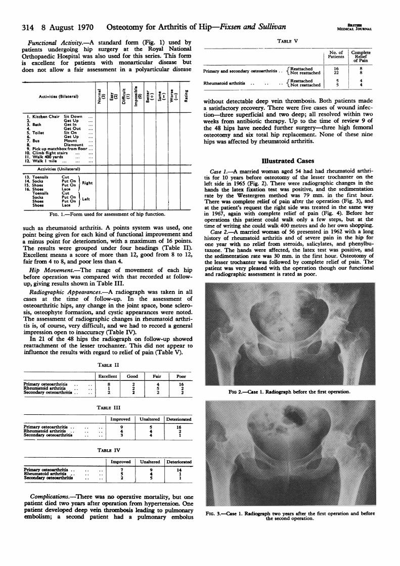
Case 1.-A married woman aged 54 had had rheumatoid arthri-tis for 10 years before osteotomy of the lesser trochanter on theleft side in 1965 (Fig. 2). There were radiographic changes in thehands the latex fixation test was positive, and the sedimentationrate by the Westergren method was 79 mm. in the first hour.There was complete relief of pain after the operation (Fig. 3), andat the patient's request the right side was treated in the same wayin 1967, again with complete relief of pain (Fig. 4). Before heroperations this patient could walk only a few steps, but at thetime of writing she could walk 400 metres and do her own shopping.
Case 2.-A married woman of 56 presented in 1962 with a longhistory of rheumatoid arthritis and of severe pain in the hip forone year with no relief from steroids, salicylates, and phenylbu-tazone. The hands were affected, the latex test was positive, andthe sedimentation rate was 30 mm. in the first hour. Osteotomy ofthe lesser trochanter was followed by complete relief of pain. Thepatient was very pleased with the operation though our functionaland radiographic assessment is rated as poor.
FIG 2.-Case 1. Radiograph before the first operation.
FIG. 3.-Case 1. Radiograph two years after the first operation and beforethe second operation.
BMnsMEICAL JOURNA
.~

8 August 1970 Osteotomy for Arthritis of Hip-Fixsen and Sullivan MUW..OUN" 315
~~~~~~~~~~.
FIG. 4.-Case 1. Radiograph three years after the first and one year afterthe second operation.
Discussion
This report aims to assess the place of a relatively minoroperation for pain in the hip caused by osteoarthritis andrheumatoid arthritis. In osteoarthritis there was completerelief of pain in 40% and partial relief in 40% of cases, but20% required further operation within three and a half years.These results compare reasonably favourably with the reliefof pain reported from high femoral osteotomy by Osborne andFahrni (1950) and Nicoll and Holden (1961). We are notadvocating this operation for osteoarthritis, but agree withBlount (1964) that it may have a place in the old and frailpatient. The results in cases of rheumatoid arthritis are veryencouraging. Of 10 cases, eight had complete relief and twopartial relief of pain. No patient has either required orwanted further surgery.There is considerable feeling that high femoral osteotomy
is not successful in cases of rheumatoid arthritis. This leaves
the main alternative operations of cup arthroplasty and totalhip replacement, both of which are major procedures. In ourexperience and that of Parsons (1969) total replacement incases of active rheumatoid arthritis has a relatively high rateof complication. Most British surgeons now hesitate to per-form total replacement in a young patient with rheumatoidarthritis. Osteotomy of the lesser trochanter, on the otherhand, is a relatively simple and safe procedure which can beperformed at any age. It does not prejudice the subsequentperformance of total replacement.We cannot explain why this procedure should be effective
in cases of rheumatoid arthritis. The work of Layani et al.(1959) suggested that tension in the iliopsoas muscle could be animportant source of pain in the hip, and, as already men-tioned, four patients with protrusio acetabulae in his serieshad good results. Benjamin (1969) showed that double osteo-tomy (osteotomy of the lower femur and upper tibia) couldcause regression of rheumatoid synovitis of the knee joint.We feel that for some obscure reason division of bone in theneighbourhood of the hip joint may cause a similar regres-sion.
We would like to thank Mr. J. N. Wilson for permission tofollow-up cases under his care and for encouragement in writingthis paper. We also wish to thank Mr. Whitley for the photo-graphs and Mrs. Glen Haig and the secretarial staff at the RoyalNational Orthopaedic Hospital for their help.
Proprietary preparations of the drugs mentioned in this artideindude: Fastocain, Lidothesin, Xylocaine (lignocaine withadrenaline); Butaphen, Butazolidin, Butazone, Flexazone, IA But(phenylbutazone).
REFERENCESBenjamin, A. (1969). Journal of Bone and Joint Surgery, 51B, 694.Blount, W. P. (1964). Journal of Bone and Joint Surgery, 46A, 1297.Cordier, G., Layani, F., and Gamier, H. (1960). Revue du Rhumatisme et des
Maladies Osteo-Articulaires, 27, 337.Layani, F., Cordier, G., Garnier, H., Roeser, J., and Paquet, J. (1959).
Rhumatologie, 11, 223.Nicoll, E. A., and Holden, N. T. (1961). Journal of Bone and Joint Surgery,
43B, 50.O'Malley, A. G. (1959). Journal of Bone and Joint Surgery, 41B, 888.Osborne, G. V., and Fahrni, W. H. (1950). Journal of Bone and Joint
Surgery, 32B, 148.Parsons, D. W. (1969). Journal of Bone and Joint Surgery, 51B, 564.Voss, C. (1956). Munchener medizinische Wochenschrift, 98, 954.
Immediate Effect on Cardiac Output of Reversion to Sinus Rhythmfrom Rapid Arrhythmias
J. S. WRIGHT,* M.B., M.R.C.P. ; J. FABIAN,t§ M.D. ; E. J. EPSTEIN,t M.D., M.R.C.P.
British Medical Journal, 1970, 3, 315-318
Summary: Cardiac output was estimated immediatelybefore and after conversion to sinus rhythm in nine
patients with rapid arrhythmias. Conversion was bysynchronized direct-current shock in eight patients, andby direct atrial wall stimulation in the other. In sevenpatients there was an immediate increase in cardiac out-put after restoration of sinus rhythm. The percentageincrease in output was directly proportional to the rate ofthe arrhythmia immediately before conversion (r=0.91,P<0.01). The critical heart rate, above which an immedi-ate increase in cardiac output might be expected on con-version to sinus rhythm, appeared in these patients to beabout 160 beats per minute.
*Senior Medical Registrar.t Late Medical Registrar.t Consultant Physician.Liverpool Regional Cardiac Centre, Sefton General Hospital, Liverpool 15.S Present address: First Medical Clinic, Charles University, Vlasska 36,
Prague 1, Czechoslovakia.
Introduction
The use of synchronized direct-current shock as described byLown et al. (1962) is now generally accepted as an Effectiveway of dealing with most cardiac arrhythmias (Friedberg,1966). Where an arrhythmia such as atrial fibrillation is slowor is readily controlled by drugs, electrical conversion is anelective procedure, and its success, in terms of a prolonged orpermanent return to sinus rhythm, is largely a matter ofselection of patients. It is now clear that lasting success willbe obtained only when the underlying cause of the arrhyth-mia has been removed, as after mitral valvotomy or the con-trol of thyrotoxicosis. At the other end of the scale are rapid,uncontrollable tachycardias of ventricular or supraventricularorigin, which are followed by obvious deterioration in thecondition of the patient, with hypotension, pulmonaryoedema, and disturbance of consciousness. Here electrical con-version is undertaken as a life-saving emergency procedure,and there is little difficulty selecting patients. We have been