outbreak of hydropericardium syndrome associated …jakraya.com/journal/pdf/4-vriarticle_2.pdf ·...
TRANSCRIPT

Veterinary Research International | April-June, 2014 | Vol 2 | Issue 2 | Pages 33-45 © 2014 Jakraya Publications (P) Ltd
VETERINARY RESEARCH INTERNATIONAL Journal homepage: www.jakraya.com/journal/vri
ORIGINAL ARTICLE
Outbreak of hydropericardium syndrome associated with ascites and liver rupture in caged broilers
Subhash Sharma1, R. K. Asrani1*, Geetanjali Singh2, Baldev R. Gulati3, P. K. Patil4 and V. K. Gupta1
1Department of Veterinary Pathology, 2Department of Veterinary Physiology and Biochemistry, Dr G C Negi College of Veterinary and Animal Sciences, CSK Himachal Pradesh Agricultural University, Palampur-176062, India. 3National Research Center on Equines, Sirsa Road, Hisar-125001, India. 4Department of Epidemiology and Preventive Veterinary Medicine, Guru Angad Dev Veterinary and Animal Science University, Ludhiana (Punjab), India.
*Corresponding Author: R. K. Asrani
Email: [email protected] Received: 11/04/2014 Revised: 09/05/2014 Accepted: 13/05/2014
Abstract An outbreak of hydropericardium syndrome (HPS) was recorded
with a sudden heavy mortality in 24-day-old caged broiler flock. An overall mortality of 57% was recorded with hydropericardium as a conspicuous lesion throughout the course of disease. Ascites and liver rupture were recorded in a large number of birds at the peak phase of outbreak. Microscopically, myocardial blood vessels showed massive disruption and vacuolar changes from day 3 of outbreak. In the liver, congestion, vacuolar changes and intranuclear inclusion bodies were observed from day 1 of outbreak. Liver hemorrhages and necrosis were more pronounced at day 5 of outbreak. Marked thickening of interlobular septae due to edema was characteristic lesion in the lungs throughout the course of disease. Renal tubules appeared markedly distended due to urate deposits in some of the birds from day 4 of outbreak. In the spleen, mild to severe reticuloendothelial cell hyperplasia was conspicuous finding. Mild to moderate depletion of lymphocytes was evident both in the bursa of Fabricius and spleen. Involvement of FAV-4 was confirmed by polymerase chain reaction coupled with restriction enzyme analysis and the disease was experimentally reproduced in 2-week old broiler chicks. Absence of direct oral-fecal cycle in the cage system of rearing suggested mechanical factors playing vital role in the spread of HPS within the flock. The occurrence of ascites in the present outbreak was linked to relatively slow development of disease process under the cage system.
Key words: Hydropericardium syndrome, ascites, liver rupture, pathogenesis, caged broilers.
Introduction Hydropericardium syndrome (HPS) is an
emerging viral disease of poultry. It is primarily a disease of broilers. HPS was reported for the first time from Angara Goth, Karachi, Pakistan, in August 1987 (Ahmad et al., 1989). Subsequently, the syndrome has spread to many parts of the world viz. Iraq (Abdul-Aziz and Al-Attar, 1991), Slovakia (Jantosovic et al., 1991), Mexico, Ecuador, Chile and Peru (Cowen et al., 1996), South and Central America (Shane, 1996), Russia (Borisov et al., 1997) and Japan (Abe et al., 1998). In India, HPS was first reported in the poultry belt of Jammu and Kashmir and Punjab in 1993 (Gowda and
Satyanarayana, 1994). The disease is locally known as “litchi heart disease” because the heart of the affected birds resembles the “deshelled litchi fruit”.
HPS is characterized by heavy mortality ranging from 20 to 75 per cent in healthy broiler flocks of 2-6 weeks age (Asrani et al., 1997; Khwaja et al., 1988). The striking postmortem findings of the disease are accumulation of clear or straw-coloured watery or jelly-like fluid in the pericardial sac, focal hepatic necrosis, swollen kidneys and congestion and oedema in lungs (Abdul-Aziz and Hasan, 1995; Asrani et al., 1997; Abe et al., 1998; Basher et al., 1999; Cheema et al., 1999). The characteristic histologic changes

Sharma et al…..Outbreak of hydropericardium syndrome associated with ascites and liver rupture in caged broilers
Veterinary Research International | April-June, 2014 | Vol 2 | Issue 2 | Pages 33-45 © 2014 Jakraya Publications (P) Ltd
34
include intranuclear basophilic or eosinophilic inclusion bodies in the hepatocytes and myocardial degeneration (Akhtar, 1994; Abe et al., 1998; Asrani et al., 1997). The histologic changes, viral isolation studies and experimental reproduction of disease have suggested group 1 fowl adenovirus (FAV) as the causative agent of HPS (Hess et al., 1999; Hess, 2000). Several studies on further molecular characterization of group 1 fowl adenovirus from the cases of HPS have suggested FAV serotype 4 as the sole cause of disease (Mazaheri et al., 1998; Toro et al., 1999; Dahiya et al., 2002).
This paper describes the sequential pathological changes in a naturally occurring outbreak of HPS in caged broiler chickens with a detail insight on some of the important aspects that might contribute in the understanding of its pathogenesis. Materials and Methods History and clinical examination
An outbreak of HPS was recorded in a broiler farm of the Department of Animal Nutrition, CSK Himachal Pradesh Agricultural University, Palampur, India. The farm had a total of 328 broiler chickens (Cobb breed) which were being reared in cages housed in a single large room. The age of the birds recorded at the time of first appearance of disease was 24 days. These birds had been vaccinated for Newcastle disease at day one of their age by F1 strain vaccine. There was no history of any other disease or mortality in the recent past. Both broiler starter (given up to 3 weeks of age) and finisher ration (being given at the time of outbreak) were prepared by the Department of Animal Nutrition, COVAS, CSK HPKV, Palampur. The ingredients composition/100 kg broiler starter was maize-48 kg, soya flakes-18 kg, ground nut cake-14 kg, sunflower cake-8 kg, fish meal-5 kg, molasses-5 kg, digestible crude protein-1 kg, lime power-1 kg while that in broiler finisher ration was maize-50 kg, soya flakes-12 kg, sunflower cake-12 kg, groundnut cake-10 kg, fish meal-5 kg, molasses-5 kg, mustard cake-4 kg, digestible crude protein-1 kg and lime power-1 kg. Both starter and finisher rations were being supplemented adequately with vitamins and minerals. During the course of outbreak, the birds were closely observed daily for clinical signs and mortality up to 3 weeks after appearance of HPS i.e. up to day 45 of their age. Gross and histopathologic changes
All those birds which died during the course of outbreak were subjected to detailed necropsy examination. Gross lesions were carefully recorded. Representative pieces of tissues from heart, lungs,
liver, kidneys, proventriculus, intestine, spleen, thymus and bursa of Fabricius were collected in 10% neutral-buffered formalin for histopathological studies from 10 birds on each day of mortality up to day 8 of the outbreak. At subsequent stages i.e. on day 9, 10 and 11 of outbreak, material for histopathological studies was collected from 5, 4 and 2 birds, respectively. Representative pieces of liver tissues from 3 to 5 birds on each day of mortality were also collected in sterile vials and preserved at -20oC until used for further studies. The formalin fixed tissue segments were processed for paraffin embedding technique, and cut sections were stained with haematoxylin and eosin stain for light microscopic studies (Luna, 1968). Agar Gel Immunodiffusion test (AGID)
AGID test was performed on tissue samples to detect the presence of viral antigen as per the method of Kumar et al. (1997). Briefly, 1% agarose (Sisco Research Lab. Pvt. Ltd., Mumbai) was prepared in phosphate buffered saline (Sigma, St. Louis, MO, USA) solution (pH 7.4) containing additional 8% sodium chloride. The agarose gel was prepared on microscopic slides. The central well was filled with freshly reconstituted freeze dried antiserum raised in chicken against FAV 4 (S-98/Gurgaon) isolated earlier from an outbreak of HPS from Gurgaon, Haryana, India (Dahiya et al., 2002). Test samples were filled in the surrounding wells along with positive control (Chicken embryo liver cell culture-propagated FAV 4 (S-98/Gurgaon) (Dahiya et al., 2002). Following incubation at 37oC for about 24-48 hr, gels were stained in 0.25 per cent Coommassie brilliant blue (BIO-RAD, USA) to visualize the line of precipitation confirming the identity. Polymerase chain reaction (PCR) and Restriction fragment length polymorphism (RFLP)
Extraction of DNA was done from 500 µl of liver tissue samples using ‘QiAamp DNA extraction kit’ (Qiagen, USA) as per the manufacturer’s instructions. Purified DNA segments were amplified using primer pairs H1/H2 (H1:5’- TGGACATTGGGGGCGACCTA-3’ H2:51- AAGGGATTGACGTTGTCCA-3’ and H3/H4 (H3:51-AACGTCAACCCCTTCAACCACC-31 H4: 51-TTGCCTGTGGCGAAAGGCG-31) specific for fowl adenovirus as per the method of Raue and Hess (1998). Following PCR, confirmation was done by observation of specific amplification product in 1 % agarose gel as per Sambrook et al. (1989). PCR products were purified using the QIA quick gel extraction kit (Qiagen, USA) and were subjected to restriction endonuclease enzyme digestion using HaeII (Fermentas Life

Sharma et al…..Outbreak of hydropericardium syndrome associated with ascites and liver rupture in caged broilers
Veterinary Research International | April-June, 2014 | Vol 2 | Issue 2 | Pages 33-45 © 2014 Jakraya Publications (P) Ltd
35
Sciences, USA) for H1/H2 amplified PCR products and HpaII (MBI, Fermentas, USA) for H3/H4 amplified PCR products according to manufacturer recommendations. Digested products along with DNA ladder were electrophoresed in 2% (w/v) agarose gel. The fragment sizes were compared with standard molecular weight marker GeneRuler DNA ladder 100 bp plus (Fermentas Life Sciences, USA).
Bacteriological isolations and serological studies
Representative pieces of liver tissues from 3 to 5 dead birds on day 5, 6, 7, and 8 of outbreak were subjected to bacteriological isolations in selenite broth and brilliant green agar (BGA) plates. Heart blood samples were collected from 10 randomly selected birds from HPS recovered flock at 45 days of their age (i.e. 3 weeks after appearance of first case of HPS); serum was separated and tested for Salmonella Gallinarum-Pullorum antibodies by a whole blood plate agglutination test using Salmonella Pullorum-coloured antigen (Indian Veterinary Research Institute, Izatnagar, U.P., India). Similarly, the serum samples were tested for antibodies to infectious bursal disease using vaccine strain as antigen source (Venkateshwara Hatcheries, India). Analysis of feed samples
Representative feed samples from broiler starter and finisher rations were submitted to Animal Feed Analytical and Quality Control Laboratory, Veterinary Hospital Campus, Trichy Road, Namakkal, Tamil Nadu, India for analysis of common mycotoxins. Experimental reproduction of HPS
Liver samples from HPS affected birds collected during outbreak were cut into small pieces, triturated in pestle and mortar with sterile sea sand to make 20 per cent (w/v) suspension in sterile PBS (pH 7.2) supplemented with streptomycin at 2 mg/ml and penicillin at 2000 IU/ml. The homogenate suspensions were centrifuged at 8000 rpm for 30 min at 4 C in the refrigerated centrifuge. The supernatant was removed and checked for its sterility on blood agar and brilliant green agar (BGA) plates followed by the presence of FAV antigen by AGID prior to inoculation in the broiler chicks. Thirteen day-old broiler chicks were procured from commercial hatchery (Uttam Hatcheries, Kangra, India) with no history of HPS or FAV infection in the flock. Before housing, the experimental rooms, brooder batteries, and cages were thoroughly cleaned with 2.5% phenol and were subsequently fumigated with formaldehyde gas. After experimental infection, cleaning of cages was done with phenol at least twice daily. The infected and control groups were
kept in separate and distantly located rooms. The control groups were always attended to first with respect to cleaning, feeding, watering, and so forth. Chicks were maintained on chick mash (Department of Animal Nutrition, COVAS, CSK HPKV, Palampur, India) from day 1 until the end of the experiment. Feed was autoclaved for 15 min at 15 pounds pressure before giving to birds. Boiled and subsequently cooled water was given to the birds throughout the experiment. Feed and water was given ad libitum, and no medication was given during the entire period of the experiment. After two weeks of rearing in the animal house of the department, the birds were randomly divided into two groups i.e. HX and CX, containing 10 and 3 birds, respectively. Each bird in group HX was injected subcutaneously (S/C) with 0.5 ml of HPS inoculum while the birds in control group CX were similarly inoculated S/C with 0.5 ml of 20% (w/v) liver homogenate from a 4-weeks-old broiler chicken tested negative to FAV antigen by AGID. The birds in both the groups were closely observed for clinical signs, mortality, gross lesions and histopatholgical changes in the liver and attempts for bacterial isolations were also made. The animal care and experimental protocol were duly approved by the University and by the Committee for the Purpose of Control and Supervision of Experiments on Animals (CPCSEA).
Results Clinical signs and mortality
The disease struck suddenly with heavy mortality in 24-days old broiler flock. Apparently the birds were healthy. The clinical signs of dullness and depression were reported with beak resting on the cage floor. The affected birds were exhibiting rapid shallow abdominal respiration and did not show any interest in feed and water. A few birds from day 5 onward also revealed mild diarrhoea but this was not a consistent observation. The mortality pattern observed during the natural disease is graphically depicted in Fig. 1.
0
5
10
15
20
25
30
35
40
45
50
1 2 3 4 5 6 7 8 9 10 11
Day of outbreak
No
of
bir
ds
die
d
Fig. 1: Mortality pattern observed during the outbreak of hydropericardium syndrome in caged broiler chickens

Sharma et al…..Outbreak of hydropericardium syndrome associated with ascites and liver rupture in caged broilers
Veterinary Research International | April-June, 2014 | Vol 2 | Issue 2 | Pages 33-45 © 2014 Jakraya Publications (P) Ltd
36
The mortality started suddenly with ten to eleven birds dying at each day of the outbreak up to first three days. The mortality graph, thereafter, showed a sharp rise and peak mortality (13.4%) was observed on day 6 of outbreak before exhibiting a declining trend from day 7 of outbreak. A total of 187 birds (57%) died during the course of outbreak. The mortality was recorded with typical gross lesions up to day 11 of the outbreak. Gross lesions Heart: Hydropericardium was conspicuous lesion observed throughout the course of disease. Most of the birds exhibited mild to moderate hydropericardium in first three days of outbreak. From day 4 onward, birds showed severe hydropericardium. During the first two days of outbreak, fluid in the pericardial sac was watery to slight gelatinous and straw-coloured, but with
the progression of the disease it became more gelatinous and yellowish coloured (Fig. 2). Besides hydropericardium, petechial haemorrhages were observed on the epicardium. On day 4 and 5, there was also slight greyish discolouration of epicardium and myocardium along with yellowish discolouration of pericardial fat. Myocardial haemorrhages were also - noticed in a few birds. The haemorrhages were more severe on day 6 of outbreak and were observed in a large number of birds frequently involving endocardium. A mild to moderate hypertrophy of the right ventricle was also observed in 13 birds on day 6 of outbreak. Yellowish discolouration of pericardial fat was a consistent finding. The vena cavae were fragile and showed yellowish discolouration in majority of the birds on day 6 of outbreak. In later days of the outbreak i.e. from day 7 onward, almost similar lesions were observed as noticed in the earlier stages. Some of the birds, however, also revealed rounding of the heart apex.
Table 1: Hydropericardium and ascites of variable intensity in broiler chickens# naturally affected with hydropericardium syndrome (HPS)*
Type of lesion Intensity
score Day of outbreak
1 2 3 4 5 6 7 8 9 10 11 Hydropericardium + 4** 4 2 3 3 1 4 6 1 1 0 ++ 4 5 4 5 10 8 3 0 0 0 0 +++ 1 1 2 8 15 26 11 5 4 2 2 Ascites + 0 0 0 0 5 10 6 1 1 0 0 ++ 0 0 0 0 0 0 6 0 0 1 0 +++ 0 0 0 0 0 0 0 0 0 0 0
+ = mild; ++ = moderate; +++ = severe; # Initial flock size = 328; *Period of HPS outbreak between 19th and 29th July, 2003. **Number of birds showing lesions of variable intensity Table 2: Gross hepatic lesions of variable intensity in broiler chickens# naturally affected with hydropericardium syndrome (HPS)*
Type of lesion Intensity
score Day of outbreak
1 2 3 4 5 6 7 8 9 10 11 Congestion and + 5** 2 2 4 6 3 7 0 0 0 0 hemorrhages ++ 0 3 2 4 8 4 6 1 2 1 0 +++ 0 5 1 12 19 31 8 4 2 1 1 Enlargement + 5 1 1 5 8 5 0 1 1 0 0 ++ 1 0 0 5 9 5 7 3 2 0 0 +++ 0 0 0 9 16 26 12 7 3 2 1 Pale + 4 4 1 2 3 5 4 1 0 0 0 discoloration ++ 1 2 1 3 3 5 4 1 0 1 0 +++ 0 1 0 1 3 6 5 2 5 2 1 Necrosis 0 0 0 3 11 11 10 5 0 2 1 Rupture 0 0 0 1 15 11 3 1 0 1 0
+ = mild; ++ = moderate; +++ = severe; # Initial flock size = 328; *Period of HPS outbreak between 19th and 29th July, 2003. **Number of birds showing lesions of variable intensity
Sharma et al…..Outbreak of hydropericardium syndrome associated with ascites and liver rupture in caged broilers
Veterinary Research International | April-June, 2014 | Vol 2 | Issue 2 | Pages 33-45 © 2014 Jakraya Publications (P) Ltd
37
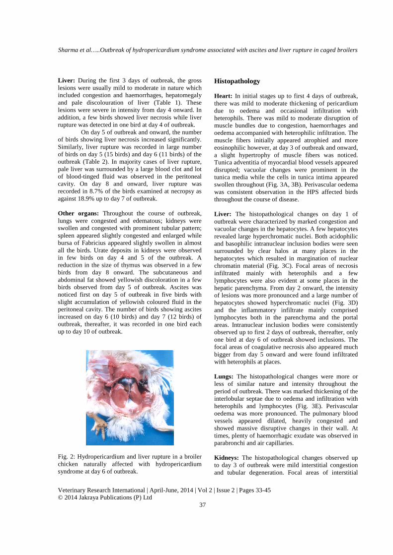
Liver: During the first 3 days of outbreak, the gross lesions were usually mild to moderate in nature which included congestion and haemorrhages, hepatomegaly and pale discolouration of liver (Table 1). These lesions were severe in intensity from day 4 onward. In addition, a few birds showed liver necrosis while liver rupture was detected in one bird at day 4 of outbreak.
On day 5 of outbreak and onward, the number of birds showing liver necrosis increased significantly. Similarly, liver rupture was recorded in large number of birds on day 5 (15 birds) and day 6 (11 birds) of the outbreak (Table 2). In majority cases of liver rupture, pale liver was surrounded by a large blood clot and lot of blood-tinged fluid was observed in the peritoneal cavity. On day 8 and onward, liver rupture was recorded in 8.7% of the birds examined at necropsy as against 18.9% up to day 7 of outbreak. Other organs: Throughout the course of outbreak, lungs were congested and edematous; kidneys were swollen and congested with prominent tubular pattern; spleen appeared slightly congested and enlarged while bursa of Fabricius appeared slightly swollen in almost all the birds. Urate deposits in kidneys were observed in few birds on day 4 and 5 of the outbreak. A reduction in the size of thymus was observed in a few birds from day 8 onward. The subcutaneous and abdominal fat showed yellowish discoloration in a few birds observed from day 5 of outbreak. Ascites was noticed first on day 5 of outbreak in five birds with slight accumulation of yellowish coloured fluid in the peritoneal cavity. The number of birds showing ascites increased on day 6 (10 birds) and day 7 (12 birds) of outbreak, thereafter, it was recorded in one bird each up to day 10 of outbreak.
Fig. 2: Hydropericardium and liver rupture in a broiler chicken naturally affected with hydropericardium syndrome at day 6 of outbreak.
Histopathology Heart: In initial stages up to first 4 days of outbreak, there was mild to moderate thickening of pericardium due to oedema and occasional infiltration with heterophils. There was mild to moderate disruption of muscle bundles due to congestion, haemorrhages and oedema accompanied with heterophilic infiltration. The muscle fibers initially appeared atrophied and more eosinophilic however, at day 3 of outbreak and onward, a slight hypertrophy of muscle fibers was noticed. Tunica adventitia of myocardial blood vessels appeared disrupted; vacuolar changes were prominent in the tunica media while the cells in tunica intima appeared swollen throughout (Fig. 3A, 3B). Perivascular oedema was consistent observation in the HPS affected birds throughout the course of disease. Liver: The histopathological changes on day 1 of outbreak were characterized by marked congestion and vacuolar changes in the hepatocytes. A few hepatocytes revealed large hyperchromatic nuclei. Both acidophilic and basophilic intranuclear inclusion bodies were seen surrounded by clear halos at many places in the hepatocytes which resulted in margination of nuclear chromatin material (Fig. 3C). Focal areas of necrosis infiltrated mainly with heterophils and a few lymphocytes were also evident at some places in the hepatic parenchyma. From day 2 onward, the intensity of lesions was more pronounced and a large number of hepatocytes showed hyperchromatic nuclei (Fig. 3D) and the inflammatory infiltrate mainly comprised lymphocytes both in the parenchyma and the portal areas. Intranuclear inclusion bodies were consistently observed up to first 2 days of outbreak, thereafter, only one bird at day 6 of outbreak showed inclusions. The focal areas of coagulative necrosis also appeared much bigger from day 5 onward and were found infiltrated with heterophils at places. Lungs: The histopathological changes were more or less of similar nature and intensity throughout the period of outbreak. There was marked thickening of the interlobular septae due to oedema and infiltration with heterophils and lymphocytes (Fig. 3E). Perivascular oedema was more pronounced. The pulmonary blood vessels appeared dilated, heavily congested and showed massive disruptive changes in their wall. At times, plenty of haemorrhagic exudate was observed in parabronchi and air capillaries. Kidneys: The histopathological changes observed up to day 3 of outbreak were mild interstitial congestion and tubular degeneration. Focal areas of interstitial
Sharma et al…..Outbreak of hydropericardium syndrome associated with ascites and liver rupture in caged broilers
Veterinary Research International | April-June, 2014 | Vol 2 | Issue 2 | Pages 33-45 © 2014 Jakraya Publications (P) Ltd
38
nephritis with lymphocytes as chief inflammatory cells were also observed in some of the birds. At subsequent stages of the outbreak, similar changes were observed, however, intensity was more pronounced. The tubules appeared markedly distended in some of the birds after day 4 of outbreak due to urate deposits infiltrated with heterophils and a few mononuclear cells (Fig. 3F). Sub capsular haemorrhages were also observed in a few birds at day 6 of outbreak. Spleen: In the first three days of outbreak, there was mild to severe reticuloendothelial cell hyperplasia associated with mild to moderate depletion of lymphoid cells. Karyorrhexis in white pulp lymphocytes was also observed. On day 4 and onward, reticuloendothelial cell hyperplasia around periarteriolar sheaths outlined the white pulp areas. Bursa of Fabricius: Mild to moderate depletion of lymphocytes from medullary region of follicles was conspicuous observation throughout. Mild heterophilic infiltration in bursal follicles was evident in some of the birds at day 3 of outbreak. Thymus: Only noticeable microscopic lesion in thymus observed at almost all stages of the outbreak was mild depletion of lymphocytes both from the cortex and the medullary region besides mild to moderate degree of congestion. Intestine: Only noticeable microscopic lesion in the intestine observed at almost all stages of the outbreak included catarrhal degeneration characterized by mild to moderate goblet cell hyperplasia. These changes were invariably accompanied with mild to moderate sloughing of villous epithelium. Molecular characterization of FAV by PCR-RFLP
Involvement of FAV was confirmed by PCR. Amplification of DNA with H1/H2 and H3/H4 primer sets resulted in a fragment of approximately 1219 bp and 1319 bp, respectively. Restriction enzyme analysis of the PCR amplicon resembled to the digestion pattern of FAV-4. Two fragments of ~1000 bp and ~200 bp were generated with HaeII digestion of H1/H2 PCR product and three fragments of ~500 bp and ~200 bp and ~100 bp were generated with HpaII digestion of H3/H4 PCR product. Experimental disease
None of the birds from the control group showed clinical signs, mortality, gross and histopathological lesions during the 10 days observation period. All the birds in the infected group
showed clinical signs of anorexia, dullness and depression just prior to death. A mortality of 100 per cent was recorded in this group. Mortality started 14 hr post-infection and all the birds had died by 34 hr post-infection. Hydropericardium due to the accumulation of sticky gelatinous straw coloured or slightly yellowish fluid in the pericardial sac was recorded in all the experimentally infected broilers. Heart gave resemblance to typical deshelled ‘litchi’ fruit 22 hr post-infection (Fig. 4A). Livers showed varying degree of congestion, haemorrhages and enlargement. Necrotic foci on liver were recorded in two birds. The lungs were congested and oedematous in all the birds. Spleen appeared slightly enlarged and congested in birds that died 16 hours post-infection. Microscopically, a large number of hepatocytes revealed large hyperchromatic nuclei and basophilic intranuclear inclusion bodies surrounded by a clear halo at 20 hr post-infection and later (Fig. 4B). Bacteriological isolations, serological studies and analysis of feed samples
No bacteria could be isolated from the liver tissues of either the naturally dead birds at the face of outbreak or from the birds in which the HPS was experimentally induced. The serum samples were also found to be free from antibodies to Salmonella Gallinarum-Pullorum and infectious bursal disease when tested by AGID. The level of aflatoxin B1 was detected at 12 ppb in broiler finisher ration only while all other feed samples were found to be free from aflatoxins, ochratoxin A, citrinin, T-2 toxin and zearalenone. Discussion
In the present study, HPS struck suddenly the fast growing healthy caged broilers of 3 weeks. The occurrence of disease in well grown and healthy broilers also suggests the possible role of some dietary factors in increasing the susceptibility to HPS. The stress of rapid growth pressure due to feeding of high-density protein rich diets in the present study may have been one of the enhancing factors associated with development of HPS. Gowda (1994) cited a maximum mortality of 60% due to HPS in chickens fed with rapeseed and fish meal and a least mortality of 12% was recorded in those fed with rice polish and maize. Both starter and finisher rations in the present outbreak revealed fish meal in addition to soya flakes and groundnut cake.
HPS spreads rapidly from flock to flock and from farm to farm (Cowen, 1992) and oral-fecal route has been suggested as the possible mechanism for the rapid spread of disease under field conditions (Shafique and Shakoori, 1994; Abdul-Aziz and Hasan, 1995;

Sharma et al…..Outbreak of hydropericardium syndrome associated with ascites and liver rupture in caged broilers
Veterinary Research International | April-June, 2014 | Vol 2 | Issue 2 | Pages 33-45 © 2014 Jakraya Publications (P) Ltd
39
Fig. 3: Microscopic lesions in an outbreak of hydropericardium syndrome in broiler chickens. (A) Myocardial blood vessel (BV). Disrupted and vacuolated tunica adventitia and tunica media (arrows) at day 2. H&E stain, x132. (B) Myocardium. Vacuolated myocardium with degenerated blood vessels (BV) at day 3. H&E stain, x66. (C) Liver. Intranuclear basophilic inclusion bodies (arrows) in the hepatocytes at day 1. H&E stain, x132. (D) Liver. A large number of hepatocytes showing karyomegaly and hyperchromatic nuclei (arrows) at day 2. H&E stain, x132. (E) Lung. Severe interlobular edema (Lb) and disruptive changes (arrows) in the wall of blood vessels (BV) at day 1. H&E stain, x33. (F) Kidney. Tubular distension due to urate deposits (arrows) at day 4. H&E stain, x33.
A
BV
BV BV
BV
B
C D
E F
Lb
BV
BV
BV

Sharma et al…..Outbreak of hydropericardium syndrome associated with ascites and liver rupture in caged broilers
Veterinary Research International | April-June, 2014 | Vol 2 | Issue 2 | Pages 33-45 © 2014 Jakraya Publications (P) Ltd
40
Fig 4: Experimentally induced hydropericardium syndrome in 2-weeks-old broiler chickens. (A) Heart. Typical litchi heart appearance at 28 hours post-infection. (B) Liver. Intranuclear basophilic inclusion bodies with margination of chromatin material (large arrow), karyocytomegaly and hyperchomatic nuclei (small arrows) at 24 hours post-infection. H&E stain, x330. Cowen et al., 1996). But the rapid spread of disease with heavy mortality under the cage system of rearing with no evidence of direct oral-faecal transmission suggested some other means of spread playing important role. Earlier we could successfully induce HPS experimentally in broiler chicks by supernatant from HPS positive liver homogenate accidentally left out at room temperature (27oC) for about a week (R K Asrani, personal communication). If the causative virus is resistant and survives outside host for longer periods, a respiratory route in the absence of direct oral-faecal cycle might explain the rapid spread of the disease under the cage system. Thakur and Grewal (1999) have earlier reported basophilic intranuclear inclusions in the alveolar and the epithelial cells of secondary parabronchi suggesting that the virus is capable of replicating in the lungs tissue. In the present study, however, there was no such evidence of virus replication or presence of inclusions in the lungs tissue. Alternatively, heavily contaminated environment might have been a direct source of infection by contaminating feed, water, equipments, and cages and spread through other mechanical means such as movement of workers cannot be ruled out. Akhtar (1992) have earlier suggested that HPS is transmitted horizontally by mechanical means. Further studies on transmission mechanisms of HPS merit serious consideration.
In the present study, involvement of group 1 FAV was confirmed by AGID and PCR using liver tissue homogenate from the HPS affected birds. Amplification of FAV hexon gene fragments in PCR for diagnosis of FAV has been employed by various research workers (Raue and Mess, 1998; Jiang et al.,
1999; Xie et al., 1999; Singh et al., 2002). Further, confirmation of FAV-4 involvement was done by restriction enzyme analysis of the PCR amplicon using HaeII and HpaII enzymes. Many workers have described hexon based PCR’s combined with restriction enzyme analysis for rapid detection and differentiation of fowl adenovirus (Raue and Hess, 1998; Hess, 2000; Singh et al., 2002). A number of workers have suggested FAV-4 as the sole cause of this syndrome (Abe et al., 1998; Dahiya et al., 2002; Ganesh et al., 2004). PCR coupled with RFLP did not give any indication of involvement of serotypes other than FAV-4 in the present outbreak.
Nakamura et al. (2003) induced HPS using serotype 8 group 1 avian adenovirus strains of inclusion body hepatitis in 3-wk-old cyclophosphamide-treated chickens while they could not induce either hydropericardium or mortality in non-treated chickens. The results of present study, however, indicated that prior immunosuppression might not be a necessary factor in the development of HPS. It is supported by the facts that the presence of mycotoxins was ruled out by the analysis of feed samples for common mycotoxins. The presence of aflatoxins in the feed at higher concentrations than 20 ppb has been found to be associated with typical lesions of inclusion body hepatitis in addition to hydropericardium (Singh et al., 1996). The level of aflatoxins in feed samples was found to be much lower (< 20 ppb) in the present study. Secondly, serum samples from HPS recovered birds were tested negative for IBD antibodies and none of the birds from the outbreak investigated for gross and histopathological lesions revealed atrophy of bursa
A B A

Sharma et al…..Outbreak of hydropericardium syndrome associated with ascites and liver rupture in caged broilers
Veterinary Research International | April-June, 2014 | Vol 2 | Issue 2 | Pages 33-45 © 2014 Jakraya Publications (P) Ltd
41
of Fabricius throughout 11 days period of outbreak. Thirdly, HPS was successfully reproduced experimentally in two weeks old broilers within 14 hr of inoculation.
An interesting observation that emerged out from the present studies on natural outbreak in caged broilers was the presence of lesions like right ventricular hypertrophy (RVH) and ascites which were observed from day 5 onward. None of the earlier workers reported either RVH or ascites in HPS. The occurrence of RVH and ascites in the present study may be attributed to relatively slow development of disease process under the cage system. The mortality graph started climbing on day 4 and the mortality peak was observed on day 6 of outbreak in the present study as compared to the previous study in which the peak mortality was observed on day 3 or 4 of outbreaks (Asrani et al., 1997). This may be partially linked to the absence of direct oral-faecal route in the spread of disease. Unlike previous reports in which hydropericardium and lung oedema have been considered as the major factors associated with sudden heavy and acute mortality due to HPS, liver rupture was the additional feature in the present study which possibly might have contributed to heavy mortality due to HPS in the caged broilers. In the birds reared under the cage system, liver tend to be rather more fragile due to the accumulation of fat and the stress of infection might have aggravated this condition as liver rupture coincided with the peak phase of HPS. The concurrence presence of fatty liver and kidney syndrome was ruled out based on the facts that not even a single case of liver rupture was recorded in the present study after day 10 of outbreak (i.e. 34 days old) until the end of the observation period in spite of the same diet and secondly, the kidney tissues never revealed fatty changes on histopathological examination. The ascites was attributed to right ventricular insufficiency following persistence pressure of the volume causing increased workload on the heart.
The yellowish discolouration of pericardial fluid may be attributed to more severe liver damage in later stages of the disease while its gelatinous appearance was possibly due to the escape of fibrinogen from the highly permeable blood vessels. The liver lesions may be ascribed to the development of hydropericardium because liver is the major organ for biosynthesis of proteins in the body. Damage to the liver lead to the decreased protein synthesis, hence, decreased colloidal osmotic pressure of plasma causing accumulation of fluid in the pericardial sac.
The oedematous disruption of myocardium and its blood vessels were suggestive of increased vascular permeability (Asrani et al., 1997; Kumar et al., 2004; Meenakshi et al., 2005) and the vascular changes were
more severe on day 5 and 6 of the outbreak. At the same time hydropericardium was also very severe.
In the present study, outbreak of HPS was recorded in well nourished caged broilers. The flow diagram hypothesizing on possible pathogenesis of HPS is presented in Fig. 5. The well growing broilers have a high basal metabolic rate owing to feeding of high-density protein rich ration. Such a pressure of rapid growth not only increases the body demand for oxygen but also the cardiac output. The workload on heart is possibly further magnified due to liver damage in HPS resulting in decreased colloidal osmotic pressure of plasma. The heart being the very active organ cannot sustain the reduced osmotic pressure for so long and possibly the vascular endothelium of myocardium is damaged. The disruption of myocardial blood vessels was a consistent observation in the present study. These changes favour the escape of fluid to collect in the pericardial sac so sudden that the death is warranted within a few hours of HPS infection possibly due to cardiac temponade and hypertensive lung oedema. As litchi heart cannot pump the blood effectively, thus increases the intravascular pulmonary pressure. The damage to pulmonary vessels and lung oedema noticed in the present study may have been the result of persistent increased intravascular pulmonary pressure. The insufficient forward pumping of blood by litchi heart is further supported by the fact that urea nitrogen values are significantly increased in the HPS-affected birds (Asrani et al., 1997) possibly because renal blood flow and glomerular filtration pressure are reduced and the excretion of urea nitrogen is impaired. Also it seems probable that a fall in fluid volume stimulates the release of adrenal cortex hormone aldosterone which in turn causes increased absorption of sodium chloride and water by the renal tubules. Hypertension due to renal malfunction possibly increases the work load on heart due to elevated sodium level and water retention (Qureshi, 1989). A reduction in the renal perfusion was possibly associated with the degenerative changes in the kidneys and increases in the serum creatinine levels (Asrani et al., 1997). Conclusion
The outbreak of hydropericardium syndrome (HPS) may be associated with heavy mortality due to the development of hydropericardium, ascites and liver rupture in the caged birds. Absence of direct oral-fecal cycle in the cage system of rearing suggested mechanical factors playing vital role in the spread of HPS within the flock. The occurrence of ascites in the present outbreak was linked to relatively slow development of disease process under the cage system. The disease pattern in the caged birds has clearly -

Sharma et al…..Outbreak of hydropericardium syndrome associated with ascites and liver rupture in caged broilers
Veterinary Research International | April-June, 2014 | Vol 2 | Issue 2 | Pages 33-45 © 2014 Jakraya Publications (P) Ltd
42
HPS agent
Infects Under rapid Healthy well grown broilers High density protein ration Growth pressure High demand for In caged birds Body O2 Already fatty & fragile liver Decreased colloid osmotic pressure of plasma Hypoxia Vascular damage (Heart, being active organ, suffers maximum vascular damage) Increased vascular permeability in heart Sudden massive Cardiac accumulation temponade
Pressure of pericardial fluid Haemoconcentration Increased cardiac Decreased cardiac output intravascular pressure
Sudden decrease in left ventricle output Reduced renal perfusion Pressure is mounted in Pulmonary veins & transmitted to capillaries Increased tubular reabsorption for water and sodium chlorides Compensatory Hypervolaemia
Increased work load on Increased pulmonary the compromised heart arterial pressure contd. next page
Degenerative changes in the walls of these vessels
Stress factors • Poor
ventilation
Hydropericardium
Extensive liver damage
Hypertensive lung edema
Kid
ney
lesi
ons
Liver rupture
Death

Sharma et al…..Outbreak of hydropericardium syndrome associated with ascites and liver rupture in caged broilers
Veterinary Research International | April-June, 2014 | Vol 2 | Issue 2 | Pages 33-45 © 2014 Jakraya Publications (P) Ltd
43
Increased work load on the compromised heart (contd. from previous page)
Hypertrophy of right ventricles Increased blood pressure in vena cava Portal hypertension Fig. 5: Flow diagram suggesting pathogenesis of hydropericardium and ascites due to
hydropericardium syndrome (HPS) in caged broilers suggested that active liver damage in HPS is not central to the pathogenesis of ascites rather diminished right ventricular output and resultant portal hypertension plays the key role. The results of present study further suggest that prior immunosuppression may not be a necessary factor in the development of HPS. It is
concluded from the study that HPS is an infectious metabolic disorder which mainly affects the fast growing healthy broilers in which the stress of rapid growth pressure due to the feeding of high-density protein rich diets may prove to be a vital contributory factor in the development of this disorder.
References Abdul-Aziz TA and Al-Attar MA (1991). New syndrome
in Iraqi chicks. Veterinary Record, 129: 272. Abdul-Aziz TA and Hasan SY (1995). Hydropericardium
syndrome in broiler chickens: its contagious nature
and pathology. Research in Veterinary Science, 59: 219-221.
Abe T, Nakamura K, Tojo H, Mase M, Shibahara T, Yamaguchi S and Yuasa N (1998). Histology,
Stress factors - In deep litter system of housing, overcrowded conditions or birds frightened by the movement of workers further contribute to increase work load on heart leading to sudden death due to massive hydropericardium and hypertensive lung edema
In cage system of rearing with limited scope for movement, birds can sustain an increase cardiac work load for relatively longer period giving time for the development of ascites
Ascites

Sharma et al…..Outbreak of hydropericardium syndrome associated with ascites and liver rupture in caged broilers
Veterinary Research International | April-June, 2014 | Vol 2 | Issue 2 | Pages 33-45 © 2014 Jakraya Publications (P) Ltd
44
immunohistochemistry and ultra-structure of hydropericardium syndrome in adult broiler breeders and broiler chicks. Avian Diseases, 42: 606-612.
Ahmad I, Afzal M, Malik MI, Hussain Z and Hanif W (1989). Studies on the disease pattern and etiology of hydropericardium syndrome (Angara disease) in broiler chickens in Pakistan. Pakistan Journal of Agricultural Research, 10: 195-199.
Akhtar S (1994). Hydropericardium syndrome in broiler chickens in Pakistan. World’s Poultry Science Journal, 50: 177-182.
Akhtar S (1992). Studies on the rate of lateral spread of hydropericardium syndrome agent (s). In: Etiology, pathogenesis and control of hydropericardium syndrome in poultry. Proceedings of the Board on Science and Technology for International Development, Washington DC.
Asrani RK, Gupta VK, Sharma SK, Singh SP and Katoch RC (1997). Hydropericardium hepatopathy syndrome in Asian poultry. Veterinary Record, 141: 271-273.
Basher HA, Al-Sadi HI and Ismail HK (1999). Pathological study of hydropericardium syndrome in broiler chickens in Ninevha Province, Iraq. Iraqi Journal of Veterinary Science, 12: 289-294.
Borisov VV, Borisov AV and Gusev AA (1997). Hydropericardium syndrome in chicken in Russia. Proceedings of the 10th International Congress of the World Veterinary Poultry Association, Budapest, Hungary. pp. 258.
Cheema AH, Ahmad J and Afzal M (1989). An adenovirus infection of poultry in Pakistan. Revue scientifique et technique (International Office of Epizootics), 8: 789-795.
Cowen BS (1992). Inclusion body hepatitis-anaemia and hydropericardium syndrome: aetiology and control. World’s Poultry Science Journal, 48: 247-253.
Cowen BS, Lu H, Weinstock D and Castro AE (1996). Pathogenicity studies of fowl adenoviruses isolated in several regions of the world. Proceedings of the International Symposium on Adenovirus and Reovirus Infections in Poultry, Rauischholzhauzen, Germany. pp.79-88.
Dahiya S, Srivastava RN, Hess M and Gulati BR (2002). Fowl adenovirus serotype 4 associated with outbreaks of infectious hydropericardium in Haryana, India. Avian Diseases, 46: 230-233.
Ganesh K, Raghavan R, Gowda RNS, Satyanarayana ML and Suryanarayana VVS (2004). Purification and characterization of the aetiological agent of hydropericardium hepatitis syndrome from infected liver tissues of broiler chickens. Tropical Animal Health and Production, 34: 7-17.
Gowda RNS (1994). ‘Leechi Disease’-A mysterious and an emerging threat to poultry industry in India. Poultry Guide, 31: 33-37.
Gowda RNS and Satyanarayana ML (1994) Hydropericardium syndrome in poultry. Indian Journal of Veterinary Pathology, 18: 159-161.
Hess M, Raue R and Prusas C (1999). Epidemiological studies of fowl adenoviruses isolated from cases of infectious hydropericardium. Avian Pathology, 28: 433-439.
Hess M (2000). Detection and differentiation of avian adenoviruses: a review. Avian Pathology, 29: 195-206.
Jantosovic J, Konard J, Saly J, Skardova I, Kusev J and Beninghausova K (1991). Hydropericardium syndrome in chicks. Veterinastvi, 41: 261-263.
Jiang P, Ojkic D, Tuboly T, Huber P and Nagy E (1999). Application of the polymerase chain reaction to detect fowl adenoviruses. Canadian Journal of Veterinary Research, 63: 124-128.
Khwaja DA, Ahmad S, Rauf AM, Zulfiqar M, Mahmood SMI and Hassan M (1988). Isolation of an adenovirus from hydropericardium syndrome in broiler chicks. Pakistan Journal of Veterinary Research, 1: 2-17.
Kumar R, Chandra R, Shukla SK and Rao VDP (2004). Pathological and electron microscopic studies in broiler chicks naturally infected with hydropericardium syndrome virus. Indian Journal of Veterinary Pathology, 28: 91-93.
Kumar R, Chandra R, Shukla SK, Agarwal DK and Kumar M (1997). Hydropericardium syndrome (HPS) in India: A preliminary study on the causative agent and control of disease by an autogenous inactivated vaccine. Tropical Animal Health and Production, 29: 158-164.
Luna LG (1968). Manual of histologic Staining Methods of the Armed Force Institute of Pathology, 3rd ed. McGraw Hill Book Co., New York.
Mazaheri A, Prusas C, Vob M and Hess M (1998). Some strains of serotype 4 fowl adenoviruses cause inclusion body hepatitis and hydropericardium syndrome in chickens. Avian Pathology, 27: 269-276.
Meenakshi, Bal MS, Kumar H and Sandhu KS (2005). Pathology of hydropericardium syndrome in an outbreak in broilers. Indian Journal of Veterinary Pathology, 29: 46-47.
Nakamura K, Shoyama T, Mase M, Imada T and Yamada M (2003). Reproduction of hydropericardium syndrome in three-week-old cyclophosphamide-treated specific-pathogen-free chickens by inclusion body hepatitis. Avian Diseases, 47: 169-174.
Qureshi AA (1989). Hydropericardium and ascites. Poultry International, 28: 44-48.
Raue R and Hess M (1998). Hexon based PCRs combined with restriction enzyme analysis for rapid detection and differentiation of fowl adenoviruses and egg drop syndrome virus. Journal of Virological Methods, 73: 211-217.
Sharma et al…..Outbreak of hydropericardium syndrome associated with ascites and liver rupture in caged broilers
Veterinary Research International | April-June, 2014 | Vol 2 | Issue 2 | Pages 33-45 © 2014 Jakraya Publications (P) Ltd
45
Sambrook J, Fritsch EF and Maniatis T (1989). Molecular Cloning: a Laboratory Manual, 2nd ed. Cold Spring Harbor Laboratory Press, Cold Spring Harbor, NY.
Shafique N and Shakoori AR (1994). Transmission of hydropericardium syndrome in poultry. Pakistan Journal of Zoology, 26:145-148.
Shane SM (1996). Hydropericardium hepatitis syndrome, the current world situation. Zootechnica International, 18: 20-27.
Singh A, Oberoi MS, Grewal GS, Hafez HM and Hess M (2002). The use of PCR combined with restriction enzyme analysis to characterize fowl adenovirus field isolates from Northern India. Veterinary Research Communications, 26: 577-585.
Singh A, Oberoi MS, Jand SK and Singh B (1996). Epidemiology of inclusion body hepatitis in
Northern India from 1990 to 1994. Revue scientifique et technique (International Office of Epizootics), 15: 1035-1060.
Thakur V and Grewal GS (1999). Pathological studies on hydropericardium syndrome in broilers. Indian Journal of Veterinary Pathology, 23: 91-92.
Toro H, Prusas C, Raue R, Cerda L, Geisse C, Gonzalez C and Hess M (1999). Characterization of fowl adenoviruses from outbreaks of inclusion body hepatitis/hydropericardium syndrome in Chile. Avian Diseases, 43: 262-270.
Xie Z, Fadl AA, Girshick T and Khan MI (1999). Detection of avian adenovirus by polymerase chain reaction. Avian Diseases, 43: 98-105.