overexpression of a membrane protein, neuropilin, in chimeric mice
TRANSCRIPT

4309Development 121, 4309-4318 (1995)Printed in Great Britain © The Company of Biologists Limited 1995DEV8278
Overexpression of a membrane protein, neuropilin, in chimeric mice causes
anomalies in the cardiovascular system, nervous system and limbs
Takashi Kitsukawa1, Akihiko Shimono2, Atsushi Kawakami1, Hisato Kondoh2 and Hajime Fujisawa1,*1Department of Molecular Biology, School of Science, Nagoya University, Fro-cho, Chikusa-ku, Nagoya 464-01, Japan2Institute for Molecular and Cellular Biology, Osaka University, 1-3 Yamadaoka, Suita-shi, Osaka 565, Japan
*Author for correspondence
Neuropilin is a type 1 membrane protein, which is highlyconserved among Xenopus frog, chicken and mouse. Theextracellular part of the neuropilin protein is composed ofthree unique domains, each of which is thought to beinvolved in molecular and/or cellular interactions. In mice,neuropilin is expressed in the cardiovascular system,nervous system and limbs at particular developmentalstages. To clarify the roles of neuropilin in morphogenesisin vivo, we generated mouse embryonic stem (ES) cellclones that constitutively expressed exogenous neuropilin,then produced chimeras using these ES cell clones. Thechimeras overexpressed neuropilin and were embryoniclethal. The chimeric embryos exhibited several morpho-logical abnormalities; excess capillaries and blood vessels,dilation of blood vessels, malformed hearts, ectopic
sprouting and defasciculation of nerve fibers, and extradigits. All of these abnormalities occurred in the organs inwhich neuropilin is expressed in normal development. Thevariety of abnormalities occurring in these chimericembryos suggested diverse functions of neuropilin inembryonic morphogesesis, which may be ascribed tomultiple interaction domains identified in the molecule.Correct spatiotemporal expression of neuropilin seems tobe essential for normal development of the cardiovascularsystem, nervous system and limbs.
Key words: cell surface molecule, embryonic stem (ES) cell,chimeric mouse, overexpression, abnormal morphogenesis,cardiovascular system, nervous system, limb.
SUMMARY
INTRODUCTION
The identification and characterization of cell surfacemolecules that mediate cell interactions is important in theunderstanding of mechanisms of organization of multicellulartissues and organs. Neuropilin (previously known as A5) is atype I membrane protein which was first identified in theXenopus tadpole nervous system (Takagi et al., 1987), then inchicken (Takagi et al., 1995) and mouse (Kawakami et al.,1996). Cloning of neuropilin cDNAs from Xenopus (Takagi etal., 1991), chicken (Takagi et al., 1995) and mouse (Kawakamiet al., 1996) has revealed that the primary structure of thismolecule is highly conserved among these vertebrate species.Neuropilin is distinctly different to other known cell surfacemolecules that mediate cell-cell interactions or adhesion. Theextracellular part of the molecule is composed of three uniquedomains referred to as a1/a2, b1/b2 and c, each of which isexpected to be involved in molecular and/or cellular interac-tions. The a1/a2 domains are homologous to the noncatalyticregions of the complement components C1r and C1s (Leytuset al., 1986; Mackinnon et al., 1987), the interaction domainsin bone morphogenetic protein-1 (BMP-1; Wozney et al.,1988) and in the Drosophila dorsal-ventral patterning proteinTolloid (Shimell et al., 1991), and the carbohydrate-bindingproteins involved in the binding of spermatozoa to the zona-
pellucida (Sanz et al., 1991, 1992a,b). The b1/b2 domains ofneuropilin are homologous to the C1 and C2 domains of thecoagulation factors V and VIII (Toole et al., 1984; Jenny et al.,1987), the discoidin 1-like domain in the receptor tyrosinekinase DDR (Johnson et al., 1993). The central portion of thec domain of neuropilin has a conserved amino acid sequencedesignated as the MAM domain contained in the metalloen-dopeptidases meprins and the receptor protein tyrosine phos-phatase µ (Beckmann and Bork, 1993). The complex domainstructure of neuropilin suggests that this cell surface moleculeserves in a variety of cell-cell interactions by binding to avariety of molecules. Recently, we showed that neuropilin canmediate cell adhesion by a heterophilic molecular interactionin an in vitro model system (Takagi et al., 1995).
Our previous studies in Xenopus have indicated several rolesof neuropilin in the development of the nervous systems. Neu-ropilin/A5 has been demonstrated to possess neuriteoutgrowth-promoting activity by in vitro model experiments.Outgrowth of the neuropilin-expressing retinal fibers andtrigeminal nerve fibers but not of the neuropilin-negativevestibulocochlear nerve fibers was promoted when they wereco-cultured with neuropilin-expressing L cells prepared bytransfection with the Xenopus neuropilin cDNA (Hirata et al.,1993). We also reported that neuropilin and the other neuronalcell surface molecule referred to as plexin (previously B2: see
4310 T. Kitsukawa and others
Takagi et al., 1987; Ohta et al., 1992, 1995) were differentiallyexpressed in the Xenopus olfactory fiber subclasses, suggest-ing that neuropilin as well as plexin may play roles in selectivefasciculation of olfactory fibers (Satoda et al., 1995). Further-more, we showed that neuropilin-expressing retinal fiberstransplanted at ectopic positions in Xenopus embryos prefer-entially grew into neuropilin-expressing brain regions(Fujisawa et al., 1989), suggesting the involvement of neu-ropilin in target recognition. Despite these findings, however,the roles of neuropilin, particularly in vivo, have remainedunclear.
To obtain better insight into the functions of neuropilin,manipulation of its gene expression in vivo seems to be aneffective approach. Our recent studies in mice in which genemanipulation is available have indicated that the expression ofneuropilin is restricted to particular neuronal circuits, and atparticular stages of development (Kawakami et al., 1996).Moreover, as will be reported in this paper, neuropilin isexpressed in a temporally restricted manner in the cardiovas-cular system, limb buds and mesenchyme of mouse embryos.Thus, ectopic or excess expression of neuropilin in developingmouse embryos is expected to cause morphological abnormal-ities in the tissues or organs in which neuropilin is expressedin normal development and thereby enable us to deduce theroles of the molecule in morphogenesis in vivo.
In this study, we generated embryonic stem (ES) cell clonesthat constitutively expressed neuropilin, by transfection withthe mouse neuropilin cDNA transcribed from β-actinpromoter, then produced chimeric mice using these ES cellclones. We report here that chimeric embryos overexpressingneuropilin showed excess capillaries and blood vessels,abnormal hearts, ectopic sprouting and defasciculation of nervefibers or extra digits, and were embryonic lethal. These resultssuggest that neuropilin is functional in vivo and playsimportant roles in the development of not only the nervoussystem but also the cardiovascular system and limbs.
MATERIALS AND METHODS
Construction of overexpression vectorsTo express neuropilin constitutively in ES cells and in chimericembryos produced using these ES cells, we prepared two constructs,Miw171KT and Miw171EP (see Fig. 1). Miw171KT: A cDNAfragment corresponding to the protein-coding region of the mouseneuropilin cDNA (348th-3119th bases of the clone M171; Kawakamiet al., 1996) was made by PCR, its nucleotide sequence was confirmedand then this cDNA was inserted into the eukaryotic expression vectorMiw (Suemori et al., 1990). Miw171EP: An insert ranging from bases175 to 3637 of the clone M171 (including 5′ and 3′ non-translatedregions of the neuropilin cDNA) was digested with restrictionenzymes EcoRI and PstI, and inserted into the Miw vector. The Miwvector contains RSV enhancer and chick β-actin promoter, and isexpected to promote insert gene expression ubiquitously.
Isolation of neuropilin-expressing ES cell linesES cell line E14 (Hooper et al., 1987; Thompson et al., 1989) derivedfrom a 129/Ola male mouse blastocyst was used. E14 cells and itssubclones were cultured on mitomycin C-treated NHL7 cells (Sawaiet al., 1993), following the procedures described by Robertson (1987).ES cells (1×10⁷ cells) were trypsinized, washed three times with ice-cold Ca2+- and Mg2+-free Hanks’ saline, then suspended in 0.5 ml ofthe saline supplemented with 1 mM of CaCl2 and MgCl2. The cell
suspension was mixed with 20 µg of the Miw171KT or Miw171EP(linearized by digestion with NdeI), and 5 µg of pSTneoB (Katoh etal., 1987; linearized by digestion with ClaI) in an electroporationcuvette, placed on ice for 10 minutes, electroporated at 200 V and 960µF using a gene pulser (Bio-Rad) on ice for 10 minutes, then platedon culture dishes. After 2 days, neomycin (G418; at 400 µg/ml) wasadded to the cultures. The surviving colonies were cloned on the 7thday. To examine the expression level of neuropilin, the ES cell cloneswere immunostained with an anti-mouse neuropilin antibody(Kawakami et al., 1996).
Blastocyst injectionFor blastocyst injection, C57B/6×C3H F1 (BCF1) were used. ICRmice were used as pseudopregnant females for injected blastocysts.Timed mating was set up under a 12 hour light/dark cycle (lights on7 a.m.-7 p.m.). Noon on the day on which a copulation plug was foundwas designated as 0.5 day postcopulation (0.5 dpc). Blastocystinjection and transfer of embryos to pseudopregnant recipient femaleswere carried out following the procedures described by Robertson(1987).
ImmunohistochemistryImmunohistochemical detection of neuropilin protein using an anti-mouse neuropilin antibody was carried out as described elsewhere(Kawakami et al., 1996). Briefly, the DNA region encoding aminoacids 483-856 of mouse neuropilin was cloned into pET-3c (Studier,1990) to obtain recombinant protein. After immunizing a rabbit withthe recombinant protein, antiserum was collected and the Ig fractionspecific for neuropilin was purified by affinity chromatography usingthe recombinant protein as a ligand (Oliver et al., 1988). Pregnantmice were deeply anesthetized with nembutal (Dinabot), thenembryos were removed and fixed with 4% paraformaldehyde in 10mM PBS (pH 7.0) at 4°C overnight. Cryostat sections (10 µm thick)were collected on poly-L-lysine-coated glass slides, and processed forimmunostaining. Endogenous peroxidase activity was quenched with0.2% hydrogen peroxidase in TBST (10 mM Tris-HCl, pH 7.5, 150mM NaCl, 0.1% Tween 20) at room temperature for 30 minutes. Afterblocking with 5% skimmed milk in TBS (10 mM Tris-HCl, pH 7.5,150 mM NaCl) for 30 minutes, the sections were incubated overnightwith the anti-mouse neuropilin antibody [1:200 dilution of the stocksolution (0.35 mg/ml) with 1% skimmed milk in TBST]. Then, thespecimens were incubated with biotinylated anti-rabbit IgG (1:200,Amersham) and with streptavidin-HRP complex (ABC elite kit, Vec-tastain). HRP activity was detected with diaminobenzidine.
Immunostaining for neurofilaments was performed using a mixtureof polyclonal antibodies against the chicken 150×103 Mr and 200×103
Mr neurofilaments (Bio Med Tek). The sections were counterstained with 0.02% toluidine blue.
In situ hybridizationDetection of neuropilin mRNA by in situ hybridization was carriedout as described elsewhere (Kawakami et al., 1996). Briefly, 35S-labeled antisense RNA probe was transcribed from the subclonedEcoRI-BamHI fragment of the neuropilin cDNA clone M51(nucleotides 1791-2913, see Kawakami et al., 1996). The RNA probewas subjected to alkaline hydrolysis to reduce its mean length to about100 bases and used as a probe at a final concentration of 108
disints/minute/ml (107 dpm per slide). After hybridization, thesections were dehydrated, coated with NTB-2 emulsion (Kodak) con-taining 1% glycerol, exposed for 7 to 10 days at 4°C and developed.The sections were counterstained with toluidine blue.
Staining of skeletal structuresEmbryos at 14.5-15.5 dpc were fixed with 3.5% paraformaldehyde at4°C overnight, then stained with 0.5% alcian green in 3% acetic acidfor 3 days. Differential destaining was achieved by dipping thespecimens into a solution containing 1% HCl and 70% methanol. The

4311Chimeric mice with excess neuropilin
Fig. 1. A schematic representation of DNA constructs forconstitutive expression of neuropilin. Mouse neuropilin cDNAsMiw171EP (including 5′ and 3′ non-translated regions) andMiw171KT (neuropilin-coding region only) were inserted betweenδ-crystallin (δ-cry) and SV termination signal (SV ter; a box shownwith vertical strips) of the Miw expression vector (Suemori et al.,1990). Both β-actin (β-act) and δ-crystallin sequences are shown bysolid lines. The exons of β-actin and δ-crystallin are shown by afilled box and a box with horizontal stripes, respectively. The RSVLTR sequence (RSV) is shown by a solid arrow and pUC18 plasmidvector sequence by a thin line.
specimens were dehydrated with 100% methanol, then cleared andstored in a 1:2 mixture of benzyil alcohol and benzyil benzoate.
Dil labelingThe spinal cord and dorsal root ganglia (DRGs) were dissected out asa single piece from the fixed embryos, then small crystals of thelipophilic dye 1,1-dioctadecyl-3,3,3′,3′-tetramethlindocarbocyanineperchlorate (DiI; Molecular Probes, Eugene, USA) were put on theDRGs. The specimens were kept in the fixative for 1-2 days, thenexamined under an epifluorecence microscope (Olympus BHT-RFK)equipped with a rhodamine filter.
RESULTS
Production of chimeric embryos with exogenousneuropilinWe transfected E14 cells with either neuropilin cDNAconstruct, Miw171KT or Miw171EP (Fig. 1) and isolated threeES cell clones that stably expressed neuropilin; KT21 andKT55 for Miw171KT, and EP40 for Miw171EP. Immunoblotanalysis indicated that the neuropilin proteins expressed inclones KT21, KT55 and EP40 had the same molecular weightas that of mouse embryos (data not shown).
These three neuropilin-expressing ES cell clones wereinjected into blastocysts of C57BL/6×C3H F1 (BCF1) togenerate chimeric mice. In this study, the chimeric miceproduced using KT21 and KT55 clones were referred to asKT21 and KT55 chimeras, respectively, or collectively as KTchimeras. The chimeric mice produced using the EP40 clonesare referred to as EP chimeras. As the BCF1 mice containedmany more melanin granules in the retinal pigmented epithe-lium than 129/Ola mice from which the ES cell line E14 isderived, chimeric mice were easily discriminated from non-chimeric siblings by external observation of the eye pigmen-tation.
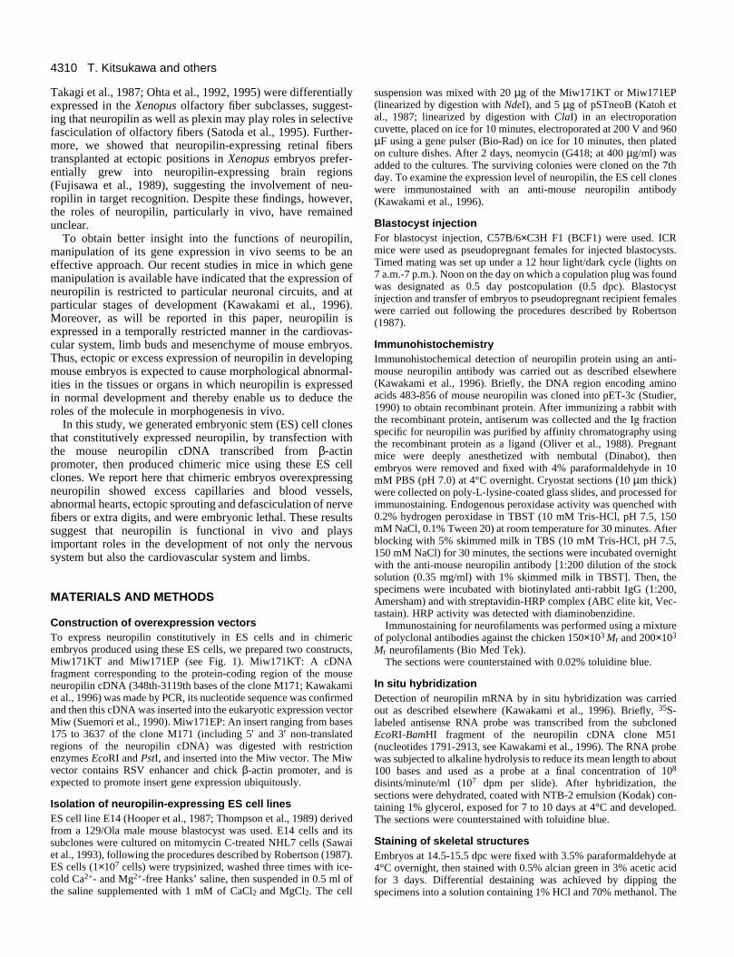
Immunostaining and in situ hybridization analyses indicatedthat the descendants of the injected ES cells within thechimeras expressed exogenous neuropilin during embryogen-esis. The exogenous expression of neuropilin was moreprominent in the KT than in the EP chimeras, although thelevels of exogenous neuropilin were different among chimerasdepending on the ratio of the ES cells contributing to embryoformation. In normal embryos at the age of 12.5 dpc, neuronalcells in the dorsolateral part of the diencephalon did notexpress neuropilin (Fig. 2A). Only capillaries were stainedwith anti-neuropilin antibody. In the EP chimeric embryos atthe same age, however, several neuropilin-positive neuronalcells were observed (Fig. 2B), indicating ectopic expression ofneuropilin. In controls, the floor plate was a normal site forneuropilin expression (Fig. 2C). In the EP chimeras, a part ofthe floor plate was often heavily stained with anti-neuropilinantibody (Fig. 2D) suggesting excess expression of neuropilin.In normal embryos at 12.5 dpc, neuropilin was expressed inlimited parts of the nervous system, cardiovascular system andmesenchymal cells (Fig. 2E; for detailed expression patterns ofneuropilin, see the later parts of this paper). While, in KT21chimeric embryos, almost all parts of the body except theepidermis and endoderm derivatives were stained strongly withanti-neuropilin antibody (compare Fig. 2F and E). In situhybridization analysis also confirmed the ectopic and excess
expression of neuropilin; in the spinal cord of the normalembryos at 12.5 dpc, in situ hybridization signals for neuropilinmRNA were prominent in the motor neuron pool but weak inthe dorsal horn (Fig. 2G), while in the KT21 chimeric embryosat same stage, in situ hybridization signals were expanded overall parts of the spinal cord (Fig. 2H).
Mortality of chimeric embryosAs listed in Table 1, we recovered a total of 26 KT chimericembryos at ages of 12.5 dpc, 14.5 dpc and 15.5 dpc (from 20pregnant dams), but no living chimeric embryos at moreadvanced stages or mice were obtained. In the uteri of thepregnant dams at 12.5-15.5 dpc, many traces of implantationand dead embryos were found. In the EP chimeras, werecovered 5 living chimeric embryos (3 at 12.5 dpc, and 2 at17.5 dpc; from 3 pregnant dams), but no living chimericembryos of more advanced stages of development or mice.These findings indicate that ectopic and/or over expression ofneuropilin is embryonic lethal.
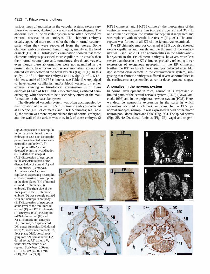
Anomalies in the cardiovascular system The cardiovascular system is a normal site of neuropilinexpression. Immunohistochemistry using anti-neuropilinantibody and in situ hybridization analyses indicated that endo-thelial cells of capillaries and blood vessels expressed neu-ropilin (Figs 2A,E, 3A,B). The immunostaining and in situhybridization signals in the walls of blood vessels were widerthan the thickness of the endothelium, indicating that mes-enchymal cells surrounding blood vessels also express neu-ropilin (Fig. 3B). The expression of neuropilin in endothelialcells and mesenchymal cells was detected as early at 8.5 dpcand persisted throughout the fetal and early postnatal period.Neuropilin was also expressed in the endocardial cells of theembryonic heart (Fig. 2E). In the adult cardiovascular system,the expression of neuropilin was extremely weak or almostnonexistent, except in the atria (data not shown).
The KT chimeric embryos collected at 12.5 dpc exhibited
4312 T. Kitsukawa and others
various types of anomalies in the vascular system; excess cap-illaries or vessels, dilation of vessels and hemorrhaging. Theabnormalities in the vascular system were often detected byexternal observation of embryos. The chimeric embryosusually appeared more red in color than their normal counter-parts when they were recovered from the uterus. Somechimeric embryos showed hemorrhaging, mainly at the heador neck (Fig. 3D). Histological examination showed that thesechimeric embryos possessed more capillaries or vessels thantheir normal counterparts and, sometimes, also dilated vessels,even though these abnormalities were not quantified in thepresent study. In embryos with severe anomalies, excess anddilated vessels deformed the brain vesicles (Fig. 3E,F). In thisstudy, 10 of 15 chimeric embryos at 12.5 dpc (4 of 6 KT21chimeras, and 6 of 9 KT55 chimeras; see Table 1) were judgedto have excess capillaries and/or blood vessels, by eitherexternal viewing or histological examination. 8 of theseembryos (4 each of KT21 and KT55 chimeras) exhibited hem-orrhaging, which seemed to be a secondary effect of the mal-formations in the vascular system.
The disordered vascular system was often accompanied bymalformation of the heart. In 5 KT chimeric embryos collectedat 12.5 dpc (4 KT21 chimeras, and 1 KT55 chimera; see Table1), the atrium was more expanded than that of normal embryos,and the wall of the atrium was thin. In 3 of these embryos (2
Fig. 2. Expression of neuropilinin normal and chimeric mouseembryos at 12.5 dpc. Neuropilinprotein was detected using anti-neuropilin antibody (A-F).Neuropilin mRNAs weredetected by in situ hybridization(G,H; dark-field images).(A,B) Expression of neuropilinin the dorsolateral part of thediencephalon of normal (A) andEP chimeric (B) embryos.Arrowheads (in A) showcapillaries expressing neuropilin.(C,D) Expression of neuropilinin the floor plates (FP) of normal(C) and EP chimeric (D)embryos. The right side of thefloor plate in the EP chimericembryo (D) was strongly stainedwith anti-neuropilin antibody.(E, F) Expression of neuropilinat the level of the forelimbs innormal (E) and KT 21 chimeric(F) embryos. (G,H) NeuropilinmRNAs in normal (G) andKT21 chimeric (H) embryos.FL, forelimb; SC, spinal cord;DF, dorsal funiculus; DH, dorsalhorn; M, motor neuron pool; FP,floor plate; DRG, dorsal rootganglion; SN, spinal nerve; DA,dorsal aorta; AT, atrium; V,ventricle; VS, ventricularseptum. Scale bars: 100 µm(A,B), 50 µm (C,D), 1 mm(E,F), 200 µm (G,H).
KT21 chimeras, and 1 KT55 chimera), the musculature of theventricles was extremely thin (compare Figs 2E and 3G). Inone chimeric embryo, the ventricular septum disappeared andwas replaced with trabecula-like tissues (Fig. 3G). The atrialseptum was formed in all KT chimeric embryos examined.
The EP chimeric embryos collected at 12.5 dpc also showedexcess capillaries and vessels and the thinning of the ventric-ular wall (see Table 1). The abnormalities in the cardiovascu-lar system in the EP chimeric embryos, however, were lesssevere than those in the KT chimeras, probably reflecting lowerexpression of exogenous neuropilin in the EP chimeras.Neither the KT nor EP chimeric embryos collected after 14.5dpc showed clear defects in the cardiovascular system, sug-gesting that chimeric embryos suffered severe abnormalities inthe cardiovascular system died at earlier developmental stages.
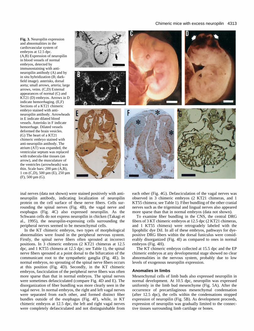
Anomalies in the nervous systemIn normal development in mice, neuropilin is expressed inlimited parts of the central nervous system (CNS) (Kawakamiet al., 1996) and in the peripheral nervous system (PNS). Here,we describe neuropilin expression in the parts in whichanomalies occurred in chimeric embryos. In the 12.5 dpcnormal embryos, neuropilin was expressed in cells of the motorneuron pool, dorsal horn and DRG (Fig. 2G). The spinal nerves(Figs 2E, 4A,D), dorsal funiclus (Fig. 2E), vagal and trigem-
4313Chimeric mice with excess neuropilin
Fig. 3. Neuropilin expressionand abnormalities in thecardiovascular system ofembryos at 12.5 dpc.(A,B) Expression of neuropilinin blood vessels of normalembryos, detected byimmunostaining with anti-neuropilin antibody (A) and byin situ hybridization (B; dark-field image). asterisks, dorsalaorta; small arrows, arteria; largearrows, veins. (C,D) Externalappearances of normal (C) andKT21 (D) embryos. Arrows in Dindicate hemorrhaging. (E,F)Sections of a KT21 chimericembryo stained with anti-neuropilin antibody. Arrowheadsin E indicate dilated bloodvessels. Asterisks in F indicatehemorrhage. Dilated vesselsdeformed the brain vesicles.(G) The heart of a KT21chimeric embryo stained withanti-neuropilin antibody. Theatrium (AT) was expanded, theventricular septum was replacedwith trabecula-like tissues (anarrow), and the musculature ofthe ventricles (arrowheads) wasthin. Scale bars: 200 µm (A,B),1 cm (C,D), 500 µm (E), 250 µm(F), 500 µm (G).
inal nerves (data not shown) were stained positively with anti-neuropilin antibody, indicating localization of neuropilinprotein on the cell surface of these nerve fibers. Cells sur-rounding the spinal nerves (Fig. 4B), the vagal nerve andesophagus (Fig. 4C) also expressed neuropilin. As theSchwann cells do not express neuropilin in chicken (Takagi etal., 1995), the neuropilin-expressing cells surrounding theperipheral nerves seemed to be mesenchymal cells.
In the KT chimeric embryos, two types of morphologicalabnormalities were found in the peripheral nervous system.Firstly, the spinal nerve fibers often sprouted at incorrectpositions. In 3 chimeric embryos (2 KT21 chimeras at 12.5dpc, and 1 KT55 chimera at 12.5 dpc; see Table 1), the spinalnerve fibers sprouted at a point dorsal to the bifurcation of thecommunicant root to the sympathetic ganglia (Fig. 4E). Innormal embryos, no sprouting of the spinal nerve fibers occursat this position (Fig. 4D). Secondly, in the KT chimericembryos, fasciculation of the peripheral nerve fibers was oftenmore sparse than that in normal embryos. The spinal nerveswere sometimes defasciculated (compare Fig. 4D and E). Thedisorganization of fiber bundling was more clearly seen in thevagal nerve. In normal embryos, the right and left vagal nerveswere separated from each other, and formed distinct fiberbundles outside of the esophagus (Fig. 4F), while, in KTchimeric embryos at 12.5 dpc, the left and right vagal nerveswere completely defasciculated and not distinguishable from
each other (Fig. 4G). Defasciculation of the vagal nerves wasobserved in 3 chimeric embryos (2 KT21 chimeras, and 1KT55 chimera; see Table 1). Fiber bundling of the other cranialnerves such as the trigeminal and lingual nerves also appearedmore sparse than that in normal embryos (data not shown).
To examine fiber bundling in the CNS, the central DRGfibers of 3 KT chimeric embryos at 12.5 dpc (2 KT21 chimeras,and 1 KT55 chimera) were retrogradely labeled with thelipophilic dye DiI. In all of these embryos, pathways for dye-positive DRG fibers within the dorsal funiculus were consid-erably disorganized (Fig. 4I) as compared to ones in normalembryos (Fig. 4H).
The KT chimeric embryos collected at 15.5 dpc and the EPchimeric embryos at any developmental stage showed no clearabnormalities in the nervous system, probably due to lowlevels of exogenous neuropilin expression.
Anomalies in limbs Mesenchymal cells of limb buds also expressed neuropilin innormal development. At 10.5 dpc, neuropilin was expresseduniformly in the limb bud mesenchyme (Fig. 5A). After theoccurrence of precartilaginous mesenchymal condensation(after 12.5 dpc), the cells within the condensations stoppedexpression of neuropilin (Fig. 5B). As development proceeds,expression of neuropilin was gradually limited to the connec-tive tissues surrounding limb cartilage or bones.

4314 T. Kitsukawa and others
Most of the KT21 and EP chimeric embryos showed extradigits (Fig. 5C) which were always observed at the anteriorside of the hindlimb. Nine of 10 KT21 chimeric embryos (5 of6 at 14.5 dpc, 3 of 3 at 15.5 dpc, and one dead embryorecovered at 17.5 dpc; see Table 1) possessed extra digits.Among these, 5 embryos had extra digits in both sides of thehindlimb. All the EP chimeric embryos at 17.5 dpc (2 livingand 1 dead; see Table 1) also possessed extra digits in theanterior side of the hindlimb. The chimeric embryos generatedusing KT55 cells (2 embryos at 15.5 dpc; see Table 1) had noextra digits.
The alcian green staining of the chimeric embryos with extradigits showed that the hindlimbs had redundant pieces ofcartilage at the anterior side; in some hindlimbs, the proximaland distal phalangeal cartilage of the first digit were duplicated(Fig. 5D) and, in the others, each of the medial cuneiformcartilage, the first metatarsus cartilage, and the first phalangealcartilage was duplicated (Fig. 5E). Some hindlimbs exhibitedduplicated medial cuneiform and first metatarsal cartilage, andtriplicated proximal and distal phalangeal cartilage of the firstdigit (Fig. 5F). The numbers of cartilage elements in the extradigits differed between the right and left hindlimbs of the sameembryo. In this study, no morphological abnormalities wereobserved in the forelimbs in chimeric embryos, except in oneKT21 chimeric embryo at 17.5 dpc in which the proximalphalangeal cartilage of the second and third fingers were fused(data not shown). No abnormalities were observed in theskeletal system other than the limbs.
DISCUSSION
In this study, we generated chimeric mouse embryos using EScells constitutively expressing the membrane protein neu-ropilin and showed that most of the chimeric embryos withexcess and/or ectopic expression of neuropilin exhibitedanomalies in the cardiovascular system, nervous system orlimbs, and died in utero. The abnormalities occurred in thepresent chimeric embryos are not due to the intrinsic proper-
Table 1. Phenotypes of chimeric embryosNumber of Abnormalities
ES cell Stages of chimeras (embryos with abnormalities / clone embryos examined embryos analyzed)
Excess capillaries (4/6)Hemorrhaging (4/6)
12.5 6 Heart malformation (4/4)†,‡KT21 Ectopic sprouting of spinal nerves (2/2)†
Defasciculation of vagal nerves (2/2)†Irregular dorsal funiculus (2/2)‡
14.5-17.5 9+1† Extra digits (9/10)Excess capillaries (6/9)Hemorrhaging (4/9)
12.5 9 Heart malformation (1/1)§KT55 Ectopic sprouting of spinal nerves (1/1)§
Defasciculation of vagal nerves (1/1)§Irregular dorsal funiculus (1/1)§
15.5 2 None12.5 3 Excess capillaries (2/3)
EP40 Heart malformation (2/3)17.5 2+1* Extra digits(3/3)
*Dead embryos; †,‡,§The same respective embryos.
ties of the ES cells, since the parental ES line E14 does notproduce any analogous abnormalities (Sawai et al., 1993).Moreover, in this study, we produced chimeric mouse embryosusing three different ES cell clones; the KT21 and KT55 clonesfor the neuropilin DNA construct Miw171KT and the EP40clone for the construct Miw171EP (see Fig. 1), and detectedsimilar anomalies in these chimeras; the KT21 and KT55chimeras showed disorganization in the cardiovascular andnervous systems, and the KT 21 and EP chimeras exhibitedextra digits. These results indicate that the anomalies observedin the chimeras were caused by the exogenous neuropilinexpressed by the ES-derived cells.
An important finding obtained in the present study is that allof the abnormalities occurred in the organs in which neuropilinis expressed in normal development. As the expression of neu-ropilin in these organs is limited at particular stages of normaldevelopment, the excess and/or persistence of neuropilinexpression after the stage when in normal development itwould have ceased appeared to results in abnormal morpho-genesis. In the normal development of limbs, neuropilin isexpressed widely in the limb mesenchymal cells, then disap-pears in the cells of the precartilaginous mesenchymal con-densations. Thus, it is likely that not only the excess expressionof neuropilin in the limb mesenchymal cells but also theprolonged expression of the molecule in the cells of the pre-cartilaginous mesenchymal condensations disturb the normallimb morphogenesis.
Another important observation obtained in this study is thatthe exogenous neuropilin expression caused embryonic death.The disordered development of the cardiovascular systemobserved in the chimeric embryos seemed to be the main causeof the lethality of embryos; chimeric embryos with moreexogenous neuropilin may suffer more severe abnormalities inthe cardiovascular system, resulting into embryonic death atearlier developmental stages. It seems worthy of note that onlyembryos with low levels of exogenous neuropilin expressionand with the resultant minor anomalies in the cardiovascularsystem and probably in the other organs survived until the laterstages of development and were analyzed in the present study.The finding that chimeric embryos collected after 14.5 dpc didnot show clear defects in the cardiovascular or nervous systemsmay support the possibility. The extra digits occurred in mostof the KT21 and EP chimeric embryos with excess neuropilin,but not in the KT55 chimeric embryos in which strongexpression of exogenous neuropilin was expected. Theseresults, however, do not indicate that the KT55 chimerasdeveloped normal limbs. Rather, only 3 living KT55 chimericembryos were obtained after 13.5 dpc suggesting that the KT55chimeras could potentially produce extra digits but died atstages before limb development due to severe abnormalities inthe cardiovascular system.
Our findings raise the question of how the overexpressionof neuropilin causes developmental aberration. The abnor-malities that occurred in the chimeric embryos affected thenervous system, cardiovascular system and limbs, which areformed as the results of quite different developmentalprocesses. Molecular cloning studies of neuropilin in Xenopus(Takagi et al., 1991), chicken (Takagi et al., 1995) and mouse(Kawakami et al., 1996) have indicated that the extracellularsegment of the neuropilin protein is composed of threedomains, each of which is expected to be involved in different
4315Chimeric mice with excess neuropilin
ression and abnormalities in the nervous system of embryos at 12.5 neuropilin in the spinal nerve (SN) of a normal embryo, detected bynti-neuropilin antibody. (B) Expression of neuropilin mRNAs in cellshe spinal nerve of a normal embryo, detected by in situ hybridizationnd B are adjacent serial sections. (C) Expression of neuropilin mRNAse esophagus (ES) and vagal nerves (VN; arrows) of a normal embryo,ridization (dark-field image). (D,E) The spinal nerves of normal (D)) embryos stained with anti-neurofilament antibody. In the chimericve was partially defasciculated (an arrow in E), and sprouteds in E). CR, communicant root; SG, sympathetic ganglion.rowheads) of normal (F) and KT21 chimeric (G) embryos around the with anti-neurofilament antibody. In the chimeric embryo, both theves were defasciculated. (H,I) DiI-filled fibers of DRGs within themal (H) and KT21 chimeric (I) embryos. In the chimeric embryo, fiber (arrowheads in I). DR, dorsal root. Scale bars: 100 µm (A-I).
molecular and/or cellular interactions, suggesting multipleroles of the molecule. Thus, it seems unlikely that a singlefunction of neuropilin is responsible for all the range of theabnormalities observed in the chimericembryos. We will discuss below theeffect of overexpressed neuropilin inmorphogenesis in each organ.
Nervous systemOur previous studies in Xenopus haveshown that neuropilin promotes neuriteoutgrowth in vitro (Hirata et al., 1993).We also showed that the subclasses of theXenopus olfactory fibers differentiallyexpressed either neuropilin or the othercell surface molecule named plexin andtheir pathways were sorted depending onthe expression levels of these twomolecules (Satoda et al., 1995). Fromthese findings, we predicted that neu-ropilin is involved in neuronal cell inter-action, and plays roles in guidance andfasciculation of nerve fibers. The obser-vation of ectopic sprouting and defascic-ulation of nerve fibers of the PNS andCNS in the chimeric embryos with excessneuropilin support the above hypothesis.
Our recent study in an in vitro modelsystem indicated that neuropilin interactswith heterotypic ligands (Takagi et al.,1995). However, very little is knownabout neuropilin ligands. Also, wecannot exclude the possibility that neu-ropilin interacts homophilically. Thus, itis open to question how neuropilinregulates interaction of nerve fibers andwhy excess neuropilin disorganizesgrowth and fasciculation of nerve fibers.At least, the present observation that neu-ropilin is normally expressed in both theperipheral nerve fibers and mesenchymalcells surrounding them enables us tospeculate that neuropilin mediates inter-action not only between nerve fibers butalso between nerve fibers and their sur-rounding cells. It is possible that, in thechimeras, excess expression of neu-ropilin in both the peripheral nerve fibersand the mesenchymal cells disrupts theappropriate balance of the fiber-fiber andfiber-mesenchymal cell interactions,resulting into ectopic sprouting or defas-ciculation of the nerve fibers. Severalstudies have indicated that non-neuronaltissues around the spinal nerves playroles in guidance and patterning of thenerves (Landmesser, 1984; Tosney,1988; Lance-Jones and Dias, 1991). Asimilar disordered cellular interactionmay occur in the CNS of the chimericembryos; excess neuropilin in the dorsal
Fig. 4. Neuropilin expdpc. (A) Expression ofimmunostaining with a(arrows) surrounding t(dark-field image). A ain cells surrounding thdetected by in situ hyband KT21 chimeric (Eembryo, the spinal nerectopically (arrowhead(F,G) Vagal nerves (aresophagus (ES) stainedright and left vagal nerdorsal funiculus of norbundling was irregular
spinal cord may alter the growth and fasciculation of the DRGfibers, resulting into disorganization of the fiber pathway in thedorsal funiculus.

4316 T. Kitsukawa and others
bnormalities in hindlimbs. (A,B) Expression of neuropilin in hindlimb (A) and 12.5 dpc (B) detected by in situ hybridization (dark-field rostrocaudal axis of the limb buds. The upper and left in each figurehe limb buds, respectively. Neuropilin mRNA was not detected in the
al condensation (PC). (C) External appearance of the right hindlimbpc. The first digit (I) was triplicated. II, III and IV indicate the second, The fifth digit is behind the fourth toe and cannot be seen in theindlimbs of KT 21 chimeric embryos at 15.5 dpc (D,E) and at 17.5, II, III, IV and V indicate the first, second, third, fourth and fiftheiform cartilage; mt, first metatarsal cartilage. Scale bars: 250 µm
Cardiovascular systemThe chimeric embryos with excess neuropilin exhibited excesscapillaries and vessels, dilation of vessels, and abnormalhearts, suggesting the involvement of neuropilin in morpho-genesis of the cardiovascular system. The capillaries andvessels are formed as a result of different morphogeneticprocesses such as the proliferation and differentiation of endo-thelial cells, the migration of endothelial cells and theformation of capillary tubes. More complicated processes arerequired for heart formation. Though the present studies didnot show which steps of the morphogenesis of cardiovascularsystem are controlled with neuropilin, some explanations arepossible for the observed abnormalities.
It has been reported that proper interaction of endothelialcells with surrounding mes-enchymal cells, smoothmuscle cells or extracellularmatrix is necessary for theproliferation, differentiationand migration of endothelialcells (Yang and Moses, 1990;Sato et al., 1990). Excessexpression of neuropilin inthe endothelial cells and/ortheir surrounding cells mightdisrupt correct cellular inter-action, resulting into theexcess capillaries andvessels, or the dilation ofvessels. The other possibilityis that neuropilin modulatesthe activities of cytokines.Several studies have shownthat the production and themigration of endothelialcells, and also capillary tubeformation are regulated bythe growth factor TGFβ(Robertis et al., 1986; Yangand Moses, 1990; Sato et al.,1990). Also, TGFβ plays arole in the epithelial-mes-enchymal cell transformationto form valves and septa ofthe embryonic heart (Pottsand Runyan, 1989). Thea1/a2 domains in the extra-cellular segment of neu-ropilin are homologous toBMP-1 (Wozney et al., 1988)and Tolloid (Shimell et al.,1991) which are expected tobind to precursors of TGFβvia these domains and prot-eolytically cleave the precur-sors into active forms (Finelliet al., 1994). Thus, it seemspossible that neuropilinmodifies the activity of TGFβor TGFβ-like cytokines. Theexcess angiogenesis and the
Fig. 5. Neuropilin expression and abuds of normal embryos at 10.5 dpcimage). Sections were cut along theare the anterior and dorsal sides of tareas of precartilaginous mesenchymof the EP chimeric embryo at 17.5 dthird and fourth digits, respectively.figure. (D-F) Cartilage patterns of hdpc (E,F), stained by alcian green. Idigits, respectively. mc, medial cun(A,B), 1 cm (C), 500 µm (D-F).
disordered hearts observed in the chimeric mice might be dueto the up- or down-regulation of the activities of TGFβ-likecytokines as a result of overexpression of neuropilin.
LimbsMost of the chimeric embryos overexpressing neuropilinshowed extra digits, suggesting that limb morphogenesis isvery sensitive to the exogenous neuropilin expression. Severalstudies have shown the importance of cell-cell and cell-sub-stratum interactions in limb morphogenesis (Leonard et al.,1991; Jiang et al., 1993; Ide and Wada, 1994). Thus, excessand/or ectopic expression of neuropilin might modify interac-tions of limb mesenchymal cells to form abnormal limbs.Growth factors belonging to the TGFβ superfamily such as

4317Chimeric mice with excess neuropilin
TGFβ1, TGFβ2, BMP-2, BMP-4 and Activin A are assumedto regulate proliferation of limb mesenchymal cells, precarti-laginous condensation of mesenchymal cells and cartilage orbone morphogenesis (Hayamizu et al., 1991; Jones at al., 1991;Leonard et al., 1991; Jiang et al., 1993; Niswander and Martin,1993). Thus, the excess or ectopic expression of neuropilin inthe limb mesenchymal cells might modify cytokines functionsand disturb normal patterns of proliferation or condensation ofthe cells, resulting in the formation of redundant pieces ofcartilage in their digits. It is still unclear why the anomalieswere limited to the anterior side of the hindlimbs.
In conclusion, the present results indicated that neuropilin isan active cell surface protein which regulates morphogenesisin vivo. Diverse morphological abnormalities occurred inchimeric mice with excess or ectopic neuropilin expressionindicating that neuropilin is a multifunctional molecule capableof regulating various aspects of morphogenesis. Althoughdetermination of the precise roles of neuropilin in each step ofmorphogenesis at the molecular level require further analysis,the correct spatiotemporal patterns of neuropilin expressionwere shown here to be essential for the growth and fascicula-tion of nerve fibers, formation of the cardiovascular system andpattern formation of limbs.
This work was funded by grants from the Ministry of Education,Science and Culture, Japan, from the Mitsubishi Foundation and fromthe Uehara Foundation, and was partly supported by Research Fel-lowships of the Japan Society for the Promotion of Science for YoungScientists (to T. K.).
REFERENCES
Beckmann, G. and Bork, P. (1993). An adhesive domain detected infunctionally diverse receptors. Trends Biochem. Sci. 18, 40-41.
Dodd, J. and Jessell, T. M. (1988). Axon guidance and the patterning ofneuronal projections in vertebrates. Science 242, 692-699.
Finelli, A. L., Bossie, C. A., Xie, T. and Padgett, R. W. (1994). Mutationalanalysis of the Drosophila tolloid gene, a human BMP-1 homologue.Development 120, 861-870.
Fujisawa, H., Otsuki, T., Takagi, S. and Tsuji, T. (1989). An aberrant retinalpathway and visual centers in Xenopus tadpoles share a common cell surfacemolecule, A5 antigen. Dev. Biol. 135, 231-240.
Hayamizu, T. F., Sessions, S. K., Wanek, N. and Bryant, S. V. (1991).Effects of localized application of transforming growth factor β1 ondeveloping chick limbs. Dev. Biol. 145, 164-173.
Hirata, T., Takagi, S. and Fujisawa, H. (1993). The membrane protein A5, aputative neuronal recognition molecule, promotes neurite outgrowth.Neurosci. Res. 17, 159-169.
Hooper, M., Hardy, K., Handyside, A., Hunter, S. and Monk, M. (1987).HPRT-deficient (Losh-Nyhan) mouse embryos derived from germlinecolonization by cultured cells. Nature 326, 292-295.
Ide, H. and Wada, N. (1994). Sorting out of cells from different parts andstages of chick limb bud. Dev. Biol. 162, 71-76.
Jenny, R. L., Pittman, D. D., Toole, J. J., Kriz, R. W., Aldape, R. A.,Hewick, R. M., Kaufman, R. J. and Mann, K. G. (1987). Complete cDNAand derived amino acid sequence of human factor V. Proc. Natl. Acad. Sci.USA 84,4846-4850.
Jiang T. X., Yi, J. R., Ying, S. Y. and Chuong, C. M. (1993). Activinenhances chondrogenesis of limb bud cells: stimulation of precartilaginousmesenchymal condensations and expression of NCAM. Dev. Biol. 155, 545-557.
Johnson, J. D., Edelman, J. C. and Rutter, W. J. (1993). A receptor tyrosinekinase found in breast carcinoma cells has an extracellular discoidin I-likedomain. Proc. Natl. Acad. Sci. USA 90, 5677-5681.
Jones, C. M., Lyons, K. M. and Hogan, B. L. M. (1991). Involvement of BoneMorphogenetic Protein-4 (BMP-4) and Vgr-1 in morphogenesis andneurogenesis in the mouse. Development 111, 531-542.
Katoh, K., Takahashi, Y., Hayashi, S. and Kondoh, H. (1987). Improvedmammalian vectors for high expression of G418 resistance. Cell Struct.Funct. 12, 575-580.
Kawakami, A., Kitsukawa, T., Takagi, S. and Fujisawa, H. (1996).Developmentally regulated expression of a cell surface protein, neuropilin, inthe mouse nervous system. J. Neurobiol. (in print).
Lance-Jones, C., and Dias, M. (1991). The influence of presumptive limbconnective tissue on motoneuron axon guidance. Dev. Biol. 143, 93-110.
Landmesser, L. (1984). The development of specific motor pathways in thechick embryo. Trends Neurosci. 7, 336-339.
Leonard, C. M., Fuld, H. M., Frenz, D. A., Downie, S. A., Massagué, J. andNewman, S. A. (1991). Role of transforming growth factor-β inchondrogenic pattern formation in the embryonic limb: stimulation ofmesenchymal condensation and fibronectin gene expression by exogenousTGF-β and evidence for endogenous TGF-β-like activity. Dev. Biol. 145, 99-109.
Leytus, P. S., Kurachi, K., Sakariassen, K. S. and Davie, E. W. (1986).Nucleotide sequence of the cDNA coding for human complement componentC1r. Biochemistry 25, 4855-4863.
Mackinnon, C. M., Carter, P. E., Smyth, S. J., Dunbar, B. and Fothergill,E. (1987). Molecular cloning of cDNA for human complement componentC1s. The complete amino acid sequence. Eur. J. Biochem. 169, 547-553.
Niswander, J. and Martin G. R. (1993). FGF-4 and BMP-2 have oppositeeffects on limb growth. Nature 361, 68-71.
Ohta, K., Takagi, S., Asou, H. and Fujisawa, H. (1992). Involvement ofneuronal cell surface molecule B2 in the formation of retinal plexiformlayers. Neuron 9, 151-161.
Ohta, K., Mizutani, A., Kawakami, A., Murakami, Y., Kasuya, Y., Takagi,S. and Fujisawa, H. (1995). Plexin: a novel neuronal cell surface moleculethat mediates cell adhesion via a homophilic binding mechanism in thepresence of calcium ions. Neuron 14, 1189-1199.
Oliver, G., Wright, C. V. E., Hardwicke, J. and De Robertis, E. M. (1988).Differential anterior-posterior expression of two proteins encoded by ahomeobox gene in Xenopus and mouse embryo. EMBO J. 7, 3199-3209.
Potts, J. D. and Runyan, R. B. (1989). Epithelial-mesenchymal celltransformation in the embryonic heart can be mediated, in part, bytransforming growth factor β. Dev. Biol. 134, 392-401.
Robertis, A. B., Sporn, M. B., Assoian, R. K., Smith, J. M., Roche, N.,Wakefield, L. M., Heine, U. I., Liotta, L. A., Falanga, V., Kehrl., H. andFauci, A. S. (1986). Transforming growth factor type β: rapid induction offibrosis and angiogenesis in vivo and stimulation of collagen formation invitro. Porc. Natl. Acad. Sci. USA. 83, 4167-4171.
Robertson, E. J. (1987). Teratocarcinomas and Embryonic Stem Cells: APractical Approach. Oxford and Washington DC: IRL Press.
Sanz, L., Calvete, J. J., Mann, K., Schäfer, W., Schmid, E. R. and Topfer-Petersen, E. (1991). The amino acid sequence of Aqn-3, a carbohydrate-binding protein isolated from boar sperm. FEBS Letters 291, 33-36.
Sanz, L., Calvete, J. J., Mann, K., Schäfer, W., Schmid, E. R. and Topfer-Petersen, E. (1992a). The complete primary structure of the boarspermadhesion AQN-1, a carbohydrate-binding protein involved infertilization. Eur. J. Biochem. 205, 645-652.
Sanz, L., Calvete, J. J., Mann, K., Schäfer, W., Schmid, E. R.,Amselsgruber, W., Sinowatz, F., Ehrhard, M. and Topfer-Petersen, E.(1992b). The complete primary structure of spermadhesion Awn, a zonapellucida-binding protein isolated from boar spermatozoa. FEBS Letters 300,213-218.
Sato, Y. Tsuboi, R., Lynos, R. Moses, H. and Rifkin, D. B. (1990).Characterization of the activation of latent TGF-β by co-cultures ofendothelial cells and pericytes or smooth muscle cells: a self-regulatingsystem. J. Cell Biol. 111, 757-763.
Satoda, M., Takagi, S., Ohta, K., Hirata, T. and Fujisawa, H. (1995).Differential expression of two cell surface proteins, neuropilin and plexin, inXenopus olfactory axon subclasses. J. Neurosci. 15, 942-955.
Sawai, S., Shimono, A., Wakamatsu, Y., Palmes, C., Hanaoka, K. andKondoh, H. (1993). Defects of embryonic organogenesis resulting fromtargeted disruption of the N-myc gene in the mouse. Development 117, 1445-1455.
Shimell, M. J., Ferguson, E. L., Childs, S. R. and O’Conner, M. B. (1991).The Drosophila dorsal-ventral patterning gene tolloid is related to humanbone morphogenetic protein 1. Cell 67, 469-481.
Studier, F. W., Rosenberg, A. H., Dunn, J. J. and Dubendorff, J. W. (1990).Use T7 RNA polymerase to direct the expression of cloned genes. Meth.Enzymol. 185, pp. 60-89.
Suemori, H., Kadokawa, Y., Goto, K., Araki, I., Kondoh, H. and Nakatsuji,

4318 T. Kitsukawa and others
N. (1990). A mouse embryonic stem cell line showing pluripotency ofdifferentiation in early embryos and ubiquitous β-galactosidase expression.Cell Differ. Dev. 29, 181-186.
Takagi, S., Tsuji, T., Amagai, T., Takamatsu, T. and Fujisawa, H. (1987).Specific cell surface labels in the visual centers of Xenopus laevis tadpoleidentified using monoclonal antibodies. Dev. Biol. 122, 90-100.
Takagi, S., Hirata, T., Agata, K., Mochii, M., Eguchi, G. and Fujisawa, H.(1991). The A5 antigen, a candidate for the neuronal recognition molecule,has homologies to complement component and coagulation factors. Neuron7, 295-307.
Takagi, S., Kasuya, Y., Shimizu, M., Matsuura, T., Tsuboi, M., Kawakami,A. and Fujisawa, H. (1995). Expression of a cell adhesion molecule,neuropilin, in the developing chick nervous system. Dev. Biol. 170, 207-222.
Thompson, S., Clarke, A. R., Pow, A. M., Hooper, M. L. and Melton, W.(1989). Germ line transmission and expression of a corrected HPRT geneproduced by gene targeting in embryonic stem cells. Cell 56, 313-321.
Toole, J. J., Knopf, J. L., Wozney, J. M., Sultzman, L. A., Buecker, J. L.,Pittman, D. D. Kaufman, R. J., Brown, E., Shoemaker, C., Orr, E. C.,Amphlett, G. W., Foster, W. B., Coe, M. L., Knutson, G. J., Fass, D. N.and Hewick, R. M. (1984). Molecular cloning of a cDNA encoding humanantihaemophilic factor. Nature 312, 342-347.
Tosney, K. W. (1988). Proximal tissues and patterned neurite outgrowth at thelumbosacral level of the chick embryo: partial and complete deletion of thesomite. Dev. Biol. 127, 266-286.
Wozney, J. M. Rosen, V., Celeste, A. J., Mitosock, L. M., Whitters, M. J.,Kriz, R. W., Hewick, R. M. and Wang, E. A. (1988). Novel regulators ofbone formation: molecular clones and activities. Science 242, 1528-1534.
Yang, E. Y. and Moses, H. L. (1990). Transforming growth factor β1-inducedchanges in cell migration, proliferation, and angiogenesis in the chickchorioallantoic membrane. J. Cell Biol. 111, 731-741.
(Accepted 18 August 1995)