overexpressionofglutaminylcyclase,theenzyme ... · responsibleforpyroglutamatea formation,induces...
TRANSCRIPT
Overexpression of Glutaminyl Cyclase, the EnzymeResponsible for Pyroglutamate A� Formation, InducesBehavioral Deficits, and Glutaminyl Cyclase Knock-outRescues the Behavioral Phenotype in 5XFAD Mice*□S
Received for publication, September 20, 2010, and in revised form, December 9, 2010 Published, JBC Papers in Press, December 10, 2010, DOI 10.1074/jbc.M110.185819
Sadim Jawhar‡1, Oliver Wirths‡1, Stephan Schilling§1, Sigrid Graubner¶, Hans-Ulrich Demuth§2,and Thomas A. Bayer‡3
From the ‡Department of Molecular Psychiatry and Alzheimer Ph.D. Graduate School, University Medicine Goettingen, 37075 Goettingen,Germany, §Probiodrug AG, 06120 Halle, Germany, and ¶Ingenium Pharmaceuticals GmbH, 82152 Munich, Germany
Pyroglutamate-modified A� (A�pE3–42) peptides are gain-ing considerable attention as potential key players in the pa-thology of Alzheimer disease (AD) due to their abundance inAD brain, high aggregation propensity, stability, and cellulartoxicity. Overexpressing A�pE3–42 induced a severe neuronloss and neurological phenotype in TBA2 mice. In vitro and invivo experiments have recently proven that the enzyme glu-taminyl cyclase (QC) catalyzes the formation of A�pE3–42.The aim of the present work was to analyze the role of QC inan ADmouse model with abundant A�pE3–42 formation.5XFADmice were crossed with transgenic mice expressinghuman QC (hQC) under the control of the Thy1 promoter.5XFAD/hQC bigenic mice showed significant elevation inTBS, SDS, and formic acid-soluble A�pE3–42 peptides andaggregation in plaques. In 6-month-old 5XFAD/hQCmice, asignificant motor and working memory impairment developedcompared with 5XFAD. The contribution of endogenous QCwas studied by generating 5XFAD/QC-KOmice (mouse QCknock-out). 5XFAD/QC-KOmice showed a significant rescueof the wild-type mice behavioral phenotype, demonstratingthe important contribution of endogenous mouse QC andtransgenic overexpressed QC. These data clearly demonstratethat QC is crucial for modulating A�pE3–42 levels in vivo andprove on a genetic base the concept that reduction of QC ac-tivity is a promising new therapeutic approach for AD.
Alzheimer disease (AD)4 is a progressive neurodegenerativedisorder characterized by the presence of extracellular amy-loid plaques composed of amyloid-� (A�) and intracellular
neurofibrillary tangles. The discovery that certain early onsetfamilial forms of AD may be caused by enhanced levels of A�peptides has led to the hypothesis that amyloidogenic A� isintimately involved in the pathogenic process (1).Besides full-length A� 40 and 42 isoforms starting with an
aspartate at position 1, a variety of different N-truncated A�peptides have been identified in AD brains. Ragged peptidesincluding phenylalanine at position 4 of A� have been re-ported as early as 1985 by Masters et al. (2). In contrast, noN-terminal sequence could be obtained from cores purified ina SDS-containing buffer, which led to the assumption that theN terminus could be blocked (3, 4).The presence of A�pE3 (N-terminally truncated A� start-
ing with pyroglutamate) in AD brain was subsequently shownusing mass spectrometry of purified A� peptides, explainingat least partially initial difficulties in sequencing A� peptidespurified from human brain tissue (5). The authors reportedthat only 10–15% of the total A� isolated by this method be-gins at position 3 with A�pE3. Saido et al. (6) and others (7)subsequently showed that A�pE3 represents a dominant frac-tion of A� peptides in AD brain.
Overexpression of A�pE3–42 in neurons of TBA2 trans-genic mice triggers neuron loss and an associated neurologicalphenotype (8). N-terminal pE formation can be catalyzed byglutaminyl cyclase (QC) and is pharmacologically inhibited byQC inhibitors, both in vitro (9) and in vivo (10). Moreover,QC expression was found up-regulated in the cortex of pa-tients with AD and correlated with the appearance of pE-modified A�. Oral application of a QC inhibitor resulted inreduced A�pE3–42 burden in two different transgenic mousemodels of AD as well as in a transgenic Drosophilamodel.Interestingly, treatment of these mice was accompanied byreductions in A�x-40/42, diminished plaque formation andgliosis, as well as improved performance in context memoryand spatial learning tests (10). Thus, A�pE3–42 reduction is apromising target for therapy of AD. In the current work, thecontribution of QC was studied for the first time using ge-netic means by human QC overexpression and endogenousQC-knock-out in an AD mouse model.
EXPERIMENTAL PROCEDURES
Transgenic and Knock-out Mice—5XFAD (11) mice havebeen described previously. All mice were backcrossed for
* This work was supported by the German Federal Department of Educa-tion, Science and Technology, Grant 3013185 to a collaborative consor-tium led by H.-U. D.’s group, including T. A. B.’s team.
□S The on-line version of this article (available at http://www.jbc.org) con-tains supplemental Methods and Figs. 1–3.
1 These authors contributed equally to this work.2 To whom correspondence may be addressed: Probiodrug AG, Weinberg-
weg 22, 06120 Halle (Saale), Germany. E-mail: [email protected].
3 To whom correspondence may be addressed: Division of Molecular Psy-chiatry, University Medicine Gottingen, Von-Siebold-Strasse 5, 37075Gottingen, Germany. E-mail: [email protected].
4 The abbreviations used are: AD, Alzheimer disease; A�, amyloid-�; A�pE3,pyroglutamate A�; APP, amyloid precursor protein; QC, glutaminyl cy-clase; hQC, human QC; QC-KO, QC knock-out.
THE JOURNAL OF BIOLOGICAL CHEMISTRY VOL. 286, NO. 6, pp. 4454 –4460, February 11, 2011© 2011 by The American Society for Biochemistry and Molecular Biology, Inc. Printed in the U.S.A.
4454 JOURNAL OF BIOLOGICAL CHEMISTRY VOLUME 286 • NUMBER 6 • FEBRUARY 11, 2011
by guest on October 13, 2018
http://ww
w.jbc.org/
Dow
nloaded from
more than 10 generations on a C57BL/6J genetic backgroundand housed at a 12-h day/12-h night cycle with free access tofood and water. For generation of hQC transgenic mice, anexpression vector containing the cDNA of human QC undercontrol of the murine Thy1 promoter sequence was con-structed, applying standard molecular biology techniques andverified by sequencing. The transgenic founder was generatedon C57BL/6J/CBA background by pronuclear injection (JSW,Graz, Austria). The resulting offspring were further character-ized for transgene integration by PCR analysis and after cross-ing to C57BL/6J wild-type mice for transgene expression byRT-PCR (more than 10 generations). QC knock-out mice(QC-KO) were generated on the basis of a classical homolo-gous recombination approach at Genoway, Lyon. The target-ing vector contained the mouse chromosomal QC regionranging from intron 3 to exon 6. This region was modified byinsertion of two LoxP sites in intron 3 and 5, respectively. Inaddition, a neomycin resistance cassette flanked by two flip-pase recognition targets was inserted immediately upstreamof the LoxP in intron 5. After homologous recombination andchimera production, the neomycin selection cassette was re-moved by breeding with Flp-expressing mice followed bybreeding of the pups with Cre-expressing mice for deletion ofQC exons 4 and 5. The deletion of exons 4 and 5 causes aframeshift in the QC open reading frame generating a stopcodon in exon 6. Successful manipulation was confirmed byPCR and Southern hybridization. Absence of murine QC in5XFAD/QC-KO comparison with 5XFAD and 5XFAD/hQCwas further confirmed by RT-PCR (supplemental Methodsand supplemental Fig. 1). Animals were handled according toGerman guidelines for animal care and studies were approvedby the local legal authorities (LAVES). Only female mice wereused.Immunohistochemistry—Mouse tissue was processed as
described previously (12). In brief, 4-�m paraffin sectionswere pretreated with 0.3% H2O2 in PBS to block endogenousperoxidases, and antigen retrieval was achieved by boilingsections in 0.01 M citrate buffer, pH 6.0, followed by a 3-minincubation in 88% formic acid. Primary antibodies were incu-bated overnight, followed by incubation with biotinylated sec-ondary antibodies (DAKO) before staining was visualizedusing the ABC method with Vectastain kit (Vector Labora-tories) and diaminobenzidine as chromogen. Alternatively,fluorochromated secondary antibodies (anti-mouse AlexaFluor 594 and anti-rabbit Alexa Fluor 488; Invitrogen) wereused for immunofluorescence detection.Antibodies—A� antibodies NT78 (against generic A�; Syn-
aptic Systems), 22C11 (APP; Millipore) and 2–48 (againstN-terminal A�pE3; Synaptic Systems) (12) were used. Anti-sera (against QC) were raised against recombinant full-lengthmouse QC (1301) and have been proven to recognize hQC(13).ELISA of A� Levels in Brain—Frozen brains (n � 4–8 per
group) were weighed and subsequently subjected to a sequen-tial A� extraction. In a first step, brains were homogenized inTBS (120 mM NaCl, 50 mM Tris, pH 8.0, containing completeprotease inhibitor (Roche Applied Science)) using a Douncehomogenizer, sonified, and subsequently centrifuged at
27,000 � g for 20 min at 4 °C. The supernatant was removedand stored at �80 °C. The pellet was dissolved in 2.5 ml of 2%SDS, sonificated, and subsequently centrifuged at 80,000 � gfor 1 h at 4 °C. Supernatants were directly frozen at �80 °C.The resulting pellets were again resuspended in 0.5 ml of 70%formic acid, sonified, and neutralized using 1 M Tris. Aliquotsof the neutralized formic acid fraction were directly frozen at�80 °C. SDS lysates were diluted at least 10-fold for determi-nation of A�x-42 and A�pE3 using ELISA. All dilutions werecarried out using EIA buffer (IBL Co.). The neutralized formicacid fraction and the TBS fraction were applied directly orafter dilution using EIA buffer. ELISA measurements wereperformed in triplicate and according to the protocol of themanufacturer (IBL Co.; catalog nos. JP27716 and JP27711).Samples were run in triplicate.Quantification of Plaque Load—Extracellular A� load was
evaluated in mouse brain using an Olympus BX-51 micro-scope equipped with an Olympus DP-50 camera and theImageJ software (V1.41; National Institutes of Health). Serialimages of 40� magnification (hippocampus) and 100� (cor-tex) were captured on six sections/animal (n � 5/group),which were at least 30 �m afar from each other. Using ImageJthe pictures were binarized to 16-bit black and white images,and a fixed intensity threshold was applied defining the DABstaining.Behavioral Testing—Spontaneous alternation rates were
assessed using Y- and cross-maze as described previously (11,14). The alternation percentage was calculated as the percent-age of actual alternations to the total number of arm entries.Balance and general motor function were assessed using thebalance beam task. A 1-cm dowel beam is attached to twosupport columns 44 cm above a padded surface. At either endof the 50-cm long beam, a 9 � 15-cm escape platform is at-tached. The animal is placed on the center of the beam andreleased. Each animal is given three trials during a single dayof testing. The time the animal remained on the beam is re-corded and the resulting latencies to fall of all three trials areaveraged. If an animal remains on the beam for whole 60-strial or escapes to one of the platforms, the maximum time of60 s is recorded (14). For the string suspension test the ani-mals are permitted to grasp the string by their forepaws andare released. A rating system from 0 to 5 is used during thesingle 60-s trial to assess each animal’s performance in thistask: 0 � unable to remain on the string; 1 � hangs only byfore- or hindpaws; 2 � as for 1, but attempts to climb ontostring; 3 � sits on string and is able to hold balance; 4 � fourpaws and tail around string with lateral movement; 5 � es-cape. The following numbers of animals were analyzed in thistask (14). The following number of female mice was used atthe age of 6 months: 5XFAD, 11; 5XFAD/hQC, 8; hQC, 11;5XFAD/QC-KO, 4; QC-KO, 6; wild type, 12.Statistical Analysis—Statistical differences were evaluated
using one-way ANOVA followed by Bonferroni post hoc testor unpaired t test as indicated. All data are given as means �S.E. All statistics were calculated using GraphPad Prism ver-sion 5.00 software.
Glutaminyl Cyclase as a Target for Alzheimer Disease
FEBRUARY 11, 2011 • VOLUME 286 • NUMBER 6 JOURNAL OF BIOLOGICAL CHEMISTRY 4455
by guest on October 13, 2018
http://ww
w.jbc.org/
Dow
nloaded from
RESULTS
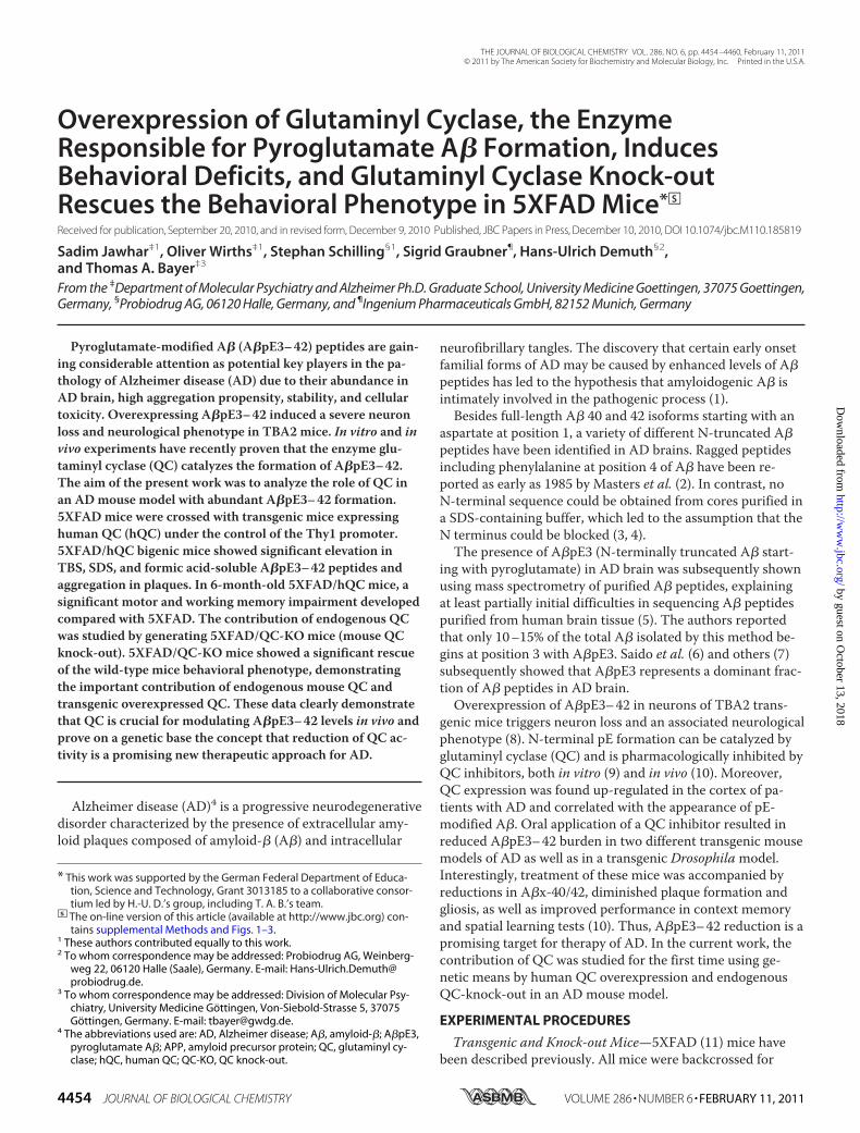
Expression and Distribution of hQC in the Brain of hQCand 5XFAD/hQC Mice—To study human QC overexpres-sion in hQC transgenic mice (hQC and 5XFAD/hQC), therabbit polyclonal antiserum 1301 recognizing human QCwas used to detect the transgene hQC expression in differ-ent brain regions (Fig. 1). Because the expression of thehQC trangene is driven by the Thy1 promoter, abundantpyramidal neurons expressing hQC were detected in vari-ous brain regions of hQC and 5XFAD/hQC mice includingthe cortex (Fig. 1, A–D), the hippocampus (Fig. 1, E and F),the midbrain and the cerebellum (data not shown). Nota-bly, a massive staining of hQC was observed in plaque-as-sociated dystrophic neurites in 5XFAD/hQC mice (Fig.1D). Abundant hQC immunoreactivity was detected inmossy fibers of hQC and 5XFAD/hQC transgenic mice(Fig. 1, E and F).
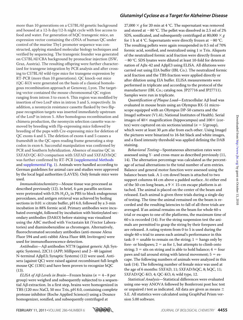
Co-localization of APP and QC in Neurons and the NeuriticComponent of Plaques—Double immunofluorescence demon-strated co-localization of hQC and APP in the same cellularcompartments in the brain of 5XFAD/hQC mice (Fig. 2). APPmarkedly labels dystrophic neurites around plaques andshows abundant co-localization with hQC suggesting thathQC is axonally transported like APP (Fig. 2).Effect of hQC Overexpression and QC Knock-out on Plaque
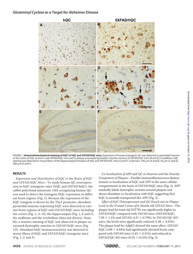
Load in the Frontal Cortex of 6-Month-old 5XFADMice—Theplaque load for total A� (NT78) was significantly higher in5XFAD/hQC compared with 5XFAD mice (5XFAD/hQC,7.99 � 1.1%; and 5XFAD, 4.27 � 0.79%). In 5XFAD/QC-KOmice, the levels were significantly reduced (1.88 � 0.43%).The plaque load for A�pE3 showed the same effect. 5XFAD/hQC (3.08 � 0.44%) had significantly elevated levels com-pared with 5XFAD mice (1.83 � 0.31%) and reduced in5XFAD/QC-KO mice (0.55 � 0.12%) (Fig. 3).
FIGURE 1. Immunohistochemical staining of hQC in hQC and 5XFAD/hQC mice. Expression of human transgenic QC was detected in pyramidal neuronsin the cortex of hQC (A and C) and 5XFAD/hQC mice and in plaque-associated dystrophic neurites (arrows) of 5XFAD/hQC mice (B and D). In addition, hQCstaining was detected in mossy fibers of the hippocampal formation of hQC and 5XFAD/hQC mice (E and F). Scale bars, 100 �m (A and B), 50 �m (C and D),200 �m (E and F).
Glutaminyl Cyclase as a Target for Alzheimer Disease
4456 JOURNAL OF BIOLOGICAL CHEMISTRY VOLUME 286 • NUMBER 6 • FEBRUARY 11, 2011
by guest on October 13, 2018
http://ww
w.jbc.org/
Dow
nloaded from
Effect of hQC Overexpression and QC Knock-out on A�x-42and A�pE3–42 Levels of 6-Month-old 5XFADMice—Proteinquantification of A�x-42 (in micrograms/gram of brainweight) and A�pE3–42 (in nanograms/gram of brain weight)levels in brain lysates of 6-month-old 5XFAD, 5XFAD/hQC,and 5XFAD/QC-KO mice revealed significant differences(Fig. 4). In the SDS�FA-soluble fraction there was a signifi-cant 48% reduction (p � 0.01) of A�x-42 levels in 5XFAD/QC-KO mice (41.07 � 4.79) compared with 5XFAD (78.52 �6.54). There was, however, no difference of A�x-42 levels inthe TBS-soluble fraction in 5XFAD/hQC (0.09 � 0.02) com-pared with 5XFAD (0.12 � 0.01) and 5XFAD/QC-KO mice(0.09 � 0.01). The effects were more pronounced onA�pE3–42 levels. There was an 86% elevation (p � 0.01) ofA�pE3–42 levels in the TBS-soluble fraction in 5XFAD/hQC(0.12 � 0.02) compared with 5XFAD (0.07 � 0.01) and unde-
tectable levels in 5XFAD/QC-KO mice. In the SDS�FA frac-tion an 84% elevation (p � 0.001) of A�pE3–42 levels wasfound in 5XFAD/hQC (114.9 � 5.89) compared with 5XFAD(62.29 � 3.66) and significantly reduced levels (�38%) in5XFAD/QC-KO mice (38.59 � 1.93, p � 0.01, compared with5XFAD) (Fig. 4). Despite the differing A�x-42 and A�pE3–42levels in 5XFAD, 5XFAD/hQC, and 5XFAD/QC-KO mice,expression levels of transgenic human APP and PS1 were un-changed (supplemental Fig. 2).Effect of hQC Overexpression and QC Knock-out on Behav-
ioral Performance in 5XFADMice—Motor coordination wasassessed by using balance beam and string suspension tasks(Fig. 5, A and B). In both tasks the 5XFAD/hQC performedsignificantly worse that 5XFAD (p � 0.001 and p � 0.01, re-spectively). Working memory was assessed using Y- andcross-maze alternation tasks. Analysis in the Y-maze revealed
FIGURE 2. Transgene human QC is co-localized with APP. Double immunostaining in the cortex of 5XFAD/hQC mice using antibodies against APP (red; Aand E), QC (green; B and F) and DAPI (blue; C and G). APP and QC showed co-localization in dystrophic neurites of plaques and in the somatodendritic com-partment of pyramidal neurons in the merged images (yellow; D, H, inset in H). Scale bars, 50 �m (A–D), 20 �m (E–H).
FIGURE 3. Effect of hQC overexpression and QC knock-out on plaque load in 5XFAD mice. A, plaque staining in the cortex using antibodies against ge-neric A� (NT78) and pyroglutamate-modified A� (2– 48) in 5XFAD, 5XFAD/hQC, and 5XFAD/QC-KO mice. B, quantification of plaque load demonstratingsignificantly elevated A� and A�pE3 levels in 5XFAD/hQC and significantly reduced levels in 5XFAD/QC-KO mouse brain. Scale bar, 200 �m. *, p � 0.05; **,p � 0.01. Error bars, S.E.
Glutaminyl Cyclase as a Target for Alzheimer Disease
FEBRUARY 11, 2011 • VOLUME 286 • NUMBER 6 JOURNAL OF BIOLOGICAL CHEMISTRY 4457
by guest on October 13, 2018
http://ww
w.jbc.org/
Dow
nloaded from

a significantly reduced alternation frequency in 5XFAD/hQCcompared with 5XFAD mice (p � 0.05). The number of armentries during the test period was not different between thegroups (Fig. 5, C and D). Assessment using the more complexcross-maze task demonstrated again a significantly reducedalternation frequency in 5XFAD/hQC compared with 5XFADmice (p � 0.05), and 5XFAD versus wild-type mice (p � 0.05).The latter finding corroborated previous results (14). More-over, the working memory deficit of 5XFAD mice was rescuedin 5XFAD/QC-KO mice (p � 0.05) showing alternation fre-quencies indistinguishable from wild-type mice. The numberof arm entries during the test period was not different amongall groups (Fig. 5, E and F).
DISCUSSION
Schilling et al. have shown that cyclization of glutamate atposition 3 of A� can be driven enzymatically by QC in vitro
(15). In addition, it has been demonstrated that QC inhibitionsignificantly reduced A�pE3 formation in vivo, emphasizingthe importance of QC activity during cellular maturation ofpyroglutamate-containing peptides. The pharmacologicalinhibition of QC activity by the QC inhibitor PQ150, whichsignificantly reduced the level of A�pE3 in vitro (16) and invivo (10), suggests that QC inhibition might serve as a newtherapeutic approach. Furthermore, the mean level of A�pE3-IgM autoantibodies was significantly decreased in AD pa-tients compared with healthy controls. In the group of mildlycognitive-impaired patients there was a significant positivecorrelation between A�pE3-IgM and cognitive decline (17).Interestingly, APP/PS1KI mice, a model with severe neuron
loss in the hippocampus, accumulate a large heterogeneity ofN-truncated A�x-42 isoforms including A�pE3 peptides co-inciding with the onset of behavioral deficits (18, 19). Morespecifically, transgenic mice expressing only A�pE3–42 de-veloped a robust and lethal neurological phenotype accompa-nied by Purkinje cell loss (TBA2 mouse line) (8).Saido et al. suggested that hypothetically the removal of
N-terminal amino acids 1 and 2 of A� might be carried out byamino or dipeptidyl peptidase(s) (6). Aminopeptidase A maybe responsible in part for the N-terminal truncation of full-length A� peptides (20).
N-truncated A�pE3 peptides have been identified by sev-eral groups in AD brains (5, 6, 21–32). N-terminal deletions ingeneral enhance aggregation of �-amyloid peptides in vitro(33). A�pE3 has a higher aggregation propensity (34, 35) andstability (36) and shows an increased toxicity compared withfull-length A� (37). It has been also suggested that N-trun-cated A� peptides are formed directly by �-secretase and notthrough a progressive proteolysis of full-length A�1–40/42(38).APP transgenic mouse models have been reported to show
no (23) or low A�pE3 levels (31). Maeda et al. have demon-strated that the localization and abundance of [11C]Pittsburghcompound B autoradiographic signals were closely associatedwith those of N-terminally truncated and modified A�pE3deposition in AD and different APP transgenic mouse brains,implying that the detectability of amyloid by [11C]Pittsburghcompound B-positron emission tomography is dependent onthe accumulation of specific A� subtypes (39). APP/PS1KI(12, 18) and 5XFAD (14) mice harbor abundant A�3pE levels.Interestingly, both models develop an age-dependent neuronloss and robust behavioral deficits, like TBA2 mice with onlyA�pE3–42 expression (8).
The findings of the present work are in good agreementwith the previous observations that A�pE3–42 levels corre-late with behavioral deficits in transgenic mouse models.Here, we demonstrate for the first time genetic evidence forQC as a major target for AD. Overexpression of human QC isco-localized with APP in the neuritic component of plaques,leading to elevated A�pE3–42 levels as detected by ELISA.This finding is corroborated by an increase in the overallplaque pathology including A�pE3–42 in 5XFAD/hQC mice.Consistently, 5XFAD/hQC mice developed a neurologicalphenotype demonstrated by learning and memory impair-ments compared with the 5XFAD mouse model at 6 months
FIGURE 4. Effect of hQC overexpression and QC knock-out on A� levelsin 5XFAD mice. Quantification of A�x-42 and A�pE3– 42 using ELISAshowed significant changes in TBS, SDS, and formic acid (FA) fractions in5XFAD, 5XFAD/hQC, and 5XFAD/QC-KO mouse brain. SDS and FA fractionswere pooled for quantification. A�x-42 levels were significantly reduced inthe SDS�FA fraction of 5XFAD/QC-KO mice. A�pE3– 42 levels were signifi-cantly elevated in all fractions in 5XFAD/hQC mice. Although in the TBSfraction of 5XFAD/QC-KO mice the levels of A�pE3– 42 were below the limitof quantitation, in the SDS�FA fractions of 5XFAD/QC-KO mice the levels ofA�pE3– 42 were significantly reduced. **, p � 0.01; ***, p � 0.001. LOQ, limitof quantitation. Error bars, S.E.
Glutaminyl Cyclase as a Target for Alzheimer Disease
4458 JOURNAL OF BIOLOGICAL CHEMISTRY VOLUME 286 • NUMBER 6 • FEBRUARY 11, 2011
by guest on October 13, 2018
http://ww
w.jbc.org/
Dow
nloaded from

of age. In addition, we could also show that knock-out of en-dogenous QC is sufficient to lower A� levels, includingA�pE3–42, leading to a concomitant rescue of behavioraldeficits in 5XFAD mice. The apparent discrepancies betweenA�x-42 and A�pE3–42 are likely because the A�x-42 pep-tides are �1000 times more abundant than A�pE3–42 (mi-crograms versus nanograms/g wet weight). Therefore it is un-likely that an hQC-dependent increase in A�pE3–42 levels isreflected in a concomitant increase of A�x-42 levels in a stoi-chiometric manner. It is, however, surprising that the level ofA�x-42 is significantly reduced in 5XFAD/QC-KO mice. This
might be due to a reduced seeding effect of A�pE3–42 onfull-length A�, which is therapeutically of interest. It is hy-pothesized that A�pE3–42 elevation does not necessarily leadto increased aggregation of A�x-42, which might be due to asaturation effect. In addition, the differences in the plaqueload of total A� and A�x-42 levels measured by ELISA mightbe because plaque load was done in the cortex whereas ELISAwas performed in whole brain lysates.Because A�pE3–42 levels were not completely reduced, we
assume that other QC-related enzymes like isoQC are respon-sible for the residual formation of pyroglutamate in QC-KO
FIGURE 5. Effect of hQC overexpression and QC knock-out on behavioral performance in 5XFAD mice. A and B, 5XFAD/hQC mice showed a signifi-cantly reduced motor performance in balance beam (A) and string suspension task (B) compared with 5XFAD mice. C, in addition, working memory deficitswere detected in 5XFAD/hQC compared with 5XFAD mice using Y- and cross-maze (E). Interestingly, the 5XFAD/QC-KO mice showed a rescue of workingmemory deficits with alternation frequencies indistinguishable from wild-type mice. D and F, the number of arm entries in Y- and cross-maze did not differamong the groups. *, p � 0.05; **, p � 0.01; ***, p � 0.001.
Glutaminyl Cyclase as a Target for Alzheimer Disease
FEBRUARY 11, 2011 • VOLUME 286 • NUMBER 6 JOURNAL OF BIOLOGICAL CHEMISTRY 4459
by guest on October 13, 2018
http://ww
w.jbc.org/
Dow
nloaded from
mice. QC and isoQC represent very similar proteins, whichare both present in the secretory pathway of cells. The func-tions of QCs and isoQC complement each other, suggesting apivotal role of pyroglutamate modification for protein andpeptide maturation (40). To analyze a possible contribution ofisoQC to the remaining QC-like activity in 5XFAD/QC-KOmice, we performed a Western blot analysis using an isoQCantibody. The protein levels of isoQC were unchanged in dif-ferent brain regions between WT and QC-KO mice. This ob-servation demonstrates that the finding of residual pyrogluta-mate A� levels in 5XFAD/QC-KO mice is likely mediated byisoQC (supplemental Fig. 3). In conclusion, reduction of QCwas sufficient to rescue the behavioral impairments in the5XFAD mouse model suggesting a crucial role of QC as atherapeutic target for AD.
Acknowledgments—We thank Petra Tucholla, Katrin Schulz andEike Scheel for technical support.
REFERENCES1. Selkoe, D. J. (1998) Trends Cell Biol. 8, 447–4532. Masters, C. L., Simms, G., Weinman, N. A., Multhaup, G., McDonald,
B. L., and Beyreuther, K. (1985) Proc. Natl. Acad. Sci. U.S.A. 82,4245–4249
3. Selkoe, D. J., Abraham, C. R., Podlisny, M. B., and Duffy, L. K. (1986) J.Neurochem. 46, 1820–1834
4. Gorevic, P. D., Goni, F., Pons-Estel, B., Alvarez, F., Peress, N. S., andFrangione, B. (1986) J. Neuropathol. Exp. Neurol. 45, 647–664
5. Mori, H., Takio, K., Ogawara, M., and Selkoe, D. J. (1992) J. Biol. Chem.267, 17082–17086
6. Saido, T. C., Iwatsubo, T., Mann, D. M., Shimada, H., Ihara, Y., and Ka-washima, S. (1995) Neuron 14, 457–466
7. Portelius, E., Bogdanovic, N., Gustavsson, M. K., Volkmann, I., Brink-malm, G., Zetterberg, H., Winblad, B., and Blennow, K. (2010) ActaNeuropathol. 120, 185–193
8. Wirths, O., Breyhan, H., Cynis, H., Schilling, S., Demuth, H. U., andBayer, T. A. (2009) Acta Neuropathol. 118, 487–496
9. Cynis, H., Scheel, E., Saido, T. C., Schilling, S., and Demuth, H. U. (2008)Biochemistry 47, 7405–7413
10. Schilling, S., Zeitschel, U., Hoffmann, T., Heiser, U., Francke, M.,Kehlen, A., Holzer, M., Hutter-Paier, B., Prokesch, M., Windisch, M.,Jagla, W., Schlenzig, D., Lindner, C., Rudolph, T., Reuter, G., Cynis, H.,Montag, D., Demuth, H. U., and Rossner, S. (2008) Nat. Med. 14,1106–1111
11. Oakley, H., Cole, S. L., Logan, S., Maus, E., Shao, P., Craft, J., Guillozet-Bongaarts, A., Ohno, M., Disterhoft, J., Van Eldik, L., Berry, R., and Vas-sar, R. (2006) J. Neurosci. 26, 10129–10140
12. Wirths, O., Bethge, T., Marcello, A., Harmeier, A., Jawhar, S., Lucassen,P. J., Multhaup, G., Brody, D. L., Esparza, T., Ingelsson, M., Kalimo, H.,Lannfelt, L., and Bayer, T. A. (2010) J. Neural Transm. 117, 85–96
13. Hartlage-Rubsamen, M., Staffa, K., Waniek, A., Wermann, M., Hoff-mann, T., Cynis, H., Schilling, S., Demuth, H. U., and Rossner, S. (2009)Int. J. Dev. Neurosci. 27, 825–835
14. Jawhar, S., Trawicka, A., Jenneckens, C., Bayer, T. A., and Wirths, O.(2010) Neurobiol. Aging, doi:10.1016/j.neurobiolaging.2010.05.027
15. Schilling, S., Hoffmann, T., Manhart, S., Hoffmann, M., and Demuth,H. U. (2004) FEBS Lett. 563, 191–196
16. Cynis, H., Schilling, S., Bodnar, M., Hoffmann, T., Heiser, U., Saido,
T. C., and Demuth, H. U. (2006) Biochim. Biophys. Acta 1764,1618–1625
17. Marcello, A., Wirths, O., Schneider-Axmann, T., Degerman-Gunnars-son, M., Lannfelt, L., and Bayer, T. A. (2009) Neurobiol. Aging,doi:10.1016/j.neurobiolaging.2009.08.011
18. Casas, C., Sergeant, N., Itier, J. M., Blanchard, V., Wirths, O., van derKolk, N., Vingtdeux, V., van de Steeg, E., Ret, G., Canton, T., Drobecq,H., Clark, A., Bonici, B., Delacourte, A., Benavides, J., Schmitz, C.,Tremp, G., Bayer, T. A., Benoit, P., and Pradier, L. (2004) Am. J. Pathol.165, 1289–1300
19. Breyhan, H., Wirths, O., Duan, K., Marcello, A., Rettig, J., and Bayer,T. A. (2009) Acta Neuropathol. 117, 677–685
20. Sevalle, J., Amoyel, A., Robert, P., Fournie-Zaluski, M. C., Roques, B.,and Checler, F. (2009) J. Neurochem. 109, 248–256
21. Saido, T. C., Yamao-Harigaya, W., Iwatsubo, T., and Kawashima, S.(1996) Neurosci. Lett. 215, 173–176
22. Kuo, Y. M., Emmerling, M. R., Woods, A. S., Cotter, R. J., and Roher,A. E. (1997) Biochem. Biophys. Res. Commun. 237, 188–191
23. Kuo, Y. M., Kokjohn, T. A., Beach, T. G., Sue, L. I., Brune, D., Lopez,J. C., Kalback, W. M., Abramowski, D., Sturchler-Pierrat, C., Staufenbiel,M., and Roher, A. E. (2001) J. Biol. Chem. 276, 12991–12998
24. Hosoda, R., Saido, T. C., Otvos, L., Jr., Arai, T., Mann, D. M., Lee, V. M.,Trojanowski, J. Q., and Iwatsubo, T. (1998) J. Neuropathol. Exp. Neurol.57, 1089–1095
25. Harigaya, Y., Saido, T. C., Eckman, C. B., Prada, C. M., Shoji, M., andYounkin, S. G. (2000) Biochem. Biophys. Res. Commun. 276, 422–427
26. Iwatsubo, T., Saido, T. C., Mann, D. M., Lee, V. M., and Trojanowski,J. Q. (1996) Am. J. Pathol. 149, 1823–1830
27. Miravalle, L., Calero, M., Takao, M., Roher, A. E., Ghetti, B., and Vidal,R. (2005) Biochemistry 44, 10810–10821
28. Piccini, A., Russo, C., Gliozzi, A., Relini, A., Vitali, A., Borghi, R., Gilib-erto, L., Armirotti, A., D’Arrigo, C., Bachi, A., Cattaneo, A., Canale, C.,Torrassa, S., Saido, T. C., Markesbery, W., Gambetti, P., and Tabaton,M. (2005) J. Biol. Chem. 280, 34186–34192
29. Piccini, A., Zanusso, G., Borghi, R., Noviello, C., Monaco, S., Russo, R.,Damonte, G., Armirotti, A., Gelati, M., Giordano, R., Zambenedetti, P.,Russo, C., Ghetti, B., and Tabaton, M. (2007) Arch. Neurol. 64, 738–745
30. Russo, C., Saido, T. C., DeBusk, L. M., Tabaton, M., Gambetti, P., andTeller, J. K. (1997) FEBS Lett. 409, 411–416
31. Guntert, A., Dobeli, H., and Bohrmann, B. (2006) Neuroscience 143,461–475
32. Tekirian, T. L., Saido, T. C., Markesbery, W. R., Russell, M. J., Wekstein,D. R., Patel, E., and Geddes, J. W. (1998) J. Neuropathol. Exp. Neurol. 57,76–94
33. Pike, C. J., Overman, M. J., and Cotman, C. W. (1995) J. Biol. Chem. 270,23895–23898
34. He, W., and Barrow, C. J. (1999) Biochemistry 38, 10871–1087735. Schilling, S., Lauber, T., Schaupp, M., Manhart, S., Scheel, E., Bohm, G.,
and Demuth, H. U. (2006) Biochemistry 45, 12393–1239936. Kuo, Y. M., Webster, S., Emmerling, M. R., De Lima, N., and Roher,
A. E. (1998) Biochim. Biophys. Acta 1406, 291–29837. Russo, C., Violani, E., Salis, S., Venezia, V., Dolcini, V., Damonte, G.,
Benatti, U., D’Arrigo, C., Patrone, E., Carlo, P., and Schettini, G. (2002) J.Neurochem. 82, 1480–1489
38. Russo, C., Salis, S., Dolcini, V., Venezia, V., Song, X. H., Teller, J. K., andSchettini, G. (2001) Neurobiol. Dis. 8, 173–180
39. Maeda, J., Ji, B., Irie, T., Tomiyama, T., Maruyama, M., Okauchi, T.,Staufenbiel, M., Iwata, N., Ono, M., Saido, T. C., Suzuki, K., Mori, H.,Higuchi, M., and Suhara, T. (2007) J. Neurosci. 27, 10957–10968
40. Stephan, A., Wermann, M., von Bohlen, A., Koch, B., Cynis, H., De-muth, H. U., and Schilling, S. (2009) FEBS J. 276, 6522–6536
Glutaminyl Cyclase as a Target for Alzheimer Disease
4460 JOURNAL OF BIOLOGICAL CHEMISTRY VOLUME 286 • NUMBER 6 • FEBRUARY 11, 2011
by guest on October 13, 2018
http://ww
w.jbc.org/
Dow
nloaded from
and Thomas A. BayerSadim Jawhar, Oliver Wirths, Stephan Schilling, Sigrid Graubner, Hans-Ulrich Demuth
Rescues the Behavioral Phenotype in 5XFAD Mice Formation, Induces Behavioral Deficits, and Glutaminyl Cyclase Knock-outβA
Overexpression of Glutaminyl Cyclase, the Enzyme Responsible for Pyroglutamate
doi: 10.1074/jbc.M110.185819 originally published online December 10, 20102011, 286:4454-4460.J. Biol. Chem.
10.1074/jbc.M110.185819Access the most updated version of this article at doi:
Alerts:
When a correction for this article is posted•
When this article is cited•
to choose from all of JBC's e-mail alertsClick here
Supplemental material:
http://www.jbc.org/content/suppl/2010/12/30/M110.185819.DC1
http://www.jbc.org/content/286/6/4454.full.html#ref-list-1
This article cites 40 references, 7 of which can be accessed free at
by guest on October 13, 2018
http://ww
w.jbc.org/
Dow
nloaded from