overview of biomolecules book - college of...
TRANSCRIPT

]
BIOCHEMISTRY REVIEW
Overview of
Biomolecules
Deborah W. Louda, Associate Professor Charles E. Schmidt College of Medicine
Florida Atlantic University Copyright 2012

Table of Contents
Chapter 1: Introduction to Biomolecules…………………………………………………1
Chapter 2: Amino Acids………………………………………………………………….. 4
Chapter 3: Peptides……………………………………………………………………….21
Chapter 4: Protein Sequence……………………………………………………………25
Chapter 5: Protein Conformation………………………………………………………..34
Chapter 6: Enzymes……………………………………………………………………....44
Chapter 7: Carbohydrates………………………………………………………………..57
Chapter 8: Lipids…………………………………………………………………………..84
Chapter 9: Nucleotides…………………………………………………………………...98
Chapter 10: Nucleic Acids………………………………………………………………108
Chapter 11: DNA Replication…………………………………………………………..118
Chapter 12: Transcription……………………………………………………………….135
Chapter 13: Protein Synthesis………………………………………………………….144
Accompanying power point slides are indicated by (PP #).

Chapter 1: Introduction to Biomolecules
Biochemistry is the study of the chemistry of cells and organisms. Thus it is
concerned with the types of molecules found in biological systems, their structure, and
their chemical properties. Biochemistry also deals with the function of these molecules,
how they interact, and what reactions they undergo.
I. Properties of Biomolecules
A. General Properties
Biomolecules are organic molecules, not fundamentally different from other,
typical organic molecules. They are the same types of molecules, react in the same
ways, and obey the same physical laws.
B. Composition and Structure
Biomolecules contain mainly carbon, which behaves as it always does in organic
compounds, forming 4 bonds, usually with a tetrahedral arrangement. (PP 2) The
carbon skeleton can be linear, branched, cyclic, or aromatic. Other important elements
are H, O, N, P and S. About 30 elements are required by biological systems, including
iodine and many metals, though most of these are needed in only trace amounts. (PP 3)
Biomolecules contain the same types of functional groups as do organic
molecules, including hydroxyl groups, amino groups, carbonyl groups, carboxyl groups,
etc. (PP 4-5) However, many biomolecules are polyfunctional, containing two or more
different functional groups which can influence each other’s reactivity. (PP 6)
Biomolecules tend to be larger than typical organic molecules. Small biomolecules
have molecular weights over 100, while most biomolecules have molecular weights in
the thousands, millions, or even billions. Because of their large size, the majority of
biomolecules have specific 3-dimensional shapes. The atoms of a biomolecule are
arranged in space in a precise way, and proper arrangement is usually needed for
proper function. The 3-dimensional shape is maintained by numerous non-covalent
bonds between atoms in the molecule. (PP 7) Because of the weak nature of most non-
covalent bonds and because of interactions between the biomolecule and the solvent,
the biomolecule’s structure is flexible rather than static.
C. Stereochemistry
As is common with organic compounds, many biomolecules exhibit
stereochemistry. When four different types of atoms or functional groups are bonded to
one carbon atom, the carbon is stereogenic (or chiral or asymmetric) and the
1
compound can exist in two different isomeric forms that have different configurations in
space. The two configurations are mirror images of each other and are not
superimposable. (PP 8) When two compounds are mirror images of each other they are
called enantiomers or optical isomers, a subclass of stereoisomers. Enantiomers
usually have identical chemical properties, and differ only in the way they rotate plane-
polarized light or interact with other chiral compounds. Most biomolecules have several
or many asymmetric carbons and so may have many diastereomers, a subclass of
stereoisomers that are non-mirror images and have different properties. (PP 9)
Stereochemistry is important because biological systems usually use only one specific
isomer of a given compound.
II. Types of Biomolecules
Biomolecules can be divided into several major classes and a few minor classes.
A. Amino Acids and Proteins
Amino acids are relatively small molecules with molecular weights around
100-200. (PP 10) They are used to produce energy, to synthesize other molecules like
hormones, and to make proteins. Proteins are polymers of amino acids. (PP 11) They
fold into specific shapes and range in molecular weight from several thousand to over a
million. (PP 12) Proteins function as enzymes (which catalyze reactions), structural
elements, transport molecules, antibodies, etc.
B. Carbohydrates (sugars & starches)
The smallest carbohydrates are the monosaccharides with molecular weights of
around 100-200. (PP 13) They are a major source of energy for biological systems.
Polysaccharides are polymers of monosaccharides with molecular weights often in the
millions. (PP 14) Polysaccharides also have definite shapes and serve as structural
elements or as stored metabolic energy. (PP 15)
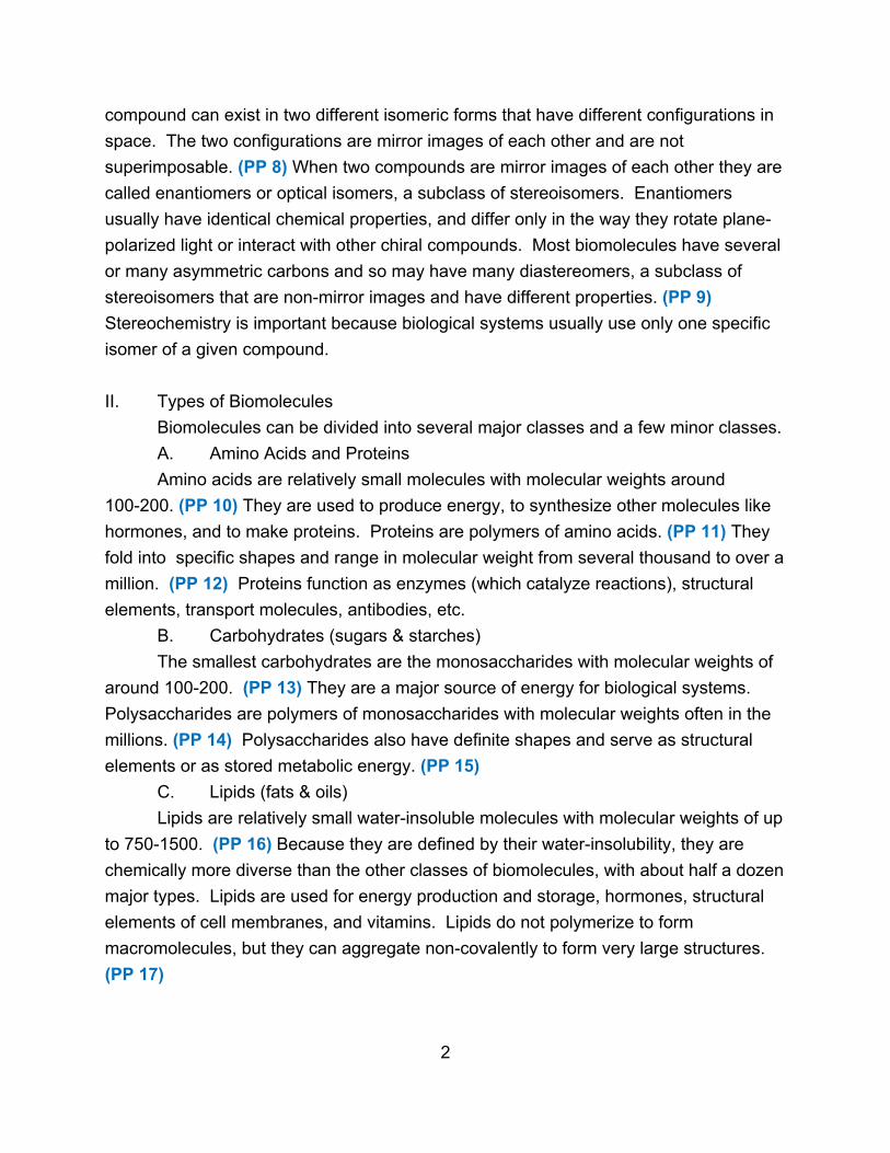
C. Lipids (fats & oils)
Lipids are relatively small water-insoluble molecules with molecular weights of up
to 750-1500. (PP 16) Because they are defined by their water-insolubility, they are
chemically more diverse than the other classes of biomolecules, with about half a dozen
major types. Lipids are used for energy production and storage, hormones, structural
elements of cell membranes, and vitamins. Lipids do not polymerize to form
macromolecules, but they can aggregate non-covalently to form very large structures.
(PP 17)
2
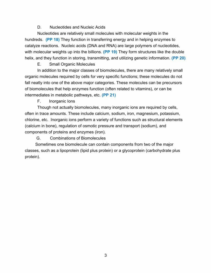
D. Nucleotides and Nucleic Acids
Nucleotides are relatively small molecules with molecular weights in the
hundreds. (PP 18) They function in transferring energy and in helping enzymes to
catalyze reactions. Nucleic acids (DNA and RNA) are large polymers of nucleotides,
with molecular weights up into the billions. (PP 19) They form structures like the double
helix, and they function in storing, transmitting, and utilizing genetic information. (PP 20)
E. Small Organic Molecules
In addition to the major classes of biomolecules, there are many relatively small
organic molecules required by cells for very specific functions; these molecules do not
fall neatly into one of the above major categories. These molecules can be precursors
of biomolecules that help enzymes function (often related to vitamins), or can be
intermediates in metabolic pathways, etc. (PP 21)
F. Inorganic Ions
Though not actually biomolecules, many inorganic ions are required by cells,
often in trace amounts. These include calcium, sodium, iron, magnesium, potassium,
chlorine, etc. Inorganic ions perform a variety of functions such as structural elements
(calcium in bone), regulation of osmotic pressure and transport (sodium), and
components of proteins and enzymes (iron).
G. Combinations of Biomolecules
Sometimes one biomolecule can contain components from two of the major
classes, such as a lipoprotein (lipid plus protein) or a glycoprotein (carbohydrate plus
protein).
3
Chapter 2: Amino Acids
I. Introduction
The major function of amino acids is to act as the building blocks of proteins.
Amino acids themselves can be used by the cell to produce energy and are the starting
point for making many nitrogen-containing compounds.
II. General Structure
A. Formula
As the name implies, amino acids contain two functional groups, a carboxylic
acid group and an amino group. The common amino acids are α-amino acids where
both functional groups are attached to the same carbon atom.
NH2
│
R — C — COOH
│
H
Also attached to the central carbon are a hydrogen atom and an R group, which
is different in each amino acid.
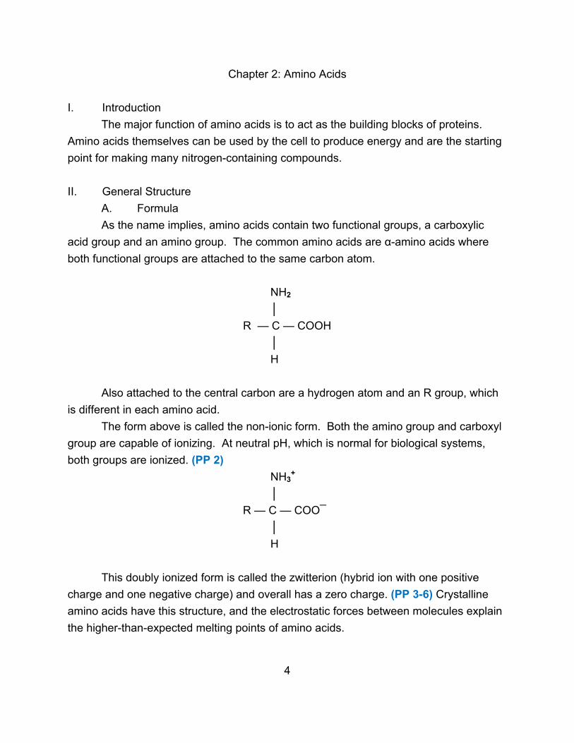
The form above is called the non-ionic form. Both the amino group and carboxyl
group are capable of ionizing. At neutral pH, which is normal for biological systems,
both groups are ionized. (PP 2)
NH3+
│
R — C — COO¯
│
H
This doubly ionized form is called the zwitterion (hybrid ion with one positive
charge and one negative charge) and overall has a zero charge. (PP 3-6) Crystalline
amino acids have this structure, and the electrostatic forces between molecules explain
the higher-than-expected melting points of amino acids.
4

B. Stereochemistry
If the R group is something other than a hydrogen atom, then the central carbon
is asymmetric and there will be two enantiomers (mirror images). (PP 7) The compound
glyceraldehyde is used as a reference compound for distinguishing stereoisomers.
(PP 8)
CHO CHO
│ │
HO ― C ― H H ― C ― OH
│ │
CH2OH CH2OH
L – glyceraldehyde D - glyceraldehyde
The prefixes L and D stand for levo (rotates light to left) and dextro (rotates light to
right). For amino acids
COOH COOH
│ │
NH2 ― C ― H H ― C ― NH2
│ │
R R
L - amino acid D - amino acid
It is the L-amino acids that are biologically important, with very few exceptions.
Amino acids found in proteins are normally L-isomers.
C. Classes
There are 20 common or major amino acids that are found in proteins. They are
divided into groups based on the nature of the R group. However, not every amino acid
falls neatly into a category, so there can be variations in how amino acids are classified.
For instance, the glycine R-group is sometimes classified as hydrophilic and sometimes
as hydrophobic. Each amino acid can be designated by a three-letter abbreviation, or
by a one-letter abbreviation. (PP 9)
1. Nonpolar aliphatic R groups
The R group of these amino acids is hydrophobic, but not the entire amino acid.
The R groups are mainly hydrocarbon in nature. (PP 10)
5

COO¯
│
+NH3 ― C ― H
│ Alanine - Ala, A
CH3
COO¯
│
+NH3 ― C ― H
│ Valine - Val, V
CH3 ― C ―H
│
CH3
COO¯
│
+NH3 ― C ― H
│
CH2
│ Leucine - Leu, L
CH3 ― C ―H
│
CH3
COO¯
│
+NH3 ― C ― H │ Isoleucine - Ile, I
H ― C ― CH3 (There are 4 possible isomers, only one
│ of which is biologically important.)
CH2
│
CH3
6
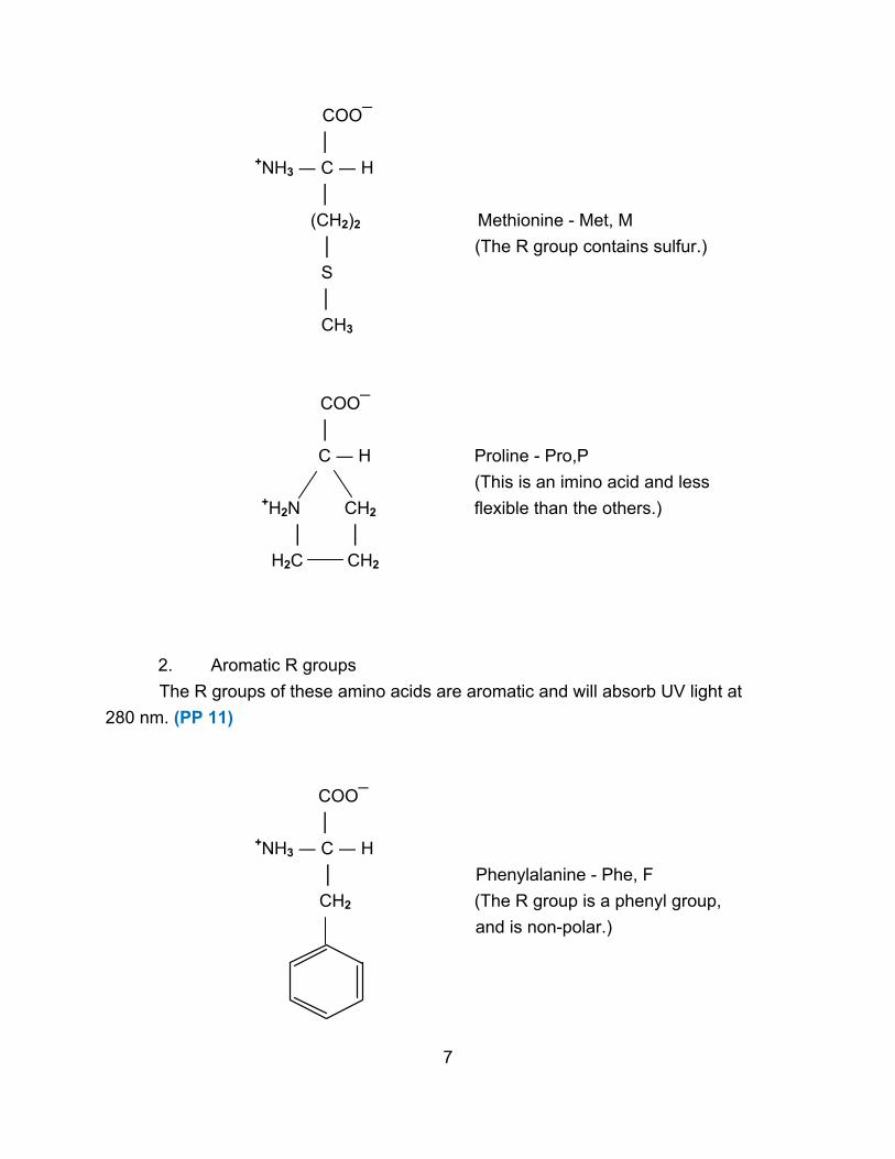
COO¯
│
+NH3 ― C ― H
│
(CH2)2 Methionine - Met, M
│ (The R group contains sulfur.)
S
│
CH3
COO¯
│
C ― H Proline - Pro,P
(This is an imino acid and less
+H2N CH2 flexible than the others.)
│ │
H2C CH2
2. Aromatic R groups
The R groups of these amino acids are aromatic and will absorb UV light at
280 nm. (PP 11)
COO¯
│
+NH3 ― C ― H
│ Phenylalanine - Phe, F
CH2 (The R group is a phenyl group,
and is non-polar.)
7
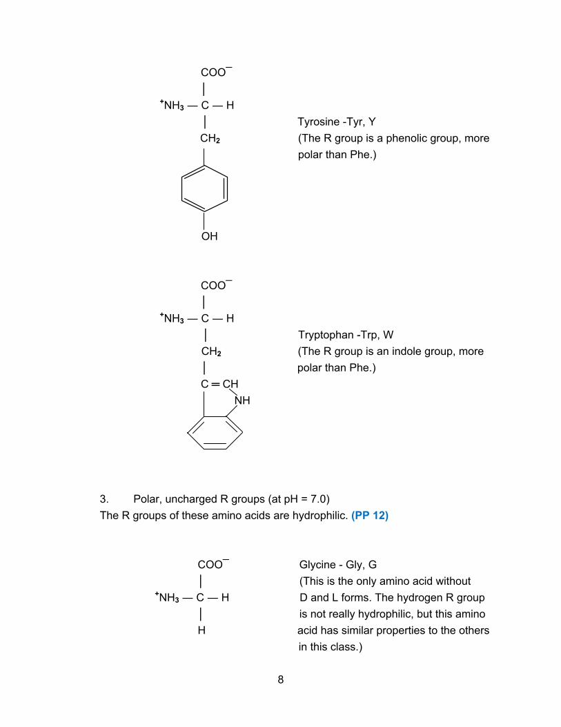
COO¯
│
+NH3 ― C ― H
│ Tyrosine -Tyr, Y
CH2 (The R group is a phenolic group, more
polar than Phe.)
OH
COO¯
│
+NH3 ― C ― H
│ Tryptophan -Trp, W
CH2 (The R group is an indole group, more
│ polar than Phe.)
C ═ CH
NH
3. Polar, uncharged R groups (at pH = 7.0)
The R groups of these amino acids are hydrophilic. (PP 12)
COO¯ Glycine - Gly, G
│ (This is the only amino acid without
+NH3 ― C ― H D and L forms. The hydrogen R group
│ is not really hydrophilic, but this amino
H acid has similar properties to the others
in this class.)
8

COO¯
│
+NH3 ― C ― H Serine - Ser, S
│ (The R group is a hydroxyl group.)
CH2OH
COO¯
│
+NH3 ― C ― H Threonine -Thr, T
│ (The R group is a hydroxyl group. There
H ― C ― OH are 4 possible isomers, only one of
│ which is biologically important.)
CH3
COO¯
│
+NH3 ― C ― H Cysteine - Cys, C
│ (The R group is a thiol or sulfydryl group.)
CH2
│
SH
COO¯
│
+NH3 ― C ― H
│ Asparagine - Asn, N
CH2 (The R group is an amide group.)
│
C ═ O
│
NH2
9

COO¯
│
+NH3 ― C ― H
│ Glutamine - Gln, Q
CH2 (The R group is an amide group.)
│
CH2
│
C ═ O
│
NH2
4. Negatively-charged R groups (at pH 7.0)
These amino acids are acidic and contain an extra carboxyl group. (PP 13)
COO¯
│
+NH3 ― C ― H Aspartate - Aspartic Acid - Asp, D
│
CH2
│
COO¯
COO¯
│
+NH3 ― C ― H Glutamate - Glutamic Acid - Glu, E
│
CH2
│
CH2
│
COO¯
10
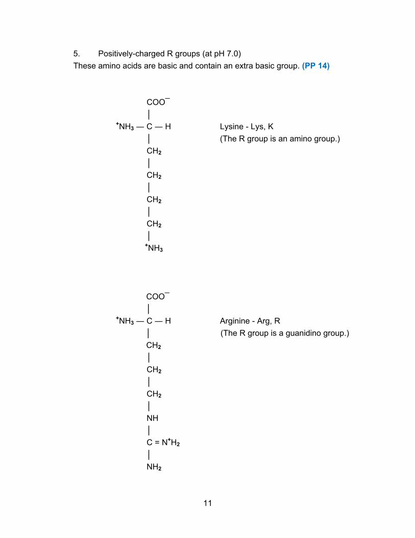
5. Positively-charged R groups (at pH 7.0)
These amino acids are basic and contain an extra basic group. (PP 14)
COO¯
│
+NH3 ― C ― H Lysine - Lys, K
│ (The R group is an amino group.)
CH2
│
CH2
│
CH2
│
CH2
│
+NH3
COO¯
│
+NH3 ― C ― H Arginine - Arg, R │ (The R group is a guanidino group.)
CH2
│
CH2
│
CH2
│
NH
│
C = N+H2
│
NH2
11

COO¯
│
+NH3 ― C ― H Histidine - His, H
│ (The R group is an imidazole group.
CH2 This is the only amino acid with an
│ R group that ionizes around neutral
C ― N ― H pH, creating two forms of the amino acid.)
C ― H
H ― C ― +N
H
6. Amino acid properties
The nature of the R-group determines the behavior of amino acids. (PP 15-18)
7. Other amino acids
In addition to the 20 standard amino acids found in proteins, some proteins contain
modified amino acids, such as 4-hydroxyproline or ε-N-methyllysine. Another 300 amino
acids have been found in biological systems that have functions other than as protein
components. They have a variety of functions as precursors in biosynthesis and
intermediates in metabolic pathways. (PP 19)
III. Acid-Base Properties
Both the carboxyl group and the amino group (and sometimes the R-group) have
acid-base properties, so amino acids have complicated acid-base behavior. These
properties are crucial in determining the behavior of both individual amino acids and the
proteins that contain them.
A. Acid-Base Theory
An acid is a proton donor relative to H2O. Weak acids do not dissociate
completely, while strong acids do. Weak acids reach an equilibrium between dissociated
and undissociated forms (generally ionizing less than 1%).
HA + H2O ↔ H3O+ + A¯
The strength of the acid is given by its Ka, the acid dissociation constant. (PP 20)
Ka = [ H3O+] [A¯] ≈ 10-5 for organic acids
[HA]
12

A base is a proton acceptor relative to H2O.
B + H2O ↔ BH+ + OH¯
The strength of a base is given by its Kb, the base dissociation constant. (PP 21)
Kb = [BH+] [OH¯] ≈ 10-4 for organic bases
[B]
In any of these reactions, an acid and a base react to form a conjugate base and
a conjugate acid. Rather than work with negative exponents, pKa and pKb are used.
pKa = -log Ka ≈ 5 for organic acids
pKb = -log Kb ≈ 4 for organic bases
To find the relationship between pKa and pKb , consider one acid-base pair. (PP 22)
HA + H2O ↔ H3O+ + A¯ and A¯ + H2O ↔ HA + OH¯
Ka = [ H3O+] [A¯] Kb = [HA] [OH¯]
[HA] [A¯]
so Ka · Kb = [ H3O+] [A¯] · [HA] [OH¯]
[HA] [A¯]
Ka · Kb = [ H3O+] [OH¯] = Kw = 10-14 at 25o C (water dissociation constant)
and -log Ka - logKb = -log Kw = 14
Therefore, pKa + pKb = 14
So rather than use pKb , only pKa is used. The strength of a base is described by the
pKa of its conjugate acid where pKa = 14 – pKb ≈ 10 for organic bases. Thus compounds
with low pKa values are good acids while compounds with high pKa values are good bases.
(PP 23-24)
13

B. Titration Curves
One of the best ways to find the pKa of a substance is to determine its titration
curve. A weak acid can be titrated with a strong base, and the pH of the resulting
solution plotted as a function of the amount of base added. (PP 25)
HA + NaOH → Na+ + A¯ + H2O
The titration curve is expressed by the Henderson-Hasselbalch equation, derived as
follows. (PP 26)
Ka = [ H3O+] [A¯]
[HA]
-log Ka = -log [ H3O+] - log [A¯] / [HA]
pKa = pH - log [A¯] / [HA]
and pH = pKa + log [A¯] / [HA]
Thus at any point on a titration curve (as shown below), the pH is determined first by the
strength of the acid HA being titrated (pKa) and second by the relative amounts of acid
(HA) and conjugate base (A¯), which is determined by the amount of NaOH added.
14

There are two important points on this curve. When there are equal amounts of HA
and NaOH present (one equivalent), all the HA has been neutralized and only A¯
remains. This is called the equivalence point, and it occurs at a basic pH since A¯ is a
base. When half as much NaOH is present as HA, then half of the HA has been
neutralized to A¯, and the other remains as HA, creating equal amounts of acid and
conjugate base. This is the half-equivalence point. The pH here can be calculated
from the Henderson-Hasselbalch equation.
pH = pKa + log [A¯] / [HA]
Since [A¯] = [HA], the ratio is 1. Since log 1 = 0, the pH = pKa. Thus the pKa can be
found from the half-equivalence point on the titration curve. (PP 27-28)
C. Amino Acid Titration
Amino acids can act as both acids and bases and so are called amphoteric.
Starting with the zwitterion, the COO¯ group can accept a proton as the pH is lowered.
The +NH3 group can lose a proton as the pH raised. Using alanine as an example,
COOH COO¯ COO¯
│ H+ │ OH¯ │
+NH3 ― C ― H ← +NH3 ― C ― H → NH2 ― C ― H
│ 2.3 │ 9.7 │
CH3 CH3 CH3
fully protonated zwitterion fully deprotonated
The pKa of the carboxyl group is 1.8 – 2.4, depending upon the amino acid. The pKa of
the amino group is 8.8 –11.0 (usually 9 – 10). The titration curve of the amino acid will
show both pKa values.
15

Equivalence points occur in steep areas, while pKa values occur in flat areas.
The form(s) of the amino acid present depends on the pH. At very low pH = 1,
the amino acid will be in the fully protonated form.
COOH
│
+NH3 ― C ― H charge = +1
│
CH3
As base is added, the COOH group will be titrated to COO¯. As the pKa is approached,
some of the amino acid will lose its proton. The exact amounts of COOH and COO¯ are
given by the Henderson-Hasselbalch equation.
When the half-equivalence point has been reached, half the COOH has been
converted to COO¯.
COOH COO¯
│ │
+NH3 ― C ― H = +NH3 ― C ― H
│ pH = pKa = 2.3 │
CH3 CH3
As more base is added, the rest of the COOH is neutralized. All of it is neutralized when
the equivalence point is reached.
COO¯
│
+NH3 ― C ― H charge = 0
│
CH3
All the amino acid is in the zwitterion form at this point. The amino acid has no net
charge, so this is called the isoelectric point or isoionic point (pI). (PP 29)
pI = pKa1 + pKa2 = 2.3 + 9.7 = 6.0
2 2
16
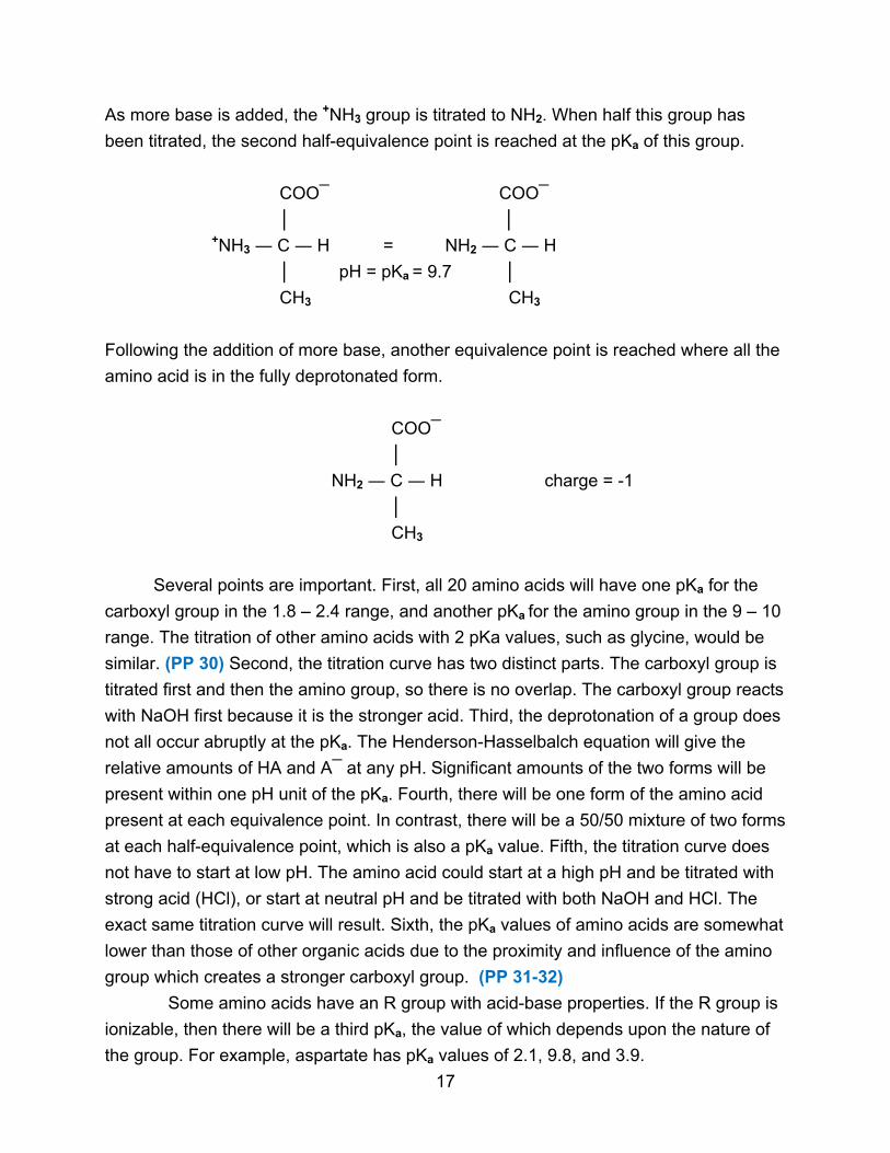
As more base is added, the +NH3 group is titrated to NH2. When half this group has
been titrated, the second half-equivalence point is reached at the pKa of this group.
COO¯ COO¯
│ │
+NH3 ― C ― H = NH2 ― C ― H
│ pH = pKa = 9.7 │
CH3 CH3
Following the addition of more base, another equivalence point is reached where all the
amino acid is in the fully deprotonated form.
COO¯
│
NH2 ― C ― H charge = -1
│
CH3
Several points are important. First, all 20 amino acids will have one pKa for the
carboxyl group in the 1.8 – 2.4 range, and another pKa for the amino group in the 9 – 10
range. The titration of other amino acids with 2 pKa values, such as glycine, would be
similar. (PP 30) Second, the titration curve has two distinct parts. The carboxyl group is
titrated first and then the amino group, so there is no overlap. The carboxyl group reacts
with NaOH first because it is the stronger acid. Third, the deprotonation of a group does
not all occur abruptly at the pKa. The Henderson-Hasselbalch equation will give the
relative amounts of HA and A¯ at any pH. Significant amounts of the two forms will be
present within one pH unit of the pKa. Fourth, there will be one form of the amino acid
present at each equivalence point. In contrast, there will be a 50/50 mixture of two forms
at each half-equivalence point, which is also a pKa value. Fifth, the titration curve does
not have to start at low pH. The amino acid could start at a high pH and be titrated with
strong acid (HCl), or start at neutral pH and be titrated with both NaOH and HCl. The
exact same titration curve will result. Sixth, the pKa values of amino acids are somewhat
lower than those of other organic acids due to the proximity and influence of the amino
group which creates a stronger carboxyl group. (PP 31-32)
Some amino acids have an R group with acid-base properties. If the R group is
ionizable, then there will be a third pKa, the value of which depends upon the nature of
the group. For example, aspartate has pKa values of 2.1, 9.8, and 3.9.
17

The curve has three distinct areas, each corresponding to the titration of one
group on the amino acid. The pKa values can be assigned to each group, with 2.1 for
the α-carboxyl group, 9.8 for the amino group, and 3.9 for the R group. The following
forms of the amino acid occur throughout the titration, with one form present at each
equivalence point and a mixture of forms present at each pKa.
One form One form One form One form
at pH < 1.0 at pH = 3.0 at pH = 6.85 at pH > 11.0
COOH COO¯ COO¯ COO¯
│ │ │ │
+NH3 ― C ― H +NH3 ― C ― H +NH3 ― C ― H NH2 ― C ― H
│ ↔ │ ↔ │ ↔ │
CH2 2.1 CH2 3.9 CH2 9.8 CH2
│ │ │ │
COOH COOH COO¯ COO¯
Two forms Two forms Two forms at pH = pKa = 2.1 at pH = pKa = 3.9 at pH = pKa = 9.8
18

Other amino acids with 3 pKa values are: (PP 33)
Glutamate – 2.2, 4.3 (R), 9.7
At pH = 4.3, the acidic R group ionizes- COOH ↔ COO¯
The titration curve of glutamate would be similar to that of aspartate. (PP 34)
Lysine – 2.2, 9.0, 10.5 (R)
At pH = 10.5, the basic R group ionizes- +NH3 ↔ NH2
Arginine – 2.2, 9.0, 12.5 (R)
At pH = 12.5, the basic R group ionizes-
│ │
NH NH
│ ↔ │
C = N+H2 C = NH
│ │
NH2 NH2
Histidine – 1.8, 6.0 (R), 9.2
At pH = 6.0, the basic R group ionizes-
│ │
CH2 CH2
│ │
C ― N ― H ↔ C ― N ― H
C ― H C ― H
H ― C ― +N H ― C ― N
H
Cysteine – 1.7, 8.3 (R), 10.8
At pH = 8.3, the R group ionizes- │ │ CH2 CH2 │ ↔ │ SH S¯
19
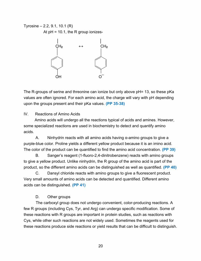
Tyrosine – 2.2, 9.1, 10.1 (R)
At pH = 10.1, the R group ionizes- │ │ CH2 ↔ CH2
OH O¯
The R-groups of serine and threonine can ionize but only above pH= 13, so these pKa
values are often ignored. For each amino acid, the charge will vary with pH depending
upon the groups present and their pKa values. (PP 35-38)
IV. Reactions of Amino Acids
Amino acids will undergo all the reactions typical of acids and amines. However,
some specialized reactions are used in biochemistry to detect and quantify amino
acids.
A. Ninhydrin reacts with all amino acids having α-amino groups to give a
purple-blue color. Proline yields a different yellow product because it is an imino acid.
The color of the product can be quantified to find the amino acid concentration. (PP 39)
B. Sanger’s reagent (1-fluoro-2,4-dinitrobenzene) reacts with amino groups
to give a yellow product. Unlike ninhydrin, the R group of the amino acid is part of the
product, so the different amino acids can be distinguished as well as quantified. (PP 40)
C. Dansyl chloride reacts with amino groups to give a fluorescent product.
Very small amounts of amino acids can be detected and quantified. Different amino
acids can be distinguished. (PP 41)
D. Other groups
The carboxyl group does not undergo convenient, color-producing reactions. A
few R groups (including Cys, Tyr, and Arg) can undergo specific modification. Some of
these reactions with R groups are important in protein studies, such as reactions with
Cys, while other such reactions are not widely used. Sometimes the reagents used for
these reactions produce side reactions or yield results that can be difficult to distinguish.
20

Chapter 3: Peptides
I. Introduction
Amino acids can join together to form polymers. When a few amino acids are
joined together (2 - ~50), the molecule is called a peptide. When more amino acids
are joined together, the molecule is called a protein.
II. Peptide Bonds
The covalent linkage between amino acids is called a peptide bond, which is
chemically an amide bond. (PP 2)
R R │ │
NH2 ― CH ― COOH + NH2 ―CH ― COOH →
R O R │ ║ │
NH2 ― CH ― C ― N ― CH ― COOH + H2O │ H
Additional amino acids can be added onto both ends. The bond is mostly planar
and rigid, with partial double bond character due to resonance forms. (PP 3-4)
H O R H Oδ−
R
│ ║ │ │ │ │
― N ― CH ― C ― N ― CH ―C ― ↔ ― N― CH ― C ― Nδ+― CH ―C ―
│ │ ║ │ │ ║ R H O R H O There is restricted rotation around the peptide bond. The configuration around a
peptide bond is normally trans. (PP 5)
III. Properties of Peptides
A. Naming
By convention, the amino acid with the free α-amino group is written on the left
and is the amino-terminal or N-terminal of the peptide. The amino acid with the free
21

carboxyl group is written on the right and is called the carboxyl-terminal or C-terminal.
(PP 6) Peptides are named according to the amino acid components, beginning with the
N-terminal. (PP 7)
Ala - Gln - Tyr - Gly - Ser - Phe - Lys
N-terminal C-Terminal
B. Chemical Properties
Peptide bonds are stable under biological conditions. Peptide bonds can be
hydrolyzed by heating in strong acid or strong base. Peptide bonds can be broken by a
group of enzymes known as proteases (exo vs. endopeptidase).
C. Optical Properties
Peptides are composed of L-amino acids, so they will be optically active. For
very short peptides, the optical activity is the sum of the optical activities of the
component amino acids. For longer peptides, it is not.
D. Acid-Base Properties
The -NH- and C = O in a peptide bond have no significant tendency to ionize or
protonate and so do not have acid-base properties. Peptides do have acid-base
properties caused by the N-terminal α-amino group, the C-terminal α-carboxyl group,
and any ionizable R-groups present. Thus a peptide may have several to many pKa
values. (PP 8) The exact pKa values may differ from their value in the free amino acids.
Peptides can be titrated, but it may be impossible to pick out individual pKa values if
several are similar and overlap. There will be some pH, however, where the negative
charges balance the positive charges, and the peptide has no net charge. This point
will be the peptide’s isoelectric point. (PP 9-12)
E. Biological Activity
Many peptides are biologically active. Some hormones are peptides, such as
insulin (51 amino acids) and glucagon (29 amino acids). Some toxins and antibiotics
are peptides.
IV. Peptide Synthesis
Peptides can be made very efficiently by the cell. A 100-amino acid protein with
a specific amino acid sequence can be made by the cell in about five seconds.
Peptide synthesis can be done in the laboratory with much more difficulty. The
problem is to join together the amino acids in a specific sequence. (PP 13)
22
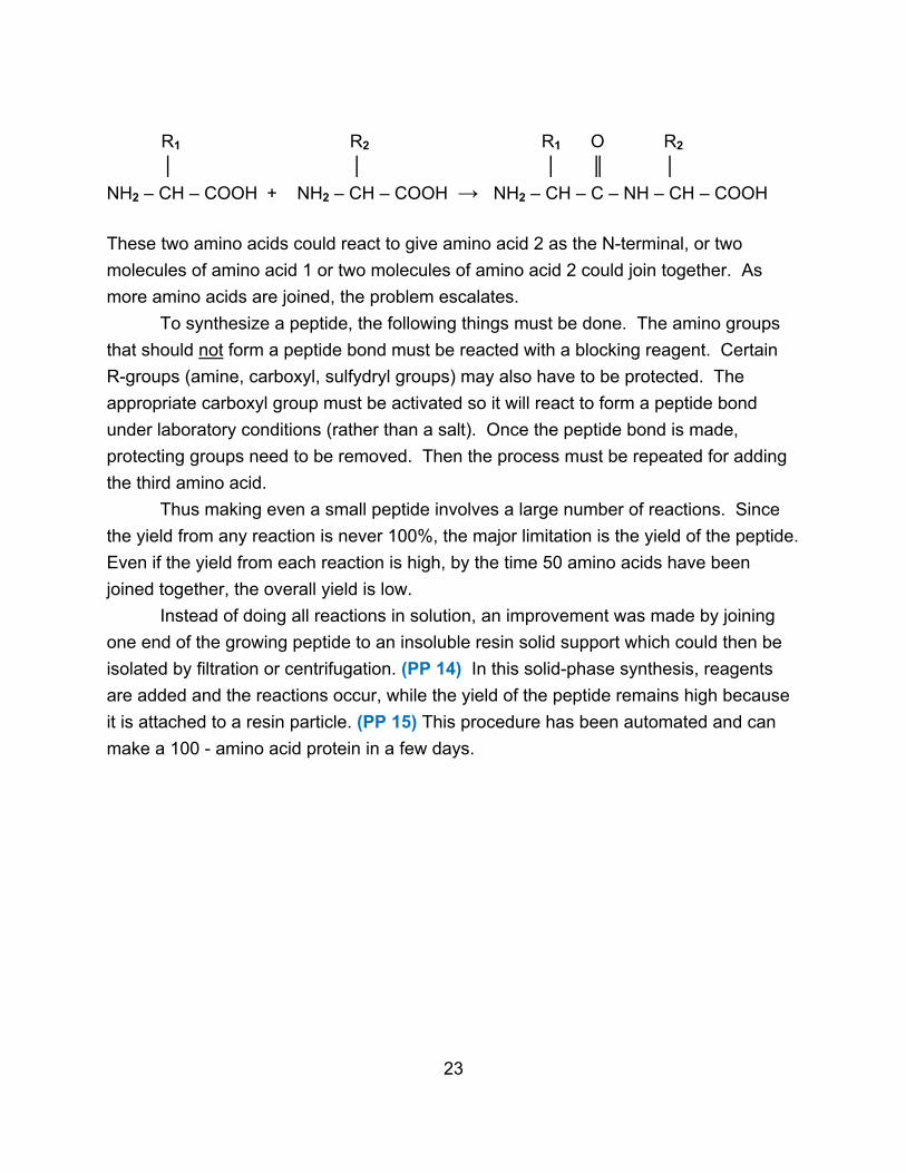
R1 R2 R1 O R2
│ │ │ ║ │
NH2 – CH – COOH + NH2 – CH – COOH → NH2 – CH – C – NH – CH – COOH
These two amino acids could react to give amino acid 2 as the N-terminal, or two
molecules of amino acid 1 or two molecules of amino acid 2 could join together. As
more amino acids are joined, the problem escalates.
To synthesize a peptide, the following things must be done. The amino groups
that should not form a peptide bond must be reacted with a blocking reagent. Certain
R-groups (amine, carboxyl, sulfydryl groups) may also have to be protected. The
appropriate carboxyl group must be activated so it will react to form a peptide bond
under laboratory conditions (rather than a salt). Once the peptide bond is made,
protecting groups need to be removed. Then the process must be repeated for adding
the third amino acid.
Thus making even a small peptide involves a large number of reactions. Since
the yield from any reaction is never 100%, the major limitation is the yield of the peptide.
Even if the yield from each reaction is high, by the time 50 amino acids have been
joined together, the overall yield is low.
Instead of doing all reactions in solution, an improvement was made by joining
one end of the growing peptide to an insoluble resin solid support which could then be
isolated by filtration or centrifugation. (PP 14) In this solid-phase synthesis, reagents
are added and the reactions occur, while the yield of the peptide remains high because
it is attached to a resin particle. (PP 15) This procedure has been automated and can
make a 100 - amino acid protein in a few days.
23
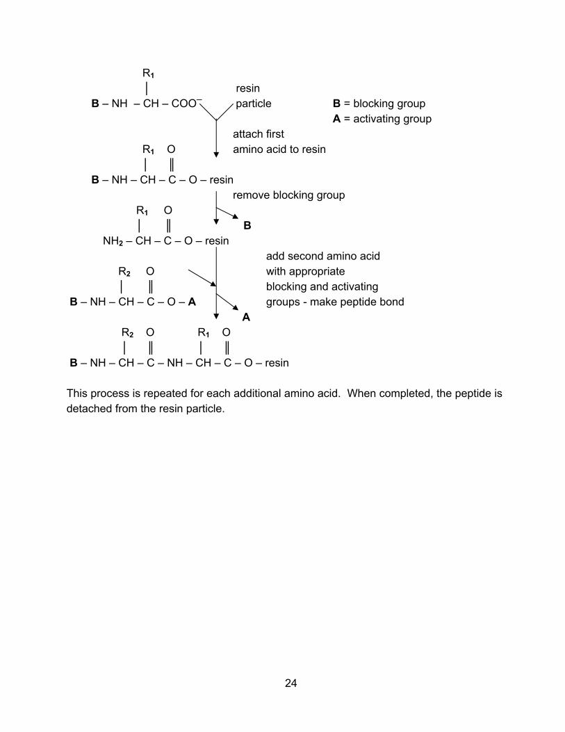
R1 │ resin B – NH – CH – COO─ particle B = blocking group
A = activating group attach first
R1 O amino acid to resin │ ║
B – NH – CH – C – O – resin remove blocking group
R1 O │ ║ B
NH2 – CH – C – O – resin add second amino acid
R2 O with appropriate │ ║ blocking and activating
B – NH – CH – C – O – A groups - make peptide bond A
R2 O R1 O │ ║ │ ║
B – NH – CH – C – NH – CH – C – O – resin This process is repeated for each additional amino acid. When completed, the peptide is detached from the resin particle.
24

Chapter 4: Protein Sequence
I. Introduction
Proteins are the same as peptides, only larger with more amino acids. The amino
acids are still joined in a long, linear chain by peptide bonds.
A. Size and Structure
Proteins can range in size from around 50 amino acids (MW = 6000) to around
8000 amino acids (MW = 1,000,000). One exceptionally large protein has 34,000 amino
acids and a molecular weight of 3.8 million. (PP 2) Some proteins consist of a single
polypeptide chain (PP 3), but many of the large proteins consist of two or more
polypeptide chains associated together. (PP 4) Such proteins are called multimeric or
multisubunit proteins, and the individual polypeptide chains are called subunits. The
subunits may be identical or they may be of different types. The number of subunits
may range from two to 50 or more. The amino acids are almost always the 20 standard
amino acids, but sometimes these are slightly modified (hydroxyproline). (PP 5-6)
B. Prosthetic Groups
Many proteins consist only of amino acids, but some contain a non-amino-acid
component called a prosthetic group. (PP 7) Some prosthetic groups are small organic
molecules related to vitamins, some are lipids (lipoproteins), some are carbohydrates
(glycoproteins), and some are metal atoms or ions (metalloproteins). (PP 8) The
prosthetic group is usually important for the protein to function properly.
C. Function
Proteins perform many different functions in biological systems. (PP 9)
1. Enzymes catalyze almost all the chemical reactions that occur in the cell.
For example, DNA polymerase is a protein that makes DNA.
2. Transport proteins carry specific molecules in the blood or through cell
membranes. Hemoglobin carries oxygen.
3. Storage proteins contain nutrients or metabolic energy. Ovalbumin in egg
white provides nutrition to the embryo.
4. Motile proteins enable a cell or organism to move. Actin is found in
muscle.
5. Structural proteins provide strength or protection. Hair is made of keratin.
6. Defense proteins include antibodies and venoms.
7. Regulatory proteins include hormones and proteins which mediate
hormonal effects.
25

D. Protein Characterization
Proteins can be separated, purified, and characterized using a variety of
methods. A fundamental property of a protein is structure. Because proteins are large
with complicated 3-dimensional shapes, protein structure is broken down into several
different levels. These levels are called primary, secondary, tertiary, and quaternary
structure. (PP 10)
II. Primary Structure
A. Definition
Primary structure refers to the amino acid sequence of a protein. It means not
merely what amino acids are in the protein, but their specific order. To know the
primary structure is to be able to list the amino acids, starting with the N-terminal amino
acid all the way through the C-terminal amino acid. (PP 11)
Asp - Lys - Ser - Thr - - - - - - - - Ala - Ile - Leu
1 2 3 4 159 160 161
The primary sequence is most important because the sequence is what makes a
protein a specific protein. All molecules of the protein lysozyme have the same
sequence (and therefore the same number of amino acids and the same molecular
weight). All molecules of the protein ovalbumin will have the same sequence, but the
ovalbumin sequence will be different from the lysozyme sequence. The sequence is of
crucial importance, for one change in the amino acid sequence can make the protein
non-functional. Some variation in sequences can occur. For instance, 30% of the
proteins in humans have some sequence differences. The same protein from different
species will usually show some variation. (PP 12-13)
Primary structure is routinely determined. It requires that the protein be pure.
Usually some preliminary experiments are done before the actual sequencing.
1. Amino acid composition
The numbers and types of amino acids in a protein (but not the sequence) can
be determined by hydrolyzing the protein with strong acid or base or breaking it down
enzymatically into its amino acids. The 20 different amino acids are then separated by
various techniques and quantitated. The result is that the protein is found to have 10
His, 12 Ala, 9 Lys, etc. (PP 14) Alternatively, each amino acid can be expressed as a
percentage of the total. This technique is now automated.
26

The problem is that no breakdown method is completely satisfactory. Strong
acid completely destroys Trp and partially destroys Ser, Thr, and Tyr. Amounts of these
last three can be estimated by measuring their disappearance over time and
extrapolating back to time = 0. In addition, Asn is converted to Asp and Gln is
converted to Glu. Base hydrolysis destroys Cys, Ser, Thr, Arg and modifies others, but
can be used to measure Trp. Since individual enzymes break only certain peptide
bonds, a mixture of enzymes must be used to ensure complete breakdown. Of course,
the enzymes digest themselves and contaminate the results, but amounts of Trp, Asn,
and Gln can be determined. (PP 15)
2. End group analysis
Determining N and C-terminal amino acids gives two reference points in the
amino acid sequence. It can also reveal (if two amino acids show up in one test) that
the protein is multimeric with different types of subunits.
The N-terminal can be found by reacting the intact protein with Sanger reagent,
dansyl chloride, etc., followed by acid hydrolysis. Only the N-terminal has an
α-amino group, so it will be modified by the reagent. After the protein is broken down,
the modified amino acid can be separated and identified. (PP 16)
The C-terminal does not undergo a similar reaction. The C-terminal amino acid
can be reduced to the amino alcohol using lithium borohydride. The protein is then
degraded and the amino alcohol identified, but this can be difficult since the amino
alcohol is not colored, fluorescent, etc. Another method is to break down the protein
with hydrazine, creating amino acyl hydrazides of all the amino acids except the C-
terminal which can then be separated and identified. However, many side reactions
occur which makes interpreting the results difficult. (PP 17)
The best method is to use enzymes which degrade the protein from the
C-terminal end. Such enzymes are called carboxypeptidases. (PP 18) However, each
enzyme has different specificity for the amino acids in the peptide bonds it cleaves. For
instance, several will not work on peptide bonds involving Pro. In addition, not all
peptide bonds are cleaved at the same rate, and once the last amino acid is cleaved off,
the next-to-the-last amino acid is susceptible to cleavage. If a mixture of
carboxypeptidases is used and the timing is correct, in most cases the C-terminal amino
acid is cleaved off and can be identified. (PP 19-20)
3. Sequencing
The sequencing itself can be done by the Edman degradation. The Edman
reagent is phenylisothiocyanate which reacts with the N-terminal amino acid. Mild acid
will then cleave off this modified amino acid leaving the rest of the protein chain intact.
27
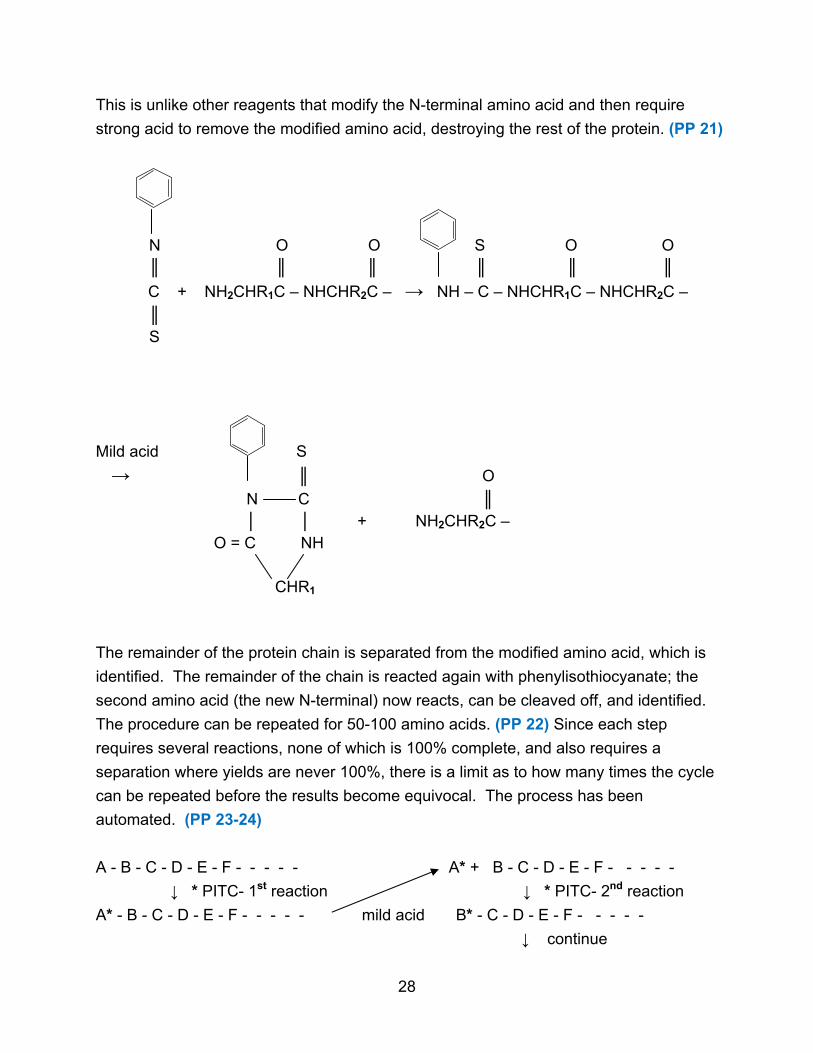
This is unlike other reagents that modify the N-terminal amino acid and then require
strong acid to remove the modified amino acid, destroying the rest of the protein. (PP 21)
N O O S O O ║ ║ ║ ║ ║ ║
C + NH2CHR1C – NHCHR2C – → NH – C – NHCHR1C – NHCHR2C –
║ S
Mild acid S
→ ║ O
N C ║ │ │ + NH2CHR2C –
O = C NH CHR1
The remainder of the protein chain is separated from the modified amino acid, which is
identified. The remainder of the chain is reacted again with phenylisothiocyanate; the
second amino acid (the new N-terminal) now reacts, can be cleaved off, and identified.
The procedure can be repeated for 50-100 amino acids. (PP 22) Since each step
requires several reactions, none of which is 100% complete, and also requires a
separation where yields are never 100%, there is a limit as to how many times the cycle
can be repeated before the results become equivocal. The process has been
automated. (PP 23-24)
A - B - C - D - E - F - - - - - A* + B - C - D - E - F - - - - -
↓ * PITC- 1st reaction ↓ * PITC- 2nd reaction
A* - B - C - D - E - F - - - - - mild acid B* - C - D - E - F - - - - -
↓ continue
28

4. Specific cleavage
Since most proteins have more than 100 amino acids, they cannot be directly
sequenced in their entirety. In such a case, the large protein is broken down into
smaller fragments of 50-100 amino acids. The fragments can then be separated and
each can be sequenced individually.
The problem is to break specific peptide bonds in the protein so that each protein
molecule is broken into the exact same fragments. If every protein molecule is broken
randomly, as by acid hydrolysis, then the fragment mixture would be impossible to
sequence. (PP 25)
________/_______/_________ _______/_______/__________
________/_______/_________ ____/_____/_________/______
________/_______/_________ __/__________/__________/__
specific cleavage gives random cleavage gives
small number of fragments many different fragments
The best way to specifically cleave the protein chain is by using enzymes. Some
enzymes will break a protein chain at specific amino acids.
O O ║ ║
– NH – CH – C – NH – CH – C – │ │ R1 R2
Trypsin will cleave a protein chain when R1 is Lys or Arg. (PP 26) Chymotrypsin will
cleave when R1 is Phe, Tyr, Trp. Staphylococcal V8 protease cleaves when R1 is Asp,
Glu. Some enzymes are specific for R2 rather than R1 . One chemical reagent which
cleaves specifically is cyanogen bromide, which cleaves when R1 is Met. (PP 27-32)
Following cleavage and sequencing of each fragment, the problem is to order the
fragments. Suppose the following fragments are found for a peptide that contains 15
amino acids, A-O. (This small peptide could be sequenced directly by the Edman
degradation without needing specific cleavage, but it illustrates the issue of ordering the
specific cleavage fragments.)
29

A B C D E F G H I J K L M N O
If A has been identified as the N-terminal amino acid and O is the C-terminal amino acid
then there are two possible sequences.
A B C D E F G H I J K L M N O
or
A B C D E H I J F G K L M N O
To establish the order a second specific cleavage is done with a second reagent that
will generate a second set of fragments.
A B C D E H I J F G K L M N O
Based on these fragments, again two orders are possible.
A B C D E H I J F G K L M N O
or
A B C J F G K D E H I L M N O
Only one order is possible from both sets of fragments. (PP 33)
5. Multimeric Proteins
If the subunits of a multimeric protein are of different types, then they must be
separated before primary structure is determined. The subunits must be purified and
each type sequenced separately.
B. Disulfide Bonds
In addition to peptide bonds, there is another type of covalent linkage which
occurs in proteins. This other type of bond is a disulfide bridge. Primary structure can
be defined as all the covalent linkages in a protein, which would include knowing the
sequence of amino acids joined by peptide bonds as well as knowing the position of
disulfide bonds. However, disulfide bonds function to maintain the 3-dimensional shape
of the protein, so they are sometimes considered a part of the higher levels of protein
structure.
Disulfide bonds occur when two cysteines are close to each other in a protein
molecule. (PP 34)
30
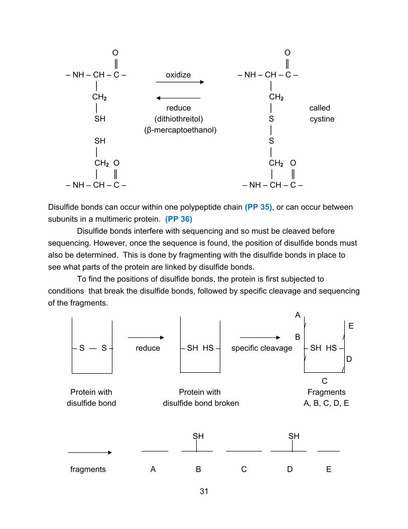
O O ║ ║
– NH – CH – C – oxidize – NH – CH – C – │ │ CH2 CH2 │ reduce │ called SH (dithiothreitol) S cystine
(β-mercaptoethanol) │ SH S │ │ CH2 O CH2 O │ ║ │ ║
– NH – CH – C – – NH – CH – C – Disulfide bonds can occur within one polypeptide chain (PP 35), or can occur between
subunits in a multimeric protein. (PP 36)
Disulfide bonds interfere with sequencing and so must be cleaved before
sequencing. However, once the sequence is found, the position of disulfide bonds must
also be determined. This is done by fragmenting with the disulfide bonds in place to
see what parts of the protein are linked by disulfide bonds.
To find the positions of disulfide bonds, the protein is first subjected to
conditions that break the disulfide bonds, followed by specific cleavage and sequencing
of the fragments.
A / E B / – S ― S – reduce – SH HS – specific cleavage – SH HS – / D / C Protein with Protein with Fragments
disulfide bond disulfide bond broken A, B, C, D, E SH SH ______ _______ ________ ________ _____ fragments A B C D E
31
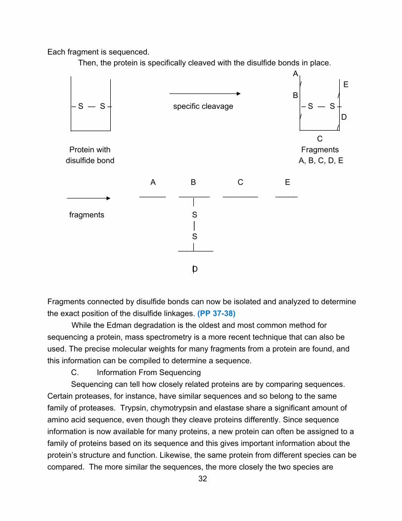
Each fragment is sequenced. Then, the protein is specifically cleaved with the disulfide bonds in place. A / E B / – S ― S – specific cleavage – S ― S – / D / C Protein with Fragments
disulfide bond A, B, C, D, E A B C E ______ _______ ________ _____ fragments S │ S
________ D
Fragments connected by disulfide bonds can now be isolated and analyzed to determine
the exact position of the disulfide linkages. (PP 37-38)
While the Edman degradation is the oldest and most common method for
sequencing a protein, mass spectrometry is a more recent technique that can also be
used. The precise molecular weights for many fragments from a protein are found, and
this information can be compiled to determine a sequence.
C. Information From Sequencing
Sequencing can tell how closely related proteins are by comparing sequences.
Certain proteases, for instance, have similar sequences and so belong to the same
family of proteases. Trypsin, chymotrypsin and elastase share a significant amount of
amino acid sequence, even though they cleave proteins differently. Since sequence
information is now available for many proteins, a new protein can often be assigned to a
family of proteins based on its sequence and this gives important information about the
protein’s structure and function. Likewise, the same protein from different species can be
compared. The more similar the sequences, the more closely the two species are
32

related. As an example, cytochrome c is a small protein of about 100 amino acids. The
sequence in humans and in chimpanzees is identical. However, cytochrome c in sheep
has 10 different amino acids, cytochrome c in fish has 18 different amino acids, and
cytochrome c in insects has 31 different amino acids. The evolutionary relationship of
the species is reflected in the protein.
The critical amino acids for a protein’s function can be found because they are the
ones that will be present in all related proteins. In cytochrome c, 28 amino acids are
invariant and therefore essential for proper function. Less crucial amino acids can vary
without affecting function.
Sometimes a protein’s primary structure provides clues about the shape of the
protein and the higher levels of the protein’s structure. For instance, certain amino acids
are more likely to be found on the surface of a protein while others are usually found in
the interior. Understanding a protein’s sequence and its relationship to other proteins
can also help in determining a protein’s function.
33

Chapter 5: Protein Conformation
I. Introduction
Primary structure includes all the covalent bonds in a protein, both peptide bonds
and disulfide bonds. However, rotation is possible around many of these covalent
bonds, so there are a large number of possible 3-dimensional shapes that a protein can
assume. The spatial arrangement of atoms in a protein is called its conformation.
Despite the almost limitless number of possible conformations of a protein, each protein
will have a specific, unique 3-dimensional structure. (PP 2) The higher levels of protein
structure describe this conformation with increasing complexity. (PP 3-5) There are
several important points about conformation.
A. Function of a protein depends upon its conformation. Protein molecules
that lose their proper shape will not be able to function properly.
B. The proper conformation for a protein is often (but not always) the one
which is the most thermodynamically stable. (PP 6) What shape is most stable depends
on what amino acids are in the protein. (PP 7) Thus conformation depends upon the
amino acid sequence. (PP 8-9) It is a goal of protein research to be able to deduce
protein conformation from a protein’s primary structure. Unfortunately, given the large
number of possible conformations and the difficulty of estimating energies and stabilities,
this cannot be done at present. Limited predictions about the structure of regions in
proteins can currently be made.
C. Conformation is maintained and held together mainly by non-covalent
forces. (Disulfide bonds and other covalent crosslinks also help to maintain protein
shape.) The various non-covalent forces include the following types. (PP 10)
1. Hydrogen bonds can form in many ways between certain R groups of
amino acids, between portions of the peptide backbone, and between polar amino acids
and the surrounding water molecules. (PP 11) Many polar amino acids tend to cluster
on the outside of a protein molecule where they can interact with the solvent.
2. Ionic interactions involve attractions between opposite charges. Two
oppositely charged amino acids can form an ionic bond known as a salt bridge. (PP 12)
Other electrostatic interactions involve induced or permanent dipoles.
3. Van der Waals forces are weak attractions between close, uncharged
atoms. A random variation in the electron cloud of one atom creates a momentary dipole
that induces an opposite dipole in another atom and causes an attraction. (PP 13)
4. Hydrophobic interactions occur when two or more hydrophobic groups
cluster together and so avoid interaction with water. (PP 14) There is no actual attraction
34

between the non-polar groups, but rather the stability comes from the thermodynamic
favorability of keeping these groups from water where they cause the water to assume a
highly structured solvation layer. Non-polar amino acids tend to cluster in the interior of
the protein where water is excluded.
While non-covalent forces are weak, there are a large number of them in a given
protein. The amino acid sequence will dictate how the chain must be spatially arranged
in order to maximize these forces. (PP 15-19)
D. Types
1. Secondary structure refers to the arrangement of neighboring amino
acids, which often occurs in a regular, repeating structure.
2. Tertiary structure refers to the complete 3-dimensional structure of the
polypeptide chain.
3. Quaternary structure occurs in multimeric proteins and refers to the spatial
arrangement of the subunits.
II. Secondary Structure
Secondary structure focuses on neighboring amino acids (20-40). The major
types of 2o structure are regular, repeating arrangements.
A. α-Helix Structure. The amino acids are arranged into a spiral shape with
the peptide backbone in a helix and the R-groups pointing out. (PP 20)
One turn of the helix is 0.54 nm and contains 3.6 amino acids. Almost all known
α-helices are right-handed (clockwise spiral) while left-handed helices (counterclockwise
spiral) are rarely observed. (PP 21)
35

What holds the α-helix together is hydrogen bonding between peptide bond
atoms in the backbone. Hydrogen bonds form between an amino acid and the fourth
amino acid further up the chain. The accumulation of many hydrogen bonds makes the
structure very stable since each N – H and C = O can form a hydrogen bond. (PP 22-24)
A protein can contain several segments of α-helix. (PP 25). However, not all
amino acid sequences can form a stable α-helix. Too many amino acids with the same
charged R group (acidic or basic) will disrupt an α-helix. Too many bulky R groups will
cause a problem (Leu, Thr). Proline cannot conform to the α-helix shape. When
present, it causes a bend in the direction of the helix. (PP 26-27)
The first R-group and the third or fourth one will interact. Often one will be
positively charged and the other negatively charged, or both will be hydrophobic.
B. β-Pleated Sheet Structure. A second type of regular 2o structure is the β-
pleated sheet. It is more extended than an α-helix and the chain is arranged in a zig-
zag. Several zig-zag chains line up to form a pleated sheet. (PP 28)
R R
side view
R R R
36
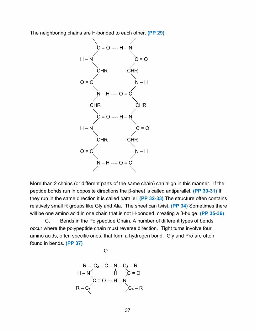
The neighboring chains are H-bonded to each other. (PP 29)
C = O ---- H – N H – N C = O CHR CHR O = C N – H N – H ---- O = C CHR CHR C = O ---- H – N H – N C = O CHR CHR O = C N – H N – H ---- O = C
More than 2 chains (or different parts of the same chain) can align in this manner. If the
peptide bonds run in opposite directions the β-sheet is called antiparallel. (PP 30-31) If
they run in the same direction it is called parallel. (PP 32-33) The structure often contains
relatively small R groups like Gly and Ala. The sheet can twist. (PP 34) Sometimes there
will be one amino acid in one chain that is not H-bonded, creating a β-bulge. (PP 35-36)
C. Bends in the Polypeptide Chain. A number of different types of bends
occur where the polypeptide chain must reverse direction. Tight turns involve four
amino acids, often specific ones, that form a hydrogen bond. Gly and Pro are often
found in bends. (PP 37)
O
║
R – C2 – C – N – C3 – R
H – N H C = O
C = O --- H – N
R – C1 C4 – R
37

D. Irregular 2o Structure. In many areas of a protein, the amino acids may not
assume a regular, repeating structure. Irregular structure can occur. This does not
mean random. The spatial arrangement is still specific, dictated by the amino acids.
E. Secondary Structure in Proteins
Different amino acids occur with different frequencies in the various types of
secondary structure. (PP 38)
Proteins can be divided into two categories with regard to 2o structure.
1. Globular proteins have a very compact 3-dimensional structure, within
which exist different areas of 2o structure. Different globular proteins have very different
amounts of 2o structure types. (PP 39)
α β irregular
myoglobin 78% 0% 22%
chymotrypsin 14% 45% 41%
lysozyme 40% 12% 48%
2. Fibrous proteins have long, extended structures consisting often of just
one main type of 2o structure. There are several categories of fibrous proteins. (PP 40)
a. α-keratins
α-keratins make up hair, wool, feathers, nails, skin, horns, etc. The
structure of α-keratins is a right-handed α-helix. They contain many hydrophobic
residues, making them insoluble and water-repellent. Adjacent chains of α-helix are
linked by disulfide bonds. The more disulfide bonds, the stronger the structure is. In
hair, two α-helices interact to form coils, two coils combine to make a protofilament, and
four protofilaments make one filament. These are packaged and wound to form
macrofibrils, many of which make up a hair strand. Disulfide cross-links occur at several
levels, producing a strong structure. In the case of hair, the structure is flexible but does
not stretch. (PP 41-42)
b. β-keratins
β-keratins include silk and spider webs that are flexible but do not stretch.
The structure is an antiparallel β-sheet with a high percentage of small amino acids (Gly,
Ala). There are no disulfide bonds. The small R groups allow pairs of the β-sheets to
stack together, forming an ordered array. (PP 43)
c. Collagen
Collagen (existing in several different types) is found in tendons, cartilage,
etc. Collagen has a unique left-handed helix with 3.3 amino acids/ turn. The helix is
more extended than an α-helix. Collagen contains 35% Gly and 21% proline plus
38
hydroxyproline, usually in a repeating sequence of Gly - X - Pro (Hyp). Three such
chains (~ 1000 amino acids) are wound together in a right-handed triple helix held
together by hydrogen bonds and covalent crosslinks between oxidized lysines. (PP 44)
│ │
H – N H N – H
│ │ │
H – C – (CH2)3 – C = C – (CH2)2 – C – H
│ │ │
O = C H – C = O C = O
│ │
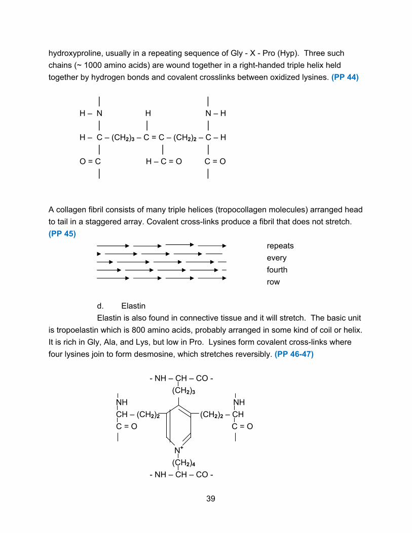
A collagen fibril consists of many triple helices (tropocollagen molecules) arranged head
to tail in a staggered array. Covalent cross-links produce a fibril that does not stretch.
(PP 45)
repeats
every
fourth
row
d. Elastin
Elastin is also found in connective tissue and it will stretch. The basic unit
is tropoelastin which is 800 amino acids, probably arranged in some kind of coil or helix.
It is rich in Gly, Ala, and Lys, but low in Pro. Lysines form covalent cross-links where
four lysines join to form desmosine, which stretches reversibly. (PP 46-47)
- NH – CH – CO -
(CH2)3
NH NH
CH – (CH2)2 (CH2)2 – CH
C = O C = O
N+
(CH2)4
- NH – CH – CO -
39

III. Tertiary Structure
Tertiary structure refers to the overall three-dimensional shape of a protein, also
called conformation.
A. General Features
1. Tertiary structure is specific for each protein. All molecules of myoglobin will
have the same tertiary structure, while all molecules of lysozyme will share a different
tertiary structure. (PP 48)
2. Tertiary structure is determined by primary structure. Amino acid chains will
fold into a shape that maximizes favorable interactions, and this will vary with amino acid
sequence. Proteins with similar sequences have similar tertiary structures. (PP 49-50)
3. Tertiary structure is maintained mainly by non-covalent forces (H – bonds,
salt bridges, hydrophobic interactions). Non-polar R groups will tend to be in the interior of
the protein while polar and charged amino acids are found mainly on the exterior.
Disulfide bonds also play a role.
4. Globular proteins have very compact structures. A protein with 600 amino
acids will be 200 nm long if arranged in a β-sheet, 90 nm long if arranged in an
α-helix, but is actually 13 nm long as a globular protein. The fraction of space occupied by
atoms in a globular protein is about 0.75, the same as for solids. (PP 51)
5. Tertiary structures are not rigid. There is some flexibility and fluctuation in
their structure. (PP 52-53)
B. Determination of Tertiary Structure
Normal chemical methods of analysis are not useful in determining higher levels of
protein structure because the forces in 2o, 3o, and 4o structure are largely non-covalent.
Thus they are easily disrupted and difficult to study.
The major method for studying higher levels of protein structure is X-ray diffraction
(X-ray crystallography). Crystals of the protein are subjected to X-rays. (PP 54) Just as
light waves diffract around an object in a microscope (and can produce an image), X-
rays will diffract around the protein’s atoms in a specific way if the atoms are arranged in
a regular array (crystals). (PP 55) The diffraction pattern yields thousands of spots where
X-rays have diffracted and positively reinforced. (PP 56) By measuring the position and
intensity of the spots, it is then possible to use a complex mathematical calculation
(a Fourier transform) to generate an image of the protein molecule. The analysis is
difficult and the resolution of the image depends on the quality of the crystals and
diffraction pattern. For thousands of proteins, the position of every atom is now known.
For other proteins, only the outline of the shape is known. (PP 57)
40
NMR is also a technique which can be used to find the tertiary structure of small
proteins. The interaction of atoms close to each other in the tertiary structure can be
seen in a 2-dimensional NMR spectrum and used to calculate a structure. (PP 58-60)
C. Example of Tertiary Structure
Myoglobin was the first protein studied by X-ray crystallography. It is a small
protein with 153 amino acids and a molecular weight of 16,700. It is found in muscle
where it binds and transports O2 for use when the muscle is working. It contains an
iron-porphyrin group called heme which binds O2. (PP 61-62)
X-ray analysis shows myoglobin is 78% α-helix in 8 segments ranging from 7 to
23 amino acids. Most of the hydrophobic R groups are in the interior. All but two of the
polar R groups are on the surface. All peptide bonds are planar and trans. Prolines
occur at bends. Other bends contain Ser, Thr, and Asn which tend to disrupt α-helices
when close to each other. The heme group rests in a crevice. The iron binds to a His.
D. Common Tertiary Structures
Certain patterns in tertiary structure are seen in many different proteins. Since
the proteins often have very different sequences and function, these patterns may have
unusual stability and so recur. Such patterns include an even number of β strands
arranged in a barrel shape. Another arrangement is four α-helices connected by
peptide loops. Several other patterns also appear commonly. (PP 63-64)
E. Protein Folding
An interesting aspect of tertiary structure is how a protein finds the right tertiary
structure. A protein is made and folds properly in about 5 seconds in a cell. It would
take a protein 1050 years to find its proper structure by chance, trying out all possible 3o
structures. Thus protein folding is not random. (PP 65)
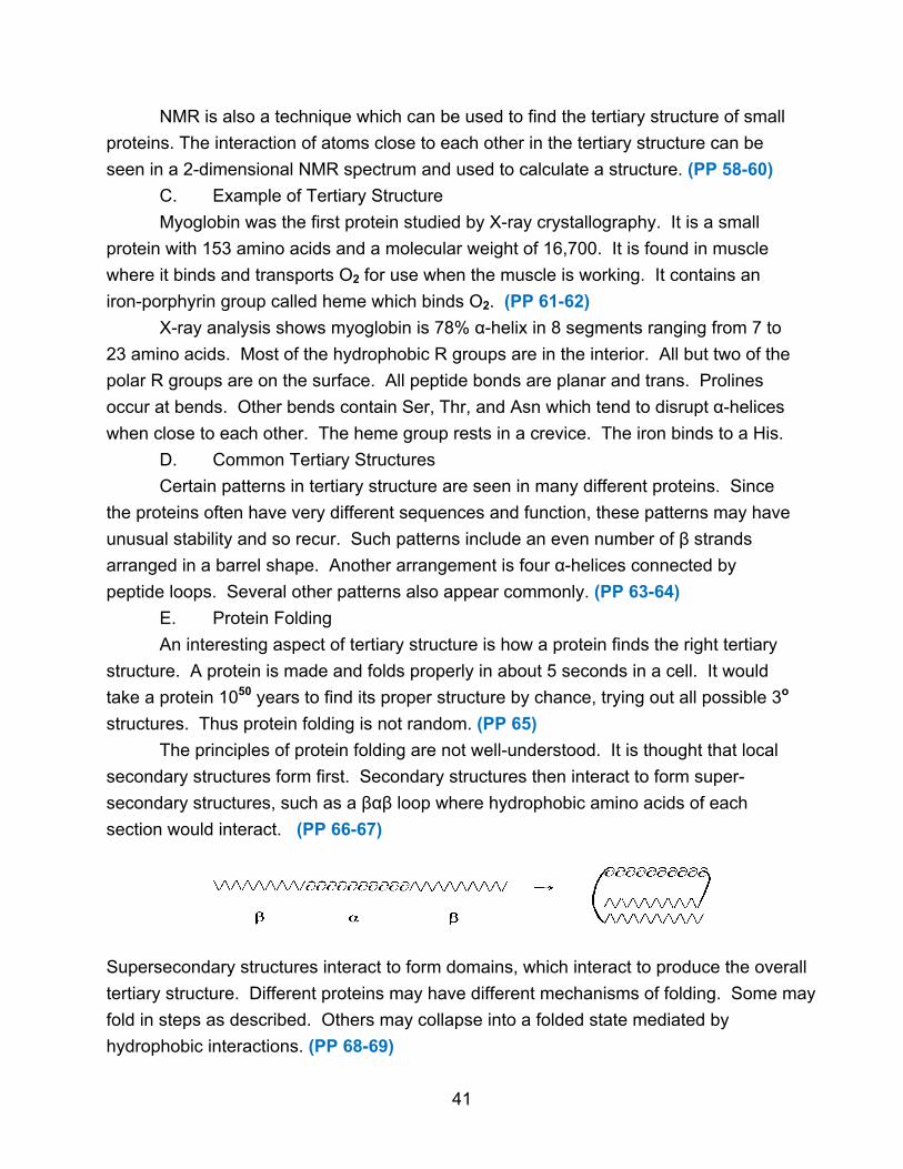
The principles of protein folding are not well-understood. It is thought that local
secondary structures form first. Secondary structures then interact to form super-
secondary structures, such as a βαβ loop where hydrophobic amino acids of each
section would interact. (PP 66-67)
Supersecondary structures interact to form domains, which interact to produce the overall
tertiary structure. Different proteins may have different mechanisms of folding. Some may
fold in steps as described. Others may collapse into a folded state mediated by
hydrophobic interactions. (PP 68-69)
41
Not all proteins fold spontaneously. Polypeptide chain binding proteins have been
found that help some proteins fold properly by preventing non-specific aggregation and
guiding the assembly of complex proteins. These helper proteins are called chaperones.
(PP 70)
F. Denaturation
Denaturation refers to the loss of proper tertiary structure caused by breaking non-
covalent bonds (but not covalent peptide bonds) in a protein. Proteins can be denatured
by heat, pH changes, and certain chemicals, any of which will disrupt H-bonds, salt
bridges, or hydrophobic interactions. To completely denature a protein, any disulfide
bonds must also be broken. It is not necessary to disrupt all the non-covalent forces in
order to disrupt conformation and destroy function. (PP 71)
Different proteins have different stabilities. Some are relatively difficult to denature
and others are relatively simple. With some proteins, denaturation is irreversible and the
protein is permanently damaged. With other proteins, the denaturation process can be
reversed if the denaturing agent is removed. This reversal is called renaturation. The
renatured protein is fully functional. Renaturation is consistent with amino acid sequence
determining 3o structure and with a precise pathway for protein folding. (PP 72-73)
IV. Quaternary Structure
When proteins contain multiple subunits (which may be identical or different), the 3-
dimensional arrangement of the subunits is called quaternary structure.
A. General Features
1. Not all proteins have quaternary structure. A protein with a single amino acid
chain cannot have quaternary structure.
2. Multimeric proteins vary from proteins with two identical subunits, to proteins
with ten different subunits, to very large protein complexes with 102 subunits of three
different types.
3. Non-covalent forces (H-bonds, salt bridges, hydrophobic interactions) are
most important in maintaining 4o structure. However, covalent disulfide bridges can also
form between subunits.
4. Quaternary structure can be determined by X-ray crystallography.
B. Example of Quaternary Structure
Hemoglobin is a well-studied protein with multiple subunits. Hemoglobin (MW =
64,500) transports oxygen in blood. It has four subunits of two different types (α2 β2).
(PP 74) The α subunit has 141 amino acids while the β subunit has 146 amino acids.
Each subunit has one heme group which can bind an O2 molecule. (PP 75) The α and
42

β chains both contain considerable α-helix and have 1o,2o, and 3o structures similar to
myoglobin. This is not surprising since all three protein chains have the same function
and are probably related through evolution.
The hemoglobin molecule is roughly spherical with the four subunits in a
tetrahedral shape. There are many contact points between the α and β chains, but few
between the two α chains or the two β chains. Hydrophobic amino acids make most of
the contact points along with a few salt bridges.
Hemoglobin has a property known as cooperative binding. The binding of one O2 to
one of the four subunits enhances the chances that O2 will bind to the other three subunits
by a factor of 500. This makes it an efficient oxygen carrier. This means that the binding
of one O2 is somehow transmitted to the other subunits. The structures of oxyhemoglobin
(with O2's) and deoxyhemoglobin (without O2's) are slightly different. The tertiary structure
of one subunit changes as the first O2 binds (PP 76), and this change is transmitted
throughout the protein with the subunits undergoing small changes in their relative
positions. (PP 77-78) As a result of the changes as the first O2 binds, the other subunits
find it progressively easier to bind O2. Specifically, certain salt bridges must be broken as
O2 binds. As the first O2 binds the most salt bridges must be broken, so binding the first
O2 is relatively hard (harder than for myoglobin). The remaining O2's require that fewer
salt bridges be broken, so their binding becomes easier and easier, producing positive
cooperativity. (There are also examples of negative cooperativity.)
Myoglobin, being a single chain, does not display cooperativity. It binds oxygen
tightly, which makes it well-designed to store oxygen, especially in muscle where the O2
concentration is relatively low. Hemoglobin, in contrast, must pick up O2 in the lungs
(where the O2 concentration is high) and release it in the peripheral tissues (where the
O2 concentration is low). (PP 79) The cooperative O2 binding ensures that as
hemoglobin leaves the lungs it will be fully and efficiently oxygenated. In the tissues
with lower O2 levels, hemoglobin will off-load a significant amount of its O2. Thus, it too
is well-designed. (PP 80-81)
Hemoglobin also demonstrates the importance of 1o structure. Sickle-cell anemia
is caused by a single mutation in hemoglobin. Amino acid 6 of the β chain is changed
from the normal Glu to Val. This creates a hydrophobic sticky spot on the outside of the
hemoglobin molecule. (PP 82) Hemoglobin molecules can then polymerize into long
chains. (PP 83) This distorts red blood cells from their normal disc shape into an
elongated sickle shape. (PP 84-85) This distorted cell is more fragile and can break,
causing anemia. Distorted cells can also clog capillaries, causing pain and tissue death.
Not all amino acid changes in hemoglobin are so detrimental. Over 300 variant
hemoglobins are known and most function with only minor problems, if any.
43

Chapter 6: Enzymes
I. Introduction
In order for a cell or organism to stay alive, thousands of chemical reactions must
occur to produce energy, make biomolecules, etc. These reactions would take place
very slowly unless catalyzed. Enzymes are biological catalysts.
A. Function - Enzymes speed up reactions 106 - 1016 times. (PP 2) They are
true catalysts required in only small amounts and fully recoverable in their original form
at the end of a reaction.
B. Structure - Most enzymes are globular proteins, although a few are nucleic
acids. Enzymes that are proteins can be multimeric. The specific tertiary and/or
quaternary structure of an enzyme is crucial in its ability to function.
C. Naming - Most enzymes end with -ase and the name indicates the
function as well as the molecule it works on (substrate), such as DNA polymerase or
glucose-6-phosphatase.
D. Prosthetic Groups and Cofactors - Many enzymes contain prosthetic
groups needed for their function. Other enzymes do not have prosthetic groups, but do
require a helper molecule in order to catalyze a reaction. This helper, known as a
cofactor, associates with the enzyme during the reaction. Cofactors are often metal
ions or organic molecules (coenzymes). A cofactor that remains permanently with the
enzyme becomes a prosthetic group. (PP 3)
E. Specificity - Enzymes are very specific regarding their substrates, working
on just one specific molecule (often one isomer) or on a group of closely related
molecules. Most enzymes will also catalyze just one reaction, so a large number of
different enzymes is needed. An enzyme will generally work on just one substrate
molecule at a time, although it can convert 105 substrate molecules into product per
second.
II. Factors in Enzyme Catalysis
In order for a chemical reaction to occur, the reacting molecules must be brought
together in the proper orientation and with sufficient energy for the reaction to take
place. Enzymes affect all these factors.
A. Orientation
During an enzyme-catalyzed reaction, enzyme and substrate bind together. This
binding automatically orients the substrate in the proper way for the reaction to occur.
Without an enzyme, proper orientation is a matter of chance during random collisions,
and only a small percentage of molecules would collide with the proper orientation.
44
With an enzyme, all substrate molecules are properly oriented which increases the rate
of the reaction. (PP 4)
B. Activation Energy
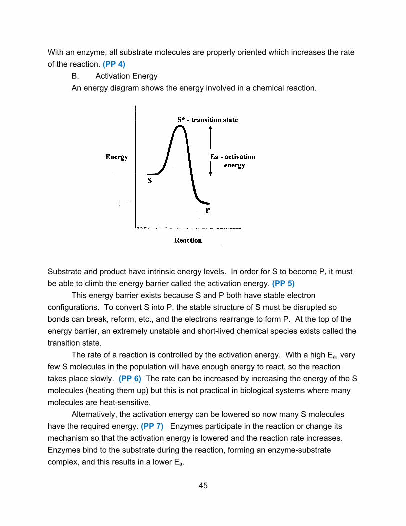
An energy diagram shows the energy involved in a chemical reaction.
Substrate and product have intrinsic energy levels. In order for S to become P, it must
be able to climb the energy barrier called the activation energy. (PP 5)
This energy barrier exists because S and P both have stable electron
configurations. To convert S into P, the stable structure of S must be disrupted so
bonds can break, reform, etc., and the electrons rearrange to form P. At the top of the
energy barrier, an extremely unstable and short-lived chemical species exists called the
transition state.
The rate of a reaction is controlled by the activation energy. With a high Ea, very
few S molecules in the population will have enough energy to react, so the reaction
takes place slowly. (PP 6) The rate can be increased by increasing the energy of the S
molecules (heating them up) but this is not practical in biological systems where many
molecules are heat-sensitive.
Alternatively, the activation energy can be lowered so now many S molecules
have the required energy. (PP 7) Enzymes participate in the reaction or change its
mechanism so that the activation energy is lowered and the reaction rate increases.
Enzymes bind to the substrate during the reaction, forming an enzyme-substrate
complex, and this results in a lower Ea.
45
Enzymes do not alter the equilibrium of the reaction which is governed by the
relative energy levels of S and P. (The energy level of P can be higher, lower, or the
same as S.) By speeding up the reaction, enzymes let the reaction reach equilibrium
more quickly. (PP 8-9)
III. Enzyme Reactions
The simplest scheme for an enzyme-catalyzed reaction is given below.
E + S ↔ E S → E + P
The substrate binds reversibly to form an enzyme-substrate complex. Formation of
product (it can be reversible) is the rate-limiting step.
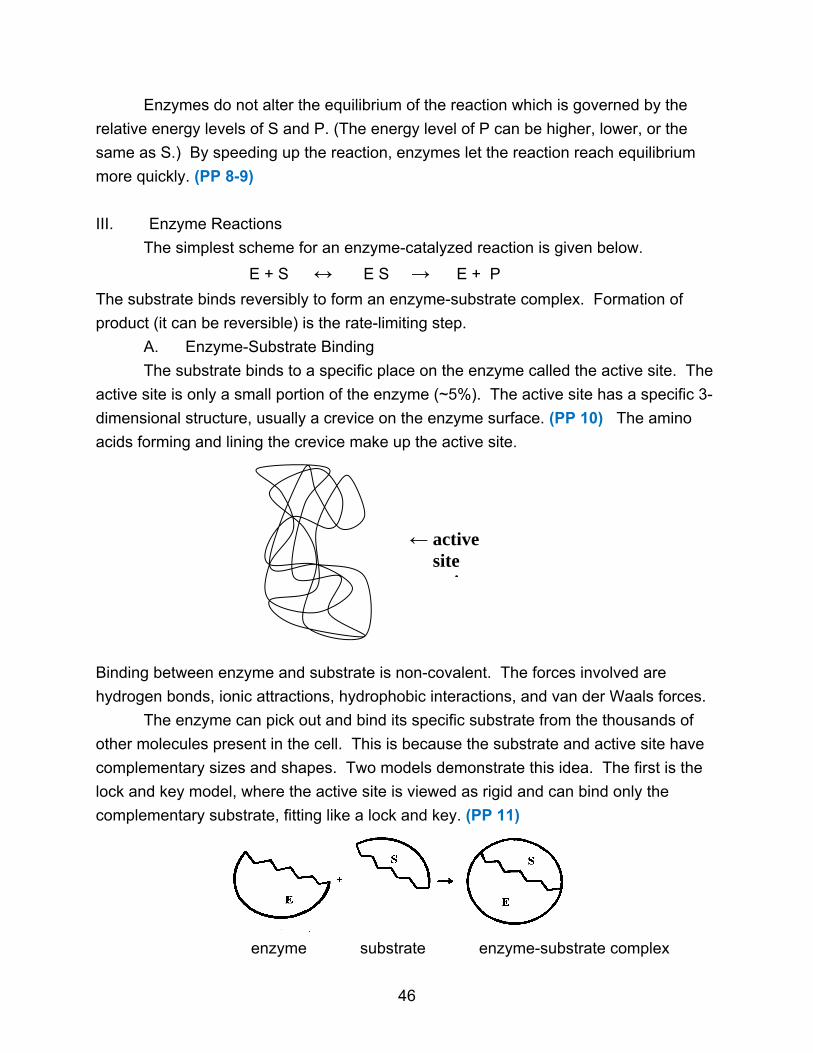
A. Enzyme-Substrate Binding
The substrate binds to a specific place on the enzyme called the active site. The
active site is only a small portion of the enzyme (~5%). The active site has a specific 3-
dimensional structure, usually a crevice on the enzyme surface. (PP 10) The amino
acids forming and lining the crevice make up the active site.
Binding between enzyme and substrate is non-covalent. The forces involved are
hydrogen bonds, ionic attractions, hydrophobic interactions, and van der Waals forces.
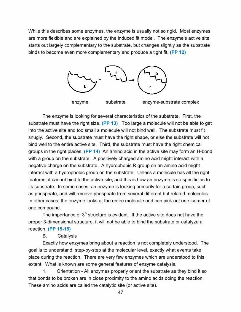
The enzyme can pick out and bind its specific substrate from the thousands of
other molecules present in the cell. This is because the substrate and active site have
complementary sizes and shapes. Two models demonstrate this idea. The first is the
lock and key model, where the active site is viewed as rigid and can bind only the
complementary substrate, fitting like a lock and key. (PP 11)
enzyme substrate enzyme-substrate complex
46
← active site
it
While this describes some enzymes, the enzyme is usually not so rigid. Most enzymes
are more flexible and are explained by the induced fit model. The enzyme’s active site
starts out largely complementary to the substrate, but changes slightly as the substrate
binds to become even more complementary and produce a tight fit. (PP 12)
enzyme substrate enzyme-substrate complex
The enzyme is looking for several characteristics of the substrate. First, the
substrate must have the right size. (PP 13) Too large a molecule will not be able to get
into the active site and too small a molecule will not bind well. The substrate must fit
snugly. Second, the substrate must have the right shape, or else the substrate will not
bind well to the entire active site. Third, the substrate must have the right chemical
groups in the right places. (PP 14) An amino acid in the active site may form an H-bond
with a group on the substrate. A positively charged amino acid might interact with a
negative charge on the substrate. A hydrophobic R group on an amino acid might
interact with a hydrophobic group on the substrate. Unless a molecule has all the right
features, it cannot bind to the active site, and this is how an enzyme is so specific as to
its substrate. In some cases, an enzyme is looking primarily for a certain group, such
as phosphate, and will remove phosphate from several different but related molecules.
In other cases, the enzyme looks at the entire molecule and can pick out one isomer of
one compound.
The importance of 3o structure is evident. If the active site does not have the
proper 3-dimensional structure, it will not be able to bind the substrate or catalyze a
reaction. (PP 15-18)
B. Catalysis
Exactly how enzymes bring about a reaction is not completely understood. The
goal is to understand, step-by-step at the molecular level, exactly what events take
place during the reaction. There are very few enzymes which are understood to this
extent. What is known are some general features of enzyme catalysis.
1. Orientation - All enzymes properly orient the substrate as they bind it so
that bonds to be broken are in close proximity to the amino acids doing the reaction.
These amino acids are called the catalytic site (or active site).
47

2. Strain - Enzymes can slightly strain or distort the substrate molecule,
making it easier to break certain bonds.
3. Transition State - An enzyme’s active site is often more complementary to
the transition state than to the original substrate. By stabilizing the transition state with
more favorable binding, the enzyme facilitates the reaction. (PP 19)
4. Covalent Intermediates - Some enzymes form an unstable covalent
intermediate with the substrate at some point during the reaction. This changes the
reaction mechanism and lowers the activation energy. The enzyme is back in its
original form by the end of the reaction. (PP 20)
5. Acid-Base Catalysis - The enzyme can act as an acid, donating protons,
or as a base, accepting protons from the substrate. This transfer of protons can
stabilize a charged intermediate to form a species that breaks down more readily into
the products. (PP 21)
6. Metal Ion Catalysis - Metal ions (Fe, Cu, Zn, Mn) function in two main
ways. They can participate in oxidation-reduction reactions, or they can be used to
stabilize or shield charges that develop on the substrate during the reaction. (PP 22)
7. Electrostatic Catalysis - Any charged group in the enzyme can stabilize an
opposite charge that develops on the substrate during the reaction.
Most enzymes use a combination of these factors to bring about a reaction.
(PP 23-26)
C. Chymotrypsin
Chymotrypsin is a well-understood enzyme. It illustrates several of the general
principles of enzymes. (PP 27)
Chymotrypsin is a proteolytic enzyme. It cleaves peptide bonds when R1 is
aromatic (Trp, Tyr, Phe). (PP 28)
O O
║ ║
- NH – CH – C – NH – CH – C -
│ ↑ │
R1 R2
48
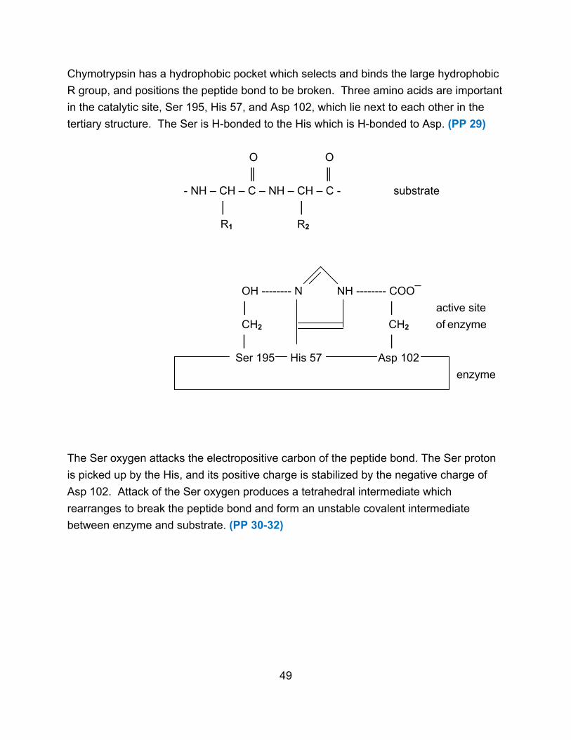
Chymotrypsin has a hydrophobic pocket which selects and binds the large hydrophobic
R group, and positions the peptide bond to be broken. Three amino acids are important
in the catalytic site, Ser 195, His 57, and Asp 102, which lie next to each other in the
tertiary structure. The Ser is H-bonded to the His which is H-bonded to Asp. (PP 29)
O O
║ ║
- NH – CH – C – NH – CH – C - substrate
│ │
R1 R2
OH -------- N NH -------- COO¯
│ │ active site
CH2 CH2 of enzyme
│ │
Ser 195 His 57 Asp 102
enzyme
The Ser oxygen attacks the electropositive carbon of the peptide bond. The Ser proton
is picked up by the His, and its positive charge is stabilized by the negative charge of
Asp 102. Attack of the Ser oxygen produces a tetrahedral intermediate which
rearranges to break the peptide bond and form an unstable covalent intermediate
between enzyme and substrate. (PP 30-32)
49
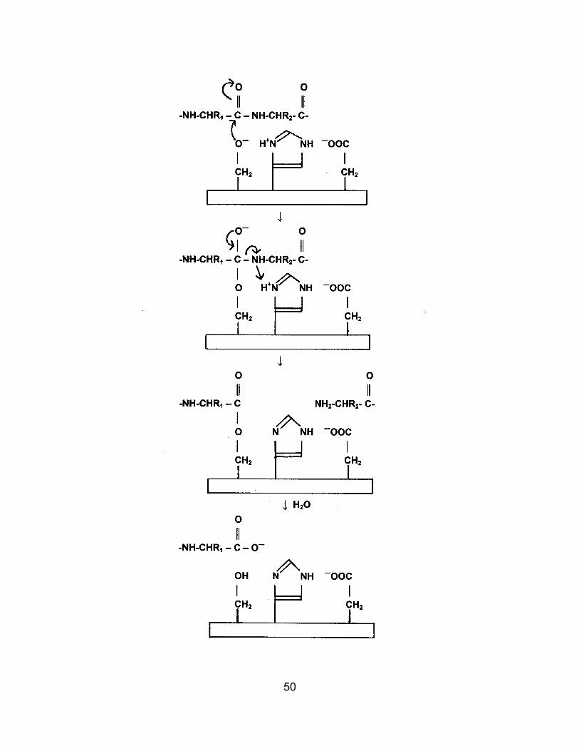
50
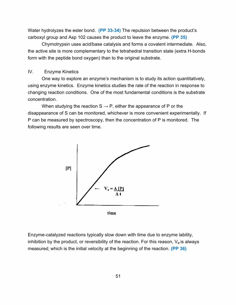
Water hydrolyzes the ester bond. (PP 33-34) The repulsion between the product’s
carboxyl group and Asp 102 causes the product to leave the enzyme. (PP 35)
Chymotrypsin uses acid/base catalysis and forms a covalent intermediate. Also,
the active site is more complementary to the tetrahedral transition state (extra H-bonds
form with the peptide bond oxygen) than to the original substrate.
IV. Enzyme Kinetics
One way to explore an enzyme’s mechanism is to study its action quantitatively,
using enzyme kinetics. Enzyme kinetics studies the rate of the reaction in response to
changing reaction conditions. One of the most fundamental conditions is the substrate
concentration.
When studying the reaction S → P, either the appearance of P or the
disappearance of S can be monitored, whichever is more convenient experimentally. If
P can be measured by spectroscopy, then the concentration of P is monitored. The
following results are seen over time.
Enzyme-catalyzed reactions typically slow down with time due to enzyme lability,
inhibition by the product, or reversibility of the reaction. For this reason, Vo is always
measured, which is the initial velocity at the beginning of the reaction. (PP 36)
51
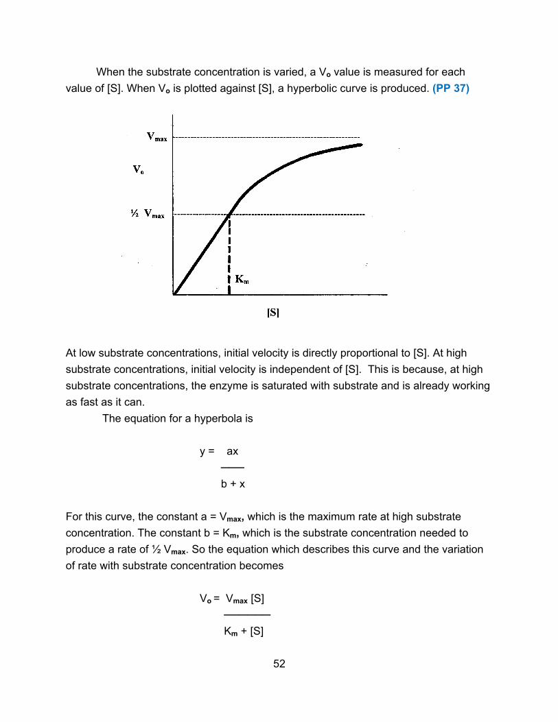
When the substrate concentration is varied, a Vo value is measured for each
value of [S]. When Vo is plotted against [S], a hyperbolic curve is produced. (PP 37)
At low substrate concentrations, initial velocity is directly proportional to [S]. At high
substrate concentrations, initial velocity is independent of [S]. This is because, at high
substrate concentrations, the enzyme is saturated with substrate and is already working
as fast as it can.
The equation for a hyperbola is
y = ax
───
b + x
For this curve, the constant a = Vmax, which is the maximum rate at high substrate
concentration. The constant b = Km, which is the substrate concentration needed to
produce a rate of ½ Vmax. So the equation which describes this curve and the variation
of rate with substrate concentration becomes
Vo = Vmax [S]
──────
Km + [S]
52

This is called the Michaelis-Menten equation. (PP 38) After this equation was found
empirically, it was then derived by a theoretical consideration of how enzymes work.
k1
E + S → ES → E + P
← k2
k -1
The theoretical treatment demonstrated that Vmax = k2eo where k2 is the rate constant
for the rate limiting step and eo is the enzyme concentration. In addition, it was found
that Km = (k -1 + k2) / k1 and can indicate the strength of the enzyme-substrate binding.
(PP 39) A small Km reflects a high affinity of the enzyme for its substrate, and means
that an enzyme will be functioning at a significant level even when a small amount of
substrate is present. An enzyme with a high Km requires higher amounts of substrate in
order to work well. (PP 40)
Vmax and Km are therefore important properties of an enzyme and are normally
determined in a study of enzyme kinetics. However, these values are difficult to find
precisely using a hyperbolic curve, so there are several algebraic rearrangements of the
Michaelis-Menten equation that yield a straight line and are used for accurate
determination of these kinetic values.
Once the basic kinetic properties of an enzyme are established, further kinetic
studies can reveal important information about the enzyme. Substrate analogs can be
used to see if the enzyme will react with them and to what extent. Such comparisons
can help determine how the enzyme binds the substrate and reacts with it. The optimum
pH for enzyme can be found, and measuring the effect of pH on enzyme activity can
indicate what groups are important in the enzyme’s active site. Using specific inhibitors
of an enzyme can also help to define the crucial characteristics of the substrate and the
mechanism of the enzyme. (PP 41-42)
V. Enzyme Regulation
If the cell is to perform efficiently, then its metabolism must be regulated. For
instance, a cell may have metabolic pathways for synthesizing alanine and also for
breaking down alanine for energy production. If the amino acid alanine is available in
large amounts from the environment, then there is no need for the cell to use its
resources making alanine. Instead, excess alanine can be broken down to produce
energy. If external levels of alanine are low, then the cell needs to make alanine and
53

should not break it down. Thus the pathways for making alanine and for breaking down
alanine should not function at the same time.
Regulation of reactions and metabolic pathways is achieved in many ways
through regulating one or more enzymes in a metabolic pathway. Enzyme activity can
be regulated in some straightforward ways, such as controlling the amount of substrate
or cofactor that is available. Changes in pH or temperature can change enzyme activity,
though these methods are rarely used inside cells. Another way of regulating reactions
is to control the presence of enzymes. Each enzyme is coded for by a gene in the cell’s
DNA. Genes can be turned on and used to make the corresponding enzyme, or turned
off so that the enzyme cannot be made. If the enzyme is not made, then the reaction
cannot occur and the pathway using that enzyme will be shut down. This saves energy
for the cell (making an enzyme consumes energy), but it is a relatively long-term
method of control since it takes time for the cell to make the enzyme when it is needed.
Another method of regulating enzymes is to control the activity of existing enzyme
molecules. Enzyme activity can be raised or lowered to quickly respond to changing
conditions. This is a relatively short-term method of control, but it does require an
investment in energy to make the enzyme molecules and have them present. It is not
necessary to regulate every enzyme in a metabolic pathway. It is usually the first
enzyme in a pathway which is regulated so that the entire pathway can be turned on
and off as needed. There are two major methods for regulating enzyme activity.
A. Allostery
Allosteric enzymes have their activity changed through the non-covalent binding
of a molecule known as an effector or modulator. In the following metabolic pathway
which converts compound A to compound X using enzymes 1-4, enzyme 1 is often an
allosteric enzyme inhibited by the end product X. (PP 43)
1 2 3 4
A → B → C → D → X
When levels of X are high and no more X is needed, the pathway will be shut down as X
binds to enzyme 1. When levels of X are low, X will not be bound to enzyme 1, so the
enzyme is active and X will be produced. The binding of the effector is non-covalent and
reversible, so that when the effector leaves, the enzyme is restored to its original form
and function.
54

Effectors can be either positive (which activate the enzyme) or negative (which
inhibit the enzyme). Many allosteric enzymes have several effectors, some positive and
some negative. A wide variety of different compounds can act as effectors, including
substrates, products, cofactors, and metabolic intermediates. Each effector will bind to a
specific site on the enzyme known as the regulatory or allosteric site. If there are
multiple effectors, there can be multiple allosteric sites. Each site will be specific for its
effector, similar to the way an active site is specific for its substrate.
Effectors work by changing the conformation of the enzyme. (PP 44) When the
effector binds, it causes a change in the tertiary and/or quaternary structure of the
enzyme which changes the active site. As a result, the enzyme works better (in
response to a positive effector) or works less well or not at all (in response to a negative
effector). (PP 45) Most allosteric enzymes are large and often have multiple subunits.
Often one type of subunit is catalytic, binding the substrate and carrying out the
reaction, while the other type of subunit is regulatory and binds the effectors.
How much enzyme is affected depend upon how much enzyme is bound to the
effector, and that in turn depends upon the concentration of the effector. An equilibrium
exists between the free enzyme, the effector, and the enzyme-effector complex.
Enzyme + Effector ↔ Enzyme-Effector Complex
The higher the concentration of the effector, the more is bound to the enzyme and the
more the enzyme will be regulated. This allows the overall activity of the enzyme to be
adjusted to whatever level is needed by the cell.
B. Covalent Modification
A second way of regulating enzymes is through the covalent joining of a
regulating group, called a covalent modifier, to a specific site on the enzyme. This
changes the enzyme conformation and so changes enzyme activity. For some
enzymes, covalent modification makes them more active. For other enzymes, covalent
modification makes them less active. The regulation is reversible since the covalent
modifier can be removed. (PP 46)
The major differences of covalent modification compared to allostery include the
following:
1. Binding of a covalent modifier is covalent while the binding of an effector
is non-covalent.
2. A few covalent modifiers, mainly phosphate, can regulate many different
enzymes while each allosteric enzyme has its own effector(s). (PP 47)
55

3. Because covalent bonds are being made and broken in covalent
modification, chemical reactions are occurring which require catalysis by additional
enzymes. Generally there is one extra enzyme to attach the covalent modifier to the
regulated enzyme, and a second extra enzyme to remove the covalent modifier.
Allostery requires nothing extra because the binding of the effector is non-covalent. The
extra enzymes involved in covalent modification must themselves be regulated in some
way (through allostery, covalent modification, hormones, etc.) The result is that the
extent of covalent modification will depend upon the activity of the extra enzymes. An
advantage of covalent modification is that many enzymes can be coordinately regulated
by simultaneous covalent modification. An enzyme can be both allosteric and covalently
modified.
C. Other mechanisms of enzyme regulation also exist, although they are
generally less common.
1. Enzymes generally work better at some pH values than others,
depending upon the pH of their normal environment. For instance, pepsin works well in
the acidic pH of the stomach where it helps digest proteins. (PP 48)
2. Some enzymes are made as inactive precursors called zymogens. The
zymogen must first be cleaved before the enzyme becomes active. This regulation is
irreversible, and occurs with certain enzymes such as digestive enzymes. These
enzymes should not be active until they reach the digestive tract or else they would
damage tissues. (PP 49)
56

Chapter 7: Carbohydrates
I. Introduction
Carbohydrates are sugars and starches. They are the major source of energy in
many organisms, serve to store energy, and are structural components in some
organisms. Because of their wide distribution, carbohydrates are the most abundant
type of biomolecule.
A. Formula and Structure
Most carbohydrates have the general formula (CH2O)n. This suggested that they
were hydrates of carbon, hence the name. However, they are not hydrates of carbon,
nor do all carbohydrates conform to this formula. Carbohydrates are fundamentally
polyhydroxy aldehydes and ketones, and some contain nitrogen, phosphorus, or sulfur.
B. Classes
There are three major classes of carbohydrates based on size. (PP 2)
1. Monosaccharides are the simplest sugars, containing 3-7 carbons and
one aldehyde or ketone group.
2. Oligosaccharides consist of ~2-10 monosaccharide units joined together.
Most are disaccharides. Some are joined to lipids, or to proteins as prosthetic groups.
3. Polysaccharides are very large, with hundreds or thousands of
monosaccharide units joined together.
II. Monosaccharides
A. General Properties (PP 3)
1. They are white solids, water-soluble (polar), and often have a sweet taste.
2. The formula is (CH2O)n where n = 3-7. The most common number of
carbons is 5 or 6.
3. The carbon skeleton is unbranched, connected by single bonds. One
carbon contains a carbonyl oxygen (aldehyde or ketone). All other carbons contain a
hydroxyl group.
4. Monosaccharides are named with the suffix - ose. Every monosaccharide
can be classified as an aldose or ketose, depending upon the functional group. (PP 4-5)
They can also be classified according to the number of carbon atoms: 3 carbons = a
triose; 4 C = a tetrose; 5 C = a pentose; 6 C = a hexose; 7 C = a heptose. The two
classifications are often combined, such as aldopentose or ketohexose. (PP 6-7)
57

B. Structure and Stereochemistry
The most important classification is aldose vs. ketose.
1. Aldoses
The simplest aldose contains three carbons with one aldehyde and two hydroxyl
groups. The name for this compound is glyceraldehyde.
CHO CHO
│ │
H ― C ― OH HO ― C ― H
│ │
CH2OH CH2OH
D-glyceraldehyde L-glyceraldehyde
Glyceraldehyde contains a chiral or asymmetric carbon, also referred to as a
stereogenic or chirality center. The central carbon is attached to four different
substituents, giving two forms that are mirror images of each other but are not
superimposable. They are designated D and L. (PP 8-9) All other monosaccharides are
classified as D or L, depending on the configuration of the asymmetric carbon farthest
from the aldehyde group.
A molecule with n chiral centers will have 2n stereoisomers. Thus 4-carbon
aldoses have two stereogenic centers and four stereoisomers. (PP 10-11)
CHO CHO CHO CHO
│ │ │ │
H ― C ― OH HO ― C ― H H ― C ― OH HO ― C ― H
│ │ │ │
H ― C ― OH H ― C ― OH HO ― C ― H HO ― C ― H
│ │ │ │
CH2OH CH2OH CH2OH CH2OH
D-erythrose D-threose L-threose L-erythrose
The first two structures are D-sugars because the configuration around the third
carbon is like that of D-glyceraldehyde. The last two structures are L-sugars because
they resemble L-glyceraldehyde. D-threose and L-threose are mirror images
58

(enantiomers) and will have all the same properties except their behavior with plane-
polarized light and their interaction with other chiral compounds. D-erythrose and D-
threose are not mirror images (are diastereomers) and are given different names
because they are different compounds with different properties.
The D-sugars are the biologically important ones. There are four D-aldopentoses
and eight D-aldohexoses (and the same number of L-isomers). (PP 12-14)
CHO CHO CHO CHO
│ │ │ │
H ― C ― OH HO ― C ― H H ― C ― OH HO ― C ― H
│ │ │ │
H ― C ― OH H ― C ― OH HO ― C ― H HO ― C ― H
│ │ │ │
H ― C ― OH H ― C ― OH H ― C ― OH H ― C ― OH
│ │ │ │
CH2OH CH2OH CH2OH CH2OH
D-ribose D-arabinose D-xylose D-lyxose
The most important aldoses and the most common in biological systems are
D-ribose (an aldopentose) and two aldohexoses, D-glucose and D-galactose.
CHO CHO
│ │
H ― C ― OH H ― C ―OH
│ │
HO ― C ― H HO ― C ― H
│ │
H ― C ― OH HO ― C ― H
│ │
H ― C ― OH H ― C ― OH
│ │
CH2OH CH2OH
D-glucose D-galactose
Sugars that differ in configuration around one carbon are called epimers. D-glucose and
D-galactose are C-4 epimers. (PP 15-19)
59
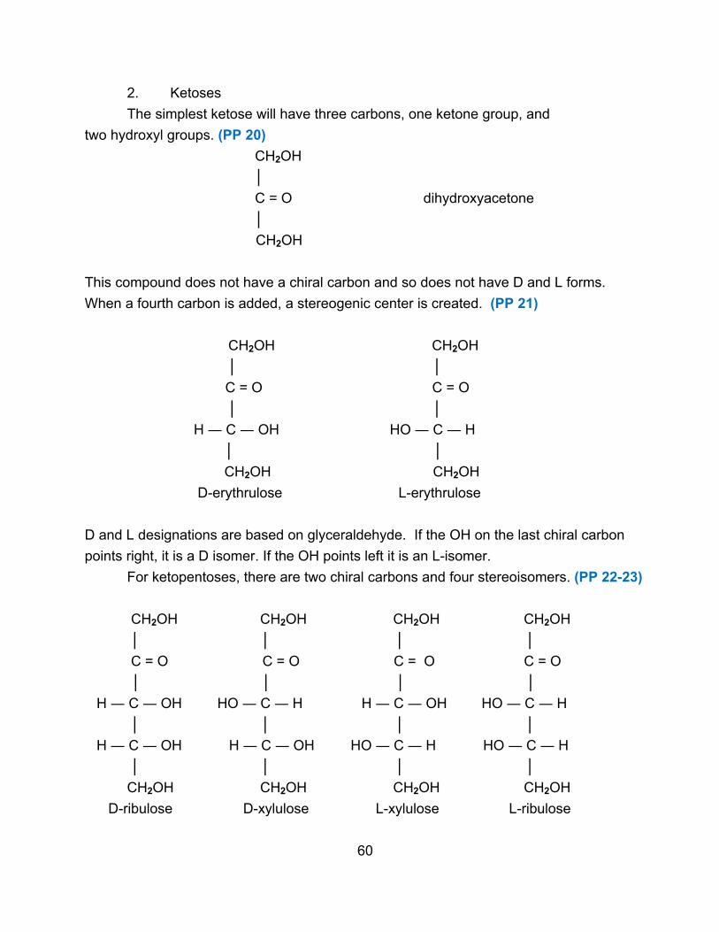
2. Ketoses
The simplest ketose will have three carbons, one ketone group, and
two hydroxyl groups. (PP 20)
CH2OH
│
C = O dihydroxyacetone
│
CH2OH
This compound does not have a chiral carbon and so does not have D and L forms.
When a fourth carbon is added, a stereogenic center is created. (PP 21)
CH2OH CH2OH
│ │
C = O C = O
│ │
H ― C ― OH HO ― C ― H
│ │
CH2OH CH2OH
D-erythrulose L-erythrulose
D and L designations are based on glyceraldehyde. If the OH on the last chiral carbon
points right, it is a D isomer. If the OH points left it is an L-isomer.
For ketopentoses, there are two chiral carbons and four stereoisomers. (PP 22-23)
CH2OH CH2OH CH2OH CH2OH
│ │ │ │
C = O C = O C = O C = O
│ │ │ │
H ― C ― OH HO ― C ― H H ― C ― OH HO ― C ― H
│ │ │ │
H ― C ― OH H ― C ― OH HO ― C ― H HO ― C ― H
│ │ │ │
CH2OH CH2OH CH2OH CH2OH
D-ribulose D-xylulose L-xylulose L-ribulose
60

The various stereoisomers again form pairs of enantiomers or diastereomers. The D-
isomers are the biologically important ones. Ketoses are generally named by inserting
‘ul’ into the name of the corresponding aldose. (ribose-ribulose). The most important
ketose is D-fructose, which is a ketohexose.
CH2OH
│
C = O
│
HO ― C ― H
│
H ― C ― OH
│
H ― C ― OH
│
CH2OH
When there are five or six carbons present, other ketoses are possible where the ketone
group is on carbon 3. However, all the biologically important ketoses have the ketone
group on carbon 2. (PP 24-25)
C. Reactions of Monosaccharides
Monosaccharides will undergo reactions characteristic of the functional groups.
Some are useful in detecting and identifying monosaccharides.
1. Oxidation
a. Fehling’s or Benedict’s reaction
Fehling’s solution is alkaline cupric ion complexed with tartrate ion, while
Benedict’s solution is complexed with citrate ion. When reacted with a sugar, the sugar
is oxidized and the copper is reduced. (PP 26)
Cu2+ Cu+
H ― C = O COOH
│ │
If the sugar is an aldose, the aldehyde is directly oxidized to the acid. If the sugar is a
ketose, an acid is formed as the carbon chain breaks. In both cases, the blue Cu2+ is
61
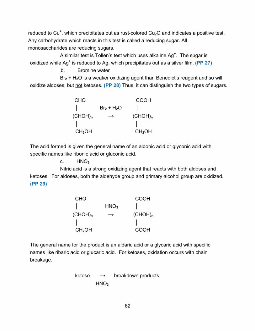
reduced to Cu+, which precipitates out as rust-colored Cu2O and indicates a positive test.
Any carbohydrate which reacts in this test is called a reducing sugar. All
monosaccharides are reducing sugars.
A similar test is Tollen’s test which uses alkaline Ag+. The sugar is
oxidized while Ag+ is reduced to Ag, which precipitates out as a silver film. (PP 27)
b. Bromine water
Br2 + H2O is a weaker oxidizing agent than Benedict’s reagent and so will
oxidize aldoses, but not ketoses. (PP 28) Thus, it can distinguish the two types of sugars.
CHO COOH
│ Br2 + H2O │
(CHOH)n → (CHOH)n
│ │
CH2OH CH2OH
The acid formed is given the general name of an aldonic acid or glyconic acid with
specific names like ribonic acid or gluconic acid.
c. HNO3
Nitric acid is a strong oxidizing agent that reacts with both aldoses and
ketoses. For aldoses, both the aldehyde group and primary alcohol group are oxidized.
(PP 29)
CHO COOH
│ HNO3 │
(CHOH)n → (CHOH)n
│ │
CH2OH COOH
The general name for the product is an aldaric acid or a glycaric acid with specific
names like ribaric acid or glucaric acid. For ketoses, oxidation occurs with chain
breakage.
ketose → breakdown products
HNO3
62

d. HIO4
Periodic acid oxidizes both aldoses and ketoses, but it does so in such a
specific way that it is used for structural determination and identification. IO4¯ cleaves a
C-C bond when both carbons carry oxidizable groups such as hydroxyls or carbonyls.
(PP 30) One molecule of HIO4 is reduced to iodate, IO3¯, and both carbons are oxidized
to the next highest oxidation state.
│ IO4¯ │
― C ― OH → ― C = O
│ + + IO3¯
― C ― OH ― C = O
│ │
│ IO4¯ │
― C ― OH → ― C = O
│ + + IO3¯
C = O ― COOH
│
The reaction of groups is ― C ― OH → ― C = O (carbonyl)
― C = O → ― COOH (acid)
― COOH → CO2
When dealing with monosaccharides, every carbon has an oxidizable group and so all
C-C bonds will be broken. The result is a mixture of one-carbon compounds, and the
following rules apply. (PP 31)
one mole of HIO4 is used per mole of C – C bonds broken
one mole of HCHO is formed per mole of 1o alcohol oxidized
one mole of HCOOH is formed per mole of 2o alcohol oxidized
one mole of HCOOH is formed per mole of aldehyde oxidized
one mole CO2 is formed per mole of ketone oxidized
63
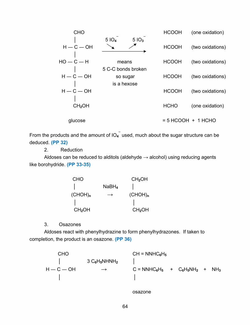
CHO HCOOH (one oxidation)
│ 5 IO4¯ 5 IO3
¯
H ― C ― OH HCOOH (two oxidations)
│
HO ― C ― H means HCOOH (two oxidations)
│ 5 C-C bonds broken
H ― C ― OH so sugar HCOOH (two oxidations)
│ is a hexose
H ― C ― OH HCOOH (two oxidations)
│
CH2OH HCHO (one oxidation)
glucose = 5 HCOOH + 1 HCHO
From the products and the amount of IO4¯ used, much about the sugar structure can be
deduced. (PP 32)
2. Reduction
Aldoses can be reduced to alditols (aldehyde → alcohol) using reducing agents
like borohydride. (PP 33-35)
CHO CH2OH
│ NaBH4 │
(CHOH)n → (CHOH)n
│ │
CH2OH CH2OH
3. Osazones
Aldoses react with phenylhydrazine to form phenylhydrazones. If taken to
completion, the product is an osazone. (PP 36)
CHO CH = NNHC6H5
│ 3 C6H5NHNH2 │
H ― C ― OH → C = NNHC6H5 + C6H5NH2 + NH3
│ │
osazone
64

Different carbohydrates form osazones with different melting points and crystal
structures allowing for identification of the initial aldose. C-2 epimers give the same
osazone. Ketoses usually react more slowly, if at all.
4. Other Reactions
Monosaccharides are stable in dilute acids. Strong acids can dehydrate or break
chains. Dilute bases can cause rearrangements around certain carbons. Strong bases
can fragment the chain.
Monosaccharides can be interconverted using several different reactions.
Epimerization around carbon-2 of an aldose can take place using pyridine. An aldose
can be shortened by one carbon using a series of reactions called the Ruff degradation,
or extended by one carbon using the Kiliani-Fischer synthesis. However, these methods
involve several steps and can create a mixture of products, so their usefulness is
limited.
D. Haworth Structures
There is a further complication in the structure of monosaccharides.
1. Unusual Properties
Certain properties of monosaccharides are inconsistent with the presence of a
normal carbonyl group.
a. The addition of HCN during the Kiliani-Fischer synthesis occurs
slowly with monosaccharides but rapidly with normal aldehydes.
b. Many aldoses fail to give a positive Schiff test, which normally
identifies an aldehyde. (A reaction occurs with sulfur dioxide and fuchsin to give a red
color.)
c. D-glucose can exist in two forms with different specific rotations. If
D-glucose is recrystallized from water, the sugar has a specific rotation [α] = + 112o
(called the α-form). If D-glucose is recrystallized from pyridine, the sugar has a specific
rotation [α] = + 19o (called the β-form). If either form is dissolved in water and allowed
to stand, the specific rotation changes until [α] = + 52.7o. This is called the mutarotation
of glucose.
2. Hemiacetals
These properties are explained by a common reaction in organic chemistry, the
formation of a hemiacetal, which occurs between an aldehyde and an alcohol. (PP 37)
O OH
║ │
R ― C ― H + R’ ― OH ↔ R ― C ― H
│
OR’
65

In an aldose, the hemiacetal formation is internal since both functional groups are in the
same molecule. For an aldohexose, the reaction is between the C-1-aldehyde and the
C-5 alcohol. (PP 38)
CHO CH2OH
│ │
H ― C ― OH C OH
│ H │ O
HO ― C ― H → │ H ║
│ C C ― H
H ― C ― OH │ OH H
│ HO │ │
H ― C ― OH C C
│ │ │
CH2OH H OH
A new asymmetric carbon is created (carbon 1), with two possible configurations. The
new OH group can point up or down. When the OH points down, this is α-D-glucose.
When the OH points up, this is β-D-glucose. This explains the two forms of D-glucose.
Since the hemiacetal is not stable and will undergo the reverse reaction readily in
solution, both the α and β forms can revert to the straight-chain form (open-chain, linear
form). Thus the α and β forms are interconvertible. If a solution of pure α (or pure β) is
allowed to stand, some of it will convert to the straight-chain form and then some to the
other cyclic form. Eventually, an equilibrium is established that is 63% β-D-glucose,
37% α-D-glucose, and a very small amount of the linear form. This explains the
66

mutarotation of glucose. Also, since only a small amount of glucose has a free aldehyde
group, this is why monosaccharides react slowly in reactions requiring an aldehyde
group.
The two forms (α and β) are called anomers and C-1 is called the anomeric
carbon. They appear to be diastereomers (non-mirror images) but are the same
compound since they interconvert. Thus they are special forms of isomers, not
enantiomers but not really diastereomers either. Hence there is the new designation of
anomers. The ring forms are named pyranoses since they resemble the 6-membered
ring compound, pyran. (PP 39)
HC O
HC CH
H2C CH
α-D-glucopyranose β-D-glucopyranose
The equilibrium concentrations of α and β glucose are explained by remembering that
these molecules exist primarily in a chair conformation. (PP 40)
α β
The α-form has four equatorial substituents and one axial substituent. The β-form has
five equatorial substituents. Since equatorial substituents are more stable than axial
ones, the β-form is favored.
67

3. Converting to Haworth structures
Any D-sugar can be transformed into a Haworth structure, starting with the
straight-chain form. First, the C-C bond involving the last chiral carbon is rotated so the
hydroxyl group points down. Next, the hemiacetal bond is formed, creating two
pseudocyclic forms. These forms are then re-written as Haworth structures. (PP 41)
CHO CHO
OH OH
HO → HO D-glucose
OH OH
OH HOH2C
CH2OH OH
H― C ― OH HO ― C ― H
OH OH
HO O HO O
OH OH
HOH2C HOH2C
α β
68
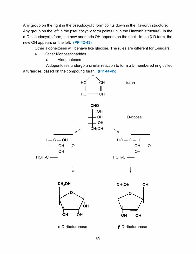
Any group on the right in the pseudocyclic form points down in the Haworth structure.
Any group on the left in the pseudocyclic form points up in the Haworth structure. In the
α-D pseudocyclic form, the new anomeric OH appears on the right. In the β-D form, the
new OH appears on the left. (PP 42-43)
Other aldohexoses will behave like glucose. The rules are different for L-sugars.
4. Other Monosaccharides
a. Aldopentoses
Aldopentoses undergo a similar reaction to form a 5-membered ring called
a furanose, based on the compound furan. (PP 44-45)
O
HC CH furan
║ ║
HC CH
CHO
OH
OH D-ribose
OH
CH2OH
H ― C ― OH HO ― C ― H
OH O OH O
OH OH
HOH2C HOH2C
α-D-ribofuranose β-D-ribofuranose
69

b. Ketohexoses
Ketoses undergo a similar reaction to form hemiketals. (PP 46)
O OH
║ │
R ― C ― R’ + R” ― OH ↔ R ― C ― R’
│
OR”
Like hemiacetals, hemiketals are unstable and this reaction is freely reversible in
solution. Ketohexoses will undergo this reaction between the C-2 ketone and the C-5
hydroxyl to form furanoses. β and α forms will follow the same conventions and will
mutarotate. (PP 47)
CH2OH CH2OH
│ │
C = O → C = O
HO HO D-fructose
OH OH
OH HOH2C
CH2OH OH
HOH2C ― C ― OH HO ― C ― CH2OH
HO O HO O
OH OH
HOH2C HOH2C
α-D-fructofuranose (OH down) β-D-fructofuranose (OH up)
70

The most stable ring forms are those with five or six atoms. Smaller
monosaccharides will not form the cyclic structures. (PP 48-49)
Some monosaccharides can form two types of ring structures.
Ketohexoses and aldopentoses can also form pyranoses when the reaction occurs with
the last OH, but the furanoses are the predominant form in biomolecules. (PP 50)
Likewise aldohexoses can form furanoses, but the pyranoses are much more common.
E. Derivatives of Monosaccharides
Several compounds that are related to monosaccharides have biological
importance.
1. Glycosides
A hemiacetal or hemiketal can be converted to an acetal or a ketal by adding
another alcohol. The acetal or ketal is stable in neutral solution, unlike the hemiacetal
or hemiketal. The acetal or ketal will not be stable in acid and then the reaction can be
reversed. (PP 51)
O OH OR’”
║ │ R’”― OH │
R ― C + R” ― OH ↔ R ― C ― H (R’) → R ― C ― H (R’)
│ │ │
H (R’) OR” OR”
hemiacetal or acetal or
hemiketal ketal
This type of reaction between a monosaccharide and any alcohol produces a glycoside.
The ring of the glycoside can no longer open. (PP 52) The sugar is now non-reducing,
and it will not oxidize, form osazones, or mutarotate. The new bond is called a
glycosidic bond and is designated α (down) or β (up). (PP 53) The compound is called a
pyranoside if the ring is 6-membered and a furanoside if the ring is 5-membered.
Glycosides are related to oligo- and polysaccharides. (PP 54-55)
71
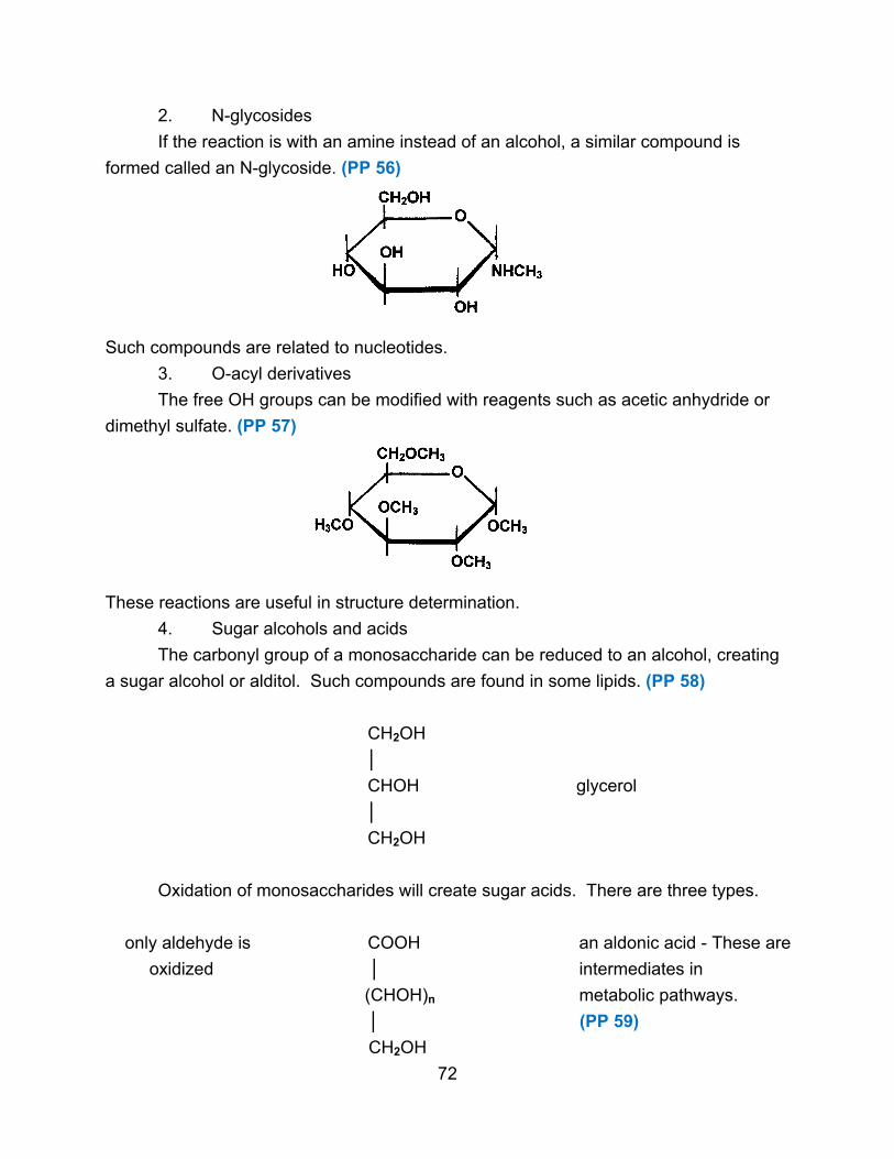
2. N-glycosides
If the reaction is with an amine instead of an alcohol, a similar compound is
formed called an N-glycoside. (PP 56)
Such compounds are related to nucleotides.
3. O-acyl derivatives
The free OH groups can be modified with reagents such as acetic anhydride or
dimethyl sulfate. (PP 57)
These reactions are useful in structure determination.
4. Sugar alcohols and acids
The carbonyl group of a monosaccharide can be reduced to an alcohol, creating
a sugar alcohol or alditol. Such compounds are found in some lipids. (PP 58)
CH2OH
│
CHOH glycerol
│
CH2OH
Oxidation of monosaccharides will create sugar acids. There are three types.
only aldehyde is COOH an aldonic acid - These are
oxidized │ intermediates in
(CHOH)n metabolic pathways.
│ (PP 59)
CH2OH
72

both aldehyde and COOH an aldaric acid - These are
1o alcohol are oxidized │ useful in
(CHOH)n structure determination.
│ (PP 60)
COOH
only 1o alcohol is CHO a uronic acid - These are
oxidized │ found in
(CHOH)n polysaccharides.
│ (PP 61)
COOH
5. Sugar phosphates and amino sugars
Some monosaccharide derivatives contain other chemical groups. (PP 62-63)
glucose-1-phosphate found in metabolic pathways
CHO
│
H ― C ― NH2
│
HO ― C ―H
│
H ― C ― OH
│
H ― C ― OH
│
CH2OH
glucosamine found in polysaccharides
73

6. Deoxysugars
Some sugars lack an OH group. (PP 64)
CHO
│
CH2
│
H ― C ― OH
│
H ― C ― OH
│
CH2OH
2-deoxy-D-ribose found in nucleotides
7. Complicated derivatives
Some sugars have several extra groups. (PP 65-67)
CHO O
│ ║
H H ― C ― NH ― C ― CH3
│ │
HOOC ― C ― O ― CH
│ │
CH3 H ― C ― OH
│
H ― C ― OH
│
CH2OH
N-acetylmuramic acid found in bacterial cell walls
III. Oligosaccharides
A. Characteristics
Oligosaccharides contain 2-10 monosaccharides joined together. The most
important are the disaccharides (two monosaccharides joined together).
74

The bond that holds the monosaccharides together is called a glycosidic bond.
The bond (acetal or ketal) is stable at neutral and basic pH but is broken by acid
hydrolysis. (PP 68)
There are two basic characteristics to any disaccharide. The first is the
identity of the monosaccharide components. These may be the same or different. The
second is the nature of the glycosidic bond. The bond is characterized by which
carbons of the monosaccharides are linked and by the orientation of the bond (α or β).
In the above example, glucose is the first sugar, mannose is the second. The bond
links carbon 1 of the glucose to carbon 4 of the mannose. Since the bond points down
off the glucose, it is an α bond (up would be β). There is no choice about the direction
of the bond from the mannose. It must be down or the sugar is not mannose. Thus this
disaccharide has an α 1,4 bond.
Since a glycosidic bond is stable, there is no interconversion between α and β.
These two monosaccharides joined by a β bond would make a different disaccharide.
Since the aldehyde group of glucose is permanently in the acetal linkage, there can be
no mutarotation or typical aldehyde reactions of the glucose unit. However, the
mannose does have a hemiacetal (potential aldehyde) so it can mutarotate, and there
will be two forms of this disaccharide (anomeric OH up or down) which will interconvert
in solution. The disaccharide will be a reducing sugar since the mannose aldehyde
group exists.
B. Examples and Analysis
Disaccharide structure can be illustrated and analyzed by looking at some
specific examples.
1. Maltose
Maltose is a common disaccharide formed from the breakdown of plant and
animal polysaccharides. Its formula is C12H22O11, which is equivalent to 2C6H12O6 - H2O,
showing water is lost when a glycosidic bond is formed. Maltose has the following
characteristics. (PP 69)
a. Acid hydrolysis produces only D-glucose, so both monosaccharides
are glucose.
75

b. Maltose is a reducing sugar, containing a free aldehyde group and
reacting with Benedict’s reagent. It also exists in two forms, α-maltose with a specific
rotation of +168o and β-maltose with a specific rotation of +112o, again indicating an
aldehyde group which allows mutarotation.
c. Maltose forms an osazone by adding two molecules of
phenylhydrazine, indicating the presence of only one aldehyde group. (Two free
aldehydes would add four molecules of phenylhydrazine.) This confirms that the C-1 of
one of the glucoses must be involved in the glycosidic bond (which is expected since
glycosidic bonds form by reacting a hemiacetal with a hydroxyl group).
d. Maltose is cleaved by the enzyme maltase, which is specific for α-
bonds. Thus the bond is in the α orientation. Emulsin is an enzyme specific for β-
bonds.
e. If maltose is oxidized by bromine water (converting free aldehyde to
an acid), exhaustively methylated with dimethyl sulfate (so all free OHs are methylated),
and then cleaved in acid, the following products result. (PP 70)
All OHs All OHs
methylated methylated
except C-1, so except C-4, so
this glucose joined this glucose joined
through C-1 through C-4
Thus the bond is from the C-1 of one glucose to the C-4 of the other. (PP 71-72)
non-reducing end reducing end
76

2. Cellobiose
Cellobiose is formed from the breakdown of cellulose. Acid hydrolysis yields only
glucose. It is a reducing sugar that can mutarotate between α and β forms (one free
reducing end). It is cleaved by emulsin but not by maltase, showing the linkage is β.
Methylation shows the linkage is 1,4. (PP 73)
3. Lactose
Lactose, found in milk, forms D-glucose and D-galactose upon acid hydrolysis. If
lactose is subjected to phenylhydrazine followed by acid hydrolysis, the products are
galactose and the osazone of glucose, indicating that only the glucose has a reducing
end. Therefore, the C-1 of galactose is involved in the glycosidic bond. Methylation
shows the bond to be 1,4. Lactase cleaves the disaccharide, showing the bond to be β.
(PP 74-75)
4. Sucrose
Sucrose (table sugar) is formed by plants. Acid hydrolysis yields D-glucose and
D-fructose. It is non-reducing, so both anomeric carbons must be in the linkage (C-1 of
glucose and C-2 of fructose). Treatment with enzymes is inconclusive regarding
orientation since enzymes cleaving α and β linkages both work. X-ray analysis was
needed to show that the bond is α with respect to glucose and β with respect to
fructose. There is only one form of sucrose, not two anomers, and no mutarotation can
occur. (PP 76-78)
77
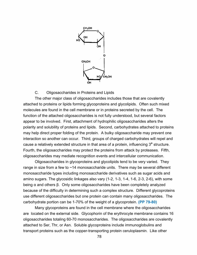
C. Oligosaccharides in Proteins and Lipids
The other major class of oligosaccharides includes those that are covalently
attached to proteins or lipids forming glycoproteins and glycolipids. Often such mixed
molecules are found in the cell membrane or in proteins secreted by the cell. The
function of the attached oligosaccharides is not fully understood, but several factors
appear to be involved. First, attachment of hydrophilic oligosaccharides alters the
polarity and solubility of proteins and lipids. Second, carbohydrates attached to proteins
may help direct proper folding of the protein. A bulky oligosaccharide may prevent one
interaction so another can occur. Third, groups of charged carbohydrates will repel and
cause a relatively extended structure in that area of a protein, influencing 3o structure.
Fourth, the oligosaccharides may protect the proteins from attack by proteases. Fifth,
oligosaccharides may mediate recognition events and intercellular communication.
Oligosaccharides in glycoproteins and glycolipids tend to be very varied. They
range in size from a few to ~14 monosaccharide units. There may be several different
monosaccharide types including monosaccharide derivatives such as sugar acids and
amino sugars. The glycosidic linkages also vary (1-2, 1-3, 1-4, 1-6, 2-3, 2-6), with some
being α and others β. Only some oligosaccharides have been completely analyzed
because of the difficulty in determining such a complex structure. Different glycoproteins
use different oligosaccharides but one protein can contain many oligosaccharides. The
carbohydrate portion can be 1-70% of the weight of a glycoprotein. (PP 79-80)
Many glycoproteins are found in the cell membrane where the oligosaccharides
are located on the external side. Glycophorin of the erythrocyte membrane contains 16
oligosaccharides totaling 60-70 monosaccharides. The oligosaccharides are covalently
attached to Ser, Thr, or Asn. Soluble glycoproteins include immunoglobulins and
transport proteins such as the copper-transporting protein ceruloplasmin. Like other
78

soluble glycoproteins, the oligosaccharide chains of ceruloplasmin end in N-acetylneuraminic
acid (a sugar acid also known as sialic acid). When these units are lost, the protein is taken
up by the liver and destroyed. Thus removal of sialic acid is one mechanism for
marking ‘old’ proteins for destruction and replacement. Within a cell, oligosaccharide
attachment often marks a protein for secretion or movement to a particular cell
organelle. Mannose -6-phosphate units are added to certain degradative enzymes so
they are moved to lysosomes where they function in degrading old molecules.
Glycolipids are found in nerve cell membranes. Lipopolysaccharides are major
components of some bacterial cell membranes.
IV. Polysaccharides
Polysaccharides, also known as glycans, have very high molecular weights.
Homopolysaccharides contain one type of monosaccharide unit.
Heteropolysaccharides contain two or more types of monomers. Polysaccharides do
not have definite molecular weights since enzymes easily add or remove
monosaccharide units. (PP 81)
A particular polysaccharide is characterized by its monomer types, the types of
glycosidic bonds present, and the degree of branching in the carbon chain.
Polysaccharides serve two main functions. First, they can serve as stores of metabolic
fuel (monosaccharides). Second, they can be structural or support elements of
organisms. (PP 82-84)
A. Starch
Starch is the storage polysaccharide of plants, occurring as granules inside cells,
heavily hydrated with water. Starch has two components.
1. α-Amylose makes up ~20% of starch. It contains only D-glucose in an
unbranched chain with the units linked by α 1,4 glycosidic bonds. Molecules have
molecular weights of 150,000-600,000 which is about 1000-4000 glucose units. The
molecular weight is determined by finding the percentage of C-4 atoms methylated,
since such methylation can occur only at the end of a chain. (PP 85)
Since virtually all the anomeric carbons are in glycosidic bonds, amylose is non-
reducing. It can be hydrolyzed by α-amylase to yield glucose and maltose. β-amylase
hydrolyzes alternate bonds to produce maltose.
79
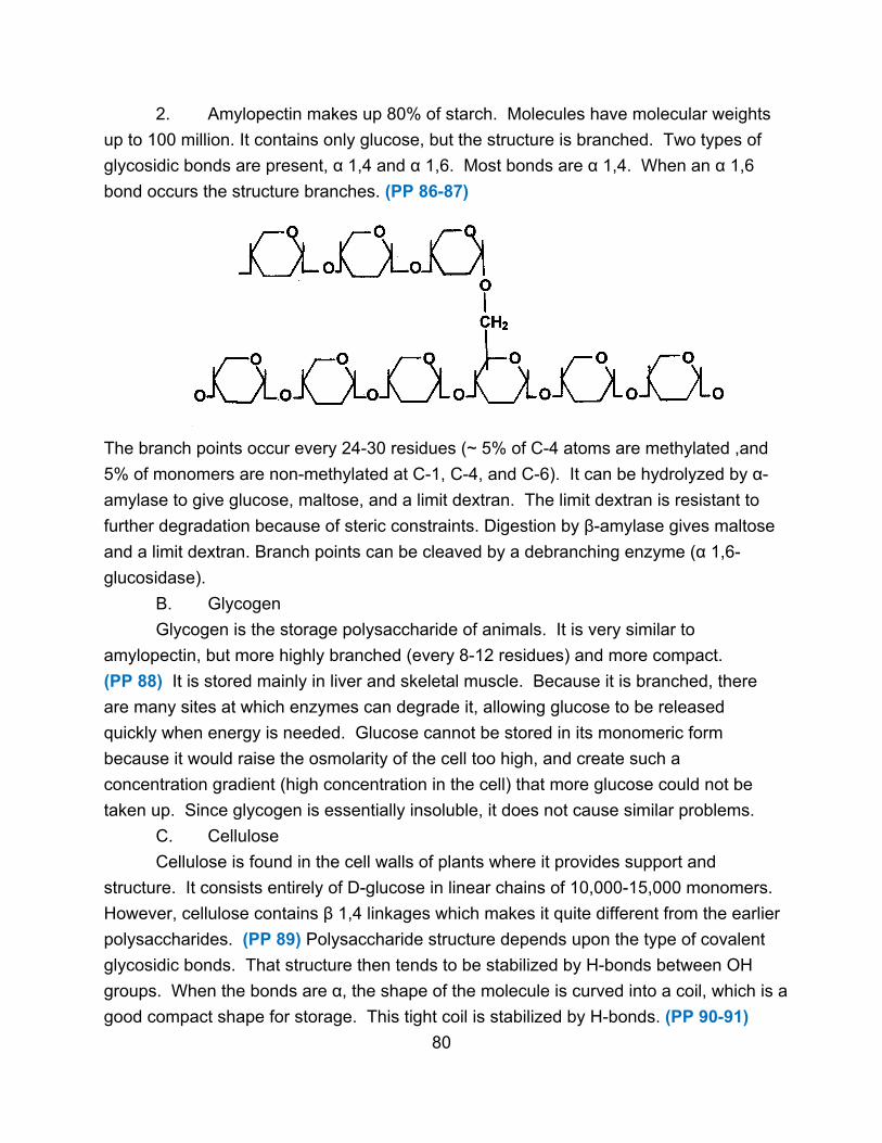
2. Amylopectin makes up 80% of starch. Molecules have molecular weights
up to 100 million. It contains only glucose, but the structure is branched. Two types of
glycosidic bonds are present, α 1,4 and α 1,6. Most bonds are α 1,4. When an α 1,6
bond occurs the structure branches. (PP 86-87)
The branch points occur every 24-30 residues (~ 5% of C-4 atoms are methylated ,and
5% of monomers are non-methylated at C-1, C-4, and C-6). It can be hydrolyzed by α-
amylase to give glucose, maltose, and a limit dextran. The limit dextran is resistant to
further degradation because of steric constraints. Digestion by β-amylase gives maltose
and a limit dextran. Branch points can be cleaved by a debranching enzyme (α 1,6-
glucosidase).
B. Glycogen
Glycogen is the storage polysaccharide of animals. It is very similar to
amylopectin, but more highly branched (every 8-12 residues) and more compact.
(PP 88) It is stored mainly in liver and skeletal muscle. Because it is branched, there
are many sites at which enzymes can degrade it, allowing glucose to be released
quickly when energy is needed. Glucose cannot be stored in its monomeric form
because it would raise the osmolarity of the cell too high, and create such a
concentration gradient (high concentration in the cell) that more glucose could not be
taken up. Since glycogen is essentially insoluble, it does not cause similar problems.
C. Cellulose
Cellulose is found in the cell walls of plants where it provides support and
structure. It consists entirely of D-glucose in linear chains of 10,000-15,000 monomers.
However, cellulose contains β 1,4 linkages which makes it quite different from the earlier
polysaccharides. (PP 89) Polysaccharide structure depends upon the type of covalent
glycosidic bonds. That structure then tends to be stabilized by H-bonds between OH
groups. When the bonds are α, the shape of the molecule is curved into a coil, which is a
good compact shape for storage. This tight coil is stabilized by H-bonds. (PP 90-91)
80
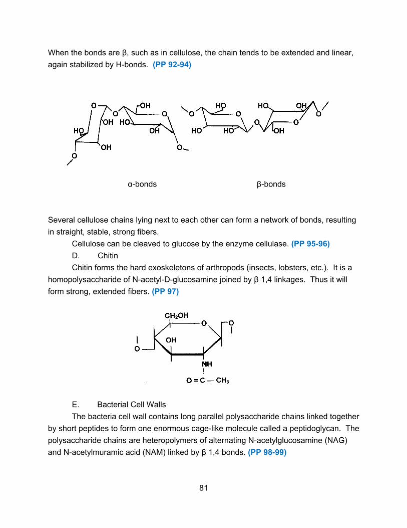
When the bonds are β, such as in cellulose, the chain tends to be extended and linear,
again stabilized by H-bonds. (PP 92-94)
α-bonds β-bonds
Several cellulose chains lying next to each other can form a network of bonds, resulting
in straight, stable, strong fibers.
Cellulose can be cleaved to glucose by the enzyme cellulase. (PP 95-96)
D. Chitin
Chitin forms the hard exoskeletons of arthropods (insects, lobsters, etc.). It is a
homopolysaccharide of N-acetyl-D-glucosamine joined by β 1,4 linkages. Thus it will
form strong, extended fibers. (PP 97)
E. Bacterial Cell Walls
The bacteria cell wall contains long parallel polysaccharide chains linked together
by short peptides to form one enormous cage-like molecule called a peptidoglycan. The
polysaccharide chains are heteropolymers of alternating N-acetylglucosamine (NAG)
and N-acetylmuramic acid (NAM) linked by β 1,4 bonds. (PP 98-99)
81
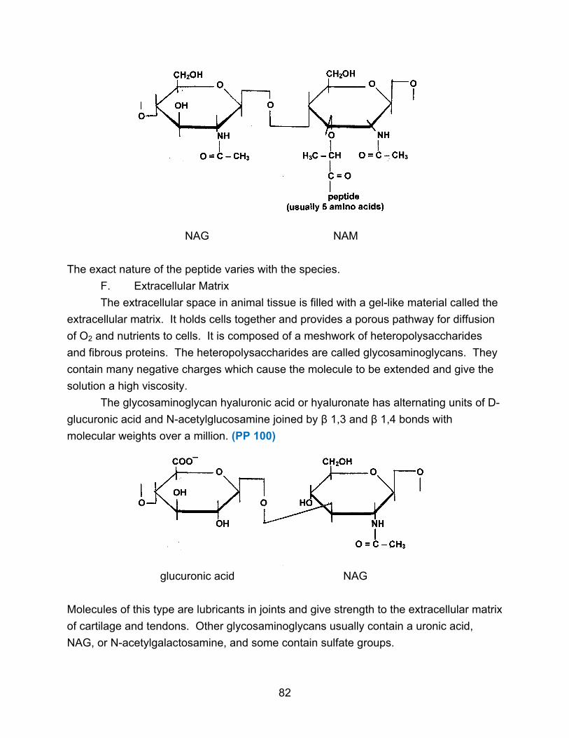
NAG NAM
The exact nature of the peptide varies with the species.
F. Extracellular Matrix
The extracellular space in animal tissue is filled with a gel-like material called the
extracellular matrix. It holds cells together and provides a porous pathway for diffusion
of O2 and nutrients to cells. It is composed of a meshwork of heteropolysaccharides
and fibrous proteins. The heteropolysaccharides are called glycosaminoglycans. They
contain many negative charges which cause the molecule to be extended and give the
solution a high viscosity.
The glycosaminoglycan hyaluronic acid or hyaluronate has alternating units of D-
glucuronic acid and N-acetylglucosamine joined by β 1,3 and β 1,4 bonds with
molecular weights over a million. (PP 100)
glucuronic acid NAG
Molecules of this type are lubricants in joints and give strength to the extracellular matrix
of cartilage and tendons. Other glycosaminoglycans usually contain a uronic acid,
NAG, or N-acetylgalactosamine, and some contain sulfate groups.
82

Proteoglycans are long glycosaminoglycans bound non-covalently to numerous
protein molecules, which in turn are covalently bound to smaller glycosaminoglycan
molecules such as chondroitin sulfate. The covalent bonds between carbohydrate and
protein are mainly through serine residues. There can be as many as 150
polysaccharide chains per protein molecule, with about 100 protein molecules per one
molecule of extended hyaluronate. This interacts with fibrous proteins like collagen and
elastin, forming a cross-linked network to which cells attach (via proteins) and along
which cell migration is directed.
83
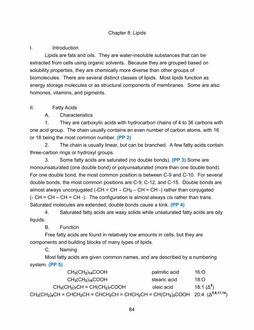
Chapter 8: Lipids
I. Introduction
Lipids are fats and oils. They are water-insoluble substances that can be
extracted from cells using organic solvents. Because they are grouped based on
solubility properties, they are chemically more diverse than other groups of
biomolecules. There are several distinct classes of lipids. Most lipids function as
energy storage molecules or as structural components of membranes. Some are also
homones, vitamins, and pigments.
II. Fatty Acids
A. Characteristics
1. They are carboxylic acids with hydrocarbon chains of 4 to 36 carbons with
one acid group. The chain usually contains an even number of carbon atoms, with 16
or 18 being the most common number. (PP 2)
2. The chain is usually linear, but can be branched. A few fatty acids contain
three-carbon rings or hydroxyl groups.
3. Some fatty acids are saturated (no double bonds). (PP 3) Some are
monounsaturated (one double bond) or polyunsaturated (more than one double bond).
For one double bond, the most common position is between C-9 and C-10. For several
double bonds, the most common positions are C-9, C-12, and C-15. Double bonds are
almost always unconjugated (-CH = CH – CH2 – CH = CH -) rather than conjugated
(- CH = CH – CH = CH -). The configuration is almost always cis rather than trans.
Saturated molecules are extended; double bonds cause a kink. (PP 4)
4. Saturated fatty acids are waxy solids while unsaturated fatty acids are oily
liquids.
B. Function
Free fatty acids are found in relatively low amounts in cells, but they are
components and building blocks of many types of lipids.
C. Naming
Most fatty acids are given common names, and are described by a numbering
system. (PP 5)
CH3(CH2)14COOH palmitic acid 16:O
CH3(CH2)16COOH stearic acid 18:O
CH3(CH2)7CH = CH(CH2)7COOH oleic acid 18:1 (∆9)
CH3(CH2)4CH = CHCH2CH = CHCH2CH = CHCH2CH = CH(CH2)3COOH 20:4 (∆5,8,11,14)
84

The first number is the number of carbon atoms, the second number is the number of
double bonds, and the numbers with ∆ describe the positions of the double bonds. (PP 6-7)
D. Reactions
Fatty acids placed in base (NaOH or KOH) form salts of fatty acids which are called
soaps. (PP 8)
CH3(CH2)16COOH + NaOH → CH3(CH2)16COO¯ Na+ + H2O
fatty acid soap
Such salts are amphipathic since they possess a polar head (ionized carboxyl group) and a
non-polar tail (hydrocarbon chain). While the original fatty acid is somewhat amphipathic,
the fatty acid salt is strongly amphipathic. In water, soaps do not truly dissolve but disperse
into micelles where the hydrophobic tails cluster to avoid water and the polar heads interact
with water. (PP 9)
Fatty acid salts of Ca2+ and Mg2+ are very insoluble and precipitate out as white solids in
hard water. Na+ and K+ salts can surround grease and disperse it, acting as soaps.
III. Triglycerides
A. Structure
Triglycerides (triacylglycerols) contain three fatty acids joined by ester bonds to a
glycerol molecule. (PP 10) The three fatty acids may be the same or different. Without
the -COOH group, they are even more non-polar than fatty acids. (PP 11)
85
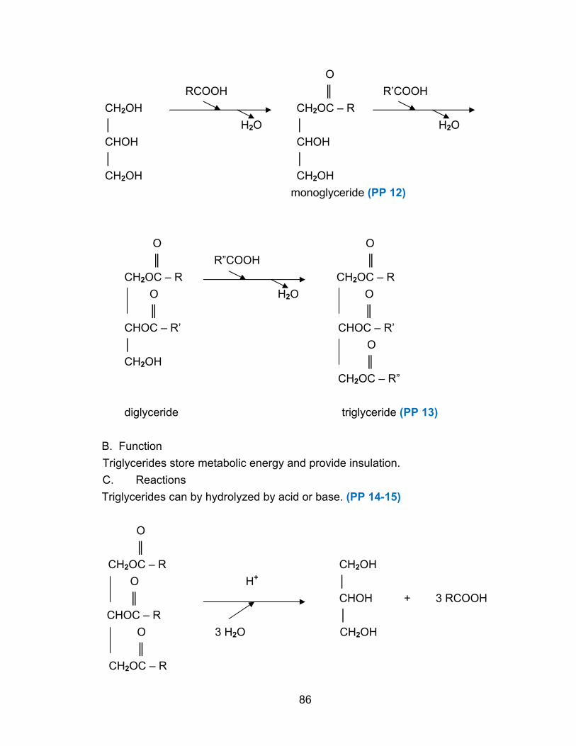
O
RCOOH ║ R’COOH
CH2OH CH2OC – R
│ H2O │ H2O
CHOH CHOH
│ │
CH2OH CH2OH
monoglyceride (PP 12)
O O
║ R”COOH ║
CH2OC – R CH2OC – R
O H2O O
║ ║
CHOC – R’ CHOC – R’
│ O
CH2OH ║
CH2OC – R”
diglyceride triglyceride (PP 13)
B. Function
Triglycerides store metabolic energy and provide insulation.
C. Reactions
Triglycerides can by hydrolyzed by acid or base. (PP 14-15)
O
║
CH2OC – R CH2OH
O H+ │
║ CHOH + 3 RCOOH
CHOC – R │
O 3 H2O CH2OH
║
CH2OC – R
86

O
║
CH2OC – R CH2OH
O │
║ CHOH + 3 RCOO¯Na+
CHOC – R │
O 3 NaOH CH2OH
║
CH2OC – R
The second reaction is called saponification since it produces soaps. Lipids can be
classified as saponifiable or non-saponifiable depending upon whether they contain fatty
acids that can be released by base hydrolysis to form soaps. (PP 16-17)
Triglycerides can also be broken down by enzymes known as lipases.
IV. Waxes
A. Structure
Waxes are esters of long-chain fatty acids (14-36 carbons) with long-chain
alcohols (16-30 carbons). (PP 18)
O
║
CH3(CH2)14C – O – CH2(CH2)28CH3
fatty acid alcohol
B. Function
Waxes function as metabolic fuels, and as protective coatings on hair, feathers,
plants, etc.
V. Glycerophospholipids (phosphoglycerides)
A. Structure
Glycerophospholipids contain glycerol, two fatty acids, and a phosphate group at
C-3 with a polar group attached to it. (PP 19-20) They are derivatives of phosphatidic
acid. (PP 21)
87
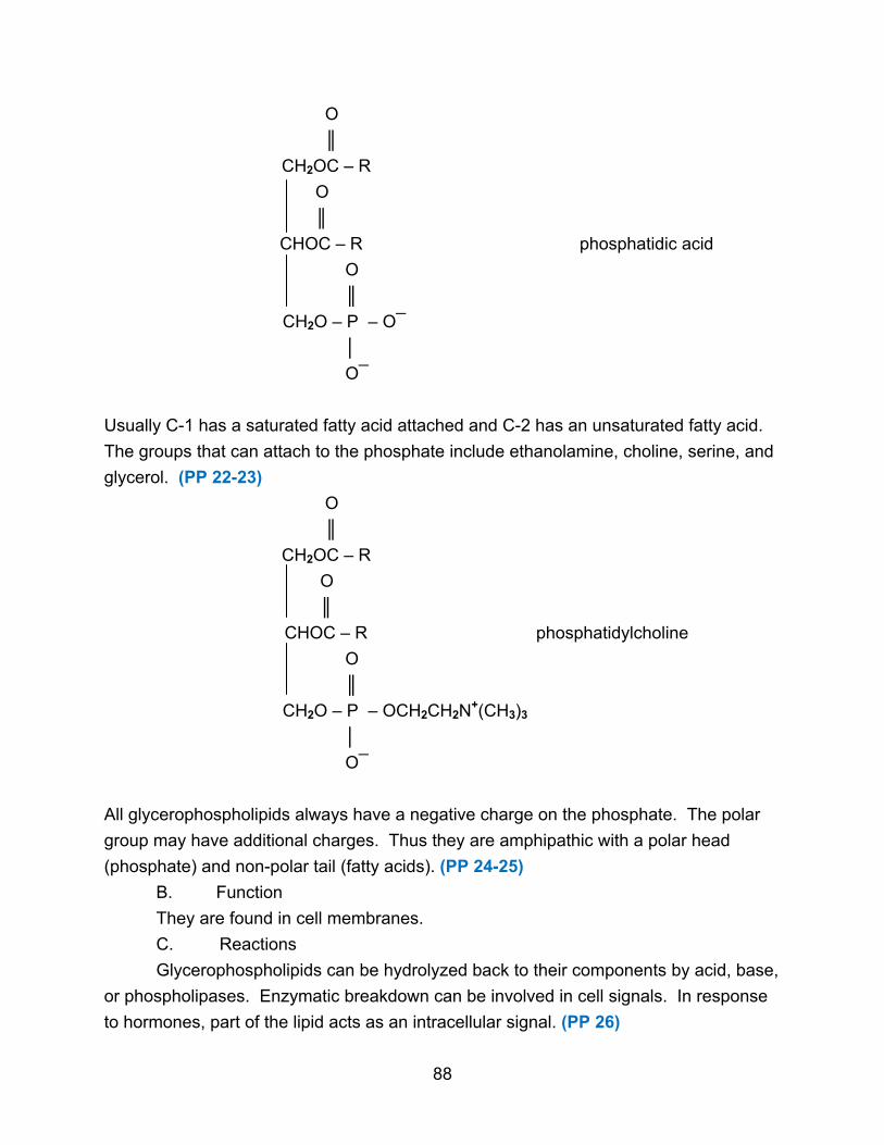
O
║
CH2OC – R
O
║
CHOC – R phosphatidic acid
O
║
CH2O – P – O¯
│
O¯
Usually C-1 has a saturated fatty acid attached and C-2 has an unsaturated fatty acid.
The groups that can attach to the phosphate include ethanolamine, choline, serine, and
glycerol. (PP 22-23)
O
║
CH2OC – R
O
║
CHOC – R phosphatidylcholine
O
║
CH2O – P – OCH2CH2N+(CH3)3
│
O¯
All glycerophospholipids always have a negative charge on the phosphate. The polar
group may have additional charges. Thus they are amphipathic with a polar head
(phosphate) and non-polar tail (fatty acids). (PP 24-25)
B. Function
They are found in cell membranes.
C. Reactions
Glycerophospholipids can be hydrolyzed back to their components by acid, base,
or phospholipases. Enzymatic breakdown can be involved in cell signals. In response
to hormones, part of the lipid acts as an intracellular signal. (PP 26)
88
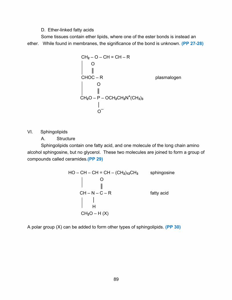
D. Ether-linked fatty acids
Some tissues contain ether lipids, where one of the ester bonds is instead an
ether. While found in membranes, the significance of the bond is unknown. (PP 27-28)
CH2 – O – CH = CH – R
O
║
CHOC – R plasmalogen
O
║
CH2O – P – OCH2CH2N+(CH3)3
│
O¯
VI. Sphingolipids
A. Structure
Sphingolipids contain one fatty acid, and one molecule of the long chain amino
alcohol sphingosine, but no glycerol. These two molecules are joined to form a group of
compounds called ceramides.(PP 29)
HO – CH – CH = CH – (CH2)12CH3 sphingosine
O
║
CH – N – C – R fatty acid
│
H
CH2O – H (X)
A polar group (X) can be added to form other types of sphingolipids. (PP 30)
89

B. Types 1. Sphingomyelins contain phosphocholine or phosphoethanolamine as the
polar group. They are found in plasma membranes and in the myelin sheath. They
have no net charge. (PP 31)
HO – CH – CH = CH(CH2)12CH3
O
║
CH – NH – C – R
O
║
CH2 – O – P – OCH2CH2 – N+(CH3)3
│
O¯
2. Cerebrosides contain a single sugar unit as the polar group. With
galactose the lipid is found in neural cell plasma membranes; with glucose the lipid is
found in nonneural membranes. They are uncharged overall. (PP 32)
HO – CH – CH = CH(CH2)12CH3
O
║
CH – NH – C – R
CH2 – O
CH2OH
O
HO OH
OH
90

3. Gangliosides contain a polar head made up of several (~ 4-6) sugar units.
They are negatively charged due to the presence of sugar acids. They are found in
various membranes including nerve cell membranes. (PP 33)
HO – CH – CH = CH(CH2)12CH3
O
║
CH – NH – C – R N-acetylneuraminic acid
CH2 – O ―
N-acetylgalactosamine
glucose galactose
4. Globosides are similar in structure to gangliosides, but the
oligosaccharide polar group contains 2-4 neutral sugar units, usually glucose,
galactose, or N-acetylgalactosamine.
C. Function
Sphingolipids are more than just structural components of membranes. They are
involved in recognition events on the cell surface, including such things as blood groups
and toxin binding. The exact functions of many sphingolipids are unknown. (PP 34-35)
VII. Sterols
A. Structure
Sterols contain the steroid ring system of four fused rings, various side groups,
and a hydroxyl group. Other steroids contain a carbonyl group. (PP 36)
HO
H
H
H
cholesterol
91
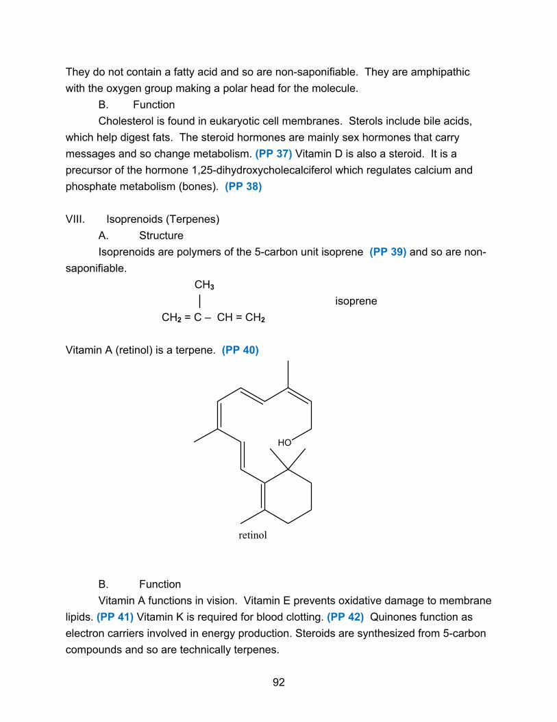
They do not contain a fatty acid and so are non-saponifiable. They are amphipathic
with the oxygen group making a polar head for the molecule.
B. Function
Cholesterol is found in eukaryotic cell membranes. Sterols include bile acids,
which help digest fats. The steroid hormones are mainly sex hormones that carry
messages and so change metabolism. (PP 37) Vitamin D is also a steroid. It is a
precursor of the hormone 1,25-dihydroxycholecalciferol which regulates calcium and
phosphate metabolism (bones). (PP 38)
VIII. Isoprenoids (Terpenes)
A. Structure
Isoprenoids are polymers of the 5-carbon unit isoprene (PP 39) and so are non-
saponifiable.
CH3
│ isoprene
CH2 = C – CH = CH2
Vitamin A (retinol) is a terpene. (PP 40)
HO
retinol
B. Function
Vitamin A functions in vision. Vitamin E prevents oxidative damage to membrane
lipids. (PP 41) Vitamin K is required for blood clotting. (PP 42) Quinones function as
electron carriers involved in energy production. Steroids are synthesized from 5-carbon
compounds and so are technically terpenes.
92

IX. Eicosanoids
A. Structure
Eicosanoids are fatty acid derivatives formed from arachidonic acid (20:4), a
polyunsaturated fatty acid. (PP 43)
B. Types
1. Prostaglandins contain a 5-membered ring.
COO¯ HO
→ COO¯
║
O OH
Prostaglandin PGD2
Prostaglandins are involved in smooth muscle contraction, regulation of blood flow,
response to hormones, and regulating body temperature, inflammation, and pain. They
are powerful biological signals.
2. Thromboxanes have a 6-membered ring with an ether.
COO¯
Thromboxane TXB2
HO O
OH
They function in formation of blood clots.
3. Leukotrienes contain three conjugated double bonds.
O
COO¯
Leukotriene A
They are potent regulatory molecules, controlling such things as contraction of muscle
linings in airways of lungs. (PP 44-45)
93
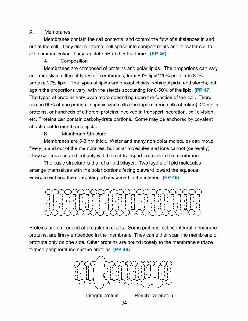
X. Membranes
Membranes contain the cell contents, and control the flow of substances in and
out of the cell. They divide internal cell space into compartments and allow for cell-to-
cell communication. They regulate pH and cell volume. (PP 46)
A. Composition
Membranes are composed of proteins and polar lipids. The proportions can vary
enormously in different types of membranes, from 80% lipid/ 20% protein to 80%
protein/ 20% lipid. The types of lipids are phospholipids, sphingolipids, and sterols, but
again the proportions vary, with the sterols accounting for 0-50% of the lipid. (PP 47)
The types of proteins vary even more depending upon the function of the cell. There
can be 90% of one protein in specialized cells (rhodopsin in rod cells of retina), 20 major
proteins, or hundreds of different proteins involved in transport, secretion, cell division,
etc. Proteins can contain carbohydrate portions. Some may be anchored by covalent
attachment to membrane lipids.
B. Membrane Structure
Membranes are 5-8 nm thick. Water and many non-polar molecules can move
freely in and out of the membranes, but polar molecules and ions cannot (generally).
They can move in and out only with help of transport proteins in the membrane.
The basic structure is that of a lipid bilayer. Two layers of lipid molecules
arrange themselves with the polar portions facing outward toward the aqueous
environment and the non-polar portions buried in the interior. (PP 48)
Proteins are embedded at irregular intervals. Some proteins, called integral membrane
proteins, are firmly embedded in the membrane. They can either span the membrane or
protrude only on one side. Other proteins are bound loosely to the membrane surface,
termed peripheral membrane proteins. (PP 49)
Integral protein Peripheral protein
94
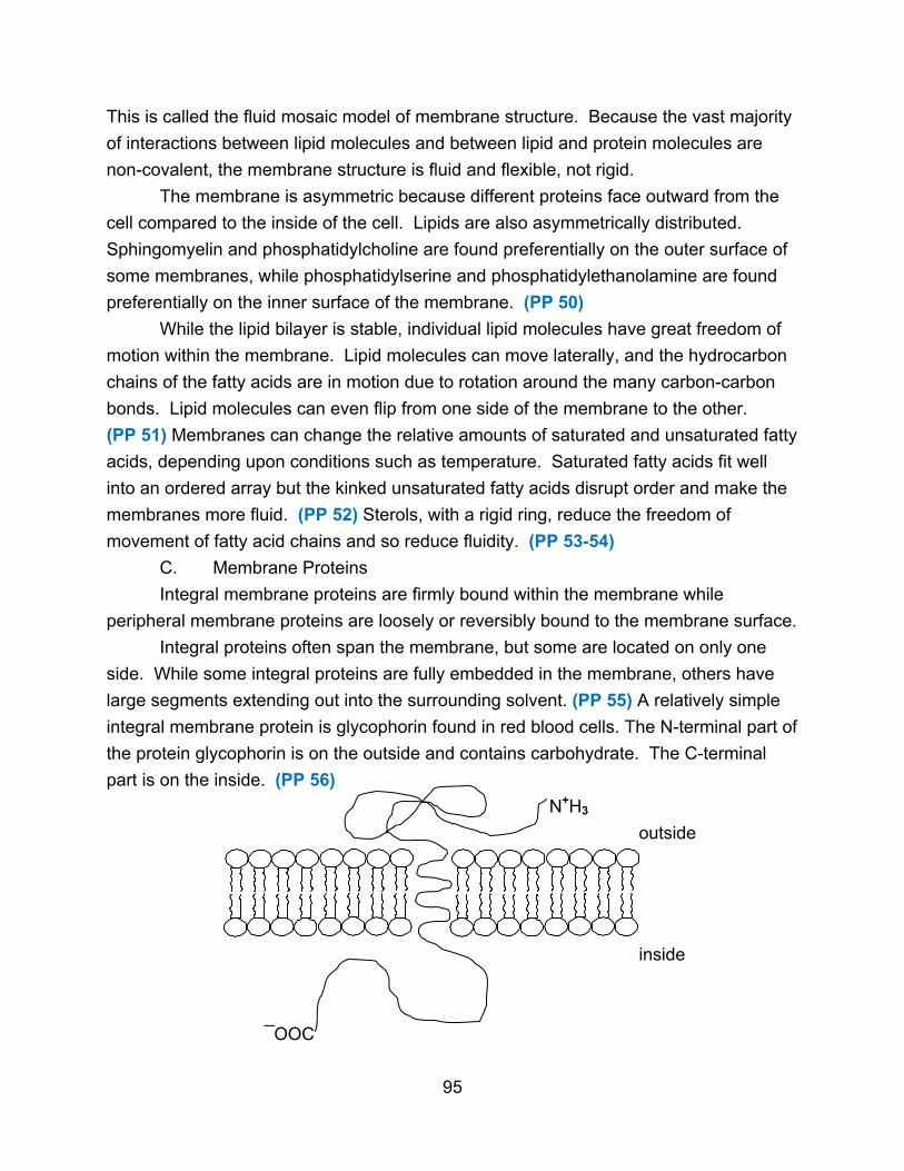
This is called the fluid mosaic model of membrane structure. Because the vast majority
of interactions between lipid molecules and between lipid and protein molecules are
non-covalent, the membrane structure is fluid and flexible, not rigid.
The membrane is asymmetric because different proteins face outward from the
cell compared to the inside of the cell. Lipids are also asymmetrically distributed.
Sphingomyelin and phosphatidylcholine are found preferentially on the outer surface of
some membranes, while phosphatidylserine and phosphatidylethanolamine are found
preferentially on the inner surface of the membrane. (PP 50)
While the lipid bilayer is stable, individual lipid molecules have great freedom of
motion within the membrane. Lipid molecules can move laterally, and the hydrocarbon
chains of the fatty acids are in motion due to rotation around the many carbon-carbon
bonds. Lipid molecules can even flip from one side of the membrane to the other.
(PP 51) Membranes can change the relative amounts of saturated and unsaturated fatty
acids, depending upon conditions such as temperature. Saturated fatty acids fit well
into an ordered array but the kinked unsaturated fatty acids disrupt order and make the
membranes more fluid. (PP 52) Sterols, with a rigid ring, reduce the freedom of
movement of fatty acid chains and so reduce fluidity. (PP 53-54)
C. Membrane Proteins
Integral membrane proteins are firmly bound within the membrane while
peripheral membrane proteins are loosely or reversibly bound to the membrane surface.
Integral proteins often span the membrane, but some are located on only one
side. While some integral proteins are fully embedded in the membrane, others have
large segments extending out into the surrounding solvent. (PP 55) A relatively simple
integral membrane protein is glycophorin found in red blood cells. The N-terminal part of
the protein glycophorin is on the outside and contains carbohydrate. The C-terminal
part is on the inside. (PP 56)
N+H3
outside
inside
¯OOC
95

Both terminal areas contain many polar amino acids and are hydrophilic, while the
segment in the middle of the membrane is hydrophobic. The result is that the protein is
asymmetrically oriented. Its orientation is specific and it does not flip-flop. Other integral
membrane proteins, such as bacteriorhodopsin in bacteria, cross the membrane several
times with hydrophobic α-helical segments. (PP 57)
H3N+
outside
COO¯ inside
Hydrophobic interactions between non-polar amino acids and the fatty acid chains
anchor the protein firmly in the membrane. Some proteins are free to diffuse around
and others are not. Certain proteins are covalently anchored to lipid molecules. Integral
membrane proteins, because they contain extensive hydrophobic regions, are generally
insoluble and so can be difficult to study. (PP 58)
Peripheral membrane proteins are held to the membrane by electrostatic
interactions and H-bonds with the polar heads of membrane lipids and hydrophilic
domains of integral membrane proteins. They are water-soluble. They may regulate
membrane-bound enzymes, connect integral proteins to intracellular structures, or limit
mobility of integral membrane proteins.
Certain proteins are covalently anchored to lipid molecules, which in turn anchor
to the membrane through hydrophobic interactions. (PP 59-61)
XI. Lipoproteins
Another important function of lipids is as components of lipoproteins. Many
lipoproteins are involved in transporting lipids around the body. Since lipids are water-
insoluble, they cannot be transported freely. Instead they form complexes with specific
proteins where the hydrophobic lipids aggregate at the core of the particle, while the
hydrophilic amino acids of the protein are on the surface along with the polar groups of
lipid molecules. (PP 62) There are four main classes of lipoproteins with different
combinations of lipid and protein. (PP 63-65)
96

A. Chylomicrons contain 2% protein and 98% lipid. They transport
triglycerides from the intestine to other tissues for use or storage.
B. Very low-density lipoproteins (VLDLs) contain 10% protein and 90% lipid.
They transport excess triglycerides from the liver to adipose tissue for storage.
C. Low-density lipoproteins (LDLs) are 25% protein and 75% lipid, mostly
cholesterol. They move cholesterol from the liver to other tissues.
D. High-density lipoproteins (HDLs) are 33% protein and 67% lipid. They
collect cholesterol remaining in other lipoproteins, and recycle the cholesterol to the
liver.
97
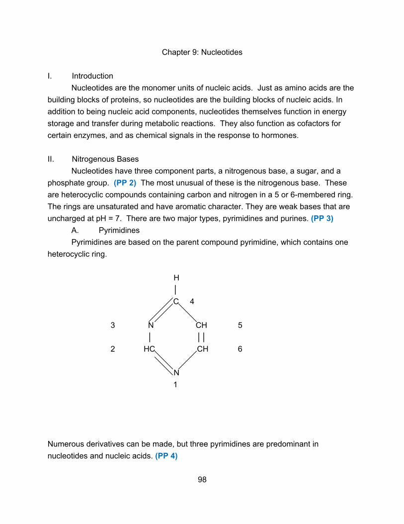
Chapter 9: Nucleotides
I. Introduction
Nucleotides are the monomer units of nucleic acids. Just as amino acids are the
building blocks of proteins, so nucleotides are the building blocks of nucleic acids. In
addition to being nucleic acid components, nucleotides themselves function in energy
storage and transfer during metabolic reactions. They also function as cofactors for
certain enzymes, and as chemical signals in the response to hormones.
II. Nitrogenous Bases
Nucleotides have three component parts, a nitrogenous base, a sugar, and a
phosphate group. (PP 2) The most unusual of these is the nitrogenous base. These
are heterocyclic compounds containing carbon and nitrogen in a 5 or 6-membered ring.
The rings are unsaturated and have aromatic character. They are weak bases that are
uncharged at pH = 7. There are two major types, pyrimidines and purines. (PP 3)
A. Pyrimidines
Pyrimidines are based on the parent compound pyrimidine, which contains one
heterocyclic ring.
H
│
C 4
3 N CH 5
│ ││
2 HC CH 6
N
1
Numerous derivatives can be made, but three pyrimidines are predominant in
nucleotides and nucleic acids. (PP 4)
98
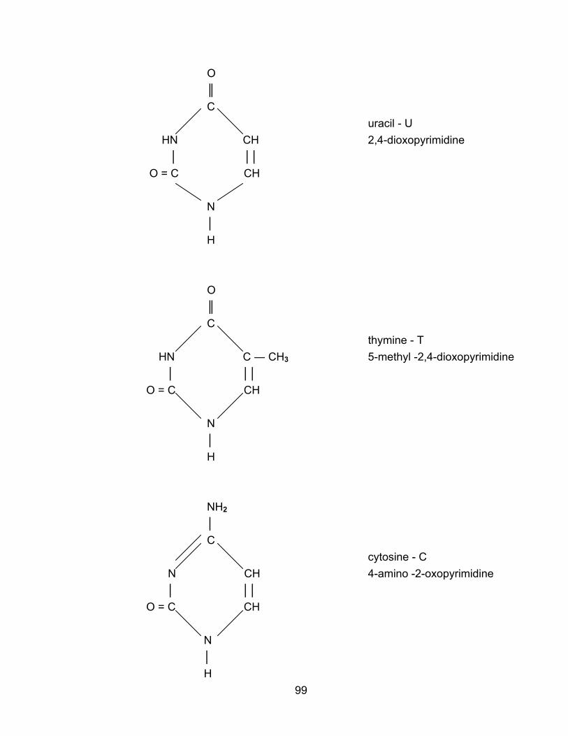
O
║
C
uracil - U
HN CH 2,4-dioxopyrimidine
│ ││
O = C CH
N
│
H
O
║
C
thymine - T
HN C ― CH3 5-methyl -2,4-dioxopyrimidine
│ ││
O = C CH
N
│
H
NH2
│
C
cytosine - C
N CH 4-amino -2-oxopyrimidine
│ ││
O = C CH
N
│
H
99

B. Purines
Purines are based on the parent compound purine, which is a derivative of
pyrimidine. Purines consist of a pyrimidine ring fused with an imidazole ring to give two
heterocyclic rings, one 5-membered and one 6-membered.
H
│
6 C 7
5 N
1 N C
│ ││ CH 8
2 HC C
4 N 9
N │
3 H
Two important purines are found in nucleic acids. (PP 5)
NH2
│
C
N adenine - A
N C 6-aminopurine
│ ││ CH
HC C
N
N │
H
100
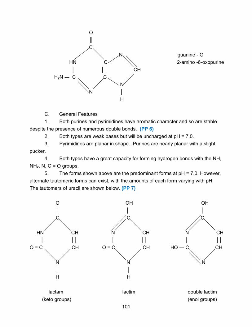
O
║
C
N guanine - G
HN C 2-amino -6-oxopurine
│ ││ CH
H2N ― C C
N
N │
H
C. General Features
1. Both purines and pyrimidines have aromatic character and so are stable
despite the presence of numerous double bonds. (PP 6)
2. Both types are weak bases but will be uncharged at pH = 7.0.
3. Pyrimidines are planar in shape. Purines are nearly planar with a slight
pucker.
4. Both types have a great capacity for forming hydrogen bonds with the NH,
NH2, N, C = O groups.
5. The forms shown above are the predominant forms at pH = 7.0. However,
alternate tautomeric forms can exist, with the amounts of each form varying with pH.
The tautomers of uracil are shown below. (PP 7)
O OH OH
║ │ │
C C C
HN CH N CH N CH
│ ││ │ ││ │ ││
O = C CH O = C CH HO ― C CH
N N N
│ │
H H
lactam lactim double lactim
(keto groups) (enol groups)
101

6. Rare bases
In addition to the five major bases, other unusual bases do exist and are sometimes
found in nucleic acids. (PP 8-9) Often these rare bases have extra methyl groups. They
still share the general characteristics of the more common bases. (PP 10-11)
NH2
│
C
N C ― CH3 5-methylcytosine
│ ││
O = C CH
N
│
H
III. Nucleosides
The second component of a nucleotide is a sugar. When a sugar is joined to a
nitrogenous base, the resulting structure is called a nucleoside.
A. Sugars
Two types of sugars are found in nucleotides, both based on D-ribose. (PP 12-13)
β-D-ribofuranose 2-deoxy -β-D-ribofuranose
(lacks OH at 2 position)
B. Nucleoside Structure
When the sugar is linked to a nitrogenous base, a nucleoside is produced. The
bond forms between the C-1 of the sugar and one of the nitrogens of the rings of the
nitrogenous base. This is an N-glycosidic bond. It is relatively stable to alkali but is
102

susceptible to acid hydrolysis (purines more than pyrimidines). The orientation of the
bond is β. In pyrimidines the bond will be 1,1. (PP 14) In purines the bond is 1,9.
(PP 15) To distinguish between the numbering of the sugar and the numbering of the
nitrogenous base, primes are added to the numbers that refer to the sugar.
uridine
(1-β-D-ribofuranosyluracil)
The N-glycosidic bond links the 1 position of the pyrimidine to the 1’ position of the
ribose.
deoxyadenosine
(9-β-2’-deoxy-D-ribofuranosyladenine)
The N-glycosidic bond links the 9 position of the purine to the 1’ position of the
deoxyribose. (PP 16)
103

The sugar and the base are both nearly planar and lie at right angles to each other. Two
conformations, syn and anti, are possible due to rotation around the N-glycosidic bond.
Due to steric hindrance, the anti conformation is favored. (PP 17-19)
C. Nomenclature
To simplify the names, nucleosides are named by adding –dine or –sine to the
name of the base. Deoxy as a prefix signifies the deoxyribose sugar. No prefix indicates
the ribose sugar.
Base Ribonucleoside Deoxyribonucleoside
adenine adenosine deoxyadenosine
cytosine cytidine deoxycytidine
guanine guanosine deoxyguanosine
thymine thymidine deoxythymidine
uracil uridine deoxyuridine
IV. Nucleotides
The third component of nucleotides is one or more phosphate groups. When a
phosphate group is added to a nucleoside, the resulting compound is a nucleotide.
A. Types
Nucleotides are distinguished by two features, where the phosphate group is
attached and how many phosphate groups are attached.
1. Position
The phosphate group will be attached to the ribose ring, but can vary as to which
OH group it is joined to. The 5'-position is by far the most common, with the 3'-position
also occurring.
adenosine -5’-monophosphate
104

deoxyguanosine-3’-monophosphate
‘Mono’ is used to indicate the presence of one phosphate group. If no number is
included in the name, such as adenosine monophosphate, then the assumption is that
the phosphate group is at the 5’ position. At pH = 7.0, phosphate will be ionized and
carry a negative charge of -2. (PP 20-25)
2. Number of phosphates
Many nucleotides have two or three phosphate groups, usually on the same
carbon (PP 26), although mixed 3',5' phosphates can occur.
adenosine-5’-diphosphate
charge = -3
105

deoxycytidine-5’-triphosphate
charge = -4
B. Nomenclature
To simplify the names, each compound is given an abbreviation. The first capital
letter indicates the base, the second letter indicates the number of phosphates, and the
third letter indicates phosphate. A small ‘d’ in front of the abbreviation signifies a
deoxyribonucleotide. Without the ‘d’, it is assumed the compound is a ribonucleotide.
5' ribonucleotides (NMP = nucleoside monophosphate)
Base NMP NDP NTP
A AMP ADP ATP
G GMP GDP GTP
C CMP CDP CTP
U UMP UDP UTP
5'-deoxyribonucleotides (dNMP = deoxyribonucleoside monophosphate)
Base dNMP dNDP dNTP
A dAMP dADP dATP
G dGMP dGDP dGTP
C dCMP dCDP dCTP
T dTMP dTDP dTTP
106

Because of the negative charges, nucleotides are often found in the cell complexed
with divalent ions like Mg2+. In the cell, phosphate groups are easily transferred by
enzymes, so there is a constant interconversion among mono, di, and triphosphates. Free
nucleosides and bases are found in cells only in very low levels. (PP 27-30)
C. Function
ATP and other nucleotides are used for energy storage and transfer in cells as
they lose or gain phosphate groups. They are also involved in enzyme regulation,
participating in both allostery and covalent modification.
Unusual nucleotides, such as 3’,5’-cyclic AMP (cAMP) mediate the action of
hormones. (PP 31)
Some nucleotides function as enzyme cofactors, like nicotinamide adenine
dinucleotide (NAD+). (PP 32)
107
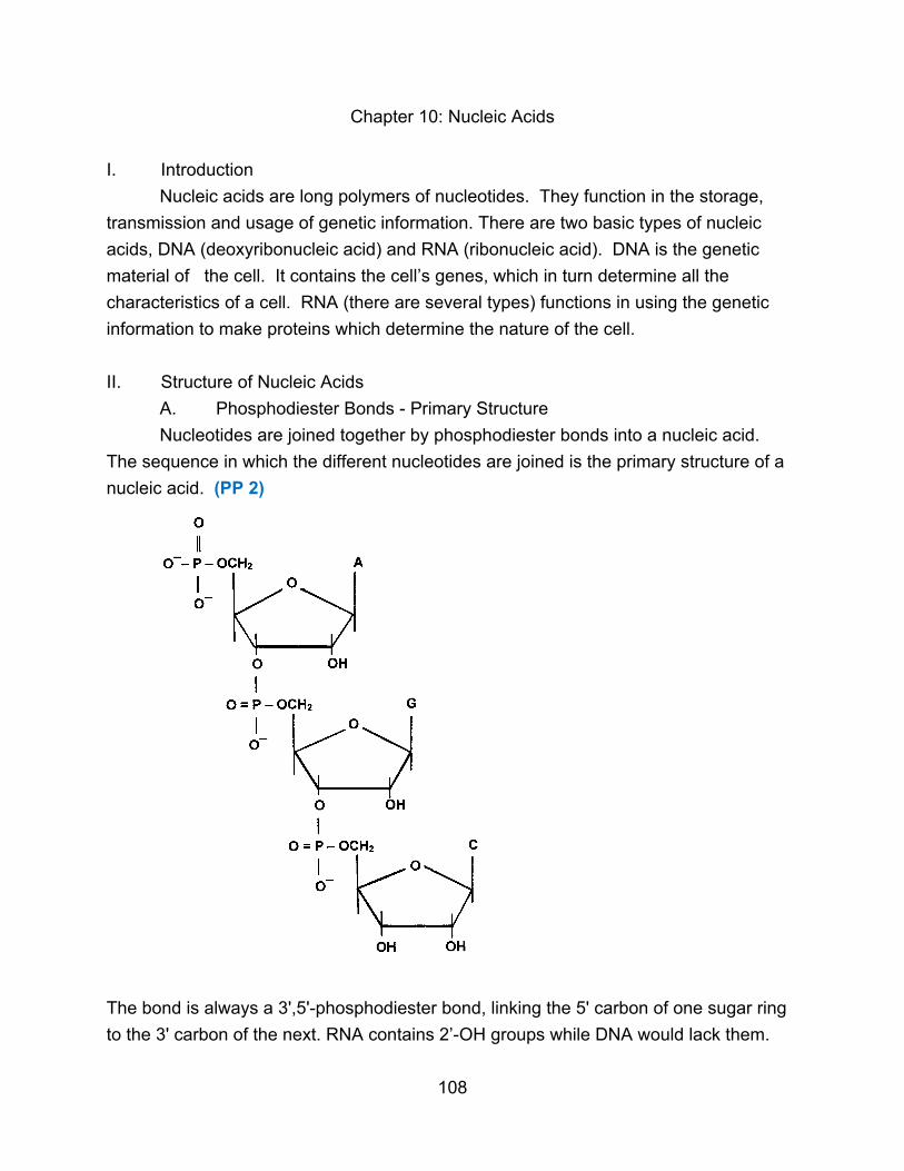
Chapter 10: Nucleic Acids
I. Introduction
Nucleic acids are long polymers of nucleotides. They function in the storage,
transmission and usage of genetic information. There are two basic types of nucleic
acids, DNA (deoxyribonucleic acid) and RNA (ribonucleic acid). DNA is the genetic
material of the cell. It contains the cell’s genes, which in turn determine all the
characteristics of a cell. RNA (there are several types) functions in using the genetic
information to make proteins which determine the nature of the cell.
II. Structure of Nucleic Acids
A. Phosphodiester Bonds - Primary Structure
Nucleotides are joined together by phosphodiester bonds into a nucleic acid.
The sequence in which the different nucleotides are joined is the primary structure of a
nucleic acid. (PP 2)
The bond is always a 3',5'-phosphodiester bond, linking the 5' carbon of one sugar ring
to the 3' carbon of the next. RNA contains 2’-OH groups while DNA would lack them.
108
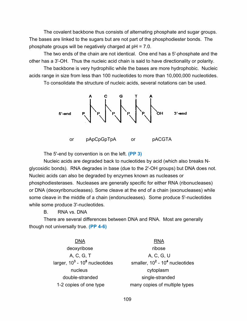
The covalent backbone thus consists of alternating phosphate and sugar groups.
The bases are linked to the sugars but are not part of the phosphodiester bonds. The
phosphate groups will be negatively charged at pH = 7.0.
The two ends of the chain are not identical. One end has a 5'-phosphate and the
other has a 3'-OH. Thus the nucleic acid chain is said to have directionality or polarity.
The backbone is very hydrophilic while the bases are more hydrophobic. Nucleic
acids range in size from less than 100 nucleotides to more than 10,000,000 nucleotides.
To consolidate the structure of nucleic acids, several notations can be used.
or pApCpGpTpA or pACGTA
The 5'-end by convention is on the left. (PP 3)
Nucleic acids are degraded back to nucleotides by acid (which also breaks N-
glycosidic bonds). RNA degrades in base (due to the 2'-OH groups) but DNA does not.
Nucleic acids can also be degraded by enzymes known as nucleases or
phosphodiesterases. Nucleases are generally specific for either RNA (ribonucleases)
or DNA (deoxyribonucleases). Some cleave at the end of a chain (exonucleases) while
some cleave in the middle of a chain (endonucleases). Some produce 5'-nucleotides
while some produce 3'-nucleotides.
B. RNA vs. DNA
There are several differences between DNA and RNA. Most are generally
though not universally true. (PP 4-6)
DNA RNA
deoxyribose ribose
A, C, G, T A, C, G, U
larger, 103 - 108 nucleotides smaller, 102 - 104 nucleotides
nucleus cytoplasm
double-stranded single-stranded
1-2 copies of one type many copies of multiple types
109
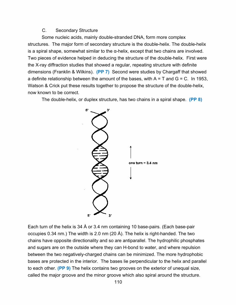
C. Secondary Structure
Some nucleic acids, mainly double-stranded DNA, form more complex
structures. The major form of secondary structure is the double-helix. The double-helix
is a spiral shape, somewhat similar to the α-helix, except that two chains are involved.
Two pieces of evidence helped in deducing the structure of the double-helix. First were
the X-ray diffraction studies that showed a regular, repeating structure with definite
dimensions (Franklin & Wilkins). (PP 7) Second were studies by Chargaff that showed
a definite relationship between the amount of the bases, with A = T and G = C. In 1953,
Watson & Crick put these results together to propose the structure of the double-helix,
now known to be correct.
The double-helix, or duplex structure, has two chains in a spiral shape. (PP 8)
Each turn of the helix is 34 Å or 3.4 nm containing 10 base-pairs. (Each base-pair
occupies 0.34 nm.) The width is 2.0 nm (20 Å). The helix is right-handed. The two
chains have opposite directionality and so are antiparallel. The hydrophilic phosphates
and sugars are on the outside where they can H-bond to water, and where repulsion
between the two negatively-charged chains can be minimized. The more hydrophobic
bases are protected in the interior. The bases lie perpendicular to the helix and parallel
to each other. (PP 9) The helix contains two grooves on the exterior of unequal size,
called the major groove and the minor groove which also spiral around the structure.
110

The two chains are held together by non-covalent forces. The first force is the
relative hydrophilic vs. hydrophobic nature of the different parts of the nucleic acid. The
second force is base stacking. The bases within a strand are parallel to each other, and
there are van der Waals forces, dipole-dipole interactions, and hydrophobic interactions
between them that stabilize the helix (and have effects on their electronic interactions
and light absorbance).
The third force is hydrogen bonding. Only certain base combinations can
hydrogen bond. (PP 10)
A – T base-pair (11.1Å wide with 2 H-bonds)
G – C base-pair (10.8Å wide with 3 H-bonds)
111

Other combinations of bases cannot fit within the helix or cannot properly hydrogen
bond. This explains why the amount of A always equals the amount of T, and the
amount of C equals the amount of G. Thus the base sequences of the strands are not
identical, but rather are said to be complementary. (PP 11) This also explains the
potential importance of tautomerization, since a base in a different tautomeric form will
hydrogen bond differently. (PP 12-15)
The double-helix is a somewhat rigid structure with fixed dimensions, but
considerable flexibility also exists. Rotation around certain bonds can occur, as can
bending and stretching of the strands. Local variations in structure occur depending
upon the base sequence. In addition, the ends of a DNA molecule are not always base-
paired, but vary with an average of 7 base-pairs frayed. The helix is not as stable at the
end of a molecule because the last base-pair is not surrounded by other base-pairs.
Even within the helix, each base-pair is not hydrogen-bonded at all times; momentary
‘breathing’ of DNA occurs.
Furthermore, the double-helix just described is not the only possible structure.
This form, called Β-DNA, is the major form of double-stranded DNA found in cells.
Another form, the A-DNA form, occurs when DNA is crystallized and dehydrated. It is also
a right-handed double-helix, but it has 11 base-pairs per turn and the bases are 20o from
perpendicular. It does not appear to occur in cells. (PP 16-18) Another possible form is
Z-DNA which is a left-handed helix with 12 base-pairs per turn and a zig-zag shape. DNA
with alternating purines and pyrimidines can form Z-DNA, and short stretches may be
found in cells. Their function is unknown but they may regulate gene expression.
Other more unusual secondary structures can also be found. When a segment
of DNA contains a palindrome or inverted repeat, other structures are possible. (PP 19)
―――TTAGCACGTGCTAA ―――
―――AATCGTGCACGATT ―――
↓
112

This double-stranded structure is called a cruciform. A single strand with this type of
structure is called a hairpin. (PP 20-21) Such structures can have non-paired bases in
the middle. These sequences tend to appear in regulatory regions of DNA. How many
palindromes actually form cruciforms in cells is not known.
H-DNA contains 3 strands and occurs when one strand contains only purines or
only pyrimidines in a long stretch. (PP 22-26)
D. Denaturation
Duplex DNA can be denatured (or melted) if the forces between the bases are
disrupted by heat, pH, or chemicals. No covalent bonds are broken. Distilled water
also denatures DNA because negative charges on phosphate groups are not shielded
by positive ions and repulsion occurs. (PP 27)
Denatured DNA can be renatured if the denaturing agent is removed slowly.
Renaturation is a two-step process. The first slow step requires that two strands find an
area of complementarity. Once these regions are H-bonded, the second fast step
occurs where the rest of the molecule zips up because complementary bases are now
aligned.
113

The ease with which a DNA molecule denatures depends upon the base content.
Since G:C base-pairs have three H-bonds, they are more stable than A:T base-pairs
with two H-bonds. Thus DNAs with high levels of G:C are more stable and more difficult
to denature.
One DNA strand and one complementary RNA strand can also associate to form
an RNA-DNA hybrid and the process is then called hybridization. (PP 28) RNA-DNA
hybrids, as well as double-stranded RNA molecules, tend to assume an A-like
conformation because the 2'-OH groups fit better in this structure. Even two nucleic
acid strands that are not entirely complementary can partially renature or hybridize. The
extent of hybridization depends on the extent of complementarity. This technique can
be used to determine the relationship of different species, isolate a gene, or detect a
certain sequence in a DNA molecule. (PP 29-30)
III. DNA as the Genetic Material
A. Evidence for DNA
In the early 1900's, proteins were better studied than nucleic acids, and it was
thought that proteins would turn out to be genetic material, providing the needed
diversity. Three different experiments showed rather that DNA was the genetic
material.
1. Griffith - 1928, Avery - 1944
Griffith was working with the bacterium Streptococcus pneumoniae. A virulent
type (S-type for smooth encapsulated colonies) would kill mice whereas a non-virulent
strain (R-type for rough nonencapsulated colonies) would not. (PP 31-32) If a heat-killed
extract of the virulent strain was mixed with live R-type bacteria and injected into a
mouse, the mouse died and live S-type bacteria could be isolated. (PP 33-34) Thus
something in the dead S-type extract transformed the non-virulent bacteria into the
virulent strain (changed its genetics). Avery later showed that the transforming factor in
the extract was not destroyed by proteases (so it was not protein) but was destroyed by
deoxyribonucleases (so it was DNA). (PP 35)
2. Hershey and Chase - 1952
Using virus T-2 which infects E. coli bacteria, they radioactively labeled the viral
protein coat with 35S and the viral DNA with 32P. (PP 36) When the virus infected the
cell, the viral parts remaining outside the E. coli cell were sheared away in a blender
(non-genetic parts) while the parts of the virus entering the cell (genetic material) were
detected by the radioactive label. (PP 37) Only the 32P-DNA entered the cell, not the 35S-protein. (PP 38)
114
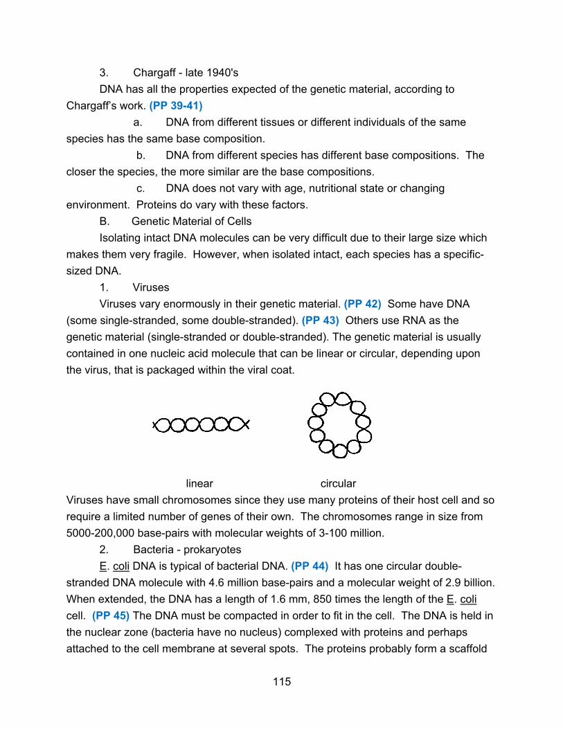
3. Chargaff - late 1940's
DNA has all the properties expected of the genetic material, according to
Chargaff’s work. (PP 39-41)
a. DNA from different tissues or different individuals of the same
species has the same base composition.
b. DNA from different species has different base compositions. The
closer the species, the more similar are the base compositions.
c. DNA does not vary with age, nutritional state or changing
environment. Proteins do vary with these factors.
B. Genetic Material of Cells
Isolating intact DNA molecules can be very difficult due to their large size which
makes them very fragile. However, when isolated intact, each species has a specific-
sized DNA.
1. Viruses
Viruses vary enormously in their genetic material. (PP 42) Some have DNA
(some single-stranded, some double-stranded). (PP 43) Others use RNA as the
genetic material (single-stranded or double-stranded). The genetic material is usually
contained in one nucleic acid molecule that can be linear or circular, depending upon
the virus, that is packaged within the viral coat.
linear circular
Viruses have small chromosomes since they use many proteins of their host cell and so
require a limited number of genes of their own. The chromosomes range in size from
5000-200,000 base-pairs with molecular weights of 3-100 million.
2. Bacteria - prokaryotes
E. coli DNA is typical of bacterial DNA. (PP 44) It has one circular double-
stranded DNA molecule with 4.6 million base-pairs and a molecular weight of 2.9 billion.
When extended, the DNA has a length of 1.6 mm, 850 times the length of the E. coli
cell. (PP 45) The DNA must be compacted in order to fit in the cell. The DNA is held in
the nuclear zone (bacteria have no nucleus) complexed with proteins and perhaps
attached to the cell membrane at several spots. The proteins probably form a scaffold
115
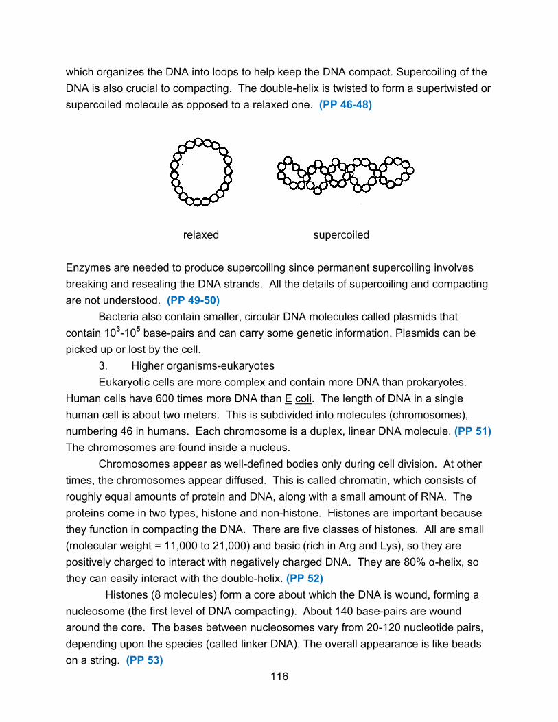
which organizes the DNA into loops to help keep the DNA compact. Supercoiling of the
DNA is also crucial to compacting. The double-helix is twisted to form a supertwisted or
supercoiled molecule as opposed to a relaxed one. (PP 46-48)
relaxed supercoiled
Enzymes are needed to produce supercoiling since permanent supercoiling involves
breaking and resealing the DNA strands. All the details of supercoiling and compacting
are not understood. (PP 49-50)
Bacteria also contain smaller, circular DNA molecules called plasmids that
contain 103-105 base-pairs and can carry some genetic information. Plasmids can be
picked up or lost by the cell.
3. Higher organisms-eukaryotes
Eukaryotic cells are more complex and contain more DNA than prokaryotes.
Human cells have 600 times more DNA than E coli. The length of DNA in a single
human cell is about two meters. This is subdivided into molecules (chromosomes),
numbering 46 in humans. Each chromosome is a duplex, linear DNA molecule. (PP 51)
The chromosomes are found inside a nucleus.
Chromosomes appear as well-defined bodies only during cell division. At other
times, the chromosomes appear diffused. This is called chromatin, which consists of
roughly equal amounts of protein and DNA, along with a small amount of RNA. The
proteins come in two types, histone and non-histone. Histones are important because
they function in compacting the DNA. There are five classes of histones. All are small
(molecular weight = 11,000 to 21,000) and basic (rich in Arg and Lys), so they are
positively charged to interact with negatively charged DNA. They are 80% α-helix, so
they can easily interact with the double-helix. (PP 52)
Histones (8 molecules) form a core about which the DNA is wound, forming a
nucleosome (the first level of DNA compacting). About 140 base-pairs are wound
around the core. The bases between nucleosomes vary from 20-120 nucleotide pairs,
depending upon the species (called linker DNA). The overall appearance is like beads
on a string. (PP 53)
116
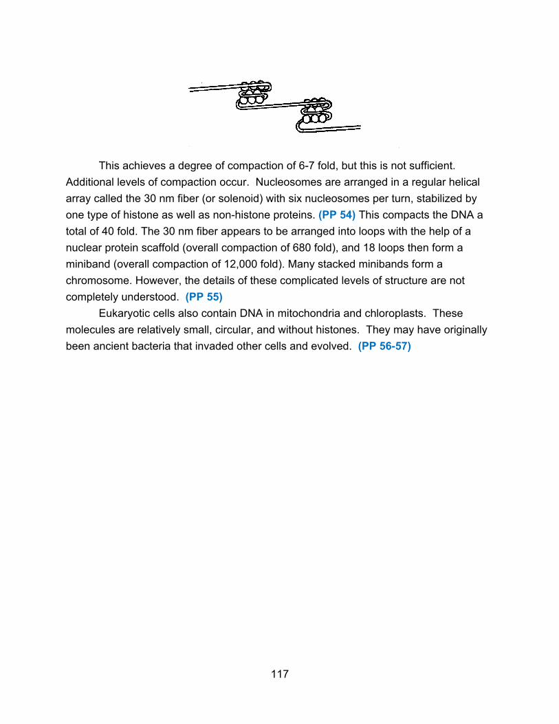
This achieves a degree of compaction of 6-7 fold, but this is not sufficient.
Additional levels of compaction occur. Nucleosomes are arranged in a regular helical
array called the 30 nm fiber (or solenoid) with six nucleosomes per turn, stabilized by
one type of histone as well as non-histone proteins. (PP 54) This compacts the DNA a
total of 40 fold. The 30 nm fiber appears to be arranged into loops with the help of a
nuclear protein scaffold (overall compaction of 680 fold), and 18 loops then form a
miniband (overall compaction of 12,000 fold). Many stacked minibands form a
chromosome. However, the details of these complicated levels of structure are not
completely understood. (PP 55)
Eukaryotic cells also contain DNA in mitochondria and chloroplasts. These
molecules are relatively small, circular, and without histones. They may have originally
been ancient bacteria that invaded other cells and evolved. (PP 56-57)
117
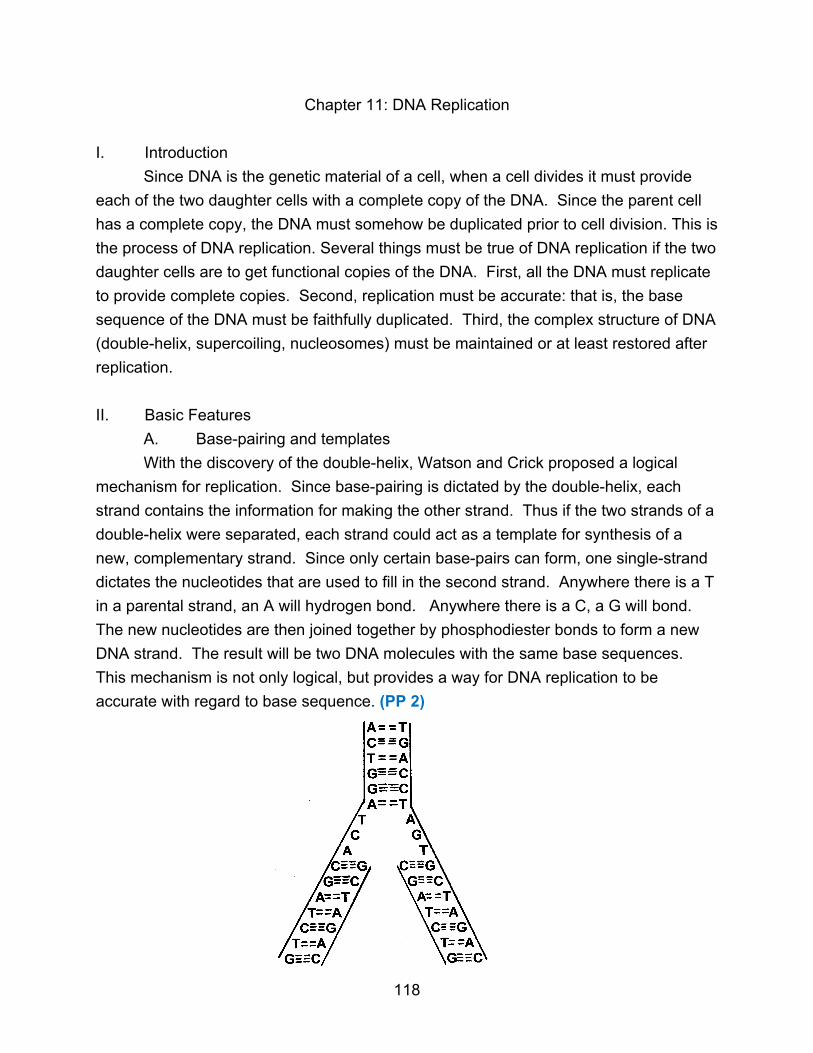
Chapter 11: DNA Replication
I. Introduction
Since DNA is the genetic material of a cell, when a cell divides it must provide
each of the two daughter cells with a complete copy of the DNA. Since the parent cell
has a complete copy, the DNA must somehow be duplicated prior to cell division. This is
the process of DNA replication. Several things must be true of DNA replication if the two
daughter cells are to get functional copies of the DNA. First, all the DNA must replicate
to provide complete copies. Second, replication must be accurate: that is, the base
sequence of the DNA must be faithfully duplicated. Third, the complex structure of DNA
(double-helix, supercoiling, nucleosomes) must be maintained or at least restored after
replication.
II. Basic Features
A. Base-pairing and templates
With the discovery of the double-helix, Watson and Crick proposed a logical
mechanism for replication. Since base-pairing is dictated by the double-helix, each
strand contains the information for making the other strand. Thus if the two strands of a
double-helix were separated, each strand could act as a template for synthesis of a
new, complementary strand. Since only certain base-pairs can form, one single-strand
dictates the nucleotides that are used to fill in the second strand. Anywhere there is a T
in a parental strand, an A will hydrogen bond. Anywhere there is a C, a G will bond.
The new nucleotides are then joined together by phosphodiester bonds to form a new
DNA strand. The result will be two DNA molecules with the same base sequences.
This mechanism is not only logical, but provides a way for DNA replication to be
accurate with regard to base sequence. (PP 2)
118
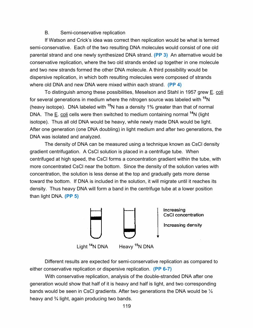
B. Semi-conservative replication
If Watson and Crick’s idea was correct then replication would be what is termed
semi-conservative. Each of the two resulting DNA molecules would consist of one old
parental strand and one newly synthesized DNA strand. (PP 3) An alternative would be
conservative replication, where the two old strands ended up together in one molecule
and two new strands formed the other DNA molecule. A third possibility would be
dispersive replication, in which both resulting molecules were composed of strands
where old DNA and new DNA were mixed within each strand. (PP 4)
To distinguish among these possibilities, Meselson and Stahl in 1957 grew E. coli
for several generations in medium where the nitrogen source was labeled with 15N
(heavy isotope). DNA labeled with 15N has a density 1% greater than that of normal
DNA. The E. coli cells were then switched to medium containing normal 14N (light
isotope). Thus all old DNA would be heavy, while newly made DNA would be light.
After one generation (one DNA doubling) in light medium and after two generations, the
DNA was isolated and analyzed.
The density of DNA can be measured using a technique known as CsCl density
gradient centrifugation. A CsCl solution is placed in a centrifuge tube. When
centrifuged at high speed, the CsCl forms a concentration gradient within the tube, with
more concentrated CsCl near the bottom. Since the density of the solution varies with
concentration, the solution is less dense at the top and gradually gets more dense
toward the bottom. If DNA is included in the solution, it will migrate until it reaches its
density. Thus heavy DNA will form a band in the centrifuge tube at a lower position
than light DNA. (PP 5)
Light 14N DNA Heavy 15N DNA
Different results are expected for semi-conservative replication as compared to
either conservative replication or dispersive replication. (PP 6-7)
With conservative replication, analysis of the double-stranded DNA after one
generation would show that half of it is heavy and half is light, and two corresponding
bands would be seen in CsCl gradients. After two generations the DNA would be ¼
heavy and ¾ light, again producing two bands.
119

Conservative
With semi-conservative replication, the DNA after one generation would be all
hybrid consisting of one heavy strand and one light strand, and one band with
intermediate density would be seen in CsCl gradients. After two generations, the DNA
would be ½ hybrid and ½ light, resulting in two bands.
Semi-Conservative
120
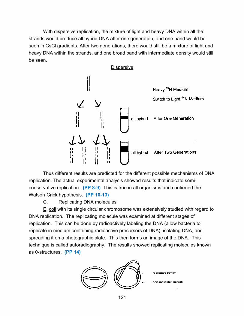
With dispersive replication, the mixture of light and heavy DNA within all the
strands would produce all hybrid DNA after one generation, and one band would be
seen in CsCl gradients. After two generations, there would still be a mixture of light and
heavy DNA within the strands, and one broad band with intermediate density would still
be seen.
Dispersive
Thus different results are predicted for the different possible mechanisms of DNA
replication. The actual experimental analysis showed results that indicate semi-
conservative replication. (PP 8-9) This is true in all organisms and confirmed the
Watson-Crick hypothesis. (PP 10-13)
C. Replicating DNA molecules
E. coli with its single circular chromosome was extensively studied with regard to
DNA replication. The replicating molecule was examined at different stages of
replication. This can be done by radioactively labeling the DNA (allow bacteria to
replicate in medium containing radioactive precursors of DNA), isolating DNA, and
spreading it on a photographic plate. This then forms an image of the DNA. This
technique is called autoradiography. The results showed replicating molecules known
as θ-structures. (PP 14)
121

Similar studies revealed two additional facts. First, replication always begins at
the same place on the E coli chromosome. This is a specific site, designated oriC, with
a specific sequence of 245 base-pairs which is always recognized as the starting place
for replication. Second, DNA replication is bidirectional, meaning that replication
proceeds in both directions around the circular chromosome until the new strands meet
180o from the origin. The alternative is unidirectional replication where replication
proceeds in only one direction, moving 360o around the circle and back to the origin.
(PP 15)
origin origin
↓ ↓
bidirectional unidirectional
All replication, except in some plasmids and viruses, is bidirectional rather than
unidirectional. Each intersection where replication is actively occurring is a replication
fork. (PP 16) With bidirectional replication, there are two such forks in a θ-structure.
← replication/ fork movement
The rate of DNA replication is very fast in E. coli, about 45,000 nucleotides added
per fork per minute at 37oC. This rate will vary with temperature, but at 37o is constant.
However, E. coli at 37oC will have different generation times depending upon the
nutritional state of the medium. In rich medium bacteria divide every 20-30 minutes,
while in minimal medium the generation time is 2-3 hours. However, DNA replication
always takes 40 minutes at 37oC. Bacteria cannot change the rate of replication, but
they can control how often they initiate DNA replication. However, once replication is
started, it continues at a fixed rate and goes to completion. Thus in minimal medium,
122

the bacteria spend 40 minutes replicating DNA and the rest of the 2-3 hours growing
and accumulating enough energy. In rich medium, a generation time of 20 minutes is
maintained by having two or more rounds of replication going on at the same time, each
of which takes 40 minutes to complete. (PP 17-18)
A round of replication is completed every 20 minutes, allowing the cell to divide, and the
two daughter DNA molecules are already undergoing subsequent rounds of DNA
replication. Controlling replication through the frequency of initiation (rather than through
varying the rate of polymerization) means that once the cell starts replication it is
committed to finishing replication. This is efficient since replication must be completed to
be beneficial to the cell. (PP 19-20)
D. DNA Polymerization
The first enzyme found in E. coli to polymerize DNA is called DNA polymerase I.
It is a single chain with a molecular weight of 103,000. The enzyme uses dNTPs in the
following reaction.
(dNMP)n + dNTP → (dNMP)n+1 + PPi
DNA DNA ↓
(longer by one 2 Pi
nucleotide)
123

The breaking of the pyrophosphate bonds of the dNTP and PPi provides energy for
making the phosphodiester bonds which explains why the enzyme will not use dNDPs
or dNMPs. It is also specific for DNA; ribonucleotides are not used.
The enzyme specifically joins the new nucleotide to the 3'-end of the existing
DNA molecule. (PP 21-23)
The DNA grows from the 5'-end toward the 3'-end (5'→3' polymerase).
Two other features of DNA polymerase are important. First, the enzyme requires
a template, an exposed single-strand of DNA to which incoming nucleotides hydrogen-
bond and so are correctly selected. Without a template, DNA polymerase I will not just
join nucleotides randomly. This is consistent with the prediction that if DNA replication
is to be accurate, base-pairing must guide the selection of each nucleotide to be
124

polymerized. Second, the enzyme requires a primer. The enzyme cannot start a new
chain, but can only add nucleotides onto an existing chain. It can elongate, but it cannot
initiate. (PP 24-26)
3’ _____________________________________ 5’ template
5’ _______ 3’
primer ↑
DNA polymerase adds nucleotides
Some properties of DNA polymerase I (also called pol I) are not consistent with a
replication enzyme. For instance, pol I adds nucleotides at a rate of 600
nucleotides/minute, which is too slow to replicate the chromosome in 40 minutes. Its
processivity (the average number of nucleotides it polymerizes before it dissociates
from the template) is 20, which is lower than expected for replication. In addition,
mutants of polymerase I can replicate DNA. Polymerase I, it turns out, functions mainly
in repair of DNA and to a limited extent in replication. Four other polymerases exist in
E. coli. (PP 27) DNA polymerase II has multiple subunits (probably 7) and a molecular
weight of about 90,000. It also has a relatively low polymerization rate and processivity,
and functions in DNA repair. Polymerase III holoenzyme has at least ten subunits and a
molecular weight of about 800,000. (PP 28-29) It polymerizes 30,000 - 50,000
nucleotides/minute, and is responsible for the vast majority of DNA replication. Three of
its subunits (αε θ) form the core enzyme that has polymerase activity. The remaining
subunits allow the enzyme to clamp onto the DNA template and result in a very high
processivity of more than 500,000. (PP 30-31)
DNA polymerases I, II, and III all have the same basic properties, including using
dNTPs, requiring a template and a primer, and polymerizing 5’→3’ to accurately copy a
DNA sequence. Polymerases IV and V function in DNA repair and are less accurate.
(PP 32-33)
III. Replication Process
The replication of the chromosome is an enormously complicated process
involving many proteins besides DNA polymerase III. (PP 34) The process can be
divided into stages.
A. Initiation
The replication origin of E. coli (oriC) is 245 base-pairs. It contains three repeated
sequences (13 bp) of one type and four or five repeated sequences (9 bp) of a second type.
(PP 35) About 8 copies of a specific replication protein, called DnaA, bind to the
125

9-bp sequences and the DNA wraps around the protein, forming a nucleosome-like
structure. This strains the A-T rich 13-bp sequences (called DUE-DNA unwinding
element) and causes them to denature. (PP 36) Protein HU, FIS, and IHF are also
needed, along with ATP. Then DnaC protein helps DnaB protein bind to the denatured
region, and DnaB further unwinds the DNA in both directions. Primers are then
synthesized so DNA can be replicated. (PP 37)
Additional proteins may also be involved. Control of the initiation process is not
well-understood, but may involve interaction of the origin with the cell membrane and
methylation of DNA within specific base sequences. (PP 38-39)
B. Elongation
Elongation is more complicated than expected because of the properties of DNA
polymerase III. First, DNA polymerase III cannot initiate a new DNA chain, so there is
the question of how replication starts. DNA chains are initiated on short primers of
RNA. Enzymes which polymerize RNA are similar to DNA polymerases, but RNA
polymerases do not require a primer and so can initiate new chains.
An additional problem is caused by the directionality of DNA polymerase III in
that it works only 5'→3'. However, in a replication fork, new DNA strands must be
synthesized both 5’→3’ and 3’→5’.
3’
5’
5’
3’ 3’
?
5’
The two template DNA strands are antiparallel to each other, and each new DNA strand
must also be antiparallel to its template. This means one new strand must be
synthesized in the 5'→3' direction, which is what polymerases can do. The other strand,
however, must be synthesized 3'→5', which no polymerases can do. The question is
how is this new strand made. (PP 40)
Okazaki proposed that this 3'→5' strand was made discontinuously, as a series
of short DNA pieces called Okazaki pieces. Each piece is synthesized 5'→3' and then
joined together to give the appearance of a strand growing 3'→5'. (PP 41)
126

3’ 3’
5’ 5’
leading strand
5’ 3’ 5’ 3’
3’ 5’ → 3’ 5’
lagging strand
3’ 3’
5’ 5’
The strand that is made continuously is called the leading strand. The strand made
discontinuously is called the lagging strand. Experiments demonstrated the presence of
Okazaki pieces, not only in E. coli but in all cells.
Okazaki’s explanation solved one problem, but created another. Since DNA
polymerases cannot initiate new chains, the problem now became to explain how the
numerous Okazaki pieces are started. Okazaki proposed that each Okazaki piece
started using an RNA primer (/\/\/\/\). (PP 42) Then, later during replication, the RNA
primers are removed and filled in with DNA.
1. Leading strand synthesis
Putting all these features together, the following mechanism exists for leading
strand synthesis. Once initiation has denatured the DNA at the origin, a short RNA
primer (10-20 nucleotides) is synthesized at the origin by an RNA polymerase known as
primase (DnaG protein). The cell’s main RNA polymerase (also called RNA
polymerase) may be involved as well. Primase binds to the DnaB protein to form a
primosome complex, and other proteins may also be needed for the primosome. Once
the primer is formed, DNA polymerase III extends this continuously by adding
nucleotides, creating a long DNA chain that extends around the chromosome. There
127

are two leading strands that must be established at the origin, one in each of the two
replication forks that create bi-directional replication. (PP 43-44)
2. Lagging strand synthesis
The primosome moves along the template with the replication fork. At various
intervals, the primase enzyme makes a short RNA primer (10-20 nucleotides). DNA
polymerase III then uses the primer to make an Okazaki piece (1000-2000 nucleotides).
(PP 45-47)
Leading and lagging strand synthesis in a replication fork are coordinated by DNA
polymerase III holoenzyme. Two core enzymes (one for the leading strand and one for the
lagging strand) are held clamped to the DNA strands by other holoenzyme subunits, and
both are connected by additional subunits. Thus both DNA strands are replicated
together. The lagging strand is probably looped so that the holoenzyme can work on both
template strands at the same time. (PP 48-51) In addition, it is possible that pol III and
other replication proteins remain attached to the cell membrane and the DNA moves
through these replication factories, rather than the proteins moving along the DNA.
128

Removing the RNA primer is carried out by DNA polymerase I. In addition to its
polymerase activity, polymerase I also has a 5'→3' exonuclease activity. Polymerase I
attaches to the 3'-end of an Okazaki fragment and begins to add deoxyribonucleotides.
When it bumps into the RNA primer of the next Okazaki piece, it removes the RNA
primer while filling in with DNA. Thus the RNA primer is replaced with DNA by pol I
using both its polymerase and exonuclease activities simultaneously. Polymerase III
does not have this exonuclease activity. (PP 52)
After the primer is removed, there remains a nick in the DNA backbone, even
though all the deoxyribonucleotides are in place and all the bases are paired. The DNA
polymerases cannot make the last phosphodiester bond because the 5'-side of the nick
has only one phosphate group as a result of removing the RNA primer. A different
enzyme, called DNA ligase, seals the nick and makes the last phosphodiester bond.
The reaction requires the input of energy from NAD+ or ATP, depending upon the
organism. (PP 53)
Following the action of DNA ligase, the Okazaki pieces are completely joined together
to create the new lagging strand.
129

3. Geometry problems
Several other things must happen for DNA to replicate. First, the double-helix
must be separated in two single strands in the replicating area and this area of
denaturation must move with the replication fork. Enzymes known as helicases, mainly
DnaB protein, unwind the DNA just ahead of replication, using the energy of ATP to
break the hydrogen bonds and separate the two parental strands. (PP 54) Second, the
DNA strands, once separated, have to be kept apart. The single-stranded regions are
prevented from renaturing by single-stranded DNA binding protein (SSB) that binds
cooperatively to single-stranded DNA. (PP 55) Third, as the double-helix unwinds in
one area of the circular DNA, supercoils build up further along the molecule. The DNA
can only twist so much before the tension must be relieved. (PP 56-57) Enzymes called
topoisomerases, specifically DNA gyrase, can nick the DNA and adjust the supercoiling.
4. Other proteins
A number of other proteins are probably involved in DNA replication but their
function is not yet defined. (PP 58-59)
5. Summary of elongation (PP 60-66)
C. Termination
The final separation of the two completed DNA molecules must be accomplished,
but details of the process are not well-understood. The termination region of the DNA
(Ter) contains a number of short repeats recognized by the Tus protein, which prevents
unwinding of the double-helix and stops the replication forks. (PP 67) Topoisomerases
then untangle the two completed DNA molecules. (PP 68)
130
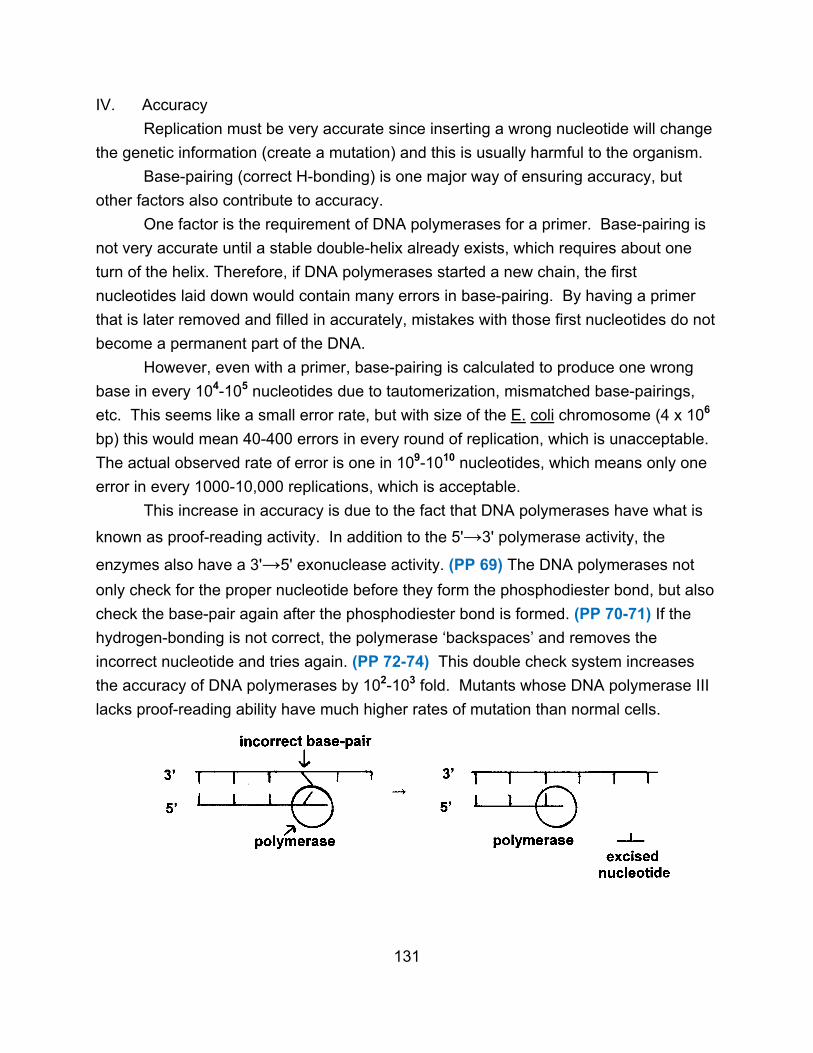
IV. Accuracy
Replication must be very accurate since inserting a wrong nucleotide will change
the genetic information (create a mutation) and this is usually harmful to the organism.
Base-pairing (correct H-bonding) is one major way of ensuring accuracy, but
other factors also contribute to accuracy.
One factor is the requirement of DNA polymerases for a primer. Base-pairing is
not very accurate until a stable double-helix already exists, which requires about one
turn of the helix. Therefore, if DNA polymerases started a new chain, the first
nucleotides laid down would contain many errors in base-pairing. By having a primer
that is later removed and filled in accurately, mistakes with those first nucleotides do not
become a permanent part of the DNA.
However, even with a primer, base-pairing is calculated to produce one wrong
base in every 104-105 nucleotides due to tautomerization, mismatched base-pairings,
etc. This seems like a small error rate, but with size of the E. coli chromosome (4 x 10⁶
bp) this would mean 40-400 errors in every round of replication, which is unacceptable.
The actual observed rate of error is one in 109-1010 nucleotides, which means only one
error in every 1000-10,000 replications, which is acceptable.
This increase in accuracy is due to the fact that DNA polymerases have what is
known as proof-reading activity. In addition to the 5'→3' polymerase activity, the
enzymes also have a 3'→5' exonuclease activity. (PP 69) The DNA polymerases not
only check for the proper nucleotide before they form the phosphodiester bond, but also
check the base-pair again after the phosphodiester bond is formed. (PP 70-71) If the
hydrogen-bonding is not correct, the polymerase ‘backspaces’ and removes the
incorrect nucleotide and tries again. (PP 72-74) This double check system increases
the accuracy of DNA polymerases by 102-103 fold. Mutants whose DNA polymerase III
lacks proof-reading ability have much higher rates of mutation than normal cells.
131
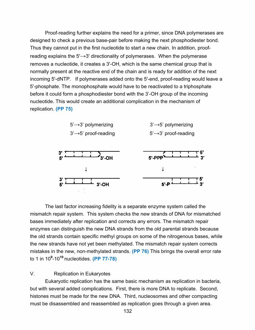
Proof-reading further explains the need for a primer, since DNA polymerases are
designed to check a previous base-pair before making the next phosphodiester bond.
Thus they cannot put in the first nucleotide to start a new chain. In addition, proof-
reading explains the 5'→3' directionality of polymerases. When the polymerase
removes a nucleotide, it creates a 3'-OH, which is the same chemical group that is
normally present at the reactive end of the chain and is ready for addition of the next
incoming 5'-dNTP. If polymerases added onto the 5'-end, proof-reading would leave a
5'-phosphate. The monophosphate would have to be reactivated to a triphosphate
before it could form a phosphodiester bond with the 3’-OH group of the incoming
nucleotide. This would create an additional complication in the mechanism of
replication. (PP 75)
5’→3’ polymerizing 3’→5’ polymerizing
3’→5’ proof-reading 5’→3’ proof-reading
The last factor increasing fidelity is a separate enzyme system called the
mismatch repair system. This system checks the new strands of DNA for mismatched
bases immediately after replication and corrects any errors. The mismatch repair
enzymes can distinguish the new DNA strands from the old parental strands because
the old strands contain specific methyl groups on some of the nitrogenous bases, while
the new strands have not yet been methylated. The mismatch repair system corrects
mistakes in the new, non-methylated strands. (PP 76) This brings the overall error rate
to 1 in 109-1010 nucleotides. (PP 77-78)
V. Replication in Eukaryotes
Eukaryotic replication has the same basic mechanism as replication in bacteria,
but with several added complications. First, there is more DNA to replicate. Second,
histones must be made for the new DNA. Third, nucleosomes and other compacting
must be disassembled and reassembled as replication goes through a given area.
132

Unlike prokaryotes, eukaryotic replication takes place at a specific time.
Eukaryotic cells have a cell cycle, with a specific series of events leading to mitosis (M).
The entire cycle takes 12-72 hours and DNA replication (S phase) takes 1-3 hours,
depending on the cell. (PP 79)
M
G2 G1
S
Eukaryotic DNA replication is slower than in prokaryotes, probably because of
the nucleosomes. Eukaryotic polymerases polymerize at the rate of 103 bp/min per
replication fork, considerably slower than in prokaryotes. If eukaryotic DNA had just
one origin, it would take 30 days to replicate the DNA at this rate. To compensate,
eukaryotes have not one origin but about 6000 origins per chromosome. The initiation
sites are irregularly spaced along the chromosome, about 30,000 base-pairs apart.
Initiations do not all occur at the same time, but occur throughout S phase. Initiation
occurs on RNA primers and replication proceeds bidirectionally from each origin. This
forms a series of replication bubbles (replicons) which eventually run into each other.
(PP 80)
133
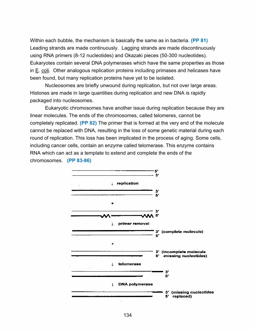
Within each bubble, the mechanism is basically the same as in bacteria. (PP 81)
Leading strands are made continuously. Lagging strands are made discontinuously
using RNA primers (8-12 nucleotides) and Okazaki pieces (50-300 nucleotides).
Eukaryotes contain several DNA polymerases which have the same properties as those
in E. coli. Other analogous replication proteins including primases and helicases have
been found, but many replication proteins have yet to be isolated.
Nucleosomes are briefly unwound during replication, but not over large areas.
Histones are made in large quantities during replication and new DNA is rapidly
packaged into nucleosomes.
Eukaryotic chromosomes have another issue during replication because they are
linear molecules. The ends of the chromosomes, called telomeres, cannot be
completely replicated. (PP 82) The primer that is formed at the very end of the molecule
cannot be replaced with DNA, resulting in the loss of some genetic material during each
round of replication. This loss has been implicated in the process of aging. Some cells,
including cancer cells, contain an enzyme called telomerase. This enzyme contains
RNA which can act as a template to extend and complete the ends of the
chromosomes. (PP 83-86)
134

Chapter 12: Transcription
I. Introduction
DNA structure and DNA replication explain the storage and transmission of
genetic information. However, for a cell to utilize its own genetic information, not only
the DNA but RNA is necessary. The overall scheme is as follows: DNA contains genes,
each of which is a particular segment of the DNA with a specific base sequence. (There
is nothing chemically distinctive about one gene as compared to another except the
base sequence.) Every gene contains the information for making a particular protein (a
polypeptide chain), and the information is held in the form of the base sequence of the
gene. The cell’s genes code for all the cell’s proteins, both structural and enzymatic.
The presence of these proteins determines what the cell looks like and what reactions
can occur. The DNA itself, though, does not direct synthesis of the proteins. An
intermediate molecule, called messenger RNA, is a go-between for the DNA and the
protein. Thus the flow of information is from DNA to mRNA, and then to protein. This is
called the central dogma. The process of making mRNA is called transcription. The
process of making a protein from mRNA is called translation. (PP 2-3)
transcription translation
DNA mRNA protein
II. Messenger RNA
The existence of mRNA was first suspected because DNA stays in the nucleus of
a cell while proteins are made in the cytoplasm. RNA was a logical candidate for
carrying the information since the amount of RNA in a cell increases when protein
synthesis increases. mRNAs are single-stranded molecules with the usual structure of a
nucleic acid, containing ribonucleotides joined by phosphodiester bonds and the bases
A, C, G, U. (PP 4) mRNA molecules vary in length from several hundred to over 10,000
nucleotides. A cell will normally have thousands of different mRNA types, each
corresponding to a particular gene and used to make a particular protein.
mRNA comes in two basic types. If the mRNA molecule contains the information
for making one polypeptide chain, it is called monocistronic. If it codes for two or more
polypeptide chains, it is said to be polycistronic. (PP 5) Eukaryotic mRNAs are usually
monocistronic while prokaryotic mRNAs are usually polycistronic. In addition, mRNA
molecules contain regions that do not carry information for making the protein, called
non-coding regions. The 5'-end of a mRNA molecule, called the leader sequence, is
135
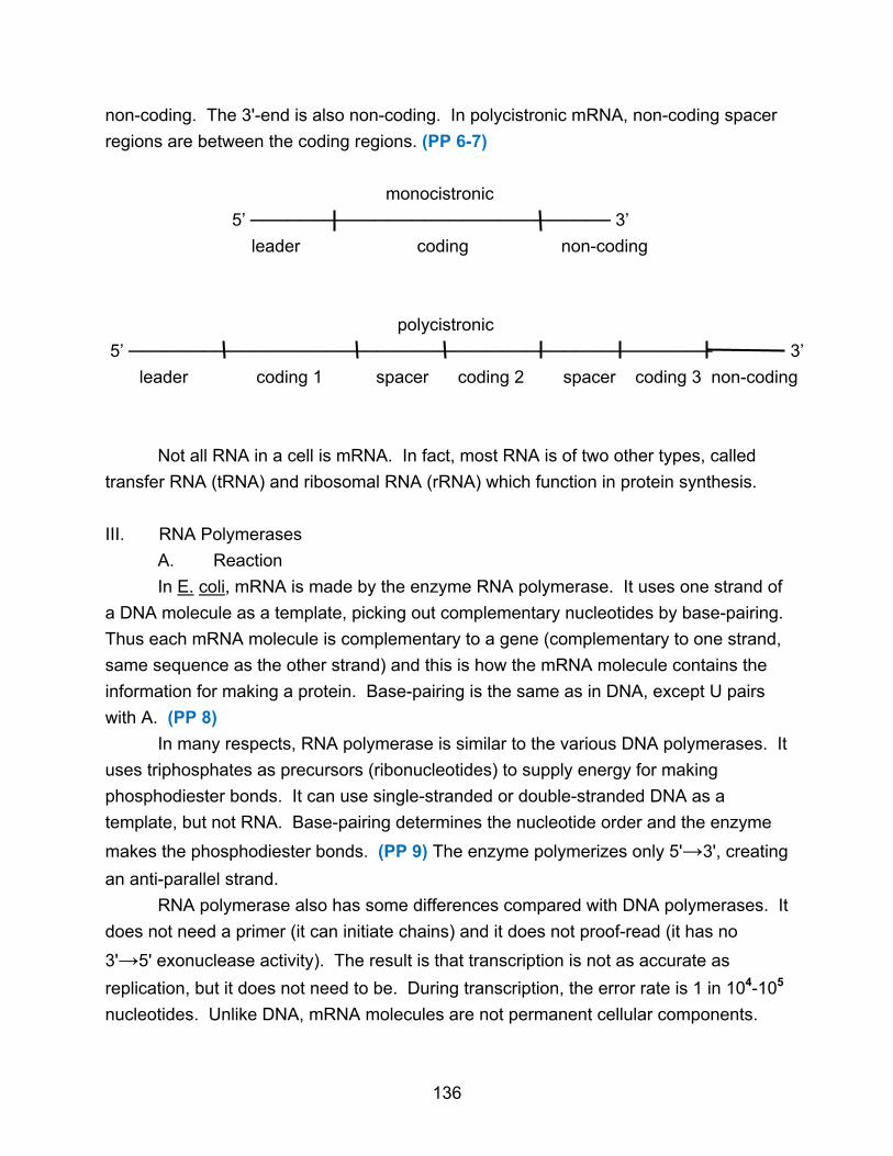
non-coding. The 3'-end is also non-coding. In polycistronic mRNA, non-coding spacer
regions are between the coding regions. (PP 6-7)
monocistronic
5’ ───────────────────────────── 3’
leader coding non-coding
polycistronic
5’ ─────────────────────────────────────────────── 3’
leader coding 1 spacer coding 2 spacer coding 3 non-coding
Not all RNA in a cell is mRNA. In fact, most RNA is of two other types, called
transfer RNA (tRNA) and ribosomal RNA (rRNA) which function in protein synthesis.
III. RNA Polymerases
A. Reaction
In E. coli, mRNA is made by the enzyme RNA polymerase. It uses one strand of
a DNA molecule as a template, picking out complementary nucleotides by base-pairing.
Thus each mRNA molecule is complementary to a gene (complementary to one strand,
same sequence as the other strand) and this is how the mRNA molecule contains the
information for making a protein. Base-pairing is the same as in DNA, except U pairs
with A. (PP 8)
In many respects, RNA polymerase is similar to the various DNA polymerases. It
uses triphosphates as precursors (ribonucleotides) to supply energy for making
phosphodiester bonds. It can use single-stranded or double-stranded DNA as a
template, but not RNA. Base-pairing determines the nucleotide order and the enzyme
makes the phosphodiester bonds. (PP 9) The enzyme polymerizes only 5'→3', creating
an anti-parallel strand.
RNA polymerase also has some differences compared with DNA polymerases. It
does not need a primer (it can initiate chains) and it does not proof-read (it has no
3'→5' exonuclease activity). The result is that transcription is not as accurate as
replication, but it does not need to be. During transcription, the error rate is 1 in 104-105
nucleotides. Unlike DNA, mRNA molecules are not permanent cellular components.
136

mRNA molecules are routinely broken down after a few minutes (bacteria), hours, or
days (eukaryotes). New mRNA molecules are then made. Thus an inaccurate mRNA
may result in making some non-functional protein molecules, but the damage will
probably not be serious or permanent. In contrast, mistakes in DNA persist and are
passed to subsequent generations.
RNA polymerase unwinds the double-helix itself, keeping a small area of the
DNA denatured (about 17 base-pairs) so the RNA can be made. (PP 10) As in
replication, unwinding the helix causes supercoils to build up in other places which must
be adjusted by topoisomerases.
Generally the RNA polymerase copies only one strand of the DNA in a given
area of the chromosome. The template strand (also called the minus (-) strand, the
non-coding strand, or the antisense strand), is copied. The other strand is called the
non-template strand, sense strand, plus (+) strand, or coding strand. (PP 11) A strand
may be (+) in one area of the chromosome and (-) in another. In some viruses, both
strands in a given area contain information and are transcribed.
B. Structure
In E. coli, RNA polymerase holoenzyme has a molecular weight of 450,000 and
contains six subunits in a α2ββ’ωσ configuration. (PP 12) The σ subunit (of which there
are several different types) binds loosely and is required only for starting transcription.
It recognizes the beginning of genes where transcription should start. There is no point
starting transcription in the middle of a gene since the mRNA will not have all the
information for making a protein, so the σ subunit ensures that transcription begins only
at the start of a gene. The core enzyme (α2ββ’ω) carries out the other functions of the
enzyme. The β subunit binds NTPs. The β’ subunit binds the enzyme to the DNA. Both
β and β’ subunits contribute to the active site. The α subunits help to assemble the
enzyme and interact with some regulatory proteins. (PP 13-14)
Whereas E. coli has only one RNA polymerase for making all mRNA, rRNA, and
tRNA, eukaryotic cells have three kinds of RNA polymerase. RNA polymerase I makes
rRNA, RNA polymerase II makes mRNA, and RNA polymerase III makes mainly tRNAs.
IV. Mechanism of Transcription
A. Initiation
Just prior to the gene in the DNA is an area of 50-100 nucleotides called the
promoter. The promoter indicates the start of a gene but is not actually transcribed, and
lies upstream relative to the start site of transcription. The start of transcription is
designated as the +1 nucleotide, and the transcribed nucleotides lying downstream are
137

given increasing positive numbers. Nucleotides in the promoter are given negative
numbers. It is here that RNA polymerase holoenzyme including the σ70 subunit (with a
molecular weight of 70,000) recognizes specific sequences indicating the start of a
gene, so RNA polymerase will bind and start transcribing in the correct position. Not all
promoters have the same base sequence. However, two areas in most E. coli
promoters are very similar. Centered at about 10 base-pairs before the start of the
gene (-10 base-pairs) is a region called the Pribnow box. (PP 15) At about -35
nucleotides is another area called the -35 sequence. RNA polymerase (with the σ70
subunit bound) recognizes and binds to these areas. (PP 16)
+1
sense ↓
───────── TTGACA ──────────────────TATAAT ──────────── A
───────── AACTGT ──────────────────ATATTA ──────────── T
antisense
-35 16-18 nucleotides -10 6-7 nucleotides
The sequences given are consensus sequences, but variations occur from promoter to
promoter. Variations probably account for different rates of transcription for different
genes. Some sequences interact better with RNA polymerase so transcription occurs
frequently while other sequences bind RNA polymerase less strongly. The difference
can be several orders of magnitude in how often transcription is initiated. This is one
mechanism for controlling gene expression. Some specialized E. coli genes have
different promoters recognized by other types of σ subunits. (PP 17)
It appears that RNA polymerase binds to the -35 sequence and the -10 sequence.
The enzyme then unwinds 12-17 base-pairs of DNA around the start site of transcription.
The A-T rich nature of this region and the σ subunit aid in unwinding. (PP 18-20)
B. Elongation
RNA polymerase then begins to make the RNA. The RNA transcript almost
always begins with an A or a G. The RNA polymerase moves along the DNA,
maintaining a small area of denatured bases. After a few nucleotides (6-10) are
polymerized, the σ subunit drops off, and the rest of the enzyme (core enzyme)
completes transcription. (PP 21) The rate of polymerization is 20-50 nucleotide/sec and
varies with the G-C content of the DNA region. Once polymerization of a nucleotide
has occurred, it rapidly dissociates from the DNA since the DNA double-helix is more
stable and tends to reform. Only about 12 base-pairs of DNA-RNA hybrid exist at any
time. (PP 22)
138
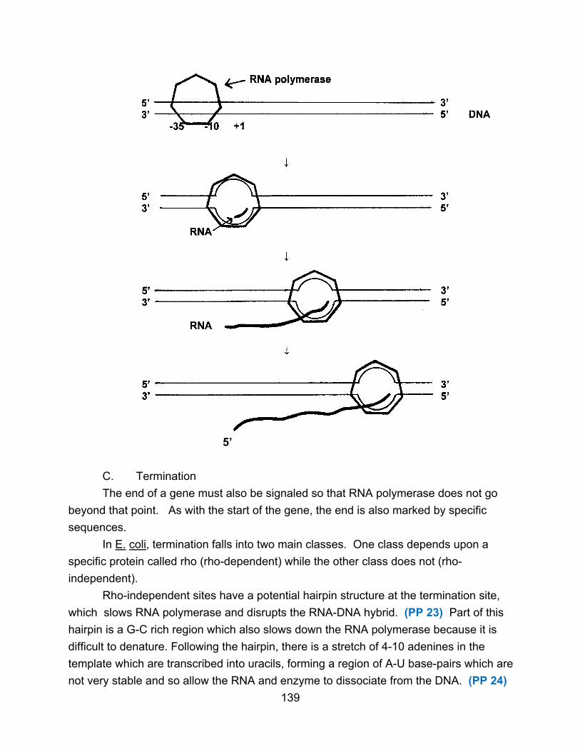
5’
C. Termination
The end of a gene must also be signaled so that RNA polymerase does not go
beyond that point. As with the start of the gene, the end is also marked by specific
sequences.
In E. coli, termination falls into two main classes. One class depends upon a
specific protein called rho (rho-dependent) while the other class does not (rho-
independent).
Rho-independent sites have a potential hairpin structure at the termination site,
which slows RNA polymerase and disrupts the RNA-DNA hybrid. (PP 23) Part of this
hairpin is a G-C rich region which also slows down the RNA polymerase because it is
difficult to denature. Following the hairpin, there is a stretch of 4-10 adenines in the
template which are transcribed into uracils, forming a region of A-U base-pairs which are
not very stable and so allow the RNA and enzyme to dissociate from the DNA. (PP 24)
139
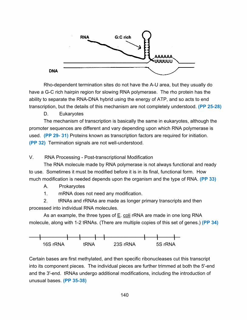
Rho-dependent termination sites do not have the A-U area, but they usually do
have a G-C rich hairpin region for slowing RNA polymerase. The rho protein has the
ability to separate the RNA-DNA hybrid using the energy of ATP, and so acts to end
transcription, but the details of this mechanism are not completely understood. (PP 25-28)
D. Eukaryotes
The mechanism of transcription is basically the same in eukaryotes, although the
promoter sequences are different and vary depending upon which RNA polymerase is
used. (PP 29- 31) Proteins known as transcription factors are required for initiation.
(PP 32) Termination signals are not well-understood.
V. RNA Processing - Post-transcriptional Modification
The RNA molecule made by RNA polymerase is not always functional and ready
to use. Sometimes it must be modified before it is in its final, functional form. How
much modification is needed depends upon the organism and the type of RNA. (PP 33)
A. Prokaryotes
1. mRNA does not need any modification.
2. tRNAs and rRNAs are made as longer primary transcripts and then
processed into individual RNA molecules.
As an example, the three types of E. coli rRNA are made in one long RNA
molecule, along with 1-2 tRNAs. (There are multiple copies of this set of genes.) (PP 34)
────────────────────────────────────────────
16S rRNA tRNA 23S rRNA 5S rRNA
Certain bases are first methylated, and then specific ribonucleases cut this transcript
into its component pieces. The individual pieces are further trimmed at both the 5'-end
and the 3'-end. tRNAs undergo additional modifications, including the introduction of
unusual bases. (PP 35-38)
140

B. Eukaryotes
1. tRNAs and rRNAs are processed as they are in prokaryotes.
2. Unlike prokaryotes, mRNA in eukaryotes is heavily modified. The RNA as
it is made in the nucleus is called heterogeneous nuclear RNA (hnRNA) or pre-mRNA.
Three things happen to this before it becomes mature mRNA.
a. 5'-cap
The 5'-end of RNA is modified by a structure known as the 5'-cap.
A 7-methyl guanosine is joined to the 5'-end via a 5'-5' triphosphate bond. This occurs
shortly after synthesis of the RNA begins. (PP 39)
3’mG5’ppp5’NpNpNp
5’ RNA 3’
The guanosine is first added using GTP, then methylated, and sometimes the first and
second nucleotides are also methylated. The cap binds to a specific protein and
participates in binding mRNA to the ribosome (which is the site of protein synthesis). In
addition, it may help to prevent premature degradation of the mRNA from the 5'-end.
b. 3'-poly(A) tail
Following transcription, a series of 80-250 adenines are added onto the
3'-end of the RNA. The adenines are added by the enzyme polyadenylate polymerase,
also called poly(A) polymerase. First the 3'-end is cleaved close to a specific sequence
(5'-AAUAAA-3'). The enzyme then adds the poly(A) tail using ATP but requiring no
template. (PP 40) The poly(A) tail binds a specific protein which protects the mRNA
from enzymatic degradation and also binds to the ribosome during protein synthesis.
The poly(A) tail gets shorter as the mRNA gets older.
c. Removal of introns - RNA splicing
Eukaryotic pre-mRNA, though monocistronic, differs from prokaryotic
mRNA in that it contains introns. Introns are non-coding regions that must be removed
before the mRNA is functional. The coding regions of the gene (exons) are interrupted
by introns. Introns occur within a gene, which distinguishes them from non-coding
spacers between genes. (PP 41)
141
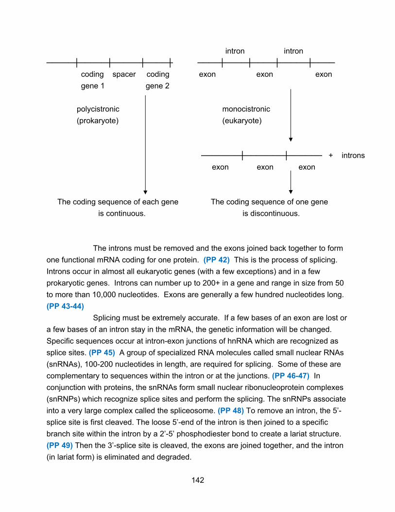
intron intron
─────┼─────┼─────┼────┼ ────┼────┼────┼─────┼────
coding spacer coding exon exon exon
gene 1 gene 2
polycistronic monocistronic
(prokaryote) (eukaryote)
───────┼───────┼────── + introns
exon exon exon
The coding sequence of each gene The coding sequence of one gene
is continuous. is discontinuous.
The introns must be removed and the exons joined back together to form
one functional mRNA coding for one protein. (PP 42) This is the process of splicing.
Introns occur in almost all eukaryotic genes (with a few exceptions) and in a few
prokaryotic genes. Introns can number up to 200+ in a gene and range in size from 50
to more than 10,000 nucleotides. Exons are generally a few hundred nucleotides long.
(PP 43-44)
Splicing must be extremely accurate. If a few bases of an exon are lost or
a few bases of an intron stay in the mRNA, the genetic information will be changed.
Specific sequences occur at intron-exon junctions of hnRNA which are recognized as
splice sites. (PP 45) A group of specialized RNA molecules called small nuclear RNAs
(snRNAs), 100-200 nucleotides in length, are required for splicing. Some of these are
complementary to sequences within the intron or at the junctions. (PP 46-47) In
conjunction with proteins, the snRNAs form small nuclear ribonucleoprotein complexes
(snRNPs) which recognize splice sites and perform the splicing. The snRNPs associate
into a very large complex called the spliceosome. (PP 48) To remove an intron, the 5’-
splice site is first cleaved. The loose 5’-end of the intron is then joined to a specific
branch site within the intron by a 2’-5’ phosphodiester bond to create a lariat structure.
(PP 49) Then the 3’-splice site is cleaved, the exons are joined together, and the intron
(in lariat form) is eliminated and degraded.
142

There are several different mechanisms for removing introns, depending
on the type and location of the RNA (mRNA, rRNA, tRNA, mitochondria, chloroplast,
etc.) Some introns are self-splicing. No proteins are required and the RNA itself acts as
an enzyme (ribozyme). (PP 50-51)
The function of introns is not understood. It may be that they allow for
recombination of segments of proteins that results in new proteins and protein evolution.
Alternatively, they may help in regulating the use of genes. Some eukaryotic primary
transcripts can be processed in two or more different ways, allowing for production of
two or more different proteins. (PP 52) Such transcripts can have two cleavage sites for
poly(A) addition, or have alternate splicing patterns for intron removal. (PP 53-54)
d. Transport
All the modifications that convert pre-mRNA into mature mRNA occur in
the nucleus. Once completed, mature mRNA must move out of the nucleus to the
cytoplasm. Pre-mRNA must not leave the nucleus. (PP 55)
143

Chapter 13: Protein Synthesis
I. Introduction
As a result of transcription, an mRNA molecule has been synthesized and is now
ready for protein synthesis (translation). The mRNA has a base sequence that contains
the same information as a gene sequence in the DNA. This sequence contains the
information for making a specific polypeptide chain. The process for making a protein
requires not only the mRNA and amino acids, but many other components as well.
II. Genetic Code
A. Breaking the Code
Since the major difference between mRNA molecules is the base sequence, the
sequence must somehow specify the order of the amino acids used to create the
corresponding protein. Since there are four bases and twenty amino acids, one base
cannot specify one amino acid. Likewise, two bases for each amino acid would not be
sufficient since 42 = 16 possible base combinations. The minimum number of bases
needed to specify an amino acid is three, since 43 = 64 possible combinations. This in
fact is the case. Groups of three bases, called triplets or codons, specify a particular
amino acid. The triplets are contiguous and do not overlap. (PP 2-3)
mRNA 5' ― G –U –A –G –C –C –U –A –C –G –G –A ― 3'
└────┘ └────┘ └────┘ └────┘
Val ― Ala ― Tyr ― Gly
Which codon sequence specifies which amino acid (the genetic code) was
determined in several ways. First, protein synthesis was performed using artificial
mRNAs. For instance, poly(U) produced polyphenylalanine, showing UUU codes for
Phe. Other mRNAs were made from different proportions of A and C. The expected
frequency of each codon, AAA, AAC, ACC, etc., could be calculated and compared to
the observed results. mRNAs with two alternating codons, like ACACACACACAC,
produced a protein with two alternating amino acids. Second, trinucleotides could be
used with the protein synthesis machinery to see which amino acid would also bind.
Combining the results of all these experiments, the genetic code was broken. (PP 4-5)
B. Characteristics of the Code
Several important features of the code became apparent.
144

1. It is of crucial importance that ‘reading’ the mRNA during protein synthesis
begins at the right point. If the reading frame of the triplets is displaced by even one
base, the codons will be different and the protein will have totally different amino acids.
(PP 6)
mRNA 5' ― G –U –A –G –C –C –U –A –C –G –G –A ― 3'
└────┘ └────┘ └────┘ └────┘
└────┘ └────┘ └────┘└────┘
The starting point of translation, after the leader sequence, is an AUG codon, which is
found by the protein synthesizing machinery at the beginning of translation.
2. Three of the 64 codons do not code for any amino acid. UAA, UAG, and
UGA are called termination or stop codons and signal the end of a protein.
3. The genetic code is degenerate, meaning that with 20 amino acids and 61
codons, most amino acids have more than one codon. (PP 7) Leucine has six codons,
glycine has four, phenylalanine has two, and only methionine and tryptophan have one
each. For amino acids with multiple codons, it is usually the last base which varies
while the first two are the same.
Glycine codons: 5' GGU 5' GGA 5' GGC 5' GGG
4. The genetic code is almost universal, with the same codons specifying the
same amino acid in both prokaryotes and eukaryotes. A few exceptions have been
found, most notably in mitochondria, where some codons vary. (PP 8)
5. It is apparent that a mutation is simply a change in the base sequence of
the mRNA (and DNA). Silent mutations result in a fully active protein, either because
the changed codon specifies the same original amino acid or because the substituted
amino acid does not hurt the protein. More often, a base change results in a different
amino acid and a non-functional or impaired protein (missense mutation). (PP 9)
Mutations can also involve insertions and deletions of bases, which change the reading
frame of the mRNA and so change the whole protein. (PP 10) If a codon is changed to
a termination codon (nonsense mutation) the protein will end prematurely. The genetic
code uses 61 of 64 codons (instead of only 20) to reduce the chance of a nonsense
mutation. It is better to have three stop codons and 61 codons for amino acids
compared to 44 stop codons and 20 codons for amino acids. A protein is more likely to
be functional with one wrong amino acid than if it ends prematurely. (PP 11-12)
145

III. Components of Protein Synthesis
In addition to mRNA, two other types of RNA are needed, tRNA and rRNA. (PP 13)
Other components are needed as well. (PP 14)
A. Transfer RNA (tRNA)
1. Structure
tRNA molecules bring the right amino acid in to pair with the right codon. Since
the codon and amino acid cannot directly recognize each other, tRNA acts as an
adapter. (PP 15) A tRNA molecule will recognize a specific codon at one end of its
structure and bind to the right amino acid (for that codon) at the other end of its
structure. (PP 16) Thus there must be numerous different tRNA molecules, at least one
for each amino acid. tRNAs are small (73-94 nucleotides) with molecular weights of
23,000 - 30,000. All have C–C–A at the 3'-end and usually pG at the 5'-end. They can
form a cloverleaf type secondary structure with intrastrand hydrogen bonds. Eight or
more bases are modified into unusual bases. (PP 17-18)
Area 1 is the TψC loop, containing ribothymidine and pseudouridine. Area 2 is the extra
arm, of variable length in different tRNAs. Area 3 is the anticodon arm. The three
middle bases are complementary to a codon. The anticodon is thus capable of base-
pairing in an antiparallel manner to a specific codon. This is how the tRNA recognizes a
codon and binds to mRNA.
146

Thus the anticodon allows the right tRNA with the right amino acid attached to align with
a codon for that amino acid.
Area 4 is the DHU arm, containing dihydrouridine. This arm can vary somewhat
in size in different tRNAs. Area 5 is the amino acid arm. This is where the amino acid
becomes covalently attached to the 3'-end of the tRNA.
The actual structure is not a flat cloverleaf. Rather it is twisted into an L-shaped
tertiary structure. (PP 19)
This structure is held together by base-stacking and some unusual H-bonds. Since
these H-bonds are not part of a double helix, base-pairs such as G-U can occur.
(PP 20-21)
2. Reaction of tRNA
The tRNAs are joined to amino acids by a group of enzymes known as
aminoacyl-tRNA synthetases. There are 20 different enzymes, one specific for each
amino acid. The enzymes are very accurate, making sure to attach its amino acid to a
tRNA with the right anticodon for that amino acid. The overall reaction is (PP 22-23)
amino acid + tRNA + ATP → aminoacyl – tRNA + AMP + PPi
The energy for joining the amino acid to the tRNA comes from breaking down
ATP. The reaction occurs in two steps. First, ATP is broken to AMP + PPi while the
AMP and amino acid are joined and bound to the enzyme. (PP 24) Then the AMP is
removed while the amino acid is moved to the tRNA. (PP 25-26)
147

The amino acid is covalently attached to either the 2' or 3' OH group depending upon
the specific enzyme.
Some aminoacyl tRNA synthetases must accurately distinguish between very
similar amino acids, such as Val and Ile (Ile has one extra CH2 group). (PP 27) The
enzymes which must distinguish two amino acids increase accuracy by proofreading
their reaction, which they do by having two active sites. (PP 28) In the first active site,
the amino acid fits snugly and is converted into the aminoacyl-AMP. Thus the enzyme
for Val cannot use Ile because Ile is too big. However, the enzyme for Ile will
occasionally use Val since Val can bind (more loosely than Ile) to the first active site.
After formation of the aminoacyl-AMP, the enzyme then attempts to put the aminoacyl-
AMP into the second active site, which is smaller. The correct Ile-AMP will not fit into
the second site, so the reaction proceeds with the tRNA. The incorrect Val-AMP will fit,
148

and when it enters the second site, it is hydrolyzed and the enzyme must start over
again. Thus the enzyme checks the amino acid twice, once to eliminate amino acids
which are too large and once to eliminate amino acids that are too small (double-sieve
mechanism). In addition, some synthetases also check the final amino acid-tRNA and
hydrolyze it if it is wrong. The result of these mechanisms is that the wrong amino acid
is attached to a tRNA only one in 3000 times. The synthetases also check for the
correct tRNA molecule, recognizing certain nucleotides (2-12) within the right tRNA
molecule. (PP 29) The important nucleotides for recognition can occur in any part of the
tRNA, depending upon the specific molecule. The specific conformation of the tRNA
molecule is also important and is recognized. (PP 30-31)
3. Wobble Hypothesis
There are some numerical contradictions to be resolved, with 20 amino acids, 20
synthetase enzymes, and 61 codons. The first question is how many tRNAs are
required, and it turns out that a minimum of 31 tRNAs are needed. However, most cells
have more. Thus some amino acids have more than one tRNA. In such a case, the
appropriate synthetase enzyme will recognize all the tRNAs for that amino acid, so only
20 synthetases are needed. A second problem is how 31 tRNAs with 31 anticodons
can recognize 61 codons. It would seem that 61 tRNAs would be needed, but this is not
the case. (PP 32) Some tRNAs can recognize more than one codon (all for the same
amino acid) and the explanation is called the wobble hypothesis. This hypothesis
assumes that the first two bases in a codon form strong, normal base-pairs with the
anticodon, but the third base ‘wobbles’ and forms looser, more flexible H-bonds,
sometimes with several different bases. (PP 33)
149

For instance, it has been found that if the first anticodon base is U, it can pair with either
A or G. The following pairings are possible. (PP 34-35)
1st anticodon base 3rd codon base
U A or G
G C or U
I(inosine) A, U, or C
C G
A U
With the wobble, 31 tRNAs can recognize 61 codons. This also explains why two
codons for the same amino acid usually differ in the 3rd base, allowing one tRNA to
recognize both codons. Codons differing in the first two bases require different tRNA
molecules. The reason for this complexity involves the mechanism of protein synthesis.
tRNAs must bind specifically to codons in mRNA, which is accomplished by two strong
base-pairs and one weak one. The codon-anticodon interaction must be strong enough
to be accurate, but it cannot be too strong either. Codons and anticodons must also
dissociate during protein synthesis as well, and if this is too difficult, it will slow down
protein synthesis. Three normal base-pairs would be too strong. Two normal base-
pairs and one ‘wobble’ base-pair are strong enough for accuracy but weak enough to
permit rapid dissociation as well. (PP 36-37)
B. Ribosomes
Ribosomes are the sites of protein synthesis. An E. coli cell contains 15,000
ribosomes, each 65% RNA and 35% protein with a molecular weight = 2.5 x 10⁶. These
RNA-protein complexes align tRNAs with codons on the mRNA and make peptide
bonds between amino acids. (PP 38)
The size of the ribosome is designated by its sedimentation coefficient, 70S. The
ribosome consists of two unequal subunits, 50S and 30S. (PP 39-40)
70S
30S 50S
contains 21 different proteins contains 33 different proteins
-one copy of each protein (S1-S21) -multiple copies of one protein (L1-L36)
one 16S rRNA one 5S rRNA, one 23S rRNA
150

The proteins have molecular weights from 6000 to 75,000. (PP 41) Although purified,
specific functions are largely unknown. The three rRNAs (5S = 120 nuc., 16S = 1542
nuc., 23S = 2904 nuc.) are all capable of extensive intrastrand hydrogen bonding and
can assume shapes like complicated tRNA molecules. (PP 42)
5S rRNA
It is known that part of the 16S rRNA is complementary to the 5'-end of mRNA and
binds the mRNA. The 23S rRNA has the catalytic ability to make peptide bonds.
Eukaryotic ribosomes are larger (80S) with more proteins and rRNA.
80S
40S 60S
~33 proteins ~ 49 proteins
18S rRNA 5S, 5.8S, 28S rRNAs
Ribosomes self-assemble. (PP 43-44)
IV. Mechanism of Protein Synthesis
Protein synthesis is a very complicated process, using up to 90% of the chemical
energy for biosynthesis and requiring ~100 different proteins and ~40 different RNA
molecules.
A. General Features
A typical protein is made in about ten seconds. The error rate is about 1 in 104
amino acids, sufficient because proteins are routinely broken down and replaced. The
mRNA is read 5'→3'. The protein is made one amino acid at a time, starting at the N-
terminal and finishing at the C-terminal. (PP 45)
151
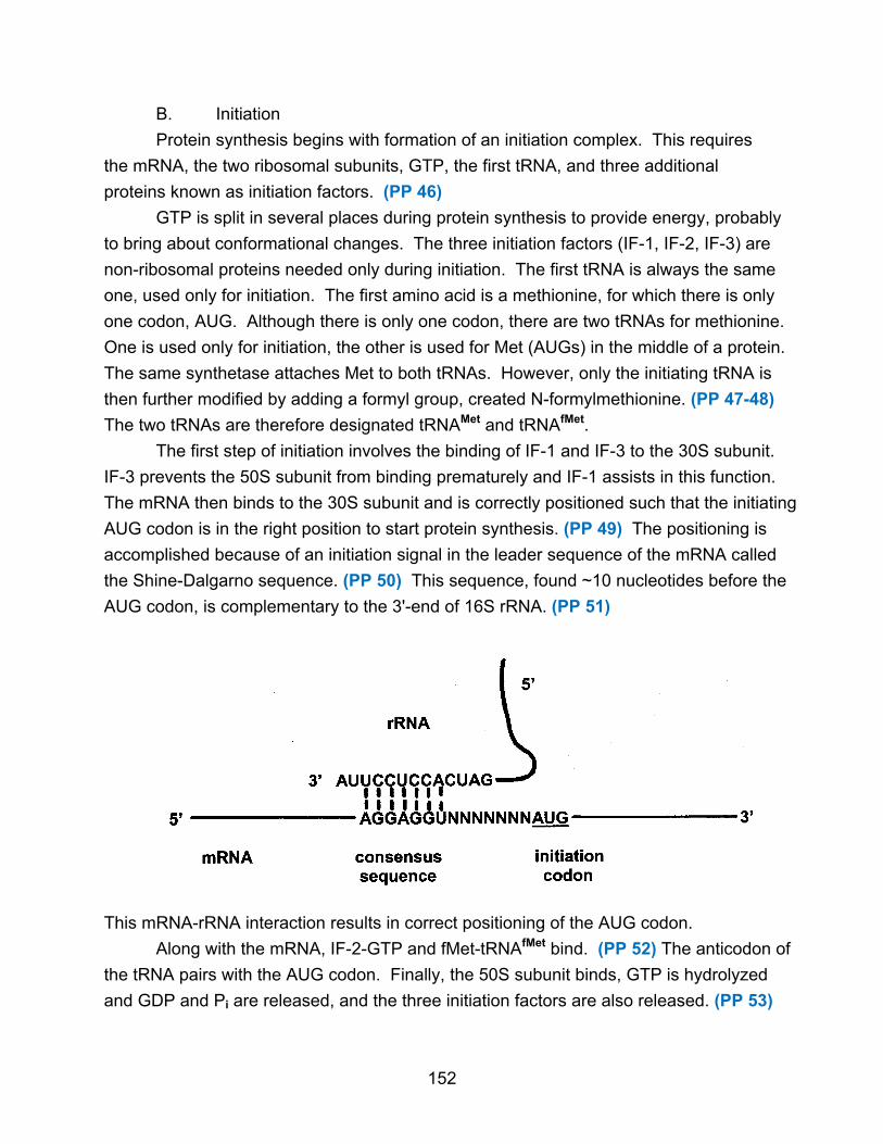
B. Initiation
Protein synthesis begins with formation of an initiation complex. This requires
the mRNA, the two ribosomal subunits, GTP, the first tRNA, and three additional
proteins known as initiation factors. (PP 46)
GTP is split in several places during protein synthesis to provide energy, probably
to bring about conformational changes. The three initiation factors (IF-1, IF-2, IF-3) are
non-ribosomal proteins needed only during initiation. The first tRNA is always the same
one, used only for initiation. The first amino acid is a methionine, for which there is only
one codon, AUG. Although there is only one codon, there are two tRNAs for methionine.
One is used only for initiation, the other is used for Met (AUGs) in the middle of a protein.
The same synthetase attaches Met to both tRNAs. However, only the initiating tRNA is
then further modified by adding a formyl group, created N-formylmethionine. (PP 47-48)
The two tRNAs are therefore designated tRNAMet and tRNAfMet.
The first step of initiation involves the binding of IF-1 and IF-3 to the 30S subunit.
IF-3 prevents the 50S subunit from binding prematurely and IF-1 assists in this function.
The mRNA then binds to the 30S subunit and is correctly positioned such that the initiating
AUG codon is in the right position to start protein synthesis. (PP 49) The positioning is
accomplished because of an initiation signal in the leader sequence of the mRNA called
the Shine-Dalgarno sequence. (PP 50) This sequence, found ~10 nucleotides before the
AUG codon, is complementary to the 3'-end of 16S rRNA. (PP 51)
This mRNA-rRNA interaction results in correct positioning of the AUG codon.
Along with the mRNA, IF-2-GTP and fMet-tRNAfMet bind. (PP 52) The anticodon of
the tRNA pairs with the AUG codon. Finally, the 50S subunit binds, GTP is hydrolyzed
and GDP and Pi are released, and the three initiation factors are also released. (PP 53)
152

The resulting initiation complex has two important sites on the ribosome called
the P site (peptidyl) and A site (aminoacyl) which can bind tRNAs. The initiator tRNA is
in the P site, the only one it can bind to. All other tRNAs can bind only to the A site
when they first interact with the ribosome. A third site for binding tRNAs, called the E
site (exit site) also forms. (PP 54-55)
Initiation Complex
In eukaryotes, the process is basically the same. The initiating tRNA, though it is
used only for initiation, binds Met, not fMet. (PP 56) At least a dozen initiation factors
are known. (PP 57) There is no Shine-Dalgarno sequence, but the 5'-cap on the mRNA
is essential.
C. Elongation
Elongation is a series of repetitive steps resulting in adding an amino acid via a
peptide bond. Besides the initiation complex, it requires the second tRNA, GTP and
three non-ribosomal proteins called elongation factors, EF-Tu, EF-Ts, and EF-G.
(PP 58)
153

The second tRNA binds to EF-Tu containing GTP. (PP 59) It then binds to the A
site and the GTP is hydrolyzed, releasing Pi and EF-Tu-GDP. The EF-Tu-GDP complex
is regenerated by EF-Ts, which allows exchange of the GDP for a GTP. Using EF-Tu
and hydrolyzing GTP allows the tRNA complex to stay on the ribosome for a few
milliseconds. If the tRNA is wrong, the codon-anticodon binding will dissociate before
the EF-Tu reactions are complete, preventing a wrong amino acid from being joined.
This is a form of proof-reading.
154

Now, a peptide bond is formed between the first and second amino acids. The
dipeptide remains attached to the second tRNA while the first tRNA now has no amino
acid. (PP 60-61)
The reaction is apparently catalyzed by the 23S rRNA, an example of an RNA enzyme.
The activity is called peptidyl transferase.
155

The next stage is called translocation, where the ribosome moves along the
mRNA by one codon. (PP 62) This movement puts the second tRNA (with attached
dipeptide) into the P site and ejects the first tRNA from the P site. The first tRNA
probably goes into the third ribosomal binding site, the E site (exit), before leaving the
ribosome. The third mRNA codon is now in the A site, while the second codon is in the
P site. This step requires EF-G. Energy for a change in conformation is supplied by
GTP hydrolysis
The elongation cycle now repeats, with the third tRNA coming into the A site. For each
amino acid added, two GTPs are used. The ribosome moves toward the 3'-end of the
mRNA, one codon at a time.
In eukaryotes the process is basically the same. Three analogous EFs are
known.
D. Termination
Elongation continues until the ribosome adds the last amino acid and encounters
a termination codon (UAA, UAG, UGA) immediately after the last amino acid codon.
Termination requires three termination or release factors. These proteins are called
RF1, RF2, and RF3. RF1 recognizes UAG and UAA, while RF2 recognizes UGA and
UAA. These bind at a termination codon and induce the peptidyl transferase to
hydrolyze the protein chain from the last tRNA. The function of RF3 is to promote
binding of RF1 and RF2, using GTP. Release of the protein causes the last tRNA to
drop off, and the ribosome dissociates into subunits. (PP 63-66)
In eukaryotes, a single releasing factor recognizes all three termination codons.
156

E. Polysomes
In both prokaryotes and eukaryotes, multiple ribosomes can attach to the same
mRNA, forming what are called polysomes. A ribosome covers about 80 nucleotides,
so many can be interspersed along a mRNA. (PP 67-68)
In bacteria, transcription and translation are tightly coupled. Translation begins at
the 5'-end of the mRNA before the 3'-end is even completed. (PP 69-70) This cannot
happen in eukaryotes because RNA processing must first occur. In addition, eukaryotic
mRNA is made in the nucleus while translation occurs in the cytoplasm.
V. Post-Translational Modification
The synthesized protein must now fold and undergo various modifications before
it becomes functional. Folding, which can begin before the protein is fully made, often
occurs spontaneously. Modifications involve several types. (PP 71)
A. The amino terminal is usually modified, removing the fMet and sometimes
additional amino acids. Often the new N-terminal is acetylated.
B. The carboxyl terminal is often modified by removing some amino acids.
C. Modification of certain amino acids can occur. Ser, Thr, and Tyr can be
phosphorylated. Lys and Glu can be methylated. (PP 72)
D. Carbohydrates can be added to make glycoproteins.
E. Other prosthetic groups are added.
F. Proteolytic cleavage is sometimes necessary for activity, including
trimming a protein, cutting it into two chains, or eliminating some internal amino acids.
157
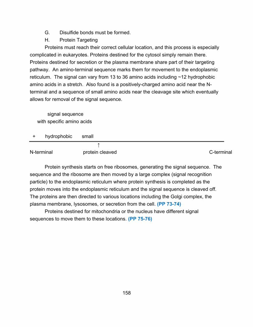
G. Disulfide bonds must be formed.
H. Protein Targeting
Proteins must reach their correct cellular location, and this process is especially
complicated in eukaryotes. Proteins destined for the cytosol simply remain there.
Proteins destined for secretion or the plasma membrane share part of their targeting
pathway. An amino-terminal sequence marks them for movement to the endoplasmic
reticulum. The signal can vary from 13 to 36 amino acids including ~12 hydrophobic
amino acids in a stretch. Also found is a positively-charged amino acid near the N-
terminal and a sequence of small amino acids near the cleavage site which eventually
allows for removal of the signal sequence.
signal sequence
with specific amino acids
+ hydrophobic small
↑
N-terminal protein cleaved C-terminal
Protein synthesis starts on free ribosomes, generating the signal sequence. The
sequence and the ribosome are then moved by a large complex (signal recognition
particle) to the endoplasmic reticulum where protein synthesis is completed as the
protein moves into the endoplasmic reticulum and the signal sequence is cleaved off.
The proteins are then directed to various locations including the Golgi complex, the
plasma membrane, lysosomes, or secretion from the cell. (PP 73-74)
Proteins destined for mitochondria or the nucleus have different signal
sequences to move them to these locations. (PP 75-76)
158