oxidative dna damage and augmentation of poly(adp-ribose ... · the international journal of...
TRANSCRIPT
B
A
(esniDbD(tscagt©
K
f
1
ARTICLE IN PRESS+ModelC-2429; No. of Pages 12
The International Journal of Biochemistry & Cell Biology xxx (2007) xxx–xxx
Oxidative DNA damage and augmentation of poly(ADP-ribose)polymerase/nuclear factor-kappa B signaling in patients
with Type 2 diabetes and microangiopathy
Antonysunil Adaikalakoteswari, Mohan Rema, Viswanathan Mohan,Muthuswamy Balasubramanyam ∗
Department of Cell and Molecular Biology, Madras Diabetes Research Foundation & Dr.Mohan’s DiabetesSpecialities Centre, 4 Conran Smith Road, Gopalapuram, Chennai 600086, India
Received 28 January 2007; received in revised form 12 April 2007; accepted 19 April 2007
bstract
Although oxidative stress and the subsequent DNA damage is one of the obligatory signals for poly(ADP-ribose) polymerasePARP) activation and nuclear factor-kappa B (NF�B) alterations, these molecular aspects have not been collectively examined inpidemiological and clinical settings. Therefore, this study attempts to assess the oxidative DNA damage and its downstream effectorignals in peripheral blood lymphocytes from Type 2 diabetes subjects without and with microangiopathy along with age-matchedon-diabetic subjects. The basal DNA damage, lipid peroxidation and protein carbonyl content were significantly (p < 0.05) highern patients with and without microangiopathy compared to control subjects. Formamido Pyrimidine Glycosylase (FPG)-sensitiveNA strand breaks which represents reliable indicator of oxidative DNA damage were also significantly (p < 0.001) higher in dia-etic patients with (19.41 ± 2.5) and without microangiopathy (16.53 ± 2.0) compared to control subjects (1.38 ± 0.85). OxidativeNA damage was significantly correlated to poor glycemic control. PARP mRNA expression and PARP activity were significantly
p < 0.05) increased in cells from diabetic patients with (0.31 ± 0.03 densitometry units; 0.22 ± 0.02 PARP units/mg protein, respec-ively) and without (0.35 ± 0.02; 0.42 ± 0.05) microangiopathy compared to control (0.19 ± 0.02; 0.11 ± 0.02) subjects. Diabeticubjects with and without microangiopathy exhibited a significantly (p < 0.05) higher (80%) NF�B binding activity compared to
ontrol subjects. In diabetic patients, FPG-sensitive DNA strand breaks correlated positively with PARP gene expression, PARPctivity and NF�B binding activity. This study provides a comprehensive molecular evidence for increased oxidative stress andenomic instability in Type 2 diabetic subjects even prior to vascular pathology and hence reveals a window of opportunity for earlyherapeutic intervention.2007 Elsevier Ltd. All rights reserved.
icroan
eywords: Oxidative DNA damage; Type 2 diabetes; NF�B; PARP; MPlease cite this article in press as: Adaikalakoteswari, A., et al., Oxipolymerase/nuclear factor-kappa B signaling in patients with TypBiochemistry and Cell Biology (2007), doi:10.1016/j.biocel.2007.0
∗ Corresponding author. Tel.: +91 44 28359048;ax: +91 44 28350935.
E-mail address: [email protected] (M. Balasubramanyam).
357-2725/$ – see front matter © 2007 Elsevier Ltd. All rights reserved.doi:10.1016/j.biocel.2007.04.013
giopathy
1. Introduction
A growing body of evidence suggests that hyper-
dative DNA damage and augmentation of poly(ADP-ribose)e 2 diabetes and microangiopathy, International Journal of4.013
glycemia associated with increased oxidative andnitrosative stress is linked to the pathogenesis ofdiabetic vascular complications (Evans, Goldfine,Maddux, & Grodsky, 2002). Although reactive oxy-
IN+Model
urnal of
ARTICLEBC-2429; No. of Pages 12
2 A. Adaikalakoteswari et al. / The International Jo
gen species (ROS) damage all macromolecules suchas lipids, proteins and nucleic acids, DNA is one ofthe most biologically significant targets of oxidativeattack. Increased superoxide generation accompaniedby increased nitric oxide generation, favors the for-mation of strong oxidant peroxynitrate (OONO−),which in turn activates nuclear transcription factor(NF�B) and damages DNA. Excessive DNA dam-age is an obligatory stimulus for the over-activationof the nuclear enzyme poly(ADP-ribose) polymerase(PARP), which in turn depletes intracellular NAD+,thus slowing the rate of glycolysis, electron transport,ATP formation and produces ADP ribosylation of manyproteins. These processes result in acute endothelialdysfunction in diabetic blood vessels contributing tothe development of diabetic complications (Ceriello,2003).
While there are lots of experimental data to sup-port various oxidative mechanisms, their relevance toclinical diabetes is still largely unexplored. The newconcept of oxidative stress, being an important trig-ger in the onset and progression of diabetes and itscomplications, is often challenged because interventionstudies with classic antioxidants, such as vitamin E,failed to demonstrate any convincing beneficial effectson cardiovascular outcomes. However, these studiespoint out several flaws in the trials among which muchemphasis is on the need for measurement of markersof oxidation to assess the degree of oxidative stress.Among various types of DNA base modifications, 8-hydroxy deoxyguanosine (8-OHdG) has been the mostwidely studied and is considered as a key biomarkerof oxidative DNA damage (Hinokio et al., 1999; Shinet al., 2001). Studies have also shown that activationof PARP and NF�B affects glucose utilization, tran-scriptional regulation and gene expression via multiplemechanisms, thus indicating PARP/NF�B interface asa novel drug target for therapeutic intervention (Hassa& Hottiger, 2002; Pacher, Obrosova, Mabley, & Szabo,2005). Since cellular systems under physiological con-dition are protected by an efficient DNA repairingmachinery, we hypothesize that (a) an increase in DNAdamage would be demonstrated in subjects with dia-betes and its vascular complications and (b) studyingdownstream effectors of DNA damage (PARP/NF�B)in a clinical setting could provide an additional linkbetween molecular intricacies of oxidative stress andthe pathogenesis of diabetes and/or its vascular com-
Please cite this article in press as: Adaikalakoteswari, A., et al., Oxipolymerase/nuclear factor-kappa B signaling in patients with TypBiochemistry and Cell Biology (2007), doi:10.1016/j.biocel.2007.0
plications. To test our hypothesis, we assessed theextent of oxidative damage and its relationship to PARPand NF�B modulation in diabetes and microangiopa-thy along with measurements of glycemic and lipid
PRESSBiochemistry & Cell Biology xxx (2007) xxx–xxx
status in clinically well-characterized groups of sub-jects.
2. Materials and methods
2.1. Sample selection
Study subjects were recruited from the Chen-nai Urban Rural Epidemiology Study (CURES), apopulation-based study in Chennai (formerly Madras) inSouthern India. The methodology of CURES has beenpublished elsewhere (Deepa, Pradeepa, & Rema, 2003;Rema, Mohan, Deepa, & Ravikumar, 2004). Details suchas age, sex and in diabetic subjects, duration of dia-betes and other details of diabetic therapy were recordedand clinical examination was done in all subjects. Sub-jects undiagnosed for diabetes underwent oral glucosetolerance tests (OGTT) using 75 g of oral glucose load(dissolved in 250 ml of water). Those who were con-firmed by OGTT to have 2 h plasma glucose value<7.8 mmol/l (140 mg/dl) were categorized as normalglucose tolerance (NGT) and those with 2 h plasma glu-cose value >7.8 mmol/l (140 mg/dl) and <11.1 mmol/l(200 mg/dl) were categorized as impaired glucose toler-ance (IGT). Diabetics were diagnosed as per the WHOcriteria. For the present study we randomly selected(using computer generated random numbers) 40 NGTsubjects and 40 diabetic subjects each with and withoutmicroangiopathy.
Microangiopathy was diagnosed if nephropathyand/or retinopathy were present. Nephropathy was diag-nosed if the patients had either persistent proteinuria(≥500 mg/day) or persistent microalbuminuria (if albu-minuria estimated by albumin creatinine ratio (ACR)exceeded 30 �g/mg of creatinine) in the absence ofurinary tract infection (Mohan et al., 2000; Varghese,Deepa, Rema, & Mohan, 2001). Retinopathy wasassessed using stereoscopic color retinal photographyas described earlier (Rema et al., 2004). Briefly, thepupils were dilated using one drop each of phenyle-pherine 10% and tropicamide 1% into both eyes andthe drops were repeated until the best possible mydri-asis was obtained. A trained photographer carried outfour-field color retinal photography with a Zeiss FF450 plus camera using 35 mm color transparencies.The photographs were graded against standard pho-tographs of the Early Treatment Diabetic RetinopathyStudy (ETDRS) grading system for severity of retinopa-
dative DNA damage and augmentation of poly(ADP-ribose)e 2 diabetes and microangiopathy, International Journal of4.013
thy. Hypertension was diagnosed if the subjects hadbeen treated with antihypertensive drugs or had systolicblood pressure (SBP) ≥ 140 mmHg or diastolic bloodpressure (DBP) ≥ 90 mmHg. Diabetic subjects without
IN+ModelB
rnal of
mo(mhmsst
2
aBmpwapswHkmsemmwhlRodaM
2
iuWtR
2
lc
ARTICLEC-2429; No. of Pages 12
A. Adaikalakoteswari et al. / The International Jou
icroangiopathy were selected on the basis of absencef retinopathy (on retinal photography) or nephropathy24 h protein excretion <100 mg/day and urinary albu-in levels <30 �g/mg creatinine). They also had no
istory of angina or myocardial infarction and had nor-al 12 lead resting ECGs and normal peripheral Doppler
tudies. Informed consent was obtained from all studyubjects and the institutional ethics committee approvedhe study.
.2. Clinical and biochemical characterization
Physical examination included height, weight, waistnd hip measurements using standardized techniques.lood pressure was recorded in the right arm with aercury sphygmomanometer (Diamond Deluxe Blood
ressure apparatus, Pune, India) while the patientsere seated. Two readings were taken 5 min apart
nd the mean of the two was taken as the bloodressure. A fasting blood sample was taken anderum separated and stored at −70 ◦C until the assaysere performed. Biochemical analyses were done onitachi-912 Autoanalyser (Hitachi, Germany) usingits supplied by Roche Diagnostics (Mannheim, Ger-any). Fasting plasma glucose (GOD–POD method),
erum cholesterol (CHOD–PAP method), serum triglyc-rides (GPO–PAP method) and HDL cholesterol (Directethod–polyethylene glycol-pretreated enzymes) wereeasured. Low-density lipoprotein (LDL) cholesterolas calculated using the Friedewald formula. Glycatedaemoglobin (HbA1C) was estimated by high-pressureiquid chromatography using the Variant machine (Bio-ad, Hercules, CA, USA). Informed consent wasbtained from all study subjects and the study was con-ucted in compliance with the Helsinki Declaration andpproved by the institutional ethics committee of theadras Diabetes Research Foundation.
.3. Assessment of intima-medial thickness (IMT)
Intima-media thickness (IMT) of the carotid arter-es was determined using a high resolution B modeltrasonography system (logic 400 GE, Milwaukee,I, USA) having an electrical linear higher frequency
ransducer (7.5 MHz) as described previously (Mohan,avikumar, ShanthiRani, & Deepa, 1998).
.4. Isolation of peripheral blood Lymphocytes
Please cite this article in press as: Adaikalakoteswari, A., et al., Oxipolymerase/nuclear factor-kappa B signaling in patients with TypBiochemistry and Cell Biology (2007), doi:10.1016/j.biocel.2007.0
Twelve milliliters of fasting peripheral blood was col-ected from each subject. Two milliliters was used toollect plasma and perform lipid and protein oxidation
PRESSBiochemistry & Cell Biology xxx (2007) xxx–xxx 3
assays. The rest was compartmentalized for lympho-cyte isolation and to perform Comet, PARP activity andNF�B–EMSA (5 ml) and PARP mRNA profiling (5 ml).Lymphocyte isolation was performed as per our previousprotocol (Balasubramanyam, Kimura, Aviv, & Gardner,1993). Briefly, freshly collected peripheral blood wasdiluted (1:1) with phosphate buffer saline (PBS) andcarefully layered on Histopaque-1077 (Sigma–Aldrich,India) gradient and centrifuged at 1600 rpm for 30 min.The buffy-coat interface representing peripheral bloodlymphocytes was aspirated, washed three times in PBS(pH 7.4) and used for various studies. FPG-sensitiveDNA damage assessment, PARP mRNA expression andactivity and NF�B binding assay were determined ina subset of randomly selected subjects (n = 20 in eachcategory).
2.5. Comet assay
DNA strand breaks and FPG-sensitive sites weredetected in peripheral blood lymphocytes by single cellgel electrophoresis, the comet assay (Singh, McCoy,Tice, & Schneider, 1988). Clear microscope slideswere pre-coated with 1% normal melting agarose. Foreach slide, 100 �l of cell suspension (approximately10,000 cells) was mixed with 200 �l of 0.5% low meltingpoint agarose, spotted as first layer onto the pre-coatedslide and covered with a coverslip. After agarose solid-ification the coverslip was gently removed; a secondlayer of 200 �l of normal melting agarose (NMA) wasadded over the first layer, covered with a coverslip andallowed to solidify. Cover slips were removed and slideswere placed in chilled lysis buffer (2.5 M NaCl, 100 mMEDTA, 10 mM Tris–HCl; pH 10, 1% Triton X-100 and10% DMSO added just before use) at 4 ◦C for 1 h. Afterlysis, the slides were placed on the platform in an elec-trophoresis tank that contains the pre-chilled (4 ◦C forat least 1 h) electrophoresis solution (300 mM NaOH,1 mM EDTA, pH 13). The buffer should just barelycover the slides and was incubated for 30 min at 4 ◦Cbefore beginning electrophoresis. The electrophoresiswas subsequently conducted at 25 V constant voltageand 300 mA for 30 min. Then slides were removed fromelectrophoresis apparatus and washed with three changesof neutralization buffer in staining jar for 5 min each at4 ◦C. Each slide was stained with 75 �l of ethidium bro-mide (20 �g/ml) and covered with a cover slip. The slideswere examined under a fluorescent microscope and ana-
dative DNA damage and augmentation of poly(ADP-ribose)e 2 diabetes and microangiopathy, International Journal of4.013
lyzed within 3–4 h. Slides were scored using an imageanalysis system (Comet Imager 1.2.13) attached to afluorescent microscope (Carl-Zeiss, Germany) equippedwith appropriate filter. The microscope was connected to
IN+Model
urnal of
PARP activity was measured by incorporation of biotiny-
ARTICLEBC-2429; No. of Pages 12
4 A. Adaikalakoteswari et al. / The International Jo
a computer through a charge coupled device (CCD) cam-era to transport images to software for analysis. The finalmagnification was 400×. Analysis of mean % DNA inthe tail, one of the reliable indicators of DNA damagewas done using image analysis software. Images from50 cells (25 from each duplicate slide) were analyzed.To show the reproducibility of our method, we mea-sured DNA damage in peripheral blood lymphocytesfrom eight subjects on two different occasions. For this,blood samples were taken twice from the same subjecton two different occasions; the respective samples wereused for the comet assay and checked for correlation(r = 0.87) and significance (p < 0.001).
2.6. Detection of FPG-sensitive sites (oxidativeDNA damage)
Additional DNA breaks formed at sites containing8-oxo-deoxyguanosine were detected by incubating theDNA with the enzyme Formamido Pyrimidine Glycosy-lase (FPG) (Collins, Duthie, & Dobson, 1993). After thelysis step in the comet assay, the slides with and withoutFPG treatment were washed three times (for 5 min each)in enzyme buffer (40 mM Hepes, 100 mM KCl, 0.5 mMEDTA, 0.2 mg/ml BSA; pH 8.0), covered with 100 �l ofeither buffer or FPG, sealed with a coverslip and incu-bated for 30 min at 37 ◦C. Slides were then continuedwith the electrophoresis till staining and scoring. FPG-sensitive sites were calculated by subtracting the percentDNA in tail obtained in the absence of enzyme from thepercent DNA in tail in the presence of enzyme.
2.7. RNA extraction
Total RNA from peripheral blood lympho-cytes (∼5 × 10⁶) was extracted using TRI reagent(Sigma–Aldrich, India). To check the integrity of thetotal RNA, 1 �g was fractionated on a 1% denaturingagarose gel. RNA concentration was quantified spec-trophotometrically and had a 280/260 OD ratio between1.8 and 2.0.
2.8. RT-PCR
Total RNA (1 �g) was reverse transcribed in a 25 �lreaction containing 5× reaction buffer, 0.2 �g ran-dom hexamer primers (Qiagen Inc., USA), 200 unitsmurine leukemia virus reverse transcriptase (GE Health-
Please cite this article in press as: Adaikalakoteswari, A., et al., Oxipolymerase/nuclear factor-kappa B signaling in patients with TypBiochemistry and Cell Biology (2007), doi:10.1016/j.biocel.2007.0
care, USA), 2.5 mm dNTPs and 50 units Ribonucleaseinhibitor in a Thermocycler (Biorad, USA) (55 min at37 ◦C, 5 min at 95 ◦C) (Dincer, Akcay, Alademir, &Ilkova, 2003).
PRESSBiochemistry & Cell Biology xxx (2007) xxx–xxx
The PARP mRNA expression PCR was probed usingspecific primers; their sequence is 5′–3′ AAGCCC-TAAAGGCTCAGAAC (nucleotide position 168–187)and TTGGGTGTCTGTGTCTTGAC (465–485). Theconditions of amplification were: 94 ◦C for 1 min, 60 ◦Cfor 1 min and 72 ◦C for 1 min for 24 cycles of amplifi-cation. The number of cycles was determined to assurethat the amplification occurs in the exponential phase.PCR products were separated by 2% agarose gel elec-trophoresis. The housekeeping GAPDH PCR productsobtained by amplifying primers were used as an internalcontrol.
2.9. Evaluation PARP gene product
The PARP gene expression was quantified using BIO-RAD gel documentation and semi-quantitative analysisusing its software. The ratio of PARP to GAPDH PCRproduct, expressed as peak density, was used as an indexof PARP gene expression (in densitometric units).
2.10. Preparation of nuclear extract
Nuclear extracts were prepared as described before(Hofmann et al., 1998). Proteinase inhibitors (leupeptin,pepstatin, aprotinin) were included in each solution atthe concentrations suggested by the manufacturer. Inbrief, 5 × 10⁵ cells were lysed in 400 �l ice-cold bufferA (10 mM Hepes (pH 8.0), 10 mM KCl, 100 �M EDTA,100 �M EGTA, 500 �M DTT, PI) and incubated on icefor 30 min. After incubation, 3%NP-40 is added andcentrifuged for 5 min at 13,000 rpm. The supernatantwas discarded and the nuclear pellet was resuspendedin 100 �l ice-cold buffer B (20 mM Hepes (pH 8.0),400 mM NaCl, 1 mM EDTA, 1 mM EGTA, 500 �MDTT, PI), incubated on ice for 2 h, vortexing every10–15 min and centrifuged for 5 min at 13,000 rpm. Thesupernatant containing nuclear proteins was immedi-ately quick-frozen at −80 ◦C. Protein concentration wasdetermined using a standard curve with bovine serumalbumin (BSA: 2–10 �g/ml) according to the Bradfordmethod.
2.11. Assay of PARP activity
For the assay, 100 �g of nuclear protein was used.
dative DNA damage and augmentation of poly(ADP-ribose)e 2 diabetes and microangiopathy, International Journal of4.013
lated poly(ADP-ribose) onto histone-coated proteins ina 96-well plate using PARP universal colorimetric assaykit (Biotech-India, India), and expressed as units ofPARP/mg protein.
IN+ModelB
rnal of
2
(rT5e7e3
pniEcpUcpc5bi7cmisg
2
oro2
2
pBa
2
uic
ARTICLEC-2429; No. of Pages 12
A. Adaikalakoteswari et al. / The International Jou
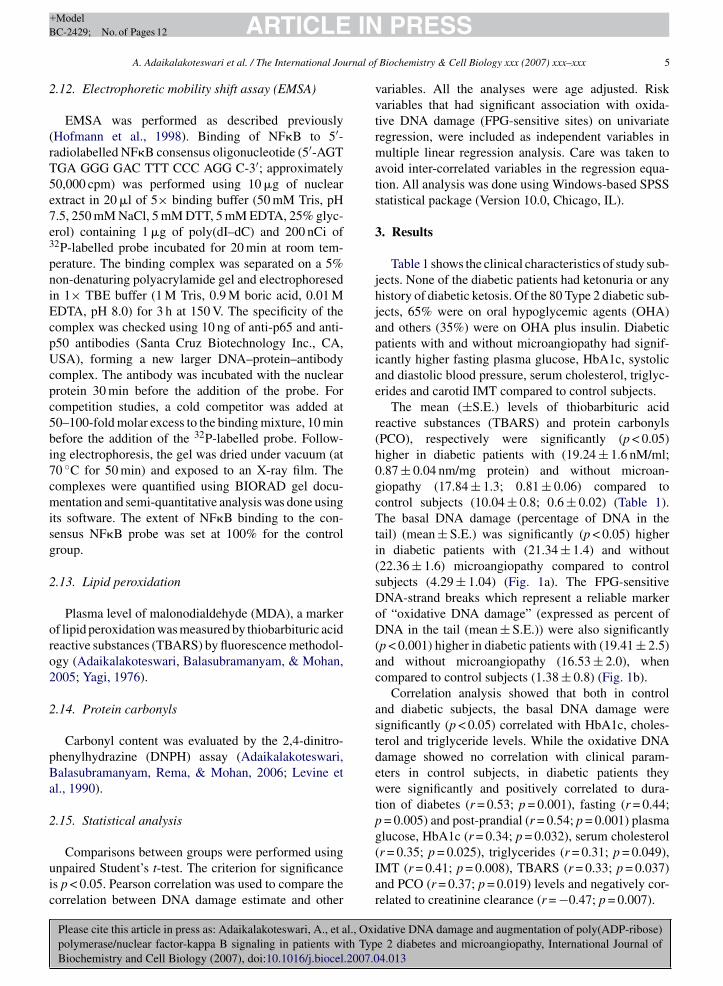
.12. Electrophoretic mobility shift assay (EMSA)
EMSA was performed as described previouslyHofmann et al., 1998). Binding of NF�B to 5′-adiolabelled NF�B consensus oligonucleotide (5′-AGTGA GGG GAC TTT CCC AGG C-3′; approximately0,000 cpm) was performed using 10 �g of nuclearxtract in 20 �l of 5× binding buffer (50 mM Tris, pH.5, 250 mM NaCl, 5 mM DTT, 5 mM EDTA, 25% glyc-rol) containing 1 �g of poly(dI–dC) and 200 nCi of2P-labelled probe incubated for 20 min at room tem-erature. The binding complex was separated on a 5%on-denaturing polyacrylamide gel and electrophoresedn 1× TBE buffer (1 M Tris, 0.9 M boric acid, 0.01 MDTA, pH 8.0) for 3 h at 150 V. The specificity of theomplex was checked using 10 ng of anti-p65 and anti-50 antibodies (Santa Cruz Biotechnology Inc., CA,SA), forming a new larger DNA–protein–antibody
omplex. The antibody was incubated with the nuclearrotein 30 min before the addition of the probe. Forompetition studies, a cold competitor was added at0–100-fold molar excess to the binding mixture, 10 minefore the addition of the 32P-labelled probe. Follow-ng electrophoresis, the gel was dried under vacuum (at0 ◦C for 50 min) and exposed to an X-ray film. Theomplexes were quantified using BIORAD gel docu-entation and semi-quantitative analysis was done using
ts software. The extent of NF�B binding to the con-ensus NF�B probe was set at 100% for the controlroup.
.13. Lipid peroxidation
Plasma level of malonodialdehyde (MDA), a markerf lipid peroxidation was measured by thiobarbituric acideactive substances (TBARS) by fluorescence methodol-gy (Adaikalakoteswari, Balasubramanyam, & Mohan,005; Yagi, 1976).
.14. Protein carbonyls
Carbonyl content was evaluated by the 2,4-dinitro-henylhydrazine (DNPH) assay (Adaikalakoteswari,alasubramanyam, Rema, & Mohan, 2006; Levine etl., 1990).
.15. Statistical analysis
Please cite this article in press as: Adaikalakoteswari, A., et al., Oxipolymerase/nuclear factor-kappa B signaling in patients with TypBiochemistry and Cell Biology (2007), doi:10.1016/j.biocel.2007.0
Comparisons between groups were performed usingnpaired Student’s t-test. The criterion for significances p < 0.05. Pearson correlation was used to compare theorrelation between DNA damage estimate and other
PRESSBiochemistry & Cell Biology xxx (2007) xxx–xxx 5
variables. All the analyses were age adjusted. Riskvariables that had significant association with oxida-tive DNA damage (FPG-sensitive sites) on univariateregression, were included as independent variables inmultiple linear regression analysis. Care was taken toavoid inter-correlated variables in the regression equa-tion. All analysis was done using Windows-based SPSSstatistical package (Version 10.0, Chicago, IL).
3. Results
Table 1 shows the clinical characteristics of study sub-jects. None of the diabetic patients had ketonuria or anyhistory of diabetic ketosis. Of the 80 Type 2 diabetic sub-jects, 65% were on oral hypoglycemic agents (OHA)and others (35%) were on OHA plus insulin. Diabeticpatients with and without microangiopathy had signif-icantly higher fasting plasma glucose, HbA1c, systolicand diastolic blood pressure, serum cholesterol, triglyc-erides and carotid IMT compared to control subjects.
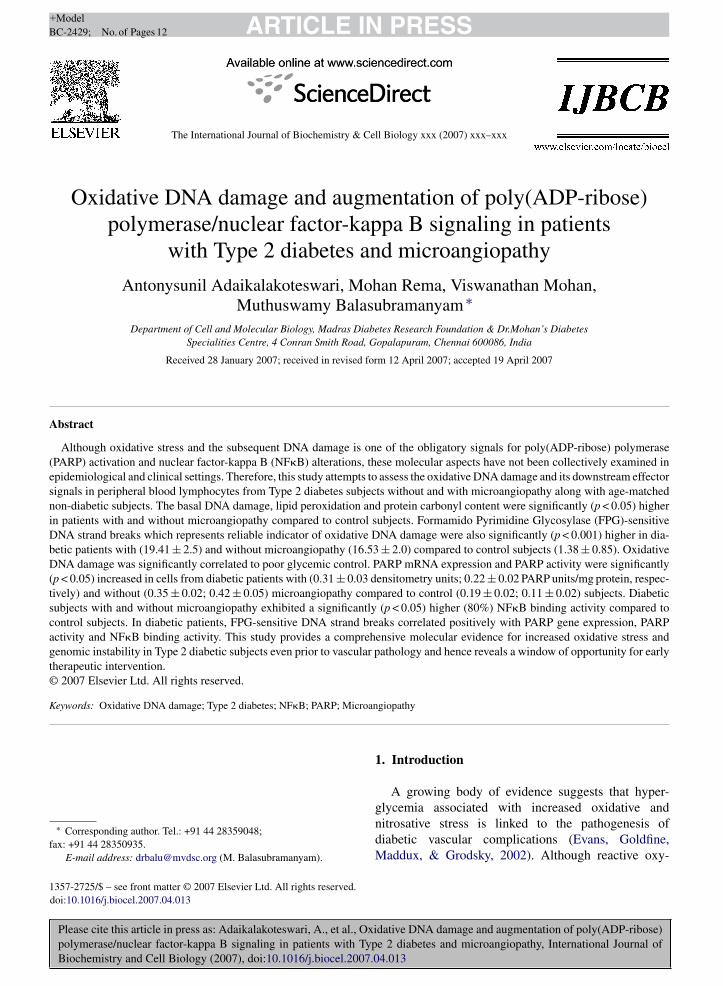
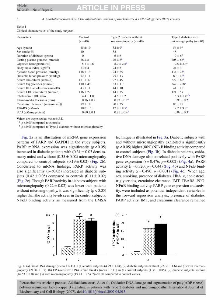
The mean (±S.E.) levels of thiobarbituric acidreactive substances (TBARS) and protein carbonyls(PCO), respectively were significantly (p < 0.05)higher in diabetic patients with (19.24 ± 1.6 nM/ml;0.87 ± 0.04 nm/mg protein) and without microan-giopathy (17.84 ± 1.3; 0.81 ± 0.06) compared tocontrol subjects (10.04 ± 0.8; 0.6 ± 0.02) (Table 1).The basal DNA damage (percentage of DNA in thetail) (mean ± S.E.) was significantly (p < 0.05) higherin diabetic patients with (21.34 ± 1.4) and without(22.36 ± 1.6) microangiopathy compared to controlsubjects (4.29 ± 1.04) (Fig. 1a). The FPG-sensitiveDNA-strand breaks which represent a reliable markerof “oxidative DNA damage” (expressed as percent ofDNA in the tail (mean ± S.E.)) were also significantly(p < 0.001) higher in diabetic patients with (19.41 ± 2.5)and without microangiopathy (16.53 ± 2.0), whencompared to control subjects (1.38 ± 0.8) (Fig. 1b).
Correlation analysis showed that both in controland diabetic subjects, the basal DNA damage weresignificantly (p < 0.05) correlated with HbA1c, choles-terol and triglyceride levels. While the oxidative DNAdamage showed no correlation with clinical param-eters in control subjects, in diabetic patients theywere significantly and positively correlated to dura-tion of diabetes (r = 0.53; p = 0.001), fasting (r = 0.44;p = 0.005) and post-prandial (r = 0.54; p = 0.001) plasmaglucose, HbA1c (r = 0.34; p = 0.032), serum cholesterol
dative DNA damage and augmentation of poly(ADP-ribose)e 2 diabetes and microangiopathy, International Journal of4.013
(r = 0.35; p = 0.025), triglycerides (r = 0.31; p = 0.049),IMT (r = 0.41; p = 0.008), TBARS (r = 0.33; p = 0.037)and PCO (r = 0.37; p = 0.019) levels and negatively cor-related to creatinine clearance (r = −0.47; p = 0.007).
ARTICLE IN PRESS+ModelBC-2429; No. of Pages 12
6 A. Adaikalakoteswari et al. / The International Journal of Biochemistry & Cell Biology xxx (2007) xxx–xxx
Table 1Clinical characteristics of the study subjects
Parameters Control(n = 40)
Type 2 diabetes withoutmicroangiopathy (n = 40)
Type 2 diabetes withmicroangiopathy (n = 40)
Age (years) 45 ± 10 52 ± 9a 54 ± 9a
Sex (male %) 40 52 48Duration of diabetes (years) 0 6 ± 6 9 ± 6b
Fasting plasma glucose (mmol/l) 88 ± 8 176 ± 8a 205 ± 66a
Glycated hemoglobin (%) 5.7 ± 0.6 8.9 ± 2.5a 9.5 ± 2.3a
Body mass index (kg/m2) 23 ± 4 24 ± 5 24 ± 3Systolic blood pressure (mmHg) 118 ± 19 124 ± 25 134 ± 25a
Diastolic blood pressure (mmHg) 72 ± 11 75 ± 13 80 ± 12a
Serum cholesterol (mmol/l) 181 ± 32 197 ± 45 222 ± 60a
Serum triglycerides (mmol/l) 110 ± 49 183 ± 113 242 ± 208a
Serum HDL cholesterol (mmol/l) 43 ± 11 44 ± 10 41 ± 10Serum LDL cholesterol (mmol/l) 116 ± 27 114 ± 35 121 ± 57Cholesterol:HDL ratio 4.4 ± 1.0 4.6 ± 1.2 5.3 ± 1.4a,b
Intima-media thickness (mm) 0.76 ± 0.2 0.87 ± 0.2a 0.93 ± 0.2a
Creatinine clearance (ml/(min m2)) 89 ± 18 90 ± 25 83 ± 28TBARS (nM/ml) 10.0 ± 5.1 17.8 ± 8.5a 19.2 ± 9.8a
PCO (nM/mg protein) 0.60 ± 0.1 0.81 ± 0.4a 0.87 ± 0.3a
Values are expressed as mean ± S.D.a p < 0.05 compared to controls.b p < 0.05 compared to Type 2 diabetes without microangiopathy.
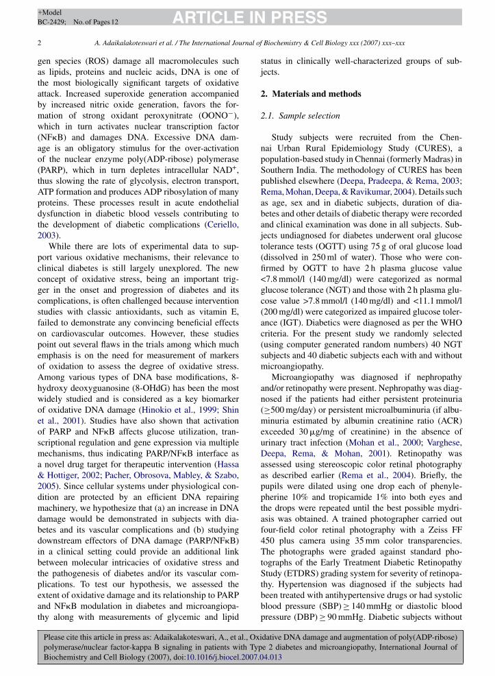
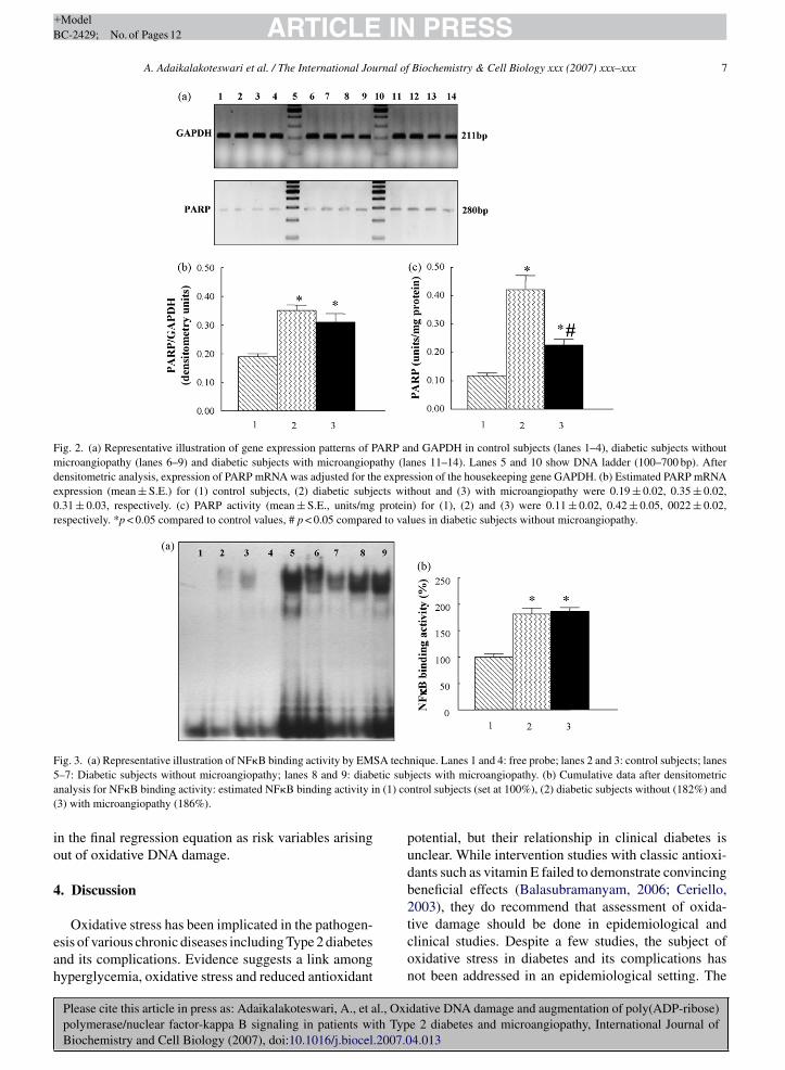
Fig. 2a is an illustration of mRNA gene expressionpatterns of PARP and GAPDH in the study subjects.PARP mRNA expression was significantly (p < 0.05)increased in diabetic patients with (0.31 ± 0.03 densito-metry units) and without (0.35 ± 0.02) microangiopathycompared to control subjects (0.19 ± 0.02) (Fig. 2b).Concurrent to mRNA findings, PARP activity wasalso significantly (p < 0.05) increased in diabetic sub-jects (0.42 ± 0.05) compared to controls (0.11 ± 0.02)(Fig. 2c). Though PARP activity in diabetes subjects with
Please cite this article in press as: Adaikalakoteswari, A., et al., Oxipolymerase/nuclear factor-kappa B signaling in patients with TypBiochemistry and Cell Biology (2007), doi:10.1016/j.biocel.2007.0
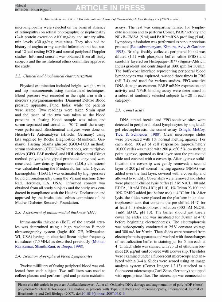
microangiopathy (0.22 ± 0.02) was lower than patientswithout microangiopathy, it was significantly (p < 0.05)higher than the activity levels seen in the control subjects.NF�B binding activity as measured from the EMSA
Fig. 1. (a) Basal DNA damage (mean ± S.E.) in (1) control subjects (4.29 ± 1giopathy (21.34 ± 1.5). (b) FPG-sensitive DNA strand breaks (mean ± S.E.(16.53 ± 2.0) and (3) with microangiopathy (19.41 ± 2.5). *p < 0.05 compare
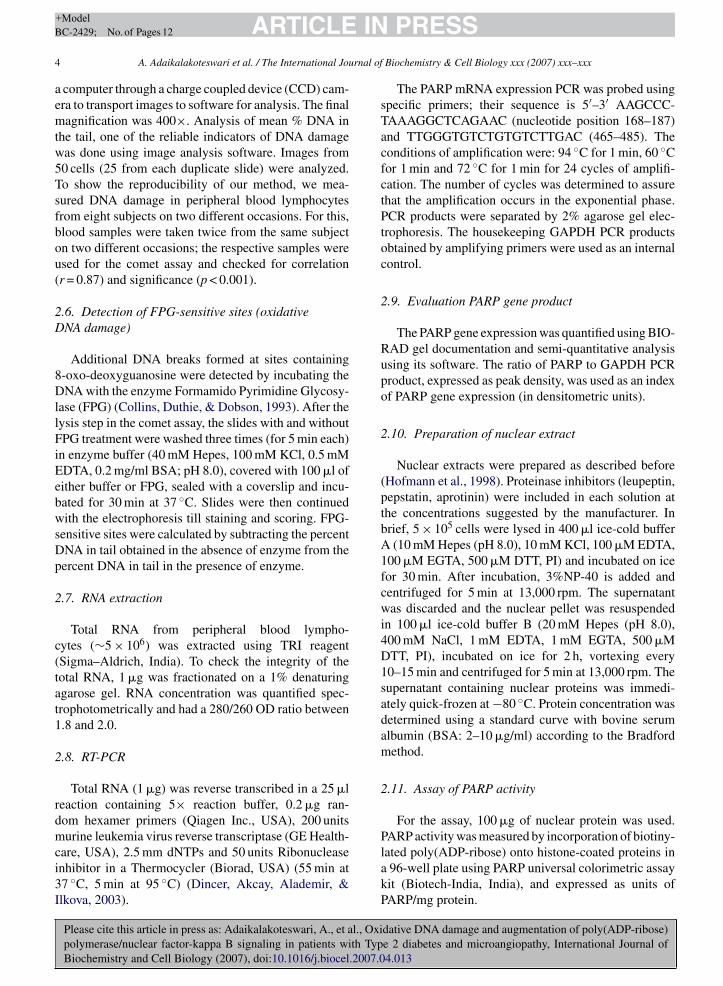
technique is illustrated in Fig. 3a. Diabetic subjects withand without microangiopathy exhibited a significantly(p < 0.05) higher (80%) NF�B binding activity comparedto control subjects (Fig. 3b). In diabetic patients, oxida-tive DNA damage also correlated positively with PARPgene expression (r = 0.476; p = 0.002) (Fig. 4a), PARPactivity (r = 0.320; p = 0.044) (Fig. 4b) and NF�B bind-ing activity (r = 0.490; p = 0.001) (Fig. 4c). When age,sex, smoking, presence of diabetes, HbA1c, cholesterol,triglycerides, creatinine clearance, IMT, TBARS, PCO,
dative DNA damage and augmentation of poly(ADP-ribose)e 2 diabetes and microangiopathy, International Journal of4.013
NF�B binding activity, PARP gene expression and activ-ity, were included as potential independent variables inthe forward regression analysis, presence of diabetes,PARP activity, IMT, and creatinine clearance remained
.04), (2) diabetic subjects without (22.36 ± 1.6) and (3) with microan-) in (1) control subjects (1.38 ± 0.85), (2) diabetic subjects withoutd to control values.
ARTICLE IN PRESS+ModelBC-2429; No. of Pages 12
A. Adaikalakoteswari et al. / The International Journal of Biochemistry & Cell Biology xxx (2007) xxx–xxx 7
Fig. 2. (a) Representative illustration of gene expression patterns of PARP and GAPDH in control subjects (lanes 1–4), diabetic subjects withoutmicroangiopathy (lanes 6–9) and diabetic subjects with microangiopathy (lanes 11–14). Lanes 5 and 10 show DNA ladder (100–700 bp). Afterdensitometric analysis, expression of PARP mRNA was adjusted for the expression of the housekeeping gene GAPDH. (b) Estimated PARP mRNAexpression (mean ± S.E.) for (1) control subjects, (2) diabetic subjects without and (3) with microangiopathy were 0.19 ± 0.02, 0.35 ± 0.02,0.31 ± 0.03, respectively. (c) PARP activity (mean ± S.E., units/mg protein) for (1), (2) and (3) were 0.11 ± 0.02, 0.42 ± 0.05, 0022 ± 0.02,respectively. *p < 0.05 compared to control values, # p < 0.05 compared to values in diabetic subjects without microangiopathy.
F SA tech5 etic suba n (1) co(
io
4
eah
ig. 3. (a) Representative illustration of NF�B binding activity by EM–7: Diabetic subjects without microangiopathy; lanes 8 and 9: diabnalysis for NF�B binding activity: estimated NF�B binding activity i3) with microangiopathy (186%).
n the final regression equation as risk variables arisingut of oxidative DNA damage.
. Discussion
Please cite this article in press as: Adaikalakoteswari, A., et al., Oxipolymerase/nuclear factor-kappa B signaling in patients with TypBiochemistry and Cell Biology (2007), doi:10.1016/j.biocel.2007.0
Oxidative stress has been implicated in the pathogen-sis of various chronic diseases including Type 2 diabetesnd its complications. Evidence suggests a link amongyperglycemia, oxidative stress and reduced antioxidant
nique. Lanes 1 and 4: free probe; lanes 2 and 3: control subjects; lanesjects with microangiopathy. (b) Cumulative data after densitometricntrol subjects (set at 100%), (2) diabetic subjects without (182%) and
potential, but their relationship in clinical diabetes isunclear. While intervention studies with classic antioxi-dants such as vitamin E failed to demonstrate convincingbeneficial effects (Balasubramanyam, 2006; Ceriello,2003), they do recommend that assessment of oxida-
dative DNA damage and augmentation of poly(ADP-ribose)e 2 diabetes and microangiopathy, International Journal of4.013
tive damage should be done in epidemiological andclinical studies. Despite a few studies, the subject ofoxidative stress in diabetes and its complications hasnot been addressed in an epidemiological setting. The

ARTICLE IN PRESS+ModelBC-2429; No. of Pages 12
8 A. Adaikalakoteswari et al. / The International Journal of Biochemistry & Cell Biology xxx (2007) xxx–xxx
RP mRetic sub
Fig. 4. Correlation of FPG-sensitive DNA strand breaks with (a) PAp = 0.044) and (c) NF�B binding activity (r = 0.490; p = 0.001) in diab
absence of epidemiologic data on oxidative damage innormal human populations represents a serious gap inour knowledge about the distribution, correlates, andcausative factors of oxidative damage. Therefore, thepresent study assumes significance for the followingreasons. First, oxidative damage to the macromolecules(lipid, protein and DNA) was comprehensively assessedin subjects from the epidemiological programme. Sec-ondly, the study establishes a significant correlationbetween oxidative DNA damage and a number of clini-cal phenotypes associated with Type 2 diabetes. Thirdly,molecular evidence for increased oxidative stress in clin-ical diabetes and microangiopathy was provided througha comprehensive analysis of oxidative DNA damage andaugmentation of PARP/NF�B signals.
Research on the cellular effects of hyperglycemiahas elucidated several molecular defects among whichoxidative damage appears to represent a central abnor-mality (Brownlee, 2001; Ceriello, 2003). Increased lipidand protein oxidation have been demonstrated in bothexperimental and clinical diabetes studies, with morerecent studies pointing to F2-isoprostane and nitrotyro-sine emerging as useful biomarkers of oxidative stress(Morrow, 2005; Pacher et al., 2005). Several studieshave also shown increased oxidative DNA damage (Choi
Please cite this article in press as: Adaikalakoteswari, A., et al., Oxipolymerase/nuclear factor-kappa B signaling in patients with TypBiochemistry and Cell Biology (2007), doi:10.1016/j.biocel.2007.0
et al., 2005; Dincer et al., 2003; Pitozzi, Giovannelli,Bardini, Rotella, & Dolara, 2003; Sampson, Winterbone,Hughes, Dozio, & Hughes, 2006) and accumulation of8-OHdG (Hinokio et al., 1999; Shin et al., 2001; Speit,
NA expression (r = 0.476; p = 0.002), (b) PARP activity (r = 0.320;jects.
Schutz, Bonzheim, Trenz, & Hoffmann, 2004) in patientswith diabetes suggesting the involvement of hyper-glycemia in oxidative DNA damage. Increased oxidativedamage related to mitochondrial DNA (mtDNA) alter-ations was also reported in Type 2 diabetes (Speit et al.,2004).
In many studies, DNA damage was assessed as DNAstrand breaks to look for endogenous oxidative dam-age to DNA. However there are some limitations tothis approach: (a) breaks can arise in a variety of waysunrelated to oxidation and (b) breaks are quite rapidlyrepaired by cells (Collins, 2004; Lin et al., 2005; Speit etal., 2004). In our study, we have used FPG to recognizealtered purines (including 8-oxo-guanine, the principalsubstrate for FPG in vivo) and thereby estimated theoxidative DNA damage. Oxidative DNA damage (FPG-sensitive DNA strand breaks) in diabetic patients wasvery well correlated to poor glycemic control (in termsof fasting and post-prandial hyperglycemia and HbA1c)suggesting that glucose toxicity may be the main culpritcontributing to enhanced oxidative stress. Since patientswith high blood glucose and high lipid levels are at anincreased risk for vascular damage, the results also sug-gest that in the presence of chronic hyperglycemia andhyperlipidemia, any increase in oxidative DNA damage
dative DNA damage and augmentation of poly(ADP-ribose)e 2 diabetes and microangiopathy, International Journal of4.013
might serve as a reliable indicator of increased predilec-tion to diabetic vascular complications.
The association of FPG-sensitive sites with lipidperoxidation and protein carbonyl levels among the
IN+ModelB
rnal of
diaeogiha2Yd
ra2tiaiHPdfC((
ra(siMswmaDpogat(eaMaopp
ARTICLEC-2429; No. of Pages 12
A. Adaikalakoteswari et al. / The International Jou
iabetic subjects in our study further substantiates thenter-relationship between lipid-, protein- and nucleiccid-damage and induction of the oxidative stress (Choit al., 2005). Additionally we also observed that bothxidative DNA damage and intimal thickening werereater in diabetics. This is in consistent with the findingsn which oxidative DNA damage was associated withuman atherosclerotic plaques that promote cell prolifer-tion, hypertrophy, growth arrest and/or apoptosis (Irani,000; Kunsch & Medford, 1999). Recently, Demirbag,ilmaz, & Kocyigit (2005) also reported increased DNAamage with severity of CAD and its risk factors.
In addition to oxidative DNA damage, our studyevealed increased PARP mRNA expression and activitynd augmented NF�B signaling in patients with Typediabetes and microangiopathy. Evidence implicates
hat PARP is involved in the onset of pathophysiolog-cal diseases at two different levels: (1) PARP acting as
sensor of DNA damage in cells and (2) PARP act-ng as transcriptional co-activator of NF�B (Hassa &ottiger, 2002). Recent studies have delineated a role of
ARP-1 activation in the pathogenesis of diabetes andiabetic complications including cardiovascular dys-unction (Pacher et al., 2002), nephropathy (Wahlberg,arlson, Wasserman, & Ljungqvist, 1985), neuropathy
Ilnytska et al., 2006; Li et al., 2004) and retinopathyZheng, Szabo, & Kern, 2004).
It is suggested that increased ROS play a criticalole in signaling the cellular inflammatory response byctivating the redox-sensitive transcription factor, NF�BKabe, Ando, Hirao, Yoshida, & Handa, 2005). In ourtudy, NF�B binding activity was significantly increasedn cells from patients with diabetes and microangiopathy.
ore importantly, oxidative DNA damage as repre-ented by FPG-sensitive sites was positively correlatedith three different molecular parameters viz., PARPRNA expression, PARP activity and NF�B binding
ctivity. This emphasizes a tight link between oxidativeNA damage and augmented PPAR/NF�B signaling inatients with diabetes. Increased DNA binding affinityf NF�B in retinal endothelial cells exposed to highlucose was demonstrated to be mediated via PARPctivity (Zheng et al., 2004) and binding of p50 subunito DNA was shown poly(ADP) ribosylation-dependentChang & Alvarez-Gonzalez, 2001). In addition, reducedxpression of NF�B dependent pro-inflammatory medi-tors was reported in PARP-1(−/−) mice (Shall & deurcia, 2000). Although DNA damage is considered as
Please cite this article in press as: Adaikalakoteswari, A., et al., Oxipolymerase/nuclear factor-kappa B signaling in patients with TypBiochemistry and Cell Biology (2007), doi:10.1016/j.biocel.2007.0
n obligatory signal for PARP activation, it may alsoccur in the absence of DNA damage through the ERK2athway (Cohen-Armon et al., 2007). Alternatively, thero-inflammatory cytokines often associate with dia-
PRESSBiochemistry & Cell Biology xxx (2007) xxx–xxx 9
betes may also stimulate ERK/NF�B signaling (Larsenet al., 2005). Therefore, it appears that the NF�B/PARPinterface might be a potential target to interrupt thevicious cycle of oxidative stress and for the develop-ment of new combinations of drugs disrupting specificprotein–protein interactions.
While a role for oxidative stress has been demon-strated in the development of microangiopathy indiabetic patients, the oxidative DNA damage seen indiabetic subjects with microangiopathy was not signif-icantly higher compared to that observed in diabeticsubjects without microangiopathy. It is unlikely to beexplained as a feedback control by DNA repair enzymesbecause vascular complications were very often relatedat the cellular level to augmentation of poly(ADP) ribo-sylation and NF�B events. The plausible explanationfor our results in diabetes patients with microangiopa-thy may be related to drug regimen in these patientsin addition to glucose-lowering agents, which couldmodulate DNA damage and interfere with PARP/NF�Bsignals (Martin-Ventura et al., 2005; Ohga et al., 2007;Schmeisser et al., 2004; Shin et al., 2005). Alternatively,reduced PARP activity may be influenced by angiogenicmilieu seen in microangiopathy conditions. For exam-ple, insulin-like growth factor-I (IGF-I) has been shownto promote angiogenesis by enhancing vascular endothe-lial growth factor (VEGF) expression via suppressionof PARP activity (Beckert et al., 2006). However, thesecausality relationships need to be tested in larger samplesand in prospective studies. Nevertheless, an increasedoxidative DNA damage and PARP activity seen in ourstudy in diabetic patients prior to the onset of vascu-lar pathology is an important observation. Since PARPactivation was also shown in healthy subjects at risk ofdeveloping diabetes as well as in established Type 2 dia-betic patients (Szabo et al., 2002), these studies revealopportunities for early therapeutic intervention. More-over, increased DNA damage in our diabetic patientswith microangiopathy compared to control subjects,emphasize a role for oxidative stress in the develop-ment and progression of microvascular complications. Infact, urinary 8-OHG has been reported as a biomarker ofoxidative DNA damage in diabetic nephropathy patients(Hinokio et al., 1999; Wu, Chiou, Chang, & Wu, 2004;Xu et al., 2004) and higher serum 8-OHG levels werereported in diabetic patients with advanced microvascu-lar complications (Shin et al., 2001). While decreasedcreatinine clearance is the earliest predictor of renal
dative DNA damage and augmentation of poly(ADP-ribose)e 2 diabetes and microangiopathy, International Journal of4.013
dysfunction, it is important to note in our study, thatoxidative DNA damage was negatively correlated to cre-atinine clearance in diabetic patients. We also noted thatthe levels of oxidative DNA damage were significantly
IN+Model
urnal of
ARTICLEBC-2429; No. of Pages 12
10 A. Adaikalakoteswari et al. / The International Jo
(p = 0.019) higher in patients with creatinine clearanceof less than 70 ml/min compared with those with valuesgreater than 70 ml/min. These observations warrant fur-ther studies so as to relate causality of oxidative DNAdamage to the severity of diabetic microvascular com-plications.
The demonstration of oxidative DNA damage andaugmentation of its downstream effectors in patientswith Type 2 diabetes and microangiopathy in this workhas several implications. Perhaps, this is one of the firstpopulation-based studies in which oxidative DNA dam-age was shown correlated to poor glycemic and lipidstatus on one hand and to augmentation of PARP/NF�Bsignals on the other hand. It is of interest to note thatoxidative DNA damage in diabetes patients was notonly correlated to HbA1c levels but also more signif-icantly to post-prandial plasma glucose levels. WhileDCCT–EDIC outcomes (Hirsch & Brownlee, 2005)claim glycemic variability in addition to HbA1c as a riskfactor for vascular complications, it has been recentlyshown that acute glucose fluctuations triggered morespecific oxidative stress than sustained chronic hyper-glycemia in patients with Type 2 diabetes (Monnier et al.,2006). In view of emerging evidence that oxidative DNAdamage to be a biomarker in molecular epidemiologi-cal studies (Dalle-Donne, Rossi, Colombo, Giustarini,& Milzani, 2006; Heilbronn et al., 2006; Ilnytska etal., 2006), development of new strategies designed todetect DNA damage caused by oxidative stress mayadvance our understanding of the roles of such typesof damage in the etiology of diabetes and its com-plications. In fact, a DNA adduct detection method(adductome approach) that uses liquid chromatogra-phy coupled with electrospray ionization tandem massspectrometry (LC/ESI-MS/MS) to detect multiple DNAadducts in human tissues as clinical biomarkers is alsounderway (Kanaly et al., 2006).
One of the limitations of this work is thatbeing a cross-sectional study, a cause-and-effect rela-tionship cannot be established. Nevertheless, it hasprovided a molecular evidence for genomic instabilityin patients with Type 2 diabetes and microangiopa-thy in Asian Indians. Asian Indians with Type 2diabetes also exhibit decreased HDL (Misra, Luthra,& Vikram, 2004), hypoadiponectinemia (Mohan etal., 2005), hypoglutathionemia (Sampathkumar etal., 2005; Sampathkumar, Balasubramanyam, Tara,Rema, & Mohan, 2006), impaired antioxidant defense
Please cite this article in press as: Adaikalakoteswari, A., et al., Oxipolymerase/nuclear factor-kappa B signaling in patients with TypBiochemistry and Cell Biology (2007), doi:10.1016/j.biocel.2007.0
(Adaikalakoteswari et al., 2006), shortened telomeres(Adaikalakoteswari et al., 2005; Adaikalakoteswari,Balasubramanyam, Ravikumar, Deepa, & Mohan, 2007)and increased premature mortality rates (Mohan et al.,
PRESSBiochemistry & Cell Biology xxx (2007) xxx–xxx
2006) all of which represent a state of increased oxida-tive stress and genomic instability. In this context, ourwork demonstrating increased oxidative DNA damagein Type 2 diabetic patients warrants further studies onthe molecular aspects of both nuclear and mtDNA alter-ations which could ultimately lead to novel drugs andnewer therapeutic options.
Acknowledgements
This work was supported by research grants from theDepartment of Science and Technology (DST & DST-FIST), Indian Council of Medical Research (ICMR) andthe Department of Biotechnology (DBT), Governmentof India, New Delhi, India. AA acknowledges the finan-cial assistance (Senior Research Fellowship) from theCouncil of Scientific & Industrial Research (CSIR), NewDelhi, India. This is paper no. 50 from the Chennai UrbanRural Epidemiology Study (CURES).
References
Adaikalakoteswari, A., Balasubramanyam, M., & Mohan, V. (2005).Telomere shortening occurs in Asian Indian Type 2 diabeticpatients. Diabet. Med., 22, 1151–1156.
Adaikalakoteswari, A., Balasubramanyam, M., Rema, M., & Mohan,V. (2006). Differential gene expression of NADPH oxidase (p22)and hemoxygenase-1 in patients with Type 2 diabetes and microan-giopathy. Diabet. Med., 23, 666–674.
Adaikalakoteswari, A., Balasubramanyam, M., Ravikumar, R., Deepa,R., & Mohan, V. (2007). Association of telomere shorteningwith impaired glucose tolerance and diabetic macroangiopathy.Atherosclerosis, in press.
Balasubramanyam, M., Kimura, M., Aviv, A., & Gardner, J. P. (1993).Kinetics of calcium transport across the lymphocyte plasma mem-brane. Am. J. Physiol., 265, C321–C327.
Balasubramanyam, M. (2006). Antioxidants and cardiovasculardisease—Where do we stand? Asian J. Diab., 8, 48–52.
Beckert, S., Farrahi, F., Perveen Ghani, Q., Aslam, R., Scheuenstuhl,H., Coerper, S., et al. (2006). IGF-I-induced VEGF expression inHUVEC involves phosphorylation and inhibition of poly(ADP-ribose)polymerase. Biochem. Biophys. Res. Commun., 341, 67–72.
Brownlee, M. (2001). Biochemistry and molecular cell biology ofdiabetic complications. Nature, 414, 813–820.
Ceriello, A. (2003). New insights on oxidative stress and diabetic com-plications may lead to a “causal” antioxidant therapy. DiabetesCare, 26, 1589–1596.
Chang, W. J., & Alvarez-Gonzalez, R. (2001). The sequence-specificDNA binding of NF-kappa B is reversibly regulated by the auto-modification reaction of poly (ADP-ribose) polymerase-1. J. Biol.Chem., 276, 47664–47670.
Choi, S. W., Benzie, I. F., Lam, C. S., Chat, S., Lam, J., Yiu, C. H., et
dative DNA damage and augmentation of poly(ADP-ribose)e 2 diabetes and microangiopathy, International Journal of4.013
al. (2005). Inter-relationships between DNA damage, ascorbic acidand glycaemic control in Type 2 diabetes mellitus. Diabet. Med.,22, 1347–1353.
Cohen-Armon, M., Visochek, L., Rozensal, D., Kalal, A., Geistrikh,I., Klein, R., et al. (2007). DNA-independent PARP-1 activation
IN+ModelB
rnal of
C
C
D
D
D
D
E
H
H
H
H
H
I
I
K
K
K
L
ARTICLEC-2429; No. of Pages 12
A. Adaikalakoteswari et al. / The International Jou
by phosphorylated ERK2 increases Elk1 activity: A link to histoneacetylation. Mol. Cell, 25, 297–308.
ollins, A. R. (2004). The comet assay for DNA damage and repair:Principles, applications, and limitations. Mol. Biotechnol., 26,249–261.
ollins, A., Duthie, S., & Dobson, V. (1993). Direct enzymatic detec-tion of endogenous oxidative base damage in human lymphocyteDNA. Carcinogenesis, 14, 1733–1735.
alle-Donne, I., Rossi, R., Colombo, R., Giustarini, D., & Milzani, A.(2006). Biomarkers of oxidative damage in human disease. Clin.Chem., 52, 601–623.
eepa, M., Pradeepa, R., Rema, M., et al. (2003). The Chennai UrbanRural Epidemiology Study (CURES) study design and method-ology (urban component) (CURES-I). J. Assoc. Phys. India, 51,863–870.
emirbag, R., Yilmaz, R., & Kocyigit, A. (2005). Relationship betweenDNA damage, total antioxidant capacity and coronary artery dis-ease. Mutat. Res., 570, 197–203.
incer, Y., Akcay, T., Alademir, Z., & Ilkova, H. (2003). Assessmentof DNA base oxidation and glutathione level in patients with type2 diabetes. Mutat. Res., 9(525), 129–130.
vans, J. L., Goldfine, I. D., Maddux, B. A., & Grodsky, G. M. (2002).Oxidative stress and stress-activated signaling pathways: A unify-ing hypothesis of type 2 diabetes. Endocr. Rev., 23, 599–622.
assa, P. O., & Hottiger, M. O. (2002). The functional role ofpoly(ADP-ribose)polymerase 1 as novel coactivator of NF-kappaBin inflammatory disorders. Cell. Mol. Life Sci., 59, 1534–1553.
eilbronn, L. K., et al. (2006). Effect of 6-month calorie restriction onbiomarkers of longevity, metabolic adaptation, and oxidative stressin overweight individuals: A randomized controlled trial. JAMA,295, 1539–1548.
inokio, Y., Suzuki, S., Hirai, M., Chiba, M., Hirai, A., & Toyota, T.(1999). Oxidative DNA damage in diabetes mellitus: Its associationwith diabetic complications. Diabetologia, 42, 995–998.
irsch, I. B., & Brownlee, M. (2005). Should minimal blood glu-cose variability become the gold standard of glycemic control? J.Diabet. Complications, 19, 178–181.
ofmann, M. A., Schiekofer, S., Kanitz, M., Klevesath, M. S.,Joswig, M., Lee, V., et al. (1998). Insufficient glycemic controlincreases nuclear factor-kappa B binding activity in peripheralblood mononuclear cells isolated from patients with type 1 dia-betes. Diabetes Care, 21, 1310–1316.
lnytska, O., Lyzogubov, V. V., Stevens, M. J., Drel, V. R., Mashtalir, N.,Pacher, P., et al. (2006). Poly(ADP-ribose) polymerase inhibitionalleviates experimental diabetic sensory neuropathy. Diabetes, 55,1686–1694.
rani, K. (2000). Oxidant signaling in vascular cell growth, death, andsurvival: A review of the roles of reactive oxygen species in smoothmuscle and endothelial cell mitogenic and apoptotic signaling.Circ. Res., 87, 179–183.
abe, Y., Ando, K., Hirao, S., Yoshida, M., & Handa, H. (2005).Redox regulation of NF-kappaB activation: Distinct redox regu-lation between the cytoplasm and the nucleus. Antioxid. RedoxSignal., 7, 395–403.
analy, R. A., Hanaoka, T., Sugimura, H., Toda, H., Matsui, S., & Mat-suda, T. (2006). Development of the adductome approach to detectDNA damage in humans. Antioxid. Redox Signal., 8, 993–1001.
Please cite this article in press as: Adaikalakoteswari, A., et al., Oxipolymerase/nuclear factor-kappa B signaling in patients with TypBiochemistry and Cell Biology (2007), doi:10.1016/j.biocel.2007.0
unsch, C., & Medford, R. M. (1999). Oxidative stress as a regulatorof gene expression in the vasculature. Circ. Res., 85, 753–766.
arsen, L., Storling, J., Darville, M., Eizirik, D. L., Bonny, C.,Billestrup, N., et al. (2005). Extracellular signal-regulated kinaseis essential for interleukin-1-induced and nuclear factor kappaB-
PRESSBiochemistry & Cell Biology xxx (2007) xxx–xxx 11
mediated gene expression in insulin-producing INS-1E cells.Diabetologia, 48, 2582–2590.
Levine, R. L., Garland, D., Oliver, C. N., Amici, A., Climent, I.,Lenz, A. G., et al. (1990). Determination of carbonyl content inoxidatively modified proteins. Meth. Enzymol., 186, 464–478.
Li, F., Szabo, C., Pacher, P., Southan, G. J., Abatan, O. I.,Charniauskaya, T., et al. (2004). Evaluation of orally activepoly(ADP-ribose) polymerase inhibitor in streptozotocin-diabeticrat model of early peripheral neuropathy. Diabetologia, 47,710–717.
Lin, T. K., Chen, S. D., Wang, P. W., Wei, Y. H., Lee, C. F., Chen, T. L.,et al. (2005). Increased oxidative damage with altered antioxidativestatus in Type 2 diabetic patients harboring the 16189 T to C variantof mitochondrial DNA. Ann. NY Acad. Sci., 1042, 64–69.
Martin-Ventura, J. L., Blanco-Colio, L. M., Gomez-Hernandez, A.,Munoz-Garcia, B., Vega, M., Serrano, J., et al. (2005). Intensivetreatment with atorvastatin reduces inflammation in mononuclearcells and human atherosclerotic lesions in one month. Stroke, 36,1796–1800.
Misra, A., Luthra, K., & Vikram, N. K. (2004). Dyslipidemia in AsianIndians: Determinants and significance. J. Assoc. Phys. India, 52,137–142.
Mohan, V., Deepa, R., Pradeepa, R., Vimaleswaran, K. S., Mohan,A., Velmurugan, K., et al. (2005). Association of low adiponectinlevels with the metabolic syndrome—The Chennai Urban RuralEpidemiology Study (CURES-4). Metabolism, 54, 476–481.
Mohan, V., Meera, R., Premalatha, G., Deepa, R., Miranda, P., & Rema,M. (2000). Frequency of proteinuria in type 2 diabetes mellitusseen at a diabetes centre in southern India. Postgrad. Med. J., 76,569–573.
Mohan, V., Ravikumar, R., ShanthiRani, S., & Deepa, R. (1998). Inti-mal medial thickness of the carotid artery in south Indian diabeticand nondiabetic subjects: The Chennai Urban Population Study(CUPS). Diabetologia, 433, 494–499.
Mohan, V., Shanthirani, C. S., Deepa, M., Deepa, R., Unnikrishnan, R.I., & Datta, M. (2006). Mortality rates due to diabetes in a selectedurban South Indian Population—The Chennai Urban PopulationStudy (CUPS-16). JAPI, 54, 113–117.
Monnier, L., Mas, E., Ginet, C., Michel, F., Villon, L., Cristol, J. P., &Colette, C. (2006). Activation of oxidative stress by acute glucosefluctuations compared with sustained chronic hyperglycemia inpatients with type 2 diabetes. JAMA, 295, 1681–1687.
Morrow, J. D. (2005). Quantification of isoprostanes as indices of oxi-dant stress and the risk of atherosclerosis in humans. Arterioscler.Thromb. Vasc. Biol., 25, 279–286.
Ohga, S., Shikata, K., Yozai, K., Okada, S., Ogawa, D., Usui, H., etal. (2007). Thiazolidinedione ameliorates renal injury in experi-mental diabetic rats through anti-inflammatory effects mediatedby inhibition of NF-{kappa}B activation. Am. J. Physiol., 292,F1141–F1150.
Pacher, P., Liaudet, L., Soriano, F. G., Mabley, J. G., Szabo, E., &Szabo, C. (2002). The role of poly(ADP-ribose) polymerase in thedevelopment of myocardial and endothelial dysfunction in diabetesmellitus. Diabetes, 51, 514–521.
Pacher, P., Obrosova, I. G., Mabley, J. G., & Szabo, C. (2005). Role ofnitrosative stress and peroxynitrite in the pathogenesis of diabeticcomplications. Emerging new therapeutical strategies. Curr. Med.
dative DNA damage and augmentation of poly(ADP-ribose)e 2 diabetes and microangiopathy, International Journal of4.013
Chem., 12, 267–275.Pitozzi, V., Giovannelli, L., Bardini, G., Rotella, C. M., & Dolara, P.
(2003). Oxidative DNA damage in peripheral blood cells in type2 diabetes mellitus: Higher vulnerability of polymorphonuclearleukocytes. Mutat. Res., 529, 129–133.
IN+Model
urnal of
ARTICLEBC-2429; No. of Pages 12
12 A. Adaikalakoteswari et al. / The International Jo
Rema, M., Mohan, V., Deepa, R., & Ravikumar, R. (2004). Associationof carotid intimal medial thickness and arterial stiffness with dia-betic retinopathy—The Chennai Urban Rural Epidemiology Study[CURES-2]. Diabetes Care, 27, 1962–1967.
Sampathkumar, R., Balasubramanyam, M., Sudarslal, S., Rema, M.,Mohan, V., & Balaram, P. (2005). Increased glutathionylatedhemoglobin (HbSSG) in type 2 diabetes subjects with microan-giopathy. Clin. Biochem., 38, 892–899.
Sampathkumar, R., Balasubramanyam, M., Tara, C., Rema, M.,& Mohan, V. (2006). Association of hypoglutathionemia withreduced Na+/K+ ATPase activity in type 2 diabetes and microan-giopathy. Mol. Cell. Biochem., 282, 169–176.
Sampson, M. J., Winterbone, M. S., Hughes, J. C., Dozio, N., &Hughes, D. A. (2006). Monocyte telomere shortening and oxidativeDNA damage in type 2 diabetes. Diabetes Care, 29, 283–289.
Schmeisser, A., Soehnlein, O., Illmer, T., Lorenz, H. M., Eskafi, S.,Roerick, O., et al. (2004). ACE inhibition lowers angiotensin II-induced chemokine expression by reduction of NF-kappaB activityand AT1 receptor expression. Biochem. Biophys. Res. Commun.,325, 532–540.
Shall, S., & de Murcia, G. (2000). Poly(ADP-ribose) polymerase-1:What have we learned from the deficient mouse model? Mutat.Res., 460, 1–15.
Shin, M. J., Cho, E. Y., Jang, Y., Lee, J. H., Shim, W. H., Cho, S. Y., et al.(2005). A beneficial effect of simvastatin on DNA damage in 242T
Please cite this article in press as: Adaikalakoteswari, A., et al., Oxipolymerase/nuclear factor-kappa B signaling in patients with TypBiochemistry and Cell Biology (2007), doi:10.1016/j.biocel.2007.0
allele of the NADPH oxidase p22phox in hypercholesterolemicpatients. Clin. Chim. Acta, 360, 46–51.
Shin, C. S., Moon, B. S., Park, K. S., Kim, S. Y., Park, S. J., Chung, M.H., et al. (2001). Serum 8-hydroxy-guanine levels are increased indiabetic patients. Diabetes Care, 24, 733–737.
PRESSBiochemistry & Cell Biology xxx (2007) xxx–xxx
Singh, N., McCoy, M., Tice, R., & Schneider, L. (1988). A simple tech-nique for quantitation of low levels of DNA damage in individualcells. Exp. Cell Res., 175, 84–191.
Speit, G., Schutz, P., Bonzheim, I., Trenz, K., & Hoffmann, H. (2004).Sensitivity of the FPG protein towards alkylation damage in thecomet assay. Toxicol. Lett., 146, 151–158.
Szabo, C., Zanchi, A., Komjati, K., Pacher, P., Krolewski, A. S., Quist,W. C., et al. (2002). Poly(ADP-ribose) polymerase is activated insubjects at risk of developing type 2 diabetes and is associated withimpaired vascular reactivity. Circulation, 106, 2680–2686.
Varghese, A., Deepa, R., Rema, M., & Mohan, V. (2001). Prevalenceof microalbuminuria in type 2 diabetes mellitus at a diabetes centrein southern India. Postgrad. Med. J., 77, 399–402.
Wahlberg, G., Carlson, L. A., Wasserman, J., & Ljungqvist, A. (1985).Protective effect of nicotinamide against nephropathy in diabeticrats. Diab. Res., 2, 307–312.
Wu, L. L., Chiou, C. C., Chang, P. Y., & Wu, J. T. (2004). Urinary8-OHdG: A marker of oxidative stress to DNA and a risk factorfor cancer, atherosclerosis and diabetics. Clin. Chim. Acta, 339, 1–9.
Xu, G. W., Yao, Q. H., Weng, Q. F., Su, B. L., Zhang, X., & Xiong, J. H.(2004). Study of urinary 8-hydroxydeoxyguanosine as a biomarkerof oxidative DNA damage in diabetic nephropathy patients. J.Pharm. Biomed. Anal., 36, 101–104.
Yagi, K. (1976). A simple fluorometric assay for lipoperoxide in blood
dative DNA damage and augmentation of poly(ADP-ribose)e 2 diabetes and microangiopathy, International Journal of4.013
plasma. Biochem. Med., 15, 212–216.Zheng, L., Szabo, C., & Kern, T. S. (2004). Poly(ADP-ribose) poly-
merase is involved in the development of diabetic retinopathyvia regulation of nuclear factor-kappaB. Diabetes, 53, 2960–2967.