oxidative stress alters base excision repair pathway and increases apoptotic response in...
TRANSCRIPT

Free Radical Biology & Medicine 46 (2009) 1488–1499
Contents lists available at ScienceDirect
Free Radical Biology & Medicine
j ourna l homepage: www.e lsev ie r.com/ locate / f reeradb iomed
Original Contribution
Oxidative stress alters base excision repair pathway and increases apoptotic responsein apurinic/apyrimidinic endonuclease 1/redox factor-1 haploinsufficient mice
Archana Unnikrishnan a,1, Julian J. Raffoul a,1, Hiral V. Patel a, Thomas M. Prychitko a, Njwen Anyangwe a,Lisiane B. Meira b, Errol C. Friedberg c, Diane C. Cabelof a,d, Ahmad R. Heydari a,d,⁎a Department of Nutrition & Food Science, College of Liberal Arts and Sciences, Wayne State University, Detroit, MI, 48202, USAb Biological Engineering Division, Massachusetts Institute of Technology, Cambridge, MA 02139, USAc Laboratory of Molecular Pathology, Department of Pathology, The University of Texas Southwestern Medical Center, Dallas, TX 75390, USAd Barbara Ann Karmanos Cancer Institute, School of Medicine, Wayne State University, Detroit, MI 48201, USA
⁎ Corresponding author. Department of Nutrition & Fooand Sciences, Wayne State University, Detroit, MI, 48202,
E-mail address: [email protected] (A.R. He1 These authors contributed equally to this work.
0891-5849/$ – see front matter © 2009 Elsevier Inc. Adoi:10.1016/j.freeradbiomed.2009.02.021
a b s t r a c t
a r t i c l e i n f oArticle history:Received 9 December 2008Revised 4 February 2009Accepted 23 February 2009Available online 3 March 2009
Keywords:Apurinic/apyrimidinic endonuclease 1/redoxfactor-1Redox activityBase excision repairOxidative DNA damageNF-κBApoptosisLiverFree radicals
Apurinic/apyrimidinic endonuclease 1/redox factor-1 (APE1/Ref-1) is the redox regulator of multiple stress-inducible transcription factors, such as NF-κB, and the major 5′-endonuclease in base excision repair (BER).We utilized mice containing a heterozygous gene-targeted deletion of APE1/Ref-1 (Apex+/−) to determinethe impact of APE1/Ref-1 haploinsufficiency on the processing of oxidative DNA damage induced by2-nitropropane (2-NP) in the liver tissue of mice. APE1/Ref-1 haploinsufficiency results in a significantdecline in NF-κB DNA-binding activity in response to oxidative stress in liver. In addition, loss of APE1/Ref-1increases the apoptotic response to oxidative stress, in which significant increases in GADD45g expression,p53 protein stability, and caspase activity are observed. Oxidative stress displays a differential impact onmonofunctional (UNG) and bifunctional (OGG1) DNA glycosylase-initiated BER in the liver of Apex+/− mice.APE1/Ref-1 haploinsufficiency results in a significant decline in the repair of oxidized bases (e.g., 8-OHdG),whereas removal of uracil is increased in liver nuclear extracts of mice using an in vitro BER assay. Apex+/−
mice exposed to 2-NP displayed a significant decline in 3′-OH-containing single-strand breaks and anincrease in aldehydic lesions in their liver DNA, suggesting an accumulation of repair intermediates of failedbifunctional DNA glycosylase-initiated BER.
© 2009 Elsevier Inc. All rights reserved.
An imbalance between pro-oxidants and antioxidants within the
cellular milieu promotes a chronic state of oxidative stress, which candamage DNA and other macromolecules within the cell [1]. Thesteady-state accumulation of oxidative damage is thought to be animportant mechanism underlying aging and age-related diseases suchas cancer [2,3]. To maintain DNA integrity, cells employ elaborate DNArepair mechanisms, of which base excision repair (BER) is the mostversatile and the pathway of choice for repairing oxidative damage,single-strand breaks, and other small, non-helix-distorting DNAdamage [4–6].Apurinic/apyrimidinic (AP) endonuclease 1 (APE1) was originallycharacterized as an endonuclease that cleaves the backbone ofdouble-stranded DNA containing AP sites [7,8]. Subsequently, APE1was shown to possess 3′-phosphodiesterase, 3′-phosphatase, and3′ → 5′-exonuclease activities [9]. APE1 was also independentlycharacterized as redox factor-1 (Ref-1), a redox activator of cellulartranscription factors [10,11]. APE1/Ref-1 participates in cellular signa-
d Science, College of Liberal ArtsUSA. Fax: +1 313 577 8616.ydari).
ll rights reserved.
ling via activation of multiple transcription factors involved in thecellular stress response, such as NF-κB [12]. Studies of NF-κB DNAbinding indicate a mechanism that is redox regulated by anddependent upon APE1/Ref-1. For example, APE1/Ref-1 was shownto enhance the DNA-binding activity of NF-κB in vitro as well as NF-κB-dependent transcriptional activation in vivo [13]. Furthermore,deletion of the redox-sensitive domain of APE1/Ref-1 significantlyinhibited TNF-induced NF-κB activation [14]. Loss of APE1/Ref-1 alsoresulted in decreased NF-κB DNA binding and transcriptional activa-tion, in addition to increased susceptibility to TNF-induced apoptosis[15,16]. These findings establish APE1/Ref-1 as an essential upstreamsignaling molecule regulating NF-κB.
Research focusedonunderstanding the role of APE1/Ref-1 in theBERresponse to oxidative stress provides insight into its multifunctionalactivity. Initially, BER was believed to be a simplistic linear pathwayinvolving damage recognition and removal, followed by base insertionand nick-sealing activity, requiring only four enzymatic reactions [17].However, recent studies have indicated that BER is a dynamic andenvironmentally responsive DNA repair pathway [18,19], with indivi-dual BER enzymes being induced by oxidizing agents [20,21]. Ourlaboratory has demonstrated that both BER activity and DNA polyme-rase β (β-Pol) levels increase in response to the oxidative stress [22].

1489A. Unnikrishnan et al. / Free Radical Biology & Medicine 46 (2009) 1488–1499
Research has also shown that APE1/Ref-1 expression is inducible byoxidative stress [23,24], whereas its downregulation increased sensi-tivity to DNA-damaging agents [25,26] and its overexpression protectedagainst oxidative stress-induced genotoxicity [27]. Recently, Fung andDemple [28] showed that APE1/Ref-1 repair activity is essential forcellular viability and indicated that APE1/Ref-1 redox activity may bedispensable. However, Izumi et al. [29] and Vasko et al. [30] presenteddata supporting the notion that both functions of APE1/Ref-1, repair andredox, are essential for cell survival.
The objective of this study was to determine the functionalimportance of APE1/Ref-1 in the repair of oxidative damage in vivo.We provide evidence that in response to in vivo exposure to oxidativestress, increases in BER activity and β-Pol and APE1/Ref-1 proteinlevels are observed. We also present data demonstrating increasedactivation of NF-κB in response to oxidative stress in vivo. Todetermine whether BER activity in response to oxidative stress isaffected by reduced APE1/Ref-1, we utilized a mouse model of APE1/Ref-1 haploinsufficiency (Apex+/−) previously characterized by ourlab [31]. We found that oxidative stress displays a differential impacton monofunctional (uracil DNA glycosylase, or UNG) and bifunctional(8-oxoguanine DNA glycosylase, or OGG1) DNA glycosylase-initiatedBER in Apex+/− mice. Oxidative stress results in a significant increasein UNG-initiated BER activity, but a significant decline in the repair ofoxidized bases (8-oxo-7,8-dihydro-2′-deoxyguanosine, or 8-OHdG).We also observed reduced DNA-binding activity of NF-κB in Apex+/−
mice exposed to oxidative stress, establishing a significant role forAPE1/Ref-1 redox function in the activation of NF-κB in response tooxidative stress in vivo. Our data have relevant translational implica-tions because APE1/Ref-1 variants have been identified in the humanpopulation [32], and variants in BER have been associated withincreased cancer risk [33,34].
Materials and methods
Animals
The experiments were performed in young (3- to 6-month), wild-type and APE1/Ref-1 heterozygous (Apex+/−) male C57BL/6 specific-pathogen-free mice in accordance with NIH Guidelines for the Use andCare of Laboratory Animals. Mice were backcrossed to the C57BL/6background. The Wayne State University Animal InvestigationCommittee approved the animal protocol. Mice were maintained ona 12-h light/dark cycle and fed standard mouse chow and water adlibitum. Mice were anesthetized in a CO2 chamber and sacrificed bycervical dislocation. Harvested liver was flash-frozen in liquid nitro-gen and stored at −70°C for further analysis.
DNA damage induction
Experimental mice were injected ip with 100 mg/kg body wt2-nitropropane (2-NP; Aldrich Chemical Co., Chem. Abstr. Serv. Reg.No. [79-46-9]) dissolved in olive oil. Control mice were injected witholive oil vehicle. Mice were sacrificed after 24 h. The dose and
Table 1Primer sequences for real-time PCR
Gene Sense primer sequence
APE1/Ref-1 5′-ATGAAGAAATTGACCTCCGTAACC-3β-Actin 5′-ACCAACTGGGACGACATGGAGAAGβ-Pol 5′-CTGGAAAAGGGCTTCACAATCAATGGADD45g 5′-AGTTCCGGAAAGCACAGCCAGGATGGAPDH 5′-GCACAGTCAAGGCCGAGA-3′OGG1 5′-CGGCTGGCATCCTAAGACATC-3′UNG 5′-AAGAGCTGTCTACAGACATCGA-3′
exposure time were based on previous studies characterizing theeffect of 2-NP on DNA damage and repair induction [22].
Gene expression profiling
The mRNA expression level of APE1/Ref-1 was quantified using areal-time PCR-based pathway focused on gene expression profiling ofmouse DNA damage. Total RNA was extracted from liver tissue ofcontrol and 2-NP-treated mice using TRIzol reagent (Gibco BRL,Rockville, MD, USA). First-strand cDNAwas synthesized from 1 μg RNAusing random primers and purified using a QIAquick PCR purificationkit (Qiagen, Valencia, CA, USA). Gene profiling was analyzed using aRealtime PCR array (SuperArray, Frederick, MD, USA) according to themanufacturer's instructions. Briefly, a cocktail of cDNA samples wasprepared using the supplied master mix and aliquotted into each wellof a 96-well plate containing primer pairs specific for 84 genesinvolved in the DNA damage pathway, including 5 housekeepinggenes. Among the 84 genes analyzed, changes in the expression ofgenes related to BER pathwaywere confirmed using real-time PCR andare reported herein. Expression of β-Pol, UNG, OGG1, and GADD45gwas also quantified using real-time PCR with RNA extracted from livertissue of control and 2-NP-treated mice. UNG primers were designedto detect both UNG1 and UNG2message. Primer sequences used for β-Pol, GADD45g, APE-1/Ref-1, UNG, OGG1, GAPDH, and β-actintranscripts are detailed in Table 1. External standards for all thegenes were prepared by subcloning the amplicons, synthesized usingthe primers listed in Table 1, into the PGEM-T Easy vector. The vectorswere linearized using EcoRI to make the standard curves. All genetranscripts were normalized to both GAPDH and β-actin.
Nuclear protein isolation
Nuclear proteins were isolated as previously described [31]. Briefly,nuclear extracts were isolated using the Transfactor extraction kit(Clontech, Mountain View, CA, USA). The kit uses a hypotonic buffer tolyse the cell, allowing the removal of cytosolic fractions, and isfollowed by the extraction of nuclear proteins by a high-salt buffer. Allsamples and tubes were handled and chilled on ice, and all solutionswere made fresh according to the manufacturer's protocol. Low-molecular-weight contaminants were removed from extracts bydialysis in 1 L dialysis buffer (20 mM Tris–HCl, pH 8.0; 100 mM KCl;10 mM NaS2O5; 0.1 mM DTT; 0.1 mM PMSF; 1 μg/ml pepstatin A) for4 h at 4°C using Slide-A-Lyzerminidialysis units (Pierce Biotechnology,Rockford, IL, USA) with a molecular weight cut-off of 3.5 kDa. Dialyzedextracts were aliquotted and flash-frozen in liquid nitrogen and storedat −70°C for subsequent analyses. Protein concentrations weredetermined according to Bradford using Protein Assay Kit I (Bio-Rad,Hercules, CA, USA).
Protein expression analysis
Western blot analysis was performed using 200 μg nuclear proteinas previously described [31]. Upon completion of SDS–PAGE, the
Antisense primer sequence
′ 5′-GTGTAAGCGTAAGCAGTGTTG-3′-3′ 5′-TACGACCAGAGGCATACAGGGACT-3′-3′ 5′-GCGCCACTGGATGTAATCAAAAATG-3′-3′ 5′-GCCAGCACGCAAAAGGTCACATTGT-3′
5′-TACGACCAGAGGCATACAGGGACT-3′5′-AACAGGCTTGGTTGGCGAAGG-3′5′- ATAAGAGCCCCAGAGGAGGAA-3′

1490 A. Unnikrishnan et al. / Free Radical Biology & Medicine 46 (2009) 1488–1499
region containing the protein(s) of interest was excised and preparedfor Western blot analysis, whereas the remaining portion of the gelwas stained with GelCode blue stain reagent (Pierce Biotechnology)to ensure equal protein loading. Manufacturer-recommended dilu-tions of antisera developed against APE1/Ref-1 (Clone 13B8E5C2;Novus Biologicals, Littleton, CO, USA), β-Pol (Ab-1 Clone 18S;NeoMarkers, Fremont, CA, USA), and p53(Pab 240; Santa CruzBiotechnology, Santa Cruz, CA, USA) were used to detect proteins ofinterest, followed by incubation with HRP-conjugated secondaryantibody (Santa Cruz Biotechnology). As an internal control to ensureequal protein transfer, membranes were reprobed with anti-lamin Bantibody (Santa Cruz Biotechnology). The bands were visualized andquantified using a ChemiImager system (AlphaInnotech, San Leandro,CA, USA) after incubation in SuperSignal West Pico chemilumines-cence substrate (Pierce Biotechnology). Data are expressed as theintegrated density value (IDV) of the band per microgram of proteinloaded.
Electrophoretic mobility shift assay (EMSA)
A nonradioisotopic EMSA was used to determine the NF-κB andCREB DNA-binding activity of nuclear extracts isolated from livertissue of control and 2-NP-treated Apex+/+ and Apex+/− miceaccording to the manufacturer's protocol (LightShift chemilumines-cence EMSA kit; Pierce Biotechnology). Briefly, 40 fmol biotin-end-labeled DNA containing an NF-κB consensus sequence (5′-AGTT-GAGGGGACTTTCCCAGG-3′ BTN from Panomics, Redwood City, CA,USA) and a CRE sequence with β-Pol flanking region (−36 …
AGCCTGGCGCGTGACGTCACCGCGCTGCGC … −7) was incubated with10 μg nuclear extract in a 20-μl reaction mixture containing 1×binding buffer (100 mM Tris, 500 mM KCl, 10 mM DTT; pH 7.5), 2.5%glycerol, 5 mM MgCl2, 50 ng/μl poly(dI–dC), and 0.05% NP-40.Negative controls (all components except nuclear extract) wereincluded in all experiments. In competitive assays, a 100× molarexcess of unlabeled oligonucleotide was added to the reactionmixture. Samples were incubated for 20 min at room temperatureand then resolved on a 6% nondenaturing polyacrylamide gel in 0.5×TBE buffer. After electrophoresis, samples were transferred from thegel to a positively charged nylon membrane and cross-linked. Biotin-labeled protein/DNA complexes were detected by chemiluminescenceand quantified using a ChemiImager system (AlphaInnotech). Data areexpressed as the IDV of the band per microgram of protein loaded.
DNA base excision repair activity assay
The G:Umismatch repair assay is developed to measure monofunc-tional glycosylase-initiated BER activity. Purified radio-end-labeled30-bp oligonucleotides (upper strand, 5′-ATATACCGCGGUCGGCCGAT-CAAGCTTATTdd-3′; lower strand, 3′-ddTATATGGCGCCGGCCGGC-TAGTTCGAATAA-5′) containing a G:U mismatch and a HpaII restrictionsite (CCGG) were incubated in a BER reaction mixture containing 50 μgnuclear protein as previously described [31]. This repair assay uses a30-bp-long oligonucleotide with G:U mismatch, as no significantdifference was seen in the catalytic efficiency of the in vitro assaywhenaplasmidoroligonucleotidewasused as a substrate [35]. Repair ofthe G:U mismatch to a correct G:C base pair was determined viatreatment of the duplex oligonucleotide with 20 U of HpaII (Promega,Madison, WI, USA) for 1 h at 37°C and analysis by electrophoresis on a20% denaturing 19:1 acrylamide:bis-acrylamide gel (SequaGel sequen-cing system; National Diagnostics, Atlanta, GA, USA). Repair activity(presenceof a 16-merband)wasvisualized andquantifiedusingaMole-cular Imager system(Bio-Rad,Hercules, CA,USA) bycalculating the ratioof the 16-mer product with the 30-mer substrate (product/substrate).Data are expressed as machine counts per microgram of protein.
The 8-OH G:C repair assay is utilized to measure bifunctionalglycosylase-initiated BER activity. Fluorescein-end-labeled 30-bp
oligonucleotides (upper strand, 5′-ATATACCGCGGGCG⁎GCCGAT-CAAGCTTATTdd-3′; lower strand, 3′-ddTATATGGCGCCGGCCGGC-TAGTTCGAATAA-5′, ⁎G, 8-hydroxydeoxyguanine) containing a HpaIIrestriction site (CCGG) were incubated in a BER reaction mixturecontaining 50 μg nuclear protein as described previously [31]. Therepair activity was determined as described above.
DNA damage analysis: random oligonucleotide-primed synthesis(ROPS) assay
The relative number of single-strand breaks containing a 3′-OHgroup was quantified using a Klenow (exo)-incorporation ROPS assayas previously described [31,36]. This assay is based on the ability ofKlenow to initiate DNA synthesis from 3′-OH ends of single-strandDNA. Incorporation of [α-32P]dCTP was quantified using a Packardscintillation counter. DNA for the ROPS assay was isolated usingQiagen gravity tip columns as described in the manufacturer's proto-col. This method generates large fragments of DNA (up to 150 kb)while minimizing shearing.
Aldehyde reactive probe-slot blot (ASB) assay
Detection of aldehydic DNA lesions (ADLs) was carried out by ASBas described previously [37] with slight modifications. DNA (8 μg)from liver tissue was incubated in 30 μl of phosphate-buffered salinewith 2 mM aldehyde reactive probe (Dojindo Laboratories, Kuma-moto, Japan) at 37°C for 10 min. DNA was precipitated by the coldethanol method and resuspended in 1× TE buffer overnight at 4°C.DNA was heat-denatured at 100°C for 10 min, quickly chilled on ice,and mixed with an equal volume of 2 M ammonium acetate. Thenitrocellulose membrane (Schleicher & Schuell) was prewet indeionized water and washed for 10 min in 1 mM ammonium acetate.DNA was immobilized on the pretreated nitrocellulose membraneusing an Invitrogen filtration manifold system. The membrane waswashed in 5× SSC for 15 min at 37°C and then baked under vacuum at80°C for 30 min. The dried membrane was incubated in a hybridiza-tion buffer (20 mM Tris, pH 7.5; 0.1 M NaCl; 1 mM EDTA; 0.5% (w/v)casein; 0.25% (w/v) bovine serum albumin; 0.1% (v/v) Tween 20) for30 min at room temperature. The membrane was then incubated infresh hybridization buffer containing 100 μl of streptavidin-conju-gated horseradish peroxidase (BioGenex, San Ramon, CA, USA) atroom temperature for 45 min. After incubation in horseradishperoxidase, the membrane was washed three times for 5 min eachat 37°C in TBS, pH 7.5 (0.26 M NaCl; 1 mM EDTA; 20 mM Tris, pH 7.5;0.1% Tween 20). The membrane was incubated in ECL (Pierce) for5 min at room temperature and visualized using a ChemiImagersystem (AlphaInnotech).
Caspase activity
Caspase-3 activity was measured using Enzchek Caspase-3 AssayKit No. 1 (Molecular Probes, Eugene, OR, USA). Briefly, Liver tissueswere homogenized, and cytosolic extracts were isolated using theTransfactor extraction kit (Clontech). The extracts (250 μg protein)were incubated for 2 h at room temperature in the working solution(25 mM Pipes, pH 7.4; 5 mM EDTA; and 0.25% Chaps) containing thesynthetic caspase-3 substrate Z-DEVD-AMC. Caspase-mediated pro-teolytic cleavage of the substrate yields a bright blue-fluorescentproduct. An additional control assay was performed using thereversible aldehyde inhibitor Ac-DEVD-CHO to confirm that thefluorescence observed in the sample assay was due to caspase activity.The fluorescence was measured using a fluorescence microplatereader (Genios Plus; Tecan) at excitation 342 nm, emission 441 nm.The caspase activity was determined using an AMC (7-amino-4-methylcoumarin) standard curve (0–100 μM) and reported asfluorescence per microgram of protein.

Fig. 2. Effect of 2-NP on APE1/Ref-1 expression levels in Apex+/− mice. The level of the37-kDa APE1/Ref-1 protein in 200 μg of liver nuclear extract was determined byWestern blot analysis. Values represent the average (±SEM) for data obtained from fivemice in each group and are representative of separate independent experiments. Thelevel of APE1/Ref-1 protein was normalized based on the amount of protein loaded oneach gel. IDV, integrated density value corresponding to the level of APE1/Ref-1 proteinas quantified by an AlphaInnotech ChemiImager. Bars with different letters indicatesignificant differences at pb0.05. Lamin B protein level served as nuclear proteinloading control. A representative sample for each group is shown.
1491A. Unnikrishnan et al. / Free Radical Biology & Medicine 46 (2009) 1488–1499
Statistical analysis
Statistical significance between means was determined usingANOVA followed by the Fisher least significant difference test whereappropriate [38]. A p value less than 0.05 was considered statisticallysignificant.
Results
Analysis of the liver tissue for APE1/Ref-1 expression and redox activityin response to 2-nitropropane
Using the hepatocarcinogen 2-NP, we analyzed the impact ofoxidative stress on the expression and redox activity of APE1/Ref-1 invivo. Metabolism of 2-NP in liver generates reactive oxygen species(ROS) and promotes oxidative DNA damage, e.g., 8-OHdG, both ofwhich are believed to be causative factors behind 2-NP-inducedcarcinogenesis [39]. 2-NP has also been shown to be genotoxic in vitro,inducing mutations in bacteria and unscheduled DNA synthesis inhepatocytes [40]. Our laboratory has demonstrated that 2-NP(100 mg/kg body wt) induces 8-OHdG (by 4- to 5-fold, pb0.01),followed by a concomitant increase in BER activity and β-Pol protein
Fig.1. Expression and activity of APE1/Ref-1 in response to 2-NP in vivo. (A) APE1/Ref-1mRNA expression was quantified using real-time PCR and the data were normalizedusing GAPDH. Values represent the average (±SEM) for data obtained from fivemice ineach group. (B) The level of the 37-kDa APE1/Ref-1 protein in 200 μg of liver nuclearextract as determined by Western blot analysis. Values represent the average (±SEM)for data obtained from five mice in each group. (C) The level of APE1/Ref-1 redoxactivation of NF-κB in 10 μg of live nuclear extract as determined using EMSA. Valuesrepresent the average (±SEM) for data obtained from five mice in each group and arerepresentative of separate independent experiments. IDV, integrated density valuecorresponding to the level of APE1/Ref-1 protein as quantified by an AlphaInnotechChemiImager. Lane A, nuclear extracts incubated in the presence of 100× molar excessof unlabeled NF-κB consensus DNA to confirm binding specificity. ⁎Significantlydifferent from control, wild-type mice at pb0.05. Lamin B protein level served asnuclear protein loading control. Representative samples from each group are depicted.

1492 A. Unnikrishnan et al. / Free Radical Biology & Medicine 46 (2009) 1488–1499
levels (50%, pb0.01) in liver tissues of mice and rats [22]. In addition,2-NP has also been shown to increase mutation frequency in livertissues of these animals [22]. Using RT-PCR andWestern blot analyses,we analyzed the expression of APE1/Ref-1 in response to 2-NPtreatment. Our data show that 2-NP induces APE1/Ref-1 mRNA(Fig. 1A) and protein levels significantly (Fig. 1B) in the liver. Thus, weconfirmed previous reports from cultured cells, e.g., HeLa S3 tumorcells andWI 38 primary fibroblasts [24], in which expression of APE1/Ref-1 was shown to be inducible by oxidative stress and extended thisobservation to in vivo study establishing APE1 as a stress-responsegene. Consequently, we examined the impact of 2-NP on the DNA-binding activity of NF-κB (Fig. 1C). As expected, NF-κB DNA-bindingactivity was significantly increased in response to oxidative stress inliver nuclear extracts. Furthermore, the increase in NF-κB DNA-binding activity was correlated with an increase in the expression/activity of APE1/Ref-1, the redox activator of NF-κB. To determine therole of APE1/Ref-1 and its redox function in this process, we evaluatedthe activation of NF-κB DNA-binding activity in response to 2-NP inmice heterozygous for the APE1/Ref-1 gene, i.e., Apex+/− mice.
Analysis of APE1/Ref-1 expression and redox activity in response tooxidative DNA damage in liver nuclear extracts of Apex+/− mice
We determined whether a reduced level of the APE1/Ref-1 gene inApex+/− mice would impact the activation of NF-κB in response tooxidative damage generated by 2-NP treatment. APE1/Ref-1 proteinlevel was significantly reduced in Apex+/− mice (Fig. 2); in addition,NF-κB DNA-binding activity was reduced in Apex+/− mice, suggestingthat the redox activation of NF-κB and its consequent DNA-bindingactivity are significantly reduced when the expression of APE1/Ref-1
Fig. 3. Effect of 2-NP on APE1/Ref-1 redox activation of NF-κB in Apex+/− mice. Thelevel of NF-κB DNA binding in 10 μg of liver nuclear extract was determined using EMSA.Values represent the average (±SEM) for data obtained from five mice in each groupand are representative of separate independent experiments. IDV, integrated densityvalue corresponding to the level of NF-κB DNA binding as quantified by anAlphaInnotech ChemiImager. Lane A, nuclear extracts incubated in the presence of a100× molar excess of unlabeled NF-κB consensus DNA to confirm binding specificity.Bars with different letters indicate significant differences at pb0.05. A representativesample for each group is shown.
Fig. 4. DNA damage analysis in liver DNA of Apex+/− mice injected with 2-NP. (A) Thelevel of 3′-OH single-strand breaks as determined by the ROPS assay in liver of wild-type (Apex+/+) and Apex+/− mice treated with 100 mg/kg body wt 2-NP. (B) The levelof aldehydic lesions as determined by the ASB assay in the liver of wild-type (Apex+/+)and Apex+/− mice treated with 100 mg/kg body wt 2-NP.Values represent the average(±SEM) for data obtained from five mice in each group and are representative ofseparate identical experiments. CPM, machine counts per minute corresponding to thelevel of [α-32P]dCTP incorporation as quantified by a Packard scintillation counter; IDV,integrated density value corresponding to the level of DNA as quantified by anAlphaInnotech ChemiImager. Bars with different letters indicate significant differencesat pb0.05.
is compromised (Fig. 3). The specificity of the NF-κB DNA-bindingactivity in our assay was established, as the shifted band is completelyabolished using an oligonucleotide containing the NF-κB unlabeledconsensus sequence (Fig. 3, lane A). In agreement with our previousdata [31], we confirmed that APE1/Ref-1 heterozygosity promotedhaploinsufficiency with respect to APE1/Ref-1 gene expression. In thisstudy we established that wild-type mice exposed to 2-NP showed asignificant increase in APE1/Ref-1 protein and redox activation of NF-κB in vivo and, although mice haploinsufficient for APE1/Ref-1showed a similar response to oxidative stress via their intact allele,the ultimate level of induction was significantly lower in theheterozygous mice, reducing the redox capacity of the protein(Figs. 2 and 3). This suggests that the induction in APE1/Ref-1expression and increased NF-κB activation in response to oxidativestress are dependent on APE1/Ref-1 genotype. Based on these data, itseems that the level of APE1/Ref-1 protein is instrumental in redoxactivation of NF-κB and its DNA-binding activity in vivo.
Analysis of DNA damage intermediates in the liver tissue of Apex+/−mice
Data from our laboratory and other labs indicate that down-regulation of APE1/Ref-1 promotes a damage-hypersensitive pheno-type [26,31]. Thus, it was essential to determine the impact of APE1/Ref-1 haploinsufficiency on the level of DNA damage. We have

Fig. 5. Effects of APE1/Ref-1 haploinsufficiency and 2-NP on G:U mismatch BER, DNApolymerase β expression, and CREB DNA-binding activity. (A) The in vitro G:U mismatchBER was conducted using nuclear extracts from liver of control and 2-NP-treated Apex+/+
and Apex+/− mice. BER reaction products were resolved on a sequencing gel andvisualized by autoradiography. Repair activity is visualized by the appearance of a16-base fragment. The relative level of BER was quantified using a Bio-Rad MolecularImager system. The data were normalized based on the amount of protein used in eachreaction and expressed as machine counts per microgram of protein. M, molecularweight standard; (−), negative control, G:Umismatch oligonucleotide incubated in theabsence of nuclear extract and treated with HpaII restriction endonuclease; (+),positive control, G:U mismatch oligonucleotide incubated with recombinant β-Pol andtreated with HpaII restriction endonuclease; G:C, positive control, G:C oligonucleotideincubatedwith nuclear extract and treatedwith HpaII restriction endonuclease. (B) Thelevel of the 39-kDa β-Pol protein in 200 μg of liver nuclear extract was determined byWestern blot analysis. The level of β-Pol proteinwas normalized based on the amount ofprotein loaded on each gel. IDV, integrated density value corresponding to the level ofβ-Pol protein as quantified by an AlphaInnotech ChemiImager. (C) The level of CREBDNA binding in 10 μg of liver nuclear extract was determined using EMSA and β-PolmRNA expression level as quantified using real-time PCR and normalized with GAPDH.Values represent the average (±SEM) for data obtained from five mice in each groupand are representative of separate identical experiments. IDV, integrated density valuecorresponding to the level of CREB DNA binding as quantified by an AlphaInnotechChemiImager. Lanes A and B, nuclear extracts incubated in the presence of a 100×molarexcess of mutated and unlabeled CRE sequence with β-Pol flanking region, respectively,to confirm binding specificity. Lamin B protein level served as nuclear protein loadingcontrol. Bars with different letters indicate significant differences at pb0.05.⁎Significantly different from wild-type (Apex+/+) mice treated with 2-NP at pb0.05.Representative samples from each group are depicted.
1493A. Unnikrishnan et al. / Free Radical Biology & Medicine 46 (2009) 1488–1499
previously reported the levels of AP sites, single-strand breaks, andaldehydic lesions in DNA isolated from liver of young Apex+/− miceunder normal conditions. Interestingly, no significant difference inDNA damage in Apex+/− mice compared to wild-type counterpartswas observed [36]. In this study, wild-type mice exposed to oxidativestress displayed a twofold induction in the level of 3′-OH-containingsingle-strand breaks (Fig. 4A) and no significant increase in the levelof aldehydic lesions (Fig. 4B). Interestingly, the level of 3′-OH-containing single-strand breaks was significantly lower in Apex+/−
mice exposed to similar treatment compared to their wild-typecounterparts (Fig. 4A). However, the level of aldehydic lesions wassignificantly higher in Apex+/− mice exposed to oxidative stresscompared to wild-type animals (Fig. 4B). We suggest that theprocessing of oxidized bases by a bifunctional DNA glycosylase suchas OGG1 could result in generation of aldehydic blocking lesions at the3′ end. Inability to pro-cess these 3′ blocking groups in the absence ofthe 3′-phosphoesterase activity of APE1/Ref-1 in Apex+/− mice [41]could result in lower detection of endonuclease-mediated single-strand breaks in the heterozygous animal.
Nevertheless, the decrease in the detection of 3′-OH-containingsingle-strand breaks in Apex+/− mice could also arise from anincrease in β-Pol-dependent BER capacity and rapid removal ofoxidized bases and their repair intermediates. Based on the findingsthat APE1 is not the rate-limiting enzyme in the uracil initiated BERpathway [31,41] and the emergence of the AP endonuclease-independent BER pathway for repair of oxidized bases [42], upregula-tion of the BER pathway could be a plausible mechanism for thedecline in 3′-OH-containing single-strand breaks in Apex+/− miceexposed to oxidative stress. Accordingly, it has become important toevaluate BER capacity using a BER assay and determine the expressionof its rate limiting enzyme, β-Pol, in Apex+/− mice in response tooxidative stress. Understanding this mechanism is important as thesedata will potentially shed light on the means by which APE1/Ref-1haploinsufficiency alters the DNA damage signal in Apex+/− mice.
Analysis of the BER response to oxidative DNA damage in the livernuclear extracts of Apex+/− mice
We examined whether loss of APE1/Ref-1 affects the BER activityin liver in response to 2-NP-induced oxidative DNA damage in vivo.Using a G:U mismatch repair assay in which APE1 endonucleaseactivity is essential, we analyzed the in vitro BER activity in Apex+/−

Fig. 6. Effects of APE1/Ref-1 haploinsufficiency and 2-NP on 8-OHdG:C BER and theconsequence of hAPE1 enrichment on the repair capacity. (A) The in vitro 8-OHdG:C BERwas conducted using nuclear extracts from liver of control and 2-NP-treated Apex+/+
and Apex+/− mice in the presence of 1.6 U of OGG1 enzyme (New England Biolabs,Beverly, MA, USA). BER reaction products were resolved on a sequencing gel. Repairactivity is visualized by the appearance of a 16-base fragment. The relative level of BERwas quantified using an Bio-Rad ChemiImager. The data were normalized based on theamount of protein used in each reaction and expressed as machine counts per micro-gram of protein. M, molecular weight standard; (−), negative control, G:U mismatcholigonucleotide incubated in the absence of nuclear extract and treated with HpaIIrestriction endonuclease. Bars with different letters indicate significant differences atpb0.05. (B) The in vitro 8-OHdG:C BER was conducted with human APE1/Ref-1(hApe1) enrichment. Values represent the average (±SEM) for data obtained from fivemice in each group and are representative of separate identical experiments. Repre-sentative samples from each group are depicted.
1494 A. Unnikrishnan et al. / Free Radical Biology & Medicine 46 (2009) 1488–1499
mice and their wild-type counterparts in response to 2-NP treatment.For this assay we utilized a 30-bp-long oligonucleotide, as nosignificant difference has been observed in the catalytic efficiency ofthe G:U mismatch assay when a plasmid or a small oligonucleotidewas used as substrate [35]. As expected, BER activity was significantlyincreased in response to oxidative stress in wild-type mice (Fig. 5A),with a concomitant increase in β-Pol protein (Fig. 5B). We confirmedprevious reports that mice haploinsufficient for APE1/Ref-1 displayreduced in vitro BER activity (Fig. 5A) and β-Pol expression (Fig. 5B).However, although the BER activity significantly declined in Apex+/−
mice, much to our surprise this activity was significantly higher inApex+/− mice treated with 2-NP compared to their wild-typelittermates. In other words, APE1/Ref-1 haploinsufficiency resultedin a significant increase in BER activity in response to oxidative stress.In addition, we provide evidence that 2-NP treatment resulted in asignificantly higher induction of β-Pol expression/protein stabilitycompared to wild-type animals (Fig. 5B).
Regulation of eukaryotic gene expression is controlled, in part,through interaction of cis-elements within promoters of the geneswith their associated DNA-binding factors. One of the candidatesresponsible for triggering induction of a specific gene by oxidativestress is the cAMP-responsive element (CRE). The CRE sequence ispresent within the promoter of both the APE1/Ref-1 and the β-Polgenes. Interestingly, mutational inactivation of the CRE sequence inthe APE1/Ref-1 promoter completely eliminates APE promoteractivity in response to oxidative stress in cells [43]. In addition, theCRE sequence in the human β-Pol promoter has been shown to play akey role in the basal expression as well as the induction of β-Pol inresponse to DNA-alkylating agents [44,45]. Based on these findings,we propose that attenuation of the redox function of APE1/Ref-1 inApex+/− mice affects the handling of oxidative stress, perhaps viaalterations in activation of factors such as NF-κB, and consequentlyresults in increased CREB-binding activity that has an impact on theexpression of the β-Pol gene. In support of this notion, we determinedwhether APE1/Ref-1 haploinsufficiency alters ATF/CREB bindingactivity to the CRE sequence within the β-Pol promoter. In gelretardation experiments, the protein binding capacity of the CRE wassignificantly increased in Apex+/−mice in response to oxidative stresscompared to their wild-type littermates (Fig. 5C). The shifted bandwas completely abolished using an oligonucleotide containing theconsensus CRE sequence as competitor (Fig. 5C, lane B), whereas usingan oligonucleotidewithmutational inactivation of the CRE site did notcompete with the shifted band (Fig. 5C, lane A). Interestingly, theincrease in CREB/CRE binding activity within the β-Pol promoter inApex+/− mice corresponds to an increase in β-Pol mRNA levels inheterozygous animals (Fig. 5C). Based on these results, it seems thatalteration in the redox function of the APE1/Ref-1 enzyme affects thehandling of oxidative stress and consequently results in an oxidativeDNA-damage repair response, e.g., increased expression of the β-Polgene.
Further, to directly determine the role of APE1/Ref-1 in the repairof oxidized bases, we used an 8-OHdG:C repair assay in which APE13′-phosphoesterase activity is reported to be rate limiting [41]. Asexpected, the wild-type animals displayed an increase in 8-OHdG:Crepair activity when exposed to 2-NP (Fig. 6A). However, although theApex+/− mice showed a similar response to 2-NP, the overall repairactivity remained considerably lower for Apex+/− mice compared totheir wild-type counterparts (Fig. 6A). Additionally, to confirm therole of APE1/Ref-1 in repairing the oxidized base, the 8-OHdG:C BERassay was performed in the presence of APE1/Ref-1 purified protein.When purified APE1/Ref-1 (Novus Biologicals, Littleton, CO, USA) wasadded to the reaction mixture, the reduced BER activity was restoredin control and 2-NP-treated Apex+/− mice, whereas the wild-typemice displayed no noticeable differences (Fig. 6B). Taken together,these results confirm that APE1/Ref-1 plays an important role in therepair of oxidized bases (e.g., 8-OHdG) and that APE1/Ref-1
haploinsufficiency results in a significant decline in this repair activity,whereas monofunctional DNA glycosylase-dependent BER activity isincreased in these mice.
Analysis of glycosylases in the liver of Apex+/− mice in response tooxidative DNA damage
To further confirm the impact of APE1/Ref-1 heterozygosity on theBER pathway, we determined the expression of two glycosylases
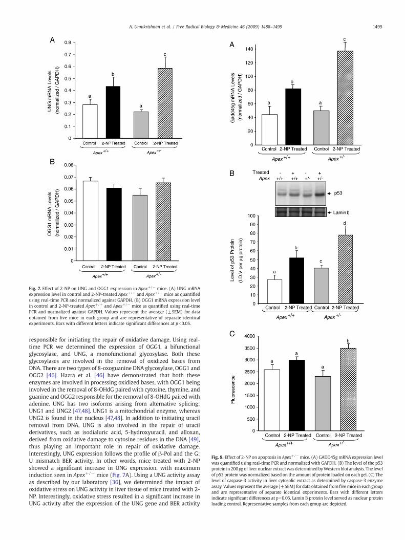
Fig. 8. Effect of 2-NP on apoptosis in Apex+/− mice. (A) GADD45gmRNA expression levelwas quantified using real-time PCR and normalized with GAPDH. (B) The level of the p53protein in200 μgof livernuclear extractwasdeterminedbyWesternblot analysis. The levelof p53 proteinwas normalized based on the amount of protein loaded on each gel. (C) Thelevel of caspase-3 activity in liver cytosolic extract as determined by caspase-3 enzymeassay. Values represent the average (±SEM) fordata obtained fromfivemice in eachgroupand are representative of separate identical experiments. Bars with different lettersindicate significant differences at pb0.05. Lamin B protein level served as nuclear proteinloading control. Representative samples from each group are depicted.
Fig. 7. Effect of 2-NP on UNG and OGG1 expression in Apex+/− mice. (A) UNG mRNAexpression level in control and 2-NP-treated Apex+/+ and Apex+/− mice as quantifiedusing real-time PCR and normalized against GAPDH. (B) OGG1 mRNA expression levelin control and 2-NP-treated Apex+/+ and Apex+/− mice as quantified using real-timePCR and normalized against GAPDH. Values represent the average (±SEM) for dataobtained from five mice in each group and are representative of separate identicalexperiments. Bars with different letters indicate significant differences at pb0.05.
1495A. Unnikrishnan et al. / Free Radical Biology & Medicine 46 (2009) 1488–1499
responsible for initiating the repair of oxidative damage. Using real-time PCR we determined the expression of OGG1, a bifunctionalglycosylase, and UNG, a monofunctional glycosylase. Both theseglycosylases are involved in the removal of oxidized bases fromDNA. There are two types of 8-oxoguanine DNA glycosylase, OGG1 andOGG2 [46]. Hazra et al. [46] have demonstrated that both theseenzymes are involved in processing oxidized bases, with OGG1 beinginvolved in the removal of 8-OHdG paired with cytosine, thymine, andguanine and OGG2 responsible for the removal of 8-OHdG paired withadenine. UNG has two isoforms arising from alternative splicing;UNG1 and UNG2 [47,48]. UNG1 is a mitochondrial enzyme, whereasUNG2 is found in the nucleus [47,48]. In addition to initiating uracilremoval from DNA, UNG is also involved in the repair of uracilderivatives, such as isodialuric acid, 5-hydroxyuracil, and alloxan,derived from oxidative damage to cytosine residues in the DNA [49],thus playing an important role in repair of oxidative damage.Interestingly, UNG expression follows the profile of β-Pol and the G:U mismatch BER activity. In other words, mice treated with 2-NPshowed a significant increase in UNG expression, with maximuminduction seen in Apex+/− mice (Fig. 7A). Using a UNG activity assayas described by our laboratory [36], we determined the impact ofoxidative stress on UNG activity in liver tissue of mice treated with 2-NP. Interestingly, oxidative stress resulted in a significant increase inUNG activity after the expression of the UNG gene and BER activity

1496 A. Unnikrishnan et al. / Free Radical Biology & Medicine 46 (2009) 1488–1499
(data not shown). However, OGG1 mRNA levels did not changesignificantly among the control and experimental groups (Fig. 7B).Interestingly, addition of purified human OGG1 (Novus Biologicals)protein to the BER reaction mixture did not increase OGG1-initiatedBER activity in 2-NP-treated Apex+/− mice (data not shown),suggesting that APE1/Ref-1 activity is essential for bifunctional DNAglycosylase-initiated BER. Thus, upregulation in UNG expression andsimultaneous increase in β-Pol protein explain the increase in G:Umismatch BER activity in 2-NP-treated Apex+/− mice (Fig. 5A).Conversely, lack of induction of OGG1-initiated BER in the liver tissueof Apex+/− mice exposed to 2-NP (Fig. 6A) is expected to increaseoxidative DNA damage that drives apoptosis.
Analysis of apoptosis in the liver of Apex+/− mice in response tooxidative stress
To characterize the effect of APE1/Ref-1 heterozygosity on cellcycle arrest and apoptosis during oxidative stress, we analyzed threefactors associated with apoptosis: GADD45g (growth arrest and DNA-damage-inducible, γ), p53, and caspase-3. GADD45g, a genotoxicstress-inducible gene associated with cell cycle arrest and apoptosis,was analyzed by real-time PCR. The mRNA level of GADD45g wassignificantly increased in Apex+/+ mice in response to 2-NP (Fig. 8A).Under control conditions, there was no effect of genotype onGADD45g expression (Fig. 8A), whereas in response to oxidativestress, expression of GADD45g was significantly greater in Apex+/−
mice compared to their wild-type counterparts. Because GADD45g is ap53-responsive gene, we wanted to evaluate the role of p53 in theGADD45g gene response. Using Western blot assay, we determinedthe effect of APE1/Ref-1 heterozygosity on p53 stability in response tooxidative stress in the liver nuclear extracts. p53, a tumor suppressorprotein, is a well-established determinant of cell cycle arrest andapoptosis. p53 protein stability followed the same trend as GADD45gexpression, with 2-NP-treated Apex+/− mice displaying the highestlevel of p53 protein stability (Fig. 8B). We further analyzed the activityof caspase-3, one of the final effectors in the apoptotic pathway, underthe same conditions using liver cytosolic extracts. Caspase-3 activitywas significantly induced in the 2-NP-treated Apex+/− mice com-pared to their untreated counterparts (Fig. 8C). Thus, loss of APE1/Ref-1 increases the apoptotic response to oxidative stress.
Discussion
APE1/Ref-1 is a multifunctional protein involved in the main-tenance of genomic integrity and in the regulation of gene expression.In addition to its role in BER as the major 5′-endonuclease and 3′-phosphoesterase, APE1/Ref-1 was independently characterized as aredox activator of cellular transcription factors. Although research hasdemonstrated that APE1/Ref-1 is involved in the cellular response tooxidative DNA damage, it is currently unclear whether this response isstrictly due to APE1/Ref-1 repair activity, APE1/Ref-1 redox regulatoryactivity, or both. A study by Fritz et al. [50] suggests that APE1/Ref-1repair activity is constitutively expressed, whereas APE1/Ref-1 redoxactivity is subject to regulation under various stimuli. Furthermore,Hsieh et al. [51] present data indicating that APE1/Ref-1 redox activityis mediated by phosphorylation in response to oxidative stress.
Reports to date have shown that APE1/Ref-1 expression isinducible by various forms of oxidative stress in vitro [23–30]. Forexample, HeLa cells exposed to damaging agents such as hypochloriteor methyl methane sulfonate show an increase in both APE1/Ref-1expression and activity [51]. Additionally, Ramana et al. [24] haveshown induction of APE1/Ref-1 mRNA and protein levels withincreased translocation of the protein into the nucleus when HeLa S3tumor cells and WI 38 primary fibroblasts were exposed to ROSgenerators. APE1/Ref-1 protein has also been shown to be translocatedfrom the cytoplasm to the nucleus during H2O2 and ATP stimulation
[52]. To determine the effect of oxidative stress on APE1/Ref-1 in vivo,we usedmice containing a heterozygous gene-targeted deletion of APE(Apex+/−). Homozygous deletion of the APE1/Ref-1 gene (Apex−/−) isembryonic lethal, but heterozygous mice survive and are fertile[26,53,54]. These APE1/Ref-1 haploinsufficient (Apex+/−) mice showtissue-specific differences in BER capacity characterized by using an invitro G:U mismatch repair assay. In addition Huamani et al. [55] havedemonstrated that Apex+/− mice show a significant increase in spon-taneous mutagenesis in lacI transgenes in liver and spleen. Further-more, embryonic fibroblasts and brain cells obtained from Apex+/−
mice have been shown to be more susceptible to oxidizing agents[26,56]. In this study we used 2-NP, a well-known hepatocarcinogen[39,57], to induce oxidative stress in vivo. 2-NP, an industrial solvent, isfound in paints, varnishes, and cigarette smoke [39,57]. 2-NP promotesformation of 8-OHdG and 8-aminoguanine through its metabolites N-isopropylhydroxylamine and hydroxylamine-O-sulfonic acid, whichare believed to be the causative factors behind 2-NP-inducedcarcinogenesis [39]. In addition, 2-NP occurs as propane 2-nitronateat physiological pH and promotes genotoxicity through production ofNO species [57]. NO is shown to induce genotoxicity by deaminatingDNA and increasing the uracil-to-cytosine ratio [48,58]. 2-NP has alsobeen shown to be genotoxic in vitro, inducing mutations in bacteriaand increasing unscheduled DNA synthesis in cultured hepatocytes[40]. Additionally, our laboratory has previously demonstrated that 2-NP-treatment results in an increase in the levels of 8-OHdG in vivo inthe liver of mice and rats, followed by a concomitant increase in β-Polexpression and BER activity [22]. Here we verified that APE1/Ref-1 isindeed an inducible protein (Fig. 1B). Although fold increase is thesame in response to oxidative stress across the genotypes, the totalcumulative level of APE1/Ref-1 protein is lower in the liver of Apex+/−
mice, i.e., even though the intact allele is induced in response to 2-NP, itdoes not compensate for the lost allele.
APE1/Ref-1 has been shown to be an essential component oftranscription complexes by directly interacting with other transcrip-tion factors such as HIF-1 and p300 [59,60]. Research has shown thatAPE1/Ref-1 enhances the DNA binding activity of NF-κB in vitro aswell as NF-κB-dependent transcriptional activation in vivo [13,14].Deletion of the redox-sensitive domain of APE1/Ref-1 has been shownto inhibit TNF-induced NF-κB activation, whereas loss of APE1/Ref-1 isshown to result in a significant decline in NF-κB activity and increasedsusceptibility to TNF-induced apoptosis [15,16]. Furthermore, down-regulation of APE1/Ref-1 by soy isoflavones in vitro and in vivoresulted in concomitant reduction of NF-κB activity, whereas atwofold increase in APE1/Ref-1 expression, obtained by cDNAtransfection, resulted in a concomitant twofold increase in NF-κBactivity, confirming the cross talk between these molecules [61]. Inline with these findings the Apex+/+ mice showed a significantincrease in APE1/Ref-1 redox activation of NF-κB when exposed to 2-NP; whereas the increase in NF-κB activation was the same inresponse to oxidative stress across the genotypes, the total NF-κBDNA-binding activity was lower in the liver of Apex+/− mice, i.e., theultimate level of NF-κB activation was significantly attenuated in theheterozygous animals. It is well established that NF-κB is amediator ofinflammatory responses, promoting cell proliferation and survival byinhibiting cell cycle arrest and apoptosis [62,63]; thus reducedactivation of NF-κB and other redox-regulated transcription factorsin response to oxidative stress in Apex+/− mice may provedetrimental owing to alterations in the signaling mechanismsnecessary to differentiate between repair and apoptosis.
We have previously reported tissue-specific differences in BERcapacity in APE1/Ref-1 haploinsufficient mice [31]. Moreover,previous studies have indicated that downregulation of APE1/Ref-1may promote a damage-hypersensitive phenotype [26–28]. There-fore, it was essential to analyze the effect of reduced APE1/Ref-1 onthe level of DNA damage production and BER. In this study, wedemonstrate that the liver tissue of Apex+/− mice expresses a BER

1497A. Unnikrishnan et al. / Free Radical Biology & Medicine 46 (2009) 1488–1499
phenotype that is more susceptible to accumulation of DNA damagein response to oxidative stress as a result of reduced APE1/Ref-13′-phoshoesterase activity in vivo. Here we show the differentialimpact of APE1/Ref-1 heterozygosity on monofunctional and bifunc-tional glycosylase-initiated BER. The traditional BER response tocertain DNA base damages, such as uracil and alkylated bases,involves a monofunctional glycosylase-mediated removal of thedamaged base resulting in the generation of an abasic site (AP site)that serves as a substrate for APE1/Ref-1 endonuclease activity.APE1/Ref-1 subsequently promotes formation of a transient single-strand break that serves as a substrate for β-Pol-mediated nucleotideinsertion and is followed by DNA ligase-mediated BER completion. β-Pol, dRP lyase activity serves as the rate-limiting step in this pathway[64]. On the other hand, other damages such as oxidized bases (e.g.,8-hydroxyguanosine and thymine glycol) require a bifunctionalglycosylase-mediated removal of the damage. These glycosylaseshave associated AP lyase activity resulting in 3′-blocking ends. DNApolymerase requires a 3′-OH group as its substrate for repairsynthesis. Therefore 3′-blocking end trimming requires the 3′-phosphoesterase activity of APE1/Ref-1. β-Pol and DNA ligasesubsequently complete the repair process. The 3′-phosphoesteraseactivity of APE1/Ref-1 is a crucial step in this bifunctionalglycosylase-mediated BER [41]. In this study, we show the differencein the requirement of APE1/Ref-1 in the BER pathway in vivo. OurApex+/- mice exposed to 2-NP show significant upregulation in theUNG message with concomitant increases in the β-Pol protein and G:U mismatch BER capacity in the liver tissue. We suggest that 2-NP, viaits NO generation, leads to increased uracil levels in the DNA resultingin the upregulation of UNG, a monofunctional glycosylase that is alsoassociated with the processing of oxidized cytosine derivatives [49].Because β-Pol is the rate-limiting enzyme in this pathway, the liverextracts of Apex+/− mice treated with 2-NP show significantinduction in the G:U mismatch repair. Apex+/− mice treated with2-NP showed increased binding activity for CREB, a stress-responsetranscriptional activator of the β-Pol promoter, potentially explainingthe induction of β-Pol expression [44]. It is widely accepted that β-Pol protein expression and activity strongly correlate with BERactivity in response to a variety of stressors [22,65–67]. Moreover, theendonuclease activity of APE1/Ref-1 is 100-fold more efficient thanthe 3′-phosphoesterase activity [68]; thus we suggest that theavailable APE1/Ref-1 protein in Apex+/− mice is adequate to sustainthe monofunctional glycosylase-initiated BER. In addition, increasedp53 level can stabilize the APE1/Ref-1 and β-Pol protein complex onthe DNA abasic site, which would enhance BER activity [69,70].
Alternatively, the liver tissues of the 2-NP-treated Apex+/− micedisplayed significantly lower 8-OHdG:C BER activity compared to thewild-type animals. OGG1 is a bifunctional glycosylase known to beinvolved in the removal of 8-OHdG. Although OGG1 mRNA andprotein levels are shown to be unaffected during oxidative stress, theactivity of this enzyme is enhanced by the increased APE1/Ref-1protein [71]. In line with these findings, our 2-NP-treated Apex+/−
mice did not show any significant difference in OGG1 mRNA levels.Furthermore, addition of purified human OGG1 protein to the BERreaction mixture did not increase OGG1-initiated BER activity in 2-NP-treated Apex+/− mice. However, addition of purified APE1/Ref-1protein to the BER reaction mix resulted in a significant increase in the8-OHdG:C BER activity in these mice, confirming the significant roleplayed by APE1/Ref-1 in the removal of 3′-blocking groups. These dataindicate that APE1/Ref-1 3′-phosphoesterase activity is critical inremoving the 3′-blocking lesion created by a bifunctional glycosylase,e.g., OGG1. Therefore, APE1/Ref-1 heterozygosity compromises thisfunction, resulting in reduced detectable single-strand breaks,increased aldehydic lesions, and reduced bifunctional DNA glycosy-lase-initiated BER activity.
Previously, we measured the presence of AP sites, single-strandbreaks, and aldehydic lesions in isolated liver DNA from APE1/Ref-1
haploinsufficient mice and observed no significant difference in DNAdamage accumulation as a result of reduced APE1/Ref-1 [31]. Thelack of damage accumulation in untreated Apex+/− mice suggestedthat APE1/Ref-1 haploinsufficiency in liver does not cause anaccumulation of genotoxic DNA repair intermediate productsunder baseline conditions. In line with previous studies from ourlaboratory [22] and others [72], we have demonstrated a significantincrease in 3′-OH-containing single-strand breaks in response tooxidative stress. However, the level of detectable single-strandbreaks in the liver tissue of 2-NP-treated Apex+/− mice wassignificantly lower than in their wild-type counterparts, whereasthe level of aldehydic lesions was significantly higher. We suggestthat the processing of oxidized bases by a bifunctional DNA glyco-sylase such as OGG1 could result in the generation of aldehydicblocking lesions at the 3′-end. Inability to process these 3′-blockinggroups in the absence of the 3′-phosphoesterase activity of Apex inApex+/− mice [41] could result in lower detection of endonuclease-mediated single-strand breaks in the heterozygous animal.
Herein, we demonstrated that APE1/Ref-1 haploinsufficiencydoes not promote a concomitant decline in G:U mismatch BERactivity in the liver in response to oxidative stress; rather it displaysa significant increase in this activity. Although the high efficiency ofAPE1/Ref-1 endonuclease activity and upregulation in UNG andβ-Pol expression seem to be the reasons behind the significantincrease in G:U mismatch repair activity, alternatively, recentstudies report an APE1/Ref-1-independent BER pathway in humancells [42]. Whereas monofunctional DNA glycosylases such asSMUG1 and TDG and bifunctional DNA glycosylases/lyases such asOGG1 and NTH require APE1/Ref-1 to process AP sites and the 3′-OH aldehydic groups, respectively, APE1/Ref-1 backup enzymeshave been identified for the repair of oxidative damage in vivo [73].The recently discovered mammalian DNA glycosylase/AP lyasesNEIL1 and NEIL2 recognize oxidative damage and generate DNAstrand breaks with 3′-phosphate termini. Removal of the 3′-phosphate termini was shown to be dependent upon polynucleotidekinase activity, not APE1/Ref-1, with NEIL1 serving as a backupenzyme. Additionally, a study by Das et al. [74] has demonstratedthat NEIL1 is induced by oxidative stress. Therefore, it is likely thatthe observed increase in BER may be due to increased processing ofDNA damage by an APE-independent β-Pol-regulated BER pathway;however, this alternative pathway does not rescue the observeddecline in 8OHdG:C BER capacity in APE1/REF-1 haploinsufficientmice.
In conclusion, we suggest that the optimal balance between thestress-response transcription factors, notably NF-κB, and the DNArepair pathway required for cell survival is attenuated in APE1/Ref-1haploinsufficient mice exposed to oxidative stress. Reduced redoxactivity of APE1/Ref-1 leads to reduced activation of NF-κB, potentiallyleading to cell cycle arrest and apoptosis. This notion is supported bythe augmentation of the expression of GADD45g, a cell cycle arrestgene, observed in the liver of 2-NP-treated Apex+/− mice. GADD45ghas also been implicated in the promotion of apoptosis underenvironmental stress via the p38K–Jun NH2-terminal kinase pathway[75,76]. Furthermore, Apex+/− mice exposed to 2-NP show significantinduction of p53 protein levels and caspase-3 activity in the liver, bothbeing markers of apoptosis. Therefore, when APE1/Ref-1 is compro-mised the cells are more susceptible to oxidative stress mainly owingto reduced redox and 3′-phosphodiestrase activity impacting cellsurvival, pushing it toward apoptosis. This finding has therapeuticimportance as increased APE1/Ref-1 levels in tumor cells have beenshown to confer resistance to chemotherapeutic drugs, perhaps via adecrease in apoptosis [77]. In line with previous findings, inhibition ofDNA repair or redox or both activities of APE1/Ref-1 using inhibitorscould potentially sensitize the tumor cells to therapeutic agents [77],making APE1/Ref-1 an attractive molecular target in the treatment ofcancer.

1498 A. Unnikrishnan et al. / Free Radical Biology & Medicine 46 (2009) 1488–1499
Acknowledgments
This work was supported by grants from the National Institutes ofHealth (R01 CA121298 to A.R.H.) and the American Institute forCancer Research (03A061 to A.R.H.).
References
[1] Evans, M. D.; Dizdaroglu, M.; Cooke, M. S. Oxidative DNA damage and disease:induction, repair and significance. Mutat. Res. 567:1–61; 2004.
[2] Mullaart, E.; Lohman, P. H. M.; Berends, F.; Vijg, J. DNA damage, metabolism andaging. Mutat. Res. 237:189–210; 1990.
[3] Jackson, A. L.; Loeb, L. A. The contribution of endogenous sources of DNA damageto the multiple mutations in cancer. Mutat. Res. 477:7–21; 2001.
[4] Sancar, A.; Lindsey-Boltz, L. A.; Unsal-Kacmaz, K.; Linn, S. Molecular mechanismsof mammalian DNA repair and the DNA damage checkpoints. Annu. Rev. Biochem.73:39–85; 2004.
[5] Slupphaug, G.; Kavli, B.; Krokan, H. E. The interacting pathways for prevention andrepair of oxidative DNA damage. Mutat. Res. 531:231–251; 2003.
[6] Friedberg, E. C.; Walker, G. C.; Siede, W.; Wood, R. D.; Schultz, R. A.; Ellenberger, T.DNA Repair and Mutagenesis. ASM Press, Washington, DC, pp. 169–226; 2006.
[7] Ljungquist, S.; Lindahl, T. A mammalian endonuclease specific for apurinic sites indouble-stranded deoxyribonucleic acid. I. Purification and general properties.J. Biol. Chem. 249:1530–1535; 1974.
[8] Ljungquist, S.; Andersson, A.; Lindahl, T. A mammalian endonuclease specific forapurinic sites in double-stranded deoxyribonucleic acid. II. Further studies on thesubstrate specificity. J. Biol. Chem. 249:1536–1540; 1974.
[9] Lindahl, T. Inroads into base excision repair. I. The discovery of apurinic/apyrimidinic (AP) endonuclease. DNA Repair 3:1521–1530; 2004.
[10] Xanthoudakis, S.; Curran, T. Identification and characterization of Ref-1, a nuclearprotein that facilitates AP-1 DNA-binding activity. EMBO J. 11:653–665; 1992.
[11] Xanthoudakis, S.; Miao, G.; Wang, F.; Pan, Y. C.; Curran, T. Redox activation ofFos–Jun DNA binding activity is mediated by a DNA repair enzyme. EMBO J. 11:3323–3335; 1992.
[12] Evans, A. R.; Limp-Foster, M.; Kelley, M. R. Going APE over Ref-1. Mutat. Res. 461:83–108; 2000.
[13] Shimizu, N.; Sugimoto, K.; Tang, J.; Nishi, T.; Sato, I.; Hiramoto, M.; Aizawa, S.;Hatakeyama, M.; Ohba, R.; Hatori, H.; Yoshikawa, T.; Suzuki, F.; Oomori, A.; Tanaka,H.; Kawaguchi, H.; Watanabe, H.; Handa, H. High-performance affinity beads foridentifying drug receptors. Nat. Biotechnol. 18:877–881; 2000.
[14] Nishi, T.; Shimizu, N.; Hiramoto, M.; Sato, I.; Yamaguchi, Y.; Hasegawa, M.; Aizawa,S.; Tanaka, H.; Kataoka, K.; Watanabe, H.; Handa, H. Spatial redox regulation of acritical cysteine residue of NF-kappa B in vivo. J. Biol. Chem. 277:44548–44556;2002.
[15] Hall, J. L.; Wang, X.; Adamson, V.; Zhao, Y.; Gibbons, G. H. Overexpression of Ref-1inhibits hypoxia and tumor necrosis factor-induced endothelial cell apoptosisthrough nuclear factor-κB-independent and -dependent pathways. Circ. Res. 88:1247–1253; 2001.
[16] Guan, Z.; Basi, D.; Li, Q.; Mariash, A.; Xia, Y. F.; Geng, J. G.; Kao, E.; Hall, J. L. Loss ofredox factor 1 decreases NF-kappaB activity and increases susceptibility ofendothelial cells to apoptosis. Arterioscler. Thromb. Vasc. Biol. 25:96–101; 2005.
[17] Lindahl, T. Keynote: past, present, and future aspects of base excision repair.Nucleic Acid Res. Mol. Biol. 68:xvii–xxx; 2001.
[18] Parikh, S. S.; Mol, C. D.; Hosfield, D. J.; Tainer, J. A. Envisioning the molecularchoreography of DNA base excision repair. Curr. Opin. Struct. Biol. 9:37–47; 1999.
[19] Wilson, S. H.; Kunkel, T. A. Passing the baton in base excision repair. Nat. Struct.Biol. 7:176–178; 2000.
[20] Wilson III, D. M.; Sofinowski, T. M.; McNeill, D. R. Repair mechanisms for oxidativeDNA damage. Front. Biosci. 8:d963–d981; 2003.
[21] Izumi, T.; Wiederhold, L. R.; Roy, G.; Roy, R.; Jaiswal, A.; Bhakat, K. K.; Mitra, S.;Hazra, T. K. Mammalian DNA base excision repair proteins: their interactions androle in repair of oxidative DNA damage. Toxicology 193:43–65; 2003.
[22] Cabelof, D. C.; Raffoul, J. J.; Yanamadala, S.; Guo, Z.; Heydari, A. R. Induction of DNApolymerase β-dependent base excision repair in response to oxidative stress invivo. Carcinogenesis 23:1419–1425; 2002.
[23] Grosch, S.; Fritz, G.; Kaina, B. Apurinic endonuclease (Ref-1) is induced inmammalian cells by oxidative stress and is involved in clastogenic adaptation.Cancer Res. 58:4410–4416; 1998.
[24] Ramana, C. V.; Boldogh, I.; Izumi, T.; Mitra, S. Activation of apurinic/apyrimidinicendonuclease in human cells by reactive oxygen species and its correlation withtheir adaptive response to genotoxicity of free radicals. Proc. Natl. Acad. Sci. U. S. A.95:5061–5066; 1998.
[25] Ono, Y.; Furuta, T.; Ohmoto, T.; Akiyama, K.; Seki, S. Stable expression in rat gliomacells of sense and antisense nucleic acids to a human multifunctional DNA repairenzyme, APEX nuclease. Mutat. Res. 315:55–63; 1994.
[26] Meira, L. B.; Devaraj, S.; Kisby, G. E.; Burns, D. K.; Daniel, R. L.; Hammer, R. E.;Grundy, S.; Jialal, I.; Friedberg, E. C. Heterozygosity for the mouse Apex generesults in phenotypes associated with oxidative stress. Cancer Res. 61:5552–5557;2001.
[27] Walker, L. J.; Craig, R. B.; Harris, A. L.; Hickson, I. D. A role for the human DNA repairenzyme HAP1 in cellular protection against DNA damaging agents and hypoxicstress. Nucleic Acids Res. 22:4884–4889; 1994.
[28] Fung, H.; Demple, B. A vital role for Ape1/Ref1 protein in repairing spontaneousDNA damage in human cells. Mol. Cell 17:463–470; 2005.
[29] Izumi, T.; Brown, D. B.; Naidu, C. V.; Bhakat, K. K.; MacInnes, M. A.; Saito, H.; Chen,D. J.; Mitra, S. Two essential but distinct functions of mammalian abasicendonuclease. Proc. Natl. Acad. Sci. U. S. A. 102:5739–5743; 2005.
[30] Vasko, M. R.; Guo, C.; Kelley, M. R. The multifunctional DNA repair/redox enzymeApe1/Ref-1 promotes survival of neurons after oxidative stress. DNA Repair 4:367–379; 2005.
[31] Raffoul, J. J.; Cabelof, D. C.; Nakamura, J.; Meira, L. B.; Friedberg, E. C.; Heydari, A. R.Apurinic/apyrimidinic endonuclease (APE/REF-1) haploinsufficient mice displaytissue-specific differences in DNA polymerase beta-dependent base excisionrepair. J. Biol. Chem. 279:18425–18433; 2004.
[32] Hadi, M. Z.; Coleman, M. A.; Fidelis, K.; Mohrenweiser, H. W.; Wilson III, D. M.Functional characterization of Ape1 variants identified in the human population.Nucleic Acids Res. 28:3871–3879; 2000.
[33] De Boer, J. G. Polymorphisms in DNA repair and environmental interactions.Mutat. Res. 509:201–210; 2002.
[34] Mohrenweiser, H. W.; Wilson III, D. M.; Jones, I. M. Challenges and complexities inestimating both the functional impact and the disease risk associated with theextensive genetic variation inhumanDNArepair genes.Mutat. Res.526:93–125; 2003.
[35] Hou, E. W.; Prasad, R.; Asagoshi, K.; Masaoka, A.; Wilson, S. H. Comparativeassessment of plasmid and oligonucleotide DNA substrates in measurement of invitro base excision repair activity. Nucleic Acids Res. 35:1–10; 2007.
[36] Cabelof, D. C.; Raffoul, J. J.; Nakamura, J.; Kapoor, D.; Abdalla, H.; Heydari, A. R.Imbalanced base excision repair in response to folate deficiency is accelerated bypolymerase beta haploinsufficiency. J. Biol. Chem. 279:36504–36513; 2004.
[37] Cabelof, D. C.; Nakamura, J.; Heydari, A. R. A sensitive biochemical assay for thedetection of uracil. Environ. Mol. Mutagen. 47:31–37; 2006.
[38] Sokal, R. R.; Rohlf, F. J. Biometry. Freeman, New York; 1981.[39] Sakano, K.; Oikawa, S.; Murata, M.; Hiraku, Y.; Kojima, N.; Kawanishi, S.
Mechanisms of metal-mediated DNA damage induced by metabolites of carcino-genic 2-nitropropane. Mutat. Res. 479:101–111; 2001.
[40] Andrae, U.; Homfeldt, H.; Vogl, L.; Lichtmannegger, J.; Summer, K. H. 2-Nitropropane induces DNA repair synthesis in rat hepatocytes in vitro and invivo. Carcinogenesis 9:811–815; 1988.
[41] Izumi, T.; Hazra, T. K.; Boldogh, I.; Tomkinson, A. E.; Park, M. S.; Ikeda, S.; Mitra, S.Requirement for human AP endonuclease 1 for repair of 3′-blocking damage atDNA single-strand breaks induced by reactive oxygen species. Carcinogenesis 21:1329–1334; 2000.
[42] Wiederhold, L.; Leppard, J. B.; Kedar, P.; Karimi-Busheri, F.; Rasouli-Nia, A.;Weinfeld, M.; Tomkinson, A. E.; Izumi, T.; Prasad, R.; Wilson, S. H.; Mitra, S.; Hazra,T. K. AP endonuclease-independent DNA base excision repair in human cells. Mol.Cell 15:209–220; 2004.
[43] Sabine, G.; Kaina, B. Transcriptional activation of apurinic/apyrimidinic endonu-clease (Ape,Ref-1) by oxidative stress requires CREB. Biochem. Biophys. Res.Commun. 261:859–863; 1999.
[44] Yau-Jan, C.; Rawson, T. Y.; Wilson, S. H. Cloning and characterization of a novelmember of the human ATF/CREB family: ATF2 deletion, a potential regulator ofthe human DNA polymerase β promoter. Gene 312:117–124; 2003.
[45] Fornance Jr., A. J.; Zmudzka, B.; Hollander, M. C.; Wilson, S. H. Induction of beta-polymerase mRNA by DNA-damaging agents in Chinese hamster ovary cells. Mol.Cell Biol. 9:851–853; 1989.
[46] Hazra, T. K.; Izumi, T.; Maidt, L.; Floyd, R. A.; Mitra, S. The presence of two distinct8-oxoguanine repair enzymes in human cells: their potential complementaryroles in preventing mutation. Nucleic Acids Res. 26:5116–5122; 1998.
[47] Nilsen, H.; Rosewell, I.; Robins, P.; Skjelbred, C. F.; Andersom, S.; Slupphaug, G.;Daly, G.; Krokan, H. E.; Lindahl, T.; Barnes, D. E. Uracil-DNA glycosylase (UNG)-deficient mice reveal a primary role of the enzyme during DNA replication. Mol.Cell 5:1059–1065; 2000.
[48] Endres, M.; Biniszkiewicz, D.; Sobol, R. B.; Harms, C.; Ahmadi, M.; Lipski, A.;Katchanov, J.; Mergenthaler, P.; Dirnagl, U.; Wilson, S. H.; Meisel, A.; Jaenisch, R.Increased postischemic brain injury in mice deficient in uracil-DNA glycosylase.J. Clin. Invest. 113:1711–1721; 2004.
[49] Dizdaroglu, M.; Karakaya, A.; Jaruga, P.; Slupphaug, G.; Krokan, H. E. Novelactivities of human uracil DNA N-glycosylase for cytosine derived products ofoxidative DNA damage. Nucleic Acids Res. 24:418–422; 1996.
[50] Fritz, G.; Grosch, S.; Tomicic, M.; Kaina, B. APE/Ref-1 and the mammalian responseto genotoxic stress. Toxicology 193:67–78; 2003.
[51] Hsieh, M.M.; Hegde, V.; Kelley, M. R.; Deutsch,W. A. Activation of APE/Ref-1 redoxactivity is mediated by reactive oxygen species and PKC phosphorylation. NucleicAcids Res. 29:3116–3122; 2001.
[52] Pines, A.; Perrone, L.; Bivi, N.; Romanello, M.; Damante, G.; Gulisano, M.; Kelley,M. R.; Quadrifoglio, F.; Tell, G. Activation of APE1/Ref-1 is dependent on reactiveoxygen species generated after purinergic receptor stimulation by ATP. NucleicAcids Res. 33:4379–4394; 2005.
[53] Ludwig, D. L.; MacInnes, M. A.; Takiguchi, Y.; Purtymun, P. E.; Henrie, M.; Flannery,M.; Meneses, J.; Pederson, R. A.; Chen, D. J. A murine AP-endonuclease gene-targeted deficiency with postimplantation embryonic progression and ionizingradiation sensitivity. Mutat. Res. 409:17–29; 1998.
[54] Xanphoudakis, S.; Smeyne, R. J.; Wallace, J. D.; Curran, T. The redox/DNA repairprotein, Ref-1, is essential for early embryonic development in mice. Proc. Natl.Acad. Sci. U. S. A. 93:8919–8923; 1996.
[55] Huamani, J.; McMahan, C. A.; Herbert, D. C.; Reddick, R.; McCarrey, J. R.; MacInnes,M. I.; Chen, D. J.; Walter, C. A. Spontaneous mutagenesis is enhanced in Apexheterozygous mice. Mol. Cell. Biol. 24:8145–8153; 2004.
[56] Friedberg, E. C.; Meira, L. B. Database of mouse strains carrying targeted mutationsin genes affecting biological responses to DNA damage version 7. DNA Repair 5:189–209; 2006.

1499A. Unnikrishnan et al. / Free Radical Biology & Medicine 46 (2009) 1488–1499
[57] Kohl, C.; Morgan, P.; Gescher, A. Metabolism of the genotoxicant 2-nitropropane toa nitric oxide species. Chem.-Biol. Interact. 97:175–184; 1995.
[58] Wink, D. A.; Kasprzak, K. S.; Maragos, C. M.; Elespuru, R. K.; Misra, M.; Dunams,T. M.; Cebula, T. A.; Koch, W. H.; Andrews, A. W.; Allen, J. S.; Keefer, L. K. DNAdeaminating ability and genotoxicity of nitric oxide and its progenitors. Science254:1001–1003; 1991.
[59] Carrero, P.; Okamoto, K.; Coumailleau, P.; O'Brien, S.; Tanaka, H.; Poellinger, L.Redox-regulated recruitment of the transcriptional coactivators CREB-bindingprotein and SRC-1 to hypoxia-inducible factor 1alpha. Mol. Cell Biol. 20:402–415;2000.
[60] Ziel, K. A.; Campbell, C. C.; Wilson, G. L.; Gillespie, M. N. Ref-1/Ape is critical forformation of the hypoxia-inducible transcriptional complex on the hypoxicresponse element of the rat pulmonary artery endothelial cell VEGF gene. FASEB J.18:986–988; 2004.
[61] Raffoul, J. J.; Banerjee, S.; Singh-Gupta, V.; Knoll, Z. E.; Fite, A.; Zhang, H.; Abrams,J.; Sarkar, F. H.; Hillman, G. G. Down-regulation of apurinic/apyrimidinicendonuclease 1/redox-factor 1 expression by soy isoflavones enhances prostatecancer radiotherapy in vitro and in vivo. Cancer Res. 67:2141–2149; 2007.
[62] Raffoul, J. J.; Wang, Y.; Kucuk, O.; Forman, J. D.; Sarkar, F. H.; Hillman, G. G.Genistein inhibits radiation-induced activation of NF-κB in prostate cancercells promoting apoptosis and G2/M cell cycle arrest. BMC Cancer 6:107;2006.
[63] Baldwin Jr., A. S. The transcription factor NF-κB and human disease. J. Clin. Invest.107:3–6; 2001.
[64] Srivastava, D. K.; Vande Berg, B. J.; Prasad, R.; Molina, J. T.; Beard,W. A.; Tomkinson,A. E.;Wilson, S. H. Mammalian abasic site base excision repair: identification of thereaction sequence and rate-determining steps. J. Biol. Chem. 273:21203–21209;1998.
[65] Cabelof, D. C.; Raffoul, J. J.; Yanamadala, S.; Ganir, C.; Guo, Z.; Heydari, A. R.Attenuation of DNA polymerase beta-dependent base excision repair andincreased DMS-induced mutagenicity in aged mice. Mutat. Res. 500:135–145;2002.
[66] Cabelof, D. C.; Guo, Z.; Raffoul, J. J.; Sobol, R. W.; Wilson, S. H.; Richardson, A.;Heydari, A. R. Base excision repair deficiency caused by polymerase beta
haploinsufficiency: accelerated DNA damage and increased mutational responseto carcinogens. Cancer Res. 63:5799–5807; 2003.
[67] Cabelof, D. C.; Yanamadala, S.; Raffoul, J. J.; Guo, Z.; Soofi, A.; Heydari, A. R. Caloricrestriction promotes genomic stability by induction of base excision repair andreversal of its age-related decline. DNA Repair 2:295–307; 2003.
[68] Suh, D.; Wilson, D. M.; Povirk, L. F. 3′-Phosphodiesterase activity of humanapurinic/apyrimidinic endonuclease at DNA double-strand break ends. NucleicAcids Res. 25:2495–2500; 1997.
[69] Bernstein, C.; Bernstein, C. H.; Payne, C. M.; Garewal, H. DNA repair/pro-apoptoticdual-role proteins in five major DNA repair pathways: fail-safe protection againstcarcinogenesis. Mutat. Res. 511:145–178; 2002.
[70] Zhou, J.; Ahn, J.; Wilson, S. H.; Prives, C. A role of P53 in base excision repair.EMBO J. 20:914–923; 2001.
[71] Saitoh, T.; Shinmura, K.; Yamaguchi, S.; Tani, M.; Seki, S.; Murakami, H.; Nojima, Y.;Yokota, J. Enhancement of OGG1 protein AP lyase activity by increase of Apexprotein. Mutat. Res. 486:31–40; 2001.
[72] Huang, D.; Shenoy, A.; Cui, J.; Huang, W.; Liu, P. K. In situ detection of AP sites andDNA strand breaks bearing 3′-phosphate termini in ischemic mouse brain. FASEB J.14:407–417; 2000.
[73] Mokkapati, S. K.; Wiederhold, L.; Hazra, T. K.; Mitra, S. Stimulation of DNAglycosylase activity of OGG1 by NEIL1: functional collaboration between twohuman DNA glycosylases. Biochemistry 43:11596–11604; 2004.
[74] Das, A.; Hazra, T. K.; Boldogh, I.; Mitra, S.; Bhakat, K. K. Induction of the humanoxidized base-specific DNA glycosylase NEIL1 by reactive oxygen species. J. Biol.Chem. 280:35272–35280; 2005.
[75] Ying, J.; Srivastava, G.; Hsieh, W. S.; Gao, Z.; Murray, P.; Liao, S. K.; Tao, Q. Thestress-responsive gene GADD45g is a functional tumor suppressor, with its res-ponse to environmental stresses frequently disrupted epigenetically in multipletumors. Clin. Cancer Res. 18:6442–6449; 2005.
[76] Takekawa, M.; Saito, H. A family of stress-inducible GADD45-like proteins mediateactivation of the stress-responsive MTK1/MEKK4/MAPKKK. Cell 95:521–530;1998.
[77] Fishel, M. L.; Kelley, M. R. The DNA base excision repair protein Ape1/Ref-1 as atherapeutic and chemopreventive target. Mol. Aspects Med. 28:375–395; 2007.