oxide for suzuki coupling 4 of pd s generated via one pot
TRANSCRIPT

1
Electronic Supporting InformationFor
Shape dependent catalytic activity of nanoflowers and nanospheres of Pd4S generated via one pot synthesis and grafted on graphene oxide for Suzuki coupling
Ved Vati Singh, Umesh Kumar, Sandeep Nath Tripathi, and Ajai Kumar Singh*
Department of Chemistry, Indian Institute of Technology Delhi, New Delhi-110016, India
Email: [email protected]
S1 Three-phase Test
For immobilization of 4-bromobenzoic acid on silica gel following standard procedure1-3
was used (Scheme S1).
Br
O
Cl
Pyridine, 12 h, 40 oC
O
OSi
OR
NH
Br
O
O
OSi
OR
NH2
Scheme S1. Immobilization of 4-bromobenzoic acid on silica
4-Bromobenzoic acid (1.99 g, 10 mmol) was mixed with dry SOCl2 (20 mL) and the
mixture refluxed for 3 h. It was cooled and thionyl chloride distilled off to give 4-
bromobenzoyl chloride as a white solid. 3-Aminopropyl trimethoxysilane-modified silica
(1 g), pyridine (0.4 mL), dry THF (10 mL) and 4-bromobenzoyl chloride (1.150 g, 5.25
mmol) were stirred at 40 °C for 12 h in a round bottom flask under N2
atmosphere. The suspension was filtered through G-4 crucible and washed with 5%
HCl (v/v) (3 × 20 mL) followed by 0.02 M solution of K2CO3 in water (2 × 20 mL) and
rinsed with distilled water (40 mL) and ethanol (40 mL). The resulting solid was washed
with dichloromethane and dried at room temperature in air, resulting in white powder.
Electronic Supplementary Material (ESI) for Dalton Transactions.This journal is © The Royal Society of Chemistry 2014
2
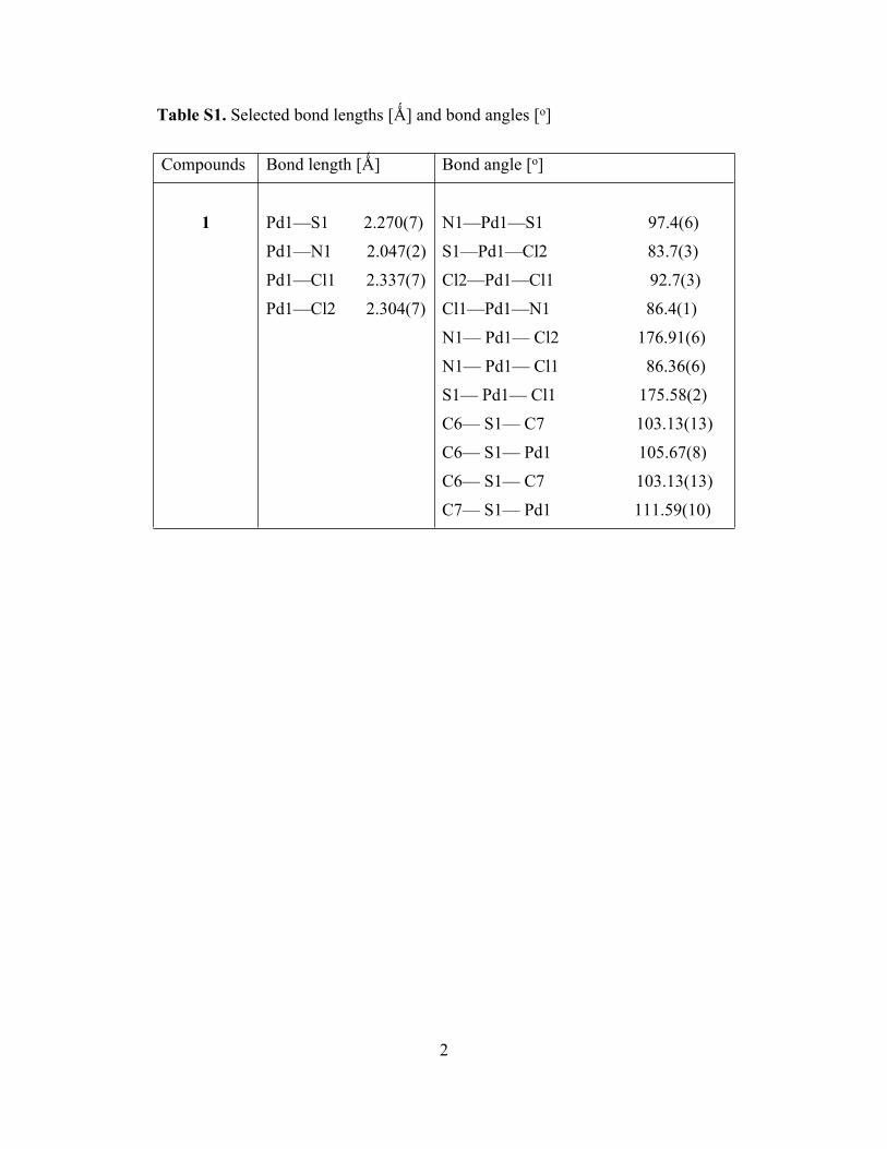
Table S1. Selected bond lengths [Ǻ] and bond angles [o]
Compounds Bond length [Ǻ] Bond angle [o]
1 Pd1—S1 2.270(7)
Pd1—N1 2.047(2)
Pd1—Cl1 2.337(7)
Pd1—Cl2 2.304(7)
N1—Pd1—S1 97.4(6)
S1—Pd1—Cl2 83.7(3)
Cl2—Pd1—Cl1 92.7(3)
Cl1—Pd1—N1 86.4(1)
N1— Pd1— Cl2 176.91(6)
N1— Pd1— Cl1 86.36(6)
S1— Pd1— Cl1 175.58(2)
C6— S1— C7 103.13(13)
C6— S1— Pd1 105.67(8)
C6— S1— C7 103.13(13)
C7— S1— Pd1 111.59(10)

3
Table S2. Crystallographic data and structure refinement summary for 1.
1
Formula C9H13 Cl2NPdSFormula weight 344.57T/K 298(2)λ/Å 0.71073Cryst system monoclinicSpace group P21/ca/Å 10.6716(18)b/Å 13.846(2)c/Å 8.2794(14)α/deg 90.00β/deg 108.956(3)γ/deg 90.00Vol/Å3 1157.0(3)Z 4Dcalcd/ g.cm-3 1.978F(000) 680 range/deg 2.02−25.00Reflections measured 2040Reflections used 1958parameters 127μ(Mo Kα) (cm-1) 2.205R1, wR2[I > 2σ(I)]a 0.0198, 0.0207R1, wR2 (all data)b 0.0486, 0.0490GOFc 1.060
aR1 = ΣFo – Fc/ΣFo; bwR2 = {Σ[w(Fo2 – Fc
2)2]/Σ[w(Fo2)2]}1/2; cS = {Σ[w(Fo
2 – Fc2)2]/(n–
p)2]}1/2
4
Table S3. Selected hydrogen bond parameters of 1 (Inter atomic distances in Å and bond
angles in deg).
D–HA D–H HA DA D–HA
N1–H1ACl2 0.90 2.70 3.14 136
C8–H8BCl2 0.97 2.95 3.69 134.
N1–H1ACl2 0.90 2.56 3.34 146
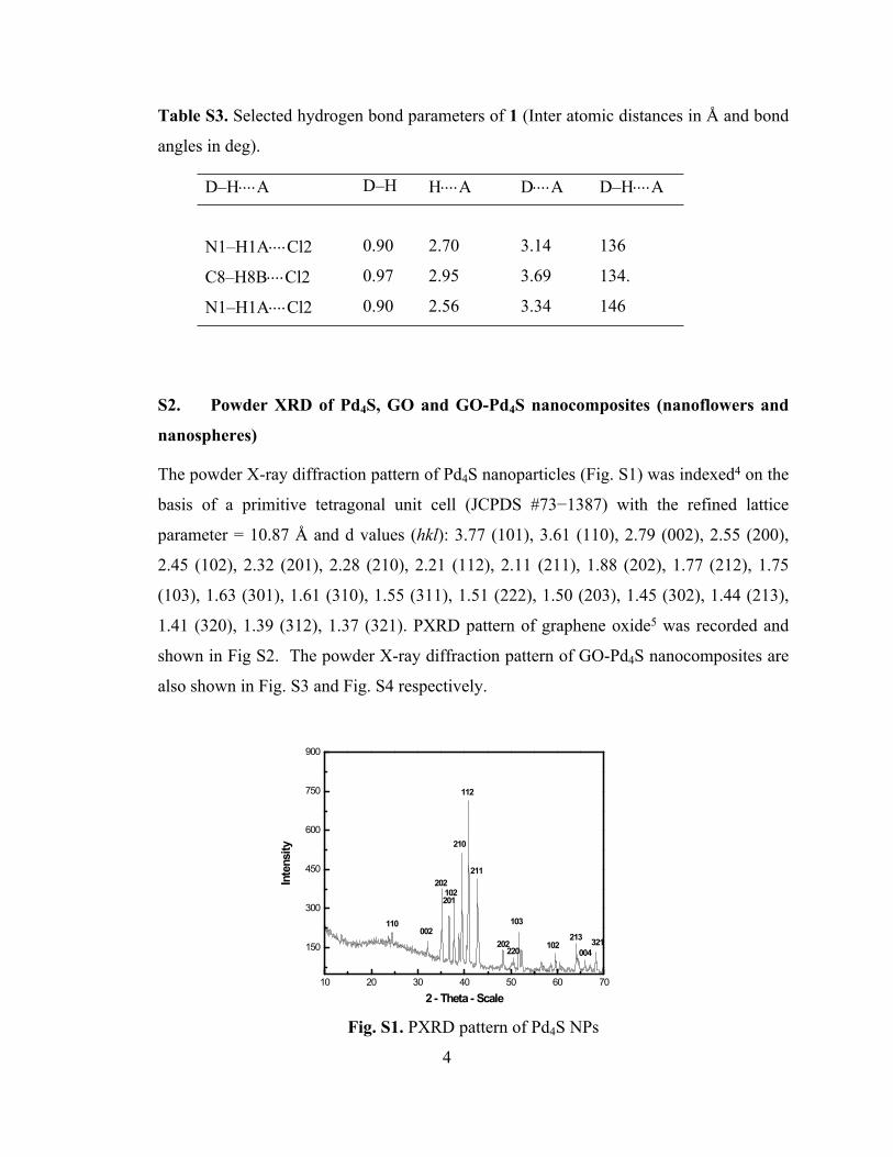
S2. Powder XRD of Pd4S, GO and GO-Pd4S nanocomposites (nanoflowers and
nanospheres)
The powder X-ray diffraction pattern of Pd4S nanoparticles (Fig. S1) was indexed4 on the
basis of a primitive tetragonal unit cell (JCPDS #73−1387) with the refined lattice
parameter = 10.87 Å and d values (hkl): 3.77 (101), 3.61 (110), 2.79 (002), 2.55 (200),
2.45 (102), 2.32 (201), 2.28 (210), 2.21 (112), 2.11 (211), 1.88 (202), 1.77 (212), 1.75
(103), 1.63 (301), 1.61 (310), 1.55 (311), 1.51 (222), 1.50 (203), 1.45 (302), 1.44 (213),
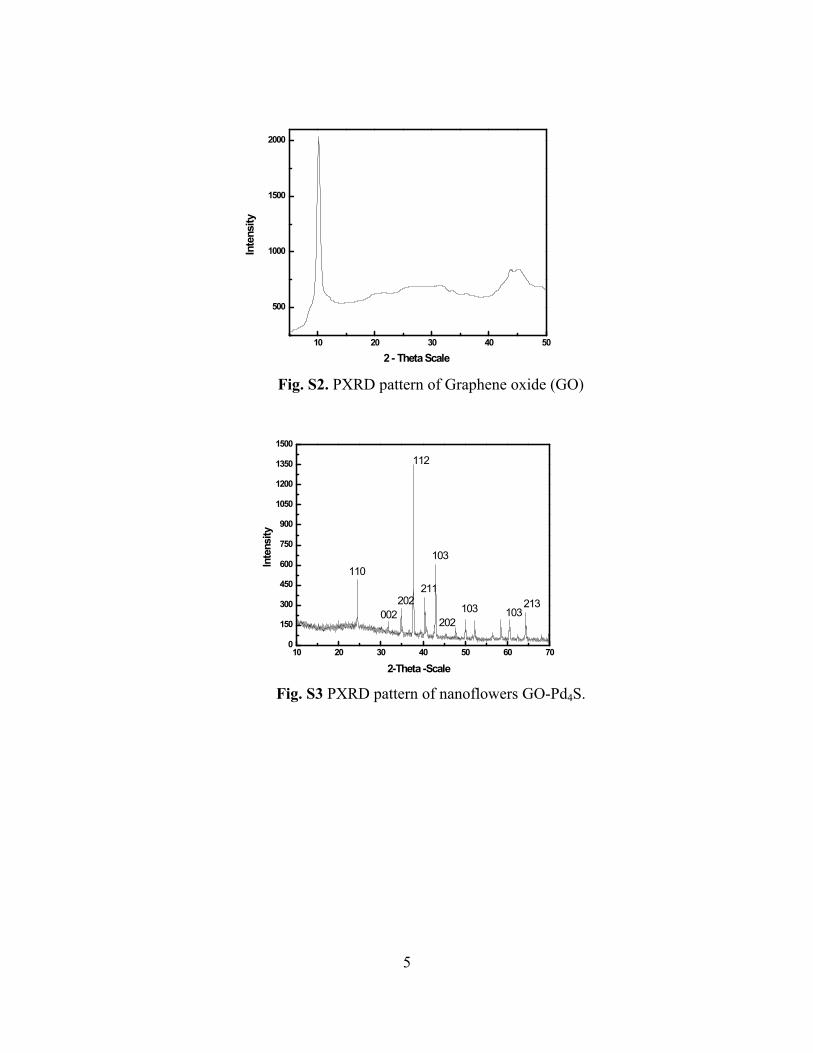
1.41 (320), 1.39 (312), 1.37 (321). PXRD pattern of graphene oxide5 was recorded and
shown in Fig S2. The powder X-ray diffraction pattern of GO-Pd4S nanocomposites are
also shown in Fig. S3 and Fig. S4 respectively.
10 20 30 40 50 60 70
150
300
450
600
750
900
Inte
nsity
2 - Theta - Scale
110002
202102201
210
112
211
202220
103
102213
004321
Fig. S1. PXRD pattern of Pd4S NPs
5
10 20 30 40 50
500
1000
1500
2000
Inte
nsity
2 - Theta Scale
Fig. S2. PXRD pattern of Graphene oxide (GO)
10 20 30 40 50 60 700
150
300
450
600
750
900
1050
1200
1350
1500
Inte
nsity
2-Theta -Scale
112
110
002202
211
103
202103 103
213
Fig. S3 PXRD pattern of nanoflowers GO-Pd4S.

6
20 30 40 50 60 70 800
100
200
300
400
500
600
700
800
002
213103103202
103
211
112
202
Inte
nsity
2- Theta Scale
110
Fig. S4 PXRD pattern of nanospheres GO-Pd4S.

7
S3. TEM−EDX of Pd4S nanocomposites
Fig. S5 TEM−EDX of nanoflowers GO-Pd4S
.
Fig. S6 TEM−EDX of nanospheres GO-Pd4S

8
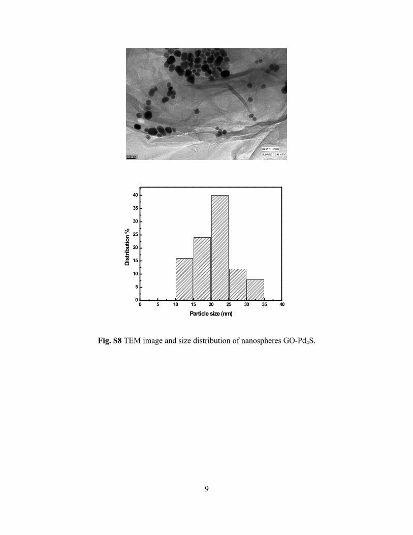
S4. Size distribution of GO-Pd4S nanocomposites
0 5 10 15 20 25 30 35 40 45 500
5
10
15
20
25
30
35
40
45
Dist
ribut
ion
%
Particle size (nm)
Fig. S7 TEM image and size distribution of GO-Pd4S nanoflowers.
9
0 5 10 15 20 25 30 35 400
5
10
15
20
25
30
35
40
Dist
ribut
ion
%
Particle size (nm)
Fig. S8 TEM image and size distribution of nanospheres GO-Pd4S.

10
S5. Spectroscopic data of coupled products
4−Phenylbenzaldehyde (light yellow solid)
OHC
1H NMR (300 MHz, CDCl3): δ 7.537−7.447 (m, 3H), 7.678−7.616 (m, 2H), 7.822 (d, J =
9.0 Hz, 2H), 7.994 (d, J = 5.4 Hz, 2H), 10.081 (s, 1H).
4-Nitrobiphenyl (Pale yellow solid)
NO2
1H NMR (300 MHz, CDCl3): δ 7.462−7.571 (m, 3H), 7.663 (d, J = 8.5 Hz, 2H), 8.050 (d,
J = 8.7 Hz, 2H), 8.225 (d, J = 8.7 Hz, 2H).
4-Phenylbenzonitrile (pale yellow solid)
NC
1H NMR (300 MHz, CDCl3): δ 7.367−7.338 (m, 3H, aromatic), 7.492−7.413 (m, 2H,
aromatic), 7.563−7.499 (m, 4H, aromatic).
4−Acetylbiphenyl (white solid)
COCH3
1H NMR (300 MHz, CDCl3): δ 2.595 (s, 3H), 7.554−7.507 (m, 3H), 7.630−7.601 (m,
4H), 7.844 (d, J = 6.6 Hz, 2H).
Biphenyl-4-carboxylic acid (White solid)O
OH
1H NMR (300 MHz, DMSO): δ 7.324−7.423 (m, 3H), 7.769 (d, J = 7.0 Hz, 2H), 7.791
(d, J = 8.3 Hz, 2H), 8.034 (d, J = 8.2 Hz, 2H)
Biphenyl (white solid)
1H NMR (300 MHz, CDCl3): δ 7.526 (t, J = 6.9 Hz, 2H), 7.596 (t, J = 7.2 Hz, 4H), 7.775
(d, J = 7.2 Hz, 4H).
11
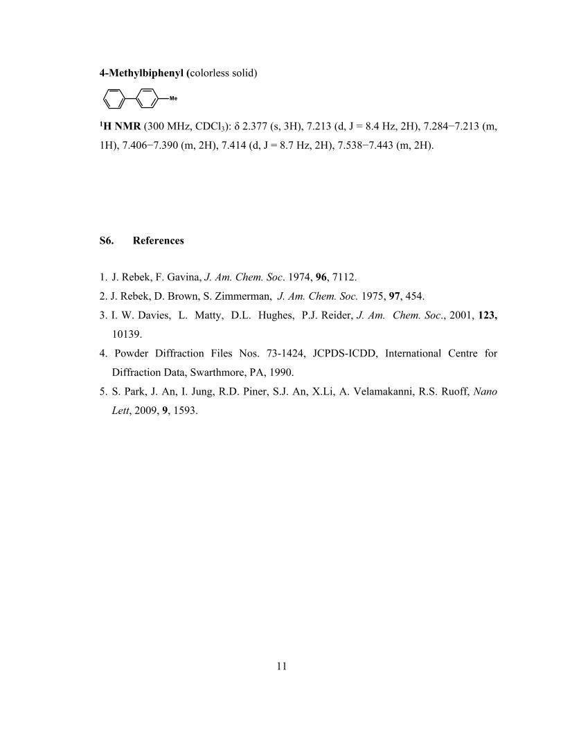
4-Methylbiphenyl (colorless solid)
Me
1H NMR (300 MHz, CDCl3): δ 2.377 (s, 3H), 7.213 (d, J = 8.4 Hz, 2H), 7.284−7.213 (m,
1H), 7.406−7.390 (m, 2H), 7.414 (d, J = 8.7 Hz, 2H), 7.538−7.443 (m, 2H).
S6. References
1. J. Rebek, F. Gavina, J. Am. Chem. Soc. 1974, 96, 7112.
2. J. Rebek, D. Brown, S. Zimmerman, J. Am. Chem. Soc. 1975, 97, 454.
3. I. W. Davies, L. Matty, D.L. Hughes, P.J. Reider, J. Am. Chem. Soc., 2001, 123,
10139.
4. Powder Diffraction Files Nos. 73-1424, JCPDS-ICDD, International Centre for
Diffraction Data, Swarthmore, PA, 1990.
5. S. Park, J. An, I. Jung, R.D. Piner, S.J. An, X.Li, A. Velamakanni, R.S. Ruoff, Nano
Lett, 2009, 9, 1593.
12
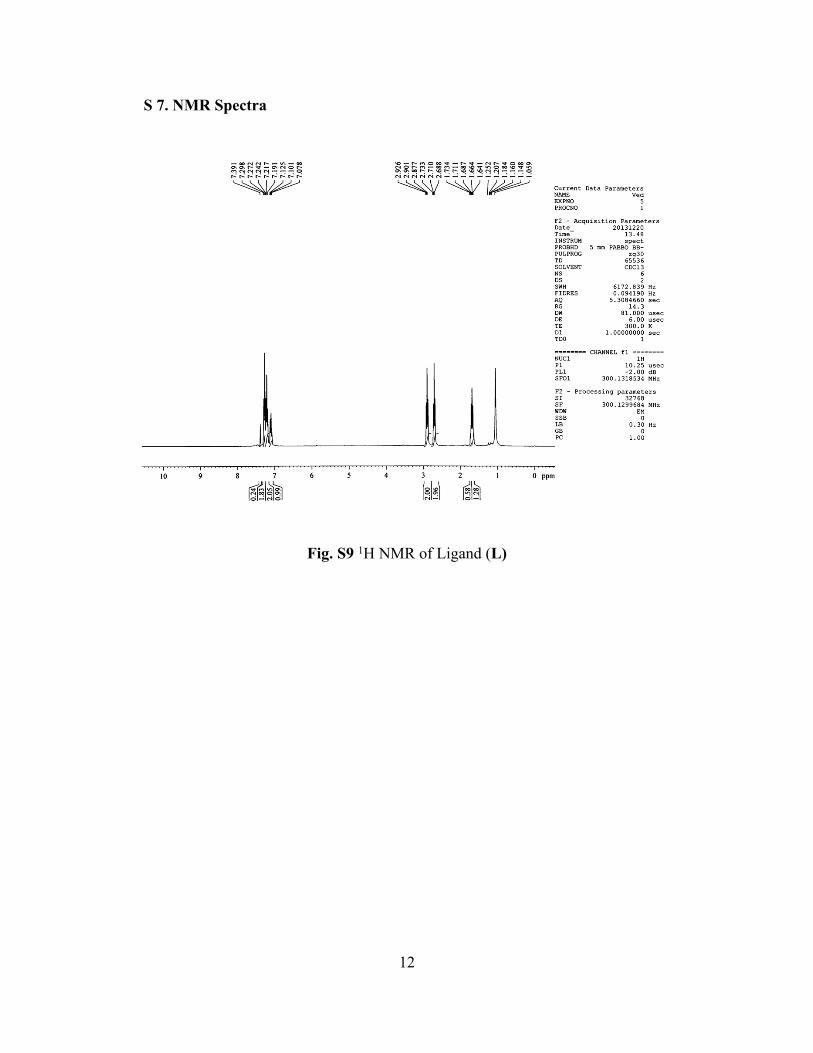
S 7. NMR Spectra
Fig. S9 1H NMR of Ligand (L)

13
Figure S10 1H NMR of Complex (1)

14
Fig. S11 13C{1H} NMR of Complex 1