oxygen-controlled automated neural differentiation of mouse embryonic stem cells
TRANSCRIPT
171ISSN 1746-075110.2217/RME.13.12 © 2013 Future Medicine Ltd Regen. Med. (2013) 8(2), 171–182
ReseaRch aRticle ReseaRch aRticle
Oxygen-controlled automated neural differentiation of mouse embryonic stem cells
Embryonic stem cells (ESC) are currently being evaluated for potential application in a number of diverse areas including regenerative medicine [1], drug discovery and development [2–4] and as routes for delivery of gene therapies [5]. The high level of interest is a consequence of the ability of ESCs to self-renew indefinitely and to differentiate into most adult cell types [6–10]. The production of differentiated cell types from ESC will be reliant upon robust, reproducible and high-yield cell culture processes [1,11,12]. Automated bioreactors have historically been used to control mammalian suspension cell culture resulting in repeatable and controlled bioprocesses [13,14]. Although a number of studies have used microcarrier technologies to differentiate ESC in suspension bioreactors [15–22], the vast majority of current differentiation protocols are based on 2D cell cultures.
A number of reports have used commercially available automation platforms for the maintenance of stem cells under 2D culture conditions [12,23–28]. By eliminating reliance on highly skilled operators, these systems can enhance the reproducibility of stem cell bioprocesses. However, these automated platforms do not control the exposure of cells to key gaseous components, such as oxygen, during the robotic liquid-handling steps of ESC culture.
Physiological oxygen levels that occur during early embryo development have been shown to maintain ESC in a pluripotent state [29–31] and to improve their directed differentiation towards cardiac [15,20], vascular [32], neural [33–37] and retinal lineages [38]. Studies from the authors’ laboratory revealed that the yield of neural cells from mouse ESC (mESC) can be greatly enhanced by manually operating at 2% O
2 [33]. However, the oxygen tension at the
growth surface rose to 21% O2 during medium
exchanges. It took approximately 5 h for the oxygen tension at the growth surface to return to 2% O
2 once the cell culture dishes had
been returned to a hypoxic environment. The biological repercussions of these transient shifts in oxygen remain unclear. In a separate study we also revealed that both the expansion and differentiation of mESCs were sensitive to shifts in pH and temperature, which were detected when cells are removed from the incubator and processed under ambient conditions [39].
In this article, we present a novel automation platform for 2D stem cell culture capable of controlling oxygen tension during the medium exchange step. By integrating an oxygen-controlled incubator into the platform we were able to maintain 2% O
2 during both the cell-
culture and liquid-handling steps. Using this system, mESC were differentiated into neural
Automation and oxygen tension control are two tools that provide significant improvements to the reproducibility and efficiency of stem cell production processes. Aim: the aim of this study was to establish a novel automation platform capable of controlling oxygen tension during both the cell-culture and liquid-handling steps of neural differentiation processes. Materials & methods: We built a bespoke automation platform, which enclosed a liquid-handling platform in a sterile, oxygen-controlled environment. An airtight connection was used to transfer cell culture plates to and from an automated oxygen-controlled incubator. Results: Our results demonstrate that our system yielded comparable cell numbers, viabilities, metabolism profiles and differentiation efficiencies when compared with traditional manual processes. Interestingly, eliminating exposure to ambient conditions during the liquid-handling stage resulted in significant improvements in the yield of MAP2-positive neural cells, indicating that this level of control can improve differentiation processes. Conclusion: This article describes, for the first time, an automation platform capable of maintaining oxygen tension control during both the cell-culture and liquid-handling stages of a 2D embryonic stem cell differentiation process.
KEYWORDS: automation n embryonic stem cells n neural differentiation n oxygen tension
Paul Mondragon-Teran1, Rui Tostoes2, Chris Mason2, Gary J Lye2 & Farlan S Veraitch*2
1Biomedical Research Division, Centro Medico Nacional ‘20 de Noviembre’ – ISSSTE. San Lorenzo 502, Del Valle, Benito Juarez, México City, 03229 México 2The Advanced Centre for Biochemical Engineering, Department of Biochemical Engineering, University College London, Torrington Place, London WC1E 7JE, UK †Author for correspondence: Tel.: +44 20 7679 2648 Fax: +44 20 7209 0703 [email protected]
For reprint orders, please contact: [email protected]
ReseaRch aRticle Mondragon-Teran, Tostoes, Mason, Lye & Veraitch
Regen. Med. (2013) 8(2)172 future science group
cells while maintaining 37°C, 5% CO2 and
2% O2 throughout the entire 8-day differentiation
protocol.
Materials & methods�n Undifferentiated cell culture
E14TG2a mESC, kindly donated by Stem Cell Sciences Ltd (Cambridge, UK), were maintained in culture at 37°C and 5% CO
2 under sterile
conditions in a Heraeus Hera-Cell 150 incubator (Jencons-PLS, West Sussex, UK). Cells were grown in 5 ml of culture medium in Iwaki T25 tissue culture-treated flasks (SLS, Nottingham, UK) coated with 0.1% gelatin (Sigma, Poole, UK). The culture medium was composed of 450 ml of Glasgow minimal essential medium supplemented with 500 µl of 0.1 M 2-b-mercaptoethanol (both Sigma), 50 ml foetal bovine serum, 5 ml nonessential amino acids, 5 ml of 200 mM l-glutamine and 5 ml of 100 mM sodium pyruvate (all Invitrogen, Paisley, UK). After sterilization by filtration, the medium was supplemented with 0.5 ml of 1 × 10⁶ units/ml leukemia inhibitory factor (Millipore, MA, USA). Cells were passaged after 2 days of culture by removing spent media followed by a gentle wash with Dulbecco’s phosphate buffered saline without Ca2+/Mg2+ (DPBS, Sigma). Cells were detached with a solution of 0.025% trypsin, 0.04% EDTA (all Invitrogen) and 0.9% chick serum (Sigma) dissolved in DPBS. After 3 min of trypsinization at 37°C, 3 ml of fresh growth media was added to quench the trypsin. Cells were centrifuged for 3 min at 300 g, resuspended as a single-cell suspension in growth medium and re-inoculated into a new T25 flask precoated with gelatin. The E14TG2a mESCs used in all experiments were between passage 15 and 22. Oct3/4 and Nanog pluripotency marker expression were used to assess the quality of the cells by immunocytochemistry (ICC) (Supplementary
Figure 1) (see online www.futuremedicine.com/doi/suppl/10.2217/RME.13.12).
�n Neural differentiation protocolMonolayer neural differentiation was based on the method reported by Ying et al. [40]. After 2 days of culture, undifferentiated stem cells were harvested by trypsinization. Trypsin was quenched with fresh culture media without leukemia inhibitory factor. The resulting cell suspension was centrifuged for 3 min at 300 g, resuspended in NDIFF–RHB-A differentiation culture medium (Stem Cells Sciences). Cells were plated in 2 ml of medium at a density of 2 × 10⁴ cells/cm2 in Iwaki tissue culture-treated
six-well plates (SLS) precoated with 0.1% gelatin (Sigma). Each well was replenished with 2 ml of NDIFF–RHB-A medium every 2 days in a laminar flow hood operating under normal atmospheric conditions over a total 8-day culture period. Manually seeded six-well plates were also transferred to hypoxic chambers and the automated incubators for processing at 2% O
2. Required cells were trypsinized and gently
resuspended with a Pasteur glass pipette to recover a single-cell suspension for further analysis.
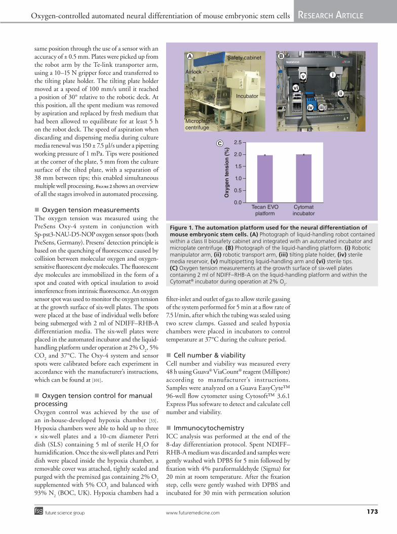
�n Description of the automation platform A Tecan Freedom EVO® 100 liquid-handling platform (Tecan, Switzerland) was used to automate medium exchanges during the neural differentiation protocol. The platform was contained within an enclosed class II biological safety cabinet (Walker Safety Cabinets, UK) (Figure 1B). This system has the capability to control temperature, O
2 and CO
2 during liquid- handling
stages. Airtight connections were used to integrate a microplate centrifuge (Hettich Rotanta 46 RSC, Switzerland) and an automated Cytomat® C450S incubator (Thermo-Fisher, UK). The incubator was equipped with a 32-plate position rack, temperature, O
2, CO
2 and humidity control
and a robotic arm for the selection and transport of culture plates to the liquid-handling platform. The liquid-handling platform was composed of the following devices: liquid-handling arm with four channels and integrated level liquid detection, robotic manipulator arm, robotic transport arm (Te-Link), tilting plate holder and a media reservoir station consisting of 2 × 500 ml and 5 × 100 ml wells designed to accept sterile disposable medium containers (Tecan) (Figure 1B). The liquid-handling arm was equipped with 1 ml syringes, which were used with 1 ml disposable tips (Tecan). Disposable tips and fresh medium were transferred onto the robot deck using an airlock which was located to the left of the platform (Figure 1A). All the robotic arms and the tilting plate holder were controlled through Freedom EVOware® 2 plus software (Tecan).
�n Automated processing parametersIn order to avoid the exposure of differentiating cells to ambient conditions during the liquid handling steps, the environment within the class II safety cabinet was maintained at a set point of 37°C, 5% CO
2 and 2% O
2. Every 48 h
plates were transported onto the liquid-handling platform by the incubator’s robotic arm with a maximum speed of 500 mm/s and placed in the
Oxygen-controlled automated neural differentiation of mouse embryonic stem cells ReseaRch aRticle
www.futuremedicine.com 173future science group
same position through the use of a sensor with an accuracy of ± 0.5 mm. Plates were picked up from the robot arm by the Te-link transporter arm, using a 10–15 N gripper force and transferred to the tilting plate holder. The tilting plate holder moved at a speed of 100 mm/s until it reached a position of 30° relative to the robotic deck. At this position, all the spent medium was removed by aspiration and replaced by fresh medium that had been allowed to equilibrate for at least 5 h on the robot deck. The speed of aspiration when discarding and dispensing media during culture media renewal was 150 ± 7.5 µl/s under a pipetting working pressure of 1 mPa. Tips were positioned at the corner of the plate, 5 mm from the culture surface of the tilted plate, with a separation of 38 mm between tips; this enabled simultaneous multiple well processing. Figure 2 shows an overview of all the stages involved in automated processing.
�n Oxygen tension measurementsThe oxygen tension was measured using the PreSens Oxy-4 system in conjunction with Sp-pst3-NAU-D5-NOP oxygen sensor spots (both PreSens, Germany). Presens’ detection principle is based on the quenching of fluorescence caused by collision between molecular oxygen and oxygen-sensitive fluorescent dye molecules. The fluorescent dye molecules are immobilized in the form of a spot and coated with optical insulation to avoid interference from intrinsic fluorescence. An oxygen sensor spot was used to monitor the oxygen tension at the growth surface of six-well plates. The spots were placed at the base of individual wells before being submerged with 2 ml of NDIFF–RHB-A differentiation media. The six-well plates were placed in the automated incubator and the liquid-handling platform under operation at 2% O
2, 5%
CO2 and 37°C. The Oxy-4 system and sensor
spots were calibrated before each experiment in accordance with the manufacturer’s instructions, which can be found at [101].
�n Oxygen tension control for manual processingOxygen control was achieved by the use of an in-house-developed hypoxia chamber [33]. Hypoxia chambers were able to hold up to three × six-well plates and a 10-cm diameter Petri dish (SLS) containing 5 ml of sterile H
2O for
humidification. Once the six-well plates and Petri dish were placed inside the hypoxia chamber, a removable cover was attached, tightly sealed and purged with the premixed gas containing 2% O
2
supplemented with 5% CO2 and balanced with
93% N2 (BOC, UK). Hypoxia chambers had a
filter-inlet and outlet of gas to allow sterile gassing of the system performed for 5 min at a flow rate of 7.5 l/min, after which the tubing was sealed using two screw clamps. Gassed and sealed hypoxia chambers were placed in incubators to control temperature at 37°C during the culture period.
�n Cell number & viabilityCell number and viability was measured every 48 h using Guava® ViaCount® reagent (Millipore) according to manufacturer’s instructions. Samples were analyzed on a Guava EasyCyte™ 96-well flow cytometer using Cytosoft™ 3.6.1 Express Plus software to detect and calculate cell number and viability.
�n ImmunocytochemistryICC analysis was performed at the end of the 8-day differentiation protocol. Spent NDIFF–RHB-A medium was discarded and samples were gently washed with DPBS for 5 min followed by fixation with 4% paraformaldehyde (Sigma) for 20 min at room temperature. After the fixation step, cells were gently washed with DPBS and incubated for 30 min with permeation solution
Incubator
Safety cabinet
i
iv
vi
Microplatecentrifuge
Airlock i
iiiiv
vi
v
ii
2.5
2.0
1.5
1.0
0.5
0.0Tecan EVO
platformCytomatincubator
Oxy
gen
ten
sio
n (
%)
Figure 1. The automation platform used for the neural differentiation of mouse embryonic stem cells. (A) Photograph of liquid-handling robot contained within a class II biosafety cabinet and integrated with an automated incubator and microplate centrifuge. (B) Photograph of the liquid-handling platform. (i) Robotic manipulator arm, (ii) robotic transport arm, (iii) tilting plate holder, (iv) sterile media reservoir, (v) multipipetting liquid-handling arm and (vi) sterile tips. (C) Oxygen tension measurements at the growth surface of six-well plates containing 2 ml of NDIFF–RHB-A on the liquid-handling platform and within the Cytomat® incubator during operation at 2% O
2.

ReseaRch aRticle Mondragon-Teran, Tostoes, Mason, Lye & Veraitch
Regen. Med. (2013) 8(2)174 future science group
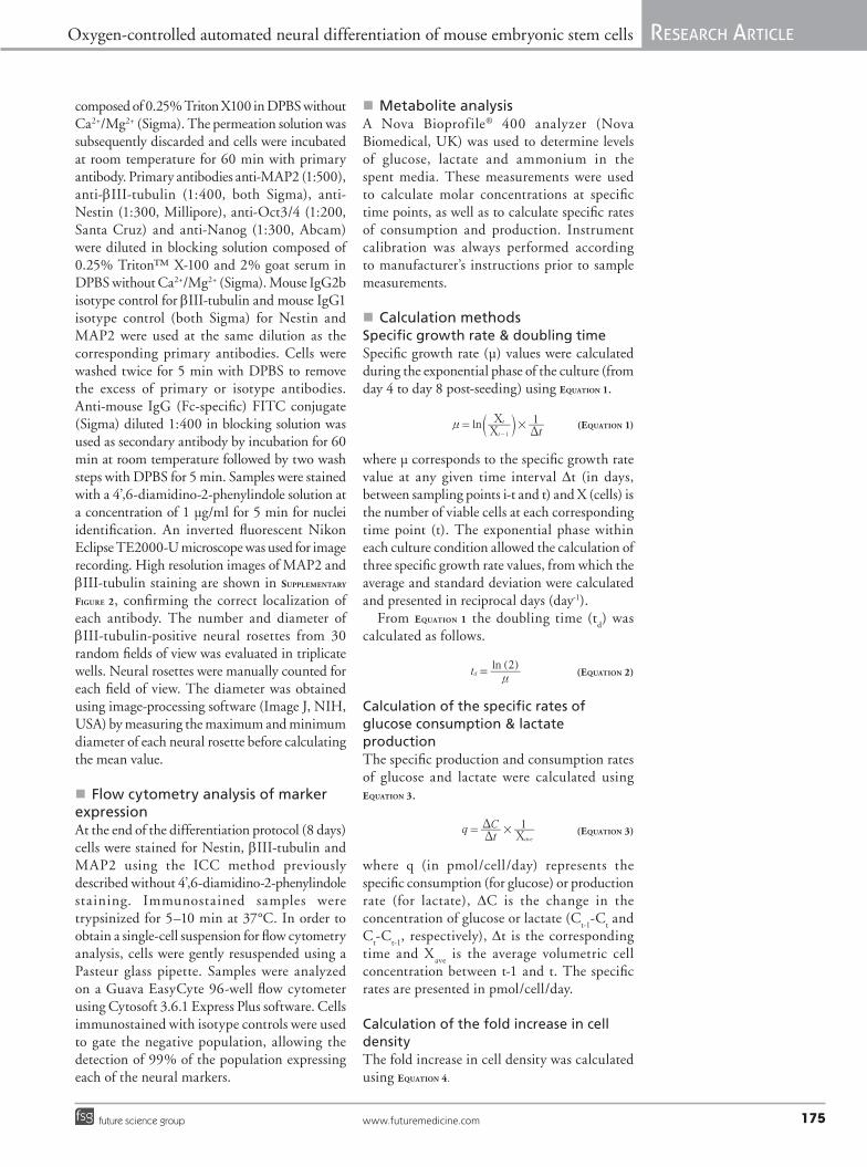
Culture CultureMedia renewal
Cytomat incubator Cytomat incubatorTecan EVO automated platform
mESCs are manually seeded onto six-well plates placed in the
incubator’s rack
Te-Link transfers the plate from the incubator to the liquid-handling platform
RoMa picks up the plate from the Te-Link and
transfers it to the tilting plate holder
Tilting plate holder is tilted 30° relative to the robot deck
RoMa removes the plate lid and places it onto a spare position on the
tilting plate holder
Liquid-handling arm picks up two sterile tips, aspirates and discards the spent media of two wells simultaneously
Incubator robot arm selects a plate and places
it on the Te-Link
Liquid-handling arm aspirates preconditioned media and dispenses it in
the microwells
Liquid-handling arm discards the used tips and
replaces them with two new sterile tips
Tilting plate holder returns to its original
position, RoMa replaces the plate’s lid and
places the plate on the Te-Link arm
Te-Link arm transports the plate
back to the incubator
Incubator’s automated arm collects and
places the plate back to its original position
Plates are manually removed from the
incubator for further analysis
48 h of culture
Media renewed in all microwells?
Has spent media been removed from
all microwells?
Has culture reached 192 h?
End
No
No
No
Yes
Yes
Yes
Plate with fresh mediais cultured for 48 h
until next media renewal
Figure 2. Standard operating procedure for automated neural differentiation of mouse embryonic stem cells. The cell culture and media renewal steps were performed by the Cytomat® incubator and Tecan EVO® automated platform, respectively. There was steady-state control of oxygen (2%), carbon dioxide (5%) and temperature (37°C). mESC: Mouse embryonic stem cell; RoMa: Robotic manipulator arm; Te-Link: Robotic transport arm.
Oxygen-controlled automated neural differentiation of mouse embryonic stem cells ReseaRch aRticle
www.futuremedicine.com 175future science group
composed of 0.25% Triton X100 in DPBS without Ca2+/Mg2+ (Sigma). The permeation solution was subsequently discarded and cells were incubated at room temperature for 60 min with primary antibody. Primary antibodies anti-MAP2 (1:500), anti-bIII-tubulin (1:400, both Sigma), anti-Nestin (1:300, Millipore), anti-Oct3/4 (1:200, Santa Cruz) and anti-Nanog (1:300, Abcam) were diluted in blocking solution composed of 0.25% Triton™ X-100 and 2% goat serum in DPBS without Ca2+/Mg2+ (Sigma). Mouse IgG2b isotype control for bIII-tubulin and mouse IgG1 isotype control (both Sigma) for Nestin and MAP2 were used at the same dilution as the corresponding primary antibodies. Cells were washed twice for 5 min with DPBS to remove the excess of primary or isotype antibodies. Anti-mouse IgG (Fc-specific) FITC conjugate (Sigma) diluted 1:400 in blocking solution was used as secondary antibody by incubation for 60 min at room temperature followed by two wash steps with DPBS for 5 min. Samples were stained with a 4’,6-diamidino-2-phenylindole solution at a concentration of 1 µg/ml for 5 min for nuclei identification. An inverted fluorescent Nikon Eclipse TE2000-U microscope was used for image recording. High resolution images of MAP2 and bIII-tubulin staining are shown in Supplementary
Figure 2, confirming the correct localization of each antibody. The number and diameter of bIII-tubulin-positive neural rosettes from 30 random fields of view was evaluated in triplicate wells. Neural rosettes were manually counted for each field of view. The diameter was obtained using image-processing software (Image J, NIH, USA) by measuring the maximum and minimum diameter of each neural rosette before calculating the mean value.
�n Flow cytometry analysis of marker expressionAt the end of the differentiation protocol (8 days) cells were stained for Nestin, bIII-tubulin and MAP2 using the ICC method previously described without 4’,6-diamidino-2-phenylindole staining. Immunostained samples were trypsinized for 5–10 min at 37°C. In order to obtain a single-cell suspension for flow cytometry analysis, cells were gently resuspended using a Pasteur glass pipette. Samples were analyzed on a Guava EasyCyte 96-well flow cytometer using Cytosoft 3.6.1 Express Plus software. Cells immunostained with isotype controls were used to gate the negative population, allowing the detection of 99% of the population expressing each of the neural markers.
�n Metabolite analysisA Nova Bioprofile® 400 analyzer (Nova Biomedical, UK) was used to determine levels of glucose, lactate and ammonium in the spent media. These measurements were used to calculate molar concentrations at specific time points, as well as to calculate specific rates of consumption and production. Instrument calibration was always performed according to manufacturer’s instructions prior to sample measurements.
�n Calculation methods Specific growth rate & doubling timeSpecific growth rate (µ) values were calculated during the exponential phase of the culture (from day 4 to day 8 post-seeding) using equation 1.
(equation 1)
where µ corresponds to the specific growth rate value at any given time interval Dt (in days, between sampling points i-t and t) and X (cells) is the number of viable cells at each corresponding time point (t). The exponential phase within each culture condition allowed the calculation of three specific growth rate values, from which the average and standard deviation were calculated and presented in reciprocal days (day-1).
From equation 1 the doubling time (td) was
calculated as follows.
(equation 2)
Calculation of the specific rates of glucose consumption & lactate productionThe specific production and consumption rates of glucose and lactate were calculated using equation 3.
(equation 3)
where q (in pmol/cell/day) represents the specific consumption (for glucose) or production rate (for lactate), DC is the change in the concentration of glucose or lactate (C
t-1-C
t and
Ct-C
t-1, respectively), Dt is the corresponding
time and Xave
is the average volumetric cell concentration between t-1 and t. The specific rates are presented in pmol/cell/day.
Calculation of the fold increase in cell densityThe fold increase in cell density was calculated using equation 4.
lnt1
t
t
1
#nVV
D=
-
c m
qtC 1
ave
#DD
V=
( )lnt
2d
n=
ReseaRch aRticle Mondragon-Teran, Tostoes, Mason, Lye & Veraitch
Regen. Med. (2013) 8(2)176 future science group
(equation 4)
where X2% O2
is the viable cell density at 2% O2
(either manual or automated); P2% O2
is the percentage of cells expressing each marker (Nestin, bIII-tubulin or MAP2) at 2% O
2
(either manual or automated); X20% O2
is the viable cell density at 20% O
2 condition; P
20% O2
is the percentage of cells expressing each marker (Nestin, bIII-tubulin or MAP2) at 20% O
2
condition.
�n Statistical analysisThree replicates were performed for all cell culture experiments. Error bars represent one standard deviation above and below the mean. Significant differences between data points were calculated using a two-tailed, paired, Student’s t-test. p-values of less than 0.05 were deemed to be significant.
ResultsIn order to investigate the impact of environ ment-control during cell culture and liquid-handling steps, the neural differentiation of mESCs was compared under three sets of conditions. Cell culture plates were either cultured in an incubator under atmospheric conditions (20% manual), in hypoxic chambers (2% manual) or in the automated incubator operating at 2% O
2
(2% automated). In the case of the 20% manual and 2% manual conditions, medium exchanges were carried out manually in a laminar flow hood operating under atmospheric conditions. Plates being cultured in the 2% automated condition were moved onto the environmentally controlled automation platform for medium exchanges as outlined in the Materials and Methods section. We also measured the oxygen tension in the liquid phase of culture plates placed in either the Cytomat incubator or on the Tecan EVO platform during operation under the 2% automated condition. 2% O
2 was measured in
both, as shown in Figure 1C. These results confirm that our automation platform was capable of operating under hypoxic conditions during both the cell-culture and liquid-handing phases of the process.
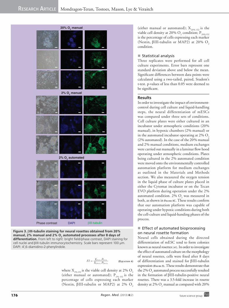
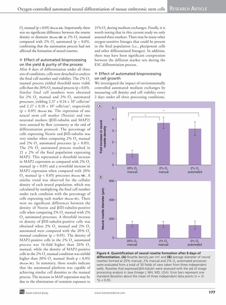
�n Effect of automated bioprocessing on neural rosette formationNeural cells obtained during the directed differentiation of mESC tend to form colonies known as neural rosettes [40]. In order to investigate the effect of automated culture on the morphology of neural rosettes, cells were fixed after 8 days of differentiation and stained for bIII-tubulin expression (Figure 3). These results demonstrate that the 2% O
2 automated process successfully resulted
in the formation of bIII-tubulin-positive neural rosettes. There was a 3.5-fold increase in rosette density at 2% O
2 manual as compared with 20%
20% O2 manual
2% O2 manual
2% O2 automated
Phase contrast DAPI βIII-tubulin
Figure 3. bIII‑tubulin staining for neural rosettes obtained from 20% manual, 2% manual and 2% O2 automated processes after 8 days of differentiation. From left to right: bright field/phase contrast, DAPI staining for cell nuclei and bIII-tubulin immunocytochemistry. Scale bars represent 100 µm. DAPI: 4’,6-diamidino-2-phenylindole.
FIX PX P
% %
% %
O O
O O
20 20
2 2
2 2
2 2=
Oxygen-controlled automated neural differentiation of mouse embryonic stem cells ReseaRch aRticle
www.futuremedicine.com 177future science group
O2 manual (p < 0.05) (Figure 4A). Importantly, there
was no significant difference between the rosette density or diameter (Figure 4B) at 2% O
2 manual
compared with 2% O2 automated (p > 0.05),
confirming that the automation process had not affected the formation of neural rosettes.
�n Effect of automated bioprocessing on the yield & purity of the process After 8 days of differentiation under all three sets of conditions, cells were detached to analyze the final cell number and viability. The 2% O
2
manual process yielded threefold more viable cells than the 20% O
2 manual process (p < 0.05).
Similar f inal cell numbers were obtained for 2% O
2 manual and 2% O
2 automated
processes, yielding 2.37 ± 0.24 × 10⁵ cells/cm2 and 2.27 ± 0.18 × 10⁵ cells/cm2, respectively (p > 0.05) (Figure 5A). The expression of one neural stem cell marker (Nestin) and two neuronal markers (bIII-tubulin and MAP2) were assessed by flow cytometry at the end of differentiation protocol. The percentage of cells expressing Nestin and bIII-tubulin was very similar when comparing 2% O2 manual and 2% O
2 automated processes (p > 0.05).
The 2% O2 automated process resulted in
21 ± 2% of the final population expressing MAP2. This represented a threefold increase in MAP2 expression as compared with 2% O
2
manual (p < 0.05) and a sevenfold increase in MAP2 expression when compared with 20% O
2 manual (p < 0.05) processes (Figure 5B). A
similar trend was observed for the cellular density of each neural population, which was calculated by multiplying the final cell number under each condition with the percentage of cells expressing each marker (Figure 5C). There were no significant differences between the density of Nestin and bIII-tubulin-positive cells when comparing 2% O
2 manual with 2%
O2 automated processes. A threefold increase
in density of bIII-tubulin-positive cells was obtained when 2% O
2 manual and 2% O
2
automated were compared with the 20% O2
manual condition (p < 0.05). The density of MAP2-positive cells in the 2% O
2 automated
process was 16-fold higher than 20% O2
manual, while the density of MAP2-positive cells in the 2% O
2 manual condition was sixfold
higher than 20% O2 manual (both p < 0.05)
(Figure 5C). In summary, these results indicate that the automated platform was capable of achieving similar cell densities to the manual process. The increase in MAP expression may be due to the elimination of transient exposure to
21% O2 during medium exchanges. Finally, it is
worth noting that in this current study we only assessed three markers. There may be many other oxygen-sensitive lineages that could be present in the final population (i.e., pluripotent cells and other differentiated lineages). In addition, there may have been significant coexpression between the different marker sets during the ESC differentiation process.
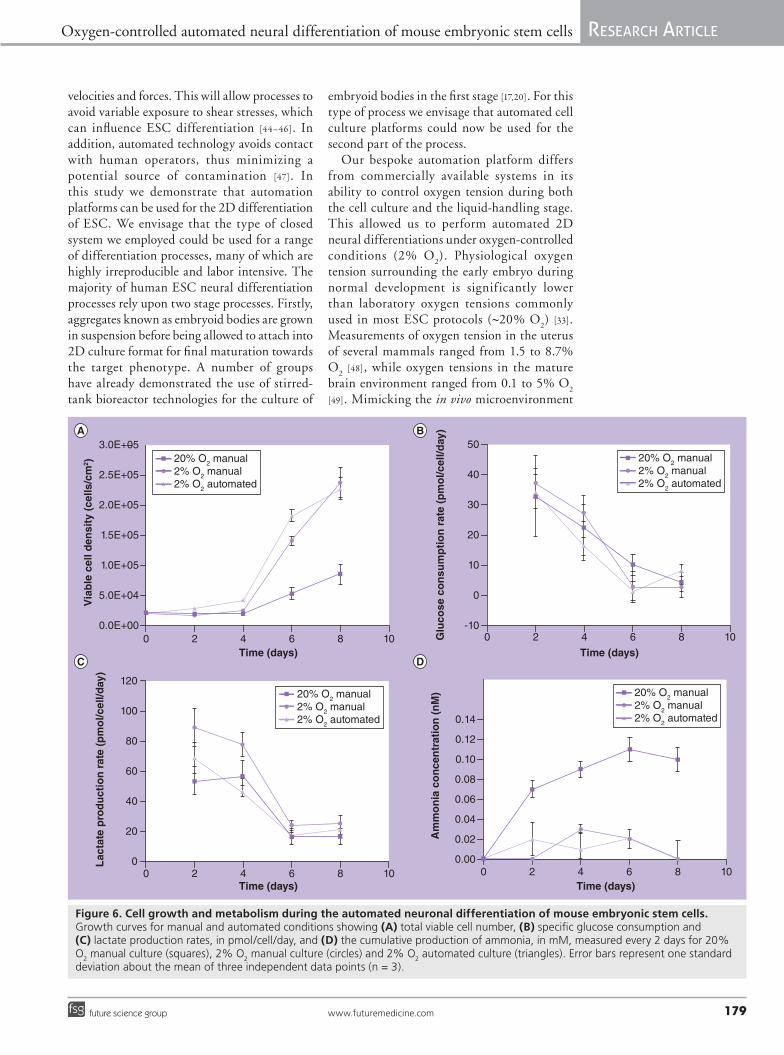
�n Effect of automated bioprocessing on cell growthWe investigated the impact of environmentally controlled automated medium exchanges by measuring cell density and cell viability every 2 days under all three processing conditions.
Fold
incr
ease
in n
eura
lro
sett
e d
ensi
tyN
eura
l ro
sett
e d
iam
eter
(µ
m)
20% O2
manual2% O2
manual2% O2
automated
20% O2
manual2% O2
manual2% O2
automated
5
4
3
2
1
0
0
200
400
600
*
*
Figure 4. Quantification of neural rosette formation after 8 days of differentiation. (A) Rosette density per cm2 and (B) average diameter of neural rosettes formed at 20% manual, 2% manual and 2% O
2 automated processes
were evaluated from a total of 30 fields of view taken from three independent wells. Rosettes that expressed bIII-tubulin were assessed with the aid of image processing analysis in Java (Image J, NIH, MD, USA). Error bars represent one standard deviation about the mean of three independent data points (n = 3). *p < 0.05.

ReseaRch aRticle Mondragon-Teran, Tostoes, Mason, Lye & Veraitch
Regen. Med. (2013) 8(2)178 future science group
At days 6 and 8 of culture, the cell density values were significantly lower in the 20% manual processing culture condition, when compared with both hypoxic cultures (Figure 6A). Comparisons of the cell growth curves revealed that the expansion of cells was very similar in both the 2% manual and 2% automated processes, although cell growth may have been slightly slower in the manual condition. To further analyze cellular metabolism, we considered the first 4 days of culture as the lag
phase followed by an exponential phase from 4–8 days, based on the growth curves. The specific growth rates were calculated for the exponential phase as indicated in equation 1, and were found to be, 0.5 ± 0.2, 0.6 ± 0.2 and 0.4 ± 0.1 day-1 for the 2% automated, 2% manual and 20% O
2 manual conditions, respectively. There
were no statistically significant differences between the growth rate of 2% automated and 2% manual O
2 culture conditions.
�n The effect of automated bioprocessing on cell metabolismIn addition to cell growth, cell metabolism was monitored for the three different culture conditions every 2 days during the 8 days of differentiation protocol. Glucose consumption, lactate production, and ammonia concentrations are shown in Figure 6B–D. A linear decrease in glucose consumption and lactate production was detected over the whole period of culture, for all processing conditions, with no statistical differences except for day 2, where lactate production was higher in the 2% O
2 manual
condition, when compared with the other culture modes (Figure 6B & C). Specific rates for glucose consumption and lactate production are in agreement with values from the literature [41,42]. These results indicate that the automation platform did not interfere with glucose metabolism. Analysis of ammonia concentrations (Figure 6D) revealed that differentiation at 20% O
2 caused the accumulation of this metabolite
to potentially toxic levels [43]. Under 2% O2
manual and automated conditions, ammonia levels remained relatively low in comparison. Lower levels of toxic ammonium may have contributed to the higher levels of viability observed at 2% O
2 [33].
DiscussionCell-culture automation has improved over the years, and systems have been integrated into production lines enhancing industrial (i.e., pharmaceutical) production in terms of efficiency and productivity. Barrier isolators and cGMP conditions have been adapted for pharmaceutical and biotechnological processing avoiding cross-contamination and operator exposure [26]. A number of studies have previously demonstrated that commercially available automation platforms can be used to develop robust stem cell production processes based on 2D cell culture systems [12,23–28]. Automated platforms enable accurate and repeatable control of plate-handling and liquid-handling
20% O2
manual2% O2
manual2% O2
automated
20% O2
manual2% O2
manual2% O2
automated
Via
ble
cel
l den
sity
(103
cells
/cm
2 )P
op
ula
tio
n (
%)
Fold
incr
ease
ince
ll d
ensi
ty
Via
bili
ty (
%)
400
350
300
250
200
150
100
50
0
100
80
60
40
20
0
*
*
*
**
*
*
*
100
80
60
40
20
0
20% O2
manual2% O2
manual2% O2
automated
0
5
10
15
20
NestinβIII-tubulinMAP2
NestinβIII-tubulinMAP2
Figure 5. Yield and purity of neural cells produced after 8 days of differentiation under 20% manual, 2% manual and 2% O2 automated conditions. (A) Viability (open circles) and viable cell number after 8 days of differentiation and (B) the percentage of cells expressing Nestin, bIII-tubulin and MAP2 measured by flow cytometry. (C) Fold increase in the number of cells/cm2 expressing Nestin, bIII-tubulin and MAP2 at 2% manual and 2% O
2 automated as
compared with 20% O2 manual controls. Error bars represent one standard
deviation about the mean of three independent data points (n = 3). *p < 0.05.
Oxygen-controlled automated neural differentiation of mouse embryonic stem cells ReseaRch aRticle
www.futuremedicine.com 179future science group
velocities and forces. This will allow processes to avoid variable exposure to shear stresses, which can influence ESC differentiation [44–46]. In addition, automated technology avoids contact with human operators, thus minimizing a potential source of contamination [47]. In this study we demonstrate that automation platforms can be used for the 2D differentiation of ESC. We envisage that the type of closed system we employed could be used for a range of differentiation processes, many of which are highly irreproducible and labor intensive. The majority of human ESC neural differentiation processes rely upon two stage processes. Firstly, aggregates known as embryoid bodies are grown in suspension before being allowed to attach into 2D culture format for final maturation towards the target phenotype. A number of groups have already demonstrated the use of stirred-tank bioreactor technologies for the culture of
embryoid bodies in the first stage [17,20]. For this type of process we envisage that automated cell culture platforms could now be used for the second part of the process.
Our bespoke automation platform differs from commercially available systems in its ability to control oxygen tension during both the cell culture and the liquid-handling stage. This allowed us to perform automated 2D neural differentiations under oxygen-controlled conditions (2% O
2). Physiological oxygen
tension surrounding the early embryo during normal development is signif icantly lower than laboratory oxygen tensions commonly used in most ESC protocols (~20% O
2) [33].
Measurements of oxygen tension in the uterus of several mammals ranged from 1.5 to 8.7% O
2 [48], while oxygen tensions in the mature
brain environment ranged from 0.1 to 5% O2
[49]. Mimicking the in vivo microenvironment
Figure 6. Cell growth and metabolism during the automated neuronal differentiation of mouse embryonic stem cells. Growth curves for manual and automated conditions showing (A) total viable cell number, (B) specific glucose consumption and (C) lactate production rates, in pmol/cell/day, and (D) the cumulative production of ammonia, in mM, measured every 2 days for 20% O
2 manual culture (squares), 2% O
2 manual culture (circles) and 2% O
2 automated culture (triangles). Error bars represent one standard
deviation about the mean of three independent data points (n = 3).
Via
ble
cel
l den
sity
(ce
lls/c
m2 )
Glu
cose
co
nsu
mp
tio
n r
ate
(pm
ol/c
ell/d
ay)
Am
mo
nia
co
nce
ntr
atio
n (
nM
)
Lac
tate
pro
du
ctio
n r
ate
(pm
ol/c
ell/d
ay)
Time (days)0 2 4 6 8 10
Time (days)0 2 4 6 8 10
Time (days)0 2 4 6 8 10
Time (days)0 2 4 6 8 10
3.0E+05
2.5E+05
2.0E+05
1.5E+05
1.0E+05
5.0E+04
0.0E+00
50
40
30
20
10
0
-10
120
100
80
60
40
20
0
0.14
0.12
0.10
0.08
0.06
0.04
0.02
0.00
20% O2 manual2% O2 manual2% O2 automated
20% O2 manual2% O2 manual2% O2 automated
20% O2 manual2% O2 manual2% O2 automated
20% O2 manual2% O2 manual2% O2 automated
ReseaRch aRticle Mondragon-Teran, Tostoes, Mason, Lye & Veraitch
Regen. Med. (2013) 8(2)180 future science group
has been a powerful tool for increasing the efficiency of ESC differentiation into a wide range of cell types [15,20,32–38,42]. Therefore, it is important that any new automation platform for ESC processing should have the ability to control this critical parameter. In a previous study we have demonstrated that the yield of neural cells from mESC can be greatly enhanced by manually operating at 2% O
2 [33]. However,
ESC differentiation processes typically require medium changes every other day (in some cases this can be daily). Using traditional hypoxic chambers, the oxygen tension at the growth surface rose to 21% O
2 during medium
exchanges [33]. It took approximately 5 h for the oxygen tensions at the growth surface to return to 2% O
2 once the cell culture dishes had
been returned to a hypoxic environment. Our automation platform was designed with this in mind, and liquid-handling steps were carried out at 2% O
2. The data presented here revealed
that our automation platform was capable of delivering a similar process to the manual control. One unexpected result was a threefold increase in the number of cells expressing the neuronal marker MAP2. We tentatively hypothesize that the formation of MAP2-positive cells is sensitive to exposure to ambient conditions and that eliminating these shifts resulted in an increase in this population. Our automated process did not affect the formation bIII-tubulin- or Nestin-positive populations, indicating that eliminating transient shifts in oxygen levels does not have a beneficial impact on all neural lineages. This is in keeping with our previous results demonstrating that oxygen had a more pronounced impact on the formation of MAP2-positive cells when compared with bIII-tubulin or Nestin [33,50]. A more detailed time–course analysis of marker expression will be needed in order to further investigate whether the automation process affected the rate of differentiation. Taken together, these results demonstrate that transient shifts in oxygen tension to ambient conditions may inhibit some of the positive effects associated with differentiation under physiological conditions.
In summary, we have demonstrated for the first time that an automation platform can be used for the neural differentiation of ESCs in a 2D format. This is of particular importance given that many human ESCs, induced pluripotent stem cell and adult stem cell-directed differentiation protocols rely upon this type of attached culture format for the generation of a wide variety of cells for regenerative medicine applications. Our system enables the transfer of labor-intensive processes, which are highly dependent upon operator variability [39], into controlled, automated bioprocesses. In addition, we exploited the system’s ability to control oxygen during the liquid-handling steps to ensure that O
2 levels
remained consistent throughout the entire 8-day process.
AcknowledgementsThe authors would like to acknowledge the engineering sup-port from Tecan and Walker Safety Cabinets for their sup-port during the design and construction of the automation platform.
Financial & competing interests disclosureP Mondragon-Teran would like to acknowledge his PhD financial support from the Mexican Science and Technology Agency (CONACYT) and from the Agency of Public Education (SEP). C Mason, GJ Lye and FS Veraitch acknowledge financial support from the Technology Strategy Board (then Department of Trade and Industry). The authors have no other relevant affiliations or financial involvement with any organization or entity with a finan-cial interest in or financial conflict with the subject matter or materials discussed in the manuscript apart from those disclosed.
No writing assistance was utilized in the production of this manuscript.
Ethical conduct of research The authors state that they have obtained appropriate insti tutional review board approval or have followed the princi ples outlined in the Declaration of Helsinki for all human or animal experimental investigations. In addi-tion, for investi gations involving human subjects, informed consent has been obtained from the participants involved.
Executive summary
� We have developed a novel automation platform capable of controlling O2 during liquid-handling steps of neural differentiation
processes.
� The platform was tested on the neural differentiation of mouse embryonic stem cells under hypoxic conditions (2% O2), resulting in
similar cellular yields when compared with the equivalent manual process.
� There was a threefold increase in the number of cells expressing the neuronal marker MAP2 following processing with the automated platform.
� Glucose, lactate and ammonia metabolism were not affected by automated processing.

Oxygen-controlled automated neural differentiation of mouse embryonic stem cells ReseaRch aRticleReseaRch aRticle
www.futuremedicine.com 181future science group
ReferencesPapers of special note have been highlighted as:n of interestnn of considerable interest
1 Mason C, Hoare M. Regenerative medicine bioprocessing: building a conceptual framework based on early studies. Tissue Eng. 13(2), 301–311 (2007).
2 McNeish JD. Embryonic stem cells in drug discovery. Nat. Rev. Drug Discov. 3, 70–80 (2004).
3 Thomson H. Bioprocessing of stem cells for drug discovery. Trends Biotech. 25(5), 224–230 (2007).
4 McNeish JD. Stem cells as screening tools in drug discovery. Curr. Opin. Pharmacol. 7(5), 515–520 (2007).
5 Conrad C, Gupta R, Mohan H et al. Genetically engineered stem cells for therapeutic gene delivery. Curr. Gene Ther. 7(4), 249–260 (2007).
6 Evans MJ, Kaufman MH. Establishment in culture of pluripotential cells from mouse embryos. Nature 292, 154–156 (1981).
7 Martin GR. Isolation of a pluripotent cell line from early mouse embryos cultured in medium conditioned by teratocarcinoma stem cells. Proc. Natl Acad. Sci. 78, 7634–7638 (1981).
8 Bradley A, Evans M, Kaufman MH, Robertson E. Formation of germ-line chimaeras from embryo-derived teratocarcinoma cell lines. Nature 309, 255–256 (1984).
9 Thomson JA, Itskovitz-Eldor J, Shapiro SS et al. Embryonic stem cell lines derived from human blastocysts. Science 282, 1145–1147 (1998).
10 Smith AG. Embryo-derived stem cells: of mice and men. Annu. Rev. Cell Dev. Biol. 17, 435–462 (2001).
11 Kirouac DC, Zandstra PW. The systematic production of cells for cell therapies. Cell Stem Cell 3(4), 369–381 (2008).
12 Thomas RJ, Chandra A, Liu Y, Hourd PC, Conway PP, Williams DJ. Manufacture of a human mesenchymal stem cell population using and automated cell culture platform. Cytotechnology 55, 31–39 (2007).
13 Platas Barradas O, Jandt U, Phan LDM et al. Criteria for bioreactor comparison and operation standardization during process development for mammalian cell culture. BMC Proc. 5(8), 47 (2011).
14 Tissot S, Zhang X, Stettler M et al. Engineering principles and cell culture performance of orbitally shaken bioreactors. ESACT Proc. 5(5), 407–412 (2012).
15 Bauwens C, Yin T, Dang S, Peerani R, Zandstra PW. Development of a perfusion fed
bioreactor for embryonic stem cell-derived cardiomyocyte generation: oxygen-mediated enhancement of cardiomyocyte output. Biotech. Bioeng. 90, 452–461 (2005).
16 Schroeder M, Niebruegge WA, Willbold E et al. Differentiaton and lineage selection of mouse embryonic stem cells in a stirred bench scale bioreactor with automated process control. Biotech. Bioeng. 92(7), 920–933 (2005).
17 Cormier JT, Zur Nieden NI, Rancourt D, Kallos MS. Expansion of undifferentiated murine embryonic stem cells as aggregates in suspension culture bioreactors. Tissue Eng. 12(11), 3233–3245 (2006).
18 McLeod M, Hong M, Sen A et al. Transplantation of bioreactor-produced neural stem cells into the rodent brain. Cell Trans. 15, 389–697 (2006).
19 Fernandes AM, Fernandes TG, Diogo MM, Lobato da Silva C, Henrique D, Cabral JMS. Mouse embryonic stem cell expansion in a microcarrier-based stirred culture system. J. Biotech. 132, 227–236 (2007).
20 Niebruegge S, Bauwens CL, Peerani R, Thavandiran N, Masse S. Sevaptisidis E. Generation of human embryonic stem cell-derived mesoderm and cardiac cells using size-specified aggregates in an oxygen-controlled bioreactor. Biotech. Bioeng. 102, 493–507 (2009).
21 Serra M, Brito C, Costa EM, Sousa MFQ, Alves PM. Integrating human stem cell expansion and neuronal differentiation in bioreactors. BMC Biotechnol. 9, 82 (2009).
22 Kehoe DE, Jing D, Lock LT, Tzanakakis SE. Scalable stirred-suspension bioreactor culture of human pluripotent stem cells. Tissue Eng. Part A 16(2), 405–421 (2010).
23 Dang SM, Gerecht-Nir S, Chen J, Iskovitz-Eldor J, Zandstra PW. Controlled scalable embryonic stem cell differentiation culture. Stem Cells 2(3), 275–282 (2004).
24 Narkilahti S, Rajala K, Pihlajamaki H, Suuroen R, Hovatta O, Skottman H. Monitoring and analysis of dynamic growth of human embryonic stem cells: comparison of automated instrumentation and conventional culturing methods. Biomed. Eng. Online 6, 11 (2007).
25 Terstegge S, Laufenberg I, Pochert J et al. Automated maintenance of embryonic stem cell cultures. Biotech. Bioeng. 96(1), 195–201 (2007).
nn� Describes the first automated platform for the culture of mouse and human embryonic stem cells using enzymatic methods for cell passaging.
26 Thomas RJ, Anderson D, Chandra A et al. Automated, scalable culture of human
embryonic stem cells in feeder-free conditions. Biotech. Bioeng. 102 (6), 1636–1644 (2009).
n� Reports an automated platform for the culture of human embryonic stem cells using an enzymatic method for cell passaging and Matrigel™ to avoid the use of mouse feeder cells.
27 Thomas RJ, Hope AD, Hourd P et al. Automated, serum-free production of CTX0E03: a therapeutic clinical grade human neural stem cell line. Biotech Lett. 31(8), 1167–1172 (2009).
28 Thomas RJ, Williams HDJ. Application of process quality engineering techniques to improve the understanding of the in vitro processing of stem cells for therapeutic use. J. Biotechnol. 136(3), 148–155 (2008).
29 Forristal C, Wright K, Hanley N, Oreffo R, Houghton F. Hypoxia inducible factors regulate pluripotency and proliferation in human embryonic stem cells cultured at reduced oxygen tensions. Reproduction 139, 85–97 (2010).
30 Forsyth NR, Musio A, Vezzoni P, Simpson AH, Noble BS, McWhir J. Physiologic oxygen enhances human embryonic stem cell clonal recovery and reduces chromosomal abnormalities. Cloning Stem Cells 8, 16–23 (2006).
31 Ezashi T, Das P, Roberts RM. Low O2
tensions and the prevention of differentiation of hES cells. Proc. Natl Acad. Sci. 102, 4783–4788 (2005).
32 Han Y, Kuang SZ, Gomer A, Ramirez-Bergeron DL. Hypoxia influences the vascular expansion and differentiation of embryonic stem cell cultures through the temporal expression of VEGF-receptors in an ARNT-dependent manner. Stem Cells 28, 799–809 (2010).
33 Mondragon-Teran P, Lye GJ, Veraitch FS. Lowering oxygen tension enhances the differentiation of mouse embryonic stem cells into neuronal cells. Biotechnol. Prog. 25(5), 1480–1488 (2009).
nn� Describes the impact of controlling oxygen at physiological levels during the neural differentiation of embryonic stem cells.
34 Morrison SJ, Csete M, Groves AK, Melega W, Wold B, Anderson DJ. Culture in reduced levels of oxygen promotes clono- genic sympathoadrenal differentiation by isolated neural crest stem cells. J. Neurosci. 20, 7370–7376 (2000).
35 Studer L, Csete M, Lee SH et al. Enhanced proliferation, survival, and dopaminergic differentiation of CNS precursors in lowered oxygen. J. Neurosci. 20, 7377–7383 (2000).
36 Milosevic J, Schwartz S, Krohn K, Poppe M, Storch A, Schwartz J. Low atmospheric

ReseaRch aRticle Mondragon-Teran, Tostoes, Mason, Lye & VeraitchReseaRch aRticle
Regen. Med. (2013) 8(2)182 future science group
oxygen avoids maturation, senescence and cell death of murine mesencephalic neural precursors. J. Neurochem. 92, 718–729 (2005).
37 Pistollato F, Chen H, Schwartz P, Basso G, Panchision D. Oxygen tension controls the expansion of human CNS precursors and the generation of astrocytes and oligodendrocytes. Mol. Cell Neurosci. 35, 424–435 (2007).
38 Bae D, Mondragon-Teran P, Hernandez D et al. Hypoxia enhances the generation of retinal progenitor cells from human induced pluripotent and embryonic stem cells. Stem Cells Dev. 21(8), 1344–1355 (2012).
39 Veraitch FS, Scott R, Wong J, Lye GJ, Mason C. The impact of manual processing on the expansion and directed differentiation of embryonic stem cells. Biotech. Bioeng. 99, 1216– 1229 (2008).
n� Describes how poorly controlled manual cell-culture techniques can affect the expansion and differentiation of embryonic stem cells.
40 Ying QL, Stavridis M, Griffiths D, Li M, Smith A. Conversion of embryonic stem cells into neuroectodermal precursors in adherent monoculture. Nat. Biotech. 21, 183–186 (2003).
n� Developed and reported a method for the neural differentiation of mouse embryonic stem cells in a monolayer format.
41 Fernandes TG, Diogo MM, Fernandez-Platzgummer A, da Silva CL, Cabral JM. Different stages of pluripotency determine distinct patterns of proliferation, metabolism, and lineage commitment of embryonic stem cells under hypoxia. Stem Cell Res. 5, 76–89 (2010).
42 Rodrigues CA, Diogo MM, da Silva CL, Cabral JM. Hypoxia enhances proliferation of mouse embryonic stem cell-derived neural stem cells. Biotech. Bioeng. 106, 260–270 (2010).
43 Butler M. Animal Cell Culture and Technology (2nd Edition). BIOS Scientific, NY, USA (2004).
44 Illi B, Scopece A, Nanni S et al. Epigenetic histone modification and cardiovascular lineage programming in mouse embryonic stem cells exposed to laminar shear stress. Circ. Res. 96(5), 501–508 (2005).
45 Zeng L, Xiao Q, Margariti A et al. HDAC3 is crucial in shear- and VEGF-induced stem cell differentiation toward endothelial cells. J. Cell Biol. 174(7), 1059–1069 (2006).
46 Stolberg S, McCloskey KE. Can shear stress direct stem cell fate? Biotechnol. Prog. 25(1), 10–19 (2009).
47 Dutton RL, Fox JS. Robotic processing in barrier isolator environments: a life cycle cost approach. Pharm. Eng. 26(5), 1–8 (2006).
48 Fischer B, Bavister BD. Oxygen tension in the oviduct and uterus of rhesus monkeys, hamsters and rabbits. J. Reprod. Fertil. 99, 673–679 (1993).
nn� Reports the physiological oxygen tension values in the oviduct of different mammals where neurulation occurs during organogenesis.
49 Andreasen A, Danscher G, Juhl SS et al. Distinct differences in partial oxygen pressure at micrometer ranges in the rat hippocampal region. J. Neurosci. Methods 72, 15–21 (1997).
50 Mondragon-Teran P, Baboo JZ, Mason C, Lye GJ, Veraitch FS. The full spectrum of physiological oxygen tensions and step-changes in oxygen tension affects the neural differentiation of mouse embryonic stem cells. Biotechnol. Prog. 27(6), 1700–1708 (2011).
�n Website101 PreSens. Precision Sensing.
www.presens.de