oxygenation and orygen therapy
TRANSCRIPT

Oxygenation


Pulmonary VentilationVentilation of the lungs is accomplished through the act
of breathing : inspiration and expiration.Adequate ventilation depends on several factors:- Clear airways- An intact central nervous system and respiratory
system- An intact thoracic cavity capable of expanding and
contracting- Adequate pulmonary compliance and recoil

Ciliary actionCough
reflex


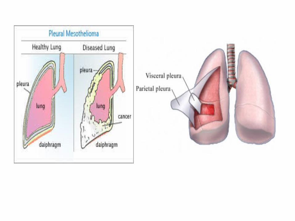
Intrapleural pressurePressure in the pleural cavity surrounding the lungs.Is always slightly negative in relation to atmospheric
pressure

Intrapulmonary pressurePressure within the lungs Always equalize with atmospheric pressure.
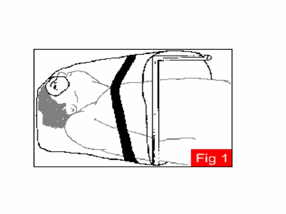
InspirationWhen the diaphragm and intercostals muscles contract ___ ↑ the size of the thoracic cavity ____ volume of the lungs ↑ ____ ↓ intrapulmonary pressure → then air moves into the lung.

ExpirationWhen the diaphragm and intercostal muscles relax ___ the size of the thoracic cavity ↓ ____ volume of the lungs ↓ ____ ↑intrapulmonary pressure → then air moves out the lung.


Tidal volume Approx. 500ml of air is inspired and expired with each
breath . - Lung complianceExpansibility or stretchability of lung tissue, plays a
significant role in the ease of ventilation .- Lung recoilThe continual tendency of the lungs to collapse away from
the chest wall. Elastic fibers in lung tissue contribute to lung recoil, also
surface tension of fluid lining the alveoli.

Surfactant, a detergent-like phospholipid, reduces the surface tension of the fluid lining the alveoli. When surfactant production is reduced, the lung becomes stiff and the alveoli collapse.

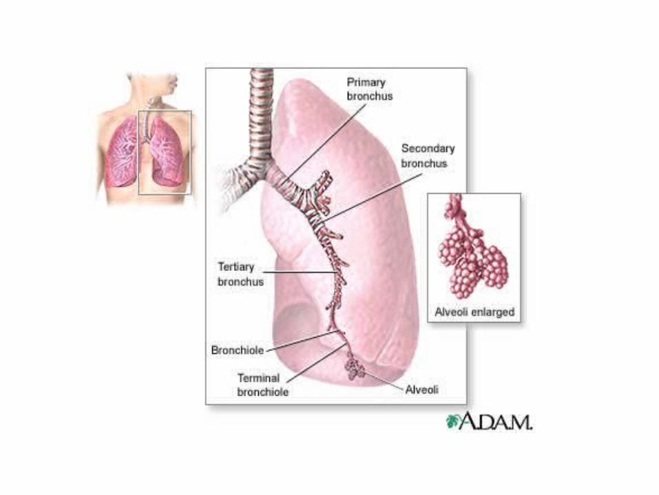
Alveolar Gas Exchange
Diffusion refers to the movement of oxygen and carbon dioxide between the air (in the alveoli) and the blood (in the capillaries). The appropriate gas moves passively from an area of higher pressure or concentration to an area of lesser pressure or concentration.

When the pressure of oxygen is greater in the alveoli than in the blood , oxygen diffuse into the blood. The PO2 in the alevoli is about is about 100mmHgm whereas the PO2 in the venous blood of the pulmonary arteries is about 60mmHg. Therefore PO2 diffuse from the alveoli to the blood. By contrast PCO2 in the venous blood entering the pulmonary capillaries is about 45mmHg , whereas PCO2 in the alevoli is about 40mmHg, Therefore CO2 diffuse from the blood into the alveoli.


Transport of Oxygen and Carbon DioxideMost of O2 97% combines loosely with hemoglobin as oxyhemoglobin. The remaining is dissolved and transported in the fluid of the plasma
and cells .Several factors affect the rate of oxygen transport from the lungs to the tissues:
-Cardiac outputAny pathologic condition that decreases cardiac output diminishes the amount of O2 delivered to the tissues.
- Number of erythrocytes and blood hematocrit Excessive ↑ in the blood hematocrit raise the blood viscosity, reducing
the C.O and therefore reducing O2 transport.

Excessive reductions in the blood hematocrit, such as occur in anemia, reduce oxygen transport.
-ExerciseIn well trained athletes , oxygen transport can be ↑ up to 20 times the normal rate, due to ↑ C.O and to ↑ use of O2 by the cells.
Carbon DioxideIs transported from the cells to the lungs in three ways. The majority (65%) is carried in the RBC as bicarbonate. A moderate amount of CO2 (30%) combines with hemoglobin as carbhemoglobin. Small amounts (5%) is transported in solution in the plasma and as carbonic acid

Respiratory regulation Respiratory regulation includes both neural and chemical
controls to maintain the correct concentration of O2 and CO2.A chemosensitive center in the medulla oblongata is highly responsive to ↑in blood CO2 or hydrogen ion concentration. This center can ↑ the activity of the inspiratory center and the rate and depth of respiration.Also there is special neural receptors sensitive to ↓ O2 concentration. ↓ in O2 concentration in carotid arteries stimulate these receptors to stimulate the respiratory center to ↑ ventilation .

FACTORS AFFECTING RESPIRATORY FUNCTION A variety of factors affect adequate respiratory
functioning. Health status In the healthy person, the respiratory system can
provide sufficient O2 to meet the body’s needs. Diseases of the respiratory system, can adversely affect the O2 of the blood.

Age
At birth the fluid filled lungs drain, the PCO2 ↑ and the neonate takes a first breath. The lungs gradually expand with each subsequent breath, reaching full inflation by 2 weeks of age .Changes of aging also affect the respiratory system
)read from the page 1362 (

Medications Opioids are chemical agents that depress the medullary
respiratory center; as a result, the rate and depth of respirations decrease. This occurs especially with the use of morphine and meperidine (Demerol).
Lifestyle see the page 1362Environmentsee the page 1362Stress
see the page 1363

Alterations in respiratory functionHypoxia is a condition insufficient oxygen anywhere in the body, from the inspired gas to the tissue. The clinical signs box lists signs of hypoxia. Page 1363
Hypoventilation that is inadequate alveolar ventilation can lead to hypoxia.
Causes -Disease of respiratory muscle
-Drugs, or anesthesiaWith hypoventilation CO2 often accumulates in the blood a condition called hypercarbia or
hypercapnia.

Hypoxemia refers to reduced oxygen in the blood and is characterized by
- low PaO2 or hemoglobin saturation.Cyanosis bluish discoloration of the skin, nailbeds, and
a mucous membranes, due to reduced hemoglobin – oxygen saturation.
Cyanosis requires these two conditions: The blood must contain about 5g or more of
unoxygeneated hemoglobin per 100ml of blood and the surface blood capillaries must be dilated.

S+S for acute hypoxia- Person appears anxious, tired, and drawn.- Person assume sitting position, often leaning forward slightly
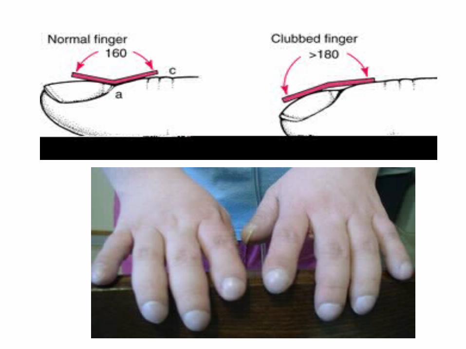
to permit greater expansion of the thoracic cavity.S+S for chronic hypoxia- Person appears fatigued and is lethargic.- Clubbed fingers and toesWith clubbing the base of the nail becomes swollen and the ends
of the fingers and toes increase in size, the angle between the nail and the base of the nail increase to more than 180 degrees.

Altered breathing Patternsbreathing Patterns refers to the rate, volume, rhythm, and relative ease or effort
of respiration Eupnea ….. Is quiet, rhythmic, and effortlessTachypnea ….. Rapid rate is seen with fever, metabolic acidosis, pain, and
hypercapnia or hypoxemia.Bradypnea ….. Slow rate is seen in clients who have taken drugs such as
morphine, metabolic alkalosis or have increased ICP (e.g., from brain injury).
Apnea ….. Is the cessation of breathing.Hyperventilation ….. Is an ↑ movement of air into and out of the lungs. The
rate and depth of respiration ↑ and more CO2 is eliminated than is produced.

One type of hyperventilation that accompanies metabolic acidosis is kussmaul’s breathing by which the body attempts to compensate by blowing off the CO2 through deep and rapid breathing . Hyperventilation can also occur in response to stress or anxiety.
Abnormal respiratory rhythms create an irregular breathing pattern. Two abnormal respiratory rhythms are:
Cheyne – strokes respiration ….very deep to very shallow breathing and temporary apnea
Causes- ↑ ICP , CHF, overdose of certain drugs
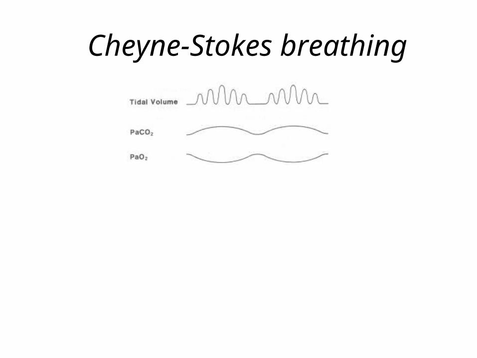
Cheyne-Stokes breathing

Biot’s (cluster) respiration …. Shallow breaths interrupted by apnea may be seen in clients with central nervous system disorders.
Orthopnea ….. Is the inability to breath except in an upright or standing position.
Dyspnea ….Difficult or uncomfortable breathingS+S- Person appear anxious and may experience SOB- Feeling of being unable to get enough air- Flared nostrils , skin appear dusky,↑ P

Obstructed airwayAn upper airway obstruction that is in the nose, pharynx, or larynxCauses- F.B such as food- Tongue falls back in unconscious- Collection of secretion in the passageways Respiration sound gurgly or bubbly Lower airway obstruction involves partial or complete occlusion the
passage in the bronchi and lungsCauses- Accumulation of mucus or inflammatory exudate.

NURSING Management Assessing The patient’s health history is an essential component
for assessing respiratory functioning. Either the patient or a family member can provide this information.
Nursing History Data should include about current and past
respiratory problems, lifestyle, presence of cough, sputum or pain, medications for breathing, and presence of risk factors for impaired oxygenation status.

Physical Examination The nurse use 4 physical examination techniques:- Inspection, palpation, percussion, and auscultation. The nurse first observes the rate, depth, rhythm, and
quality of respirations, noting the position the client assumes for breathing. Also inspects for variations in the shape of the thorax that may indicate adaptation to chronic respiratory conditions. e.g., client with emphysema frequently develop a barrel chest.


The nurse palpates the thorax for bulges, tenderness, or abnormal movement, detect vocal fremitus. Perform percussion posteriorly as the patient pulls the shoulders forward. Then continue with the examination proceeding down the patient’s back, comparing one side to the other. Examine the anterior and lateral thorax with the patient in a supine position.
Listen carefully to the intensity and quality of each sound as the chest wall and underlying structures are percussed.

Using the diaphragm of a stethoscope, move from apex to base, comparing one side with the other side while listening to a complete respiratory cycle, inspiration and expiration. While auscultating, ask the patient to breathe through an open mouth slowly because breathing through the nose can produce falsely abnormal breath sounds. Breathing too quickly, such as with hyperventilation, may cause syncope and patient distress. If any abnormal breath sound is detected, instruct the patient to cough and auscultate again for at least two complete respiratory cycles. Record location, change in breath sounds after coughing, and phase of respiration (e.g., expiration) when any abnormal sound is noted.

Diagnostic Studies
There are various diagnostic tests to assess respiratory status included:-
Sputum specimens, throat cultures, visualization procedures, VBG, ABG, Pulmonary function test.
Pulmonary function tests measures lung volume and capacity.
See table 50-1

Pulmonary function tests measure the following lung volumes and capacities:
• Tidal volume (TV): the amount of air inspired and expired in a normal respiration. Normal is 500 mL.
• Inspiratory reserve volume (IRV): Maximum amount of air that can be inhaled over and above a normal breath . Normal is 3,100 mL.
• Expiratory reserve volume (ERV): Maximum amount of air that can be exhaled following a normal exhalation. Normal is 1,200 mL.
• Residual volume (RV): the amount of air remaining in the lungs after a maximal expiration. Normal is 1,200 mL.

•Total lung capacity (TLC): the total volume of the lungs at maximum inflation calculated by adding the TV, IRV, ERV, and RV. Normal is 6,000 mL.
• Vital capacity (VC): the amount of air that can be exhaled after a maximal inspiration. Calculated by adding the TV,IRV, AND ERV. Normal is 4,800 mL.
• Inspiratory capacity (IC): the total amount of air that can be inhaled following normal quiet exhalation. Calculated by adding the TV,IRV . Normal is 3,600 mL.
• Functional residual volume (FRV): The volume left in the lungs after normal exhalation Calculated by adding the ERV and RV. Normal is 2,400 mL.


DiagnosingSee page 1365, 1366
PlanningSee page 1366
Implementing -Promoting Oxygenation
-Deep breathing and coughing One common breathing exercise is abdominal (diaphragmatic)
and pursed – lip breathing. Advantage of this exercise

Abdominal breathing permits deep full breaths with little effort. Pursued lip breathing helps the client develop control over breathing, also create resistance to the air flowing out of the lungs, thereby prolonging exhalation and preventing airway collapse by maintaining positive airway pressure also this tightening abdominal muscles to exhales more effectively. The client usually inhales to a count 3 and
exhales to a couunt of 7.


-Hydration Adequate hydration maintains the moisture of the respiratory mucous membranes. When the client is dehydrated or when environment has a low humidity, the respiratory secretions can become thick and tenacious.
Humidifiers are devices that add water vapor to inspired air, to prevent mucous membranes from drying and becoming irritated and to loosen secretions for easier expectoration.

Humidifiers


-Medication A number of types of medication can be used for clients
with oxygenation problem . *Bronchodilators
Reduce bronchospasm, opening tight or congested airways and facilitating ventilation. Route P.O, IV but
the prefered route is by inhalation .Side effect include ↑ P, ↑ BP, anxiety, restlessness .


Anti- inflammatory drugs such as glucocorticoidsRoute : PO, IV, Inhaler.
Action : ↓ edema and inflammation in the airways and allowing a better air exchange .
*Leukotriene modifiers These medications suppress the effects of Leukotriene on the smooth muscle of the respiratory tract. Leukotriene cause bronchoconstriction, mucous production, edema of the respiratory
tract . *Expectorants
Help breakup mucus, making it more liquid and easier to expectorate. E.g., Guaifenesin
When frequent or prolonged coughing interrupts sleep, a cough suppressant such as codeine.
*Digitalis glycosides act directly on the heart to improve the strength of contraction and slow the heart rate.
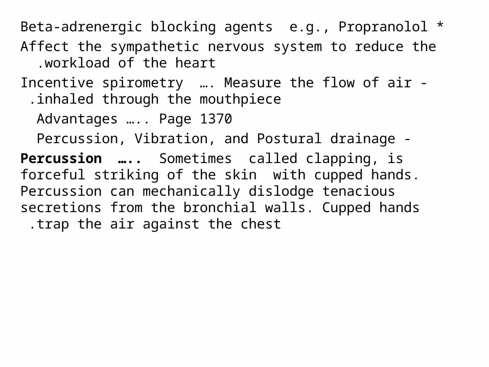
*Beta-adrenergic blocking agents e.g., Propranolol Affect the sympathetic nervous system to reduce the workload of the
heart . -Incentive spirometry …. Measure the flow of air inhaled through the
mouthpiece .Advantages ….. Page 1370
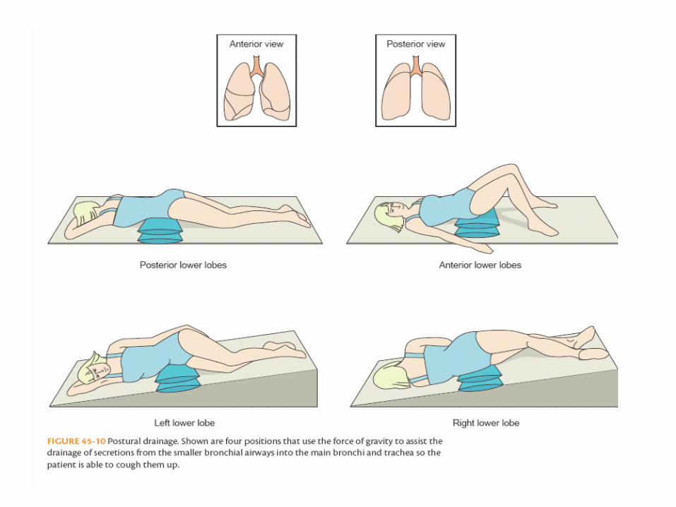
-Percussion, Vibration, and Postural drainage Percussion ….. Sometimes called clapping, is forceful striking of the
skin with cupped hands. Percussion can mechanically dislodge tenacious secretions from the bronchial walls. Cupped hands trap the
air against the chest.


Vibration …… is a series of vigorous quiverings produced by hands that are placed flat against the
client’s chest wall .Postural drainage ….. Is the drainage by gravity of secretions from various lung segments. A wide variety of positions is necessary to drain all segments of the lungs . The lower lobes require drainage most frequently because the upper lobes drain by gravity.


Oxygen Therapy Clients who have difficulty ventilating all areas of
their lungs, those whose gas exchange is impaired, or people with heart failure may benefit from oxygen therapy to prevent hypoxia.
Oxygen therapy safety precautions … Page 1373

Oxygen delivery systemsThe choice of system depends on the client’s oxygen
needs, comfort, and developmental considerations.- Cannula (nasal prongs) Advantage- Does not interfere with the client’s ability to
eat or to talk. It also is relatively comfortable, permits some freedom of movement and is well tolerated by the client.

-It delivers a relatively low concentration of O2 (24% to 45% ) at flow rates of 2 to 6 L /min . Above 6 L/min the client tends to swallow air and the Fio2 is
not increased .Disadvantage
-Inability to deliver higher concentrations of O2, and it can be drying and irritating to mucous membranes .
-Face mask Simple face mask delivers O2 concentrations from
40% to 60% at flow rates of 5 to 8 L/min .Partial rebreather mask delivers O2 concentrations of 60% to 90% at liter flows of 6 to 10 L/min .

The partial rebreather bag must not totally deflate during inspiration to avoid carbon dioxide buildup.

Nonrebreather mask delivers the highest oxygen concentration possible 95% to 100% at liter flows of 10 to 15 L/min. One way bag valves on the mask and between the reservoir bag and the mask prevent the room air and the client’s exhaled air from entering the
bag so only the oxygen in the bag is inspired .

Venturi mask delivers oxygen concentration varying from 24% to 40% or 50% at liter flows of 4 to 10 L/min.

-Face tent Can replace oxygen masks when masks are poorly tolerated by clients. Face tents provide varying concentrations of O2 , for example, 30% to 50% concentration of oxygen at 4 to 8 L/min.

Artifical airways Are inserted to maintain a patent air way passage for clients whose airway has become or may become
obstructed. Four common types of airways are-:1 -Oropharyngeal and nasopharyngeal airways
are used to keep the upper air passage open when they may become obstructed by secretions or the tongue .
Oropharyngeal airways stimulate the gag reflex and are only used for clients with altered levels of consciousness (e.g., general anesthesia, overdose, or head injury).


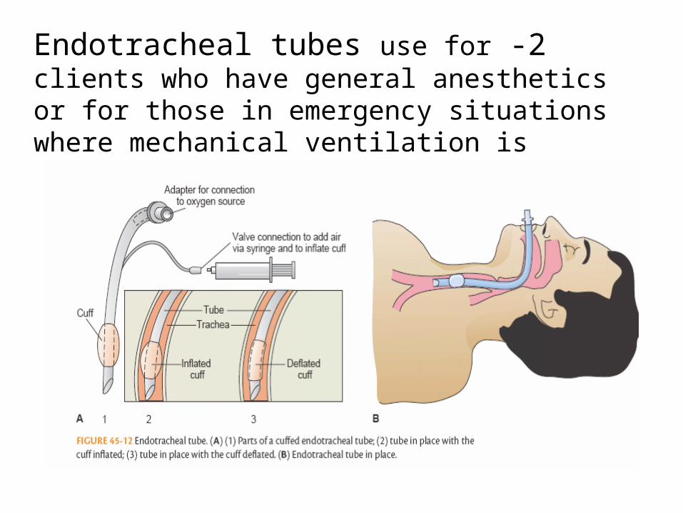
2 -Endotracheal tubes use for clients who have general anesthetics or for those in emergency
situations where mechanical ventilation is required .


3 -Tracheostomy. Clients who need long term airway support may have a tracheostomy .
Tracheostomy is an opening into the trachea through the neck


SuctioningIs aspirating secretions through a catheter connected to a suction machine or wall suction outlet.
Indication of suction -Signs of respiratory distress
-The client unable to cough up and expectorate secretions -Dyspnea, bubbling, rattling breath sounds, poor skin
color, ↓ oxygen saturation levels Complications : hypoxemia, trauma to the airway, nosocomial
infection, cardiac dysrhythmia .

The following techniques are used to minimize or decrease these complications :
-Hyperinflation -Hyperoxygenation
Chest Tubes and drainage systems If the thin, double layered pleural membrane is disrupted by lung disease, surgery, or trauma, the negative pressure between the pleural layers may be lost. The lung then may be collapses . Chest tubes may be inserted into the pleural cavity to restore
negative pressure and drain collected fluid or blood .


Managing Chest TubesPatients with fluid (pleural effusion), blood (hemothorax), or air (pneumothorax) in the pleural space require a chest tube to drain these substances and allow the compressed lung to reexpand.. Once inserted, the tube is secured with a suture and tape, covered with an airtight dressing, and attached to a drainage system that may or may not be attached to suction .


Heimlich chest drain valve

Pneumostat chest drain