p-glycoprotein and breast cancer resistance protein
TRANSCRIPT

JPET # 154781
1
P-glycoprotein and Breast Cancer Resistance Protein
Influence Brain Distribution of Dasatinib
Ying Chen, Sagar Agarwal, Naveed M. Shaik, Cliff Chen, Zheng Yang, William F. Elmquist
Department of Pharmaceutics, University of Minnesota, Minneapolis, MN, U.S.A. (Y.C., S.A.,
N.M.S. and W.F.E.)
Metabolism and Pharmacokinetics, Pharmaceutical Candidate Optimization, Bristol-Myers
Squibb Co., Princeton, NJ, USA (C.C. and Z.Y.)
JPET Fast Forward. Published on June 2, 2009 as DOI:10.1124/jpet.109.154781
Copyright 2009 by the American Society for Pharmacology and Experimental Therapeutics.
This article has not been copyedited and formatted. The final version may differ from this version.JPET Fast Forward. Published on June 2, 2009 as DOI: 10.1124/jpet.109.154781
at ASPE
T Journals on D
ecember 14, 2021
jpet.aspetjournals.orgD
ownloaded from
JPET # 154781
2
Running Title:
Active efflux limits CNS distribution of dasatinib
Corresponding Author
William F. Elmquist, Department of Pharmaceutics, University of Minnesota, 9-177 Weaver
Densford Hall, 308 Harvard Street SE, Minneapolis, MN 55455, USA. Phone: +001-612-625-
0097; Fax: +001-612-626-2125; e-mail: [email protected]
Number of text pages – 16
Number of figures – 6
Number of tables – 2
Number of references – 29
Number of words in abstract – 243
Number of words in introduction – 593
Number of words in discussion – 1464
List of Abbreviations
CML, Chronic myelogenous leukemia; Ph, philadelphia chromosome; ALL, acute
lymphoblastic leukemia; CNS, central nervous system; BBB, blood-brain-barrier; Mdr1, multi-
drug resistance protein 1; P-gp, p-glycoprotein; BCRP, breast cancer resistance protein; BCR,
breakpoint cluster region; ABL, Abelson; PDGFR, platelet-derived growth factor; TKI,
tyrosine kinase inhibitors; A-to-B, apical-to-basolateral; B-to-A, basolateral-to-apical; Peff,
effective permeability; B/P , brain-to-plasma; ER, efflux ratio; GBM , glioblastoma
multiforme.
This article has not been copyedited and formatted. The final version may differ from this version.JPET Fast Forward. Published on June 2, 2009 as DOI: 10.1124/jpet.109.154781
at ASPE
T Journals on D
ecember 14, 2021
jpet.aspetjournals.orgD
ownloaded from

JPET # 154781
3
Abstract
The novel tyrosine kinase inhibitor dasatinib is approved for use in imatinib resistant or
intolerant chronic myelogenous leukemia (CML) and may be useful for other tumors in the CNS.
The objective of this study was to investigate the role of p-glycoprotein (P-gp) and breast cancer
resistance protein (BCRP) in modulating the CNS penetration of dasatinib. Results from the in
vitro studies indicate that cellular delivery of dasatinib is significantly limited by active efflux
due to both P-gp and BCRP. Permeability studies indicated greater permeability in the
basolateral-to-apical direction than in the apical-to-basolateral direction due to active efflux by
P-gp or BCRP. Selective inhibitors of P-gp and BCRP, such as LY335979 and Ko143, were
able to restore the intracellular accumulation and abolish the directionality in net flux of
dasatinib. In vivo brain distribution studies showed that the CNS distribution of dasatinib is
limited with the brain-to-plasma concentration ratios less than 0.12 in wild-type mice, which
increased approximately 8-fold in Mdr1a/b-/- Bcrp1-/- mice. Dasatinib brain distribution was
significantly increased in Mdr1a/b-/- mice and when wild-type mice were pretreated with
LY335979. Simultaneous inhibition of P-gp and BCRP by GF120918 resulted in a 5-fold
increase in brain concentration. These in vitro and in vivo studies demonstrate that dasatinib is a
substrate for the important efflux transporters p-glycoprotein and BCRP. These transport
systems play a significant role in limiting the CNS delivery of dasatinib and may have direct
implications in the treatment of primary and metastatic brain tumors.
This article has not been copyedited and formatted. The final version may differ from this version.JPET Fast Forward. Published on June 2, 2009 as DOI: 10.1124/jpet.109.154781
at ASPE
T Journals on D
ecember 14, 2021
jpet.aspetjournals.orgD
ownloaded from

JPET # 154781
4
Chronic myelogenous leukemia (CML) accounts for 15-20% of all cases of adult leukemia in
western populations (Quintas-Cardama et al., 2007). Imatinib (Gleevec, STI 571) is a first-
generation tyrosine kinase inhibitor (TKI) that was approved for use in the treatment of CML
and gastrointestinal stromal tumor (GIST) (Druker, 2003a). Imatinib inhibits the BCR-ABL, c-
kit, platelet-derived growth factor (PDGFR) and the ARG tyrosine kinases (Druker, 2003b).
CNS involvement is a common complication seen in CML and most patients with CML and Ph+
acute lyphoblastic leukemia (Ph+ ALL) develop extramedullary involvement during the course of
their disease. CNS failure has been reported in approximately 20% of imatinib-treated patients
with CML or Ph+ ALL (Leis et al., 2004). CNS relapses were observed in patients despite a
complete hematological response (Leis et al., 2004). This can be attributed to the poor
penetration of imatinib across the blood-brain barrier (BBB) resulting in sub-therapeutic drug
levels in the brain. This limited delivery of imatinib to the brain is primarily a result of active
efflux at the BBB by drug eluting P-glycoprotein (P-gp) and the breast cancer resistance protein
(BCRP). Our laboratory and several others have reported that imatinib is a good substrate for
these two drug transporters which limit CNS distribution (Dai et al., 2003; Breedveld et al.,
2005). An inadequate concentration of imatinib in the CNS can make the brain a sanctuary for
chronic myelogenous leukemia (Leis et al., 2004; Neville et al., 2004; Rajappa et al., 2004).
Dasatinib (Sprycel;BMS-354825) is a second-generation TKI approved for use in
imatinib-resistant CML patients (Shah, 2007). Dasatinib is an extremely potent BCR-ABL
inhibitor and has shown significant activity in imatinib-resistant or intolerant CML patients
(Cortes et al., 2007; Kantarjian et al., 2007; Ottmann et al., 2007). It effectively inhibits the
proliferation of tumor cells expressing nearly all imatinib-resistant isoforms of BCR-ABL
(2006). In addition to this, dasatinib also inhibits the SRC tyrosine kinase (Lombardo et al.,
2004) which has been identified as a potential target for glioblastoma therapy (Du et al., 2009).
Given that dasatinib was designed to overcome the molecular resistance seen with imatinib, one
question that arises is whether dasatinib can also overcome the CNS delivery problem and
prevent CNS metastasis. There is little information available about the delivery of dasatinib
across the BBB, including the action of relevant BBB transporters in modulating this delivery.
As a promising multi-targeted TKI, adequate CNS delivery of dasatinib is important for the
prevention of CNS metastases in CML as well as treatment of other CNS malignancies.
This article has not been copyedited and formatted. The final version may differ from this version.JPET Fast Forward. Published on June 2, 2009 as DOI: 10.1124/jpet.109.154781
at ASPE
T Journals on D
ecember 14, 2021
jpet.aspetjournals.orgD
ownloaded from

JPET # 154781
5
It was recently suggested that dasatinib can cross the BBB to achieve therapeutic
response in a mouse model of CML and in Ph(+) ALL and CML patients with CNS involvement
(Porkka et al., 2008). However, dasatinib brain concentrations were on average 12- to 31-fold
lower than the plasma concentrations and the therapeutic benefit was attributed to the extremely
high potency of dasatinib against the BCR-ABL and SRC therapeutic targets. The fact that this
low level of brain penetrance was able to result in anti-tumor activity may be true in this case,
but may not hold for other tumors of the CNS, such as those driven by less sensitive tyrosine
kinase targets, e.g., c-kit. In general, the limited CNS distribution of dasatinib may be a
consideration in developing new therapeutic strategies for the treatment of brain tumors.
Therefore, the objective of this study was to investigate the extent to which dasatinib can
distribute to the brain and to establish the influence of active efflux by P-gp and BCRP at the
blood-brain barrier on the brain distribution of dasatinib.
This article has not been copyedited and formatted. The final version may differ from this version.JPET Fast Forward. Published on June 2, 2009 as DOI: 10.1124/jpet.109.154781
at ASPE
T Journals on D
ecember 14, 2021
jpet.aspetjournals.orgD
ownloaded from

JPET # 154781
6
Methods
Chemicals and Reagents
Dasatinib and [14C]-dasatinib (31.9 µCi/mg) were kindly provided by Bristol-Myers Squibb
Company (Princeton, NJ). [3H]-vinblastine sulfate and [3H]-mitoxantrone were purchased from
Moravek Biochemicals (La Brea, CA). LY335979 (zosuquidar, (R)-4-((1aR,6R,10bS)-1,2-
difluoro-1,1a,6,10b-tetrahydrodibenzo-(a,e)cyclopropa(c) cycloheptan-6-yl)-α-((5-
quinoloyloxy)methyl)-1-piperazineethanol, trihydrochloride) was a gift from Eli Lilly and Co.
(Indianapolis, IN), GF120918 (elacridar, N-[4-[2-(6, 7-Dimethoxy-3, 4-dihydro-1H-isoquinolin-
2-yl) ethyl]-5-methoxy-9-oxo-10H-acridine-4-carboxamide) was generously given by
GlaxoSmithKline (Research Triangle, NC). Dr. Alfred H. Schinkel (The Netherlands Cancer
Institute, Amsterdam, The Netherlands) kindly provided Ko143 (Allen et al., 2002). Cell culture
reagents were purchased from Invitrogen (Carlsbad, CA). All other reagents and chemicals were
purchased from Sigma-Aldrich (St. Louis, MO).
In vitro studies - Cell lines
Epithelial Madine Darby Canine Kidney (MDCKII) cells were used in all in vitro studies. Wild-
type (WT) and MDR1-transfected cells were a gift from Dr. Piet Borst (Netherlands Cancer
Institute), and WT and Bcrp1-transfected cells were kindly provided by Dr. Alfred H. Schinkel
(The Netherlands Cancer Institute, Amsterdam, The Netherlands). Cells were cultured in
DMEM media supplemented with 10% fetal bovine serum (SeraCare Life Sciences, Inc.,
Oceanside, CA), penicillin (100 U/ml), streptomycin (100 µg/ml) and amphotericin B 250ng/mL
(Sigma-Aldrich, St. Louis, MO) and maintained at 37°C with 5% CO2 under humidifying
conditions. Cells between passages 5 and 15 were used in all experiments.
Intracellular Accumulation Studies in MDCKII Cells
The wild-type and MDR1- or Bcrp1-transfected cells were seeded in clear polystyrene 12-well
plates (TPP cell culture plate; Sigma-Aldrich, St. Louis, MO) at a seeding density of 2 × 10⁵
cells/well. The medium was changed on alternate days until the cells formed confluent
monolayers. On the day of the experiment, the medium was aspirated and the monolayer was
washed twice with 1 mL prewarmed (37°C) assay buffer. The cells were preincubated with 1 ml
This article has not been copyedited and formatted. The final version may differ from this version.JPET Fast Forward. Published on June 2, 2009 as DOI: 10.1124/jpet.109.154781
at ASPE
T Journals on D
ecember 14, 2021
jpet.aspetjournals.orgD
ownloaded from

JPET # 154781
7
of assay buffer for 30 min, after which the buffer was aspirated and the experiment was initiated
by adding tracer solution of radiolabeled drug (1 μg [14C] dasatinib, or tracer vinblastine or
mitoxantrone for positive controls) in 1 ml assay buffer per well. The plates were continuously
agitated at 60 rpm in an orbital shaker maintained at 37°C for the duration of the experiment. At
the end of the 3-hour accumulation period, the assay buffer containing the radiolabeled drug was
aspirated from all the wells and the cells were washed three times with 2 ml of ice-cold
phosphate-buffered saline. Cells were then solubilized using 1 ml of mammalian protein
extraction reagent (M-PER®, Pierce Biotechnology, Inc., Rockford, IL). A 200 μl sample of
solubilized cell fractions was drawn from each well in triplicate, 4 ml of scintillation fluid
(ScintiSafe Econo cocktail; Fisher Scientific Co., Pittsburgh, PA) was added to each sample and
counted using liquid scintillation counting (LS-6500; Beckman Coulter, Inc., Fullerton, CA) to
determine the radioactivity associated with the cell fractions. The protein concentration was
determined using the BCA protein assay (Pierce Biotechnology, Inc., Rockford, IL) to normalize
the radioactivity in each well for cell number. For inhibition studies, the cells were treated with
the selective inhibitors 1 μM LY335979 for P-gp and 200 nM Ko143 for BCRP during both the
preincubation and the accumulation periods. Drug accumulation in cells was expressed as a
percentage of the accumulated radioactivity measured in the wild-type control cells (dpm) per
microgram of protein. The stock solutions for all the inhibitors used were prepared in dimethyl
sulfoxide (DMSO) and diluted using assay buffer to obtain working solutions, so that the final
concentration of DMSO was less than 0.1%.
Directional Flux across MDCKII Monolayers
The methods used for directional flux were similar to that previously described by Dai et al. (Dai
et al., 2003). Briefly, cells were seeded at a density of 2 × 10⁵ cells/well on polyester
semipermeable membrane supports of the inserts in six-well transwells (Corning Inc., Corning,
NY). The medium was changed on alternate days and the cells formed confluent polarized
epithelial monolayers in 3-4 days. The representative transepithelial electrical resistance was
300 ± 8 ohm·cm2 (n=6) in the WT MDCKII monolayers, 275 ± 26 ohm·cm2 (n=6) in the MDR1-
transfected MDCKII monolayers and 248± 27 ohm·cm2 (n=6) in the Bcrp1-transfected MDCKII
This article has not been copyedited and formatted. The final version may differ from this version.JPET Fast Forward. Published on June 2, 2009 as DOI: 10.1124/jpet.109.154781
at ASPE
T Journals on D
ecember 14, 2021
jpet.aspetjournals.orgD
ownloaded from

JPET # 154781
8
monolayers. Mannitol flux across the monolayer was also measured to confirm the existence of
tight junctions with approximately 1% per hour flux (Peff = 9 × 10⁻⁸ cm/s). The monolayers were
washed with 2 mL prewarmed (37°C) assay buffer, and after a 30-min preincubation period, the
experiment was initiated by adding a tracer solution of radiolabeled drug (1.5 μg or 2.6 μg 14C-
dasatinib) in assay buffer to the donor side (apical side, 1.5 ml; basolateral side, 2.6 ml). Fresh
assay buffer was added to the receiver side and 200 μl was sampled from the receiver
compartment at 0, 10, 20, 30, 45, 60, and 90 min. The volume sampled was immediately
replaced with fresh assay buffer. Additional samples were drawn at 0 and 90 min from the donor
compartment. The amount of radioactivity in the samples was determined using liquid
scintillation counting. The apical-to-basolateral (A-to-B) flux was determined by addition of
radiolabeled drug solution to the apical compartment and sampling the basolateral compartment,
whereas for basolateral-to-apical (B-to-A) flux, the donor was the basolateral compartment and
the apical compartment was sampled at the mentioned times. When an inhibitor was used in the
flux study, the cell monolayers were preincubated with the inhibitor (1 µM LY335979 for P-gp
and 200 nM Ko143 for Bcrp1) for 30 min, followed by determination of A-to-B and B-to-A flux
with the inhibitor being present in both compartments throughout the course of the experiment.
Permeability Calculation
The effective permeability (Peff) was calculated by the following equation,
where Q is the amount of radiolabeled drug transported though the cell monolayer, t is time,
dQ/dt is the mass transport rate, A is the apparent surface area of the cell monolayer (4.67 cm2),
and C0 is the initial donor concentration. The efflux ratio (ER) is defined as the ratio of Peff in
the B-to-A direction to the Peff in the A-to-B direction and gives an indication of the magnitude
of P-gp or Bcrp1 mediated efflux.
In vivo studies
Animals
0*
)(CA
dt
dQ
Peff =
This article has not been copyedited and formatted. The final version may differ from this version.JPET Fast Forward. Published on June 2, 2009 as DOI: 10.1124/jpet.109.154781
at ASPE
T Journals on D
ecember 14, 2021
jpet.aspetjournals.orgD
ownloaded from

JPET # 154781
9
Animals used in this study were male Mdr1a/b-/- (P-gp knockout), Bcrp1-/- (Bcrp1 knockout),
Mdr1a/b-/- Bcrp1-/- (triple knockout) and wild-type mice of a FVB genetic background. All mice
were purchased from Taconic Farms. Inc. (Germantown, NY) and were between 8 to 10 weeks
old at the time of experiment. Animals were maintained under temperature-controlled conditions
with a 12-h light/dark cycle and unlimited access to food and water. All mice were allowed to
acclimatize for a minimum of one week upon arrival. All studies were approved by the
Institutional Animal Care and Use Committee of the University of Minnesota (IACUC).
Dasatinib Brain and Plasma Distribution Studies in Mice.
All mice received dasatinib (1:1 propylene glycol: water, pH 4.6) by intravenous administration
in the tail vein at a dose of 5 mg/kg or an oral dose of 10 mg/kg. The study involved the
following five study groups.
(i) Wild-type and Mdr1a/b-/- Bcrp1-/- mice received i.v. dasatinib following which blood and
brain were sampled at 5, 20, 60, 120, 180 min postdose, n=4 at each time point.
(ii) Wild-type mice received 25 mg/kg LY335979, 10 mg/kg Ko143 or 10 mg/kg GF120918
intravenously 30 minutes before i.v. dasatinib, and brain and blood were sampled at 20 and
120 minutes, n=4 at each time point.
(iii) Wild-type, Mdr1a/b-/-, Bcrp1-/- , Mdr1a/b-/- Bcrp1-/- mice were administered i.v. dasatinib,
blood and brain were sampled at 20 and 120 minutes post-dose, n=4 at each time point.
(iv) Wild-type mice received 25 mg/kg LY335979, 10 mg/kg Ko143 or 10 mg/kg GF120918
intravenously 30 minutes before an oral dose of dasatinib, brain and blood were sampled at
120 minutes, n=4.
(v) Wild-type, Mdr1a/b-/-, Bcrp1-/- , Mdr1a/b-/- Bcrp1-/- mice were administered dasatinib by
oral gavage, blood and brain were sampled at 120 minutes post-dose, n=4.
The animals were euthanized at predetermined time points by using a CO2 chamber. Blood was
immediately harvested via cardiac puncture and collected in tubes preloaded with potassium
EDTA (BD, Franklin Lakes, NJ). Whole brain was immediately harvested, rinsed with ice-cold
saline to remove extraneous blood and flash frozen using liquid nitrogen. Plasma was isolated
from blood by centrifugation at 3000 rpm for 10 min at 4°C. All plasma and whole brain
samples were stored at -80°C until analysis by LC-MS/MS.
Determination of Dasatinib Concentrations in Plasma and Brain using LC-MS/MS
This article has not been copyedited and formatted. The final version may differ from this version.JPET Fast Forward. Published on June 2, 2009 as DOI: 10.1124/jpet.109.154781
at ASPE
T Journals on D
ecember 14, 2021
jpet.aspetjournals.orgD
ownloaded from

JPET # 154781
10
Mouse plasma and brain samples were analyzed by LC-MS/MS. Sample preparation included
addition of 3 volumes of acetonitrile containing internal standard (IS) d6-dasatinib to the plasma
samples. Whole brain samples were first homogenized in water with a volume ratio of 1:3
followed by the addition of acetonitrile containing IS. After centrifugation to remove
precipitated proteins, 10 µL of the clear supernatant was analyzed by LC/MS/MS.
Chromatographic separation was obtained using an Atlantis® dC18 column (2.1 mm x 50 mm)
packed with a 3µM stationary phase (Waters Corporation, Milford, MA). The mobile phase was
composed of 10 mM ammonium acetate with 0.1% formic acid (A) and acetonitrile (B). A
gradient elution method was used with the initial mobile phase consisting of 90% A and 10% B.
After sample injection, the mobile phase was held at the initial condition for 1 min and then
changed to 10% solvent A and 90% solvent B over 1 min and held at that composition for an
additional 0.8 minutes. The mobile phase was then returned to initial conditions and the column
was re-equilibrated for one minute. The total analysis time was 4 minutes. The HPLC was
interfaced to a Micromass Quattro LC triple quadrupole mass spectrometer (Waters, Milford,
MA, USA) equipped with electrospray ionization interface. The desolvation temperature was
350°C and the source temperature was 120°C. Data acquisition employed selected reaction
monitoring (SRM). Positively charged ions representing the [MH]+ for dasatinib and the internal
standard (IS) were selected in MS1 and dissociated with argon at a pressure of 2.5 x 10⁻³ Torr to
form specific product ions which were subsequently monitored by MS2. All dwell times were
100 ms. The SRM transitions monitored were m/z 488 → 401 for dasatinib, m/z 496 → 409 for
the internal standard. The cone voltage was optimized at 35 V for both dasatinib and IS, while
the collision energy was 35 eV. The retention time for dasatinib and IS was 2.05 and 2.04 min,
respectively. The lower limit of quantification was 1 ng/mL.
Statistical Analysis
Statistical analysis was conducted using SigmaStat, version 3.1(Systat Software, Inc., Point
Richmond, CA). Statistical comparisons between two groups were made by using two-sample t-
test at p < 0.01 significance level. If groups failed the normality test, then the nonparametric
alternative of two-sample t test, the Mann-Whitney rank sum test was used. Multiple groups
were compared by one-way analysis of variance with the Holm-Sidak post-hoc test for multiple
This article has not been copyedited and formatted. The final version may differ from this version.JPET Fast Forward. Published on June 2, 2009 as DOI: 10.1124/jpet.109.154781
at ASPE
T Journals on D
ecember 14, 2021
jpet.aspetjournals.orgD
ownloaded from

JPET # 154781
11
comparisons at a significance level of p < 0.01. When groups failed the normality test, the
Kruskal-Wallis one way analysis of variance on ranks was used.
Results
Intracellular Accumulation Studies in MDCKII Cells
Intracellular accumulation of [14C] dasatinib was studied in MDCKII-Bcrp1, MDCKII-MDR1
and MDCKII wild-type cells. [3H] Vinblastine, a prototypical P-gp substrate, was included as a
positive control in all accumulation studies done in MDR1-transfected cells and showed
significantly lower accumulation in the MDR1-transfected cells compared with wild-type cells
(~13% of WT control, p < 0.01) (Figure 1). Similar to the positive control, accumulation of
[14C] dasatinib in the MDR1-transfected cells was significantly lower than that in the wild-type
cells (~ 15% of WT control, p < 0.01). Treatment with 1 μM LY335979, the P-gp selective
inhibitor, completely abolished P-gp efflux activity such that there was no significant difference
in the accumulation of [14C] dasatinib between the two cell types (Figure 1). Accumulation of
dasatinib in the Bcrp1-transfected cells was about 25-fold lower than the accumulation in wild-
type cells (p<0.01), similar to the positive control for BCRP, mitoxantrone. Use of the BCRP
selective inhibitor Ko143 completely abolished this difference in dasatinib accumulation
between the Bcrp1-transfected and wild-type cells (Figure 2).
Directional Flux across MDCKII Cell Monolayers
Transport of [14C]-dasatinib was studied in MDCKII cells and the effective permeabilities were
calculated. In the MDCKII wild-type cells, effective permeability of dasatinib was significantly
greater in the B-to-A direction than in the A-to-B direction, yielding an efflux ratio (ER) of 4.8.
Similarly, in the MDR1-transfected cells, the effective permeability was significantly enhanced
in the B-to-A direction compared to the A-to-B direction with an ER of 25.6. Treatment with 1
µM LY335979 completely abolished this directionality in dasatinib transport by P-gp, such that
there was no significant difference in permeabilities of dasatinib in the two directions in either
cell type. Use of 1 μM LY335979 dramatically reduced the ER to 0.9 in the wild-type cells and
1.2 in the MDR1-transfected cells (Table 1A). Transport of [14C] dasatinib in the wild-type and
Bcrp1-transfected cells also showed significant directionality with the effective permeability
significantly increased in the B-to-A direction in both cell types. The ER was 51.5 in the Bcrp1-
This article has not been copyedited and formatted. The final version may differ from this version.JPET Fast Forward. Published on June 2, 2009 as DOI: 10.1124/jpet.109.154781
at ASPE
T Journals on D
ecember 14, 2021
jpet.aspetjournals.orgD
ownloaded from

JPET # 154781
12
transfected cells compared to 4.1 in the wild-type cells. The BCRP inhibitor Ko143 (200nM)
was not able to completely abolish this directionality in transport. Permeability of dasatinib in
the B-to-A direction decreased resulting in a reduction of the ER to approximately 9 in the
Bcrp1-transfected cells compared to 4.1 in the wild-type (Table 1B).
Dasatinib Brain Distribution in Mice
Brain distribution of dasatinib after an i.v. dose was determined in wild-type and Mdr1a/b-/-
Bcrp1-/- mice. There was no significant difference in plasma concentrations between the wild-
type and Mdr1a/b-/- Bcrp1-/- mice (Figure 3A), however, brain concentrations in the Mdr1a/b-/-
Bcrp1-/- triple-knockout mice were significantly greater (p<0.001) than that in wild-type mice
(Figure 3B). The brain-to-plasma concentration ratio (B/P), a measure of the brain distribution,
was significantly greater (p<0.001) in the triple-knockout mice at all of the five measured time
points (Figure 4). The B/P concentration ratio was lower than 0.12 in the wild-type mice at all
the time points, suggesting that less than 12% of dasatinib in blood crosses the blood-brain
barrier to reach brain. In the triple knockout mice this ratio reached an “equilibrium” value of 1
after 60 mins.
The brain concentrations of dasatinib were also measured in wild-type mice that were pretreated
with the efflux inhibitors LY335979, Ko143 or GF120918. P-gp inhibition by LY335979
significantly increased brain dasatinib concentrations in the wild-type mice (p<0.01) (Figure
5A). Pretreatment with the BCRP inhibitor Ko143 was not able to increase brain distribution of
dasatinib to any significant extent. Interestingly, simultaneous inhibition of both P-gp and BCRP
by the dual inhibitor GF120918 resulted in a dramatic increase in brain dasatinib concentrations,
the B/P ratio increasing by more than 5-fold at both time points (p<0.001). A study was then
conducted in wild-type, Mdr1a/b-/-, Bcrp1-/- and Mdr1a/b-/- Bcrp1-/- to replicate pharmacological
inhibition with genetic deletion of drug efflux proteins. Results from this study were similar to
those seen when P-gp, BCRP, or P-gp and BCRP were inhibited using pharmacological
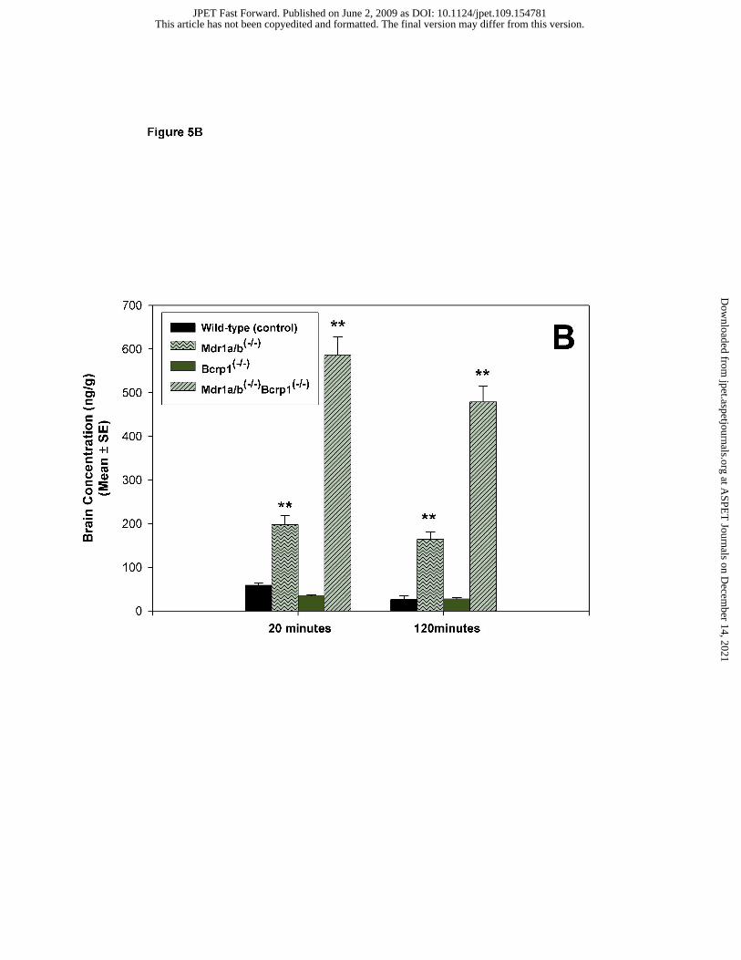
inhibitors (Figure 5B). Brain dasatinib concentrations increased significantly in Mdr1a/b-/- mice
compared with those in wild-type mice (p<0.001). While brain concentrations in Bcrp1-/- mice
did not increase to any significant extent, brain dasatinib concentrations increased by greater than
10-fold in the Mdr1a/b-/- Bcrp1-/- mice (p<0.001).
This article has not been copyedited and formatted. The final version may differ from this version.JPET Fast Forward. Published on June 2, 2009 as DOI: 10.1124/jpet.109.154781
at ASPE
T Journals on D
ecember 14, 2021
jpet.aspetjournals.orgD
ownloaded from

JPET # 154781
13
Dasatinib brain distribution was also studied after oral administration of drug in FVB mice.
Brain concentrations after a 10 mg/kg oral dose of dasatinib were significantly lower than the
plasma concentrations. Similar to the intravenous study, brain distribution was enhanced when
FVB wild-type mice were treated with efflux inhibitors LY335979, Ko143 or GF120918 prior to
receiving oral dasatinib. The brain-to-plasma ratio increased 2-fold on treatment with the P-gp
inhibitor, though this difference did not reach significance. Treatment with Ko143 did not
change the B/P ratio, however GF120198 dramatically increased the B/P ratio by over 6-fold
(p<0.01) (Figure 6). In the gene knockout animals, the B/P concentration ratio increased
approximately 4-fold in the Mdr1a/b-/- mice and by over 9-fold in the Mdr1a/b-/- Bcrp1-/- mice
(p<0.01). The brain-to-plasma concentration ratio did not change in the Bcrp1-/- mice (Figure
6).
Discussion
Imatinib mesylate showed initial remarkable efficacy in patients with CML (Druker et al., 2001a;
Druker et al., 2001b). However, this success was relatively short-lived with reports of rapid
relapse and CNS metastases in imatinib treated patients (Ottmann et al., 2002; Sawyers et al.,
2002). This led to the development of dasatinib – an extremely potent inhibitor of the BCR-ABL
tyrosine kinase, for use in imatinib resistant and intolerant CML patients. In addition to its
potent inhibitory effects against BCR-ABL, dasatinib also inhibits the SRC family proteins
(Lombardo et al., 2004; Nam et al., 2005), which have been implicated in the development of
resistance and the extramedullary expansion of disease in CML (Donato et al., 2003; Dai et al.,
2004; Ptasznik et al., 2004). Taking into consideration the role of SRC in tumors of the CNS and
the potent inhibitory effect of dasatinib against SRC, dasatinib may be a promising drug for
treatment of brain tumors such as GBM (Du et al., 2009; Milano et al., 2009). However, one
caveat behind the use of dasatinib in CNS tumors is its ability to cross the BBB and achieve
therapeutic concentrations in the brain.
Drug penetration into the CNS is often limted by active efflux proteins in the BBB, such as Pgp
and BCRP. In the current study, we have shown that dasatinib is a substrate for both P-gp and
BCRP using in vitro Pgp- and Bcrp-transfected cell models. In vivo transport studies
This article has not been copyedited and formatted. The final version may differ from this version.JPET Fast Forward. Published on June 2, 2009 as DOI: 10.1124/jpet.109.154781
at ASPE
T Journals on D
ecember 14, 2021
jpet.aspetjournals.orgD
ownloaded from

JPET # 154781
14
demonstrate that these two efflux proteins significantly limit the amount of drug that traverses
the BBB. Moreover, our studies have evaluated the possibility that the two drug efflux
transporters act in association with each other to extrude substrate drugs out of the brain.
In a recent report, Porkka et al. suggested that dasatinib can cross the BBB to result in
therapeutic response in CNS leukemia (Porkka et al., 2008). However, the group also reported
that dasatinib concentrations in brain were on average 12 to 31-fold lower than in plasma. A
brain penetrance of less than 10% reported in this study indicates that dasatinib does not readily
cross the BBB. The group has concluded that, given the greater potency of dasatinib against
BCR ABL, the CNS penetration seen was sufficient to produce antitumor activity against CML
in the CNS. However, it remains to be tested that this level of CNS distribution can result in
therapeutic response for other tumors of the CNS. Furthermore, considering the fact that
dasatinib is being evaluated for treatment in other tumors of the brain, adequate CNS distribution
of dasatinib is important. Therefore, the CNS distribution kinetics of dasatinib was evaluated in
vivo using the FVB mouse model.
This study assessed the importance of the efflux transporters P-gp and BCRP, individually and in
combination, on dasatinib brain distribution using transporter gene knockout mouse models, such
as Mdr1a/b-/-, Bcrp1-/- and Mdr1a/b-/- Bcrp1-/- mice. After intravenous administration in FVB
wild-type mice, dasatinib demonstrated low brain penetration, with brain-to-plasma
concentration ratios lower than 0.12 during the entire period of study (Figure 4). This is
consistent with the findings by Porkka et al. and confirms that dasatinib brain distribution is
limited. In Mdr1a/b-/- Bcrp1-/- mice, this brain-to-plasma ratio increased by greater than 10-fold
and reached an “equilibrium” value of 1 after one hour. This increase in the B/P ratios in the
absence of P-gp and BCRP confirms the role of these two transporters in limiting brain
distribution of dasatinib. To delineate the relative involvement of P-gp and BCRP in limiting
dasatinib delivery across the BBB, we studied the brain concentrations in the Mdr1a/b-/- and
Bcrp1-/- mice. Surprisingly, brain concentrations in the Bcrp1-/- mice were not different from
those in the wild-type mice. Brain concentrations in the Mdr1a/b-/- mice were increased by
greater than 6-fold (120 min) when compared to the wild-type mice (Figure 5A). These findings
were confirmed by the results from the pharmacological inhibition studies in FVB wild-type
This article has not been copyedited and formatted. The final version may differ from this version.JPET Fast Forward. Published on June 2, 2009 as DOI: 10.1124/jpet.109.154781
at ASPE
T Journals on D
ecember 14, 2021
jpet.aspetjournals.orgD
ownloaded from

JPET # 154781
15
mice. Dasatinib brain concentration in FVB wild-type mice increased 3-fold (120 min)
compared to control, when they were pretreated with the P-gp selective inhibitor LY335979.
However, pretreatment with the selective and potent BCRP inhibitor Ko143 was unable to result
in any increase in dasatinib brain concentrations (Figure 5B). One common observation from
the studies in gene knockout animal group and the pharmacological inhibited animal group is
that the greatest increase in brain concentration was observed when both P-gp and BCRP were
knocked out or inhibited. Brain concentrations increased by greater than 18-fold in the Mdr1a/b-
/- Bcrp1-/- mice (Figure 5A) and by greater than 10-fold in FVB wild-type mice pretreated with
GF120918, the dual inhibitor of P-gp and BCRP (Figure 5B). Similar results were seen on oral
administration of dasatinib, the brain-to-plasma ratios increasing by 6-fold compared to control
in presence of GF120918 and by over 9-fold in the Mdr1a/b-/- Bcrp1-/- mice (Figure 6). These
results show that while P-gp might limit the delivery of dasatinib across the BBB, BCRP alone
has no such effect on dasatinib transport. Moreover, these two transporters in combination exert
the highest efflux activity and brain distribution is enhanced to a significant extent when they
both are pharmacologically inhibited or both are genetically deleted.
It is interesting to see that inhibition or genetic deletion of BCRP alone does not enhance brain
penetration of dasatinib in vivo, since in vitro results show that dasatinib is a good BCRP
substrate. One possibility could be that P-gp compensates for the loss of BCRP and is still able
to extrude drugs out of the brain. Even more interesting is the dramatic increase in brain
concentrations in the Mdr1a/b-/- Bcrp1-/- mice and the GF120918 pretreated FVB mice. This
suggests that the two transporters work in concert in limiting delivery of dasatinib across the
BBB. Remarkably, similar results have been reported for imatinib, the brain distribution
increasing in absence of both P-gp and BCRP (Bihorel et al., 2007a; Bihorel et al., 2007b). In a
recent study, De Vries et al. also reported such additive effect of the two transporters for
topotecan, another dual substrate of P-gp and BCRP (de Vries et al., 2007). The latest report of
“synergistic” activity of P-gp and BCRP in limiting CNS distribution was in a study by Polli et
al. The group reported that brain-to-plasma ratio of lapatinib increased by greater than 40-fold in
the Mdr1a/b-/- Bcrp1-/- mice as compared to a 4-fold increase in the Mdr1a/b-/- mice (Polli et al.,
2009). The authors explained this greater than additive effect in the triple knockout mice by a
possible “synergistic” mechanism in the action of the two transporters. The possibility of the
This article has not been copyedited and formatted. The final version may differ from this version.JPET Fast Forward. Published on June 2, 2009 as DOI: 10.1124/jpet.109.154781
at ASPE
T Journals on D
ecember 14, 2021
jpet.aspetjournals.orgD
ownloaded from

JPET # 154781
16
two efflux transporters acting synergistically at the BBB is a relatively new finding and
highlights the need for future studies trying to elucidate the mechanism by which the two
transporters interact with each other to limit delivery of macromolecules across the BBB.
Moreover, these results demonstrate the great impact that this combinatorial action of P-gp and
BCRP has on the delivery of drugs that are dual substrates to the brain.
The finding that P-gp and BCRP have a combined effect on delivery of dasatinib to the CNS has
important potential consequences for the treatment of CNS metastasis in chronic myeloid
leukemia and other CNS tumors. Although it was introduced to circumvent the resistance seen
with imatinib therapy, dasatinib is now being evaluated for use in other tumors of the CNS.
Limited CNS distribution of dasatinib may yield concentrations insufficient to adequately treat
some CNS tumors, whether they are primary or secondary metastases. This could be of great
importance in primary tumors of the brain like glioblastoma multiforme, where inadequate
delivery of dasatinib to a highly heterogeneous tumor may significantly limit therapy. These
findings also provide insight into novel therapeutic strategies, such as concurrent administration
of a dual P-gp and BCRP inhibitor like elacridar, to improve brain and tumor delivery of
dasatinib and other dual substrates (Breedveld et al., 2006). Similar to the reports by Polli et al.
and de Vries et al., this study warrants further investigation of the possible mechanisms by which
the two transporters interact with each other to limit drug delivery to the brain. The findings also
highlight the need to better understand the role of BCRP at the blood-brain barrier.
In summary, dasatinib is a substrate of efflux transporters P-gp and BCRP and these two
transport proteins play a central role in limiting CNS delivery of dasatinib. The use of potent
inhibitors of both P-gp and BCRP should be able to improve CNS delivery and may provide a
potential therapeutic strategy to improve delivery and efficacy of dasatinib against CNS tumors.
This finding will have direct clinical implications in the treatment of primary and metastatic
brain tumors with dasatinib and is one mechanism that may be exploited to improve delivery
across the blood-brain barrier and the tumor cell barrier.
This article has not been copyedited and formatted. The final version may differ from this version.JPET Fast Forward. Published on June 2, 2009 as DOI: 10.1124/jpet.109.154781
at ASPE
T Journals on D
ecember 14, 2021
jpet.aspetjournals.orgD
ownloaded from

JPET # 154781
17
References
(2006) Dasatinib: BMS 354825. Drugs R D 7:129-132.
Allen JD, van Loevezijn A, Lakhai JM, van der Valk M, van Tellingen O, Reid G, Schellens JH,
Koomen GJ, Schinkel AH (2002) Potent and specific inhibition of the breast cancer
resistance protein multidrug transporter in vitro and in mouse intestine by a novel
analogue of fumitremorgin C. Molecular Cancer Therapeutics 1:417-425.
Bihorel S, Camenisch G, Lemaire M and Scherrmann JM (2007a) Influence of breast cancer
resistance protein (Abcg2) and p-glycoprotein (Abcb1a) on the transport of imatinib
mesylate (Gleevec) across the mouse blood-brain barrier. J Neurochem 102:1749-1757.
Bihorel S, Camenisch G, Lemaire M and Scherrmann JM (2007b) Modulation of the brain
distribution of imatinib and its metabolites in mice by valspodar, zosuquidar and
elacridar. Pharm Res 24:1720-1728.
Breedveld P, Beijnen JH and Schellens JH (2006) Use of P-glycoprotein and BCRP inhibitors to
improve oral bioavailability and CNS penetration of anticancer drugs. Trends Pharmacol
Sci 27:17-24.
Breedveld P, Pluim D, Cipriani G, Wielinga P, van Tellingen O, Schinkel AH and Schellens JH
(2005) The effect of Bcrp1 (Abcg2) on the in vivo pharmacokinetics and brain
penetration of imatinib mesylate (Gleevec): implications for the use of breast cancer
resistance protein and P-glycoprotein inhibitors to enable the brain penetration of
imatinib in patients. Cancer Res 65:2577-2582.
Cortes J, Rousselot P, Kim DW, Ritchie E, Hamerschlak N, Coutre S, Hochhaus A, Guilhot F,
Saglio G, Apperley J, Ottmann O, Shah N, Erben P, Branford S, Agarwal P, Gollerkeri A
and Baccarani M (2007) Dasatinib induces complete hematologic and cytogenetic
This article has not been copyedited and formatted. The final version may differ from this version.JPET Fast Forward. Published on June 2, 2009 as DOI: 10.1124/jpet.109.154781
at ASPE
T Journals on D
ecember 14, 2021
jpet.aspetjournals.orgD
ownloaded from

JPET # 154781
18
responses in patients with imatinib-resistant or -intolerant chronic myeloid leukemia in
blast crisis. Blood 109:3207-3213.
Dai H, Marbach P, Lemaire M, Hayes M and Elmquist WF (2003) Distribution of STI-571 to the
brain is limited by P-glycoprotein-mediated efflux. Journal of Pharmacology &
Experimental Therapeutics 304:1085-1092.
Dai Y, Rahmani M, Corey SJ, Dent P and Grant S (2004) A Bcr/Abl-independent, Lyn-
dependent form of imatinib mesylate (STI-571) resistance is associated with altered
expression of Bcl-2. J Biol Chem 279:34227-34239.
de Vries NA, Zhao J, Kroon E, Buckle T, Beijnen JH and van Tellingen O (2007) P-glycoprotein
and breast cancer resistance protein: two dominant transporters working together in
limiting the brain penetration of topotecan. Clin Cancer Res 13:6440-6449.
Donato NJ, Wu JY, Stapley J, Gallick G, Lin H, Arlinghaus R and Talpaz M (2003) BCR-ABL
independence and LYN kinase overexpression in chronic myelogenous leukemia cells
selected for resistance to STI571. Blood 101:690-698.
Druker BJ (2003a) David A. Karnofsky Award lecture. Imatinib as a paradigm of targeted
therapies. Journal of Clinical Oncology 21:239s-245s.
Druker BJ (2003b) Overcoming resistance to imatinib by combining targeted agents. Mol Cancer
Ther 2:225-226.
Du J, Bernasconi P, Clauser KR, Mani DR, Finn SP, Beroukhim R, Burns M, Julian B, Peng XP,
Hieronymus H, Maglathlin RL, Lewis TA, Liau LM, Nghiemphu P, Mellinghoff IK,
Louis DN, Loda M, Carr SA, Kung AL and Golub TR (2009) Bead-based profiling of
tyrosine kinase phosphorylation identifies SRC as a potential target for glioblastoma
therapy. Nat Biotechnol 27:77-83.
This article has not been copyedited and formatted. The final version may differ from this version.JPET Fast Forward. Published on June 2, 2009 as DOI: 10.1124/jpet.109.154781
at ASPE
T Journals on D
ecember 14, 2021
jpet.aspetjournals.orgD
ownloaded from

JPET # 154781
19
Kantarjian H, Pasquini R, Hamerschlak N, Rousselot P, Holowiecki J, Jootar S, Robak T,
Khoroshko N, Masszi T, Skotnicki A, Hellmann A, Zaritsky A, Golenkov A, Radich J,
Hughes T, Countouriotis A and Shah N (2007) Dasatinib or high-dose imatinib for
chronic-phase chronic myeloid leukemia after failure of first-line imatinib: a randomized
phase 2 trial. Blood 109:5143-5150.
Leis JF, Stepan DE, Curtin PT, Ford JM, Peng B, Schubach S, Druker BJ and Maziarz RT
(2004) Central nervous system failure in patients with chronic myelogenous leukemia
lymphoid blast crisis and Philadelphia chromosome positive acute lymphoblastic
leukemia treated with imatinib (STI-571). Leukemia & Lymphoma 45:695-698.
Lombardo LJ, Lee FY, Chen P, Norris D, Barrish JC, Behnia K, Castaneda S, Cornelius LA, Das
J, Doweyko AM, Fairchild C, Hunt JT, Inigo I, Johnston K, Kamath A, Kan D, Klei H,
Marathe P, Pang S, Peterson R, Pitt S, Schieven GL, Schmidt RJ, Tokarski J, Wen ML,
Wityak J and Borzilleri RM (2004) Discovery of N-(2-chloro-6-methyl- phenyl)-2-(6-(4-
(2-hydroxyethyl)- piperazin-1-yl)-2-methylpyrimidin-4- ylamino)thiazole-5-carboxamide
(BMS-354825), a dual Src/Abl kinase inhibitor with potent antitumor activity in
preclinical assays. J Med Chem 47:6658-6661.
Milano V, Piao Y, Lafortune T and de Groot J (2009) Dasatinib-induced autophagy is enhanced
in combination with temozolomide in glioma. Mol Cancer Ther 8:394-406.
Nam S, Kim D, Cheng JQ, Zhang S, Lee JH, Buettner R, Mirosevich J, Lee FY and Jove R
(2005) Action of the Src family kinase inhibitor, dasatinib (BMS-354825), on human
prostate cancer cells. Cancer Res 65:9185-9189.
Neville K, Parise RA, Thompson P, Aleksic A, Egorin MJ, Balis FM, McGuffey L, McCully C,
Berg SL and Blaney SM (2004) Plasma and cerebrospinal fluid pharmacokinetics of
This article has not been copyedited and formatted. The final version may differ from this version.JPET Fast Forward. Published on June 2, 2009 as DOI: 10.1124/jpet.109.154781
at ASPE
T Journals on D
ecember 14, 2021
jpet.aspetjournals.orgD
ownloaded from

JPET # 154781
20
imatinib after administration to nonhuman primates. Clinical Cancer Research 10:2525-
2529.
Ottmann O, Dombret H, Martinelli G, Simonsson B, Guilhot F, Larson RA, Rege-Cambrin G,
Radich J, Hochhaus A, Apanovitch AM, Gollerkeri A and Coutre S (2007) Dasatinib
induces rapid hematologic and cytogenetic responses in adult patients with Philadelphia
chromosome positive acute lymphoblastic leukemia with resistance or intolerance to
imatinib: interim results of a phase 2 study. Blood 110:2309-2315.
Ottmann OG, Druker BJ, Sawyers CL, Goldman JM, Reiffers J, Silver RT, Tura S, Fischer T,
Deininger MW, Schiffer CA, Baccarani M, Gratwohl A, Hochhaus A, Hoelzer D,
Fernandes-Reese S, Gathmann I, Capdeville R and O'Brien SG (2002) A phase 2 study of
imatinib in patients with relapsed or refractory Philadelphia chromosome-positive acute
Polli JW, Olson KL, Chism JP, John-Williams LS, Yeager RL, Woodard SM, Otto V, Castellino
S and Demby VE (2009) An unexpected synergist role of P-glycoprotein and breast
cancer resistance protein on the central nervous system penetration of the tyrosine kinase
inhibitor lapatinib (N-{3-chloro-4-[(3-fluorobenzyl)oxy]phenyl}-6-[5-({[2-
(methylsulfonyl)ethy l]amino}methyl)-2-furyl]-4-quinazolinamine; GW572016). Drug
Metab Dispos 37:439-442.
Porkka K, Koskenvesa P, Lundan T, Rimpilainen J, Mustjoki S, Smykla R, Wild R, Luo R,
Arnan M, Brethon B, Eccersley L, Hjorth-Hansen H, Hoglund M, Klamova H, Knutsen
H, Parikh S, Raffoux E, Gruber F, Brito-Babapulle F, Dombret H, Duarte RF, Elonen E,
Paquette R, Zwaan CM and Lee FY (2008) Dasatinib crosses the blood-brain barrier and
is an efficient therapy for central nervous system Philadelphia chromosome-positive
leukemia. Blood 112:1005-1012.
This article has not been copyedited and formatted. The final version may differ from this version.JPET Fast Forward. Published on June 2, 2009 as DOI: 10.1124/jpet.109.154781
at ASPE
T Journals on D
ecember 14, 2021
jpet.aspetjournals.orgD
ownloaded from

JPET # 154781
21
Ptasznik A, Nakata Y, Kalota A, Emerson SG and Gewirtz AM (2004) Short interfering RNA
(siRNA) targeting the Lyn kinase induces apoptosis in primary, and drug-resistant, BCR-
ABL1(+) leukemia cells. Nat Med 10:1187-1189.
Quintas-Cardama A, Kantarjian H and Cortes J (2007) Flying under the radar: the new wave of
BCR-ABL inhibitors. Nat Rev Drug Discov 6:834-848.
Rajappa S, Uppin SG, Raghunadharao D, Rao IS and Surath A (2004) Isolated central nervous
system blast crisis in chronic myeloid leukemia. Hematological Oncology 22:179-181.
Sawyers CL, Hochhaus A, Feldman E, Goldman JM, Miller CB, Ottmann OG, Schiffer CA,
Talpaz M, Guilhot F, Deininger MW, Fischer T, O'Brien SG, Stone RM, Gambacorti-
Passerini CB, Russell NH, Reiffers JJ, Shea TC, Chapuis B, Coutre S, Tura S, Morra E,
Larson RA, Saven A, Peschel C, Gratwohl A, Mandelli F, Ben-Am M, Gathmann I,
Capdeville R, Paquette RL and Druker BJ (2002) Imatinib induces hematologic and
cytogenetic responses in patients with chronic myelogenous leukemia in myeloid blast
crisis: results of a phase II study. Blood 99:3530-3539.
Shah NP (2007) Medical Management of CML. Hematology Am Soc Hematol Educ Program
2007:371-375.
This article has not been copyedited and formatted. The final version may differ from this version.JPET Fast Forward. Published on June 2, 2009 as DOI: 10.1124/jpet.109.154781
at ASPE
T Journals on D
ecember 14, 2021
jpet.aspetjournals.orgD
ownloaded from

JPET # 154781
22
Footnotes
This work was supported by a grant from the Leukemia Research Fund to the University of
Minnesota.
This work was originally presented at the 15th North American Regional ISSX Meeting, October
2008. Drug Metab Rev. 2008; 40 Suppl 3:1-308.
Reprint Requests:
William F. Elmquist, Department of Pharmaceutics, University of Minnesota, 9-177 Weaver
Densford Hall, 308 Harvard Street SE, Minneapolis, MN 55455, USA. Phone: +001-612-625-
0097; Fax: +001-612-626-2125; e-mail: [email protected]
This article has not been copyedited and formatted. The final version may differ from this version.JPET Fast Forward. Published on June 2, 2009 as DOI: 10.1124/jpet.109.154781
at ASPE
T Journals on D
ecember 14, 2021
jpet.aspetjournals.orgD
ownloaded from

JPET # 154781
23
Legends for Figures:
Figure 1. Accumulation of [14C] dasatinib and [3H] vinblastine (positive control) in wild-
type (black bar) and MDR1-transfected (gray bar) MDCKII cells and the effect of P-gp
inhibitor LY335979 (1µM). Results are presented as mean ±S.D. (as percentage of wild-type
control), n =4 to 12. (**, p<0.01 compared to wild-type control, ††, p<0.01 compared to
treatment control). Vinblastine was used as positive control for P-gp function. Treatment with
LY335979 restored dasatinib accumulation in the MDR1 cells such that it was not different than
wild-type control.
Figure 2. Accumulation of [14C] dasatinib and [3H] mitoxantrone (positive control) in wild-
type (black bar) and Bcrp1-transfected (gray bar) MDCKII cells and the effect of BCRP
inhibitor Ko143 (200nM). Results are expressed as mean ±S.D. (as percentage of wild-type
control), n =6 to 12. (**, p<0.01 compared to wild-type control, ††, p<0.01 compared to
treatment control). Mitoxantrone was used as positive control for BCRP function. Treatment
with Ko143 restored dasatinib accumulation in the Bcrp1-transfected cells such that it was not
different than wild-type control.
Figure 3. Brain distribution of dasatinib in wild-type and Mdr1a/b(-/-)Bcrp1(-/-) FVB Mice
after an intravenous dose of 5 mg/kg dasatinib via tail vein injection. Plasma and whole
brain tissue were collected at 5, 20, 60, 120 and 180 minutes postdose ( n=4 at each time point)
and analyzed by LC-MS/MS. Dasatinib concentration-time profiles in plasma (A) and brain (B)
in wild-type (●) and Mdr1a/b(-/-)Bcrp1(-/-) (▲) FVB mice. Brain concentrations in the Mdr1a/b(-/-
This article has not been copyedited and formatted. The final version may differ from this version.JPET Fast Forward. Published on June 2, 2009 as DOI: 10.1124/jpet.109.154781
at ASPE
T Journals on D
ecember 14, 2021
jpet.aspetjournals.orgD
ownloaded from

JPET # 154781
24
)Bcrp1(-/-) were significantly greater than the wild-type at all time points. Results are represented
as mean ± SE. (* p<0.001 compared to wild-type control).
Figure 4. Comparison of brain-to-plasma concentration ratios (Cb/Cp) between wild-type
(●) and Mdr1a/b(-/-)Bcrp1(-/-)(▲) FVB mice. Cb/Cp ratios for dasatinib in the Mdr1a/b(-/-
)Bcrp1(-/-) were significantly greater than the Cb/Cp ratios in wild-type at all time points. Results
are represented as mean ± SE. (* p<0.001 compared to wild-type control).
Figure 5. (A) Effect of pharmacological inhibition of drug efflux transporters on brain
distribution of dasatinib in wild-type FVB mice. Wild-type mice received 5mg/kg dasatinib
via tail vein injection 30 minutes after i.v. administration of 25 mg/kg LY335979, 10 mg/kg
Ko143 or 10 mg/kg GF120918. (B) Effect of genetic deletion of drug efflux transporters on
brain distribution of dasatinib in FVB mice. Wild-type, Mdr1a/b-/-, Bcrp1-/- , Mdr1a/b-/-
Bcrp1-/- mice were administered 5 mg/kg dasatinib via tail vein injection. Whole brain tissue
were collected at 20 and 120 minutes postdose (n=4 at each time point) and analyzed for
dasatinib. The values are presented as mean ± S.E. (** p<0.001, compared to wild-type control,
†† p<0.05, compared to wild-type control).
Figure 6. Effect of pharmacological inhibition and genetic deletion of drug efflux
transporters on brain-to-plasma distribution ratio of dasatinib after an oral dose in FVB
mice. Wild-type mice were administered an oral dose of 10 mg/kg dasatinib 30 minutes after i.v.
administration of 25 mg/kg LY335979, 10 mg/kg Ko143 or 10 mg/kg GF120918. For the
genetic deletion study, wild-type, Mdr1a/b-/-, Bcrp1-/- , Mdr1a/b-/- Bcrp1-/- mice were
This article has not been copyedited and formatted. The final version may differ from this version.JPET Fast Forward. Published on June 2, 2009 as DOI: 10.1124/jpet.109.154781
at ASPE
T Journals on D
ecember 14, 2021
jpet.aspetjournals.orgD
ownloaded from

JPET # 154781
25
administered a 10 mg/kg oral dose of dasatinib. Plasma and brain were collected at 120 minutes
postdose (n=4 at each time point). Similar to the i.v. study, the brain-to-plasma ratio increased
when P-gp was inhibited or genetically deleted. While absence of BCRP did not cause any
change, the greatest change in brain distribution was seen when both P-gp and BCRP was
inhibited or absent. The values are presented as mean ± S.E. (** p<0.01, compared to wild-type
control).
This article has not been copyedited and formatted. The final version may differ from this version.JPET Fast Forward. Published on June 2, 2009 as DOI: 10.1124/jpet.109.154781
at ASPE
T Journals on D
ecember 14, 2021
jpet.aspetjournals.orgD
ownloaded from

JPET # 154781
26
Table 1A. Effective permeabilities (Peff) and efflux ratios (ER) of [14C] dasatinib across
wild-type and MDR1-transfected MDCKII cell monolayers, in presence and absence of
P-gp inhibitor LY335979 (1 µM).
MDCK wild-type (control) MDCK MDR1- transfected
A-to-B Peff B-to-A Peff ER A-to-B Peff B-to-A Peff ER (Mean ± SD ) x 10-6 cm/s (Mean ± SD ) x 10-6 cm/s
Treatment control 2.02 ± 0.33 9.64 ± 0.98* 4.8 0.45 ± 0.05 11.58 ± 0.65* 25.6 1 µM LY335979 5.16 ± 0.15† 4.83 ± 0.19† 0.9 3.79 ± 0.37† 4.53 ± 0.10† 1.2
* p<0.001 compared to A-to-B, † p<0.001 compared to treatment control
This article has not been copyedited and formatted. The final version may differ from this version.JPET Fast Forward. Published on June 2, 2009 as DOI: 10.1124/jpet.109.154781
at ASPE
T Journals on D
ecember 14, 2021
jpet.aspetjournals.orgD
ownloaded from

JPET # 154781
27
Table 1B. Effective permeabilities (Peff) and efflux ratios (ER) of [14C] dasatinib across
wild-type and Bcrp1-transfected MDCKII cell monolayers, in presence and absence of
BCRP inhibitor Ko143 (200 nM).
MDCK wild-type (control) MDCK Bcrp1- transfected
A-to-B Peff B-to-A Peff ER A-to-B Peff B-to-A Peff ER (Mean ± SD ) x 10-6 cm/s (Mean ± SD ) x 10-6 cm/s
Treatment control 3.09 ± 0.61 12.73 ± 0.60* 4.1 0.31 ± 0.25 16.20 ± 0.66* 51.6 200 nM Ko143 2.60 ± 0.10 10.68 ± 0.63* 4.1 1.66 ± 0.10† 15.19 ± 0.56* 9.2
* p<0.001 compared to A-to-B transport, † p<0.001 compared to treatment control `
This article has not been copyedited and formatted. The final version may differ from this version.JPET Fast Forward. Published on June 2, 2009 as DOI: 10.1124/jpet.109.154781
at ASPE
T Journals on D
ecember 14, 2021
jpet.aspetjournals.orgD
ownloaded from

This article has not been copyedited and formatted. The final version may differ from this version.JPET Fast Forward. Published on June 2, 2009 as DOI: 10.1124/jpet.109.154781
at ASPE
T Journals on D
ecember 14, 2021
jpet.aspetjournals.orgD
ownloaded from

This article has not been copyedited and formatted. The final version may differ from this version.JPET Fast Forward. Published on June 2, 2009 as DOI: 10.1124/jpet.109.154781
at ASPE
T Journals on D
ecember 14, 2021
jpet.aspetjournals.orgD
ownloaded from

This article has not been copyedited and formatted. The final version may differ from this version.JPET Fast Forward. Published on June 2, 2009 as DOI: 10.1124/jpet.109.154781
at ASPE
T Journals on D
ecember 14, 2021
jpet.aspetjournals.orgD
ownloaded from

This article has not been copyedited and formatted. The final version may differ from this version.JPET Fast Forward. Published on June 2, 2009 as DOI: 10.1124/jpet.109.154781
at ASPE
T Journals on D
ecember 14, 2021
jpet.aspetjournals.orgD
ownloaded from

This article has not been copyedited and formatted. The final version may differ from this version.JPET Fast Forward. Published on June 2, 2009 as DOI: 10.1124/jpet.109.154781
at ASPE
T Journals on D
ecember 14, 2021
jpet.aspetjournals.orgD
ownloaded from

This article has not been copyedited and formatted. The final version may differ from this version.JPET Fast Forward. Published on June 2, 2009 as DOI: 10.1124/jpet.109.154781
at ASPE
T Journals on D
ecember 14, 2021
jpet.aspetjournals.orgD
ownloaded from
This article has not been copyedited and formatted. The final version may differ from this version.JPET Fast Forward. Published on June 2, 2009 as DOI: 10.1124/jpet.109.154781
at ASPE
T Journals on D
ecember 14, 2021
jpet.aspetjournals.orgD
ownloaded from

This article has not been copyedited and formatted. The final version may differ from this version.JPET Fast Forward. Published on June 2, 2009 as DOI: 10.1124/jpet.109.154781
at ASPE
T Journals on D
ecember 14, 2021
jpet.aspetjournals.orgD
ownloaded from