p r i m a r y r e n a l c e l l c a r c i n o m a
TRANSCRIPT
P R I M A R Y R E N A L C E L L C A R C I N O M A
St. Joseph’s Hospital and Medical Center CyberKnife® Team:Radiation Oncologist: John Kresl, M.D., Ph.D.
Medical Physicist: Raymond F. Rodebaugh, Ph.D.
Radiation Therapist: William Howe Jr., R.T.(T.)
CyberKnife Center: St. Joseph’s Hospital and Medical Center Phoenix, AZ
CASE STUDY
P R I M A R Y R E N A L C E L L C A R C I N O M A
Case HistoryThis 75 year-old male presented with abdominal pain. He had a medical history of hypertension & coronary artery disease and a surgical history including triple bypass, cardiac stent placement, partial colectomy for diverticulitis, and multiple laminectomies. Abdominal CT revealed a 7.5 x 8.0 cm mass in the lower pole of the left kidney and a 4.7 x 4.1 cm mass in the mid to upper pole of the right kidney, felt to be consistent with bilateral renal cell carcinoma. A bone scan revealed no indications of metastatic disease and chest CT was normal.
Thepatientunderwentembolizationoftheleftrenalarteryfollowedby left radical nephrectomy for the large left renal mass. Upon pathological examination, the mass proved to be a clear cell carcinoma involving the renal capsule, renal parenchyma and large blood vessels. The patient was staged as T3a, N0, M0. The patient had a prolonged post-operative course because of gastroparesis and new onset of gouty arthritis, which gradually resolved. Post-operatively the patient was noted to have an eleveated serum creatinine of 2.1 mg/dl and elevated BUN level of 49 mg/dl. Subsequent CT revealed 2 lesions in the right remaining kidney, a large superior mass measuring 86.4 cm3 and a 2.8 cm3 inferior lesion. The patient underwent CT-guided biopsy of the right renal mass which confirmed clear cell renal cell carcinoma, Fuhrman Grade I-II.
CyberKnife® Treatment Rationale The patient was evaluated by surgery and radiation oncology for his right renal cell carcinoma. The patient was not considered a good candidate for partial nephrectomy due to his recent history of his left nephrectomy, elevated post-operative renal function tests and multiple co-morbidities putting him at high risk for dialysis. High dose-per-fraction, conformal stereotactic radiosurgery has been shown to achieve local control of small renal cell carcinomas.1 Pre-clinical studies have demonstrated that hypofractionation schemes delivered by the CyberKnife® System can ablate renal cell carcinomas in vivo.2,3 Furthermore, renal cell carcinoma metastases to the spine have been responsive to CyberKnife radiosurgery.4 In this case, the patient refused surgery and opted for CyberKnife treatment of the right renal masses.
DEMOGRAPHICSSex: MaleAge: 75 Histology: Clear Cell Renal Cell Carcinoma
CLINICAL HISTORYReferred by: UrologistPast Medical History: Triple bypass (1997), Partial colectomy (1980), Cardiac stent placement (2000), and laminectomies (1975, 1976, and 1999)
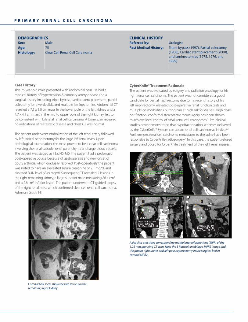
Axial slice and three corresponding multiplanar reformations (MPR) of the 1.25 mm planning CT scan. Note the 5 fiducials in oblique MPR2 image and the patent right ureter and left post nephrectomy in the surgical bed in coronal MPR2.
Coronal MRI slices show the two lesions in the remaining right kidney.
Treatment Planning ProcessThe patient was prepared for planning as follows: i) 5 fiducials were placed around the right renal masses using CT guidance ii) planning CT image was obtained with patient in the prone position using an alphacradleforimmobilization.Fiducialswereidentifiedandtherightsuperior and inferior lesions were outlined on the scans resulting in target volumes of 86.4 cm3 and 2.8 cm3 respectively. A treatment plan was developed which combined treatment plans for the large and small tumors into a composite plan. The final plan was created to deliver 30 Gy in 3 fractions to the 72% isodose line at the margin of the tumor using both the 7.5 and 20 mm collimators.
P R I M A R Y R E N A L C E L L C A R C I N O M A
Tumor Volume: 86.4 cc and 2.8 ccImaging Technique(s): CTRx Dose & Isodose: 30 Gy to 72%Conformality Index: 1.39Tumor Coverage: 98.7%Number of Beams: 282
Fractions: 3 Path Template: 1 path 900_1000 mmTracking Method: Synchrony & FiducialCollimator(s): 7.5 mm and 20 mm
TREATMENT DETAILS
Treatment DeliveryThe patient underwent CyberKnife® treatment which consisted of 10 Gy times 3 fractions using 282 beams from 77 nodes. The prescribed dose covered 98.7% of the combined target volumes with a homogeneity index score of 1.39 and a conformality index score of 1.39. The amount of remaining normal kidney was maximally spared and the patient tolerated the procedure well.
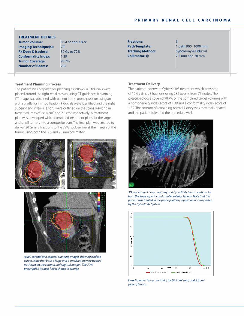
Dose Volume Histogram (DVH) for 86.4 cm3 (red) and 2.8 cm3 (green) lesions.
3D rendering of bony anatomy and CyberKnife beam positions to both the large superior and smaller inferior lesions. Note that the patient was treated in the prone position, a position not supportedby the CyberKnife System.
Axial, coronal and sagittal planning images showing isodose curves. Note that both a large and a small lesion were treated as shown on the coronal and sagittal images. The 72% prescription isodose line is shown in orange.
P R I M A R Y R E N A L C E L L C A R C I N O M A
ST. JOSEPH’S HOSPITAL AND MEDICAL CENTER / BARROW NEUROLOGICAL INSTITUTESt. Joseph’s Hospital and Medical Center/Barrow Neurological Institute, Phoenix, AZ (www.stjosephs-phx.org) is a highly regarded 690 bed not-for-profi t medical center founded in 1895 by the Sisters of Mercy and is part of the Catholic Healthcare West (CHW) system. Dr. Kresl, the Co-Medical Director of the St. Joseph’s Barrow Neurological Institute’s CyberKnife Center and a member of Arizona Oncology Services, one of the largest radiation oncology treatment programs in the Southwest, actively participates in national and regional Clinical Oncology Treatment Protocols that offer the newest and most advanced forms of treatment for tumors of all disease sites. CyberKnife radiosurgery began at St. Joseph’s Hospital and Medical Center/Barrow Neurological Institute in September 2003. The CyberKnife is used on those patients for whom traditional radiosurgery is not possible or in situations where patients specifi cally request this procedure over other treatment.
Outcome and Follow-Up Three months after CyberKnife® treatment, CT scan of the abdomen revealed an unchanged 3-4 cm lesion in the upper pole of
the right kidney with no evidence of retroperitoneal adenopathy – there was no evidence of recurrent tumor in the left renal fossa; a PET scan confirmed no evidence of metastatic disease – creatinine (2.3 mg/dl) and BUN (36 mg/dl) levels remained stable• NinemonthsafterCyberKnifetreatment,thepatienthadaCTscanrevealinganunchanged4cmlowdensitymasslocatedin the upper pole of the right kidney and a negative chest X-ray; creatinine (2.2 mg/dl) and BUN (37 mg/dl) levels remain stable No acute or chronic radiation-induced toxicities were noted 9 months following treatment
Conclusion and CyberKnife Advantages This patient had an excellent initial outcome with the CyberKnife System using Synchrony® Motion Tracking in the treatment of renal cell
carcinoma, while preserving renal function of the right remaining kidney• TheCyberKnifeSystemcandelivercomplextreatmentplanstomultiplelesionswhileminimizingirradiationtothesurroundinghealthytissue, thereby decreasing the risk of complications• TheCyberKnifeSystemhasthepotentialtobeanexcellenttreatmentmodalityforrenalcancerpatientswithrenalcellcarcinomasorpatients with bilateral renal cell carcinoma who refuse surgery or are medically inoperable
References1. Beitler, JJ, Makara D, Silverman P, Lederman G. Definitive, high-dose-per-fraction, conformal, stereotactic external radiation for renal cell carcinoma. Am J of Clin Oncol 27:646-648, 2004.2. Walsh L, Stanfield JL, Cho LC, Chang CH, Forster K, Kabbani W, Cadeddu JA, Hsieh JT, Choy H, Timmerman R, Lotan Y. Efficacy of Ablative High-Dose-per-Fraction Radiation for Implanted Human Renal Cell Cancer in a Nude Mouse Model. Eur Urol. Mar 29, 2006, In Press.3. Ponsky LE, Crownover RL, Rosen MJ, Rodebaugh RF, Castilla EA, Brainard J, Cherullo EE, Novick AC. Initial Evaluation of CyberKnife Technology for Extracorporeal Renal Tissue Ablation. Urology 61(3):498-501, March 2003.4. Gerszten PC, Burton SA, Ozhasoglu C, Vogel WJ, Welch WC, Barr J, Friedland DM. Stereotactic Radiosurgery for Spinal Metastases from Renal Cell Carcinoma. J Neurosurg Spine 3(4):288-295, Oct 2005.
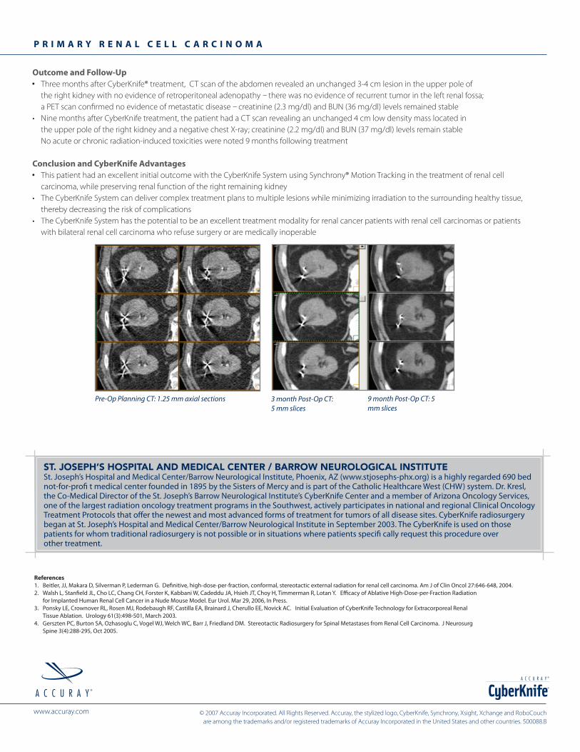
3 month Post-Op CT: 5 mm slices
9 month Post-Op CT: 5 mm slices
Pre-Op Planning CT: 1.25 mm axial sections
©2007AccurayIncorporated.AllRightsReserved.Accuray,thestylizedlogo,CyberKnife,Synchrony,Xsight,XchangeandRoboCouch are among the trademarks and/or registered trademarks of Accuray Incorporated in the United States and other countries. 500088.B
www.accuray.com
®