p38 andp38 mediateoncogenic ras-inducedsenescence ... requirement of p38 for oncogene-induced...
TRANSCRIPT
p38� and p38� Mediate Oncogenic ras-induced Senescencethrough Differential Mechanisms*□S
Received for publication, October 31, 2008, and in revised form, February 27, 2009 Published, JBC Papers in Press, February 27, 2009, DOI 10.1074/jbc.M808327200
Jinny Kwong‡§1, Lixin Hong¶1, Rong Liao‡, Qingdong Deng‡, Jiahuai Han¶, and Peiqing Sun‡§2
From the ‡Department of Molecular Biology and §Kellogg School of Science and Technology, The Scripps Research Institute,La Jolla, California 91037 and the ¶Key Laboratory of Ministry of Education for Cell Biology and Tumor Cell Engineering, School ofLife Sciences, Xiamen University, Xiamen 361005, China
Oncogene-induced senescence is a tumor-suppressive de-fense mechanism triggered upon activation of certain onco-genes in normal cells. Recently, the senescence response tooncogene activation has been shown to act as a bona fide barrierto cancer development in vivo. Multiple previous studies haveimplicated the importance of the p38 MAPK pathway in onco-gene-induced senescence. However, the contribution of each ofthe four p38 isoforms (encoded by different genes) to senes-cence induction is unclear. In the current study, we demon-strated that p38� and p38�, but not p38�, play an essential rolein oncogenic ras-induced senescence. Both p38� and p38� areexpressed in primary human fibroblasts and are activated upontransduction of oncogenic ras. Small hairpin RNA-mediatedsilencing of p38� or p38� expression abrogated ras-inducedsenescence, whereas constitutive activation of p38� and p38�caused premature senescence. Furthermore, upon activation byoncogenic ras, p38� stimulated the transcriptional activity ofp53 by phosphorylating p53 at Ser33, suggesting that the abilityof p38� tomediate senescence is at least partly achieved throughp53. However, p38� contributed to ras-inducted senescence viaa p53-indepdendent mechanism in cells by mediating ras-in-duced expression of p16INK4A, another key senescence effector.These findings have identified p38� and p38� as essential com-ponents of the signaling pathway that regulates the tumor-sup-pressing senescence response, providing insights into themolecular mechanisms underlying the differential involvementof the p38 isoforms in senescence induction.
The ras proto-oncogenes encode small GTP-binding pro-teins that transduce growth signals from cell surface (1–3).Aberrant activation of ras is a crucial step in tumor formation.Constitutive activation of ras genes, either through pointmuta-tions or overexpression, is associated with a wide variety ofhuman tumors at high frequency and contributes to the initia-tion and maintenance of multiple tumorigenic phenotypes inthese cancers (4–11). However, in early-passage primaryhuman and rodent cells, activated ras causes a permanent pro-
liferative arrest known as premature senescence, because of itsphenotypic similarities to replicative senescence observed inlate-passage cells (12). Other oncogenes, such as E2F1 and raf,or inactivation of certain tumor suppressor genes, also inducesenescence in normal human cells (13–15). The existence of thepremature senescence response to oncogene activation impliesthat like apoptosis, oncogene-induced senescence serves as ananti-tumorigenic defense mechanism. Indeed, it has been welldocumented that cellular transformation by ras requires coop-eration from immortalizing oncogenes that overcome thesenescence response, such as those inactivating p53 (8, 16, 17).Recent studies have also demonstrated that senescent cells canbe detected in early-stage premalignant lesions of lung, pan-creas, skin, and prostate in both human cancer patients andmouse tumor models and that disruption of senescence accel-erates the development of malignant tumors(18–23). Thesefindings indicate that oncogene-induced senescence occurs invivo and serves as a barrier to tumorigenesis.Although the downstream effectors of the oncogenic activity
of ras have been studied extensively, relatively little is knownabout the signaling pathways that mediate the ras-inducedsenescence response. Studies have indicated that the ability ofras to induce senescence depends on activation of the Raf/MEK/ERKMAPK pathway (13, 24) and is accompanied by up-regulation of several inhibitors of cell proliferation, includingp16INK4A, p53, p14/p19ARF, and p21WAF1 (12, 25), and silencingof E2F target genes (26). In some cells, senescence is triggered asa result of ras-induced production of reactive oxygen species(27). In addition, it has been reported that oncogene inducedsenescence is mediated by DNA damage responses generatedby aberrant DNA replication (28, 29). Recently, studies fromour laboratory and others have shown that ras-induced senes-cence relies on activation of the p38 MAPK3 (30–33). p38 andits upstreamMAPK kinasesMKK3 andMKK6 (34, 35) are acti-vated by oncogenic ras as a result of persistent MEK/ERK(mitogen-activated protein kinase/extracellular signal-regu-lated kinase kinase/extracellular signal-regulated kinase) acti-vation in senescent cells. Constitutive activation of p38 causespremature senescence, whereas pharmacological inhibition ofp38 prevents ras-induced senescence (30).
* This work was supported, in whole or in part, by Grant CA106768 from theNational Institutes of Health (to P. S.). This is Scripps Manuscript 19841.
□S The on-line version of this article (available at http://www.jbc.org) containssupplemental Fig. S1.
1 Both authors contributed equally to the study.2 To whom correspondence should be addressed: Dept. of Molecular Biology,
MB-41, The Scripps Research Inst., 10550 N. Torrey Pines Rd., La Jolla, CA91037. Tel.: 858-784-9710; Fax: 858-784-9067; E-mail: [email protected].
3 The abbreviations used are: MAPK, mitogen-activated protein kinase; PRAK,p38-regulated/activated protein kinase; shRNA, small hairpin RNA; GFP,green fluorescent protein; shGFP, shRNA against GFP; SA-�-gal, senes-cence-associated �-galactosidase; PD, population doubling; GST, glutathi-one S-transferase; MBP, myelin basic protein; MOPS, 4-morpholinepro-panesulfonic acid.
THE JOURNAL OF BIOLOGICAL CHEMISTRY VOL. 284, NO. 17, pp. 11237–11246, April 24, 2009© 2009 by The American Society for Biochemistry and Molecular Biology, Inc. Printed in the U.S.A.
APRIL 24, 2009 • VOLUME 284 • NUMBER 17 JOURNAL OF BIOLOGICAL CHEMISTRY 11237
by guest on June 10, 2018http://w
ww
.jbc.org/D
ownloaded from

The requirement of p38 for oncogene-induced senescencesuggests that the p38 pathway has a tumor-suppressing func-tion, in addition to its previously known roles in inflammatoryand stress responses (36–38). Indeed, target deletion of p38� orPRAK, a downstream substrate kinase of p38, accelerates can-cer development in mouse models (23, 39, 40). Moreover, dele-tion ofWip1, a p38 phosphatase frequently amplified in humanbreast tumors, leads to p38 activation and reduced mammarytumorigenesis in mice (41, 42). Therefore, the p38 pathway islikely to play an important role in tumor suppression by medi-ating the senescence response to oncogene activation.Four mammalian isoforms of p38 (�, �, �, and �), each
encoded by a different gene, have been identified; they differ intissue-specific expression and affinity for the upstream regula-toryMAPK kinases (43–49). Among these isoforms, only p38�has been shown essential for inflammatory and stress responsesby genetic analysis in murine models (50), whereas the physio-logical roles of the other p38 isoforms in inflammation or othercellular functions are still unclear (51, 52).We have shown pre-viously that SB203580, a chemical compound that inhibitsp38� and p38�, prevents ras-induced senescence in primarycells (30), indicating that p38�/� or both might be required forsenescence induction. However, this compound also inhibitsthe activity of other p38 isoforms and other protein kinasesalthough with lower affinity. The specific involvement of eachp38 isoform in senescence has never been investigated. In thecurrent study, we examined the role of the p38 isoforms inoncogenic ras-induced senescence in primary human cells.Ourdata demonstrate that p38� and p38�, but not p38�, are essen-tial components of the signaling pathway that mediates ras-induced senescence and that p38� and p38� contribute tosenescence induction through different mechanisms. Whereasp38� mediates ras-induced senescence at least partly by stim-ulating the transcriptional activity of p53 through direct phos-phorylation, p38� appears to regulate senescence in a p53-in-dependent, p16INK4A-dependent manner.
EXPERIMENTAL PROCEDURES
Cell Culture—BJ human foreskin fibroblasts were main-tained in minimum essential medium supplemented with 10%fetal calf serum, non-essential amino acids, glutamine, and anti-biotics. WI38 and IMR90 human fibroblasts and LinX-A retro-viral packaging cells were grown inDulbecco’s modified Eagle’smedium supplemented with 10% fetal calf serum, glutamine,and antibiotics.Plasmids—The Ha-RasV12 expression vectors were ob-
tained from Dr. Scott Lowe. Retroviral vectors for FLAG-tagged wild type p38 isoforms were constructed by subcloningthe respective cDNA into WZLHygro. Retroviral vectors forhemagglutinin-tagged wild type and intrinsically activemutants of p38 isoforms (53) were constructed by subcloningthe respective cDNA into pBabePuro. Oligonucleotides forshRNA targeting p38�-758 (AAATTCTCCGAGGTCTAAT),p38�-550 (GCGCTAAGGTGGCCATCAA), p38�-1023 (GCG-TGTTACTTACAAAGAG) and GFP (54) were cloned intopSUPER.retro according to the published protocol (55). Oligo-nucleotides for shRNA targeting p38�-577 (CGCGGTTACT-TAAACATATGAA), p38�-756 (CACCAAATTCTCCGAG-
GTCTAA), p38�-319 (CCCCTGATGGGCGCCGACCTGA),and p38�-661 (ACCCTCTTCCCGGGAAGCGACT) werecloned into pSM2C according to the published protocol (56).Retroviral vectors for MKK3E and MKK6E (30), and retroviralp53- reporter PG-Luc and its non-p53-binding control, MG-Luc (57), have been reported previously.Retrovirus-based Gene Transduction—This was carried out
as described previously (58). Transduced cells were purifiedwith 120 �g/ml hygromycin B, 400 �g/ml G418, 5 �g/ml blas-ticidin, and/or 1.2 �g/ml puromycin.Analysis of Senescence—This was performed in cell cultures
by measuring the rate of proliferation and the expression of thesenescence-associated �-galactosidase (SA-�-gal) senescencemarker as described previously (30). Population doublings (PD)were calculatedwith the formula PD� log(N2/N1)/log2, whereN1 is the number of cells seeded and N2 is the number of cellsrecovered (59). To quantify SA-�-gal positives, at least 200 cellswere counted in random fields in each of the duplicated wells.Each experiment was performed in triplicates or duplicates.Western Blot Analysis—Western blot analysis was per-
formed with lysates prepared 7–10 days after transduction ofRas or MKK3/6E from subconfluent cells as described (30).Primary antibodies were from Covance (HA-11), Sigma(FLAG-M5, FLAG-F7425, and actin), Santa Cruz Biotechnol-ogy (Ras C-20, MKK3 C-19, p53 FL-393, p21WAF1 C-19), CellSignaling (phospho-p38-Thr180/Tyr182, phospho-p53-Ser15and -Ser33, and phospho-ATF2-Thr71). Antibodies againstp38�, -�, -�, and -� were generated previously in our labora-tory. Signalswere detected using enhanced chemiluminescenceand captured by using the FluorChemTM-8900 imaging system(AlphaInnotech).p53 Reporter Assays—BJ cells were stably transduced with a
retroviral luciferase reporter driven by a promoter containingmultiple copies of a functional p53-binding sites (PG-Luc) or amutant p53-binding site (MG-Luc) (57). These cells were trans-duced with shRNA for GFP, p38�, or p38� at PD28–32 andsubsequently with Ha-RasV12 or vector at PD30–34. Cellswere split into 12-well plates onday 7 or 8 post-ras transductionand lysed on day 8 or 9. Luciferase activity was determinedusing a luciferase assay system (Promega) according to themanufacturer’s instructions and normalized to protein concen-trations as determined by the Bradford assay. Each experimentwas performed in triplicates or duplicates.Recombinant Proteins—Recombinant GST-ATF2, GST-
MKK6E, andHis-p38 isoformswere prepared as described pre-viously (60, 61). Wild type and mutant hp53 (1–61), co-ex-pressed with the ZZTAZ2 domain of CREB-binding proteinfrom a bicistronic vector to enhance protein stability, werepurified as described (62).Myelin basic protein (MBP) was pur-chased from Sigma.Immunoprecipitation-coupled Kinase Assays for p38—BJ
cells were lysed at PD30–40 on day 6–8 post-ras/MKK3/6Etransduction in a buffer containing 50 mM HEPES, pH 7.5, 2.5mM EGTA, 1 mM EDTA, 1% Triton X-100, 150 mM NaCl, 10%glycerol, 1 mM phenylmethylsulfonyl fluoride, 50 mM NaF, 1mM sodium vanadate, 1 mM �-glycerophosphate, 1 mM dithio-threitol, and Complete protease inhibitors. 100–300 �g oflysate were incubated with 60 �l of agarose-conjugated anti-
p38 Isoforms and Oncogene-induced Senescence
11238 JOURNAL OF BIOLOGICAL CHEMISTRY VOLUME 284 • NUMBER 17 • APRIL 24, 2009
by guest on June 10, 2018http://w
ww
.jbc.org/D
ownloaded from
FLAG antibody M2 (Sigma) at 4 °C for 2 h. The beads werewashed three timeswith 1ml of lysis buffer and three timeswith1� kinase buffer (50 mMHEPES, pH 7.5, 0.5 mM EGTA, 10 mMMgCl2, 0.1 mM phenylmethylsulfonyl fluoride, 1 mM NaF, 0.1mM sodium vanadate, 0.1 mM �-glycerophosphate, and 1 mMdithiothreitol). The reactions were performed in 20 �l of 1�kinase buffer (above) with 10�MATP, 0.5�l of [�32P]ATP, and10 �g of hp53 (1–61) or 2 �g of GST-ATF2 at 30 °C for 45min.The reactions were stopped by 7 �l of 4� Laemmli buffer,heated at 95 °C, and separated by SDS-PAGE. Radioactive sig-nals were detected by using a PhosphorImager. Part of theimmunoprecipitates, as well as the total protein lysates, weresubjected toWestern blot analysis to ensure equal efficiency ofimmunoprecipitation and equal input of proteins.Kinase Assays with Recombinant p38—Assays with recombi-
nant kinases were performed in two sequential steps, with thefirst step being the phosphorylation of His-p38 by MKK6E andthe second the phosphorylation of substrates by p38. The firststep was carried out at 30 °C for 10 min in 14 �l of 1� kinasebuffer (20 mM Tris-HCl, pH 7.5, 20 mM NaCl, 10 mM MgCl2, 1mM dithiothreitol, 20 �M cold ATP, and 1 mMNaF) containing0.4 �g of His-p38 with or without 50 ng of GST-MKK6. Subse-quently, 6 �l of substrate mix in 1� kinase buffer (same asabove) containing 20 �g of hp53 (1–61) (wild type or S33A orS46A mutant) or 10 �g of MBP and 2 �Ci of [�-32P]ATP wasadded to each reaction. The resulting 20 �l of reaction wasincubated at 30 °C for 30min, stopped by the addition of 7�l of4� Laemmli buffer, and heated at 95C° for 10 min. The reac-tions were separated on 4–20% gradient SDS-polyacrylamidegels. Radioactive signals were detected by PhosphorImager.Northern Blot Analysis—Total RNA was isolated from cells
usingTRIzol reagent (Invitrogen) according to themanufactur-er’s instructions. 10 �g of RNA was separated on a 1% agarosegel containing 3.7% formaldehyde in 1� MOPS buffer (20 mMMOPS, 5 mM NaOAc, and 1 mM EDTA, pH 7.0), transferred toHybondN�nylonmembranes in 10� SSC (1.5MNaCl and 150mM Na3 citrate, pH 7.0), and hybridized at 65 °C in Church-Gilbert buffer (1% bovine serum albumin, 400 mM NaPO4, pH7.0, 15% formamide, 1 mM EDTA, and 7% SDS) to a 800-basepair human p16 cDNA probe labeled with [�-32P]dATP and[�-32P]dCTP by randompriming. After extensive washing with0.2� SSC/0.1% SDS buffer at 65 °C, the signals were visualizedand quantitated by phosphorimaging.
RESULTS
Expression and Activation of the p38 Isoforms during Onco-gene-induced Senescence in Primary Human Fibroblasts—Weshowed previously that oncogenic ras fails to induce senes-cence in primary BJ human fibroblast cells treated with thep38�- and p38�-specific inhibitor SB203580 (30), suggestingthat at least one of these two isoforms might be required forsenescence induction. However, this compound also inhibitsother p38 isoforms and even other protein kinases, althoughwith a lower affinity. To investigate the specific involvement ofeach p38 isoforms, in the present work we initially examinedthe expression and activity of these isoform in senescent cells.Although all four p38 isoforms contain similar numbers ofamino acid residues, they displayed distinct rates ofmobility on
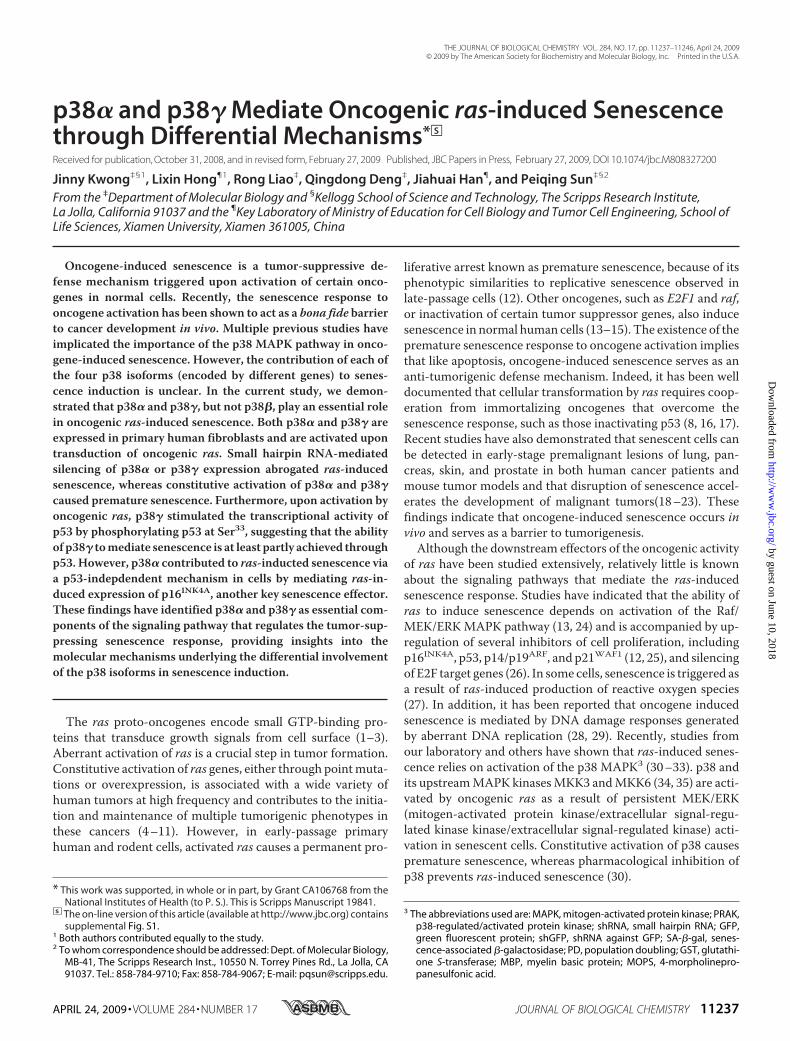
SDS-PAGE. Whereas the other isoforms had apparent molec-ular mass of 38–42 kDa, p38� migrated closely to the 49-kDamarker (Fig. 1A). p38� had a slightly faster mobility than p38�and p38�. These differences allowed us to differentiate theseisoforms from each other.Upon transduction of oncogenic ras (HarasV12) or active
mutant of MKK3 (MKK3E), the phosphorylation of ectopicallyexpressed p38�, p38�, p38�, and p38� in their activation loopwas greatly enhanced in BJ cells as detected by a phospho-spe-cific antibody (Fig. 1B). These p38 isoforms also displayedincreased protein kinase activity toward ATF2 in vitro whenimmunoprecipitated from BJ cells transduced with oncogenicras as compared with those from control cells (Fig. 1C). Theseresults indicate that all four of the p38 isoforms can be activatedduring ras-induced senescence. Using isoform-specific anti-bodies, we were able to show that p38�, p38�, and p38� wereexpressed in primary BJ human fibroblasts (Fig. 1D). In addi-
FIGURE 1. The p38�, -�, and -� isoforms are expressed in primary humanfibroblast cells and activated by oncogenic ras during senescence induc-tion. A, Western blot analysis of BJ cells (PD26) transduced with FLAG-taggedp38 isoforms detecting FLAG-p38 and actin. B, Western blot analysis of BJ cellstransduced with FLAG-p38 isoforms and Ha-RasV12 (Ras), MKK3E or vector(WH), detecting phospho-p38, FLAG, Ras, MKK3, and actin. Cells were lysed onday 8 post-Ras transduction at PD30. C, induction of the kinase activity of theFLAG-p38 isoforms toward ATF2 by ras. FLAG-p38 isoforms were immunopre-cipitated from BJ cells transduced with FLAG-p38 and Ha-RasV12 or vector atPD30 on day 8 post-Ras transduction (the same lysates as in B) using an aga-rose-conjugated anti-FLAG M2 antibody and incubated with GST-ATF2 in thepresence of [�-32P]ATP. Phosphorylated ATF2 were detected by autoradiog-raphy. The input of ATF2 was determined by staining with Coomassie BrilliantBlue R. D, Western blot analysis of BJ cells transduced with Ha-RasV12, MKK3E,or vector, detecting phospho-p38, p38�, p38�, and p38� using specific anti-bodies. Cells were lysed on day 8 post-Ras transduction at PD25. Identical setsof lysates, each set containing lysates from BJ cells transduced with vector,Ha-RasV12, or MKK3E were resolved side-by-side on the same SDS-polyacryl-amide gel and transferred to a nitrocellulose membrane. The membrane wascut into pieces, each containing one set of lysates. These pieces of membranewere then hybridized to the antibody against phospho-p38, -p38�, -p38�,and -p38�, respectively. The chemiluminescence signals were captured afterthe membranes were re-aligned into the original position. The positions ofp38�, p38�, and p38� are marked by arrows.
p38 Isoforms and Oncogene-induced Senescence
APRIL 24, 2009 • VOLUME 284 • NUMBER 17 JOURNAL OF BIOLOGICAL CHEMISTRY 11239
by guest on June 10, 2018http://w
ww
.jbc.org/D
ownloaded from

tion, both Ras and MKK3E induced the activating phosphoryl-ation of the endogenous p38 proteins that co-migrated withp38� and p38� (Fig. 1D). We confirmed that the protein bandsdetected by the phospho-specific antibody and co-migratingwith p38� and p38� indeed represent the phosphorylated p38�and p38� isoforms, respectively, because these bands wereabolished in cells expressing p38� (Fig. 2A) or p38� shRNA(Fig. 3A). Therefore, these findings indicate that oncogenic rasactivates not only the endogenous p38�, as we demonstratedpreviously (30), but also the endogenous p38� during senes-cence induction. Interestingly, in BJ human fibroblasts, onco-genic ras activated p38� through phosphorylation withoutaltering its expression level (Fig. 1D). This is in contrast to aprevious finding that oncogenic ras induces p38� expressionbut not its phosphorylation in rat intestinal epithelial cells(IEC-6) (63). This raises the possibility that Ras may stimulatethe activity of p38� through different mechanisms in a species-or cell type-dependent manner.Although p38� was expressed in primary human fibroblasts,
we failed to detect an obvious phospho-p38 band co-migratingwith p38� (Fig. 1D). However, based on the induction of phos-phorylation and kinase activity of ectopically expressed p38� byoncogenic ras (Fig. 1, B and C), we reasoned that p38� wasactivated during ras-induced senescence. Because the mobilityof p38� was only slightly slower than that of p38� on SDS-PAGE, it is possible that the signal for phospho-p38� wasobscured by that of phospho-p38� because of the relativelylower abundance of p38� as compared with p38�. Moreover,although the ectopically expressed p38� could be activated by
both oncogenic ras and MKK3E(Fig. 1, B and C), the level of p38�was barely detectable in primaryhuman fibroblasts (data not shown).Thus, our study focused on p38�,-�, and -� isoforms.p38� and p38�, butNot p38�, Are
Essential for Oncogenic ras-inducedSenescence—The detectable expres-sion and activation of p38�, -�,and -� in senescent cells promptedus to examine the requirement ofthese p38 isoforms for oncogene-in-duced senescence. Three p38�shRNA (shp38�-577, -756, and-758), two p38� shRNA (shp38�-319 and -661), and two p38� shRNA(shp38�-550 and -1023) were con-structed. When stably transducedinto BJ cells via retrovirus, shp38�-577 failed to inhibit the expressionlevels of p38� (Fig. 2A), whereas allof the other shRNAs efficientlysilenced the expression of appropri-ate p38 isoforms without affectingthe other isoforms (Figs. 2A and3A).When stably expressed in BJ cells,
the p38�-shRNAs (shp38�-756 and-758), which efficiently knocked down p38�, prevented onco-genic ras-induced growth arrest (Fig. 2B) and accumulation ofSA-�-gal, a biomarker for senescence (Fig. 2C). In contrast,shRNA for GFP or the p38� shRNA (shp38�-577), which failedto silence p38� expression, had no effect on senescence induc-tion (Fig. 2, B and C). Furthermore, two shRNAs that silencedp38� expression (shp38�-550 and -1023) also blocked the abil-ity of ras to induce growth arrest (Fig. 3B) and greatly inhibitedras-induced expression of SA-�-gal (Fig. 3C) as compared withthe GFP shRNA. On the other hand, the p38� shRNA thateffectively silenced p38� expression did not disrupt oncogenicras-induced senescence (data not shown). These results werereproduced in WI38 primary human fibroblast cells derivedfrom normal embryonic lung tissue, in which ras-inducedsenescence was inhibited by shRNA for p38� and p38� but notby shRNA for p38� (supplemental Fig. S1). ShRNA for p38�also delayed the onset of ras-induced senescence in the IMR90primary human lung fibroblasts (data no shown). These resultsdemonstrate that both p38� and p38� are essential for ras-induced senescence, whereas p38� is dispensable for senes-cence induction.Constitutive Activation of p38� or p38�, but Not p38�, Leads
to Premature Senescence—To analyze the effect of p38 activa-tion on senescence induction, we took advantage of the intrin-sically active mutants of the p38 isoforms recently constructedin Drs. Engelberg and Livnah’s groups (Hebrew University ofJerusalem (53, 53)). These mutants have acquired spontaneousprotein kinase activity in vitro and in vivo and maintain speci-ficity toward substrates and inhibitors similar to that of thewild
FIGURE 2. p38� is essential for ras-induced senescence but not for phosphorylation of p53 at Ser33 orinduction of p21WAF1 expression. A, BJ cells transduced with shRNA against GFP (shGFP) or p38� (shp38�-577, -756, or -758) and Ha-RasV12 (Ras) or vector (WH) were subjected to Western blot analysis detecting theindicated proteins. Cells were lysed on day 10 post-Ras transduction at PD34 (left panel) or PD35 (right panel).B, the population doublings of BJ cells transduced with shGFP or shp38�-577, -756, or -758 and Ha-RasV12 orvector were followed over a period of 16 days, starting at day 5 post-Ras transduction at PD34 (top panel) orPD35 (bottom panel). Values are mean � S.D. for duplicates. C, BJ cell lines (described in B) were stained for theSA-�-gal senescence marker on day 15 post-Ras transduction. Values are mean � S.D. for duplicates.
p38 Isoforms and Oncogene-induced Senescence
11240 JOURNAL OF BIOLOGICAL CHEMISTRY VOLUME 284 • NUMBER 17 • APRIL 24, 2009
by guest on June 10, 2018http://w
ww
.jbc.org/D
ownloaded from

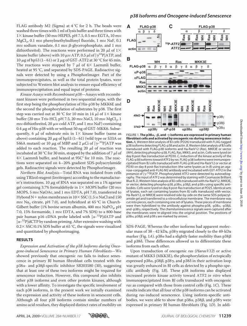
type p38 isoforms.When transduced into BJ cells via retrovirus,the active formof p38� (p38�-D176A), p38� (p38�-D176A), orp38� (p38�-D179A) and its wild type counterpart wereexpressed at comparable levels (Fig. 4, A and B). However, theactive mutants displayed a much higher level of autophospho-rylation in the activation loop than the corresponding wild typeproteins as detected by the phospho-specific antibody inWest-ern blot analysis (Fig. 4,A and B). These results were consistentwith previous reports in other cell lines and indicated that thesemutants of p38 isoforms were indeed constitutively active.Moreover, similar to oncogenic ras, expression of p38�-
D176A or p38�-D179A led to growth arrest (Fig. 4C, top andmiddle panels) and accumulation of the SA-�-gal marker (Fig.4D) in BJ cells, whereas wild type p38� and p38� did not signif-icantly inhibit cell proliferation or induce SA-�-gal expression.In contrast, the active mutant of p38� (p38�-D176A) did notcause inhibition of proliferation in primary BJ fibroblasts (Fig,4C, bottompanel). Therefore, constitutive activation of p38� orp38�, but not p38�, is sufficient to induce premature senes-cence in primary human fibroblasts.p38�, but Not p38�, Is Required for ras-induced Activation of
p53 in Senescent Cells—In an attempt to investigate the molec-ular mechanism underlying the essential roles of p38� andp38� in senescence, we examined the ability of these iso-forms to regulate the activity of p53, a key effector of ras-induced senescence. For this purpose, we used a retrovirus-based, stable, luciferase reporter system for p53 (57). In BJ cells
stably transduced with this stable p53 reporter (PG-Luc), lucif-erase activity was stimulated significantly (3–4-fold) in thepresence of Ha-RasV12 (Fig. 5,A andB, top panels), confirmingthe induction of p53 transcriptional activity during senescence.However, ras-induced p53 activity was greatly reduced in BJcells expressing the effective p38� shRNA (shp38�-550 and-1023) (Fig. 5B, top panel), whereas the p38� shRNA had noobvious effect on the p53-dependent luciferase activity insenescent cells. Neither ras nor the p38� or p38� shRNA sig-nificantly altered the transcription of MG-Luc, a controlreporter containing mutant p53-binding sites (57) (Fig. 5A andB, bottom panels), indicating that the effects we observedwere specific for p53. These data indicate that p38�, but notp38�, is required for the activation of p53 during ras-in-duced senescence.Consistent with the essential role of p38� in p53 activation
and in senescence induction, shRNA for p38� inhibited ras-mediated induction of p21WAF1, an endogenous transcriptionaltarget of p53 and a key effector of senescence (Fig. 5C). Bycontrast, although the effective p38� shRNA (shp38�-756 and-758) blocked ras-induced senescence (Fig. 2, B and C), it didnot reduce the p21WAF1 induction by ras, as compared with theGFP shRNA (Fig. 5D). A similar observation was made inIMR90 cells (Fig. 5E). Moreover, although the constitutivelyactive mutants of both p38� (p38�-D176A) and p38� (p38�-D179A) induced senescence (Fig. 4, C and D), only p38�-D179A, but not p38�-D176A, increased the expression ofp21WAF1 (Fig. 5F). The active mutant of p38� also failed tostimulate the level of p21WAF1 (Fig. 5F), consistent with theinability of this mutant to induce premature senescence. Takentogether with the results from the luciferase reporter assays,these data demonstrate that although the role of p38� in ras-induced senescence correlates with its ability to stimulate p53activity and subsequently to induce the expression of p21WAF1,p38� mediates senescence induction through a p53/p21WAF1-independent mechanism.p38�, but Not p38�, Mediates Phosphorylation of p53 in Vivo
during Senescence Induction by Oncogenic ras—The activity ofp53 is regulated through phosphorylation of its N-terminaltranscriptional activation domain. The differential effects ofp38� and p38� on ras-induced p53 activity suggest that theseisoforms may play different roles in p53 phosphorylation dur-ing senescence induction. It has been shown that Ser33 andSer46 of p53 are direct substrates of p38� in vitro and that phos-phorylation of these sites contributes to the activation of p53uponDNAdamage (64). The ability of the other p38 isoforms tophosphorylate p53 was unknown. We found that in vitro,recombinant p38� phosphorylated the N-terminal transcrip-tional activation domain of p53, as well as the positive control,MBP, in an MKK6E-dependent manner (Fig. 6A), indicatingthat p53 is a substrate of activated p38�. Mutation of Ser33 toAla essentially abolished the phosphorylation of p53 by p38�,whereas mutation of Ser46 to Ala had little or no effect on p53phosphorylation (Fig. 6B). In contrast, mutation of either Ser33or Ser46 to Ala greatly diminished phosphorylation of p53 byp38� (Fig. 6B). Therefore, p38� phosphorylates p53 mainly atSer33 in vitro, whereas p38� phosphorylates p53 at both Ser33and Ser46, as demonstrated previously.
FIGURE 3. p38� is essential for ras-induced senescence, phosphorylationof p53 at Ser33, and induction of p21WAF1 expression. A, BJ cells trans-duced with shGFP or shp38�-550 or -1023 and Ha-RasV12 (Ras) or vector (WH)were subjected to Western blot analysis detecting the indicated proteins.Cells were lysed on day 8 post-Ras transduction at PD36. B, the populationdoublings of BJ cells transduced with shGFP or shp38�-550 or -1023 andHa-RasV12 or vector were followed over a period of 12 (left panel) or 23 (rightpanel) days, starting at day 5 post-Ras transduction at PD36 (top panel) orPD35 (bottom panel). Values are mean � S.D. for duplicates. C, BJ cell lines(described in B) were stained for the SA-�-gal senescence marker on day 17post-Ras transduction. Values are mean � S.D. for duplicates.
p38 Isoforms and Oncogene-induced Senescence
APRIL 24, 2009 • VOLUME 284 • NUMBER 17 JOURNAL OF BIOLOGICAL CHEMISTRY 11241
by guest on June 10, 2018http://w
ww
.jbc.org/D
ownloaded from
To investigate the phosphorylation of p53 by p38� duringsenescence induction, we compared the p53 kinase activitiesof p38� and p38� immunoprecipitated from control andsenescent BJ cells. Correlating with its increased phospho-rylation in the activation loop (Fig. 1B), p38� immunopre-cipitated from Ras- and MKK3E-expressing, senescent BJcells phosphorylated p53 at much higher levels as comparedwith that from control cells (Fig. 6C, right panel). On theother hand, although the phosphorylation of p38� in theactivation loop was induced in Ras- or MKK3E-expressingcells to levels comparable with those of p38� (Fig. 1B), p38�barely phosphorylated p53 when immunoprecipitated fromthese cells (Fig. 6C, compare left and right panels, which werederived from the same exposure). The phosphorylation ofp53 by p38� immunoprecipitated from Ras- and MKK3E-expressing cells could still be detected upon overexposure,but the signals were almost negligible when compared withthe signals derived from p38� on the same exposure (Fig. 6C,32P-p53overexposure (OverExp)).These results demonstrate thatwhereas oncogenic Ras and MKK3E activate both p38� andp38� during senescence, only activated p38�, but not p38�, isable to phosphorylate p53.The Ras-induced kinase activity of p38� toward p53 was
almost completely abolished when Ser33 was mutated, but itwas unaltered by the mutation of Ser15 or Ser46 (Fig. 6D).Thus, upon activation in senescent cells, p38� phosphoryl-ates p53 mainly at Ser33, consistent with the results obtained
with recombinant p38� activatedby MKK6E in vitro (Fig. 6B).We further investigated the effect
of p38� and p38� on phosphoryla-tion of the endogenous p53 proteinat Ser33 in vivo during senescenceinduction by ras. As with activatedras, intrinsically active p38� (p38�-D179A) induced phosphorylationof p53 at Ser33 in BJ cells, whereasactive p38� (p38�-D176A) did notincrease p53-Ser33 phosphoryla-tion, although it induced senes-cence (Fig. 5F). Wild type p38� andp38� had little effect on p53-Ser33phosphorylation. Wild type andconstitutively active p38� also failedto cause phosphorylation of p53-Ser33 (Fig. 5F), which correlateswith their inability to induce senes-cence. Furthermore, the effectivep38� shRNA (shp38�-550 and-1023) greatly diminished ras-in-duced phosphorylation of p53 atSer33 but not the phosphorylation ofp53-Ser15, a site that is not a p38substrate (Fig. 5C). However, theeffective p38� shRNA (shp38�-756and -758) blocked ras-inducedsenescence but not p53-Ser33 phos-phorylation (Fig. 5D). Consistent
with these findings in BJ cells, oncogenic ras-induced phospho-rylation of p53-Ser33 and the increase in p21WAF1 expressionwere also inhibited by the p38� shRNA, but not by the p38�shRNA, in IMR90 primary human fibroblast cells (Fig. 5E).Taken together, our data indicate that oncogenic ras activatesboth p38� and p38�, which in turn mediate senescence induc-tion through different mechanisms. Upon activation by onco-genic ras, p38� induces p53 activity by directly phosphorylatingSer33, a residue that is required for p53 to mediate ras-inducedsenescence (23). Activation of p53 by p38� leads to increasedexpression of a key senescence effector, p21WAF1. By contrast,p38� contributes to senescence induction through a mecha-nism independent of p53.p38�, but Not p38�, Is Essential for ras-induced Expression of
p16INK4A—We demonstrated previously that oncogenic rasstimulates the transcript level of p16INK4A, anothermajor effec-tor of senescence, through activation of the p38 pathway (30).To gain insights into the p53-independent role of p38� insenescence,we examined the requirement of p38� andp38� forp16INK4A expression in senescent cells. As shown previously(30), oncogenic ras induces a 3-fold increase in themRNA levelof p16INK4A in BJ cells (Fig. 7). However, this induction wasabolished by the p38� shRNA but not the p38� shRNA. Thus,p38� and p38� mediate oncogene-induced senescence byinducing two major senescence effectors, p53 and p16INK4A,respectively.
FIGURE 4. Constitutively active p38� and p38�, but not p38�, induce premature senescence in primaryhuman fibroblast cells. A and B, Western blot analysis of BJ cells transduced with vector control (Babe Puro,BP). Ha-RasV12 (Ras), hemagglutinin-tagged wild type p38�, p38�, or p38� (WT), or their active mutants(p38�D176A, p38�D179A, or p38�D176A) was performed to detect the indicated proteins. Cells were lysed onday 8 post-transduction at PD33. C, the population doublings of the BJ cell lines described in A and B werefollowed for 6 (left panel) or 12 (middle and right panels) days, starting on day 5 post-transduction at PD32.Values are mean � S.D. for duplicates. *, p � 0.001; **, p � 0.01; #, p � 0.05, versus vector control by Student’st test. D, BJ cell lines (described in A) were stained for the SA-�-gal senescence marker on day 14 post-trans-duction. Values in are mean � S.D. for duplicates. *, p � 0.01; #, p � 0.05, versus vector control by Student’s t test.
p38 Isoforms and Oncogene-induced Senescence
11242 JOURNAL OF BIOLOGICAL CHEMISTRY VOLUME 284 • NUMBER 17 • APRIL 24, 2009
by guest on June 10, 2018http://w
ww
.jbc.org/D
ownloaded from

DISCUSSION
The existence of multiple p38 isoforms with differences intissue distribution and affinity for upstream regulators suggeststhat these isoforms may have distinct functions. Although thep38� isoform has been shown to be required for inflammatory
and stress responses in vivo (50,65–67), the physiology roles of theother p38 isoforms have beenunclear. Using SB203580, a phar-macological inhibitorwith relativelyhigher affinity for p38� and p38� ascompared with p38� and p38�, wewere able to demonstrate a key roleof p38 in oncogene-induced senes-cence. However, the relative contri-bution of the specific p38 isoformsto senescence had never beendefined. In the present study, wehave shown that p38� and p38�, butnot p38�, mediate senescenceinduction by oncogenic ras. Thesestudies have identified a novel func-tion of p38� in the regulation ofoncogene-induced senescence. Tak-ing these findings together withour previous report demonstrat-ing the involvement of p38� in�-radiation-induced G2 cell cyclearrest and DNA damage check-point control (68), we concludethat a major function of p38� maybe to suppress tumorigenesis andmaintain genome stability.The requirement of both p38�
and p38� suggests that the func-tions of these p38 isoforms are notredundant during senescenceinduction and that p38� and p38�may target different downstreamsubstrates in the senescence path-way. Indeed, our data demonstratethat p38� and p38� contribute tosenescence induction through dif-ferent mechanisms, with p38�transducing the senescence signalvia the p53-p21WAF1 pathway andp38� via a p53-independent, butp16INK4A-dependent, route. Thep53-p21WAF1 circuit is one of thekey effector pathways known to beessential for almost all types ofsenescence. The ability of p38�shRNA to disrupt ras-inducedp53-Ser33 phosphorylation, p53transcriptional activity, andp21WAF1 expression, as well as theability of constitutively active p38�to mimic ras in the induction of p53
phosphorylation and activity, indicates that p38� mediatessenescence induction at least partly by regulating the p53-p21WAF1 pathway. Thep16INK4A-Rbpathway is the othermajoreffector of senescence. We previously demonstrated that con-stitutive activation of p38 by active MKK3 or MKK6 leads to
FIGURE 5. p38�, but not p38�, is essential for oncogenic ras-induced transcriptional activity of p53. A, BJcells stably transduced with a retroviral luciferase reporter driven by a promoter containing multiple copies ofa functional p53-binding site (PG-Luc, top panel) or a mutant p53-binding site (MG-Luc, bottom panel) weretransduced with retroviruses encoding shGFP or shp38�-756 or -758 at PD33 and with Ha-RasV12 (Ras) orvector (WH) at PD35. Cells were lysed on day 8 post-Ras transduction. B, BJ cells stably transduced with aretroviral luciferase reporter driven by a promoter containing multiple copies of a functional p53-binding site(PG-Luc, top panel) or a mutant p53-binding site (MG-Luc, bottom panel) were transduced with retrovirusesencoding shGFP or shp38�-550 or -1023 at PD28 and with Ha-RasV12 or vector at PD30. Cells were lysed on day8 post-Ras transduction. In A and B, luciferase activity was measured and normalized to protein concentration.Values are mean � S.D. for triplicates. Note that the luciferase values are not comparable between cells withPG-Luc and those with MG-Luc, as the luciferase activities were measured with different volumes of lysatesunder different settings of sensitivity of the luminometer. C and D, Western blot analysis of BJ cells transducedwith shGFP or shp38�-550 or -1023 and Ha-RasV12 or vector (C) or with shGFP or shp38�-756 or -758 andHa-RasV12 or vector (D), detecting the indicated proteins. Cells were lysed on day 8 (C) or day 10 (D) post-transduction at PD36 (C) or PD35 (D). E, Western blot analysis of IMR90 cells transduced with shRNA for p38�(sh�756, -758) or p38� (sh�1023) and Ha-RasV12 or vector control detecting the indicated proteins. Cell lysateswere prepared 11 days post-infection with ras at PD36. F, Western blot analysis of BJ cells transduced withvector control (Babe Puro, BP). Ha-RasV12, hemagglutinin-tagged wild type p38�, p38�, or p38� (WT), or theiractive mutants (p38�D176A, p38�D179A, or p38�D176A), detecting the indicated proteins. Cells were lysedon day 8 post-transduction at PD33.
p38 Isoforms and Oncogene-induced Senescence
APRIL 24, 2009 • VOLUME 284 • NUMBER 17 JOURNAL OF BIOLOGICAL CHEMISTRY 11243
by guest on June 10, 2018http://w
ww
.jbc.org/D
ownloaded from

increased expression of p16INK4A at both protein and mRNAlevels (30). The results from our current study indicate thatras-induced increase in p16INK4A expression is mediated byp38�.It has been reported that recombinant p38� phosphorylates
p53-Ser33 in vitro (64). We confirmed this finding and furtherdemonstrated that activated recombinant p38� also phospho-rylated p53 at Ser33 in vitro as efficiently as p38�. However,when immunoprecipitated from senescent cells, only p38�, butnot p38�, could phosphorylate p53-Ser33. In addition, ras-in-duced phosphorylation of p53-Ser33 in senescent cells wasgreatly reduced by p38� shRNA but not by p38� shRNA. Con-stitutively active p38�, but not active p38�, consistentlyinduced p53-Ser33 phosphorylation in cells. These findings
indicate that in cells, phosphorylation of p53-Ser33 is mainlymediated by p38�, but not p38�, upon senescence induction.The mechanism underlying this discrepancy between the invitro and in vivo activities of p38 toward p53 is currentlyunknown. It is possible that during ras-induced senescence invivo, the kinase activity of p38� toward p53 is repressed as aresult of posttranslational modification or binding to an inhib-itory protein. Alternatively, the p53 kinase activity of p38� maybe enhanced by posttranslational modification or an associatedprotein in senescent cells.In vitro, recombinant p38� and p38� seem to have different
affinities for the substrate sites on a same protein. Whereasp38� phosphorylates both Ser33 and Ser46 of p53, p38� phos-phorylates only Ser33. It has been shown previously that p38�and p38� have different substrate selectivity in vitro. MAP-KAPK2, MAPKAPK3, and PRAK are preferred substrates ofp38� over p38�, whereas p38� has higher kinase activitytoward the microtubule-associated protein Tau and scaffoldproteins SAP90 and SAP97 than does p38� (60, 69). These dif-ferences in substrate selectivity for p53 and other proteins areconsistent with the fact that these two isoforms belong to dif-ferent subgroups within the p38 MAPK family (35, 69). P38�shares lower identity in amino acid sequence with p38� thanother isoforms, and the structure of the ATP-binding pocketdiffers between the � and � isoforms.Although our study has suggested the importance of the
p38�/p53-Ser33/p21WAF1 cascade in ras-induced senescence,there are almost certainly other pathways that act in a parallelor partially overlapping fashion to mediate senescence induc-tion. Supporting this notion, we found that constitutively activep38� increased phosphorylation of p53-Ser33 to a significantlyhigher level than oncogenic ras but that the p21 level wasinducedmore robustly by ras as compared with the active p38�(Fig. 4A). It is highly likely that besides phosphorylation of Ser33by p38�, oncogenic ras induces additional posttranslationalmodifications on p53, leading to a further increase in the p53activity and p21WAF1 expression. Oncogenic ras may also
FIGURE 6. Phosphorylation of p53 by recombinant or immunoprecipi-tated p38� and p38� in vitro. A, recombinant p38� phosphorylates p53.His-p38� was first incubated with GST-MKK6E (�) or buffer (�) and cold ATPand then with the substrate MBP or p53 (1– 61) in the presence of [�-32P]ATP.B, recombinant p38� phosphorylates p53 at Ser33 and Ser46, whereas recom-binant p38� phosphorylates Ser33 only. His-p38� or -p38� was first incubatedwith GST-MKK6E and cold ATP and then with p53 (1– 61) (WT, wild type) orp53 (1– 61) carrying the S33A or S46A mutation in the presence of [�-32P]ATP.C, p38� immunoprecipitated from senescent cells displays much higherkinase activity toward p53 than p38� does. FLAG-p38� or -p38� was immu-noprecipitated from BJ cells transduced with FLAG-p38� or -p38� and Ha-RasV12 (Ras), MKK3E, or vector (WH) at PD30 on day 8 post-Ras transductionusing an agarose-conjugated anti-FLAG M2 antibody and incubated with p53(1– 61) in the presence of [�-32P]ATP. The same cell lysates as described in thelegend for Fig. 1B were used for immunoprecipitation. Part of the immuno-precipitates was subjected to Western blot analysis to detect FLAG-p38.D, p38� immunoprecipitated from senescent cells phosphorylates p53 atSer33. FLAG-p38� immunoprecipitates from control (WH) or Ras-expressingBJ cells (Ras), as described in C, were incubated with wild type or mutant(S15D, S33D, or S46D) p53 (1– 61) in the presence of [�-32P]ATP. A–D, thereactions were separated by SDS-PAGE. Phosphorylated MBP, p53, and p38were detected by using a PhosphorImager. The input of the substrates wasdetermined by staining with Coomassie Brilliant Blue R.
FIGURE 7. p38�, but not p38�, is essential for oncogenic ras-inducedincrease in p16INK4A mRNA levels. Total RNA was isolated from BJ cellstransduced with shGFP, shp38�-756, or shp38�-550 and Ha-RasV12 (Ras) orvector (WH) on day 8 after transduction with Ras, separated on an agarose gel,transferred to nylon membrane, and hybridized to a human p16INK4A cDNAprobe labeled by random priming. The signals were visualized and quantifiedwith a PhosphorImager. The numbers represent the relative intensities ofp16INK4A signals from Ras cells after being normalized to the signals fromvector control cells.
p38 Isoforms and Oncogene-induced Senescence
11244 JOURNAL OF BIOLOGICAL CHEMISTRY VOLUME 284 • NUMBER 17 • APRIL 24, 2009
by guest on June 10, 2018http://w
ww
.jbc.org/D
ownloaded from

induce p53-independent signaling pathways that contribute toincreased p21WAF1 expression. It has been shown that senes-cence induction is accompanied by phosphorylation of othersites on p53 in addition to Ser33, such as Ser15 and Ser37 (23, 25).We found that all of these sites (Ser15, Ser33, and Ser37) arerequired for p53 to be able to mediate senescence (23) and forthe ras-induced activation of p53 (data not shown). Therefore,it is likely that p53 needs to be phosphorylated at multiple sitesto be fully activated during senescence and to function as asenescence effector.The key role of the p38 pathway in inflammation has
prompted efforts to develop anti-inflammatory drugs targetingthis pathway.Most suchdrug candidates currently under devel-opment inhibit p38�. However, the essential role of p38� in thetumor-suppressing senescence response to activated onco-genes, as demonstrated in this study, suggests that these drugswould potentially increase the risk of initiating cancer. It is thusimperative to determine the functional specificity of the signal-ing components of the p38 pathway so that the anti-inflamma-tory drugs can be designed to target the signaling moleculesthat are specifically involved in inflammation but not in tumorsuppression.
Acknowledgments—We thank Dr. Hannon for the pSM2C plasmid,Dr. Agami for the pSUPERretro plasmid, Drs. Engelberg and Livnahfor the constitutively active mutants of the p38 isoforms, and Drs.MariaMartinez-Yamout, Josephine Ferreon, and PeterWright for therecombinant p53 proteins.
REFERENCES1. Barbacid, M. (1987) Annu. Rev. Biochem. 56, 779–8272. Medema, R. H., and Bos, J. L. (1993) Crit. Rev. Oncog. 4, 615–6613. Cahill, M. A., Janknecht, R., and Nordheim, A. (1996)Curr. Biol. 6, 16–194. Bos, J. L. (1989) Cancer Res. 49, 4682–46895. Bos, J. L. (1988)Mutat. Res. 195, 255–2716. Weinberg, R. A. (1989) Cancer Res. 49, 3713–37217. Ruley, H. E. (1990) Cancer Cells 2, 258–2688. Hahn,W. C., Counter, C.M., Lundberg, A. S., Beijersbergen, R. L., Brooks,
M. W., and Weinberg, R. A. (1999) Nature 400, 464–4689. Elenbaas, B., Spirio, L., Koerner, F., Fleming,M. D., Zimonjic, D. B., Dona-
her, J. L., Popescu, N. C., Hahn, W. C., and Weinberg, R. A. (2001) GenesDev. 15, 50–65
10. Hunter, T. (1991) Cell 64, 249–27011. Vogelstein, B., and Kinzler, K. W. (1993) Trends Genet. 9, 138–14112. Serrano, M., Lin, A. W., McCurrach, M. E., Beach, D., and Lowe, S. W.
(1997) Cell 88, 593–60213. Zhu, J.,Woods, D.,McMahon,M., and Bishop, J.M. (1998)Genes Dev. 12,
2997–300714. Olsen, C. L., Gardie, B., Yaswen, P., and Stampfer, M. R. (2002) Oncogene
21, 6328–633915. Dimri, G. P., Itahana, K., Acosta, M., and Campisi, J. (2000)Mol. Cell. Biol.
20, 273–28516. Land, H., Parada, L. F., and Weinberg, R. A. (1983) Nature 304, 596–60217. Seger, Y. R., Garcia-Cao, M., Piccinin, S., Cunsolo, C. L., Doglioni, C.,
Blasco, M. A., Hannon, G. J., and Maestro, R. (2002) Cancer Cell 2,401–413
18. Narita, M., and Lowe, S. W. (2005) Nat. Med. 11, 920–92219. Collado, M., Gil, J., Efeyan, A., Guerra, C., Schuhmacher, A. J., Barradas,
M., Benguria, A., Zaballos, A., Flores, J. M., Barbacid, M., Beach, D., andSerrano, M. (2005) Nature 436, 642
20. Michaloglou, C., Vredeveld, L. C., Soengas, M. S., Denoyelle, C., Kuilman,T., van der Horst, C. M., Majoor, D. M., Shay, J. W., Mooi, W. J., and
Peeper, D. S. (2005) Nature 436, 720–72421. Braig, M., Lee, S., Loddenkemper, C., Rudolph, C., Peters, A. H., Schlegel-
berger, B., Stein, H., Dorken, B., Jenuwein, T., and Schmitt, C. A. (2005)Nature 436, 660–665
22. Chen, Z., Trotman, L. C., Shaffer, D., Lin, H. K., Dotan, Z. A., Niki, M.,Koutcher, J. A., Scher, H. I., Ludwig, T., Gerald, W., Cordon-Cardo, C.,and Pandolfi, P. P. (2005) Nature 436, 725–730
23. Sun, P., Yoshizuka, N., New, L., Moser, B. A., Li, Y., Liao, R., Xie, C., Chen,J., Deng, Q., Yamout, M., Dong, M. Q., Frangou, C. G., Yates, J. R., III,Wright, P. E., and Han, J. (2007) Cell 128, 295–308
24. Lin, A.W., Barradas,M., Stone, J. C., Van Aelst, L., Serrano,M., and Lowe,S. W. (1998) Genes Dev. 12, 3008–3019
25. Ferbeyre, G., de Stanchina, E., Lin, A. W., Querido, E., McCurrach, M. E.,Hannon, G. J., and Lowe, S. W. (2002)Mol. Cell. Biol. 22, 3497–3508
26. Narita, M., Nunez, S., Heard, E., Narita, M., Lin, A. W., Hearn, S. A.,Spector, D. L., Hannon, G. J., and Lowe, S. W. (2003) Cell 113, 703–716
27. Lee, A. C., Fenster, B. E., Ito, H., Takeda, K., Bae, N. S., Hirai, T., Yu, Z. X.,Ferrans, V. J., Howard, B. H., and Finkel, T. (1999) J. Biol. Chem. 274,7936–7940
28. Di, M. R., Fumagalli, M., Cicalese, A., Piccinin, S., Gasparini, P., Luise, C.,Schurra, C., Garre, M., Nuciforo, P. G., Bensimon, A., Maestro, R., Pelicci,P. G., and d’Adda di Fagagna, F. (2006) Nature 444, 638–642
29. Bartkova, J., Rezaei, N., Liontos,M., Karakaidos, P., Kletsas, D., Issaeva, N.,Vassiliou, L. V., Kolettas, E., Niforou, K., Zoumpourlis, V. C., Takaoka,M.,Nakagawa, H., Tort, F., Fugger, K., Johansson, F., Sehested, M., Andersen,C. L., Dyrskjot, L., Orntoft, T., Lukas, J., Kittas, C., Helleday, T., Halazone-tis, T. D., Bartek, J., and Gorgoulis, V. G. (2006) Nature 444, 633–637
30. Wang,W., Chen, J. X., Liao, R., Deng, Q., Zhou, J. J., Huang, S., and Sun, P.(2002)Mol. Cell. Biol. 22, 3389–3403
31. Iwasa, H., Han, J., and Ishikawa, F. (2003) Genes Cells 8, 131–14432. Haq, R., Brenton, J. D., Takahashi, M., Finan, D., Finkielsztein, A., Dama-
raju, S., Rottapel, R., and Zanke, B. (2002) Cancer Res. 62, 5076–508233. Nicke, B., Bastien, J., Khanna, S. J., Warne, P. H., Cowling, V., Cook, S. J.,
Peters, G., Delpuech, O., Schulze, A., Berns, K., Mullenders, J., Beijersber-gen, R. L., Bernards, R., Ganesan, T. S., Downward, J., and Hancock, D. C.(2005)Mol. Cell 20, 673–685
34. Cohen, P. (1997) Trends Cell Biol. 7, 353–36135. Ono, K., and Han, J. (2000) Cell. Signal. 12, 1–1336. Han, J., and Sun, P. (2007) Trends Biochem. Sci. 32, 364–37137. Nebreda, A. R., and Porras, A. (2000) Trends Biochem. Sci. 25, 257–26038. Johnson, G. L., and Lapadat, R. (2002) Science 298, 1911–191239. Hui, L., Bakiri, L., Mairhorfer, A., Schweifer, N., Haslinger, C., Kenner, L.,
Komnenovic, V., Scheuch, H., Beug, H., and Wagner, E. F. (2007) Nat.Genet. 39, 741–749
40. Ventura, J. J., Tenbaum, S., Perdiguero, E., Huth,M., Guerra, C., Barbacid,M., Pasparakis, M., and Nebreda, A. R. (2007) Nat. Genet. 39, 750–758
41. Bulavin, D. V., Demidov, O. N., Saito, S., Kauraniemi, P., Phillips, C.,Amundson, S. A., Ambrosino, C., Sauter, G., Nebreda, A. R., Anderson,C.W., Kallioniemi, A., Fornace, A. J., Jr., andAppella, E. (2002)Nat. Genet.31, 210–215
42. Bulavin, D. V., Phillips, C., Nannenga, B., Timofeev, O., Donehower, L. A.,Anderson, C. W., Appella, E., and Fornace, A. J., Jr. (2004)Nat. Genet. 36,343–350
43. Han, J., Lee, J. D., Bibbs, L., andUlevitch, R. J. (1994) Science 265, 808–81144. Jiang, Y., Chen, C., Li, Z., Guo,W., Gegner, J. A., Lin, S., and Han, J. (1996)
J. Biol. Chem. 271, 17920–1792645. Li, Z., Jiang, Y., Ulevitch, R. J., and Han, J. (1996) Biochem. Biophys. Res.
Commun. 228, 334–34046. Jiang, Y., Gram, H., Zhao, M., New, L., Gu, J., Feng, L., Di Padova, F.,
Ulevitch, R. J., Han, J., Han, J., Lee, J. D., Bibbs, L., and Ulevitch, R. J. (1997)J. Biol. Chem. 272, 30122–30128
47. Enslen, H., Brancho, D.M., andDavis, R. J. (2000) EMBO J. 19, 1301–131148. Tanoue, T., Yamamoto, T.,Maeda, R., andNishida, E. (2001) J. Biol. Chem.
276, 26629–2663949. Shi, Y., and Gaestel, M. (2002) Biol. Chem. 383, 1519–153650. Kang, Y. J., Chen, J., Otsuka, M., Mols, J., Ren, S., Wang, Y., and Han, J.
(2008) J. Immunol. 180, 5075–508251. Beardmore, V. A., Hinton, H. J., Eftychi, C., Apostolaki, M., Armaka, M.,
p38 Isoforms and Oncogene-induced Senescence
APRIL 24, 2009 • VOLUME 284 • NUMBER 17 JOURNAL OF BIOLOGICAL CHEMISTRY 11245
by guest on June 10, 2018http://w
ww
.jbc.org/D
ownloaded from

Darragh, J., McIlrath, J., Carr, J. M., Armit, L. J., Clacher, C., Malone, L.,Kollias, G., and Arthur, J. S. (2005)Mol. Cell. Biol. 25, 10454–10464
52. Sabio, G., Arthur, J. S., Kuma, Y., Peggie, M., Carr, J., Murray-Tait, V.,Centeno, F., Goedert, M., Morrice, N. A., and Cuenda, A. (2005) EMBO J.24, 1134–1145
53. Avitzour, M., Diskin, R., Raboy, B., Askari, N., Engelberg, D., and Livnah,O. (2007) FEBS J. 274, 963–975
54. Brummelkamp, T. R., Bernards, R., and Agami, R. (2002) Science 296,550–553
55. Brummelkamp, T. R., Bernards, R., and Agami, R. (2002) Cancer Cell 2,243–247
56. Paddison, P. J., Cleary, M., Silva, J. M., Chang, K., Sheth, N., Sachidanan-dam, R., and Hannon, G. J. (2004) Nat. Methods 1, 163–167
57. Deng, Q., Li, Y., Tedesco, D., Liao, R., Fuhrmann, G., and Sun, P. (2005)Cancer Res. 65, 8298–8307
58. Sun, P., Dong, P., Dai, K., Hannon, G. J., and Beach, D. (1998) Science 282,2270–2272
59. Shay, J. W., and Wright, W. E. (1989) Exp. Cell Res. 184, 109–11860. New, L., Jiang, Y., Zhao, M., Liu, K., Zhu, W., Flood, L. J., Kato, Y., Parry,
G. C., and Han, J. (1998) EMBO J. 17, 3372–338461. New, L., Jiang, Y., and Han, J. (2003)Mol. Biol. Cell 14, 2603–261662. Legge, G. B., Martinez-Yamout, M. A., Hambly, D. M., Trinh, T., Lee,
B. M., Dyson, H. J., and Wright, P. E. (2004) J. Mol. Biol. 343, 1081–109363. Tang, J., Qi, X., Mercola, D., Han, J., and Chen, G. (2005) J. Biol. Chem.
280, 23910–2391764. Bulavin, D. V., Saito, S., Hollander, M. C., Sakaguchi, K., Anderson, C.W.,
Appella, E., and Fornace, A. J., Jr. (1999) EMBO J. 18, 6845–685465. Tamura, K., Sudo, T., Senftleben, U., Dadak, A.M., Johnson, R., and Karin,
M. (2000) Cell 102, 221–23166. Allen, M., Svensson, L., Roach, M., Hambor, J., McNeish, J., and Gabel,
C. A. (2000) J. Exp. Med. 191, 859–87067. Adams, R. H., Porras, A., Alonso, G., Jones, M., Vintersten, K., Panelli, S.,
Valladares, A., Perez, L., Klein, R., and Nebreda, A. R. (2000)Mol. Cell 6,109–116
68. Wang, X., McGowan, C. H., Zhao, M., He, L., Downey, J. S., Fearns, C.,Wang, Y., Huang, S., and Han, J. (2000)Mol. Cell. Biol. 20, 4543–4552
69. Cuenda, A., and Rousseau, S. (2007) Biochim. Biophys. Acta 1773,1358–1375
p38 Isoforms and Oncogene-induced Senescence
11246 JOURNAL OF BIOLOGICAL CHEMISTRY VOLUME 284 • NUMBER 17 • APRIL 24, 2009
by guest on June 10, 2018http://w
ww
.jbc.org/D
ownloaded from

Jinny Kwong, Lixin Hong, Rong Liao, Qingdong Deng, Jiahuai Han and Peiqing SunMechanisms
-induced Senescence through Differentialras Mediate Oncogenic γ and p38αp38
doi: 10.1074/jbc.M808327200 originally published online February 27, 20092009, 284:11237-11246.J. Biol. Chem.
10.1074/jbc.M808327200Access the most updated version of this article at doi:
Alerts:
When a correction for this article is posted•
When this article is cited•
to choose from all of JBC's e-mail alertsClick here
Supplemental material:
http://www.jbc.org/content/suppl/2009/03/04/M808327200.DC1
http://www.jbc.org/content/284/17/11237.full.html#ref-list-1
This article cites 69 references, 27 of which can be accessed free at
by guest on June 10, 2018http://w
ww
.jbc.org/D
ownloaded from