p53 family proteins in thyroid cancer
TRANSCRIPT

REVIEWEndocrine-Related Cancer (2007) 14 43–60
p53 family proteins in thyroid cancer
R Malaguarnera, V Vella, R Vigneri and F Frasca
Endocrinologia, Dipartimento di Medicina Interna e Medicina Specialistica, Universita di Catania, PO Garibaldi Nesima, Via Palermo
636, 95122 Catania, Italy
(Requests for offprints should be addressed to R Vigneri; Email: [email protected])
Abstract
At variance with other human malignancies, p53 mutations are not frequent in thyroid cancer andare believed to be responsible mainly for cancer progression to poorly differentiated andaggressive phenotype. p63 and p73, two proteins with a high degree of homology with p53, areoverexpressed in thyroid cancer, but their role in cancer initiation or progression is controversial.Regulation of p53 family protein function depends on: (1) the balance between the expression oftranscriptionally active (p53, TAp63, and TAp73) and inactive isoforms (DNp63 and DNp73); (2)their interaction and competition at DNA-responsive elements; (3) their interaction with regulatoryproteins, either inhibitory or activating. In thyroid cancer, therefore, although mutations of the p53oncosuppressor protein family are rare, other mechanisms are present, including aberrantexpression of p53 family dominant negative isoforms, up-regulation of inhibitory proteins, andfunctional inhibition of activating proteins. The overall result is a defective oncosuppressor activity.These inactivating mechanisms may be present in the early stages of thyroid cancer and indifferent cancer histotypes. A better understanding of this complex network may not onlyameliorate our comprehension of cancer biology, but also open the possibility of innovativediagnostic procedures and the development of targeted therapies.
Endocrine-Related Cancer (2007) 14 43–60
p53 in thyroid cancer
Oncogene gain of function is the most frequent
molecular alteration described in thyroid cancer. It
mainly includes the aberrant activation of the
RAS/RAF/MEK/ERK pathway (Kroll 2004, Hunt
2005). These alterations regard the rearrangements of
Ret/PTC and Trk tyrosine kinase receptors and point
mutations of RAS or BRAF genes (Kroll 2004, Hunt
2005). Recently, mutations in the PI3KCA gene,
resulting from the constitutive activation of the PI-3K
pathway, have also been described in anaplastic
thyroid carcinomas (Garcia-Rostan et al. 2005). Loss
of function of tumor suppressor proteins may also
occur in thyroid cancer and includes PAX-8/PPARgrearrangement, PTEN down-regulation, b-catenin, and
p53 mutations (Kroll 2004, Hunt 2005).
While inactivating mutations of the p53 gene is very
frequent in human cancers (50% of all human
malignancies), they have been found in only 10%
of thyroid carcinomas (Olivier et al. 2002; Fig. 1A)
and mainly in poorly differentiated and aggressive
histotypes. These observations and the indolent
progression of most thyroid carcinomas have brought
Endocrine-Related Cancer (2007) 14 43–60
1351–0088/07/014–043 q 2007 Society for Endocrinology Printed in Great
to the paradigm that p53 role is minor in thyroid cancer
initiation and early stages, while it is important only in
the rare case of progression to aggressive phenotypes
(Harvey et al. 1995). However, the role of p53
functional inactivation in thyroid cancer has never
been carefully studied and remains controversial.
The p53 protein has three major functional domains,
all conserved in the p53 homologs p63 and p73:
the N-terminal transactivation domain (TAD), the
C-terminal oligomerization domain (OD), and the core
domain, which has DNA-binding activity (DNA-binding
domain, DBD; Fig. 2A) (Murray-Zmijewski et al. 2006).
The vast majority of p53 mutations occurs within the
DBD domain (Olivier et al. 2002), thus impairing p53
binding to DNA (Fig. 1B). p53 is an important tumor
suppressor, as it integrates multiple stress signals and
regulates cell response to DNA damage by the induction
of a series of target genes, which regulate cell cycle
arrest. This allows DNA damage repair or apoptosis
when cells are severely damaged (Murray-Zmijewski
et al. 2006). These biological effects are elicited by p53
binding to responsive promoters which, in turn, activate
the transcription of genes likep21 (G1 cell growth arrest),
Britain
DOI:10.1677/erc.1.01223
Online version via http://www.endocrinology-journals.org

Figure 1 p53 inactivating mutations. (A) Frequency of p53 mutations among tumors from different human tissues. Mutations mostfrequently occur in the DNA-binding domain. (B) Frequency of mutations among the different codons of the p53 gene. Data areobtained from the indicated reference (Olivier et al. 2002).
R Malaguarnera et al.: p53 tumor suppressor family in thyroid cancer
www.endocrinology-journals.org44

Figure 2 p53 family members and isoforms. (A) Functional domains of p53 family member genes. Blue, transactivation domain(TAD); orange, proline-rich region (PR); green, DNA-binding domain (DBD); pink, oligomerization domain (OD); yellow, sterile amotif (SAM); hatched, post-SAM basic domain. (B) Intron/exon structure. Introns are depicted in gray, while exons are coloredaccording to the functional domains. All three genes express multiple splice variants and contain different internal promoters. Thep53 isoforms include: TAp53,D40p53 (generated by either alternative splicing of intron 2 or alternative initial translation site in exon 4at codon 40), Dp53 (produced by alternative splicing of exons 7/9 with a deletion of 66 amino acids within DBD), and D133p53(generated using an internal promoter in intron 4). The alternative splicing of intron 9 gives rise to a, b and, g isoforms. Altogether,p53 gene can transcribe ten different variants: p53 (a, b, g), D40p53 (a, b, g), D133p53 (a, b, g) and Dp53 (Courtois et al. 2004,Bourdon et al. 2005, Mills 2005, Rohaly et al. 2005, Mills 2006). In the case of p63 and p73, the proximal promoter P1 yields the TAisoforms, while the distal promoter P2, in intron 3, gives rise to DNp63/p73-truncated variants. In addition, the COOH-terminalsplicing leads to p73 a, b, g, d, 3, z, h, f and p63 a, b, g for both TA and DN forms. Moreover, p73 can use an additional NH2-terminalsplicing site, within exon 2, that produces DN like proteins Ex2p73, Ex2/3p73, and DN’p73 (Moll & Slade 2004).
Endocrine-Related Cancer (2007) 14 43–60
Bax, and PUMA (apoptosis; Murray-Zmijewski et al.
2006). Another p53major target gene isMdm2, encoding
for an ubiquitin ligase that binds to the N-terminus of the
p53 protein and causes p53 inactivation, nuclear export,
and degradation. Mdm2 induction in response to p53 is
the major negative feedback loop aimed at restraining
p53 proapoptotic function and thus allowing cell repair.
p53 is also a major regulator of cell senescence. Indeed,
telomere shortening caused by cell replications triggers
p53 activation, thereby blocking cell cycle and favoring
cell entry into senescence. As a consequence, p53
inactivation may contribute to the increased number of
cell replications and, eventually, to the accumulation of
further genetic abnormalities and the acquisition of the
immortalized phenotype in cultured cells (Stewart &
Weinberg 2006). Finally, the p53 gene can use different
www.endocrinology-journals.org
internal promoters and/or internal translation start sites to
produce a variety of N-terminally truncated isoforms,
with a variable degree of dominant negative activity. As
depicted in Fig. 2B, the p53 gene contains two upstream
promoters (P1 and P1’) that transcribeD40p53 andDp53.
Furthermore, the use of an internal promoter in intron 4
leads to an NH2-truncated p53 protein termed D133p53
(Courtois et al. 2004, Bourdon et al. 2005, Mills 2005,
Rohaly et al. 2005, Mills 2006).
While in anaplastic carcinomas (Salvatore et al.
1996, Quiros et al. 2005), mutations of both RAS/BRAF
and p53 genes may be observed, in well-differentiated
thyroid cancer, BRAF and RAS mutations, but not p53
mutations, are frequent. This suggests the possibility
that p53 mutations may occur as a secondary event,
concomitant with the loss of cell differentiation.
45

R Malaguarnera et al.: p53 tumor suppressor family in thyroid cancer
Factors favoring p53 mutations in thyroid cancer are
not fully understood. In vitro experiments have shown
that exposure of immortalized human thyroid cells to
either a-particles or g-rays causes p53 mutations and
the acquisition of cell ability to be tumorigenic in nude
mice (Gamble et al. 1999). In vivo, however, the
relationship between exposure to radiation and p53
mutation is less clear. Studies performed in the Belarus
population after the Chernobyl accident did not prove a
definitive correlation between radiation exposure and
p53 mutations in thyroid cancer (Nikiforov et al. 1996,
Smida et al. 1997, Suchy et al. 1998, Pisarchik et al.
2000). Recent evidences suggest that p53 mutations in
thyroid cancer may be favored by the genomic
instability occurring during the tumor progression
process (Shahedian et al. 2001).
Other studies on p53 protein expression in a large
series of thyroid tumor specimens suggest that,
although not mutated, p53 activity may be inhibited
in thyroid cancer by other mechanisms. Indeed,
increased p53 protein levels were observed by
immunohistochemistry not only in anaplastic and
poorly differentiated thyroid cancer, where p53
mutations are frequent, but also in well-differentiated
cancers, in the absence of any p53 mutation (Soares
et al. 1994, Pollina et al. 1996, Park et al. 1998).
Nonfunctioning p53 cannot induce Mdm2, its major
degrading protein, and consequently accumulates in
the cell nucleus. A strong p53 staining in paraffin-
embedded specimens, therefore, is considered indirect
evidence of nonfunctioning p53. Nuclear accumulation
of wild-type p53 protein and reduced p53 tumor
suppressor function in some differentiated thyroid
cancers is also suggested by the observation of a
correlation between elevated p53 protein content and
poor clinical outcome (Dobashi et al. 1993, Gerasimov
et al. 1995, Nishida et al. 1996, Ruter et al. 1996, Hosal
et al. 1997, Godballe et al. 1998, Chen et al. 1999).
Wild-type p53 inactivation is also suggested by in vitro
studies in thyroid cancer cells: H-RAS-transformed rat
thyrocytes display wild-type p53 protein accumulation
(Burns et al. 1992) and isolated rat thyroid cancer cells
in culture display both wild-type p53 overexpression
and a defect in G1 arrest in response to DNA damage
(Wyllie et al. 1995). Several independent mechanisms
may be hypothesized to explain the wild-type p53
inactivation in thyroid carcinomas, including p53
cytoplasmic retention (Zedenius et al. 1996) and
Mdm2 overexpression (Jennings et al. 1995, Zou
et al. 1995). Indeed, in a large series of differentiated
thyroid cancer specimens, immunohistochemistry
indicated that Mdm2 is overexpressed and its
expression level directly correlates with a poor clinical
46
outcome (Jennings et al. 1995, Zou et al. 1995, Czyz
et al. 2001, Horie et al. 2001).
In addition to its role in tumor progression in poorly
differentiated thyroid cancer, p53 inactivation has also
been implicated in chemoresistance (Blagosklonny
et al. 1998, Ceraline et al. 2003, Hassan et al. 2006).
Several reports indicate that wild-type p53 gene
delivery into anaplastic thyroid cancer cells induces a
partial differentiation, with the re-expression of thyroid
specific-genes, and makes cells more vulnerable to the
effect of chemotherapy (Fagin et al. 1996, Moretti et al.
1997, Blagosklonny et al. 1998). This effect may be
increased by the concomitant use of histone deacetyl-
ase inhibitors, which stimulate p53 acetylation
and functional activation (Imanishi et al. 2002).
More recently, in anaplastic thyroid cancer, p53
mutations were used as a target for oncolytic viruses.
For instance, the E1B gene-defective adenovirus
(ONYX-15), which is able to replicate only in cells
that are p53 defective, was used to kill anaplastic
thyroid cancer cells both in vitro and in vivo (Portella
et al. 2003, 2002). These approaches of gene therapy,
although novel and intriguing, are unlikely to be
available for clinical use in the next future because of
some concerns about cell-killing efficiency.
In conclusion, although p53 is rarely mutated in
thyroid cancer and mainly in undifferentiated tumors, the
paradigm that it is only involved in advanced cancer
progression is today contradicted by a series of evidences
indicating that it may also play a role in the early stages of
thyroid cancer. In these tumors, wild-type p53 activity
may be inhibited by several inactivating mechanisms
acting on the protein and/or its signaling pathway.
Other members in p53 family: p63 and p73
Two novel genes, named p63 and p73, have recently
been discovered as members of the p53 tumor
suppressor family because of their remarkable simi-
larity with p53 structure and functional domains
(Lohrum & Vousden 2000). Structural homology
between these three proteins suggests that they also
share similar tumor suppressor functions. However,
recent studies have demonstrated that the functions of
these proteins are not entirely redundant since each of
them can behave as a p53 agonist or antagonist, and
can also play its own unique biological tasks (Courtois
et al. 2004, Moll & Slade 2004).
Like p53, p63 and p73 also contain three major
domains: the TAD, the DBD, and the (OD) (Moll et al.
2001). Similarly, p63 and p73 also are expressed as
multiple isoforms, the products of alternative splicing,
and the use of two different promoters. P1 promoter,
www.endocrinology-journals.org

Endocrine-Related Cancer (2007) 14 43–60
upstream of exon 1, yields full-length proteins (TAp63
and TAp73, containing the TA domain), while P2,
spanning intron 3, produces NH2-terminally truncated
forms DNp63 and DNp73 (missing TAD). The latter
isoforms may play a dominant negative function (Moll
& Slade 2004) by both competition for DNA binding
and oligomerization with isoforms containing the TA
domain. Moreover, p73 can undergo alternative
splicing of exon 2, producing additional isoforms
lacking the TAD (Ex2p73, Ex2/3p73, and DN’p73).
Additional complexity to this network of protein
isoforms arises from multiple splicing of the COOH
terminus, skipping one or several exons (Moll & Slade
2004). Thus far, several COOH-terminus transcripts
have been identified for p53 family members: a, b, gfor p53 (Fig. 2B); a, b, g for p63; and a, b, g, d, 3, z, h,
f for p73. Interestingly, the a isoforms of p73 and p63
(but not of p53) contain a sterile a motif (SAM;
Fig. 2A) that mediates specific protein–protein
interactions (Ghioni et al. 2002, Blandino & Dobbel-
stein 2004). With respect to p63 and p73 activity, it
depends on many factors, including their expression
pattern and the functional crosstalk between full-length
protein and other isoforms.
Several reports have indicated that, whereas p53
protein is ubiquitously expressed, p63 and p73
expression is more restricted and dependent on cell
differentiation and development stage (Blandino &
Dobbelstein 2004). For instance, p63 is expressed in
proliferating basal cells of the epidermis and squamous
epithelia (Mills et al. 1999) and p63-deficient mice
exhibit defects in the epithelia as well as craniofacial
malformations. In humans, p63 germ-line missense
mutations cause Ectrodactyly, Ectodermal displasia,
facial Clefts (EEC) and Ankyloblepharon, Ectodermal
displasia, Clefting (AEC) syndromes (Celli et al. 1999,
Mills et al. 1999, Fomenkov et al. 2003).
Like p63, p73 also has its own distinct development
functions (Yang et al. 2000). p73 knockout mice show
site-specific development defects in hippocampus,
immune system, and behavior (Yang et al. 2000,
Benard et al. 2003). Moreover, DNp73 has been shown
to inhibit neuronal apoptosis by blocking p53 pro-
apoptotic function (Pozniak et al. 2000, Casciano et al.
2002). These observations suggest that p73 and DNp73
play an important role in development and differen-
tiation, by protecting neuronal precursors from
apoptotic death (Pozniak et al. 2000).
Different isoforms of p53 family members can play
opposite roles, depending on various conditions.
Ectopic TAp63 and TAp73 are able to bind to p53-
responsive promoters and cause p53-like functions
(tumor suppressor) in human cells, whereas the
www.endocrinology-journals.org
N-terminally truncated DNp63 and DNp73 isoforms
may have an oncogenic role by antagonizing full-
length TAp63, TAp73, and TAp53 (Benard et al.
2003). Thus, the tumor suppressor function of these
proteins depends on the balance between the different
isoforms expressed in different cells and tissues.
Recent findings suggest that TAp63 and TAp73,
isoforms that have potential tumor suppressor activity,
under certain conditions may also be involved in the
acquisition and maintenance of the transformed
phenotype.
TAp63 and TAp73 are very rarely mutated in
cancer cells and often overexpressed. These obser-
vations are in concert with a possible role of
these proteins in tumorigenesis (Ikawa et al. 1999,
Yang et al. 2000, Moll 2003, Flores et al. 2005).
Interestingly, in some tumors, this overexpression is
concomitant with that of the dominant negative
isoforms DNp63 and DNp73, which may override
the effects of the transcriptionally active isoforms
explaining why, as a final effect, p63 and p73 may
function as oncogenes (Moll 2003). A possible role of
p63 and p73 in cancer progression might also be
explained by the ability of each protein isoform to
affect the functions of the other members of the family
(Lohrum & Vousden 2000). For instance, p53 mutants
can oligomerize with p63 and p73 transcriptionally
active isoforms via the DBD and inhibit their
transcriptional activity. On the other hand, p63 and
p73 can antagonize wild-type p53 binding to the
DNA-responsive promoters (Strano et al. 2001). In
this way, p63 and p73 isoforms may differentially
interfere with the biological properties of wild-type
p53, inhibiting its tumor suppressor activity and
thereby conferring a selective survival advantage to
cancer cells (Benard et al. 2003). These observations
provide a possible explanation for the negative role of
p63 and p73 overexpression in many human cancers,
(Hibi et al. 2000, Benard et al. 2003, Mills 2006).
In conclusion, the discovery of p53 homologs, p63
and p73, has clarified that p53 exerts its tumor
suppressor function in association with its homologs,
within a complex network of physical and functional
interactions, where the specific activities of TAp63 and
TAp73 are still less understood than those of p53,
because their expression level and the function of
different isoforms and their regulation have not yet
been studied. Undoubtedly, understanding the relation-
ship between these three proteins and their isoforms
with either overlapping or opposite roles will provide
intriguing insights into new and exciting therapeutic
approaches for cancer.
47

R Malaguarnera et al.: p53 tumor suppressor family in thyroid cancer
p63 and thyroid cancer
A possible role of p63 in the malignant transformation
of thyroid follicular cells is supported by two lines of
evidence: (1) p63 is involved in epithelial cell
differentiation (Yang et al. 1998, Reis-Filho et al.
2003, Reis-Filho & Schmitt 2002); and (2) a number of
human epithelial malignancies express a high levels of
p63 isoforms (Hibi et al. 2000, Marin & Kaelin 2000,
Moll et al. 2001, Massion et al. 2003, Moll & Slade
2004). How p63 isoform expression pattern may
influence tumor progression, however, remains unclear.
A specific role of p63 in thyroid cancer was first
denied by studies performed in paraffin-embedded
specimens, which did not detect p63 in either normal or
malignant thyroid tissues (Di Como et al. 2002).
Subsequent studies, however, found p63 immunostain-
ing in a small subset of papillary and anaplastic thyroid
carcinomas (Preto et al. 2002, Reis-Filho et al. 2003,
Reis-Filho & Schmitt 2002), but not in normal thyroid
tissue, nodular goiter, and oncocytic follicular adeno-
mas (Unger et al. 2003, Burstein et al. 2004).
The latter results were confirmed by the immuno-
histochemistry experiments performed in frozen
thyroid tissues, where the TAp63a protein was found
in a high percentage of thyroid carcinomas, but, again,
not in normal thyroid cells and benign adenomas
(Malaguarnera et al. 2005.). In the same study,
RT-PCR, western blot and immunohistochemistry
indicated that TAp63a is the predominant isoform
expressed in both thyroid cancer tissues and malignant
thyroid cells in permanent culture (Malaguarnera et al.
2005). One possible explanation for the discrepancy
between these studies and the previous ones is that the
paraffin-embedding process could damage the p63
epitopes, thereby preventing its detection by immuno-
histochemistry (Malaguarnera et al. 2005).
Although the high prevalence of TAp63a expression
in thyroid cancer suggests its possible role in thyroid
carcinogenesis, no clue is available on the specific
function of TAp63a in thyroid cancer initiation and/or
progression. TAp63a, in fact, may display bi-
functional properties (either tumorigenic or tumor
suppressor) depending on the cellular context (Mills
2006), its binding to different target gene promoters,
and the interaction with specific proteins. When
functional studies on p63 were performed in order to
evaluate p63 effects on target genes, at variance with
other human cell types where p63 exerts some p53-like
functions (Zhu et al. 1998, Sasaki et al. 2001), in
thyroid cancer cell lines, no effect was observed in
terms of transactivation of p53-responsive promoters
p21, bax, and Mdm2 (Malaguarnera et al. 2005). In
48
thyroid cancer cells, therefore, the tumor suppressor
activity of endogenous TAp63a is absent. Moreover,
ectopic TAp63a also failed to induce p21, bax, and
Mdm2 genes, thereby suggesting the existence of an
inhibiting mechanism, not due to the TAp63amolecule but due intrinsically to thyroid cancer cells
(Malaguarnera et al. 2005).
A defect downstream of TAp63a was ruled out
because the ectopic expression of p53 (which activates
similar signaling pathways) was still able to transacti-
vate target genes and to exert tumor suppressor activity
in these cells (Malaguarnera et al. 2005). Since the
inhibition of target gene expression was restricted to
p63, a specific p63 inhibitor, i.e.DNp63a, which is often
up-regulated in cancers (Yang et al. 1998, Crook et al.
2000, Hibi et al. 2000, Park et al. 2000, Yamaguchi et al.
2000, Massion et al. 2003) was evaluated, but was not
detected in thyroid cancer tissues and cell lines by
RT-PCR (Malaguarnera et al. 2005).
Finally, co-immunoprecipitation experiments
excluded that the inhibitory mechanism was the direct
interaction between the core domains of TAp63a and p53
mutants (Gaiddonet al.2001, Strano et al.2001), with the
formation of complexes that impair TAp63a transcrip-
tional activity (Malaguarnera et al. 2005), a mechanism
that is anyway unlikely in well-differentiated thyroid
carcinomas, which seldom harbor p53 mutations.
Interestingly, thyroid cancer cell transfection with
p63 isoforms different from TAp63a (i.e. TAp63b and
TAp63g) was able to elicit the activation of p63 target
genes (Malaguarnera et al. 2005). Recently, a
C-terminal inhibitory domain was identified in
TAp63a, but not in TAp63b and TAp63g isoforms.
This domain is both necessary and sufficient for
TAp63a transcriptional activity inhibition, since it
binds to the TAD and masks the residues that are
important for the protein transactivation activity (Moll
et al. 2001, Serber et al. 2002). The TAp63a C-terminal
domain also includes a SAM domain, which is a
protein–protein interaction sequence (Thanos & Bowie
1999, Ghioni et al. 2002). It is possible to speculate,
therefore, that one or more co-repressor protein(s) are
present in thyroid cancer cells and may interact with this
domain, present in the TAp63a isoform but not in
TAp63b and TAp63g isoforms, and, consequently,
specifically inhibit only TAp63a transcriptional activity
(Malaguarnera et al. 2005). This possibility is under
investigation in our laboratory.
As an additional mechanism for TAp63a tumori-
genic activity in thyroid cancer, we have to consider
that TAp63a, devoid of oncosuppressor activity, may
bind DNA at the same sites than p53 (Yang et al. 1998)
and, therefore, antagonize p53 oncosuppressor activity
www.endocrinology-journals.org

Endocrine-Related Cancer (2007) 14 43–60
by competition, preventing the binding of more
transcriptionally active p53 family members (Mala-
guarnera et al. 2005). Indeed, in thyroid cancer cells,
TAp63a prevents the effect of p53 on thyroid cancer
cell viability and foci formation at an extent
comparable with that of DNp63a (Fig. 3). This
antagonistic role of TAp63a against p53 in thyroid
cancer is confirmed by gene silencing experiments:
suppression of p63 expression by siRNA causes an
improvement of p53 transcriptional function and
oncosuppressor activity (Malaguarnera et al. 2005).
These data indicate that TAp63a has an oncogenic
rather than oncosuppressor role in thyroid cancer by a
variety of mechanisms, including counteraction of p53
tumor suppressor function. Recent evidence, indicating
that in mice TAp63 overexpression is able to accelerate
skin tumorigenesis by up-regulating pro-tumorigenic
proteins, confirms the potential tumorigenic role of
TAp63a (Koster et al. 2006).
The mechanisms underlying this aberrant p63
expression in malignant thyroid cells are not clear.
One possible explanation is that p63 expression is a
direct consequence of cancer cell origin. A subset of
papillary thyroid carcinomas may originate from
pluripotent, p63-positive embryonic remnants, more
biologically labile than mature thyrocytes and thereby
more susceptible to undergo oncogenic changes
leading to papillary thyroid carcinomas (Burstein
et al. 2004). In concert with this possibility is the
observation of a similar p63 immunostaining in solid
cell nests of the normal thyroid (remnants of the
ultimo-brachial body) and in some papillary thyroid
cancers (Reis-Filho et al. 2003, Burstein et al. 2004).
Since in thyroid cancer cells alterations of p53
signaling and abnormal p63 expression may be present
at the same time, it is not possible to attribute to p63
expression a causative role in thyroid tumor develop-
ment (Hibi et al. 2000, Di Como et al. 2002, Westfall
Figure 3 In thyroid cancer cells, TAp63a prevents p53 tumorwere transfected with either empty vector (white bars), p53 (black babars). Transfected cells were then subjected to antibiotic selectioninhibited the number of foci formation, whereas co-transfection withsuppression on colony formation, as expected. Surprisingly, also T
www.endocrinology-journals.org
& Pietenpol 2004). On the other hand, there is no
evidence that the pro-oncogenic role of p63 can be
attributed only to p53 tumor suppressor activity
inhibition. As already mentioned, the possibility that
p63 may confer oncogenic properties to pluripotent
stem cells cannot be excluded.
In conclusion, we know that p63 may have an
oncogenic rather than oncosuppressor function in thyroid
cancer, but mechanisms and specific role in the p53
family protein network require a better understanding.
p73 and thyroid cancer
Although a large amount of data is available on p73
expression in human malignancies, data on human
thyroid cancer are scanty. A quantitative RT-PCR
analysis indicates that both TAp73 and DNp73
transcripts are present in a consistent number of
human thyroid carcinomas, although no correlation
was found with the tumor clinical and pathological
characteristics (Ferru et al. 2006). At immunohisto-
chemistry, the presence of p73 and DNp73 staining
was observed in human thyroid cancer specimens,
predominantly at nuclear level (Frasca et al. 2003, Ito
et al. 2006), and RT-PCR analysis confirmed that both
TAp73a and DNp73a are expressed in malignant, but
not in normal, thyroid tissues.
Data in tissues were confirmed by in vitro studies:
RT-PCR and Western blot detected TAp73a and
DNp73a isoforms in the large majority of thyroid
cancer cell lines from different histotypes (papillary,
follicular, and anaplastic), but not in normal cultured
thyrocytes. These data suggest that p73 expression in
cancer cells is related to thyroid malignant transfor-
mation (Frasca et al. 2003, Vella et al. 2003, Ito et al.
2006). Similarly to TAp63a, the presence of TAp73a,
a protein with potential tumor suppressor activity,
is also counter-intuitive in malignant thyroid cells.
suppressor function. The indicated thyroid cancer cell linesrs), p53 plus DNp63a (gray bars), or p53 plus TAp63a (hatchedwith G418. Compared with empty vector, p53 dramaticallythe dominant negative p63 isoform DNp63a prevented the p53Ap63a co-transfection inhibited p53-driven foci suppression.
49

Figure 4 Restoration of c-Abl nuclear import activatesp73 tumorsuppressor function. Diagram of AblNuk, AblNuk-FKBP, andAbl-FKBP. To render Abl constitutively nuclear, its nuclearexport sequence was inactivated with a single point mutation(NES*), whereas three copies of SV40 nuclear localizationsignals (SV40NLS(3X)) were inserted in-frame downstream ofthe nativeNLSsof c-Abl (AblNuk). To conditionally activate c-Abltyrosine kinase through dimerization, two copies of FKBP werefused to theC terminusof c-Abl andAP20187wasemployedasadimerizer (Abl-FKBP). ARO (p73-positive) and C-643 (p73-negative) thyroid cancer cells were transfected with eitherAblNuk-FKBP or Abl-FKBP and treated (black bars), with 50 nMAP20187 (dimerizer) to activate Abl tyrosine kinase (Abl TK).Restoration of c-Abl nuclear import by AblNuk-FKBP transfec-tion inducedapoptosis in p73-positive thyroid cancer cells (ARO,left), but not in p73-negative cancer cells (C-643, right).
R Malaguarnera et al.: p53 tumor suppressor family in thyroid cancer
To clarify this issue, functional studies were carried out
and indicated that, in thyroid cancer cells, TAp73a is
not functional in response to the DNA-damaging agent
doxorubicin and fails to cause G1 arrest and/or
apoptosis (Frasca et al. 2003, Vella et al. 2003).
TAp73 mutations, which rarely occur in human
tumors (including central nervous system, lung, and
breast; Melino et al. 2002), were never found in thyroid
cancer cells by p73 mRNA sequence analysis. Moreover,
ectopic TAp73a activity was also blunted in thyroid
cancer cells, suggesting that inactivating mechanisms
other than mutations are present (Melino et al. 2002).
Previous studies had shown that the apoptotic
function of p73 is regulated by the c-Abl tyrosine
kinase (Agami et al. 1999, Gong et al. 1999).
Differences in c-Abl expression and activity were not
found in either normal or malignant thyroid cells (Vella
et al. 2003). In the latter, however, indirect immuno-
fluorescence staining and nuclear/cytoplasmic fraction
separation revealed a predominant cytoplasmic local-
ization of c-Abl and a reduced nuclear import,
suggesting that the subcellular segregation of c-Abl
and p73 could contribute to the defective activation of
p73 tumor suppressor function. To explore this
hypothesis, we used an Abl construct with constitutive
nuclear localization (AblNuk; Fig. 4) and bound to a
FK506-binding protein (FKBP) sequence, to allow
dimerization and conditioned activation (McWhirter
et al. 1993, McWhirter & Wang 1997, Klemm et al.
1998, Yang et al. 1998, Smith & Van Etten 2001).
When the nuclear AblNuk-FKBP was activated by the
presence of the dimerizer AP20187 (Fig. 4), apoptosis
occurred in p73-positive, but not in p73-negative
thyroid cancer cells (Fig. 4), indicating that when
c-Abl can reach p73 in the nucleus, the oncosuppressor
function will follow. Co-expression of either p73DD
(which interferes with p73 oligomerization) or the
baculovirus p35 protein, a potent inhibitor of caspases
(Clem 2001), reduced the apoptotic response. These
results indicate that in thyroid cancer cells, the
restoration of c-Abl nuclear import can induce a p73-
and caspase-dependent apoptosis (Fig. 4).
Thyroid cancer cell lines used in these studies are
derived from poorly differentiated cancer and harbor p53
mutations (Frasca et al. 2003). Previous reports
demonstrated that p53 mutants may interact with
TAp73 and inhibit its function (Gaiddon et al. 2001,
Frasca et al. 2003). Indeed, co-immunoprecipitation
experiments performed in these thyroid cancer cells
showed that, at variance with TAp63a, TAp73a interacts
with p53 mutants. This is an additional p73 inactivating
mechanism, which may occur in poorly differentiated
thyroid cells where p53 is frequently mutated. In
50
addition, previous studies suggested that in ovarian
cancer and leukemia cells, TAp73a antagonizes p53
transcriptional activity (Vikhanskaya et al. 2000, 2001,
Freebern et al. 2003): this is another possibility that
cannot be excluded in thyroid cancer cells. Finally,
co-immunoprecipitation experiments indicated that
TAp73a may also interact with its dominant negative
isoform DNp73a, which, at variance with DNp63a, is
highly expressed in thyroid cancer cells.
Overall, these data indicate that the presence of
either p53 mutants or DNp73a contributes to the
functional inactivation of TAp73a in thyroid cancer
cell lines. In many human tumors including thyroid
cancer, in fact, TAp73a is co-expressed with the
dominant negative isoform DNp73a (Frasca et al.
2003), as a consequence of the fact that TAp73a is a
www.endocrinology-journals.org

Endocrine-Related Cancer (2007) 14 43–60
major inducer of DNp73a (Nakagawa et al. 2003).
In some thyroid cancer, DNp73a is expressed at a
higher level than TAp73a (resulting in an increased
DN/TAp73 expression ratio), a condition that in other
tumors (i.e. ovarian cancer) is related to an increased
aggressiveness and chemoresistance (Casciano et al.
2002, Zaika et al. 2002, Concin et al. 2004, 2005, Ugur
et al. 2004, Guan & Chen 2005, Muller et al. 2005,
Dominguez et al. 2006). DNp73a is also expressed in
well-differentiated thyroid cancers that have a wild-
type p53. In these well-differentiated tumors, DNp73aexpression may represent an additional mechanism for
inactivating the tumor suppressor function of p53. In
these cases, TAp73a expression may be considered
indirectly responsible for wild-type p53 inactivation
via the induction of DNp73a expression (Fagin et al.
1993, Frasca et al. 2003).
In summary, (a) p73 expression is a marker of thyroid
cell malignant transformation, (b) overall, the biologi-
cal role of TAp73a in malignant thyroid cells may be
ambivalent and depends on many different factors, and
(c) the interactions of the various p73 isoforms within
the molecular network of p53 family members are
complex and await additional investigation.
All p53 family members are inhibited byHMGA1up-regulation in thyroid cancer cells
The high mobility group A factors (HMGA1a,
HMGA1b, and HMGA2) are non-histone proteins,
with several different functions, including gene
transcription, malignant transformation promotion,
and metastatic progression (Reeves 2001, Sgarra
et al. 2004). All members of the HMGA family
contain multiple copies of a DNA-binding site called
the ‘AT-hook’, which binds to the narrow minor
groove of stretches of AT-rich DNA sequences. In
addition, the HMGA proteins participate in specific
protein–DNA and protein–protein interactions that
induce both structural changes in chromatin and
formation of stereospecific complexes called ‘enhan-
ceosomes’ on the promoter/enhancer regions of genes.
The expression level of HMGA genes is maximal
during the embryonic development, while it is very low
in well-differentiated and adult cells (Zhou et al. 1995,
Chiappetta et al. 1996, Hirning-Folz et al. 1998).
HMGA proteins are rapidly induced by growth factor
stimulation and are involved in the control of cell
proliferation (Ayoubi et al. 1999). Consistent with such
a growth regulatory role, homozygous mutations in the
Hmga2 gene in mice result in the pygmy or ‘mini-
mouse’ phenotype (Zhou et al. 1995).
www.endocrinology-journals.org
Increased expression of HMGA proteins may
promote a transformed phenotype in cell lines (Reeves
2001, Wood et al. 2000a, 2000b, Treff et al. 2004a,
2004b) and transgenic mice overexpressing HMGA
proteins develop tumors (Fedele et al. 2002, 2005, Xu
et al. 2004). In thyroid cancer, as in several human
cancers (including colorectal, prostate, breast, cervi-
cal, lung), the HMGA1 protein level is high and
correlates with the increasing degree of malignancy
and metastatic potential (Sgarra et al. 2004).
Interestingly, the use of HMGA2 antisense vector
results in the suppression of both HMGA1 and
HMGA2 synthesis and in the prevention of the
retrovirus-mediated neoplastic transformation of rat
thyroid cells (Berlingieri et al. 1995). The requirement
for HMGA1 expression during rat thyroid cell
transformation is further confirmed by antisense
techniques specific for HMGA1 (Berlingieri et al.
2002). Moreover, suppression of HMGA1 protein
synthesis by adenoviruses causes apoptosis in
anaplastic human thyroid cancer cell lines leading to
a drastic decrease in tumor growth in vivo (Scala et al.
2000). Despite these numerous lines of evidence
regarding the pro-tumorigenic role of HMGA, the
mechanism(s) underlying this effect are still poorly
understood.
In thyroid cancer cells, all the three p53 family
members are present with an expression pattern that is
complex and different in different tumors (Ruter et al.
1996, Frasca et al. 2003, Vella et al. 2003, Malaguar-
nera et al. 2005). In general, the tumor suppressor
activity of these proteins is kept latent by several
mechanisms, including interaction with p53 mutants,
dominant negative isoform overexpression, and
impaired activation mechanisms. Since HMGA over-
expression is very common in thyroid cancer,
the possibility of an interference of HMGA proteins
on p53 family oncosuppressor function was investi-
gated. In several thyroid cancer cells of different
histotypes, HMGA1 gene silencing indicated that
HMGA1 protein has an inhibitory effect on both ectopic
and endogenous p53 family member activity (Fig. 5A).
Moreover, fluorescence activated cell sorting (FACS)
analysis indicated that HMGA1 is able to inhibit p53
activity on G1 cell cycle arrest and apoptosis, both under
basal conditions and after exposure to the DNA-
damaging agent doxorubicin (Fig. 5B). Co-immuno-
precipitation experiments showed that HMGA1 protein
directly interacts with all members of the p53 family.
Further experiments with deletion mutants demon-
strated that the C-terminal oligomerization domain of
the p53 family members is required for the direct
interaction with the HMGA1 protein (Frasca et al.
51

Figure 5 HMGA1 inhibits the function of p53, TAp63a, andTAp73a in thyroid cancer cells. TPC-1 thyroid cancer cells weresilenced for HMGA1 by siRNA. In these cells, ectopic p53transcriptional activity was increased by HMGA1 depletion(compare black with white bars in A). Depletion of HMGA1resulted in increased p53-mediated G1 arrest and apoptosis inresponse to DNA damage (compare black with white bars in B).
R Malaguarnera et al.: p53 tumor suppressor family in thyroid cancer
2006). Indeed, HMGA1 silencing resulted in increased
p53 oligomerization in response to doxorubicin in
thyroid cancer cells. Moreover, electrophoretic mobi-
lity shift assays indicated that the interaction of
HMGA1 with p53 attenuates p53 DNA-binding activity
(Frasca et al. 2006).
These data indicate that HMGA1 overexpression is
another mechanism by which p53 family member
function is kept latent in thyroid cancer cells and that
HMGA1 directly interacts with the oligomerization
domain of p53 family transcription factors, thereby
preventing proper oligomerization, DNA binding, and,
as a consequence, transcriptional and tumor suppressor
activity (Frasca et al. 2006). Since HMGA1 over-
expression is very common in thyroid cancer and
occurs also in well-differentiated histotypes, it is
reasonable to suppose that the p53-blunted function
due to overexpressed HMGA1 is also a very common
mechanism and may explain the positive correlation
between HMGA1 expression and poor prognosis in
some thyroid carcinomas. These observations also
suggest that p53 network inactivation may be an
important prerequisite for oncogene-driven thyroid
cancer progression.
The specific mechanism, leading to HMGA1
up-regulation during thyroid malignant transformation,
remains to be elucidated.
52
Clinical applications of p53 family memberexpression in thyroid cancer
Possible role of p53 family proteins as diagnostic
markers
Since p53 family protein expression is mostly
restricted to thyroid malignant tissue, it may represent
a hallmark of thyroid cell malignant transformation
(Dobashi et al. 1993, Nishida et al. 1996, Ruter et al.
1996, Zedenius et al. 1996, Preto et al. 2002, Frasca
et al. 2003, Unger et al. 2003, Vella et al. 2003, Hunt
et al. 2004, Malaguarnera et al. 2005).
In the last 10 years, several attempts have been made to
identify by immunohistochemistry protein markers
(single or in combination) that are able to differentiate
benign from malignant thyroid nodules. These marker
proteins have included Galectin-3 (Orlandi et al. 1998),
CD44v6 (Bartolazzi et al. 2001), HBME-1 (Miettinen &
Karkkainen 1996), CK19 (Schelfhout et al. 1989),
hTERT (Wood et al. 2000a), and S100A4 (Ito et al.
2004). The recent availability of microarray techniques
has allowed a different approach, through the identifi-
cation of clusters of genes, whose expression is specific
of some types of thyroid tumors and malignancies
(Lubitz et al. 2005, Musholt et al. 2006). Since a
consistent series of thyroid malignant tissues display a
positive immunostaining for at least one of the p53 family
members (Table 1; Frasca et al.2003, Malaguarnera et al.
2005, unpublished results), it is reasonable to
hypothesize a possible use of these proteins in thyroid
nodule differential diagnosis by immunostaining in both
tissue specimens and cytoaspirates. Commercially
available anti-p53 family protein antibodies have not
yet been extensively tested and their possible use for this
purpose requires further investigations.
Possible role of p53 family proteins in thyroid
cancer chemoresistance
In several cancer types, chemoresistance has been
attributed to the presence of p53 mutants (Koike et al.
2004). Thyroid carcinomas are very resistant to
chemotherapy agents, including doxorubicin and
cisplatin (Asakawa et al. 1997), even when p53
mutations are not present in these tumors. One possible
explanation is that p53 family members with dominant
negative function, that are often expressed in thyroid
cancer, may inhibit p53 activity and account for
chemoresistance (Frasca et al. 2003, Vella et al.
2003, Malaguarnera et al. 2005). Indeed, in other
tumors (i.e. ovary cancer), TAp73a is believed to be
responsible for chemoresistance by antagonizing p53
activities (Vikhanskaya et al. 2001), a similar effect has
www.endocrinology-journals.org
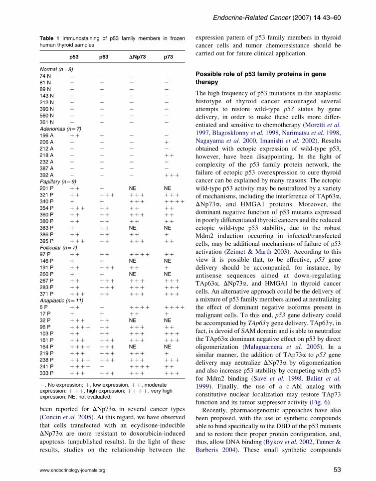
Table 1 Immunostaining of p53 family members in frozen
human thyroid samples
p53 p63 DNp73 p73
Normal (nZ8)
74 N K K K K
81 N K K K K
89 N K K K K143 N K K K K
212 N K K K K
390 N K K K K560 N K K K K
361 N K K K K
Adenomas (nZ7)
196 A CC C K K206 A K K K C
212 A K K K K
218 A K K K CC
232 A K K K K387 A K K K K
392 A K K K CCC
Papillary (nZ9)
201 P CC C NE NE
321 P CC CCC CCC CCC
340 P C C CCC CCCC
354 P CCC CC CC CC360 P CC CC CCC CC
380 P CC CC CC CC
383 P C CC NE NE
386 P CC CC CC C395 P CCC CC CCC CC
Follicular (nZ7)
97 P CC CC CCCC CC
146 P C C NE NE
191 P CC CCC CC C
260 P C C NE NE
267 P CC CCC CCC CCC283 P CC CCC CCC CCC
371 P CCC CC CCC CCC
Anaplastic (nZ11)
6 P CC K CCCC CCCC17 P C C CC C
32 P CCC CC NE NE
96 P CCCC CC CCC CC
103 P CC CC CCC CCC161 P CCC CCC CCC CCC
164 P CCCC CCC NE NE
219 P CCC CCC CCC C238 P CCCC CCC CCC CCC
241 P CCCC K CCCC CC
333 P CCC CCC CCC CCC
K, No expression; C, low expression, CC, moderateexpression: CCC, high expression; CCCC, very highexpression; NE, not evaluated.
Endocrine-Related Cancer (2007) 14 43–60
been reported for DNp73a in several cancer types
(Concin et al. 2005). At this regard, we have observed
that cells transfected with an ecydisone-inducible
DNp73a are more resistant to doxorubicin-induced
apoptosis (unpublished results). In the light of these
results, studies on the relationship between the
www.endocrinology-journals.org
expression pattern of p53 family members in thyroid
cancer cells and tumor chemoresistance should be
carried out for future clinical application.
Possible role of p53 family proteins in gene
therapy
The high frequency of p53 mutations in the anaplastic
histotype of thyroid cancer encouraged several
attempts to restore wild-type p53 status by gene
delivery, in order to make these cells more differ-
entiated and sensitive to chemotherapy (Moretti et al.
1997, Blagosklonny et al. 1998, Narimatsu et al. 1998,
Nagayama et al. 2000, Imanishi et al. 2002). Results
obtained with ectopic expression of wild-type p53,
however, have been disappointing. In the light of
complexity of the p53 family protein network, the
failure of ectopic p53 overexpression to cure thyroid
cancer can be explained by many reasons. The ectopic
wild-type p53 activity may be neutralized by a variety
of mechanisms, including the interference of TAp63a,
DNp73a, and HMGA1 proteins. Moreover, the
dominant negative function of p53 mutants expressed
in poorly differentiated thyroid cancers and the reduced
ectopic wild-type p53 stability, due to the robust
Mdm2 induction occurring in infected/transfected
cells, may be additional mechanisms of failure of p53
activation (Zeimet & Marth 2003). According to this
view it is possible that, to be effective, p53 gene
delivery should be accompanied, for instance, by
antisense sequences aimed at down-regulating
TAp63a, DNp73a, and HMGA1 in thyroid cancer
cells. An alternative approach could be the delivery of
a mixture of p53 family members aimed at neutralizing
the effect of dominant negative isoforms present in
malignant cells. To this end, p53 gene delivery could
be accompanied by TAp63g gene delivery. TAp63g, in
fact, is devoid of SAM domain and is able to neutralize
the TAp63a dominant negative effect on p53 by direct
oligomerization (Malaguarnera et al. 2005). In a
similar manner, the addition of TAp73a to p53 gene
delivery may neutralize DNp73a by oligomerization
and also increase p53 stability by competing with p53
for Mdm2 binding (Save et al. 1998, Balint et al.
1999). Finally, the use of a c-Abl analog with
constitutive nuclear localization may restore TAp73
function and its tumor suppressor activity (Fig. 6).
Recently, pharmacogenomic approaches have also
been proposed, with the use of synthetic compounds
able to bind specifically to the DBD of the p53 mutants
and to restore their proper protein configuration, and,
thus, allow DNA binding (Bykov et al. 2002, Tanner &
Barberis 2004). These small synthetic compounds
53

Figure 6 Schematic representation of a possible network among p53 family members in thyroid cancer cells. Normal thyrocytesexpress a functional wild-type p53, whichmay exert a tumor suppressor activity in response to DNA damage (left). Well-differentiatedthyroid carcinomas express not only wild-type p53, but also other p53 family members with a dominant negative function (middle). Asa result of different mechanisms, the oncosuppressor activity may be blunted: (1) TAp63a, which may be regarded as ade-differentiation marker, may antagonize p53 transcriptional activity by occupying p53-responsive promoters; (2) TAp73a tumorsuppressor activity may be kept latent by c-Abl nuclear exclusion; (3) DNp73a, on the one side, may inactivate TAp73a by botholigomerization and promoter occupancy and, on the other side, may antagonize p53 by promoter occupancy. In addition, HMGA1overexpression may inhibit all transcriptionally active p53 family members (p53, TAp63a, and TAp73a) by interfering with proteinoligomerization. In undifferentiated and anaplastic thyroid cancer, p53 may be inactivated by point mutations (right). p53 mutants notonly have lost their tumor suppressor function, but may also inhibit TAp73a by direct interaction via the core domain.
R Malaguarnera et al.: p53 tumor suppressor family in thyroid cancer
could be used in combination with traditional
chemotherapy to sensitize poorly differentiated thyroid
cancer cells to the cytotoxic effect of these agents.
Conclusions
Although p53 mutations are rare in thyroid cancers,
p53 functional inactivation by a variety of different
mechanisms is apparently very common. The
involved mechanisms are based on the very complex
network of p53 family member isoforms and include
the expression of proteins with dominant negative
function (i.e. DNp73a and TAp63a). Malfunction or
overexpression of regulatory proteins is other,
independent, inhibitory mechanisms. p53 inactivation
by these mechanisms may be an important prerequi-
site for oncogene-driven thyroid cell transformation
(in the early stages of cancer) and in cancer
progression and may render thyroid cancer cells
resistant to the common chemo- and gene therapy
approaches. The unraveling of such a complex
network may result helpful not only for better
understanding of the important aspects of thyroid
tumor progression, but also to design novel multi-
54
targeted therapies for the poorly differentiated and
most aggressive thyroid carcinomas.
Acknowledgements
This work was partially supported by grants and
fellowships from Associazione Italiana per la Ricerca
sul Cancro (AIRC), MIUR-PRIN 2004 and American
Italian Cancer Foundation (AICF).
References
Agami R, Blandino G, Oren M & Shaul Y 1999 c-Abl and
p73 interact and collaborate to induce apoptosis. Nature
399 809–813.
Asakawa H, Kobayashi T, Komoike Y, Maruyama H,
Nakano Y, Tamaki Y, Matsuzawa Y & Monden M 1997
Chemosensitivity of anaplastic thyroid carcinoma and
poorly differentiated thyroid carcinoma. Anticancer
Research 17 2757–2762.
Ayoubi TA, Jansen E, Meulemans SM & Van de Ven WJ
1999 Regulation of HMGIC expression: an architectural
transcription factor involved in growth control and
development. Oncogene 18 5076–5087.
www.endocrinology-journals.org

Endocrine-Related Cancer (2007) 14 43–60
Balint E, Bates S & Vousden KH 1999 Mdm2 binds p73 a
without targeting degradation. Oncogene 18 3923–3929.
Bartolazzi A, Gasbarri A, Papotti M, Bussolati G, Lucante T,
Khan A, Inohara H, Marandino F, Orlandi F, Nardi F et al.
2001 Application of an immunodiagnostic method for
improving preoperative diagnosis of nodular thyroid
lesions. Lancet 357 1644–1650.
Benard J, Douc-Rasy S & Ahomadegbe JC 2003 TP53 family
members and human cancers.HumanMutation21182–191.
Berlingieri MT, Manfioletti G, Santoro M, Bandiera A,
Visconti R, Giancotti V & Fusco A 1995 Inhibition of
HMGI-C protein synthesis suppresses retrovirally
induced neoplastic transformation of rat thyroid cells.
Molecular and Cellular Biology 15 1545–1553.
Berlingieri MT, Pierantoni GM, Giancotti V, Santoro M &
Fusco A 2002 Thyroid cell transformation requires the
expression of the HMGA1 proteins. Oncogene 21
2971–2980.
Blagosklonny MV, Giannakakou P, Wojtowicz M, Romanova
LY, Ain KB, Bates SE & Fojo T 1998 Effects of p53-
expressing adenovirus on the chemosensitivity and differ-
entiation of anaplastic thyroid cancer cells. Journal of
Clinical Endocrinology and Metabolism 83 2516–2522.
Blandino G & Dobbelstein M 2004 p73 and p63: why do we
still need them? Cell Cycle 3 886–894.
Bourdon JC, Fernandes K, Murray-Zmijewski F, Liu G, Diot
A, Xirodimas DP, Saville MK & Lane DP 2005 p53
isoforms can regulate p53 transcriptional activity. Genes
& Development 19 2122–2137.
Burns JS, Blaydes JP, Wright PA, Lemoine L, Bond JA,
Williams ED & Wynford-Thomas D 1992 Stepwise
transformation of primary thyroid epithelial cells by a
mutant Ha-ras oncogene: an in vitro model of tumor
progression. Molecular Carcinogenesis 6 129–139.
Burstein DE, Nagi C, Wang BY & Unger P 2004
Immunohistochemical detection of p53 homolog p63 in
solid cell nests, papillary thyroid carcinoma, and
hashimoto’s thyroiditis: a stem cell hypothesis of
papillary carcinoma oncogenesis. Human Pathology 35
465–473.
Bykov VJ, Issaeva N, Shilov A, Hultcrantz M, Pugacheva E,
Chumakov P, Bergman J, Wiman KG & Selivanova G
2002 Restoration of the tumor suppressor function to
mutant p53 by a low-molecular-weight compound.
Nature Medicine 8 282–288.
Casciano I, Mazzocco K, Boni L, Pagnan G, Banelli B,
Allemanni G, Ponzoni M, Tonini GP & Romani M 2002
Expression of DeltaNp73 is a molecular marker for
adverse outcome in neuroblastoma patients. Cell Death
and Differentiation 9 246–251.
Celli J, Duijf P, Hamel BC, Bamshad M, Kramer B, Smits
AP, Newbury-Ecob R, Hennekam RC, Van Buggenh-
out G, van Haeringen A et al. 1999 Heterozygous
germline mutions in the p53 homolog p63 are the
cause of EEE syndrome. Cell 99 143–153.
Ceraline J, Deplanque G, Noel F, Natarajan-Ame S, Bergerat
JP & Klein-Soyer C 2003 Sensitivity to cisplatin
www.endocrinology-journals.org
treatment of human K1 thyroid carcinoma cell lines with
altered p53 function. Cancer Chemotherapy and
Pharmacology 51 91–95.
Chen BK, Ohtsuki Y, Furihata M, Takeuchi T, Iwata J, Liang
SB & Sonobe H 1999 Co-overexpression of p53 protein
and epidermal growth factor receptor in human papillary
thyroid carcinomas correlated with lymph node metas-
tasis, tumor size and clinicopathologic stage. Inter-
national Journal of Oncology 15 893–898.
Chiappetta G, Avantaggiato V, Visconti R, Fedele M,
Battista S, Trapasso F, Merciai BM, Fidanza V, Giancotti
V, Santoro M et al. 1996 High level expression of the
HMGA1 gene during embryonic development. Oncogene
13 2439–2446.
Clem RJ 2001 Baculoviruses and apoptosis: the good, the bad,
and the ugly. Cell Death and Differentiation 8 137–143.
Concin N, Becker K, Slade N, Erster S, Mueller-Holzner E,
Ulmer H, Daxenbichler G, Zeimet A, Zeillinger R, Marth
C et al. 2004 Transdominant DeltaTAp73 isoforms are
frequently up-regulated in ovarian cancer. Evidence for
their role as epigenetic p53 inhibitors in vivo. Cancer
Research 64 2449–2460.
Concin N, Hofstetter G, Berger A, Gehmacher A, Reimer D,
Watrowski R, Tong D, Schuster E, Hefler L, Heim K
et al. 2005 Clinical relevance of dominant-negative p73
isoforms for responsiveness to chemotherapy and
survival in ovarian cancer: evidence for a crucial p53-
p73 cross-talk in vivo. Clinical Cancer Research 11
8372–8383.
Courtois S, de Fromentel CC & Hainaut P 2004 p53 protein
variants: structural and functional similarities with p63
and p73 isoforms. Oncogene 23 631–638.
Crook T, Nicholls JM, Brooks L, O’Nions J & Allday MJ
2000 High level expression of deltaN-p63: a
mechanism for the inactivation of p53 in undiffer-
entiated nasopharyngeal carcinoma (NPC)? Oncogene
19 3439–3444.
Czyz W, Kuzdak K, Pasieka Z, Timler D & Brzezinski J 2001
p53, MDM2, bcl-2 staining in follicular neoplasms of the
thyroid gland. Folia Histochemica et Cytobiologica 39
167–168.
Di Como CJ, Urist MJ, Babayan I, Drobnjak M, Hedvat
CV, Teruya-Feldstein J, Pohar K, Hoos A & Cordon-
Cardo C 2002 p63 Expression profiles in human
normal and tumor tissues. Clinical Cancer Research 8
494–501.
Dobashi Y, Sakamoto A, Sugimura H, Mernyei M, Mori M,
Oyama T & Machinami R 1993 Overexpression of p53 as
a possible prognostic factor in human thyroid carcinoma.
American Journal of Surgical Pathology 17 375–381.
Dominguez G, Garcia JM, Pena C, Silva J, Garcia V,
Martinez L, Maximiano C, Gomez ME, Rivera JA,
Garcia-Andrade C et al. 2006 DeltaTAp73 upregulation
correlates with poor prognosis in human tumors: putative
in vivo network involving p73 isoforms, p53, and E2F-1.
Journal of Clinical Oncology 24 805–815.
55

R Malaguarnera et al.: p53 tumor suppressor family in thyroid cancer
Fagin JA, Matsuo K, Karmakar A, Chen DL, Tang SH &
Koeffler HP 1993 High prevalence of mutations of the p53
gene in poorly differentiated human thyroid carcinomas.
Journal of Clinical Investigation 91 179–184.
Fagin JA, Tang SH, Zeki K, Di Lauro R, Fusco A &
Gonsky R 1996 Reexpression of thyroid peroxidase in a
derivative of an undifferentiated thyroid carcinoma cell
line by introduction of wild-type p53. Cancer Research
56 765–771.
Fedele M, Battista S, Kenyon L, Baldassarre G, Fidanza V,
Klein-Szanto AJ, Parlow AF, Visone R, Pierantoni GM,
Outwater E et al. 2002 Overexpression of the HMGA2
gene in transgenic mice leads to the onset of pituitary
adenomas. Oncogene 21 3190–3198.
Fedele M, Pentimalli F, Baldassarre G, Battista S, Klein-
Szanto AJ, Kenyon L, Visone R, De Martino I,
Ciarmiello A, Arra C et al. 2005 Transgenic mice
overexpressing the wild-type form of the HMGA1 gene
develop mixed growth hormone/prolactin cell pituitary
adenomas and natural killer cell lymphomas. Oncogene
24 3427–3435.
Ferru A, Denis S, Guilhot J, Gibelin H, Tourani JM,
Kraimps JL, Larsen CJ & Karayan-Tapon L 2006
Expression of TAp73 and DeltaNp73 isoform transcripts
in thyroid tumours. European Journal of Surgical
Oncology 32 228–230.
Flores ER, Sengupta S, Miller JB, Newman JJ, Bronson R,
Crowley D, Yang A, McKeon F & Jacks T 2005 Tumor
predisposition in mice mutant for p63 and p73: Evidence
for broader tumor suppressor functions for the p53 family.
Cancer Cell 7 363–373.
Fomenkov A, Huang YP, Topaloglu O, Brechman A, Osada
M, Fomenkova T, Yuriditsky E, Trink B, Sidransky D &
Ratovitski E 2003 P63 alpha mutations lead to aberrant
splicing of keratinocyte growth factor receptor in the Hay-
Wells syndrome. Journal of Biological Chemistry 278
23906–23914.
Frasca F, Vella V, Aloisi A, Mandarino A, Mazzon E,
Vigneri R & Vigneri P 2003 p73 tumor-suppressor
activity is impaired in human thyroid cancer. Cancer
Research 63 5829–5837.
Frasca F, Rustighi A, Malaguarnera R, Altamura S, Vigneri P,
Del Sal G, Giancotti V, Pezzino V, Vigneri R & Manfioletti
G 2006 HMGA1 inhibits the function of p53 family
members in thyroid cancer cells. Cancer Research 66
2980–2989.
Freebern WJ, Smith JL, Chaudhry SS, Haggerty CM &
Gardner K 2003 Novel cell-specific and dominant
negative anti-apoptotic roles of p73 in transformed
leukemia cells. Journal of Biological Chemistry 278
2249–2255.
Gaiddon C, Lokshin M, Ahn J, Zhang T & Prives C 2001 A
subset of tumor-derived mutant forms of p53 down-
regulate p63 and p73 through a direct interaction with the
p53 core domain. Molecular and Cellular Biology 21
1874–1887.
56
Gamble SC, Cook MC, Riches AC, Herceg Z, Bryant PE &
Arrand JE 1999 p53 mutations in tumors derived from
irradiated human thyroid epithelial cells. Mutation
Research 425 231–238.
Garcia-Rostan G, Costa AM, Pereira-Castro I, Salvatore G,
Hernandez R, Hermsem MJ, Herrero A, Fusco A,
Cameselle-Teijeiro J & Santoro M 2005 Mutation of the
PIK3CA gene in anaplastic thyroid cancer. Cancer
Research 65 10199–10207.
Gerasimov G, Bronstein M, Troshina K, Alexandrova G,
Dedov I, Jennings T, Kallakury BV, Izquierdo R,
Boguniewicz A, Figge H et al. 1995 Nuclear p53
immunoreactivity in papillary thyroid cancers is associ-
ated with two established indicators of poor prognosis.
Experimental and Molecular Pathology 62 52–62.
Ghioni P, Bolognese F, Duijf PH, Van Bokhoven H,
Mantovani R & Guerrini L 2002 Complex transcriptional
effects of p63 isoforms: identification of novel activation
and repression domains. Molecular and Cellular Biology
22 8659–8668.
Godballe C, Asschenfeldt P, Jorgensen KE, Bastholt L,
Clausen PP, Hansen TP, Hansen O & Bentzen SM 1998
Prognostic factors in papillary and follicular thyroid
carcinomas: p53 expression is a significant indicator of
prognosis. Laryngoscope 108 243–249.
Gong JG, Costanzo A, Yang HQ, Melino G, Kaelin WG, Jr,
Levrero M & Wang JY 1999 The tyrosine kinase c-Abl
regulates p73 in apoptotic response to cisplatin-induced
DNA damage. Nature 399 806–809.
Guan M & Chen Y 2005 Aberrant expression of DeltaNp73
in benign and malignant tumours of the prostate:
correlation with Gleason score. Journal of Clinical
Pathology 58 1175–1179.
Harvey M, Vogel H, Lee EY, Bradley A & Donehower LA 1995
Mice deficient in both p53 and Rb develop tumors primarily
of endocrine origin. Cancer Research 55 1146–1151.
Hassan I, Wunderlich A, Burchert A, Hoffmann S & Zielke A
2006 Antisense p53 oligonucleotides inhibit proliferation
and induce chemosensitivity in follicular thyroid cancer
cells. Anticancer Research 26 1171–1176.
Hibi K, Trink B, Patturajan M, Westra WH, Caballero OL,
Hill DE, Ratovitski EA, Jen J & Sidransky D 2000 AIS is
an oncogene amplified in squamous cell carcinoma. PNAS
97 5462–5467.
Hirning-Folz U, Wilda M, Rippe V, Bullerdiek J &
Hameister H 1998 The expression pattern of the Hmgic
gene during development. Genes, Chromosomes &
Cancer 23 350–357.
Horie S, Maeta H, Endo K, Ueta T, Takashima K & Terada T
2001 Overexpression of p53 protein and MDM2 in papillary
carcinomas of the thyroid: correlations with clinicopatho-
logic features. Pathology International 51 11–15.
Hosal SA, Apel RL, Freeman JL, Azadian A, Rosen IB,
LiVolsi VA & Asa SL 1997 Immunohistochemistry
localization of p53 in human thyroid neoplasms:
correlation with biological behavior. Endocrine Path-
ology 8 21–28.
www.endocrinology-journals.org

Endocrine-Related Cancer (2007) 14 43–60
Hunt J 2005 Understanding the genotype of follicular thyroid
tumors. Endocrine Pathology 16 311–321.
Hunt JL, LiVolsi VA & Barnes EL 2004 p63 expression in
sclerosing mucoepidermoid carcinomas with eosinophilia
arising in the thyroid. Modern Pathology 17 526–529.
Ikawa S, Nakagawara A & Ikawa Y 1999 P53 family genes:
structural comparison, expression and mutation. Cell
Death and Differentiation 6 1154–1161.
Imanishi R, Ohtsuru A, Iwamatsu M, Iioka T, Namba H, Seto
S, Yano K & Yamashita S 2002 A histone deacetylase
inhibitor enhances killing of undifferentiated thyroid
carcinoma cells by p53 gene therapy. Journal of Clinical
Endocrinology and Metabolism 87 4821–4824.
Ito Y, Yoshida H, Tomoda C, Uruno T, Miya A, Kobayashi K,
Matsuzuka F, Kakudo K, Kuma K & Miyauchi A 2004
S100A4 expression is an early event of papillary
carcinoma of the thyroid. Oncology 67 397–402.
Ito Y, Uramoto H, Funa K, Yoshida H, Jikuzono T, Asahi S,
Higashiyama T, Tomoda C, Takamura Y, Miya A et al.
2006 Delta Np73 expression in thyroid neoplasms
originating from follicular cells. Pathology 38 205–209.
Jennings T, Bratslavsky G, Gerasimov G, Troshina K,
Bronstein M, Dedov I, Alexandrova G & Figge J 1995
Nuclear accumulation of MDM2 protein in well-differ-
entiated papillary thyroid carcinomas. Experimental and
Molecular Pathology 62 199–206.
Klemm JD, Schreiber SL & Crabtree GR 1998 Dimerization
as a regulatory mechanism in signal transduction. Annual
Review of Immunology 16 569–592.
Koike M, Fujita F, Komori K, Katoh F, Sugimoto T,
Sakamoto Y, Matsuda M & Fujita M 2004 Dependence of
chemotherapy response on p53 mutation status in a panel
of human cancer lines maintained in nude mice. Cancer
Science 95 541–546.
Koster MI, Lu SL, White LD, Wang XJ & Roop DR 2006
Reactivation of developmentally expressed p63 isoforms
predisposes to tumor development and progression.
Cancer Research 66 3981–3986.
Kroll TG 2004 Molecular events in follicular thyroid tumors.
Cancer Treatment and Research 122 85–105.
Lohrum MA & Vousden KH 2000 Regulation and function of
the p53-related proteins: same family different rules.
Trends in Cell Biology 10 197–202.
Lubitz CC, Gallagher LA, Finley DJ, Zhu B & Fahey TJ, III
2005 Molecular analysis of minimally invasive follicular
carcinomas by gene profiling. Surgery 138 1042–1048
(discussion 1048–9).
Malaguarnera R, Mandarino A, Mazzon E, Vella V,
Gangemi P, Vancheri C, Vigneri P, Aloisi A, Vigneri R
& Frasca F 2005 The p53-homologue p63 may promote
thyroid cancer progression. Endocrine-Related Cancer
12 953–971.
Marin MC & Kaelin WG, Jr 2000 p63 and p73: old members
of a new family. Biochimica et Biophysica Acta 1470
M93–M100.
Massion PP, Taflan PM, Jamshedur Rahman SM, Yildiz P,
Shyr Y, Edgerton ME, Westfall MD, Roberts JR, Pietenpol
www.endocrinology-journals.org
JA, Carbone DP et al. 2003 Significance of p63
amplification and overexpression in lung cancer develop-
ment and prognosis. Cancer Research 63 7113–7121.
McWhirter JR & Wang JY 1997 Effect of Bcr sequences on
the cellular function of the Bcr-Abl oncoprotein.
Oncogene 15 1625–1634.
McWhirter JR, Galasso DL & Wang JY 1993 A coiled-coil
oligomerization domain of Bcr is essential for the
transforming function of Bcr-Abl oncoproteins. Molecu-
lar and Cellular Biology 13 7587–7595.
Melino G, De Laurenzi V & Vousden KH 2002 p73: friend or
foe in tumorigenesis. Nature Reviews. Cancer 2 605–615.
Miettinen M & Karkkainen P 1996 Differential reactivity of
HBME-1 and CD15 antibodies in benign and malignant
thyroid tumours. Preferential reactivity with malignant
tumours. Virchows Archiv 429 213–219.
Mills AA 2005 p53: link to the past, bridge to the future.
Genes & Development 19 2091–2099.
Mills AA 2006 p63: Oncogene or tumor suppressor? Current
Opinion in Genetics & Development 16 38–44.
Mills AA, Zheng B, Wang XJ, Vogel H, Roop DR & Bradley
A 1999 p63 is a p53 homologue required for limb and
epidermal morphogenesis. Nature 398 708–713.
Moll UM 2003 The role of p63 and p73 in tumor formation
and progression: coming of age toward clinical useful-
ness. Clinical Cancer Research 9 5437–5441.
Moll UM & Slade N 2004 p63 and p73: roles in
development and tumor formation. Molecular Cancer
Research 2 371–386.
Moll UM, Erster S & Zaika A 2001 p53, p63 and p73 – solos,
alliances and feuds among family members. Biochimica
et Biophysica Acta 1552 47–59.
Moretti F, Farsetti A, Soddu S, Misiti S, Crescenzi M, Filetti
S, Andreoli M, Sacchi A & Pontecorvi A 1997 p53
re-expression inhibits proliferation and restores differen-
tiation of human thyroid anaplastic carcinoma cells.
Oncogene 14 729–740.
Muller M, Schilling T, Sayan AE, Kairat A, Lorenz K,
Schulze-Bergkamen H, Oren M, Koch A, Tannapfel A,
Stremmel W et al. 2005 TAp73/LNp73 influences
apoptotic response, chemosensitivity and prognosis in
hepatocellular carcinoma. Cell Death and Differentiation
12 1564–1577.
Murray-Zmijewski F, Lane DP & Bourdon JC 2006
p53/p63/p73 isoforms: an orchestra of isoforms to
harmonise cell differentiation and response to stress. Cell
Death and Differentiation 13 962–972.
Musholt TJ, Brehm C, Hanack J, von Wasielewski R &
Musholt PB 2006 Identification of differentially
expressed genes in papillary thyroid carcinomas with and
without rearrangements of the tyrosine kinase receptors
RET and/or NTRK11. Journal of Surgical Research 131
15–25.
Nagayama Y, Yokoi H, Takeda K, Hasegawa M, Nishihara
E, Namba H, Yamashita S & Niwa M 2000 Adenovirus-
mediated tumor suppressor p53 gene therapy for
57

R Malaguarnera et al.: p53 tumor suppressor family in thyroid cancer
anaplastic thyroid carcinoma in vitro and in vivo.
Journal of Clinical Endocrinology and Metabolism 85
4081–4086.
Nakagawa T, Takahashi M, Ozaki T, Watanabe K, Hayashi S,
Hosoda M, Todo S & Nakagawara A 2003 Negative
autoregulation of p73 and p53 by DeltaNp73 in regulating
differentiation and survival of human neuroblastoma cells.
Cancer Letters 197 105–109.
Narimatsu M, Nagayama Y, Akino K, Yasuda M, Yamamoto T,
Yang TT, Ohtsuru A, Namba H, Yamashita S, Ayabe H et al.
1998 Therapeutic usefulness of wild-type p53 gene
introduction in a p53-null anaplastic thyroid carcinoma cell
line. Journal of Clinical Endocrinology and Metabolism 83
3668–3672.
Nikiforov YE, Nikiforova MN, Gnepp DR & Fagin JA 1996
Prevalence of mutations of RAS and P53 in Benign and
Malignant thyroid tumors from children exposed to
radiation after the chernobyl nuclear accident. Oncogene
13 687–693.
Nishida T, Nakao K, Hamaji M, Nakahara MA & Tsujimoto
M 1996 Overexpression of p53 protein and DNA content
are important biologic prognostic factors for thyroid
cancer. Surgery 119 568–575.
Olivier M, Eeles R, Hollstein M, Khan MA, Harris CC &
Hainaut P 2002 The IARC TP53 database: new online
mutation analysis and recommendations to users. Human
Mutation 19 607–614.
Orlandi F, Saggiorato E, Pivano G, Puligheddu B, Termine A,
Cappia S, De Giuli P & Angeli A 1998 Galectin-3 is a
presurgical marker of human thyroid carcinoma. Cancer
Research 58 3015–3020.
Park KY, Koh JM, Kim YI, Park HJ, Gong G, Hong SJ &
Ahn IM 1998 Prevalences of Gs alpha, ras, p53 mutations
and ret/PTC rearrangement in differentiated thyroid
tumours in a Korean population. Clinical Endocrinology
49 317–323.
Park BJ, Lee SJ, Kim JI, Lee CH, Chang SG, Park JH &
Chi SG 2000 Frequent alteration of p63 expression in
primary bladder carcinomas. Cancer Research 60
3370–3374.
Pisarchik AV, Ermak G, Kartel NA & Figge J 2000
Molecular alterations involving p53 codons 167 and 183
in papillary thyroid carcinomas from chernobyl-
contaminated regions of belarus. Thyroid 10 25–30.
Pollina L, Pacini F, Fontanini G, Vignati S, Bevilacqua G &
Basolo F 1996 bcl-2, p53 and proliferating cell nuclear
antigen expression is related to the degree of differen-
tiation in thyroid carcinomas. British Journal of Cancer
73 139–143.
Portella G, Scala S, Vitagliano D, Vecchio G & Fusco A 2002
ONYX-015, an E1B gene-defective adenovirus, induces
cell death in human anaplastic thyroid carcinoma cell
lines. Journal of Clinical Endocrinology and Metabolism.
87 2525–2531.
Portella G, Pacelli R, Libertini S, Cella L, Vecchio G,
Salvatore M & Fusco A 2003 ONYX-015 enhances
58
radiation-induced death of human anaplastic thyroid
carcinoma cells. Journal of Clinical Endocrinology and
Metabolism 88 5027–5032.
Pozniak CD, Radinovic S, Yang A, McKeon F, Kaplan DR &
Miller FD 2000 An anti-apoptotic role for the p53 family
member, p73, during developmental neuron death.
Science 289 304–306.
Preto A, Reis-Filho JS, Ricardo S & Soares P 2002 P63
expression in papillary and anaplastic carcinomas of the
thyroid gland: lack of an oncogenetic role in tumorigen-
esis and progression. Pathology, Research and Practice
198 449–454.
Quiros RM, Ding HG, Gattuso P, Prinz RA & Xu X 2005
Evidence that one subset of anaplastic thyroid carcinomas
are derived from papillary carcinomas due to BRAF and
p53 mutations. Cancer 103 2261–2268.
Reeves R 2001 Molecular biology of HMGA proteins: hubs
of nuclear function. Gene 277 63–81.
Reis-Filho JS & Schmitt FC 2002 Taking advantage of basic
research: p63 is a reliable myoepithelial and stem cell
marker. Advances in Anatomic Pathology 9 280–289.
Reis-Filho JS, Preto A, Soares P, Ricardo S, Cameselle-
Teijeiro J & Sobrinho-Simoes M 2003 p63 expression in
solid cell nests of the thyroid: further evidence for a stem
cell origin. Modern Pathology 16 43–48.
Rohaly G, Chemnitz J, Dehde S, Nunez AM, Heukeshoven J,
Deppert W & Dornreiter I 2005 A novel human p53
isoform is an essential element of the ATR-intra-S phase
checkpoint. Cell 122 21–32.
Ruter A, Dreifus J, Jones M, Nishiyama R & Lennquist S
1996 Overexpression of p53 in tall cell variants of
papillary thyroid carcinoma. Surgery 120 1046–1050.
Salvatore D, Celetti A, Fabien N, Paulin C, Martelli ML,
Battaglia C, Califano D, Monaco C, Viglietto G, Santoro M
et al. 1996 Low frequency of p53 mutations in human
thyroid tumours; p53 and Ras mutation in two out of fifty-
six thyroid tumours. European Journal of Endocrinology
134 177–183.
Sasaki Y, Morimoto I, Ishida S, Yamashita T, Imai K &
Tokino T 2001 Adenovirus-mediated transfer of the p53
family genes, p73 and p51/p63 induces cell cycle arrest
and apoptosis in colorectal cancer cell lines: potential
application to gene therapy of colorectal cancer. Gene
Therapy 8 1401–1408.
Save V, Nylander K & Hall PA 1998 Why is p53 protein
stabilized in neoplasia? Some answers but many more
questions? Journal of Pathology 184 348–350.
Scala S, Portella G, Fedele M, Chiappetta G & Fusco A 2000
Adenovirus-mediated suppression of HMGI(Y) protein
synthesis as potential therapy of human malignant
neoplasias. PNAS 97 4256–4261.
Schelfhout LJ, Van Muijen GN & Fleuren GJ 1989
Expression of keratin 19 distinguishes papillary thyroid
carcinoma from follicular carcinomas and follicular
thyroid adenoma. American Journal of Clinical Path-
ology 92 654–658.
www.endocrinology-journals.org

Endocrine-Related Cancer (2007) 14 43–60
Serber Z, Lai HC, Yang A, Ou HD, Sigal MS, Kelly AE,
Darimont BD, Duijf PH, Van Bokhoven H, McKeon F
et al. 2002 A C-terminal inhibitory domain controls the
activity of p63 by an intramolecular mechanism.
Molecular and Cellular Biology 22 8601–8611.
Sgarra R, Rustighi A, Tessari MA, Di Bernardo J,
Altamura S, Fusco A, Manfioletti G & Giancotti V 2004
Nuclear phosphoproteins HMGA and their relationship
with chromatin structure and cancer. FEBS Letters 574
1–8.
Shahedian B, Shi Y, Zou M & Farid NR 2001 Thyroid
carcinoma is characterized by genomic instability:
evidence from p53 mutations. Molecular Genetics and
Metabolism 72 155–163.
Smida J, Zitzelsberger H, Kellerer AM, Lehmann L, Minkus
G, Negele T, Spelsberg F, Hieber L, Demidchik EP,
Lengfelder E et al. 1997 p53 mutations in childhood
thyroid tumours from Belarus and in thyroid tumours
without radiation history. International Journal of Cancer
73 802–807.
Smith KM & Van Etten RA 2001 Activation of c-Abl kinase
activity and transformation by a chemical inducer of
dimerization. Journal of Biological Chemistry 276
24372–24379.
Soares P, Cameselle-Teijeiro J & Sobrinho-Simoes M 1994
Immunohistochemical detection of p53 in differentiated,
poorly differentiated and undifferentiated carcinomas of
the thyroid. Histopathology 24 205–210.
Stewart SA & Weinberg RA 2006 Telomeres: cancer to
human aging. Annual Review of Cell and Developmental
Biology 22 531–557.
Strano S, Munarriz E, Rossi M, Castagnoli L, Shaul Y, Sacchi
A, Oren M, Sudol M, Cesareni G & Blandino G 2001
Physical interaction with Yes-associated protein enhances
p73 transcriptional activity. Journal of Biological
Chemistry 276 15164–15173.
Suchy B, Waldmann V, Klugbauer S & Rabes HM 1998
Absence of RAS and p53 mutations in thyroid
carcinomas of children after Chernobyl in contrast to
adult thyroid tumours. British Journal of Cancer 77
952–955.
Tanner S & Barberis A 2004 CP-31398, a putative p53-
stabilizing molecule tested in mammalian cells and in
yeast for its effects on p53 transcriptional activity.
Journal of Negative Results in Biomedicine 3 5.
Thanos CD & Bowie JU 1999 p53 Family members p63 and
p73 are SAM domain-containing proteins. Protein
Science 8 1708–1710.
Treff NR, Dement GA, Adair JE, Britt RL, Nie R, Shima JE,
Taylor WE & Reeves R 2004a Human KIT ligand
promoter is positively regulated by HMGA1 in breast and
ovarian cancer cells. Oncogene 23 8557–8562.
Treff NR, Pouchnik D, Dement GA, Britt RL & Reeves R
2004b High-mobility group A1a protein regulates
Ras/ERK signaling in MCF-7 human breast cancer cells.
Oncogene 23 777–785.
www.endocrinology-journals.org
Ugur H, Sayan AE, Ozdamar SO, Kanpolat Y & Ozturk M
2004 Expression of TAP73 and DeltaNP73 in malignant
gliomas. Oncology Reports 11 1337–1341.
Unger P, Ewart M, Wang BY, Gan L, Kohtz DS & Burstein
DE 2003 Expression of p63 in papillary thyroid
carcinoma and in Hashimoto’s thyroiditis: a pathobiologic
link? Human Pathology 34 764–769.
Vella V, Zhu J, Frasca F, Li CY, Vigneri P, Vigneri R &
Wang JY 2003 Exclusion of c-Abl from the nucleus
restrains the p73 tumor suppression function. Journal of
Biological Chemistry 278 25151–25157.
Vikhanskaya F, D’Incalci M & Broggini M 2000 p73
competes with p53 and attenuates its response in a
human ovarian cancer cell line. Nucleic Acids Research
28 513–519.
Vikhanskaya F, Marchini S, Marabese M, Galliera E &
Broggini M 2001 p73a overexpression is associated with
resistance to treatment with DNA-damaging agents in a
human ovarian cancer cell line. Cancer Research 61
935–938.
Westfall MD & Pietenpol JA 2004 p63: molecular
complexity in development and cancer. Carcinogenesis
25 857–864.
Wood LJ, Maher JF, Bunton TE & Resar LM 2000a The
oncogenic properties of the HMGI gene family. Cancer
Research 60 4256–4261.
Wood LJ, Mukherjee M, Dolde CE, Xu Y, Maher JF, Bunton
TE, Williams JB & Resar LM 2000b HMG-I/Y, a new
c-Myc target gene and potential oncogene. Molecular and
Cellular Biology 20 5490–5502.
Wyllie FS, Haughton MF, Blaydes JP, Schlumberger M &
Wynford-Thomas D 1995 Evasion of p53-mediated
growth control occurs by three alternative mechanisms
in transformed thyroid epithelial cells. Oncogene 10
49–59.
Xu Y, Sumter TF, Bhattacharya R, Tesfaye A, Fuchs EJ,
Wood LJ, Huso DL & Resar LM 2004 The HMG-I
oncogene causes highly penetrant, aggressive lym-
phoid malignancy in transgenic mice and is over-
expressed in human leukemia. Cancer Research 64
3371–3375.
Yamaguchi K, Wu L, Caballero OL, Hibi K, Trink B, Resto
V, Cairns P, Okami K, Koch WM, Sidransky D et al. 2000
Frequent gain of the p40/p51/p63 gene locus in primary
head and neck squamous cell carcinoma. International
Journal of Cancer 86 684–689.
Yang A, Kaghad M, Wang Y, Gillett E, Fleming MD, Dotsch
V, Andrews NC, Caput D & McKeon F 1998 p63, a p53
homolog at 3q27-29, encodes multiple products with
transactivating, death-inducing, and dominant-negative
activities. Molecular Cell 2 305–316.
Yang A, Walker N, Bronson R, Kaghad M, Oosterwegel M,
Bonnin J, Vagner C, Bonnet H, Dikkes P, Sharpe A et al.
2000 p73-deficient mice have neurological, pheromonal
and inflammatory defects but lack spontaneous tumours.
Nature 404 99–103.
59

R Malaguarnera et al.: p53 tumor suppressor family in thyroid cancer
Zaika AI, Slade N, Erster SH, Sansome C, Joseph TW, Pearl
M, Chalas E & Moll UM 2002 DeltaNp73, a dominant-
negative inhibitor of wild-type p53 and TAp73, is
up-regulated in human tumors. Journal of Experimental
Medicine 196 765–780.
Zedenius J, Larsson C, Wallin G, Backdahl M, Aspenblad U,
Hoog A, Borresen AL & Auer G 1996 Alterations of p53
and expression of WAF1/p21 in human thyroid tumors.
Thyroid 6 1–9.
Zeimet AG & Marth C 2003 Why did p53 gene therapy fail in
ovarian cancer? Lancet Oncology 4 415–422.
60
Zhou X, Benson KF, Ashar HR & Chada K 1995 Mutation
responsible for the mouse pygmy phenotype in the
developmentally regulated factor HMGI-C. Nature 376
771–774.
Zhu J, Jiang J, Zhou W & Chen X 1998 The potential tumor
suppressor p73 differentially regulates cellular p53 target
genes. Cancer Research 58 5061–5065.
Zou M, Shi Y, al-Sedairy S, Hussain SS & Farid NR
1995 The expression of the MDM2 gene, a p53
binding protein, in thyroid carcinogenesis. Cancer 76
314–318.
www.endocrinology-journals.org