p53 tumor suppressor gene: understanding p53 based dietary anti cancer therapies utilizing dietary...
TRANSCRIPT

P53 – Tumor Suppressor Gene:Understanding P53 – Based
Anticancer Therapies UtilizingDietary Agents
by Serge Jurasunas,N.D., M.D. (Hom)
www.sergejurasunas.comLisbon, Portugal
1

P53 – Tumor Suppressor Gene: Understanding P53 –Based Anticancer Therapies Utilizing Dietary Agents
(At the end of this paper we made an announcementconcerning the “20th World Congress on Advances inOncology – 18th International Symposium of MolecularMedicine”, featuring a program presenting several topicsrelated to our therapies.)
Abstract
The P53 tumor suppressor gene which has been dubbed both the “Guardian ofthe Genome” (Lane 1992) and Science “Molecule of the Year”, is directlyinvolved in the initiation of apoptosis and programmed cell death, to preventan accumulation of abnormal cells. However apoptosis evasion is acharacteristic feature of human cancers that promote tumor formation andprogression (1). Presently, P53 is known to play a key role in practically alltypes of human cancers, and the mutation or loss of P53 gene function, canbe identified in more than 50% of all human cancer cases worldwide (2).
Frequency of P53 mutations70% in lung cancer60% in cancers of colon, head, neck, ovary, bladder45% in stomach cancer35% - 40% in breast cancer
Recent data has shown in addition to losing transcriptional function, mutantP53 gains oncogenic functions termed GOF (Gain of Function) that drive cellmigration, invasion, and metastases (3-4). The notion for mutant P53 GOFtheory is supported by recent studies using mutated P53-blocked mice whichdisplay a broader tumor spectrum, increased aggressiveness and metastaticpotential as compared with their P53–null counterparts (5-6). Similarly inhuman cancers mutant P53 expression has been linked with a poor prognosis(7).
Therefore mutant P53 function raises the possibility that the mutant proteinmay be a good target for designing novel therapies.
The P53 pathway seems to play a critical role in therapeutic response andboth as a diagnostic and marker in the prognosis of therapeutic treatmenteffects.
The inability of most cancers to undergo apoptosis in response to appropriatestimuli is a key cause of treatment failure, presenting one of the major, yetunsolved problems in Oncology (8).
2
Introduction
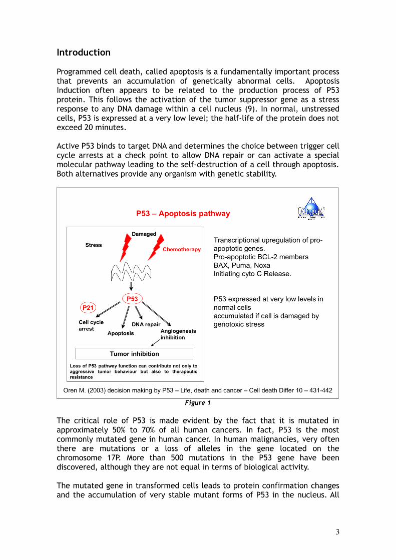
Programmed cell death, called apoptosis is a fundamentally important processthat prevents an accumulation of genetically abnormal cells. ApoptosisInduction often appears to be related to the production process of P53protein. This follows the activation of the tumor suppressor gene as a stressresponse to any DNA damage within a cell nucleus (9). In normal, unstressedcells, P53 is expressed at a very low level; the half-life of the protein does notexceed 20 minutes.
Active P53 binds to target DNA and determines the choice between trigger cellcycle arrests at a check point to allow DNA repair or can activate a specialmolecular pathway leading to the self-destruction of a cell through apoptosis.Both alternatives provide any organism with genetic stability.
P53 – Apoptosis pathway
Transcriptional upregulation of pro-apoptotic genes.Pro-apoptotic BCL-2 membersBAX, Puma, NoxaInitiating cyto C Release.
P53 expressed at very low levels innormal cellsaccumulated if cell is damaged bygenotoxic stress
Oren M. (2003) decision making by P53 – Life, death and cancer – Cell death Differ 10 – 431-442
Stress
Damaged
Chemotherapy
P53
P21
Cell cyclearrest
Apoptosis
DNA repairAngiogenesisinhibition
Tumor inhibition
Loss of P53 pathway function can contribute not only to aggressive tumor behaviour but also to therapeuticresistance
Figure 1
The critical role of P53 is made evident by the fact that it is mutated inapproximately 50% to 70% of all human cancers. In fact, P53 is the mostcommonly mutated gene in human cancer. In human malignancies, very oftenthere are mutations or a loss of alleles in the gene located on thechromosome 17P. More than 500 mutations in the P53 gene have beendiscovered, although they are not equal in terms of biological activity.
The mutated gene in transformed cells leads to protein confirmation changesand the accumulation of very stable mutant forms of P53 in the nucleus. All
3

types of mutated P53 are likely to be ineffective in maintaining a non-tumorigenic cellular phenotype when compared to a wild type P53.
Wild Type P53, which is a nuclear phosphoprotein, has been shown to be asequence specific transcription factor which induces the expression of P21,WAF1/C1P1/Sdi-1, leading to a G1 arrest checkpoint to step up repair beforeDNA replication and contributes to normal cell proliferation (10) unless DNAreplication is successful, the cells will be induced to undergo apoptosis.
However during a stress response from its P53 gene to any damage, recentfindings suggest that P53 induces apoptosis by trans-activating expression ofthe BAX gene MRna (11) to increase BAX protein and simultaneously inhibit thefunction of BCL2. RNA proteins are homologs, through BAX acts as anaccelerator of apoptosis while BCL2 serves to prolong life survival (12).
This suggests that P53 mutation not only serves to inactivate the pro-apoptotic P53 pathway, but that may also play an additional role in tumorprogression. Mutant P53 itself provides a selective advantage to tumor cellsand promotes tumor growth. Recent data suggests that expression of mutantP53 is not the equivalent of P53 loss, where mutant P53 can acquire newfunctions.
BCL2 activity up-regulates in many types of cancer and correlates with cancercell resistance to a wide spectrum of chemotherapy agents (13). Over-expression of the anti-apoptotic BCL2 proteins blocks cytochrome C release inmitochondria in response to a variety of stimuli, whereas the pro-apoptoticBAX protein releases cytochrome C that in turn activates an apoptoticcascade, while the loss of BAX associates with tumor progression and withshorter survival in metastatic breast cancer (14).
Figure 2
4

BAX was the first identified P53–regulated, pro-apoptotic BCL2 familymember. P53–responsive elements have been unequivocally identified in theBAX gene (15). BAX is specifically required for Puma mediated apoptosis, andit also participates in the death response as an indirect target of P53 throughPuma (16) and Noxa, both implicated in P53–dependent apoptosis.
Some studies show that the loss of BAX is responsible for nearly half of theaccelerated tumor growth in brain tumors that are related to loss of P53function (17). BAX is inactive in approximately one third of invasive breastcancers, where in a study of 119 women with metastatic breast cancer, it wasfound that patients whose tumors had lost BAX activity, had poor responses tocombination chemotherapy, faster time to tumor progression, and shorteroverall survival (18).
This may suggest that turning on this pro-apoptotic gene may be important forchemotherapy response, where one of the factors that can regulate BAX geneactivity is the P53 tumor suppressor gene, which also simultaneously inhibitsBCL2 during the process of apoptosis. Nevertheless because BAX proteinsantagonize BCL2 anti-apoptotic function, it is likely that the BCL2/BAXbalance ratio determines both the susceptibility of a cell to apoptosis as wellas therapeutic response to apoptosis stimuli (19).
If apoptosis signaling is not initiated by nuclear P53 and/or the presence of amutated P53 gene, loss of BAX and over-expressed BCL2, this allows somecancer cells to divide unchecked after radiation or chemotherapy treatment;associating with cancer cell resistance, increased rate of tumor recurrence,and shorter patient survival (20).
5

Figure 3
Another profound feature of malignant cells is their ability to induceangiogenesis necessary for tumor growth. Again, there is a clear correlationbetween mutant P53 GOF that facilitates angiogenesis by increasing theexpression of VEGF (21), via interaction with E2F1 that induces the expressionof ID4, which in turn promotes the expression of pro-angiogenic factors suchIL and GROa, thus eventually leading to increasing angiogenesis in canceroustissues (22). Other novel functions of mutant P53 GOF are shown through theactivation of specific target genes EGFR/1, RAS, Myc, and interference withthe TGFB growth arrest control pathway, down-regulation of the E-Cadherincell-cell adhesion molecules to enhance motility and tumor cell migration andinvasion (23).
We have new clues and important conceptualizations that indicate tumorscannot be viewed simply as an uncontrolled proliferative mass, but rather as acellular community, interacting with a microenvironment (24-25). This is whytargeting mutant P53 remains an urgent need to improve cancer treatment byincreasing a cancer cell’s sensitivity to apoptosis.
Therapeutic Strategy
Targeting mutant P53 and restoring the wild type function of P53 tumorsuppressor gene in tumor cells would be of potential therapeutic benefit andan attractive strategy for anticancer treatments (26-27). Upon restoration ofP53 transcriptional activity, the apoptosis pathway would predominate.
Many cancer cells escape apoptosis and become resistant to chemotherapyradiation or from destruction by the immune cells by endogenous cytotoxic T-cells and natural killer cells (NK). If the oncogene BCL2 is highly expressed itconfers greater resistance to cancer cells from attacking immune cells,increasing the urgent need for effective cancer therapies.
Furthermore some of the P53 apoptosis targets such as BAX, Puma, Noxa, andP21 could potentially be used as targets for gene therapy to increase theeffectiveness of chemotherapy.
Dietary Agents that Induce Apoptosis with Chemo Preventive Effects
A large number of dietary agents can exert effects on the human genomeeither directly or indirectly to modulate gene expression. Extensive researchduring the last half century demonstrated that numerous agents identifiedfrom fruits and vegetables can interfere with several signaling pathways, andwere validated as apoptosis inducers in research experiments.
6
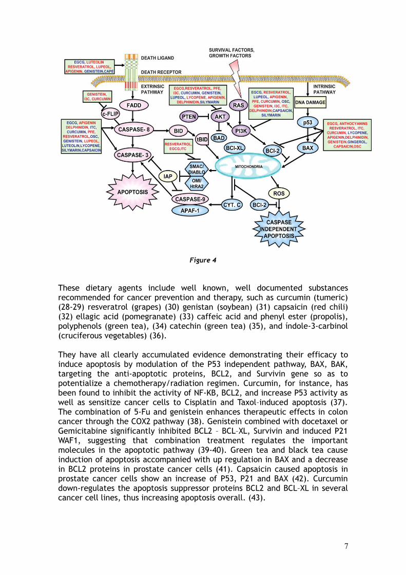
Figure 4
These dietary agents include well known, well documented substancesrecommended for cancer prevention and therapy, such as curcumin (tumeric)(28-29) resveratrol (grapes) (30) genistan (soybean) (31) capsaicin (red chili)(32) ellagic acid (pomegranate) (33) caffeic acid and phenyl ester (propolis),polyphenols (green tea), (34) catechin (green tea) (35), and índole-3-carbinol(cruciferous vegetables) (36).
They have all clearly accumulated evidence demonstrating their efficacy toinduce apoptosis by modulation of the P53 independent pathway, BAX, BAK,targeting the anti-apoptotic proteins, BCL2, and Survivin gene so as topotentialize a chemotherapy/radiation regimen. Curcumin, for instance, hasbeen found to inhibit the activity of NF-KB, BCL2, and increase P53 activity aswell as sensitize cancer cells to Cisplatin and Taxol–induced apoptosis (37).The combination of 5-Fu and genistein enhances therapeutic effects in coloncancer through the COX2 pathway (38). Genistein combined with docetaxel orGemicitabine significantly inhibited BCL2 – BCL–XL, Survivin and induced P21WAF1, suggesting that combination treatment regulates the importantmolecules in the apoptotic pathway (39-40). Green tea and black tea causeinduction of apoptosis accompanied with up regulation in BAX and a decreasein BCL2 proteins in prostate cancer cells (41). Capsaicin caused apoptosis inprostate cancer cells show an increase of P53, P21 and BAX (42). Curcumindown-regulates the apoptosis suppressor proteins BCL2 and BCL–XL in severalcancer cell lines, thus increasing apoptosis overall. (43).
7

In human breast cancer cells curcumin, induces apoptosis through P53dependent BAX induction (44) curcumin resveratrol and green tea polyphenolsare also known to down-regulate the expression of apoptosis suppressorproteins, such as BCL2 and BCL-X in several cancer cell lines.
In human prostate carcinoma, LNCaP cells, treatment with EGGG inducedapoptosis, was associated with stabilization of P53, with an accompanyingdown regulation of NF.KB activity resulting in a decreased expression of theanti-apoptotic BCL2. Overall dietary agents synergise with chemotherapeuticdrugs, thereby reducing the toxicity of chemotherapeutic agents. (45)
Numerous studies continue to report that resveratrol exerts its anticancereffects by causing cell cycle arrest and inducing apoptosis in many differentcancers (46). These include colon adenocarcinoma cells (Caco-2), esophagealcarcinoma cells, medulloblastoma cells, the highly invasive and metastaticbreast cancer cell line MDA-MB-231, melanoma cells, pancreatic carcinomacells, esophageal squamous carcinoma cells, as well as lung cancer cells.
A complete document concerning the modulation of apoptosis by activecompounds for cancer therapy and their synergy with chemotherapeutic agentis available at www.sergejurasunas.com .
Conclusion
Deregulation of P53 has enormous influence on carcinogenesis as mutant P53,which can induce an increased epigenetic instability of tumor cells,facilitating and accelerating tumor evolution.
Increasing body of investigation has shown that inhibitor of apoptosis protein(IAP’s) as BCL2, Survivin, etc. is now seen as diagnostic markers for early-stage malignancy and novel prognostic markers (47). In addition thesemolecules have been validated as therapeutic targets. Accumulated evidenceclearly indicates that dietary agents may play a critical role by targeting P53and IAP’s and improve chemotherapy regimen. Despite significant advances incancer diagnosis and therapy, there is still little progress in the treatment ofadvanced disease.
Most modern medicines currently available for treating cancer are veryexpensive, toxic, and less effective in treating the disease. Therefore neweffective ways to treat cancer have become a priority. Thus, one mustinvestigate further in detail, dietary agents derived from natural sourceswithout toxicity. Hopefully they will find a place in the clinical managementof patients with malignancy.
Case Reports
We have been measuring the activity of P53 pathway, BAX, BCL2, Survivin andP21 gene expression with a large number, variety, and grades of cancerpatients, developing a targeting therapy that could restore mutant P53 to anormal wild type function as a first step after gaining results to modulate
8

BAX, inhibit the BCL-2 and Survivin anti-apoptotic proteins. The targetingtherapy includes dietary agents such as curcumin and other compoundsempirically experimented upon by the author. It includes an extract of fishoils rich in oligopeptide, that contain short-chain of amino acids shown tohave efficiency to target mutant P53 (48), fermented chlorella in tablets richin vitamins, minerals and nucleic acids. The fermenting process increases thelevel of nutrients and absorptive power. Finally an antioxidant compound wasderived from modified vegetables and seeds, with low molecular weighthaving an S.O.D. - like activity (49).
This targeting therapy is known as PSJ-53 therapy, had been first utilized inexperiments to restore mutant P53, and were proven by P53 gene expressionand P53 protein testing. Later experimentation in modulating BAX geneexpression and targeting BCL2 and Survivin anti-apoptotic proteins (50), wereshown to potentialize the efficiency of chemotherapy and radiation. Survivin,a unique member of the IAP’S inhibits Caspase 7-9 and promotes both cellproliferation and angiogenesis (51). Measuring and targeting Survivin remainsa major goal in response to antineoplasic agents (52).
We present three cancer cases with blood analysis reports of P53 geneexpression and mutated protein levels before and after the treatment, alongwith two cases with complete figures of the pro-apoptotic and anti-apoptoticgenes before and after treatment.
1 – A case of multi-cancer recurrence.2 – Remission of breast cancer3 – Pancreatic cancer4 – Recurrence of colon cancer5 – Lung cancer6 - Glioma
Methodology
P53 pathway activity and other pro-apoptotic and anti-apoptotic genes wereevaluated by measuring protein concentration and the level of P53 geneexpression, BCL2, BAX, Survivin , and P21 in the same peripheral venous bloodobtained from patients in the clinic.
The enzyme-link immunosorbent assay (Elisa) was used together with thePolymer chain reaction (PCR) for the evaluation of the level of P53, BAX,Survivin, P21 gene expression and for the qualitative detection of P53 protein.Blood samples were collected in sterile tubes and sent to a laboratoryspecializing in molecular marker tests, which offers complete reports anddiscussion about each test.
9

Table 1 - Effects of PSJ-53 Therapy on the Tumor Suppressor P53 Pathway
M – Case of Multiple Cancer Recurrence
P53 protein level units/mlof plasma
No
Date ofblood
samplecollection
Wild P53Ref.range*
<0.33units/ml of
plasma
Mutated P53Ref.range
N.D.**
P53 gene (wildexpression levelRef.range * <106
copies/ml ofplasma
Comments
1 2 Feb 2009 N.D.** 26.1 2.7 x 10⁵ The blood sample was collected prior to PSJ-53 therapy
2 18 May 2009 16.8 N.D.** 8.9 x 10¹¹ The blood sample was collected after a 3 month course of PSJ-53 therapy
3 21 Sep 2009 156.0 N.D.** 1.5 x 10¹³ The blood sample was collected 4 months after completion of the PSJ-53 therapy during which time no further treatment was given.
Table 2 – F – 48 years old: Breast Cancer – Breast Cancer in Remission 2009
P53 protein level units/mlof plasma
No
Date ofblood
samplecollection
Wild P53Ref.range*
<0.33units/ml of
plasma
Mutated P53Ref.range
N.D.**
P53 gene (wildexpression levelRef.range * <106
copies/ml ofplasma
Comments
1 2 Feb 2010 N.D.** 52.5 52.245 The blood sample was collected prior to PSJ-53 therapy
2 19 Apr 2010 10.99 N.D.** 170.000 The blood sample was collected after a 2 month after completion of the PSJ-53 therapy
The results clearly show the presence of mutated P53 prior to PSJ-53 therapy.However after 2 months of the treatment we reversed the mutant P53 to anormal wild type function, associated with an increase of the P53 geneexpression and protein level during this period of time.
10

Table 3 – F – 56 years old – Pancreatic Cancer – 5 Years of Remission
P53 protein level units/mlof plasma
No
Date ofblood
samplecollection
Wild P53Ref.range*
<0.33units/ml of
plasma
Mutated P53Ref.range
N.D.**
P53 gene (wildexpression levelRef.range * <106
copies/ml ofplasma
Comments
1 4 May 2009 N.D.** 52.5 52.245 The blood sample was collected prior to PSJ-53 therapy
2 4 July 2009 10.99 N.D.** 170.000 The blood sample was collected after a 2 month after completion of the PSJ-53 therapy
3 17 Nov 2009 67.4 N.D.** 1.2 x 10⁶ The blood sample was collected after a 4 month after completion of the PSJ-53 therapy
The results clearly show the presence of mutated P53 prior to the PSJ-53therapy. However after 2 months of therapy followed by 4 months oftreatment, we reversed the mutant P53 to a normal wild type function andgradually the P53 wild protein production had risen to a high level leading toincreased self-destruction of cancer cells.
Table 4 – M – 81 years old – Recurrence of Colon Cancer – Liver Metastases
The patient refused chemotherapy but agreed to take some radiation therapy.He was sent by his medical doctor to take molecular markers testing to firstcheck if radiotherapy will be efficient or not. P53, BAX or either P21 shouldbe active, sensitive to radiation and increase self-destruction of cancer cells.
New Reference range: P53 protein level wild – 0.10 – 1.00 units/ml of plasma
P53 gene expression – 10-50 units/ml of plasmaBCL2 gene expression - 10 unitsBAX gene expression - 10 – 100 unitsSurvivin gene expression - 10 unitsP21 gene expression - 10 – 50 units
11

P53 protein levelunits/ml of plasma
No
Date ofblood
samplecollection
Wild P53Ref.range*
<0.33units/ml of
plasma
MutatedP53
Ref.rangeN.D.**
P53 gene(wild
expressionlevel
Ref.range *<106
copies/ml ofplasma
BCL-2 BAX Survivin P21
1 1 Mar 2011 N.D.** 10.88 N.D.** 390 N.D.** 129 N.D.**
2 11Jul 2011 N.D.** N.D.** 1.180 N.D.** 409 N.D.** N.D.**
The results clearly show after 4 months of treatment a significantimprovement and reversal of the mutant P-53 tumor suppressor gene.However the P53 gene didn’t induce the level of normal protein (Oftenbecause of a blockage of Puma). BAX gene expression is now active as apathway to destroy cancer cells and the Oncogene BCL2 and Survivin are notactive due to the applied treatment. Therefore the new pattern showed thatcancer cells were destroyed and that radiotherapy would be efficient,increasing the destruction of cancer cells. The first report showed BCL2 andSurvivin were slightly active (at risk) but after the treatment, were totallyinhibited which contributed to increase the efficiency ofchemotherapy/radiation regimen.
After radiation therapy and further treatment with natural compounds, thepatient was free from liver metastases.
Table 5 – M – 50 years old – Lung Cancer
P53 protein levelunits/ml of plasma
No
Date ofblood
samplecollection
Wild P53Ref.range*
<0.33units/ml of
plasma
MutatedP53
Ref.rangeN.D.**
P53 gene(wild
expressionlevel
Ref.range *<106
copies/ml ofplasma
BCL-2 BAX Survivin P21
1 7Jan 2013 N.D.** 16.26 N.D.** 340 330 1.028 552
2 11Mar2013 0.1 N.D.** 3 2 5 5 4.527
12

Ratio of the 1st Analysis – BCL/BAX - 0.89- Survivin/P21 – 0.53
Ratio of the 2nd Analysis – BCL/BAX – 2.5- Survivin/P21 – 905.4 – Too high to make a ratio
The results clearly show after 2 months with PSJ-53 therapy that we reversedmutant P53 to a wild type function, however P53 protein was produced onlyto a certain extent. However we have targeted BCL2 and especially the highexpression of Survivin to a normal range and eliminated resistance in somepopulation of cancer cells and increased the self-destruction of cancer cellsthrough chemotherapy/radiation with a resultant decrease in lung nodulesize. P21 gene expression is very highly active (4.527) and promotes the self-destruction of cancer cells. P21 is a P53-independent channel to apoptosis andcan be independent of P53 activated by another channel such as the TGF-B.P21 is very sensitive to radiation in destroying cancer cells.
Table 6 – F – 8 years old – Glioma
Postponed Chemotherapy after 3 surgeries with poor results.
P53 protein levelunits/ml of plasma
No
Date ofblood
samplecollection
Wild P53Ref.range*
<0.33units/ml of
plasma
MutatedP53
Ref.rangeN.D.**
P53 gene(wild
expressionlevel
Ref.range *<106
copies/ml ofplasma
BCL-2 BAX Survivin P21
1 6Jan 2013 0.2 N.D.** 1.344 2.066 1.714 1.734 2.192
2 11Mar2013 16.4 N.D.** 820 131 N.D.** N.D.** 229
We have clearly demonstrated that mutant P53 can be targeted together withother pro-apoptotic and anti-apoptotic genes using dietary compounds, whichfor many patients has been proven with many scientific examples. This is onlyone example and not the publication of cancer cases followed under 1 or 2year period as we have done with many patients (This child has now beentreated for over 2 years with excellent results, and has taken 5 bloodanalyses,Which each time indicated what treatment should be done. However, my lastarticle in the Townsend Letter 2014 (p.68-74) showed complete cases relativeto breast cancer with molecular markers testing done over a one year period
13

and more. Step by step it showed improvement and the normal balancebetween the pro-apoptotic and anti-apoptotic genes (pp.68-74).
New avenues are now focusing on targeting apoptosis in cancer, which includeOncogenes, BCL2, and Survivin that increases cancer cell resistance tochemotherapy/radiation regimen while scientific literature today has alreadyaccumulated thousands of articles on laboratory reports, theories, andstudies.
We urgently need to put into clinical practice what we have discovered andlearned. Targeting P53 and other genes remain one of the greatest challengesin the treatment of cancer. We have been working now for over 8 years withmolecular markers as a diagnostic, prognosis, and follow up to treatment,selected the appropriate bioactive dietary compounds or anticancer agents,exceeding 1000 cases, blood tests, and successes. This may be an incentivefor more doctors to venture into this new direction in order to achieve morebeneficial results with their patient treatment, especially in cases where wecan verify the ones who would be refractory to chemotherapy and have a poorresponse. It is always best to first check through patient testing, to determinewhether or not chemotherapy would be beneficial.
20th World Congress on Advances in Oncology 18th International Symposium of Molecular Medicine – October 8-10, 2015Metropolitan Hotel – Athens – Greece
Participants from nearly 36 countries
In October we will participate in this important event that focuses onmolecular medicine and opens the door to a new conception of futurediagnostics and personalized treatment that predicts outcome in advance. Thepurpose of the Congress is to offer a significant opportunity to share theknowledge that cancer cells are “addicted” to certain altered genes, avulnerability that can be exploited therapeutically. These driver genescorrespond to Oncogenes or tumor suppressor genes causing the tumor togrow and proliferate. Oncogenes as BCL2 or Survivin are two anti-apoptoticgenes mostly expressed in cancer. The program includes at least 2 lectures onBCL-2.
The preliminary presentation includes new Biomarkers for molecularmedicine, the role of angiogenesis, the Japanese Kampa Medicine, lecture onherbal extract including curcuma which is a real opening in a Congress ofOncology.
It is now more than 8 years since I have been involved with the study andclinical practice of Molecular Markers and cancer, including P53 mutation andwe are able to modulate treatment of patients according to the results of thetest and predict better how patients react to chemotherapy.
14

This Congress is a real opportunity for M.D., N.D.'s that are looking for abetter way to treat patients and enter now in the future which has nowarrived.
REFERENCES
1 – Hollsteen M., Sidrausky D., Volgestein B., Harris C.C. – P53 mutations inhuman cancers – Science 1991, July 5, 253 (5015) 49-53.
2 – Fulda S. – Tumor resistance to apoptosis - Int. J. Cancer 2009, 124, 511-515.
3 – Oren M., Rather V. – Mutant P53 gain of function in cancer – Cold spring –Perspect Biol 2010 – February: 2 (2).
4 – Monique G.C.T., Van Oijen and Piete J. Slootweg – Gain of functionmutations in the tumor suppressor gene P53 – Clin Cancer – Res. June 2006:2138.
5 – Patricia A. J. Muller, Karen H. Vousden and Jim C. Norman – P53 and itsmutants in tumor cell migration and invasion – JCB Home, January 2011, Vol.2209, 218.
6 – David Walerych, Marco Napoli, Licio Collavin and Giannino Del Sal – Therebel angel: mutant P53 as the driving oncogene in breast cancer –Carcinogenesis Vol.00 no O.P. 1 of 11 2012.
7 – L. Baker et al – P53 mutation, deprivation and poor prognosis in primarybreast cancer – British Journal of Cancer (2010) 102 – 719 – 726.
8 – J.C. Reeds – BCL-2 and the regulation of programmed cell death – J. CellBiol (1994), 124, 1-6.
9 – Nicholas D Lakin and Stephen P. Jackson – Regulation of P53 in response toDNA damage – Oncogene 13 December 1999, volume 18, nº 53 – 7644-7655.
10 – K.F. Macleod, N. Sherry, G. Hannon et al – P53-dependent andindependent expression of P21 during cell growth, differentiation and DNAdamage – Genes development – August 25 – 2014.
11 – Miyashita T. and Red J.C. (1995) – Tumor suppressor P53 is a directtranscriptional activator of the human BAX gene – Cell, 80, 293-299.
12 – Miyashita T. and Red J.C. (1995) – Tumor suppressor P53 is a directtranscriptional activator of the human BAX gene – Cell, 80, 293-299.
15

13 – Donatello Del Bufalo, Annamaria Biraccio, Carlo Leonetti and GabriellaZupi – BCL-2 overexpression enhances the metastatic potential of humanbreast cancer line - Fase B, Oct.1997, 950, Vol.11.
14 – Krajewski C., Blomquist K., Franssila M., Krajewski V.M., Wasenius E.,Niskanens Nordling, J.C. Reed – Reduced expression of pro-apoptotic Bax isassociated with poor response rate to combination chemotherapy and shortersurvival in women with metastatic breast adenocarcinoma – Cancer Res. 55,1995, 4471 – 4478.
15 – Miyashita T., Krajewski S., Krajewska M., Wong H.G., Lin H.K., LiehemannD.A., Red J.C. – Tumor suppressor or P53 is a regulator of BCL-2 and BAX geneexpression in vitro and in vivo – Oncogene 1994 June 9 (6) 1799-805.
16 – Yu J., Zhang L – No Puma no death: implications for P53 – dependentapoptosis - Cancer Cell 4, 2003, 248-249.
17 – Juan Fueyo, Candelaria Gomez, Manzano and Timothy J, Mc Donnell –Regulation of cell cycle and apoptosis in Human brain tumors – contemporarycancer research – Brain Tumors – Edited by F. Ali-Osman – Humana Press Inc.Totowa N.J. 249-264.
18 – Krajewski C., Blonquist K., Franssila M., Krajewska, V.M. Wasenius, E.Niskamen, S. Nordling, J.C. Reed – Reduced expression of pro-apoptotic geneBAX is associate with poor response rate to combination chemotherapy andshorter survival in women with metastatic breast adenocarcinoma, CancerRes. (1995), 55, 4471 – 4478.
19 – Scopa, Chriscula, Vagronas, Cosnstantine, Kardinaki, Dimitris, Kourelis,Athanasias C. – BCL-2/BAX ratio as a predictive marker for therapeuticresponse to radiotherapy in patient with colorectal cancer -Immunotochemistry x Molecular Morphology, Dec.2001 – Vol 9, issue 4, 329-334.
20 – Harina Y., Harina K., Shikata N., Oka A., Ohnishi T., Tanaka Y. - Bax andBCL-2 expression predict response to radiotherapy in human cervical cancer –J. of cancer Res and clinical oncology, 1998, 124, 503–510.
21 – M. Farthang Ghabreman, S. Goossens et al – P53 promotes VEGFexpression and angiogenesis in the absence of an intact P21 – Rb pathway –Cell death and differentiation (2013) 20.888.897.
22 – Fontemaggi G. Dell Orsos Triscinoglo D. et al – The execution of thetranscriptional axis mutant P53, E2F1 and LD4 promotes tumor angiogenesis –Nature Struct Mol Biol, 2009, 16, 086-1093.
23 – Patricia A.J. Muller, Karen H., Vousden and Jim C. Norman – P53 itsmutants in tumor cell migration and invasion – JBC Home – January 2011, Vol.2209.218.
16

24 – Allinen M., Berouklim R., Car L., Brennan C., Lahti-Domenici J. Huaqng,Hu M. Clin, Richardson A., Schnitt S., Sellers W.R., Polyak K. – Molecularcharacterization of the microenvironment in breast cancer cell – 2004, 6, 17-32.
25 – Howlett A.R., Bissell M.J. – The influence of tissue microenvironment(stroma and extracellular matrix) on the development and function ofmammary epithelium – Epithelium cell biol, 1993, 2, 79 – 89.
26 – Ling Bai and Wer Guo Zhu – P53 structure, functions and therapeuticapplications – Journal of cancer molecules, 2006, 2 (4), 141-153.
27 – Aaron D. Schimmer – Inhibitor of apoptosis proteins: Translating Basicknowledge into clinical practice - Cancer Research, October 15 – 2004, 64,7183–7190.
28 – Aggarwal B.B., Kumar A., Bharti A.C. – Anticancer potential of curcumin:preclinical and clinical studies – Anticancer Res. 2003: 23-363-398.
29 – Dorai Y. C. Cao, B, Dorai R., Buttyan and A.E. Katz – Therapeutic potentialof curcumin in prostate cancer III – Curcumin inhibits proliferation, inducesapoptosis and inhibits angiogenesis of LNCAP prostate cancer cells in vivo –Prostate, 2001, 47, 293-303.
30 – Aggarvwal B.B., Bhardway A., Aggarwal R.S., Seeram N.P., Shishodia S.,Takada Y. – Role of resveratrol in prevention and therapy of cancer: preclinicaland clinical studies – Anticancer Research, 2004, 24 (5A) 2783-840.
31 - Li M., Zhang Z., Hill D.L., Chen X., Wang H., Zhang R. – Genistein adietary isoflavone down-regulates the MDM2 oncogene at both transcriptionaland posttranslational levels – Cancer Res. 2500, 65 (18), 8200 – 8.
32 – Oyagbemi A.A., Saba A.B., Azeez O.I. – Capsaicin: a novelchemopreventive molecule and its underlying molecular mechanism of action– Indian J. cancer, Jan/March 2010, 47 (1) 53-8.
33 – Bhagavathi A, Narayana, Otto Geoffray, Mark C., Willingham, Gian G. Ree,Daniel W. Nixon – P53/P2 (WAF1/C1P1) expression and its possible role in G1arrest and apoptosis in ellagic acid treated cancer cells – Cancer Letters,March 1999, Vol.136, issue 2 – P 215-221.
34 – K. Natarajan, Sanjaya Singh, Terrence R., Burke Jr., Deizder Grimbergerand Bharat B. Aggarwal – Caffeic acid phenetlyl ester is a potent and specificinhibitor of activation of nuclear transcription factor NF.KB – Proc. Nath AcadSci USA, immunology, August 1996, Vol 33, P. 990-9095.
35 – Baliga M., Meleth S., Katiyar S. – Growth inhibitory and antimetastaticeffect of green tea polyphenols on metastasis – Specific mouse mammarycarcinoma AT1 cells in vitro and in vivo systems. – Clin Cancer Res 11-1918 –1927 – 2005.
17

36 – Hee-Sook Choi, Min-Chul Cho Hee Gu Lee, Do-Young Yoan. – Indole-3-Carbinol induces apoptosis through P53 activation of caspases 8 – pathway inlung cancer – A 5 49 – cells. – Food and chemical toxicology, March 2010,vol.48, issue 88, pages 883–890.
37 – Han S.S., Chung S.T., Robertson D.A., Ranjan D., Bondada S. – Curcumincauses the growth arrest and apoptosis of B-cell lymphoma by down regulationof egr/1 – C-myc, bcl-xl, NF Kappa B and P53, BAX – Clin Immunol, 1999, 93(2), 152-61
38 - Li Y., Bhuiyan M., Sarkar F. – Induction of apoptosis in breast cancer cellsMDA-MB-231 by genistein. – Oncogene 18: 3166-3172 - 1999.
39 – David J.N., Shingh B., Bhuiyan M., Sharkar F.H. - Genistein – induced up-regulation of P21/WAF1, down-regulation of cyclin B and induction ofapoptosis in prostate cancer cells – Nutr. Cancer, 1998, 32-123-31.
40 – Philip P.A., Abbruzzere J. and Sarkar FH – Molecular evidence forincreased antitumor activity of gemicitabine by genistein in vitro and in vivousing an orthotropic model of pancreatic cancer – Cancer Res., 2005, 65, 9064– 9072.
41 – Tomonari Nakazato, Keisuke Ito, Yasno Ikedo and Masahiro Kizahi - Greentea component, catechin, induce apoptosis of human malignant B cells viaproduction of reactive oxygen species – Clin cancer res, 2005, 7-79-09-11 (16)6049-9.
42 – Chio-Han Lin, Wei-Cheng Lu, Che-Wei Wang, Ya-Chi Chan and Mu-KuanChen – Capasaicin induces cell cycle arrest and apoptosis in human KB cancercells – BMC Complementary and Alternative Medicine, 2013, 13, 46 dor:10-1186/1472-6882 – 13-46.
43 – Zhi-Dong Lu, Xiang-Ping Liu and Bin Kong – Curcumin induces apoptosis inbreast cancer cells and inhibits tumor growth in vitro in vivo. – Int. Journal ofclinical x experimental pathology 2014 7 (6): 2818.2824.
44 – Choudhuri, Pal S., Agwarwal M.L., Das T., SA G. – Curcumin inducedapoptosis in human breast cancer cells through P53 – dependent BAX induction– FEBS Letter Feb 13-512 (1-3): 334-40.45 – Bharat B. Aggarwal, Shishir Shishodia – Molecular targets of dietary agentsfor prevention and therapy of cancer – Bioch. Pharma 71 (2006) 1397-1421
46 – Ahmand N., Adhami VM, Afaq F., Feyes DK., Mukhtar H. – Resveratrolcauses WAF-1/P21 – mediated G (1) – phase arrest of cell cycle and inductionof apoptosis in human epidermoid carcinoma A 431 cells Clin Cancer Res.2001: 7 (5): 1466-73.
18

47 – Xiaoynan C., Longhang C., Jinghua W. et al – Survivin: a potentialprognostic marker and chemoradiotherapeutic target for colorectal cancerHR. J. Med Sc 2010 – 174-327.35.
48 – Jurasunas S., Galkina Taylor O. – How to target mutant P53 in a case ofmultiple cancer recurrence – Townsend Letter, August/Sept 2010, issue 325/26p. 68-71.
49 – Jurasunas S. – Therapeutic application of a new low molecular antioxidantcompound: Int Symposium on ROS and Nitrogen Species: Diagnostic,Preventive and Therapeutic value, 2002 – July 8-12 – St. Petersburg , Russia.
50 – Jurasunas Serge – Complementary approach to breast cancer targetingBCL2, BAX and Survivin gene expression – Townsend Letter August/Sept 201468-72.
51 – Shin S., Sung B.J., Cho Y.S., et al – Anti-apoptotic protein human survivingis a direct inhibitor of caspase 3 and 7 – 2001 – Biochemistry 40: 1117-23.
52 – Mita AC., Mita MM., Nawrocki ST., Giles FJ. – Survivin: Key regulator ofmitosis and apoptosis and novel target for cancer therapeutics – Clin. CancerRes. 2008: 14.5000.5.
Author Contact Information:
Professor Serge Jurasunas, N.D., M.D. (Hom)HoliterapiasRua da Misericordia, 137 – 1º1200-272 Lisboa -PortugalEmail: [email protected]
19