pa baseo components o *l mhhh////hh .nll l,,,h mmm. … · knowledge baseo components o expert is...
TRANSCRIPT

AD-AI05 753 PITTSBURGH UNIV PA LEARNING RESEARCH AND OEVELOPNEN--ETC F/0 6/5KNOWLEDGE BASEO COMPONENTS O EXPERT IS IN MEDICAL DIAGNOSZS.(U)SEP 81 P J FELTOVICH N0001I79-C-0215
UNCLASSIFIED LRDC-8l/PDS-2 NL1 3mmm.mllllnsoonmhhh////hh*l .nll l,,,h

LEVEVU 6j
LEARNING RESEARCHAND DEVELOPMENT CENTER VW
* KNOWLEDGE BASED COMPONENTS OFEXPERTISE IN MEDICAL DIAGNOSIS f
Paul J. FltovichLowno Rawarch and Dowlopmont C*ntr
Unhmwoely of Pitsburgh
TechIol Aeport No. ODS4L
now
a Ab'
YfwC-4
dk~
L7tk ( 4,,

Unclassified'-ECU-11T'v CLASSIFICATION 0r Twis PAGE (i~h.. Date Entered)
REPORT DOCUMENTATION PAGE BFRE COMPTCTORM. REPORT umE '2. GOVT ACCISSION NO.pCINT CAALG UMETecnic lport No. PDS-2 "7.,45 '
7. TITLE (and Subtitle) ..ZiLX"54 0*.Y-A a -C~OVERED
Knowledge Based Components of Expertise 7 Technical Asect.in Medical Diagnosis. -X
-- 11/ -R- ER7. AUTNOR(eJ
Paul J. JFeltovich NO479C-02l5
9. PERFORMING ORGANIZATION NAME AND ADDRESS I.PROGRAM LMNPOET TASK
Learning Research and Development Center Ar ,1 ,IM ~u egns
University of Pittsburgh NR 157-430 /Pittsburgh,_Pennsylvania__15260 ______________
It. CONTROLLING OFFICE NAMIE AND ADDRESS 91. REPORT OATSt
Personnel and Training Research Programs 1.9abWI81Off ice of Naval Research (Code 458)
4. Al~Piton VA~2227 _______________265_
14. ~ ~ ~ AM *f%4 WN AM i ADDRESS(If diftletefi h emtiad office) IS. SECURITY CLASS. (of ti epeuw)
UnclassifiedIS&. ORCLASS1 IIICATION/ DOWNGRADING V
SCHEDULE
14. DISTRIOUTION STATEMENT (of ti Xapert)
Approved for public release;distribution unlimited
17. DISTRI§UTION STATEMENT (of Me abamat eered in AllcA 20. it 41iequnt hern RAspe)
III. SUPPLEMENTARY NOTES
1S. KEY WORDS (C"WImme. on revaee side it oeme*ar and Is0.ttIff by we@& smbe)
EXPetise, skill, huan performance, skill acquisition, diagnosis,troubleshooting, medicine.
20. ABSTRACT (COtiMM1110 e - ,rn* Old& 110000t 4Msl.-, iatgp5 b~a inm~e
The report investigates the contribution of case-related medical knowledgeto clinical diagnosis and differences in this knowledge among individualswith different amounts of experience in a subspeciality of medicine. Subjectsdiagnosed clinical cases while thinking aloud.* Each case was designed to"ssess a different aspect of medical knowledge. Conistent differences in
performance among diagnosticians at different levels of experience were foundand inferences made to sources of meodical knowledge responsible for
DO Un 90#10 47 EDTorO I Nov go Is OBSOLETE Ucasfes/H 0102.LF.0144601
SECURITY CLASSIFICATION OF THIS PAGE (91wo baaoomwd

20. -performance. Recurrent sources of error (bugs) vere identified forthe less experienced diagnosticians..

• ii
ACKNOWLEDGMENTS
This report is based on a doctoral dissertation sub-
mitted to the Graduate School of the University of Min-
nesota. Fundamental contributions were made by my advisor
for the work, Paul E. Johnson of the Center for Research
in Human Learning, and by James H. Moller of the University
of Minnesota Medical School. The contributions by other
committee members, Norman L. Chervany,William P. Fox,
Thomas J. Hummel, and Herbert L. Pick and by David B.
Swanson of the American Board of Internal Medicine are also
appreciated. Gratitude is also extended to the physicians
who participated in the study and to the Division of Pediatric
Cardiology of the University of Minnesota Medical School.
The study was completed during my residence at the Learning
Research and Development Center, University of Pittsburgh,
where my appointment was supported in part by the Office of
Naval Research. The paper has benefitted greatly from my
collaboration with Alan M. Lesgold, Robert Glaser, and
Michelene T. H. Chi. Portions of the study were reported at
the annual meeting of the American Educational Research
Association, Boston, 1980.
ii.
I,- . . .________________.__._'-_______ --',,./' -
ABSTRACT
The report investigates the contribution of case-
related medical kniowledge to clinical diagnosis and
differences in this knowledge among individuals with
different amounts of experience in a subapeciality ofmedicine. Subjects diagnosed clinical cases while think-
ing aloud. Each case was designed to assess a different
aspect of medical knowledge. Consistent differences in
performance among diagnosticians at different levels ofexperience were found and inferences made to sources ofmedical knowledge responsible for performance. Recurrent
sources of error (bugs) were identified for the less
experienced diagnosticians.
___ ___ ___ ___W__ _7 4t.-.-

r ------- - ---- --- ________________
TABLE OF CONTENTS
CHAPTER ONE, Introduction 1
CHAPTER TWO: Method 22Materials 22Subjects 37Procedure 141Data and Analysis 145
CHAPTER THRBEa Results 50Case 1 (Subvalvular Aortic Stenosis) 56Case 2 (Total Anomalous PulmonaryVenous Connection) 76
Case 3 (Patent Ductus Arteriosus) 91(Pulmonary Atresia) 96
Case 5 (Coarctation of the Aorta plus) 111Summary of Results 137
CHAPTER FOUR: Discussion 152
BIBLIOGRAPHY 172
Appendix A 183
Appendix B 184
Appendix C 202
Appendix D 206
Appendix 3 213
Appendix F 216
Appendix G 231
Appendix H 233
Appendix 1 249
Appendix J 261
%pedx 6

Knowledge Based Components of Expertise
in Medical Diagnosis
1. INTRODUCTION
The present research investigates the effects of med-
ical knowledge on clinical diagnosis and the differences in
such knowledge possessed by individuals with different ex-
perience in the task of medical diagnosis and with a sub-
speciality of medicine. The study in useful because questions
exist among medical practitioners concerning the relative con-
tribution to diagnostic ability of medical knowledge in com-
parison to skills of clinical reasoning or "problem solving
skills" (e.g., Barrows, 1979). In this regard, the study com-
plements a recent report which demonstrated the importance of
medical knowledge in diagnosis but addressed this issue less
directly (Lesgold, Feltovich, Glaser. & Wang, 1981). The
present work also contributes more generally to the theory of
problem solving where the role of domain knowledge has recently
gained emphasis (Greeno, 1980).
The Imyortange of Knowledge in Cogmitive Skills
In recent studies distinctions between knowledge and
cognitive skills have blurred. That knowledge affects the
quality and nature of reasoning, problem solving, and other
cognitive skills has been demonstrated. That these skills use
knowledge as a substrate appears evident, and even the idea that
reasoning is embedded within forms of knowledge has

been advanced.
Recent laboratory research has indicated that
knowledge contributes to even the most fundamental cog-
nitive skills. Glaser and others, investigating basic
skills of human intelligence such as induction, have
suggested that even these are strongly dependent on a
person's conceptual knowledge of the domain (e.g.. con-
ceptual knowledge of numbers in number analogy and num-
ber series tasks) to which the intellectual skill is
applied (Corsale & Gitomer, 1979; Glaser & Pellegrino,
in press: Pellegrino, Chi, & Majetic, 1978). An in-
dication of the importance of knowledge in skills tra-
ditionally considered to measure intelligence is that of
the three major components of analogical reasoning pro-
posed by Glaser & Pellegrino (in press) two are directly
knowledge related.
The knowledge base possessed by an individual
has also been shown to influence fundamental mechanisms
of learning, for example, the spontaneous use by sub-
jects of memory strategies such as grouping and rehear-
sal (e.g., Chi, 19781 Ornstein & Corsale, in press), the
ability to use such strategies even under experimental
prompting (Chi, 1979), and the amount of information (
that can be held in short-term memory (Chi, 1978).
Voss and colleagues (Chiesi, Spilich & Voss, 197912

Spilich, Vesonder, Chiesi, & Voss, 1979) have ex-
tended work of this sort beyond basic memory tasks
into domains of subject-matter learning. Within a
given subject matter, high-knowledge individuals
have greater recognition and recall memory for new
material than do low-knowledge individuals, can make
useful inferences from smaller amounts of partial in- 4formation, and are better able to integrate new ma-
terial within a coherent and interconnected framework
of knowledge (organized, for example, around a common
goal structure).
Reasoning, itself, has been shown to be highly
dependent on an individual's knowledge base for the task
environment in which the reasoning occurs. Wason and
Johnson-Laird (1972) present considerable evidence that
individuals perform poorly in testing the implications
of logical inference rules (e.g., if p then q) when
these rules are stated abstractly. However, perform-
ance is greatly improved for concrete instances of the
swe rules (eg., every time I go to Manchester, I
travel by train). Rumelhart (1979) reports an exten-
sion of this work in which nearly five times as many
subjects were able to test appropriately the implica-
tions of a simple, single-conditional logical expression
3
I . .... .... . . ....._

when the expression was couched in terms of a familiar
setting (eog.. a work setting; every purchase for over
thirty dollars must be approved by the regional manager)
versus when the expression was stated in an understand-
able but loe richly semantic form (e.g., Every card
with a vowel on the front must have an integer on the
back).
An explanation for this context sensitivity of
reasoning ability is that particular situations engage
in an individual an infrastructure of related knowledge
concerning such things as characteristics of entities
or people involved, models of causality or temporal se-
quence, conventions of conduct, or even records of per-
sonal involvement and activity in situations of a simi-
lar type. These extralogical knowledge factors dominate
formal rules in reasoning. When, for a given setting,
these other factors are consistent with formal logic, a
person will appear formally rational (In the Rumeilart,
1979, "regional manager" task mentioned above, a ration-
ale consisting ofta "So what if the manager signs a few
more forms than he really needs to" is enough to ex-
clude from this task the most prevalent type of error
subjects make on the companion vowel-integer task, that
is, testing that cards with an integer on the back have
a vowel on the front).4

In situations where extralogical knowledge is inconsis-
tent with formal reasoning or, perhaps more convincingly,
where its contribution is taken away altogether as in
the vowel-integer task, the relatively anemic nature of
content-free, "pure" reasoning is exposed.
Under the assumption that, in the long run,
nature rewards logically accurate and punishes logical-
ly inaccurate reasoning, one might expect that as indi-
viduals acquire extensive experience functioning in
particular task environments, their related knowledge
will be shaped along logically appropriate lines. Con-
vergent evidence that the resultant reasoning profl-
ciencyis highly situation-specific again comes from
Wason (Wason & Johnson-Laird, 1972) who reports very
little transfer from inference training in one context
to proficiency in others. This content-constrained con-
ception of formal reasoning is in contrast to struc-
tural developmental theories (e.g., Piaget, 1972) which
claim cross-situational, content free, and maturational-
ly determined, general reasoning skills. Yet, even
within these theories, evidence is emerging for the im-
port of accumulated knowledge as a contributor to these
abilities (e.g., Carey, 1973).
5

These developments in the psychology of reason-
irg have been mirrored in artificial intelligence re-
search which has shown an evolution from systems in
which knowledge (declarative) and reasoning (procedures)
were clearly separated to systems in which these com-
ponents strongly interact or are indistinct. Early
systems such as Green's QA3 (Green, 1969) and Quillian's
TLC (Quillian, 1969) relied on data bases of uniformly
formatted declarative knowledge and a few general pur-
pose reasoning algorithms for operating on these know-
ledge bases. These systems have given way to ones in
which the separation between knowledge and reasoning
components is less distinct and in which general reason-
ing algorithms have considerably less status in com-
parison to narrowly applicable reasoning strategies em-
bedded in procedures for operating within specific do-
mains of knowledge (e.g., Norman, Rumelhart & LNR,1975;
Sacerdoti, 19771 Van Lehn & Seely Brown, 1979). Again,
reasoning is treated not so much as a general but as a
task and content specific skill.
Research in problem solving has shown a similar
evolution from an emphasis on generality to relative
task-specificity and its relationship to bodies of know-
ledge. Early problem solving theories proposed quite
general domain-independent problem solving methods
for example, 6
I •*" '"
"means-ends analysis," that were envisioned sufficient
to capture cross-situational problem solving abilities
(e.g., Ernest & Newell, 19691 Fikes, 1969). Such
theories fared reasonably well so long as problem
solving was restricted to domains (e.g., puzzles) which
involved little domain knowledge and in which the pri-
mary obstacle to successful problem solving involved de-
termining the appropriate sequence for a small number of
state-changing operators (e.g., Newell & Simon, 1972).
As problem solving researchers havw addressed tasks
more like those encountered in the professions and for
which proficiency requires years of training and learn-
ing, the critical role of domain knowledge has been rec-
ognized. Recent major problem solving systems depend on
extensive stores of knowledge both about the particular
problem solving domain and particular problem solving
strategies effective in the domain (e.g., Buchanan &
Feigenbaum, 19781 Friedland, 19791 Shortliffe, 19761
Stevens, Collins, & Goldin, 1979). Such systems have
come to be known as "knowledge-based systems* (Barnett,
1977) and the enterprise of harnessing large-scale know-
ledge bases within these systems for the purpose of sol-
ving complex problems has been termed "knowledge en-
gineering" (Feigenbaum, 1977).
7

Knowledge and Expert-Novice Differences in Problem Solving
The role of knowledge and its organization have
been implicated in recent work on expertise and expert-
novice differences in problem solving within complex do-
mains. Perhaps the best established characteristic of
expert problem solvers is their ability to recognize
quickly meaningful events in their problem solving en-
vironment. The pioneering demonstrations of this
phenomenon were for chess experts and were conducted by
deGroot (1965) and later by Simon and Chase (Chase &
Simon, 1973al Simon & Chase, 1973). These investigators
found that, after very brief exposure to chess boards
extracted from real games, chess experts were able to
reproduce much more of a board than were novices. This
ability was not due to general superiority of memory,
since experts reproduced random boards no better than
novices, but rather to the experts' ability to see
entire configurations of pieces as single units or
mchunksm. Expert perceptual and cognitive chunking
has been replicated many times in chess (e.g., Frey &
Adesman, 19761 Goldin, 1978), in a wide variety of other
problem solving fields (e.g., Charness, 1979, in bridge:
Egan & Schwartz, 1979, in electronics; Reitman, 1976,
the game of GO), and is not limited to visually loaded
8

tasks (Chase & Chi,1980)..
The usual explanation for the expert's
recognition ability is that with experience experts
establish in long-term memory a very large "vocabulary"
of memory structures, each representing a recurring
problem solving pattern or event, which can then be used
to encode subsequent problem solving situations (e.g.,
Chase & Simon, 1973a,b). Novices, with less exposure
to recurrent patterns, have a much smaller store of
familiar memory structures, and are more often forced
to deal with problems in a novel and piecemeal manner.
Simon and Chase speculated that expert memory was or-
ganized hierarchically (i.e,, specific representations
embedded within layers of more general representations)
but had no direct evidence for this claim. More recent
research has contributed more direct evidence for the
hierarchical nature of expert memory utilized in the
recognition of problem situations (Akin, 1980% Egan &
Schwartz, 1979).
How does expert recognition memory contribute
to problem solving? A plausible interpretation of prior
research results is that for experts, memory represen-
tations for familiar problem settings have associated
with them corresponding sequences of good actions. One
9
source of evidence for this interpretation comes again
from chess research which has shown that experts con-
sider no more alternative moves from a given position
than do novices, nor do they investigate more conse-
quences of any particular moves experts simply consider
* better moves. The expert recognizes the situation and
calls forth actions that have proved efficacious in the
same or analogous situations.* Other evidence for memory
representations driving solution, comes from recent re-
search in physics problem solving. Larkin, 1980, has
proposed a construct of "chunked procedures" to account
for the fact that expert solvers generate solution
equations in grouped *bursts" while more novice solvers
generate equations in a more sporadic and isolated man-
ner. A chunked procedure is a relatively integrated sol-
ution plan associated with expert categorization or typ-
ing of a problem. Similarly, Chi, Feltovich, & Glaser,
1980, have shown that differences in problem solving pro-
cesses of expert and novice physics problem solvers re-
sult both from differences in the internal structure of
memory representations for problem types and from dif-
ferences In memory organization among these types.
While their work has not addressed expert-novice dif-
ferences, Simon and colleagues (Hinsley, Hayes & Simon,
19761 Paige & Simon, 1966) have shown that schemata,
10

knowledge structures representing problem types, also
strongly influence the nature of problem solving pro-
cesses in algebra.
A characterization of expertise in problem solv-
ing that follows from the findings outlined above Is
that expertise is largely a matter of the content and or-
ganization of knowledge in long term-memory and mechanisms
for engaging appropriate knowledge In situations where
it is needed. This characterization of expertise, that
is, a largq,organized long-term memory vocabulary of
problem representations, mechanisms for mapping problem
events into these representations, and associated courses
of action, was at one time proposed for expertise by
Newell and termed the "big switch" (Newell, 1973). It
has since gained empirical support.
A final characteristic of expertise and expert
problem solving that needs to be addressed is its task-
specificity. There appears to be little transfer from
high-level proficiency in one domain to proficiency in
other domains - even when the domains seem, intuitively,
very similar. For example, in tasks similar to those
used in the chess board studies, Eisenstadt and Kareev,
1975, have studied the memory for brief displays of
expert GO and Gomoku players. Even though these two

games are played on the same board and with the same
pieces, GO players showed quite poor performance on
Gomoku displays and vice versa. In another expert-
novice study, some of the counter intuitive results of
Thorndyke and Stasz, 1980, are explained by the fact
that some of their designated experts were slightly out
of their realm, for example, an individual proficient in
working with high altitude aerial maps involved in a
task of learning city street maps. Task specificity is
also characteristic of motor skills (e.g., Fitts & Posner,
19671 Martenink, 1974), and to the extent that motor
and cognitive skill development are similar (for ex-
ample, they both share the Maine learning curve - Newell
and Rosenbloom, in press), this can be taken as further
evidence for the task specificity of cognitive skills
such as problem solving. Task specificity is what one
would expect if, in fact, high-level skill development
is largely a matter of knowledge base development
(cef. Chase & Chi, 1980).
The literature on cognitive skills, reasoning,
problem solving and expertise overviewed thus far has
several implications for the present .research on
diagnostic expertise. First, the medical knowledge base
is likely to constitute a major component of expertise,
12
___
and differences in this knowledge base between highly
experienced diagnosticians and those less experienced
should largely account for differences in proficiency.
The study must have means for assessing knowledge com-
ponents in detail - for example, the ways diagnos-
ticians partition their problem space into categor-
ies or types of subproblems and the effects of these
partitionings on problem solving episodes. Second, the
details of problem solving, as problem solving relates
to the content and organization of the knowledge base,
are more likely to be important than the general form
of problem solving. And finally, expertise and its
constituents must be studied in problem domains where
subjects have exercised practice and have adapted -
using tasks reasonably similar to those they normally
encounter. Expertise has little transfer; it is the
grindstone to which the expert has had his nose that
counts. These implications from non-medically oriented
literature are further supported from research on med-
ical diagnosis.
Research on Medical Diagnosis
Recent research in clinical diagnosis (Barrows,
Feightner, Neufeld, & Norman, 1978 1 Elstein, Shulman,
& Sprafka, 19781 Kassirer & Gorry, 19781 McGuire &
13
iMa

Bashook, 1978) has contributed to a growing consensus
about the general form of the process of diagnostic
reasoning. Cues In patient data (signs, symptoms,
laboratory test findings, etc.) suggest diagnostic
hypotheses which are, in turn, tested against subsequent
data of the case. This basic hypothetico-deductive
process is shared by experienced arnd inexperienced diag-
nosticians alike, as are numerous parametric character-
istics of the process such as the percentage of data
items to first hypotheses, the average number of hy-
potheses maintained in active consideration, etc.
Despite their prevalent findings of lack of dif-
ferences in the form of diagnosis as a function of ex-
perience, these research efforts have pointed the way
to where important differences may lie and again, it is
the knowledge base that is implicated. The Michigan
State group (Elstein et. al., 1978) found that, with ex-
perience, physicians differ in the "acuracy of inter-
pretation" of patient data with respect to the hypotheses
they considers experienced physicians are more likely
to interpret findings appropriately as positive or nega-
tive evidence for the existence of a disease. This
finding would appear to implicate the importance of
knowledge of patient data that present in patients with
particular diseases. The group at McMaster (BarrowsJ
14

et. al., 1978) have found that experience can be dis-
criminated by the actual hypotheses (as opposed to numn-
ber etc.) that physicians consider during the diagnosis
of a case. This suggests that experienced and less ex-
perienced physicians differ in their knowledge store of
diseases or the cues by which they judge that particu-
lar diseases are likely to apply to a case. The same
projects have also affirmed the problem-specificity of
skill in diagnosis. The same physician may show differ-
ent profiles of competence depending on his particular
experiential history with different types of cases, a
further indication that diagnostic skill, like other
cognitive skills, is not entirely general, but rather is
strongly dependent on the contents of problem-related
knowledge.
Research at the University of Minnesota has con-
centrated on diagnosis in the mnedical subspecialty of
pediatric cardiology and has resulted in a theory of
diagnosis in this field that attempts to explicate know-
ledge and knowledge organization necessary for expert
diagnostic performance (Johnson, Peltovich, Moller, &
Swanson, 1979). Extensive experimentation and consul-
tation with an expert pediatric cardiologist has re-
sulted in a computer-runnable instantiation of the theory
for this subject that represents knowledge explicitly

and shows strong correspondence to the subject's per-
formance over a broad range of cases (Swanson, 19781
Swanson, Feltovich,Johnson & Roller, 1979). The doc-
umentation of this expert subject's knowledge-base
yields some guidance regarding the content and organiz-
ation of medical knowledge in the highly experienced
diagnostician. However, the knowledge base of less ex-
perienced individuals has not yet been studied.
The Present Study
Within the constructs of the Minnesota theory,
the present -esearch attempts to assess differ-
ences in the medical knowledge base of individuals with
increasing degrees of training and clinical experience
in pediatric cardiology and the consequences of these
knowledge differences for diagnostic performance. These
cross-sectional differences provide evidence of how the
knowledge base changes and develops as individuals pro-
gress from noviceness to expertise. In this section,
Odisease knowledge, the particular knowledge-base con-
struct to be investigated, Is discussed first. Some
speculations about the nature of developmental differen-
ce in the disease knowledge base are then presented
which provide guidance .for the design of the diagnostic
tasks used in the itudy to assess disease knowledge and
16

I I .
its diagnostic implications.
The particular knowledge base construct of focus
in the present work is "disease knowledge." Disease
knowledge refers to a memory story of disease models.
Each disease model is a memory structure that repre-
sents a disease. The disease model includes the patho-
physiology of the disease and the signs, symptoms, and
other clinical findings to be expected in a patient who
has the disease. The model represents the physician's
physiologic and clinical "picture" of the disease and
can be thought of as a "schemaO or "frame" as these
constructs are used in cognitive psychology (e.g., Minsky,
19751 Rumelhart and Ortony, 1977). Constructs similar
to the disease model have also appeared in other cog-
nitive theories of medical diagnosis, for example,
Rubin's (1975) "disease templates" and Pople's (1977)
"disease entities."
1The term "disease knowledge" refers to the same con-struct that in earlier expositions of the Minnesotatheory has been termed "prototype knowledge" (e.g.,Johnson et. al., 1979a). It was decided to abandon thislatter designation because of its suggestion of en-tities typical of a class (e.g., Rosch and Mervis,1975). While some disease models are prototypic, notall of them are.
17

In the theory of the expert, the set of disease
models is extensive (see also de Groot, 1965, Simon &
Chase, 1973) and organized hierarchically in groups
and subgroups (see also Wortman, 19721 Pople, 1977).
At upper (more general) levels of the hierarchy are
disease categories, sets of diseases that present simi-
larly because of physiologic or clinical similarity.
Particular diseases occupy middle ranks of the hierarchy
and these, in turn, are differentiated at the lowest
hierarchical levels into numerous specific variants of
each disease. Each disease variant may present differ-
ently in the clinic for reasons of subtle underlying
difference in pathoanatomy, severity, or age of pre-
sentation in a patient.
While our previous work provides some guidance
about the nature of disease knowledge in the expert,
little is known about the knowledge base of the novice
and how it changes toward expertise* Speculations about
characteristics of the novice.'h disease knowledge can
be garnered from analysis of the training experiences
that novices encounter, the training materials they use,
as well as psychological theory pertaining more gener-
postulate for the novice's store of diseases is that it
Is classically cetrd Initial training materials
(e.g., Moller, 1978) as well as the probability distri-
bution of diseases presenting in the hospital, accen-
tuate the most common diseases and the most common
versions of diseases. These "training diseases" con-
stitute "anchorage points" for subsequent elaboration
of the store of diseases (see also, Ausubel, 19641 Rosch,
Mervis, Gray, Johnson, & Boyes-Braem, 1978, "basic ob-
jects"). A second postulate for novices is that the
disease store is sparse in the sense that it lacks ex-
tensive cross-referencing and connection among the dis-
eases in memory (Chi et.al., 19801 Elstein, Loupe, &
Erdmann, 19711 Shavelson, 1972; Thro, 1980). It is
with experience that the starting-point store of dis-
eases is augmented, and both generalized into categor-
ical clusters, as similarities among diseases are dis-
covered, and discriminated into finer distinct entities
as differentiation points among and within diseases are
learned (Reed, 1978 ; Roach & Mervis, 1975; Wortman &
Greenberg, 1971). A third postulate about the novice
disease store refers to the internal structure of the
disease models themselves; this involves I jjg in
the patient findings (signs, symptoms, etc.) to be ex-
pected clinically in a disease. Given that there is a
range of natural variability associated with the clini-
cal findings that can occur with any disease, large
19
T

sampling, through clinical experience or other training
devices, is probably necessary to 'tune" (Anderson, Kline
& Beasley, 1979; Rumelbart & Norman, 1977) clinical ex-
pectations in disease models to the naturally occurring
range. Novice expectations may be either overly general,
tolerating clinical findings in a patient that should
not occur, or overly specific, not allowing the legit-
imate range.
In contrast to the novice, whose disease s~tore
is assumed sparse, imprecise, and classical, thu expert
store of diseases, by converse arguments, aa well as our
prior research findings, is speculated to be densep
precise, and penumbral. "Density" refers to extensive
cross-referencing and interconnection among diseases
in memory. This is partly achieved through the coexis-
tence of diseases within categorical groupings. A "pe-
numbral" memory store of diseases includes less common
and less stereotypic diseases and disease variants in
addition to those included in the more standard "train-
ing set". "Precision" refers to clinical expectations
within disease models which are tuned to be neither too
tolerant nor overly restrictive.
20
-7
The device for studying these knowledge claims
in the present experimental study in the careful selec-
tion for diagnosis by subjects of naturally occurring
patient cases each of which, through the structure of
patient data it contains, provides a focused test of a
different aspect of disease knowledge. An attempt was
made to create diagnostic problems capable of show-
ing where a diagnostician lies on various dimensions
of knowledge base development. In a laboratory setting,
these cases were diagnosed by subjects at different
levels of experience with pediatric cardiology.
21

2.* MHOD
Materials
Stimulus materials for the study were sets of
patient data, each representing a different patient
case, extracted from medical records of clinical cases
seen at the University of Minnesota Hospitals. Clini-
cal findings from the medical record for each case
were assembled in a typed "patient file." The file
arranged these data in the typical clinical order of
history findings, followed by those from physical ex-
amination, x-ray, and electrocardiogram.
Cases. Five cases were used in the present
study, each of which was chosen to assess a different
characteristic of subjects' disease knowledge, for ex-
ample, the differentiation of a disease into subtypes.
In addition, the case design employed a Ogarden -path"
methodology in that some chosen cases showed an early
strong cue for an erroneous disease but had later crit-
ical, disconfirmatory evidence for this same disease.
This device had three important functions. First, it
enabled bringing all subjects to a common, comparable
point in their thinking about possible explanations
(hypotheses) for a case. Secondly, the garden-path
allowed assessment of the precision in a subject's
22

model for the originally induced disease. For a sub-
ject to appropriately reject this disease, his cor-
rect evaluation of certain patient data items would be
germane. Thirdly, because the true disease in a case
was physiologically and clinically similar to the in-
itially induced disease (a kind of Ofoila). the gar-
den-path established an environment for assessing the
diseases that subjects considered as plausible com-
petitors to the initial (foil) disease. The logical
and temporal relationship between a subject's hypo-
thesis generation of the initial disease and other
reasonable candidates (including the true disease)
could be studied.
The rationale which guided the selection of
cases to be included in the study will now be dis-
cussed. Assessing the precision in subjects' dis-
ease models was a general objective that was to be ad-
dressed in all cases to be chosen - where. precision re-
fers to the accuracy of interpretation of patient data
items as confirmatory or disconfirmatory evidence for
a disease* For subjects to be compared on their pre-
cision, it would be helpful if the case caused all sub-
jects to generate or consider a particular disease hy-
pothesis in common during the course of the case. It
was also useful for this shared hypothesis to be wrong,23

forcing successful diagnosticians to interpret cor-
rectly the discrepancy between the hypothesis and
certain data items.
This strategy would fail in a case if, in fact,
the "foil" were not generated as a hypothesis by most,
if not all, subjects in the study - from the least to
the most experienced. Given our presumptions about
the "classicality" of disease knowledge in inexperi-
ienced subjects, it was judged that the "foil" should
be a common or "classic" disease to ensure that even
the least experienced subjects would create it as a2
hypothesis. In addition, such a "foil" would enable
us to address another objective of the study which
was to assess the relative "classical dependency" of
subjects with different degrees of experience. From
2While a formal definition of a "classic" disease ordisease variant cannot be given, the "spirit" of thisdesignation can be discussed. Classic diseases or var-iants are ones that have a high clinical incidence(i.e., are relatively common) among patients with con-genital heart disease, and/or receive the bulk of em-phasis in introductory training materials. For exam-ple, in the introductory textbook used by subjects,Valvular Aortic Stenosis occupies five pages while Sub-valvular Aortic Stenosis has one and one-half pages andSupravalvular Aortic Stenosis has two (Moller, 1978,pp. 95-103). In addition, Valvular Aortic Stenosisis the first of the three presented*
224i
I-t* - -- ~ ~ ---

the above arguments,* a selected case was to have an
early strong cue for a "classic" disease but yet con-
tain subsequent data that could fairly unequivocally
prove this disease wrong. (For particular reasons.
some cases deviated slightly from this general form
and these deviations will be discussed under the de-
scriptions of the particular cases In the study)
If cues were to exist in a case such that the
classic foil and the true disease would, in fact,
both be strong candidates, at least for a time, then
these diseases would almost necessarily be structural-
ly (anatomically and physiologically) similar, and,
hence, constitute good candidates for existing in some
subjects' memories as members of a disease category
or grouping. The existence of various memory groupings
(Ouppe.r ,nd "bottom" levels of the hierarchical struc-
ture ol ad.-V-ease knowledge discussed earlier) in
The "strength of a cue (patient finding) for a diseaseis related to how reliably the cue is produced by thedisease and to the number of other diseases that alsoproduce the cue* Hlence, a "strong" cue for a diseaseis one that presents reliably in patients with thisdisease and does not present in many other diseases(See Beach, 1964~, on "cue validity" or any expositionof Bayes' theorem), While cue strength was not treatedformally in the design of cases, the general guide-line given here was sufficient for our purposes.
25

subjects could be tested by examining subjects' use of
other diseases structurally, and hence, clinically
similar to both the "foil" and true disease. The
method for defining these target disease groupings
will be presented later under the section on data
and analvsis.
The actual selection of cases was carried out
with considerable help and guidance from an expert4
pediatric cardiologist and consultant to the project.
The consultant was first approached with a general
rationale for each case, for example:
A case where the patient's true con-dition is an uncommon variant of adisease and where there are early cuesfor a more classic variant of the samedisease, but later data are clearly -dis-crepant with this classic interpretation.
The consultant often had immediate hunches as to the
kind of case that would fit the description, a.t other
times he would think about the problem for a number of
days. In either event, the consultant eventually pro-
duced a set of candidate cases from the Medical School
patient files, The author and consultant ultimately
This person is on the faculty of the Division ofPediatric Cardiology and has been a collaborator inthe study of diagnosis in congenital heart diseasesince the inception of the project at Minnesota.
26
chose from among these candidates after discussions,
medical literature investigations, and trial
diagnoses on the part of the consultant.
Synopses for the five cases selected are given
below,
Case 1. The operative (true) disease in this
case is Subvalvular Aortic Stenosis, an uncommon var-
iant of Aortic Stenosis, the "classic" or most com-
mon version of which is Valvular Aortic Stenosis. The
case is meant to assess subjects' differentiation of
diseases into subtypes and the precision in their mod-
els of the classical variant..
Case 2. The operative disease in this case is
Total Anomalous Pulmonary Venous Connection (TAPYC).
The case contains classic auscultatory findings for
Atrial Septal Defect (other findings are discrepant),
a highly comon congenital heart disease, findings
that are also perfectly consistent with TAPVC, and,
in fact, also consistent with any disease in the cate-
gory of diseases with "volume overload in the right
side of the heart.= (Including, in addition to dis-
eases mentioned, Partial Anomalous Pulmonary Venous
Connection and some forms of Endocardial Cushion De-
fect). The case is designed to assess subjects' know-
ledge of and use of disease clusters corresponding to27

disease categories, along with the standard precision
issues.
Case 3. This case is a straightforward presen-
tation of the operative disease, Patient Ductus Arter-
iosus, a highly common congenital heart disease. The
case is intended to assess the relationships of this
disease to other diseases in the subject's disease
store and the diagnostic use of these related diseases
in a case where the true diagnosis seems clear. This
case was included so that there would be a case in the
study which involved no wfoil.*
Case 4. The operative disease in this case is
Pulmonary Atresia, one of a group of physiologically
similar diseases (including, in addition, Tricuspid
Atresia and Ebstein's Malformation) that constitute a
category of "cyanotic diseases with decreased pulmonary5
blood flow." Like Case 2, this case is designed to
assess subjects' knowledge and use of disease clusters
corresponding to categories.
Case 5. The true condition in the case involves
multiple" diseases. The case contains an early
5The medical reader may wonder about the absence ofTetralogy of Fallot from this group. Since it, un-like the other diseases in the group, has its shunt atthe ventricular level, it is not so clearly a memberof this group as are the other diseases included -
for the purposes of this study.28
1~|

confirmatory cue for the disease, Coarctation of the
Aorta. Later data are discrepant with this disease in
isolation and require not a shift to a different dis-
ease nor disease variant, but rather, the inclusion of
additional diseases in a conjoint diagnosis. The or-
iginal objective of this case was as a test of pre-
cision in subjects' disease models for Coarctation.
The taxing procedure for choosing cases should
not be interpreted as meaning that the cases involvet
in the study are somehow "freaks". Although the true
diseases, on the whole, are not among the most common
congenital heart diseases, neither are they rare (e.g.,
all, except the Case 5 combination, receive at least
some discussion in a standard introductory textbook of
the field; Moller, 1978) and the clinical data used in
the study to represent the diseases are not atypical.
The fact is that all cases of TAPVC will, in some
sense, have ASD as a "foil", and so forth for the other
cases. The procedure for selecting cases was complex
because of our attempt to find the clearest and best
case to address each issue of interest.
Patient Piles. The patient files to be presen-
ted to subjects were assembled after the choice of
patient cases was made. The "patient file" is a
29
listing of the clinical signs and symptoms and the
laboratory test results (from x-ray and EKG) that
were observed in a real patient and recorded in the
patient's medical record by the original attending
physicians. Some of the guidelines used in assem-
bling the patient file will be discussed first, fol-
lowed by the actual procedure that was used.
An objective of the study was to be able to
compare the inferences, interpretations# and eval-
uations of subjects in a uniform stimulus environment.
For this reason, the order and content of patient
findings to be presented to subjects was fixed.
While this eliminated some components of the diagnos-
tic process, primarily those associated with data col-
lection and first-order interpretations of patient
data (e.g., reading x-rays), "fixing" of the stimulus
was crucial to the control needed to study the know-
ledge-based issues of interest. Because we wanted the
variability among subjects to be primarily a function
of their disease knowledge, attempts were also made to
eliminate extraneous sources of variability arising
from the patient data themselves, for example, ambig-
uity, lack of clarity, or conspicuous omissions. The
intent van to make the statements of patient findings
30

4
as high quality as possible, conisn _ a case of
_ particular kind. (The inherent quality of findings
that can be achieved in some types of diseases and
patients, e.g.. newborns, is lower than in others)
To this end, several precautions were taken in assem-
bling information from the medical records.
First, cases were chosen for inclusion in the
study only if the original attending physician was an
established staff pediatric cardiologist (as opposed to,
say, an intern or resident). This was an attempt to
ensure the quality of the history and physical exam-
ination data. Secondly, the x-ray and EKG findings for
a case were taken verbatim from the "formal reports" of
these lab tests included in the medical record. The
*formal report" is the hospital's official interpre-
tation of an x-ray or EKG. Formal reports are created
only by a select group of x-ray or EKG readers. Finally,
minor modifications and additions were made to the
findings as stated in the medical file if, in the judg-
ment of the author and consultant, these were necessary
to eliminate non-task-relevant sources of confusion.
More will be said of the nature of these modifications
below.
31

In assembling a "patient file". the consultant
reviewed the medical record and extracted the first
(verbatim) listing of patient findings. This first
step involved a level of screening. There are some
findings in a medical record that are routinely col-
lected on a patient but are not generally important
for the diagnostic work of a pediatric cardiologist.
These include things like Ofinancial history*. 'soc-
ial history" (e.g., parent occupation, number of bed-
rooms In the house), and routine physical examina-
tion of HPE1V (head, eyes, ears, nose and throat).
Items of this type were included only if the consul-
tant felt they were important in a particular case.
The general guideline the consultant followed in choos-
Ing information was to include those items that he
would send to another pediatric cardiologist as a de-
scription of a case. The types of findings that were
included are given in Appendix A.
The first listing of findings was then ordered
and segmented into small, meaningful units, each unit
containing information on a different topic. The gross-
est level of ordering. History, Physical Exam, x-ray,
and NO was not problematic since it conforms to stan-
dard clinical procedure and, In fact, the medical files
32

are arranged this way (x-ray and MKG may be reversed).
The ordering of findings within history and physical
was determined by the consultant who chose an order he
judged would be reasonably consistent with the order
of information-gathering most practitioners In his
field would use. Ordering was not an Issue within
x-ray and EKG as the formal reports were short and
were used intact.
Within history and physical, data were segmented
into small groups or units that could be presented to
subjects, one unit at a time. Each unit might con-
tain a few signs and symptoms, but the characteristic
of a unit is that all of its information is about a
logically coherent topic. For example, Ofirst heart
soundso and *systolic murmurs" were included together
because both give information pertaining to the same
segment of the cardiac cycle and a diagnostician
would be handicapped in analyzing this cycle compon-
ent without both sources of information. Appendix A
represents a good guide to the general segmentation
structure built into the cases.
Data were segmented into small presentation
units for two reasons. First, it encouraged fairly uni-
form, an opposed to sporadic, responding by subjects
over the course of a case. Secondly, it provides focus
for determining the functional stimulus for particular
subject responses (eog., creation of hypotheses or in-
terpretations of findings) during the diagnostic pro-
coos*
After patient findings were ordered and segmen-
ted, all findings were reviewed by the author and con-
sultant together for their clarity, precision, and
completeness. The intent here was not to change the
absolute character of the findings as the attending
K physician originally reported them, but, rather, to
eliminate any troublesome artifacts that may have en-
tered the findings through the mechanics, conventions,
and implicit assumptions Involved in transforming what
was observed In the clinic into a medical record re-
port.
As a result of this review, two primary types of
modifications were made to the patient data. First,
in some instances, data were added to the case. Data
may be excluded from a medical record sometimes simply
because of negligent reporting procedure but, more com-
monly, because of a tendency for physicians to report
what they consider to be the *eventful" findings and to
assume that the reader of the report will infer unre-
ported findings to have been unimportant (e.g.,normali0.
34

If some item in ouratemplate" of findings for a case
(Appendix A) did not appear in the medical report,
the consultant fabricated a finding consistent with
the other findings in the case and with the true diag-
nosis (examples of data sometimes added were birth
weights and pregnancy histories). The attempt was to
anticipate situations where a subject might feel that
"if he only knew x", he could clarify some issue. The
second type of modification to data items involved
minor embellishments or clarifications to the state-
ments of particular findings. Again, most of these
were necessary because of the implVi"it assumptions of
those who created the medical reports. For example.
if a physician hears a very soft murmur he may not
say anything in the report about the direction of
radiation of the murmur because soft murmurs do not
radiate much. It was our decision not to leave such
inferences to chance but rather to state them explic-
itly. e.g., "There is little radiation from the mur-
mur."
No addition or change to the findings as re-
ported in the medical record was made if, in the
judgment of the consultant, some suboptimal compon-
ent of the record was due, not to some aspect of
35

reporting, but, rather, to some inherent characteris-
tic of the case. Characteristics of patients and
particular diseases set constraints on how 'good* the
findings can be. For example, precise findings are
hard to get from newborn patients in general. Hence,
in Case 4, Pulmonary Atresia in a 4-day old child, the
murmur descriptions are not as precise as some subjects
would have liked, however, the consultant's judgment
was that they are probably as well specified as they
could have been in the clinic for a child of this sort.
The final content, segmentation structure, and or-
dering for findings in each of the "patient files"
representing the five cases used in the study are giv-
en as Appendix B.
36
V *-- _ _ _ _

Sub~iects
Subjects were twelve volunteer (ioe.. un-
paid) individuals from the University of Minnesota
Medical School who were chosen to span a dimension of
clinical experience in the diagnosis and management of
congenital heart disease. Two were female (both
"trainees") and the remainder male. There were four
subjects from each of the following three groups:
Students. These were fourth-year medical stu-
dents who had just completed a six-week course in ked-
iatric cardiology. This course has a five-day per
week classroom component in which most congenital
heart diseases are covered, although not all are given
the same emphasis nor commitment of time (see Moller,
1978). As part of this training, each student held
primary responsibility (with supervision) for diagno-
sis and management of 25-50 patients with congenital
heart disease.
Trainees. Subjects in this group were either in
the third year of a general pediatrics residency or
were beginning their first year of fellowship in pod-
iatric cardiology. General pediatrics residents are
individuals who have completed medical school and, in
their residency, acquire considerable clinical
37

experience in the diagnosis and management of chil-
dren with a broad range of disorders, including dis-
orders of congenital heart disease. The residency
usually lasts three years. Residents used in the
study had just completed a rotation In pediatric
cardiology. Fellows are individuals who have com-
pleted a pediatrics residency. Their training dur-
ing fellowship is focused on the diagnosis and
management of patients with congenital heart dis-
ease. The fellowship is the academic experience de-
signed to train practitioners of the pediatric card-
iology subspecialty. Subjects in this group estima-
ted that they had held primary responsibility for
about 150 patients with congenital heart disease.
Residents anid fellows did not differ in their estimates.
Experts. This group was composed of two faculty
members in the Division of Pediatric Car~iology with
upwards of twenty years of practice as pediatric car-
diologists anid two fourth-year fellows in pediatric
cardiology. One fellow had just become Board certi-
fied as a pediatric cardiologist and was appointed as
an instructor in the University of Minnesota Medical
School.
38

The two fellows estimated that they had held
primary responsibility for about 4100 patients with
congenital heart disease. The best estimates the
faculty subjects could give were somewhere between
five and ten thousand. The experience discrepancy
within the "expert" group was intentional. We
wanted to be able to compare subjects of extreme
levels of experience (faculty) with those who, al-
though prepared to practice the subspecialty, lacked
such extensive experience (fellows). It was hoped
that this comimarison would yield insight into how
the knowledge base is "fine-tuned" after it is
basically established.
Sampling of subjects within the experience levels
was not a functional issue. The Division of Pediatric
Cardiology is a very small subspecialty unit of the
Medical School. Except for the two faculty subjects,
other subjects represented nearly all people who ex-
isted at the experience levels of interest. In fact,
this was the reason that both residents and first-
year fellows had to be combined to form a "middle-
experience" group. Additional individuals existed at
the faculty level. The two who were chosen were se-
lected because of availability, willingness, and
39
because the consultant considered them to be out-
standing.
The relatively small *sample-size" (12) was usc-
essary partly because of the small subject pool and
partly because of the nature of the research. The
research is idiographic in nature in the sense that it
requires anticipation and analysis of each subject's
knowledge base as this interacts with a carefully ana-
lyzed and structured information environment. The
preparation of materials, the time commitments required
from subjects to carry out the tasks (3-4 hours per
subject), as well as the analyses of data required to
do such work set severe limits on the number of sub-
jects that can be handled.
Subjects used in the study were contacted and re-
cruited in the following manner. The consultant pro-
vided the author with a list of names of people at the
prespecified experience levels. At the same time, the
consultant sent a note to each potential subject, intro-
ducing the author and informing the candidate that the
author would be calling to solicit his or her help.
All potential subjects subsequently contacted by the
author agreed to participate without remuneration.
40
I[ -

Procedure
All subjects participated during a one-month per-
iod. Two two-hour sessions were scheduled for each
subject. The length of time between sessions varied
across subjects from one day to a week, depending up-
on the subject's work or school schedule. Subjects
were asked not to discuss the study or cases with
anybody else until after the month in which the ex-
periment was conducted.
Each subject diagnosed all five cases and every
subject diagnosed the cases in the ame order. Diag-
nosis of each case required approximately one-half
hour. During the first session, the subject worked
fases one (Patent Ductus Arteriosus), four (Pulmonary
Atresia), and three, (Total Anomalous Pulmonary Venous
Connection), in that order. In the second session,
subjects diagnosed in order, ease five (Coarctation-
plus) and 6ass one (Subvalwular Aortic Stenosis). No
discussion of cases or a subject's performance was con-
ducted with subjects and, in particular, the correct
"answers' (diagnoses) were not given to subjects until
the end of the second session, after all cases had
been diagnosed. After the final case, there was an in-
formal "debriefing' session during which the correct
, 41

diagnoses for all of the cases and the subjects'
performance and impressions of the cases were discussed6
Debriefing sessions varied greatly in length, from thir-
ty minutes to four hours, depending on the subject's
willingness and time commitments. Where time permit-
ted, an attempt was made to informally "quiz" subjects
about various aspects of a case (e.g., "Can you discuss
the EKG Axis in Pulmonary Atresia, Tricuspid Atresia,
and Ebstein's Malformation?") before correct diagnoses
were given. Information from these debriefing sessions
was not used in any formal way in the study, but did,
at times, contribute to an understanding of subjects'
performance on a case.
A quiet, comfortable, private office was pro-
vided by the Division of Pediatric Cardiology for con-
ducting the sessions. Subjects were given the option
of using this office or any other place they wished,
provided that it was quiet and sessions would not be
6
Except for Case 5, the correct diagnosis for a casewas established by cardiac catheterization, an in-vasive probing procedure of the heart which in usuallydefinitive for diagnosis. The exception for Case 5will be discussed under the "resultso section forthat case.
4j2
1~ -~i

interrupted. Alternative settings used ranged from
professional offices to private homes.
The subject was seated at a desk or table* The
experimenter (the author) sat facing in the same dir-
ection as the subject, off to one side, and slightly
behind the subject. This was so that the experimuen-
ter would not be in the subject's view as the sub-
ject worked.
The subject himself,first read the instructions
for the task"(Appendix C). The experimenter than
read the instructions aloud. (Second sessions in-
volved only the silent reading). Pollowing clarifi-
cations requested by the subject, the subject was pre-
sented the "patient file" for a case. Instructions
directed the subject to read aloud each numbered data
segment (see Appendix B) in the order in which data
were given in the patient file and to report aloudg
any thoughts he had toward formulating a diagnosis for
the patient's condition. If he had no thoughts follow-
ing an item, he was to pass on to the next* The sub-
ject warn free to review any item he had already seen,
but was asked not to skip ahead* When reviewing, the
subject was asked to re-read the item aloud so that we
would know what he was attending to.
143

At four points in the case, after history, phy-
sical examination, x-ray, and EKG, the subject was
asked for an explicit reporting of any OhunchesO he
might have about the patient's condition. This in-
volved a short, standard interjection from the experi-
menters e.g., "Hunches after history please." At
the end of the case, the subject was also asked for a
primary diagnosis and as many as two alternatives.
Except for asking for *hunches" and the final diag-
noses, the only other interaction between the experi-
menter and a subject, once a case had begun, was an
occasional request to Oreport your thoughts" when it
was clear that a subject was thinking but not talking.
Sessions were tape recorded for later transcrip-
tion.
4A4

Data and Analysis
Basic data from the study were typed transcrip-
tions (protocols) of tape recordings made while sub-
jects diagnosed the cases and reported aloud their
thinking toward a diagnosis for each case.
Particular analyses of these data vary somewhat
according to the objectives of each case. In general,
analyses are organized around a concept of "Logical
Competitor Sets* (LCS) which are sets of diseases tar-
geted as important from the choice of cases for the
study (see *Materials" above). Diseases in the compet-
itor set for most cases share underlying physiology
with the operative or true disease in the case and,
hence, have similar clinical presentation.
In concentrating analyses on the Logical Compet-
itor Sets for each case, a commitment was made to
focus analyses on diseases specified in advance to be
plausible alternatives for the case and that are likely
to be more difficult to discriminate among themselves
than are diseases within this group with those out-
side it. Hence, they constitute a set of OgoodO hypo-
theses to be considered in a case. One major motiva-
tion for restricting analyses in this manner comes
from prior work on expert-novice differences which
45

suggests that unless a dimension of quality is built
into the *dependent variables* considered, expert-
novice differences are not likely to be revealed
(Chase & Simon, l973as Barrows et, al., 1978).
Another motivation is the case design itself for the
study. Althoughi it was assumed that disease hypo-
theses outside the LCS for each case could be con-
sidered by subjects at various times, it was an-
ticipated that the structure designed into the stimu-
li (case materials) would greatly control and re-
strict subjects' performance and, most importantly,
that the important dynamics of each case would center
around the prespecified hypotheses (the LCS) and their
management.
The LCS for each case was developed from two
major sources. First, for the operative (true) disease
in each case, the expert in pediatric cardiology and
collaborator on the project, was asked to specify the
set of alternative diseases most similar to the true
disease and. likely to be confused with it. Because
these are diseases that are highly similar in clinical
presentation, he was also asked to specify items of
patient data which, if interpreted correctly, could be
used to discriminate among diseases in the LCS * These
i46
judgments were then cross-checked against a major dis-
ease reference for pediatric cardiology (Moss, Adams &
ftmanouilides, 1977). Specifically, for each disease
described in this reference, the authors provide a
"differential diagnosie section which discusses dis-
eases similar to and difficult to discriminate from
the target disease, as well as differential data points.
Based on the reference, no diseases were deleted from
the consultant's list although some were added.
For each case, protocols were coded for the two
general kinds of uses of the Logical Competitor Set.
The first of these is the use of LCS members as hy-
potheses by subjects at each patient data point of the
case. To the extent LCS members are used together.
this is taken as evidence that these diseases are being
used as competitors and are clustered in memory. The
second is the evaluations of LCS members with respect to
a set of selected data items. These evaluations yield
evidence of the precision in subjects' individual dis-
ease models and also can be used to discern character-
istic kinds of errors among the subjects and the loci
of these errors in disease knowledge.
47
I

All tables and interpretations of subject pro-
tocol reported In the body of this paper are based
on the coding and judgments of the author, a person
who has worked In the subject matter of pediatric car-
diology and with data of this sort for over five years.
The author believes such knowledge and experience con-
tributes greatly to the understanding of subjects'
overt behavior. These coding. and interpretations
were *blind* neither to the identities of subjects
nor to the objectives of the study.
Recently the laboratory which sponsored the pres-
ent study has conducted an investigation of coding re-
liability on the protocols generated from the present
study* The protocol coding addressed in that inves-
tigation was of the extraction of al hypotheses used
in a case by subjects which include as a subset the
hypotheses of interest in the present study, that is,
the Logical Competitor Set members. It was found that
the hypotheses used by subjects could be extracted with
respectable reliability by multiple scorers (See Appen-
dix D for a description of this investigation and its
results).
In the present study, the major protocol coding
for one case, Case 1, was submitted to an alternative
scorer to establish a degree of agreement between a
48

second person and the author. The coding involved
the identification of LOS members used by subjects
during the course of the case and evaluations by
subjects of target data items. The second scorer
was a senior college student with some interest in
medicine and some knowledge of pediatric cardiology.
The coding by this subject was Oblind" to the ident-
ity of subjects* The general scoring procedure used
by the second scorer is given as Appendix E. More
detailed accounts of this alternative scoring and
discussions of the agreement between the author
and the second scorer will be introduced at appro-
priate points under the *results* section for
Case .
i&9

3.* RESULTS
In this section, the results of the study will
be presented in a case by case mnners The presenta-
tion of results for each case will follow the same gen-
eral format. First, there is a brief description of
the objectives of the case in terms of the knowledge
base issue it is intended to address. The disease
representing the operative disease in the case (the
true diagnosis) is then discussed along with other
diseases in the Logical Competitor Set for this con-
dition. The basic structural proporties (anatomy,
physiology) of these diseases are presented along with
their important clinical findings, especially the
patient data items which can potentially be used to
discriminate among the LCS members. These differential
data items were the ones considered in advance to be
the case findings on which subjects' diagnoses of the
case would Oturnos that is, it was anticipated that sub-
jects0 handling of these items would have much to do
with their successful or unsuccessful diagnoses of the
case.
Two kinds of results are then presented for each
case. The first of these, the OUse of Logical Cam-
petitor Seto, presents the members of the Logical
50
Competitor Set that were used by subjects as hypotheses
at each patient data point of the ease. To the -%tent
LCS members were used by a subject an diagnostic hy-
potheses and were used in close proximity to the impor-
tant cies for any one LCS member, this is taken as evi-
dence that the LCS members exist within memory in some
form of interconnected cognitive unit (Anderson, 1980),
for example, a category. This bears on the issue of
Odensity* in disease knowledge. In those cases that
have a classic *foil", the use as hypotheses of the
other, less classic, LCS members in addition to the
foil, bears on the issue of an extended or "penumbralo
disease knowledge base versus one that is more clas-
sically dependent. The second type of result, "Diag-
nostic Errorse, is an analysis of errors subjects made
in diagnosis of the case. This is a kind of "bugo
analysis (Brown & Burton, 1978) in that it attempts to
identify sources of misconception in subjects' know-
ledge responsible for error. An attempt is made to ex-
plain the performance of each subject misdiagnosing the
case by identifying the critical issue or issues of the
case that led the subject astray and, in addition, to
identify characteristic commonalities of error and
disease knowledge deficiency among groups of subjects.
51
-, -
In many instances, critical errors pertain to sub-
jectsO evaluations of prespecified patient data items
in relation to LS memberss however, in those in-
stances where the critical error is outside the pre-
specified data item or diseases, other items are intro-
duced. In large part, error analyses provide evidence
for types of imprecision*, within subjects' disease
models, associated with clinical expectations for a
disease. However, other sources of error are also
identified,
Because the presentation and discussion of re-
suits within this section make extensive reference to
the heart and cardiovascular system, a very brief in-
troduction to the anatomy and physiology of the cardio-
vascular system is given as a prelude to the section -
in order to help the reader better understand what
comes thereafter.
52

The Normal Cardiovascular System
Figure 1 shows the normal heart and other major7
components of the cardiovascular system. Starting on
the right side of the heart, the right ventricle (RY)
of the heart pumps blood across the pulmonary valve
(PV), through the pulmonary artery (PA), and into the
lungs where the blood receives oxygen. Blood then re-
turns to the heart via the pulmonary veins (PVn) into
the left atrium (LA), From the left atrium, oxygenated
blood proceeds across the mitral valve (MV) into the
left ventricle (LV), where it is pumped across the
aortic valve (AV), through the aorta (Ao), and to the
body. In the body, oxygen is extracted from the blood
which then flows back to the right atrium (RA) of the
heart via the vena cavae (VC). Deoxygenated blood from
the right atrium flows across the tricuspid valve (TV)
into the right ventricle and the cycle repeats. The
7In Figure 1 and all figures like it presented in thereort, lefto and Oright"are from the "patient's=point of view# i.e., the reverse of the perspective ofthe reader viewing the figure. Also, all major ana-tomical components are given a symbol in Figure 1, andthe names of components referenced by the symbols aregiven explicitly. Only the symbols for anatomy germaneto the discussion will be used in subsequent figurest ifneed be, the reader can consult Figure 1 for their mean-ings.
!53
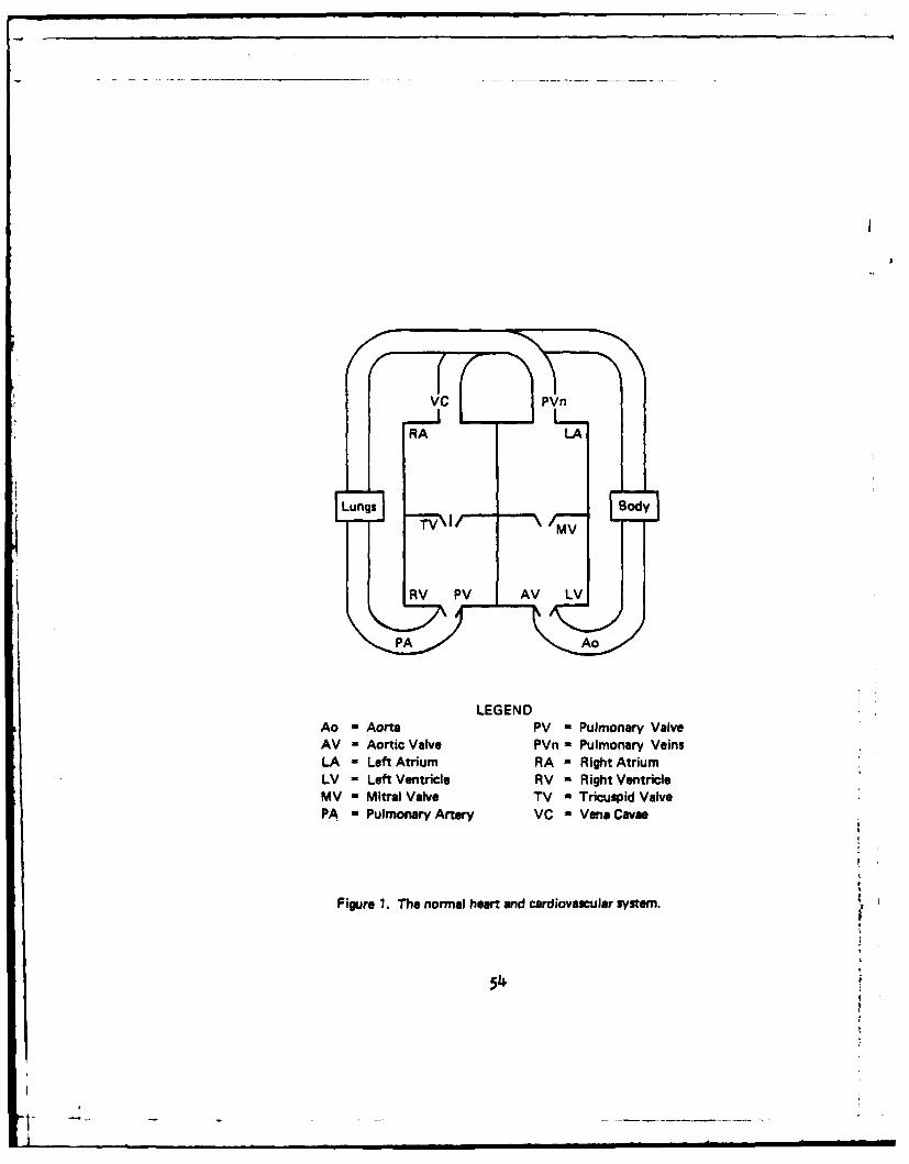
VC PVn
RA LA
Lungs BMV ody
TV MV
LEGENDAo -Aorta PV - Pulmonary ValveAV - Aortic Volvo PVn - Pulmonary VeinsLA -Left Atrium RA -Right AtriumLV - Left Ventricle RV - Right VentricleMV - Mitral Valve TV aTricuspid ValvePA -Pulmonary Artery VC aVena Cavae
Figure 1. The normal heart and card iovascular system.
514

upperachambers or the heart, the atria, are normally
separated by the atrial septum, while the Olower"
chambers, the ventricles,* are normally separated by the
ventricular septum.
Congenital heart diseases are anatomic or physio-
logic abnormalities within the heart and cardiovascular
system (e.g., holes in heart septa, tight valves, or
electrical conduction problems). These basic abnormal-
ities alter the flow, pressure, or resistance patterns
of the system and produce the patient manifestations(signs, symptoms, laboratory test results) that the
physician must utilize in diagnosis. Particular die-
eases, pertinent to cases presented in this section,
will be described under the statement of results for
each case.
55
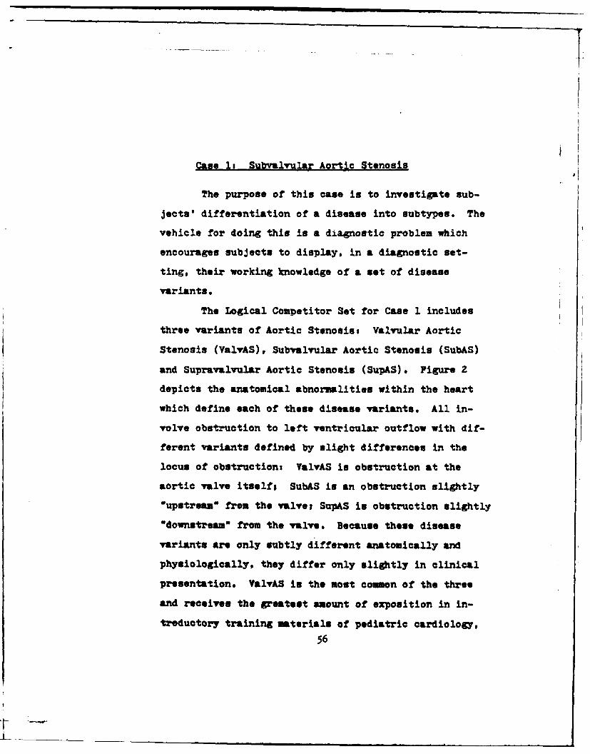
CaSe l. Subvalvular Aortic Stenosis
The purpose of this case is to investigate sub-
jects' differentiation of a disease into subtypes. The
vehicle for doing this is a diagnostic problem which
encourages subjects to display, in a diagnostic set-
ting, their working knowledge of a set of disease
variants.
The Logical Competitor Set for Case 1 includes
three variants of Aortic Stenosiss Valvular Aortic
Stenosis (YalvAS), Subvalvular Aortic Stenosis (SubAS)
and Supravalvular Aortic Stenosis (SupAS). Figure 2
depicts the anatomical abnormalities within the heart
which define each of these disease variants. All in-
volve obstruction to left ventricular outflow with dif-
ferent variants defined by slight differences in the
locus of obstruction, YalvAS is obstruction at the
aortic valve itselft SubAS is an obstruction slightly
"upstream" from the valvel SupAS is obstruction slightly
"downstream" from the valve. Because these disease
variants are only subtly different anatomically and
physiologically, they differ only slightly in clinical
presentation. ValvAS is the most common of the three
and receives the greatest amount of exposition in in-
troductory training materials of pediatric cardiology,
56
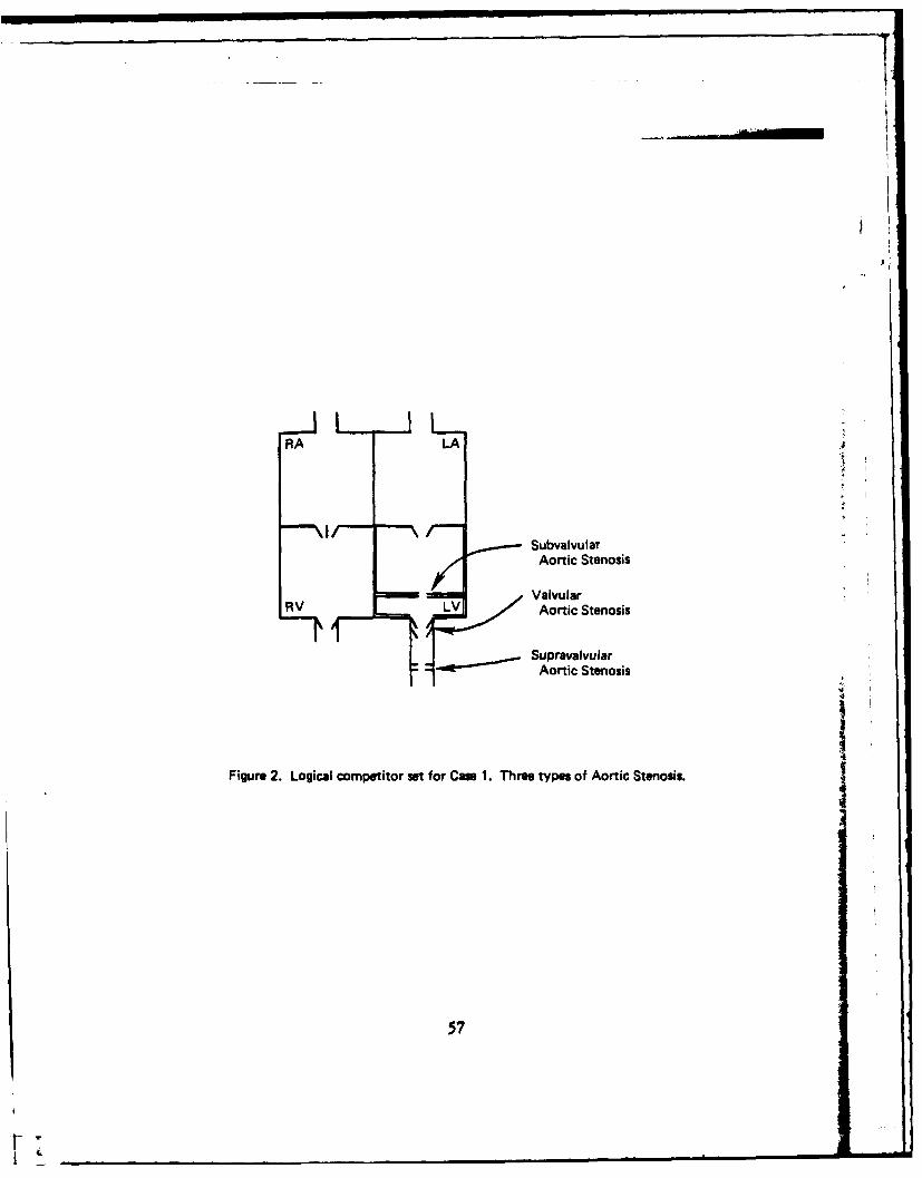
RA LA
Subvalvular
Aortic Stenosis
VavularRV LV Aortic Stenosis
SupravalvularAortic Stenosis I
Figure 2. Logical competitor set for Case 1. Three types of Aortic Stenosis.
57
tMONO

(e.g., Moller, 1978). Hence, it might be expected
that subjects' knowledge for ValvAS will develop more
rapidly than for the others and that ValvAS may function
as a "foil" for some subjects. SubAS, however, is the
operative disease in the case and the correct diagnosis.
In the "patient file" presented to subjects for
Case 1 (Appendix B, Case 1), patient data items 17 and
19, a "thrillo and a "murmur" respectively, are strong
cues for Valvular Aortic Stenosis although they are
compatible with the other variants. Hence it was sus-
pected that all subjects would raise at least ValvAS
as a hypothesis by the time of these data points.
Data item 18, a finding of "no systolic ejection
click", is very strong evidence against YalvAS. Data
items 10, "normal facies," and 22, "prominent aorta,"
are evidence against SupAS. All data of the case are
compatible with the operative disease, SubASe
Use of the Logical Comoetition Set - Case 1
Table 1 shows the variants of Aortic Stenosis
that were used as hypotheses by individual subjects8
at all patient data points where any variant was
USubjects 33 and 34 are the faculty subjects with up-wards of twenty years of experience. They are notedwith an asterisk in this and all subsequent tables.
58
__ __
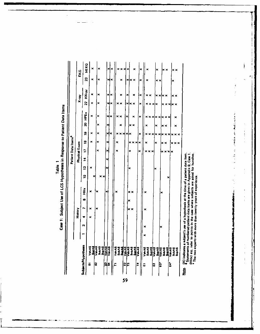
LU X X XX XX X X X X X XXX
wU
X Nxx XX XX XXX X Xxx x xx
x x xx xx x x x XXXxX
cX XI x XX X X xXx
c X X
0 X XX xxx xx
cc-
C.,L
XX
X X XX X fx.~.C~W X
E
-~ X .jC t
I.-- L*
59

_ -7
mentioned by any subject and at the four points of the
case where *hunches* were actively solicited from the
subjects (MHx, hunches after historyl HPEx, hunches
after physicals HXray, hunches after Xray: HEKG, hun-
ches after EKG). Data items from the "patient filem
(Appendix B, Case 1) are listed across the top in the
left to right order in which they were presented to sub-
Jects. An Ox" in this table simply indicates that the
subject mentioned a particular Aortic Stenosis variant
in his protocol at the data point where the "x" ap-
pears.
Instructions and guidelines for coding the var-
iants of Aortic Stenosis used by subjects in protocol
and for building Table 1 are given as Appendix F. 9
9In reading Appendix P and Appendix H, the reader willnote that in coding for Table 1 the author and alter-nate scorer coded not only for the simple mention ofAortic Stenosis variants at data points but, in ad-dition, for the *tone" with which the variant was men-tioned when it was mentioned, that is, whether it wasspoken of as a good" or "bad* explanation for thepatient's condition. ft use of these particular "tone*coding. was made in the study and the reader needonly attend to the aspects of Appendices F and H thatbear on deciding whether or not a variant was men-tioned at all These appendices are included in thereport as they are because the alternate scorer usedthem in this form in following the procedure for cod-ing (Appendix E).
60
,
II

The coding of the alternate scorer and agreement with
the author are addressed in Appendix G. An entire pro-
tocol eor one subject. T3, is given as Appendix H
along w:'th a complete discussion of the author's codine
of this subject for Table 1.
Table 1 shows an increase in the use of variants
of Aortic Stenosis, other than ValvAS, from medical
students to experts in pediatric cardiology. In par-
ticular, only one student, S2, ever raised both of the
less classic variants of Aortic Stenosis at all, during
the entire course of the case, and he mentioned SubAS
and SupAS only once each. Two trainees (T1, T3) and
three experts (El, E3, E4) used all three variants at
some time during the case. If one considers the number
of subjects in each group who not only used all three
variants, but used each more than once, no students,
one trainee (Ti) and, again, three experts meet this
criterion.
While simple mention (as reflected in Table 1)
of the Aortic Stenosis variants as hypotheses Is one
indication of whether these were oonsidered by subjects,
a measure of how actively these hypotheses were con-
sidered is the prevalence with which they were eval-
uated with respect to data items. Table 2 shows all
61
I 1

0C4 .4f + +
+ +. +'- + + 4- + '4- + + +.4
13
U,,
, + 44.0
,,6 4 + + + +.4.4.IM)
+. 0 +4 +to
4- "--
J " + 0 + +
+ +
CLL
+ +.- " I,, I, , -,J t/J 4 0I ~
)uu0 za0
+ + + + ++
V V
62E

evaluations by subjects of the Aortic Stenosis variants
with respect to the set of data items which are central
to successful solution of the case. A mark ( 0 - )
under a disease variant and data item in this table in-
dicates that the data item was judged to be positive,
negative, or ambivalent evidence for the disease variant
as a hypothesis. 10For example, a negative evaluation
of NO CLICK with respect to ValvAS would bet "The lack
of a systolic ejection click is against Valvular Aortic
Stenosis.* Instructions and guidelines for the coding
4 in this table are given as Appendix 1. The coding of
10Tere is no absolute correspondence between the use of
an hypothesis at the point of a particular data item(Table 1) and the evaluation of the hypothesis with re-spect to that data item (Table 2). Subjects can eval-uate an hypothesis with respect to a data item longpast (e~g., evaluate with respect to data it.m 10 whenhe has reached, say, data point 17 of the case) and canalso mention an hypothesis at a data point without
~1necessarily evaluating the hypothesis with respect tothat data item. Hence, for example, even though sub-ject S2 mentioned all three variants at data point 10,he only ever evaluated one of these (SupAS) with re-spect to data item 10. The mention of the other var-iants at 10 was as part of a puzzled attempt to recall
the variants of Aortic Stenosis.
63

the alternative scorer on Table 2 and agreement with
the author are addressed in Appendix ..
Table 2 shows an increase, from students to ex-
perts, in the active evaluation of data items as evi-
dence for or against the variants of aortic stenosis.
In particular, no student evaluated all three of the
variants with respect to a data Item (of course, only
one student, S29 ever mentioned all three variants at
all). The two trainees (Tl, T3), and three experts
(2l, 13. 3L4) who used all three variants in the case
also evaluated all three variants with respect to at
least one data item. While this suggests activeness
In the evaluation of variants by more experienced sub-
jects, it does not necessarily reflect comparative eval-
uation. However, when a subject evaluates all variants
with respect to the same data item, this is an indi-
cation that the subject is actively attempting to weigh
the variants against each other to determine which is
the best explanation for the data item and case. In
this regard, no students, the two trainees (Tl. T3),
and again, the three experts (El, E3, 34) evaluated all
three variants with respect to a comon (the same) data
item. These same experts, but not the trainees, eval-
uated all variants in relation to mor than one data
64

item in common (E, 5 items, E3, 2 items: E., 2 items).
The analysis thus far suggests that with in-
creasing diagnostic experience subjects know and ac-
tively utilize non-classical variants of a disease as
hypotheses in a diagnostic setting. Examination of the
two most experienced subjects. E3 and 94, yields some
clue as to the knowledge structure that supports this
performance. Figure 3 shows the protocols of these sub-
jects at two data points, 17 which is the first strong
evidence for Yalvular Aortic Stenosis and other variants,
and 18 which is the strongest evidence against ValvAS.
23 raises all three variants together (in the same
"breath" so to speak) at the time of the first strorn
evidence. These hypotheses are then available to be
evaluated comparatively against subsequent data, in par-
ticular, data item 18. This same form characterizes
subject E1 (see Table 1). Subject EL4, however, aggres-
sively focuses on the "classic" member of the competitor
set at 17, but immediately expands to the full gjt upon
receiving strong negative evidence at 18. This form Is
shared by subject T3 and, loes clearly, subject T1 (see
Table 1).
One explanation for these patterns is that in the
expert a disease and its set of subtle variations come
65
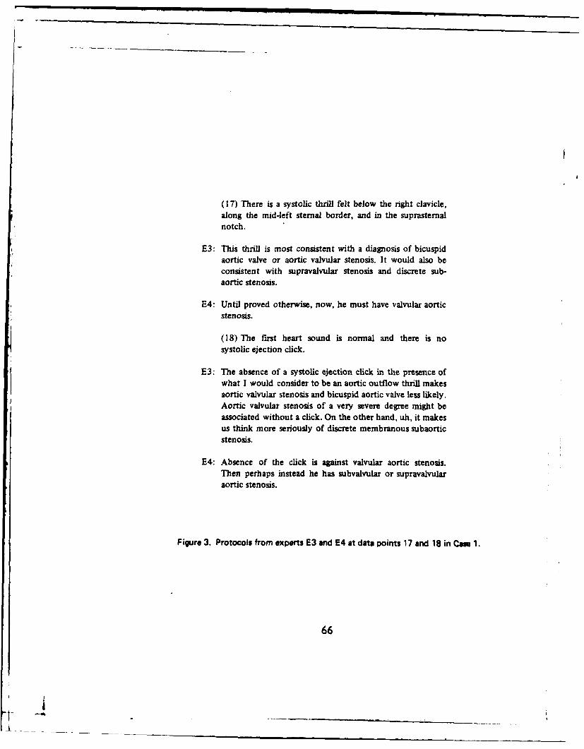
(17) There is a systolic thrill felt below the right clavicle,along the mid-left sternal border, and in the suprasternalnotch.
E3: This thrill is most consistent with a diagnosis of bicuspidaortic valve or aortic valvular stenosis. It would also beconsistent with supravalvular stenosis and discrete sub-aortic stenosis.
E4: Until proved otherwise, now, he must have valvular aorticstenosis.
(18) The first heart sound is normal and there is nosystolic ejection click.
E3: The absence of a systolic ejection click in the presence ofwhat I would consider to be an aortic outflow thrill makesaortic valvular stenosis and bicuspid aortic valve less likely.Aortic valvular stenosis of a very severe degree might beassociated without a click. On the other hand, uh, it makesus think more seriously of discrete membranous subaorticstenosis.
E4: Absence of the click is against valvular aortic stenosis.Then perhaps instead he has subvalvular or supravalvularaortic stenosis.
Figure 3. Protocols from experts E3 and E4 at data points 17 and 18 in Case 1.
66
rA
to constitute an interconnected memory unit, a kind of
categoryl when one of the members is strongly activated
in memory, the category and other members are also ac-
tivated. The expert can then choose to consider cate-
gory members in two modes. In the first mode, he tests
all members simultaneously. This first mode might be
termed "precautionary" since if any hypothesis encoun-
ters disconfirmatory evidence, alternative explanations
for which the same evidence might be compatible are
already under consideration. In the second mode, the
expert tests only the most likely (in his current judg-
ment) member. This mode might be termed one of Oex-
tractiono because its general success depends heavily
on rejection" of the target disease when appropriate
which, in turn, depends heavily on the precision in the
diagnostician's model for the disease. Once the target
disease is rejected, other category members provide a
ready "back-up" set of alternative hypotheses. Further
evidence for these speculations will be addressed asresults from other oases are presented.
Diagnostic Errors - Case 1
A final analysis of the results of this case in-
volves an attempt to discern the causes for subjects'
errors in final diagnosis. Table 3 gives the final
67
J......
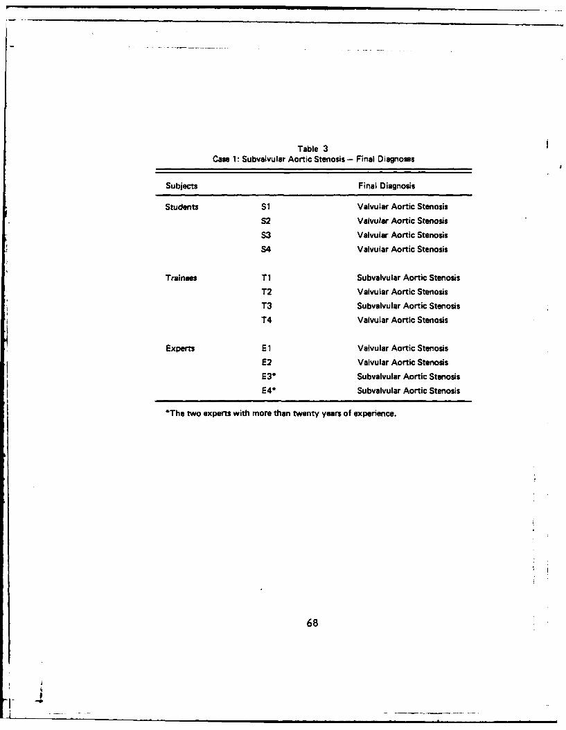
Table 3Case 1: Subvalvular Aortic Stenosis - Final Diagnoses
Subjects Final Diagnosis
Students Si Valvular Aortic Stenosis
S2 Valvular Aortic Stenosis
S3 Valvular Aortic Stenosis
S4 Valvular Aortic Stenosis
Trainees Ti Subvalvular Aortic Stenosis
T2 Valvular Aortic Stenosis
T3 Subvalvular Aortic Stenosis
T4 Valvular Aortic Stenosis
Experts El Valvular Aortic Stenosis
E2 Valvular Aortic Stenosis
E3* Subvalvular Aortic Stenosis
E4* Subvalvular Aortic Stenosis
*The two experts with more than twenty years of experience.
68
primary diagnosis for each subject. Six subjects (Si,
S3, S4, T2, T4, E2) never considered Subvalvular Aortic
Stenosis at all (see Table 1) although all generated
Valvular Aortic Stenosis. At least three explanations
could apply to this lack of activation* First, and
most basically, it could be that subjects do not know
about SubAS at all. However, post-experimental inter-
views with all these subjects confirmed that they had
some knowledge of this disease and could describe it.
A second possible explanation is that these subjects
have built up no strong "bottom-up" association in mem-
ory between any data item of the case and the subalvu-
lar disease. Even lacking such a "trigger" for SubAS
itself, it would have been possible for subjects to
generate SubAS as a side effect of their activation of
ValvAS, if these two diseases were related in a memory
unit, through a process of Ospreading activation"
(Anderson, 1976) or "top-down" activation (Rumelhart &
Ortony, 1977)1 Bobrow & Norman, 1975). This suggests
the third explanation - that for these subjects, their
knowledge representations for the variants of Aortic
8tenosis exist more in Isolation than they do in the
more experienced subjects. This is the issue of
sparseness in disease knowledge.
69
1~ - - - --..- n-- .,I.-,-------___

For those subjects who generated YalvAS as a hy-
pothesis but failed to abandon it in the face of strong
negative evidence, examination of their handling of this
disconfirmatory evidence yields Insight into the nature
and precision of their disease models for ValvAS. Dis-
cussion will focus on data item 18, the strongest evi-
dence against ValvAS.
Two students (S2, S3), evaluated 18, NO CLICK,
as confirmatory for ValvAS (Table 2). This appears to
reflect, simply, an error in important factual know-
ledge about this disease. Two subjects (S4, T2) did
not evaluate 18 at all with respect to ValvAS (Table 2).
Significantly, they also did not generate any variant of
Aortic Stenosis until after data item 18 (Table 1).
This suggests that the memory store of bottom-up asso-
ciations between data items and Aortic Stenosis variants
for these ubjects is not as extensive as for other sub-
jects and, in particular, that data item 17 is not rec-
ognized as a strong cue for Aortic Stenosis type diseases.
A further implication is that the physical examination
finding of a uystolic ejection click* and its import
in ValvAS are not represented in the ValvAS disease
models of these subjects since, if they were, the model
itself should have lead the subjects to re-examine
this finding.70
... . __ *
Finally, there were four subjects (S, T4, El,
S2) who, although evaluating 18 as negative for ValvAS,
maintained ValvAS as a final diagnosis. The protocols
of subjects S1 and T4 yield some insight into an ex-
planation for these subjects. Figure 4 shows the pro-
tocols for these two subjects at data points 18 and
22, the latter consisting primarily of the finding of
a *prominent aorta" on x-ray. Both subjects question
YalvAS at 18, but are much more satisfied with this
diagnosis at 22 and thereafter. Why might this be?
Figure 5 shows the causal relationship between
a "tight" or stenotic aortic valve and an enlarged or
prominent aorta. To open a tight valve, the left ven-
tricle (Lv) of the heart must generate abnormally high
pressure. Blood expelled under this high pressure
forces against the aortic wll and expands it. For the
two subjects under discussion, it appears that their
causal knowledge attributes the "systolic ejection
click" in YalvAS to the enlarged aorta itself# that is.,
the click is caused by the large chamber into which the
valve is opening, perhaps some kind of resonance phe- 0
nomenon. For these subjects the causal chain from the
valve to the "click" in as follows.
tight valve -+ big aorta -. click
71
1 II - - | -1 1 1- -I

(18) The first heart sound is normal and there is nosystolic ejection click.
SI: Ah, well this, the fact that there is no systolic ejectionclick present, tells us that there is probably not a poststenotic dilitation of the aorta which one would expectwith the presence of aortic stenosis and some aortic insuf-ficiency. However, this does not necessarily rule it out.
T4: Love it. Urn, well, okay. I wonder if there is.. ., no click,that's funny. I would expect if he had AS. I wish they hadsaid whether the murmur went up into his neck, Okay.
X-RAY(22) The chest X-ray shows normal cardiac size andcontour and normal vascularity, but prominence of theascending aorta.
SI: Ah, well this is what one would expect with ah, aorticstenosis with secondary aortic insufficiency. One wouldexpect that the aorta, ascending aorta distal to the ah, tothe stenosis, would be dilated due to the changes in thewall tension across the gradient. Therefore, ah, the factthat ah, a click was not heard on physical exam, mayhave been a subjective finding of the person examining.But, the X-ray does indeed suggest that there is some poststenotic dilitation.
T4: Ha ha! AS-Al.
Figure 4. Protocols from subjects S1 and T4 at data points 18 and 22 in Case 1.
72

Aorta enlargedfrom blood force /Normal
Aortac
Valve
LV
Figure 5. Aorta enlarged from the force of blood ejecting from a stenotic aorticValve.j
73

Hence, for these subjects, the Obig aorta" itself
is predominant over the *click* as evidence for ValvAS,
with the click just additional evidence for a big aorta.
Once they receive their best evidence for a "big aortam,
22, they are no longer worried about the lack of a
"click".
The true state c affairs appears to be that
tight valve causes both the "clicko and the enlarged
aorta at the same level of cause (Friedman & Kirk-
patrick, 1977, p. 180). The systolic ejection click is
associated with the opening of the tight valve itself
as shown below,
tight valve rclickEbig aorta
Hence, both of these effects must be proved. Why might
a number of subjects have misconstrued this relation-
ship? One need look no further than the introductory
textbook these subjects use (Moler, 1978, p. 96)
where the erroneous causal relationship is stated, or
at least strongly implied.
The subjects just discussed raise two important
issues. First, they demonstrate how Osmall knowledge
errors can have major repercussions for the handling of
74
a case and shed some insight into the case -specificity
of a clinician's diagnostic performance found elsewhere
(e.g., Blstein et. al., 1978). Secondly, they suggest
a sensitivity in less experienced clinicians to specifictraining experiences, e.g,, training materials, particu-
lar patient cases, etc. An experience increases, so
does the sample of "inputs" and the effects of particu-
lar experience might be expected to lessen.
75

Case 21 Total Anomalous Pulmonary Venous Connection
The purpose of this case is to investigate the
aggregation by subjects of a set of physiologically sim-
ilar diseases into a memory grouping or category. The
case is different from Case 1 in that while Case 1
dealt with a set of variants of one disease. Case 2 is
concerned with a set of diseases.
The Logical Competitor Set for Case 2 includes
four diseases: Total Anomalous Pulmonary Venous Con-
nection (TAPVC), Partial Anomalous Pulmonary Venous
Connection (PAPVC), Atrial Septal Defect (ASD), and
Endocardial Cushion Defect (ECD). 11Figure 6 shows the
anatomical and physiologic abnormalities within the
heart which define each of these diseases.
In TAPVC, all four pulmonary veins (PYn in
Figure 6) connect to the right atrium (RA) of the
heart rather than to the left atrium (LA). their nor-
mal site of connection. All oxygenated blood coming
back to the heart from the lungs mixes with deoxygenated
blood coming back to the heart from the "body". Hence,
11All of these diseases have variants as in Case 1.
Variants of these diseases were treated as equivalentfor Case 2 since interest in this case was in memoryclustering at the disease level.
76
V | I | I-

VC Pn
NA RAS LAA
Lungsod/-TV-
RV PV LV
PVn VCWTAPVn
RA ASO LA
'TVD
NV PV LV vIFiue8P oia optio e o ae2 APCnAVA!,EO
P~77
__ IS

all blood subsequently pumped back to the body is a
mixture of oxygenated and deoxygenated blood which
causes the patient to appear cyanotic, that is# to
take on a mildly Oblueo skin coloration.
In PAPYC, only a subset of the pulmonary veins
connect abnormally to the right atrium, with the re-
mainder connecting, as they should, to the left atrium.
A result is that some already oxygenated blood is re-
circulated' through the lungs. Blood pumped to the
body, however, is oxygenated and the patient retains
his normal Opink" coloration.
Both ASD and ECD consist of a defect (a hole)
in the atrial septum of the heart. They differ in the
particular site of defecti ASD is a defect in the "upper'
portion of the septum (the ostium secundum) while ECD
is a defect in the lower portion of the septum (the
ostium primum). In both diseases, the presence of the
OholeO in the septum allows blood to shunt from the left
atrium to the right atrium. While some oxygenated blood
shunts to the right side to be recirculated to the lungs,
blood expelled to the body is oxygenated and the patient
is pink. Both TAPVC and PAPVC may also contain a hole in
the atrial septuml this defect is necessary in TAPYC
and optional in PAPVC.
78

A feature common to all four diseases in the LCS
is an increased volume of blood in the right-sided
chambers of the heart. This common element is a can-
didate feature on which diagnosticians might base a
disease category, e.g., "diseases with right-sided
volume overload". A clinical manifestation related to
volume overload that all these diseases produce in
common is a set of three auscultation findings. One is
a murmur associated with increased blood flow across
the tricuspid valve (TV). The second is a murmur as-
sociated with increased flow across the pulmonary
valve (PV). The third is wide, fixed splitting of the
second heart sound. The third finding is nearly path^
ognomonic for conditions of this type.
Of the four diseases, ASD is more com-
mon than the others. Hence, it might be expected that
subjects' knowledge for this disease would develop more
rapidly than for the others. More importantly, ASD is
the disease that is used instructionally to introduce
the concepts of atrial level left-to-right shunting of
blood in the heart and right-sided volume overload.
Therefore, it might be expected that the three aus-
cultation findings (especially the "splittingo) reflect-
ing overload would be more strongly associated with ASD
than with the other diseases. TAPVC, however is the
79
operative disease in the case.
There are six particularly important data items
in the apatient fileO presented to subjects for Case 2
(Appendix B. Case 2). Data items 17. 18, and 19 con-
tain the set of three findings discussed above which
are salient results of increased right-sided heart flow.
Item 17 contains the *wide, fixed split second heart
sound*. Hence, it was expected that all subjects would
raise at least ASD, the classic instance of this type
of disease, oy the time of these data points. Data
item 7 (also 11), which reports that the patient is
cyanotic, represents disconfirmatory evidence for all
members of the LCS except TAPYC. Data item 21. which
contains an x-ray description of "an unusual vascular
shadow on the right side" is evidence against ASD and
simultaneously constitutes a classic cue for PAPVC. In
fact, one variant of PAPVC, "scimitar syndrome', de-
rives its name from its presentation of such a finding
on x-ray (Lucas & Schmidt, 1977, pp. 442). The EKG.
item 22, contains a finding of "right axis deviation"
on the EKG and constitutes strong disconfirmatory evi-
dence for BCD. All data of the case are compatible
with the operative disease, TAPVC.
80
A• - w n

Use of the Logical Cometitor Set - Case 2
Table 4 shows all uses by all subjects of the
four diseases in the Logical Competitor Set for Case 2
at all patient data points (Appendix B, Case 2) where
any of the four was mentioned by any subject. The
nature of this table is the same as Table 1 in that
an ox" intersecting a disease and data item indicates
that the subject mentioned the disease in his protocol
at the time of the data item.
For reasons discussed above, it was assumed
that most subjects would consider ASD in relation to
the three data items, 17. 18, and 19. The use of other
LCS members at these points is taken as evidence that
the other diseases are associated in memory with ASD
and this set of cues. Table 4 shows a decrease from
students to experts, in the number of subjects who con-
sidered only ASD at these points. All of the students
considered only ASD, the disease we presume to be the
classic exemplar of right sided volume overload, at
data items 1?-19. Three of four trainees (Ti, T2, T3)
also considered only ASD while the fourth considered
both ASD and TAPVC. The two least experienced experts
also considered only ASD. Of the two highly experienced
81
-- -

Table 4Case 2: Subject Use of LCS Hypotheses in Response to Patient Data Items
Patient Data Itemsa
History Physical Exam X-ray EKG
Subiect/HYPothem 1 3 5 7 HHx 17 18 19 20 HPEx 21 HXray 22 HEKG
S1 ASO X X XPAPVC X X X X
S2 ASO X X XECD X X X
PAPVC XS3 ASO X X X X
ECD X XPAPVC X X
S4 ASD X X X X XECD X
TAPVC X
T1 ASO X XPAPVC X X X X
T2 ASO XECO X X
TAPVC X X XT3 ASO X X X X X X X X
PAPVC XTAPVC X X
T4 ASO X X X XECD X X X X X
PAPVC XTAPVC X X X
El ASO X X X X X X X XPAPVC X X X
E2 ASD X X XPAPVC X XTAPVC X
E3* ASO X X X XECD X X X X
PAPVC X XTAPVC X X X X X X X X X
E4° ASO X X XECD X X
PAPVC XTAPVC X X
Notea. IX) indicates a subject's use of a hypothesis at the time of a patient data item.Statements of numbered patient data items are given in Appendix B, Caw 2.
(HHx) etc. refers to points in the case where subjects ar asked for hunches.The two experts with more than twenty years of experience.
82
4-
experts. 23 utilized three LCS members (ASD, PAPVC,
TAPVC) and 34 used two (ASD, ECD) at these points.
From the point of view of the entire case, no
students, one trainee (T.), and two experts (E3, E4)
generated all four members of the LCS during the course
of the case. While this shows no obvious general trend
toward incrotased use of the LCS with experience, it is
perhaps significant that the full competitor set was
used by the two high-level experts, E3 and F.
In utilizing the full Logical Competitor Set,
the two most experienced subjects, 33 and E4, demonstra-
ted the same patterns of "precautionO and wextractionw
respectively as they did in Case 1. 33 considered three
of the four LOS members (ASD, PAPVC, TAPVC) at item 1?,
the first strong cue for right sided volume overload.
34 raised only ASD at this point and maintained this
hypothesis until data item 21 which contains strong evi-
dence against ASD. At this point, he expanded to the
remainder of the LCS.
Diagnostic Errors - Case 2
Table 5 gives the final primary diagnoses for all
subjects on Case 2. Only four subjects, trainees T2 and
T3, and the two most experienced experts, E3 and EL,
e3
Table 5Case 2: Total Anomalous Pulmonary Venous Connection - Final Diagnoses
Subjects Diagnosis
Students S1 Partial Anomalous Pulmonary Venous ConnectionS2 Transposition of the Great Vessels
+ Pulmonary Stenosis+ Atrial Septal Defect+ Partial Anomalous Pulmonary Venous Connection
S3 Endocardial Cushion DefectS4 Pulmonary Stenosis
+ Atrial Septal Defect+ Ventricular Septal Defect
Trainees T1 Partial Anomalous Pulmonary Venous ConnectionT2 Total Anomalous Pulmonary Venous ConnectionT3 Total Anomalous Pulmonary Venous ConnectionT4 Atrial Septal Defect
Experts El Partial Anomalous Pulmonary Venous ConnectionE2 Partial Anomalous Pulmonary Venous ConnectionE3* Total Anomalous Pulmonary Venous ConnectionE4* Total Anomalous Pulmonary Venous Connection
The two experts with nore than twenty years of experience.
84
diagnosed the case correctly. Two of these four sub-
jects are ones who considered the full LCS in the case.
Subjects who diagnosed the case incorrectly demonstrate
informative types of errors.
Student S3 diagnosed the case as Endocardial
Cushion Defect (BCD). The strongest evidence against
this disease is the finding of aright axis deviationo
on the EKG (item 22). BCD uniformly presents with left
axis deviation and, in fact, is one of a very few con-
genital heart diseases that doesi hence, left axis de-
viation is a nearly pathognomonic finding for BCD.
S3 not only evaluated the Oright axis" as positive evi-
dence for BCD, but, in addition "triggered* or proposed
BCD for the first time at this point (see Table 4).
This is, simply, Imprecision in the subject's disease
model for BCD. It is as though the subject remembered
that the EKG axis is important in BCD but could not re-
member the details.
The final diagnosis of subject T4 was ASD, even4
though she had considered TAPYC during the case. She
correctly evaluated cyanosis (blueness - items 7 and 11)
as negative for ASD, but maintained ASD nonetheless.
Her primary difficulty was that she did not believe
85
that TAPVC could present in a child as old as the one
in the case (5 years old) although it certainly can -
as the case itself, a real case, attests. This suggests
that the allowable age range specified in the subject's
disease model for TAPVC is overly restrictive, prob-
ably reflecting a limited sample of experiences with
this disease.
Four subjects (Si, TI, El, E2) diagnosed the case
as PAPVC. Three of these subjects (S1, Ti, El) show a
pattern in which only ASD (among the LCS members) is
considered prior to item 21, a classic x-ray cue for
PAPVC, and only PAPYC is considered at that point and
thereafter (see Table 4). This indicates a strong data-
driven dependence in the diagnosis by these subjects;
that is, the subjects are pushed from hypothesis to hy-
pothesis depending on the most recent strong disease
cue in the data, and when new hypotheses are generated,
these are not strongly enough associated in memory with
other LCS members to activate these other diseases.
Some support for this claim can be seen in subject Ti's
protocol taken from the point in the case where he of-
fers his final diagnosis,
86
MEN
Tl, I an sort of drawing a blank onhow to fit all this information to-gether. And ah, I am just sort ofguessing right now. I would sayjust Scimitar Syndrome (PAPVC) pri-marily based on the chest x-ray andah. I'm not really sure whether thewhole thing fits together well.That is all I can say.
Of the four subjects, student Si never evaluated PAPVC
with respect to cyanosis; hence, this finding had no
opportunity to detract from his PAPYC hypothesis. Sub-
ject T1 evaluated cyanosis as confirmatory evidence for
PAPVC and this erroneous evaluation reinforced this dis-
ease interpretation. Expert subjects El and E2 eval-
uated cyanosis appropriately as negative evidence for
PAPVC, but this evaluation was probably overridden by
the strength of the cue for PAPVC on the x-ray.
Finally, two students (S2, S4) proposed config-
urations of multiple diseases as explanation for the
case. Both of these composite diagnoses included the1
disease Pulmonary Stenosis (PS) and it is this compon-
ent of the final diagnosis that is the key to under-
standing the performance of these two subjects. Table 6
shows the interpretations by all subjects of data item
18, a 0systolic murmur0 in auscultation of the heart.
Such a murmur results whenever there is too much flow
87
I-k
Table 6Case 2: Interpretations of Data Item 18
Interpretation
Increased FlowSubject Pulmonary Stenosis Pulmonary Valve
Students S1 +S2 +
S3 + +S4 +
Trainees TIT2 +
T3 + +T4 +
Experts El + +
E2E3* +
E4 + +
Note. (+) indicates that a subject interpreted the murmur of data item 18as Pulmonary Stenosis or increased flow over the pulmonary valve.
The two experts with more than twenty years of experience.
88
p- -

over the pulmonary valve, relative to its orifice size.
This situation prevails In either of two conditions#a
(1) When there is normal amount of flow but an abnormal-
ly sma1l orifice. This is the disease Pulmonary Stenosis
which refers to an abnormally tight valve. (2) When
there is a normal sized orifice but abnormally high
flow, the situation which prevails in the diseases of
the LCS. Am+" under one of these two interpretations
in Table 6 indicates that a subject attributed this in-
terpretation to the murmur of data item 18.
Table 6 shows that most of the students (3 of 4)
interpreted the murmur only as Pulmonary Stenosis while
most of the expert group (3 of 4) interpreted the murmur
as increased flow or a tight valve. While student S1.
(also subject T2) was eventually able to extract him-
self from his Interpretation, students S2 and S4i were
not. Once these students introduced PS into their di-
agnoses, they were forced to propose rather unusual
combinations of multiple diseases to account for some
of the findings of the case. For example, subject S2,
in order to reconcile PS with other data of the case
indicating Increased blood flow in the lungs, simply
transposed the great vessels of the hearts that is, hei
89

detached the pulmonary artery from its normal mooring at
the pulmonary valve, and reattached it at the aortic valve
and did the opposite with the other great vessel, the
aorta. While this transposition is a congenital heart
disease, Transposition of the Great Vessels, it is high-
ly unlikely that a child with the combination of abnor-
malities proposed by the subject could have lived for
five years untreated.
The interpretations of the systolic murmur by the
students in Case 2 is another example of error, or at
least imitation, in causal knowledge. It represents a
situation where there are multiple causes for a finding
and the novice considers only a subset. This is not
unlike what has been shown at the disease and disease
variant levels: that is, when multiple diseases in the
Logical Competitor Set can produce a finding, the nov-
ice seems limited to the most salient members. This sug
gests the import of grouped or clustered memory organ-
ization not only for diseases, but also for "low-level,
pathophysiologic interpretations for data.
90

AD-A1bS 753 PITTSBURG U jNIV PA LEARNING RESEARCH AND OEVELOP TEN-EC 7/ i
KNOWLEDGE BASED COMPONENTS OF EXPERTISE IN MEDIA NAGNOSIS.0 U/SEP l P J1 FELTOVICH NOGW79CAL I SS.UNCLASSIFIED LROC-SI/PDS-2
NL2*3fflfll**.111E2hmhh3hh
MENEMhhmonsoolmhhhhmhhmmhhhu0mhhhhhhhhhhhhlmhhhhhhhhmhhlo

Case 3s Patent Ductus Arteriosus
The purpose of this case is to test the robust-
ness of expert grouping of hypotheses in a straight-
forward case in which there are no data discrepant
with an initially induced disease interpretation.
Interest is in whether subjects, even in a case with a
highly common disease, strong cues for this disease,
and no data discrepant with this interpretation, still
investigate a related set of physiologically similar
alternatives.
The operative disease in the case is Patent Duct-
us Arteriosus (PDA), a schematic for which is shown in
Figure 7. This disease is an extra-cardiac shunt,
that is, an abnormal communication between vessels,
the aorta (Ao) and pulmonary artery (PA), outside the
heart. There are four other "disease' conditions in
the Logical Competitor Set. Congenital heart dis-
eases, Arterio-venous Fistula (AYF) and Aorto-
Pulmonary Window (APW) are other extra-cardiac shunts.
Venous Hum (VH) is a benign condition that presents a
murmur similar to PDA, and Ruptured Sinus of Valsalva
(RSV) is a heart condition that has a similar clLn-
ical presentation to PDA.
91
VC PVn
RA LA
RV LV
PA Ao
Figure 7. Patent Ductus Arteriosus.
92

In the patient file presented to subjects for
Case 3 (Appendix B. Case 3), the most important patient
data item is number 19. a classic murmur of Patent
Ductus Arteriosus. It was assumed that all subjects
would generate PDA as a hypothesis no later than this
point. No data of the case are incompatible with PDA.
Use of the Logical Copetitor Set - Case I
Table 7 shows all uses of members of the Logi-
cal Competitor Set by all subjects during the course
of the case. (It is like Table 1 only the diseases
recorded and the referents for the "patient file"
data items listed across the top are changed). It is
clear that only one subject, 33, one of the two high-
level experts, considered the full competitor set,
although expert E2 considered three of the five -
more than any of the remaining subjects.
If one inspects the subjects who used even one
additional LCS member other than PDA, it is found that
three students (Si, S3, S), one trainee, Ti, and
three experts (El, 32, 33) meet this criterion. Most
student uses of a second LCS member were isolates,
so that if one requires that the additional member be
used more than once, this condition holds for only one
student, S3, one trainee, Ti, but still three of the
93

Table 7Case 3: Subject Use of LCS Hypotheses in Response to Patient Data Items
Patient Data Itemnsa
History Physical Exam X-ray EKG
Subjects/Hypotheses 3 4 5 7 HHx 14 19 20 HPEx 21 HXray 22 HEKG
Si PDA X X X X X X XAPW X
S2 PDA X X X X X X XS3 PDA X XX X X X X X X X
AVF X XS4 PDA X X X X X X X X
AVF X
Ti PDA X X XX X X X XAPW X X X X X
T2 PDA X X X X X XT3 PDA X XX X X X XT4 PDA X X X XX X X X X
El PDA X X X X X X
E2 PDA X X X X X X XAVF X X
VH X XE3* PDA X X X X
AVF XVH X
APW X X XRSV X
E4* PDA X X X X X
Note. IX) indicates a subject's use of a hypothesis at the time of a patient data item.a Statements of numbered patient data items are given In Appendix B, Can 3.(HHx) etc. refer to points in the came where subjects are asked for hunches.*The two experts with more than twenty years of experience.
94~

exPerts (El, E2, E3).
Expert E3 considered the full LCS in a "pre-
cautionary" pattern consistent with his performance on
other cases (see Table 7). He used three of the five
LCS members as hypotheses at data item 19, a strong cue
for PDA. The remainder of the LCS was filled out two
items later, after an intervening, uninformative data
item, at the point where the subject was asked for
Ohunches", The other high-level expert, E4, looks in
all respects like a novice in this case in that he con- Isidered only PDA. However, if our earlier interpreta-
tions of an Oextractiono method are correct for this
subject, we would not expect him to expand to other mem-
bers of the competitor set unless he encountered data
discreoant with his target hypothesisl of course, there
are none in this case.
The diseases in this case constitute a category
of "Extra-Cardiac Communicationso and related conditions.
An interpretation of the results of this case is that
with high-level experience, it is this category, and not
isolated individual members, that is generated and
tested when a strong cue for a category member is en-
countered. No subject diagnosed this case incorrectly,
hence analysis of subject errors is uninformative.
95
I
Case 41 Pulmonary Atresia
The objective of this case is similar to that of
Case 2# that is, to assess subjects' aggregation of
physiologically similar diseases into categories.
Case 4 is different from Case 2 in that, unlike Case 2,
no single cue serves to distinguish the members of the
Logical Competitor Set from diseases outside it (as
did *wide. fixed split second heart soundo in Case 2).
In Case 4 the diagnostician must arrive at the LCS by
partitioning the space of diseases, using multiple
data items from widely separated parts of the case.
The Logical Competitor Set for Case 4 includes
three diseases, Pulmonary Atresia (PAT), Tricuspid
Atresia (TAT), and Ebstein's Malformation of the Tri-12cuspid Valve (EBST). Figure 8 depicts the anatom-
ical abnormalities within the heart which define each
of these diseases., In Pulmonary Atresia and Tricuspid
Atresia, the pulmonary and tricuspid valves respective-
ly are "shut" (only tissue exists where the valves
should be), In Ibstein's disease, a diminutive and
12The abbreviations PAT and TAT will be used instead of
the more conventional abbreviations PA and TA in orderto avoid confusion between "Pulmonary Atresia" andOpulmonary artery" which we have abbreviated PA.
96
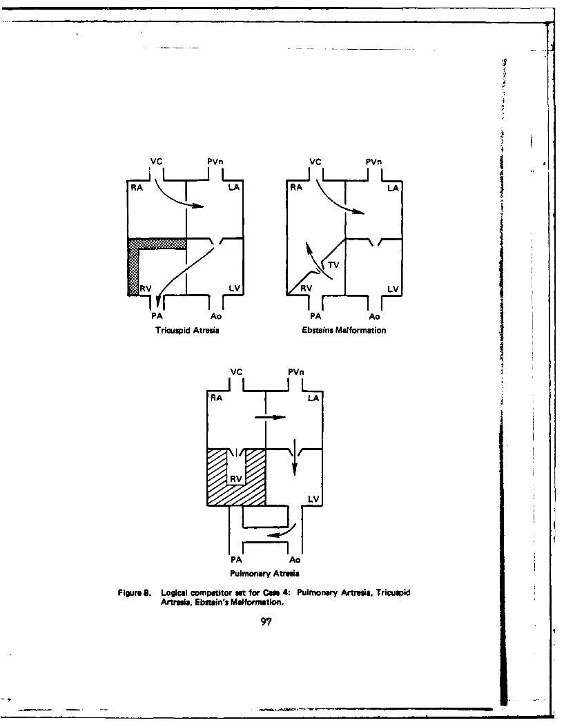
E
VC PVn VC PVn
RA LA RA LA- I -
x(TV
R LV RV LV
PA Ao PA Ao
Tricuspid Atresia Ebsteins Malformation
VC PVn
RALA
RV
LV
Im IPA Ao
Pulmonary Atrusia
Figure 8. Logical competitor st for Cam 4: Pulmonary Artresla, TricuspidArtrasla, Ebstein's Malformation.
97
non-compliant right ventricle (RV) restricts inflow
of blood to that ventricle and an incompetent tri-
cuspid valve (TV) allows some regurgitation of blood
from the right ventricle back to the right atrium(RA).
The net physiology of all these diseases is one of
obstruction to blood flow on the right side of the
heart resulting in reduced blood flow to the lungs
and right-to-left shunting of blood at the atrial
level within the heart. The right-to-left shunting
and diminished blood flow to the lungs cause the
patient to be cyanotic (blue skin coloration). In
short, these diseases constitute a physiologic cate-
gory of "Ocyanotic diseases with decreased pulmonary
blood flow."
Pulmonary Atresia is the operative (or true)
disease in the case. The three members of the LCS
are best discriminated on the EKG. Tricuspid Atresia
produces a finding of "left axis deviation" on the EKG
while Pulmonary Atresia produces a normal EKO axis.
Ebstein's, unlike the other two, produces an EKG
finding of "right bundle branch blocking." All other
clinical manifestations of the three diseases are
quite similar.
98
A'. No -ll
There are several key data items in the "patient
file" presented to subjects for Case 4 (Appendix B,
Case 4). The subject receives evidence of cyanosis
during history and early physical examination (1, 3,
8). The x-ray, item 17, contains evidence of dimin-
ished blood flow to the lungs and, with the cyanosis
evidence, could enable the subject to narrow diag-
nosis to the three members of the LCS. The EKG, item
18, contains information to discriminate among these.
Use of the Logical Competitor Set - Case 4
Table 8 shows all uses of members of the Logi-
cal Competitor Set by all subjects during the course
of the case. (It is like Table 1i only the diseases
recorded and the referents for the "patient file"
data items listed across the top are changed,)
Table 8 shows a clear increase in the use of the
full Logical Competitor Set from students to train-
ees, but no clear difference in this regard between
trainees and experts* in particular, no student con-
sidered the full LCS, and two students (SI, S3) con-
sidered only one member. All four trainees and three
experts (Zl, Z2, 33) used all of the diseases in the
LCS. Two experts (2, E3) used all three diseases
more than once while no trainee did - suggesting
99
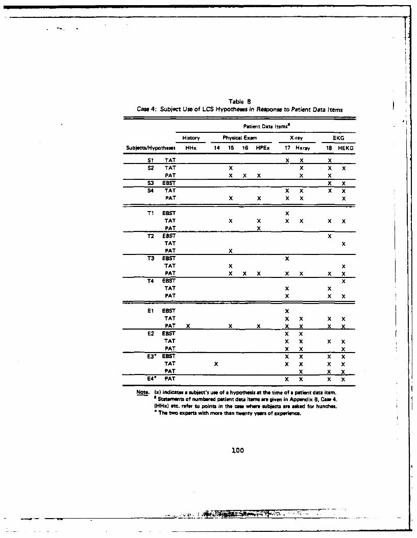
Table 8Case 4: Subject Use of LCS Hypotheses in Response to Patient Data items
Patient Data Itemns'
History Physical Exam X-ray EKG
Subjects/Hypothesas HHx 14 15 16 HPEx 17 Hxray 18 HEKG
S1 TAT X X XS2 TAT X X X X
PAT X X X X XS3 EBST X X54 TAT X X X X
PAT X X X X X
Ti EBST XTAT X X X X X XPAT X
T2 EBST XTAT XPAT X
T3 EBST XTAT X XPAT X X X X X X X
T4 EBST XTAT X XPAT X X X
El EBST XTAT X X X XPAT X X X X X X X
E2 ESST X XTAT X X X XPAT X X X
E3* EBST x x X XTAT X X X X XPAT X X X
E4* PAT X X X X
Note. lx) indicates a subject's use of a hypothesis at the time of a patient data item.a Statements of numbered patient data Items are given in Appendix B, Casn 4.lM4x) etc. refer to points in the case where subjects are alted for hunches.The two experts with more than twenty years of experience.
100

somewhat more active consideration of the Logical Com-
petitor Set by these experts.
While both trainees and experts considered the
full Logical Competitor Set, their patterns of use of
these diseases were different. Three of the four ex-
perts used all members of the LCS at data point 17
(the x-ray) or at the immediately succeeding point where
subjects reported hunches. Since item 17 is the data
item that allows specification of the category t cyan-
otic heart disease into the category Ocyanotic dis-
eases with decreased pulmonary blood flow," this pat-
tern suggests that the expert subjects were using this
category. In contrast, ng trainees used all three
LCS members at either of these points, suggesting that
these three diseases do not, at least to the same ex-
tent. constitute a functional diagnostic category for
these subjects. 4Regarding the expert diagnostic modes of Opre-
!caution" and *extraction," expert E3 again considered
all three LCS members together before the onset of data
useful for discriminating among them. Expert E4 con-
sidered explicitly only Pulmonary Atresia, the correct
disease, at data item 17e However, his protocol from
the immediately succeeding data point, HXray (hunches
101 I

after x-ray), shows explicit consideration of the
category of "cyanotic disease with decreased pul-
monary blood flow" with targeting for active con-
sideration of the particular LCS member he judged
most likely,
Experimenters At the end of x-ray canyou tell me about hunches?
ELI At this point the picture would bemore likely that of cyanotic heartdisease involving decreased pul-monary blood flow. The specificdefect would seem to be pulmonaryatresia with intact septum.
Since no succeeding data are discrepant with his
target hypothesis, his performance is consistent
with the "extraction" mode as we have proposed it.
In addition, 34's overt consideration of the LCS
category here lends credence to a speculation we have
made about the "extraction" mode in Case 1 and Case 2,
that is, that the subject covertly considered the LCS
category in those oases before he overtly articulated
the members.
Diaanootic Errors - Case 4
Table 9 gives the final primary diagnoses for
all subjects. The final diagnoses of the students on
this case are outside the LCS and the full explanation
for their performance is not transparent. However, a
102
v--. .-- -

Table 9Case 4: Pulmonary Atresia - Final Diagnoses
Subjects Diagnosis
Students Si Truncus ArteriosusS2 Hypoplastic Right VentricleS3 Truncus ArteriosusS4 Pulmonary Atresia
Trainees Ti Tricuspid AtresiaT2 Tricuspid AtresiaT3 Tricuspid AtresiaT4 Pulmonary Atresia
Experts El Pulmonary AtresiaE2 Pulmonary AtresiaE3* Ebstein's Malformation
4E4* Pulmonary Atresia
*The two experts with more than twenty years of experience.
103
partial explanation can be given.
Two students (Si, S3) gave a final diagnosis of
Truncus Arteriosus. Truncus is a congenital heart dis-
ease in which the aorta and pulmonary artery, the two
great vessels which normally lead out of the heart, are
merged into one large outlet vessel. This single ves-
sel is associated with only one outlet valve from the
heart. The single valve results in a patient finding
of Osingle second heart sound" on auscultation since
these "heart soundsO are produced by the closing of
heart valvest in a normal heart there are two compon-
ents to the second heart sound.
While Truncus produces a single heart sound, so
do a number of other diseases, including all members
of the Logical Competitor Set. It is not even neces-
sary that only one valve exist for only a Osingle
sound" to be produced: the same finding is produced
when there are two outlet valves but the blood flow
across one of them is substantially diminished -
the situation in Ebstein's and Tricuspid Atresia.
One explanation for the performance of students
Si and S3 is that they judged the "single sound" to be
more discriminating for Truncus than it really is.
Some evidence for this interpretation can be seen in
104
V' . . . ..
protocols from these two subjects showing interpreta-
tions of the patient finding of a "single sound"
(Figure 9). It is clear that this finding had a sub-
stantial influence on the final diagnoses of these
subjects.
If our interpretation for these subjects is
true, it would be another example of how the beginning
practitioner is restricted in the number of alternative
explanations he can bring to bear on a finding, either
at the level of alternative pathophysiological causes
or alternative disease explanations. Whether "single
second sound" is a more "classic" cue for Truncus than
for other diseases is open to question. However, it
would seem reasonable that the "common trunk" which
defines this disease would serve to accentuate the
single sound as an expected finding in the disease.
S2, the other student who misdiagnosed Case 4,
gave as a final diagnosis (Hypoplastic Right Ventricle)
one of the patient findings presented in the case(the
EKG)i that is, the subject used one of the patient
data items as a final diagnosis. An explanation for
this student's performance will not be ventured.
The trainees and experts are nicely split on
this case, with most trainees (3 of 4) Judging Tri-
-cuspid Atresia and most experts (3 of 4) judging
105

(IS) The second heart sound is single and perhaps slightlyincreased in intensity. There is no gallop or diastolicmurmur.
SI: Well, this is a significant finding because ah, the fact thatt- second heart sound is not split ah, suggests that ah, we
uld be dealing with a truncus.
S3: It could be ah, ah. There is a single outflow tract ah. Itcould be truncus arteriosus. Ah, that would fit with thesingle S2 (second heart sound)... So, Ill go with numberone on my list as ah, truncus arteriosus and I'm not surewhat type. I'd have to do an angio. I guess, or I meanarteriography.
Figure 9. Protocols from subjects S1 and S3 showing interpretations of "singlesecond heart sound" - Cas 4.
106

Pulmonary Atresia, the correct disease. Recall that
TAT and PAT are distinguishable on the axis of the
EKG where TAT presents =left axis deviation" and PAT
presents a normal, undeviated axis. It is on the
subjects' evaluations of this particular data item
that we might expect to find an explanation for the
performance of these two groups.
Table 10 shows all explicit evaluations by sub- ijects of the EKG axis as confirmatory (+), discom-
firmatory(-),or ambivalent(o)evidence with respect to
Pulmonary Atresia and Tricuspid Atresia. All subjects
below the expert level, who explicitly evaluated the
axis with respect to either of these two diseases,
evaluated the axis as confirmatory evidence for Tri-
cuspid Atresia. Subject Ti proceeded to conclude TAT
as a final diagnosis. While subjects S4 and T4 ulti-
mately chose Pulmonary Atresia over Tricuspid Atresia
on other grounds, their explicit evaluations of the
axis as confirmatory for TAT contribute to an ex-
planation of what happened to other non-expert sub-
jects who maintained Tricuspid Atresia. In particu-
lar, even though T2 did not overtly evaluate the EKG
axis with respect to Tricuspid Atresia, imediately
after the EKG she generated TAT for the first time in
10?
1!.

Table 10Case 4: Evaluations of EKG Axis in Relation to Tricuspid Atresia
and Pulmonary Atresia
Hypotheses
Subjects Tricuspid Atreia Pulmonary Atresia
Students SiS2S3S4 +
Trainees T1 +T2T3T4 +
Experts El 0 +E2E3°
E4o
Note. (+, -, 0) indicate that the subject evaluated the EKG axis as con-firmatory, disconfirmatory, or ambivalent evidence respectivelyin relation to the hypothsis
The two experts with more than twenty years of experience.
1
108
-. .--- -

the case as her only "hunch". Likewise, subject T3
raised TAT at this same point as a "hunch" after not
having used this hypothesis since early physical exam-
ination (see Table 8). All expert subjects who ex,
plicitly evaluated the axis, evaluated it as either
disconfirmatory for Tricuspid Atresia or confirmatory
for Pulmonary Atresia.
The EKG axis as presented in the case is +50
degrees which technically represents left axis dev-
iation (for a 4 day old child, as presented in the
case - Moller 1978, p. 24) as one would expect in Tri-
cuspid Atresia, so that if one were using the "text-
book" rule for discriminating PAT and TAT (e.g., Mol-
ler, 1978, p. 137), Tricuspid Atresia would be the
diagnosis of choice in the case. However, the expert
evaluations of this finding, as well as post-experi-
mental discussions with these subjects, confirmed that
the experts judged +50 degrees to be "just not farI
enough leftward* for Tricuspid Atresia and that these
subjects would require the axis to be "down around zero
or negatives before they would choose TAT over PAT. We
see here a nice example of overly general, textbook-
like rules of evaluation and clinical expectations in
109
<1 ___ __ _ _ _ _ _ _ _ _
- _ _ _ __ _ _ _ _ __ _ _ _ __ _ _ _ _

loe experienced subjects (imprecise disease models)
and pin-point refinement of these in more experienced
diagnosticians, probably just reflecting their greater
clinical experience with the two diseases and their
manifestations*
110

I
Case 5, Coaretation. of the. Aortait'ral Insufficieny. Dicuspid Aortic Valve
This case was chosen as a test of subjects' abil-
ity to detect the need for and to include in a diagnosis
additional abnormalities beyond a strongly induced in-
itial disease interpretation. Unlike other cases in
the study, logical competitors for the principle diag-
nosis are not in question, the main disease interpre-4
tation is clear-cut. What is at issue is the addition
of other abnormalities to this interpretation.
The case is one in which there is a very strong
early cue for the principle diagnosis, Coaretation of
the Aorta (Coarc)o this disease has a pathognomonic cue
which appears among the patient findings presented in
Case 5. In our initial conception, later findings were
believed to be inconsistent with this interpretation
alone, requiring not a switch to another disease nor to
a variant of Coarctation. but rather the conjoining of
two additional diseases to the original Coarctation in-
terpretation. The two additional abnormalities be-
lieved necessary were itral Insufficiency (I) and
Bicuspid Aortic Valve (BAV, a kind of Aortic Stenosis).
The case was seen as a test of the precision of subjects'
diseasemodels for the initially induced disease, Co-
arctations subjects would need to recognise the111
1-

discrepancy between this disease, taken alone, and
certain findings of the case. As will be seen, the
results from this case are sufficiently complicated,
perhaps counter intuitive, that a discussion of the
background of our prior analysis seems warranted.
For the other four cases included in the study
the Ocorrecto answer, that is, the correct final
diagnosis was established by cardiac catheterization,
an invasive, probing procedure of the heart which is
generally definitive. For the present case, no cathet-
erization was ever carried out. For this reason, the
correct diagnosis for this case was not available in
the same sense as for the other cases of the study,
Alternatively, we relied on the following body of ex-
ternal evidence for establishing our analysis of the
cases
1) When the patient represented in the case was
originally diagnosed at the University of Minnesota
Hospitals, the three-part diagnosis of Coarctation,
Mitral Insufficiency, and Bicuspid Aortic Valve was made.
2) The patient was later rediagnosed at the Mayo Hos-
pitals in Rochester, Minnesota where the sane diagnosis
ws Madeo.
3) On two different occasions, separated by a year,
the expert censultant to the project diagnosed the ease
112

in the same experimental format and with the same patient
data used in the patient study. On both occasions, his
final diagnosis included all of the components, that is.
Coarc, KI. and BAV. While our interpretation of the
case is not based on a catheterization, we continue to
believe that it is sound.
Figure 10 depicts the anatomic abnormalities
within the heart that define the correct diagnosis for
Case 5. Coarctation of the Aorta is a constriction in
the aorta (Ao), down-stream of the left ventricle (LV).
This constriction has a Ostenotic" effect on blood
flow, that is, it increases resistance to blood flow
and forces the left ventricle to work harder to eject
blood. Bicuspid Aortic Valve is a condition in which
the aortic valve (AV) has two cusps rather than three.
While there can be a subtle semantic and physiologic
distinction between BAY and valvular "Aortic Stenosis",
the two conditions are so similar that they were
treated as equivalent in all analyses of the case. Ni-
tral Insufficiency is a condition in which the mitral
valve (NY), between the left atrium (LA), and left ven-
tricle, remains partly open during the phase of the
heart cycle when the left ventricle is ejecting blood.
During this phase a normal aitral valve is closed, pre-
venting back-flow of left ventricular blood to the left
113

rRA
LA
Mitral Valve)I nsufficiencyRVAL
RV LV Bicuspid Aortic Valve
Coarctation of the Aorta
Figure 10. Components of the correct diagnosis for Case 5: Coarctation of theAorta; Bicuspid Aortic Valve, Mitral Insufficiency.
114
atrium. An "insufficient" mitral valve allows regurg-
itation or back-flow of blood into the left atrium.
This regurgitant blood causes the left atrium to en-
large and also produces a murmur.
In the "patient file" presented to subjects for
Case 5 (Appendix B, Case 5), data item 12 contains a
description of substantial discrepancy between blood
pressures taken in the arms and legs. This is very
nearly pathognomonic for Coarctation and it was assumed
all subjects would establish Coarctation as a hypothesis
by this point in the case. Data items 18 and 20 are
cues for Bicuspid Aortic Valve (Aortic Stenosis). Eigh-
teen is a systolic ejection click. Twenty is a murmur of
Valvular Aortic Stenosis. Data item 19 contains a rather
classic description of the murmur of Mitral Insufficiency
although the murmur could reasonably be interpreted as
emanating from the Coarctation of the Aorta. Data items
22 and 23, the x-ray and the MMG respectively, contain
three findings that, in our view, require that Mitral
Insufficiency be included in an optimal diagnosis.
These are findings of prominent pulmonary vascularity on
x-ray, right ventricular hypertrophy on EKG and left
atrial enlargement on both x-ray and EKG.
115

Use of the Components of the Correct Diagnosis - Case J
Table 11 shows all uses by subjects of the
three components (Coarc, MI, RAV) of the correct diag-
nosis during the course of the case. (The table is like
Table 1; only the diseases recorded and the referents
for the patient data items listed across the top are
changed). An "x" in the intersection of a data item
and disease component in this table indicates that the
subject mentioned the disease as a hypothesis at that
patient data point.
Table 11 shows that two students (S3, S4), all
four trainees, and two experts (3.2, E3) ever considered
all three components of the correct diagnosis. From
the point of view of subjects entertaining all of the
correct components, regardless of whether or not they
ultimately included these in a final diagnosis, it is
clear that such sinple consideration did not increase
uniformly with experience.
Since all subjects generated Coarctation as a
hypothesis, it is useful to examine separately the
other two components of the correct diagnosis. Two
students (S3, S14), all four of the trainees, and
all of the experts used Bicuspid Aortic Valve as an ad-
ditional hypothesis to Coarctation during the course
116
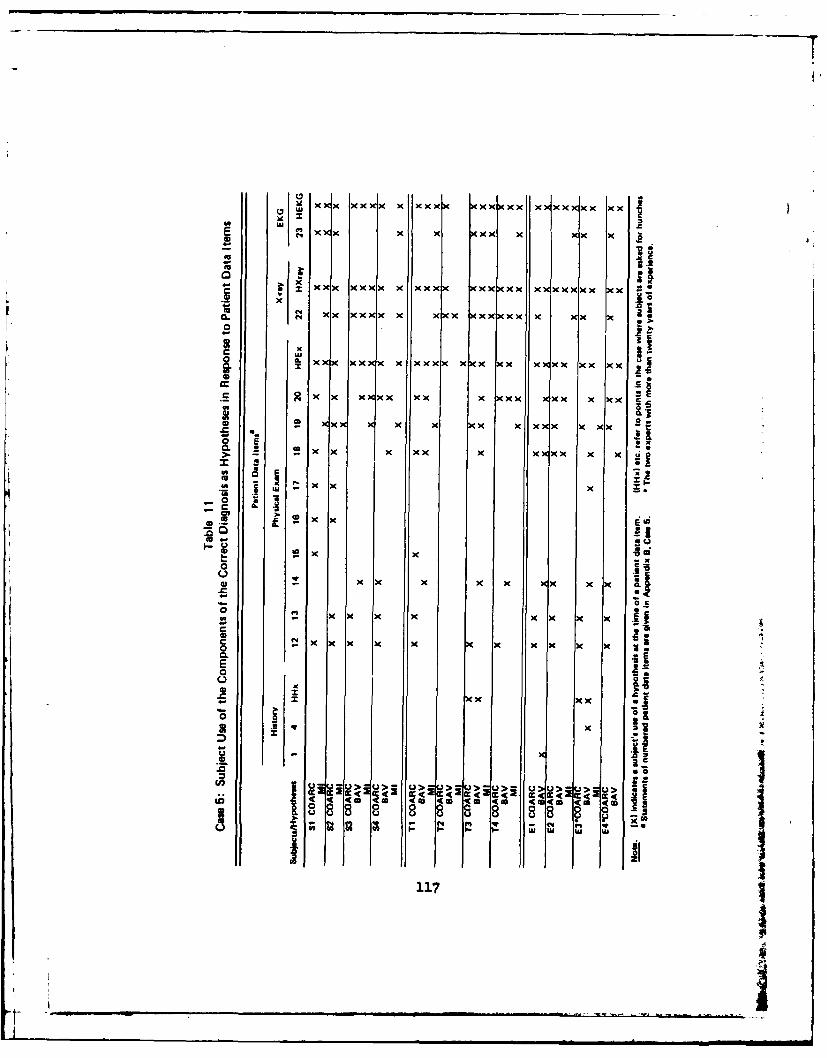
xxx xxxx x xxxx xxx XXXxx XXxxIt
tOfxx xx X X XX XX x X x
a'U
a. xxx xxxx x xxxx xxx xx xxxx x x
uJ ic ~ x x x xxxx xx x xxx xxxx xx Z
* 0- x x x x xxx x xx
x xx x xx x xxxx x x -
A! x x x'~A.
x x ix
0 .3
* x x x x x x x x
00
Ixx xx xxxx
4- xxO
8 k88 8 8 88 8 U8 0
117

of the case. Clearly it is not the BAV component of
the correct diagnosis that accounts for the somewhat
counter-intuitive results for the case. The only sub-
jects who failed to utilize the cues for BAY were stu-
dents. The explanation for the unexpected results in
the case is to be found in subjects' handling of Mi-
tral Insufficiency. In particular, all four students,
all trainees, but only two experts (E2, E3), ever gen-
erated this component as a hypothesis for the case.
Moreover, expert E3 mentioned Mitral Insufficiency
only once and can hardly be judged to have given this
hypothesis serious contemplation.
Diagnostic Errors - Case 5
The experts in this study did not judge it nec-
essary to add a comDonent of Mitral Insufficiency to
their Coaretation diagnosis although some less experi-
enced subjects did. This is illustrated in the final
diagnoses subjects gave for the Case (Table 12). Only
three subjects (S3, T1, T4) offered the correct final
diagnosis and Mitral Insufficiency was obviously the
impediment, since no expert included it in the final
diagnosis - although all experts included Bicuspid
Aortic Valve.
118

Table 12Case 5: Coarctation, Mitral Insufficiency, Bicuspid Aortic Valve - Final Diagnoses
Subjects Diagnosis
Students S1 Coarctation+ Mitral Insufficiency
S2 Coarctation+ Ventricular Septal Defect
S3 Coarctation+ Mitral Insufficiency+ Aortic Stenosis
S4 Coarctation+ Mitral Insufficiency
Trainees T1 Coarctation+ Mitral Insufficiency+ Aortic Stenosis
T2 Coarctation+ Ventricular Septal Defect
T3 Coarctation+ Mitral Insufficiency+ Bicuspid Aortic Valve+ Atrial Septal Defect
T4 Coarctation+ Mitral Insufficiency+ Bicuspid Aortic Valve
Experts El Coarctation+ Bicuspid Aortic Valve
E2 Coarctation
+ Bicuspid Aortic ValveE3• Coarctation
+ Bicuspid Aortic ValveE4• Coarctation
+ Bicuspid Aortic Valve
* The two experts with more than twenty years of experience.
119
Although the case did not turn out as expected,
it does offer the clearest discrimination between the
expert group and other subjects of any case in the
study. Ignoring the BAY component of the diagnosis,
experts basically arrived at a *Coarctation only* di-
agnosis (BAY occurs in about 50% of Coarcs and might
be thought of as a defining component of one variation
of Coarctation). Since the case was originally de-
signed as a test of augmentation to Coarctation in a
diagnosis, the overall results beg an explanation.
Perhaps the best explanation for the general
result lies in the Interpretation of the murmur, data
item 19, which was originally thought to be the
strongest evidence for Mitral Insufficiency. Table 13
shows the interpretation of this murmur by subjects in
relation to Witral Insufficiency and Coarctation of the
Aorta. A *plus" in this table indicates that a subject
interpreted data item 19 as confirmatory evidence for
the interpretation (COARC or MI) under which it is
listed. A Ozero" indicates an ambivalent interpreta-
tion of the murmur with regard to the column headingi
an example of such an interpretation is given below,
Tt It doesn't say if this murmur Isaudible over the back which wouldbe, ah, well occur, if this werejust . . . if this were his murmurof the coaro
120
-- i-e 4

Table 13Case 5: Evaluation of a Murmer as Coarctation
of the Aorta or Mitral Insufficiency
Interpretation of Murmur
Mitral CoarctationSubjects Insufficiency of the Aorta
Students S1 +S2 + 0S3 +S4 +
Trainees T1 +T2T3 + 0T4 + 0
Experts E 1 +E2 +E3° + +E4 +
Note. (+) indicates that the subject judged the murmur description to beconsistent with an interpretation of Mitral Insufficiency orCoarctation of the Aorta.(0) indicates that the subject judged the murmur description to beambivalent with rard to an interpretation.
The two expr - . ith more than twenty years of experience.
1
121
I- ..... ....... . ... ... -..... ...

Seven of the eight subjects below the expert level
interpreted the murmur given in item 19 as Mitral Insuf-
ficiency. Three of these raised the possibility that the
murmur represented Coarctation, but this interpretation
was clearly not their preferred choice. In contrast,
all four experts interpreted the murmur solidly as eman-
ating from the Coarctation of the Aorta. Expert E3 was
the only exper who even raised the possibility of Mi-
tral Insufficiency for the murmur. After this murmur,
however, he never mentioned Mitral Insufficiency again.
Although expert E2 eventually brought Mitral Insuffic-
iency into his diagnostic thinking, his only explicit
evaluation of the murmur of data item 19 was as Coarc-
tation.
The evaluations of this murmur go a long way
toward explaining the results of Case 5. Students and
trainees generally had no problem adding Mitral Insuf-
ficiency into their case interpretation while experts
did. By way of accounting for this result, Figure 11
shows three murmur descriptions. One of these is data
item 19, the murmur description given in the case.
The other two are introductory textbook (Moller, 1978)
descriptions of the murmurs of Mitral Insufficiency
and Coarctation of the Aorta. It seems that the mur-
mur as presented in Case 5 is a *textbook" murmur of
122

(a) Murmur - Data item 19 - Case 5There is a grade 3/6 soft systolic murmur at the apexwhich radiates to the axilla.
(b) Textbook Mitral Insufficiency Murmur"An apical (apex) pan systolic murmur ... This murmurradiates to the axilla and may be associated with a thrill."
(c) Textbook Coarctation Murmur"An (systolic) ejection type murmur is present along thesternal border, at the apex, and over the back between thescapulae. The murmur is generally of grade 2-3/6intensity."
Figure 11. Murmur as described in data item 19, Cam 5(a). Textbook (Moller, I1978, p. 81) description of the murmur of Mitral Insufficiency (b).Textbook (Molter, 1978, p. 91) description of the murmur of Coarct-ation of the Aorta c).
1
t
123
_____________________________________

MI and that this may well explain the performance of
less experienced subjects. While the experts' in-
terpretations of this murmur surely contributed to
their erroneous diagnoses for the case. it is not clear
why they interpreted the murmur as they did.
The handling of the Mitral Insufficiency murmur
does not explain all of the final diagnoses for Case 5.
If one ignores the BAY (Aortic Stenosis) component of
the final diagnoses in Table 12, these diagnoses can be
partitioned into three basic groups. The first group
involves Coarctation only and includes all of the ex-
perts and no other subjects. The second group of diag-
noses involves Coarctation plus MI and includes subjects
S1, S3, S4, T1. and T4. Two subjects (S2. T2) had a
third class of diagnosis, involving Coarctation plus an
intracardiac shunt (Ventricular Septal Defect). The re-
maining subject (T3) included both an intracardiac
shunt (ASD) and MI in his final diagnosis. The con-
joining of shunts with Coaretation in some diagnoses can
be used to gain some interesting insights into the dy-
namics of the diagnosis of this case.
In addition to the murmur already discussed, there
are three patient findings presented in the case which
might lead a subject to add components, including a shunt,
to his main diagnosis of Coarctation. These are the
124

findings of increased pulmonary vascularity (x-ray),
left atrial enlargement (x-ray and EKG) and right ven-
tricular hypertrophy (EKG). These are findings that,
from our a Driori interpretation of the case, argued
strongly for the presence of Mitral Insufficiency. in-
dependently of a subject's interpretation of the
murmur.
The set of subjects, taken as a whole, raised
three basic arguments or interpretations in relation to
these three findings* The rationale behind each of
these arguments will be outlined below, as a prelude to
a final set of results from the case:
1) The pure "obstruction to emptying" arguments a
schematic for this argument is given as Figure 12(a)
and reference to that figure may help the reader under-
stand the verbal discussion. The pure obstruction ar-
gument involves only Coarctation and secondary effects
of this condition. Under this argument, the Coarctation
makes it difficult for the left ventricle to eject all
of its blood, and residual blood *pools" in the left
ventricle. Because of pooled blood in the left ven-
tricle, the left atrium has difficulty emptying all of
its blood, blood pools in the left atrium and this
chamber enlarges. FoPr analogous reasons,* blood pools
In the pulmonary veins causing pulmonary venous1.25

RA LA Blood Flow
U Pooled Blood
(a) A
RA LA (b) A
I ncreased/-MV-Blood Flow
-~-.......-Coarctation ofthe Aorta
*Figure 12. Schematic for the "pure obstruction" argument-Can 5(a). Schematicfor the "augmented obstruction" argument (b). Schematic for the"shunt" agrument (c).
126

congestion. The right ventricle, working hard to pushii
blood through this stagnated system, thickens its mus-
culature, that is. hypertrophies.
An example protocol showing this argument ap-
plied to left atrial enlargement and prominent pulmon-
ary vasculature (the x-ray, item 22), is given in
Figure 13(a). Experts must have relied on something
akin to the "pure obstruction to emptying" argument
to justify their basic interpretations of the case.
While this interpretation is generally reasonable, it
azmears to involve a compitment to at least a partially
"failingO left ventricle (congestive heart failure),
that is, to a left ventricle that has dilated and lost
the capacity to eject its normal component of blood
(Hartmann, Goldring, Strauss, Hernandez, McKnight, and
Weldon, 1977, p. 201-2041 Moller, Amplatz and Edwards,
1971, p. 49). Substantial signs of congestive heart
failure are not present among the patient findings of
Case 5.
2) The "augmented obstruction to emptying argument.
A schematic for this argument is given as Figure 12(b).
This is the interpretation that includes Mitral Insuf-
ficiency with a Coarctation and would lead to the cor-
rect diagnosis of the case. The augmented argument
127

F
(22) The x-ray shows moderately enlarged heart and leftatrial enlargement. The pulmonary vasculature is prom-inent and perhaps slightly increased.
(a) The pure "Obstruction to Emptying" argumentE3: Dilatation of the left ventricle is presumably present.
Dilatation of the left atrium is, uh, secondary to elevatedend-diastolic pressure in the left ventricle. The prominentpulmonary vasculature, uh, is, uh, most likely related toincreased, uh, pulmonary venus markings, uh. Thesefeatures are consistent with significant left-sided ob-structive lesion and would be consistent with a severecoarctation of the aorta.
(b) The "Augmented Obstruction "argumentS3: Ok, the moderately enlarged heart and left atrial enlarge-
ment ah, that fits with the regurgitation across the A-V(Mitral) valve and the enlarged heart probably could fitwith the ah, there probably is hypertrophy. We'll see thaton the EKG most likely. Pulmonary vasculature is prom-inent. Now let's see that ah, I guess that could be due toah, back flow. Back pressure would be on the venous sideso you would look for exactly the characterization of thevasculature.
(c) The "Shunt" argumentS2: Ok, the ah, x-ray showing a moderately enlarged heart
could be ah, evidence of a ah, coarc, ah, with the increasedpressure transmitted to the left ventricle. But, ah, with theleft atrial enlargement it would also point to me that thereis probably a volume overload. And the fact that the pul-monary vasculature is prominent is ah, very ah, significant,I think, in that you wouldn't expect to fimd that in a purecoarc. And ah, one would have to be, I think, a little more,bit more suspective that maybe it may be a combination oflesions with a coarc being in evidence but also the a ah,VSD (Ventricular Septal Defect) which would account forboth the murmur that is heard and also the increased flowto the right side and having, and thus giving you the leftatrial enlargement and also the increased pulmonaryvasculature.
Figure 13. Protocol examples of three basic arguments used to explain LeftAtrial Enlargement and other key findings of Case 5.
128

follows the same fundamental logic as the pure obstruc-
tion argument in that its basic ingredient is one of
obstruction of inflow to the left side of the heart.
Under the augmented interpretation, blood back-flows
or regurgitates from the left ventricle through the
insufficient m~tral valve to the left atrium. The re-
sulting increased blood volume in the left atrium
causes the chamber to enlarge and also poses obstruc-
tion to emptying of the pulmonary veins. The ex-
planations for pulmonary venous congestion and right
ventricular hypertrophy are the same as under the pure
obstruction argument.
This argument has all the advantages of the pure
obstruction interpretation without requiring an ele-
ment of congestive heart failure. An example of this
argument applied to the x-ray is given in Figure 13(b).
3) The "shunto argument. A schematic for the shunt
interpretation is given in Figure 12(c). This inter-
pretation is fundamentally different from the others.
Under this argument the left-sided heart effects under
discussion are the result, not of obstruction to inflow
of blood to the left side, but rather, result from an
abnormally large volume of blood flowing into the left-
sided chambers, from the right side of the heart.
129

According to the shunt argument. some kind of abnor-
mal communication (e.g., a hole in the ventricular sep-
tumn, VSD, or an open ductus arteriosis, PDA) exists
between the left and right-sided heart systems. This
communication allows blood to shunt or cross over from
the left to the right side during certain parts of the
cardiac cycle. During other parts of the cycle, this
augmented right-sided blood is pushed through the
lungs by the right ventricle into the left-sided cham-
bers of the heart. Right ventricular hypertrophy, in-
creased pulmonary vascularity, and left atrial enlarge-
ment are related to this increased volume of blood flow.
A protocol example of this argument applied to
the x-ray is given in Pigure 13(c). While the shunt
explanation is a rather poor explanation for Case 5
taken as a whole (among other things, the murmurs are
wrong),* a shunt is a reasonable wlocalw explanation for
the target findings under discussion. Subjects who
added a shunt to their final Coarctation diagnosis did
so under the argument just outlined.
The three arguments just discussed suggested a
kind of unanticipated Logical Competitor Set for Case 5,
a set of plausible "local" alternatives for a small
group of findings in the case. This LCS is reminiscent
of the alternative explanations for the finding of
130
systolic murmur" in Case 2 repirted in Table 6. In the
spirit of the investigations of the use of Logical Com-
petitor Sets carried out on other cases of the study, it
was decided to investigate application of alternative
causal arguments to the three target patient findings
discussed above for Case 5 (left atrial enlargement.
right ventricular hypertrophy, increased pulmonary vas-
cularity).
Because the Opure" obstruction argument and the
"augmented" obstruction argument greatly overlap in
their causal dynamics, it proved impossible, in gener-
al, to discriminate these arguments in protocols given
by subjects (the relatively clear examples of Figure 13
notwithstanding). However, discrimination between the
two obstruction arguments and the shunt argument was,
in general, straightforward, Hence, it was decided to
analyze the application of a general "obstructionO ar-
gument (pure or augmented) versus a shunt argument to
the target items of Case 5.
Table l4 shows the results of this analysis. An
Ox" under one of the findings and arguments in this
table indicates that a subject applied the argument dir-
ectly to the interpretation of the finding, regardless
of the evaluative Otone" of this interpretation, that
is, whether or not the subject judged the argument to
131
-1-, , , ,

x xx
C
U.00
.00om C
Cx -xx C..3
78.
CLC
4- rb c
.S ~E
z CL
* .00
*13

yield a good or poor explanation for the finding.
The intent of Table 14 is to determine which
subjects raised or gave a "hearing" to the two main al-
ternative explanations for each finding. One way to
address this question is to ask which subjects raised
both arguments in relation to any one target data item.
Only one student (S2) applied both arguments to a com-
mon target data item while all trainees and experts
meet this minimal criterion for comparative consider-
ation. Once again we see evidence that a consequence
of increasing experience is the grouping together of
reasonable alternative explanations. Students, in gen-
eral, appear not to have as many interpretive options
available to them. It is as though, for inexperienced
subjects, each finding has an idiosyncratically salient
and lone causal interpretation. For example, even
though subject S2 raised two arguments in response to
"increased vascularityw, his protocol in response to
this finding (Figure 13c) suggests he did not recog-
nize the general merit of the obstruction argument in
the interpretation of this finding.
The results of Case 5 have raised several gen-
oral issues to be discussed in conclusion to this
case.* The case was originally intended to be a test of
of whether subjects would add multiple components to a
133
diagnosis in a case where, from a prior analysis, such
addition seemed necessary. In general, less experienced
subjects added these components while the most exper-
ienced subjects did not.
Another case in the study. Case 2 - TAPVC, showed
students who created piecemeal, unintegrated combin-
ations of abnormalities as diagnoses even in a case
where these fragments could easily be integrated with-
in a single, more global interpretation. We have also
seen, among less experienced subjects, a kind of "local
reactiveness" to particular data items of a case. Re-
call the PAPVC diagnoses in 8ase 2 that seemed to be
based largely on a single x-ray finding, the student
difficulties with Pulmonary Stenosis on that same case,
and the Truncus diagnoses in reaction to the heart
sound finding in Case 4. Other cases have shown novice
dependence on "textbook" or classic descriptions of
patient findings. Since the correct diagnosis for
Case 5 is, in a sense, fragmented, and because the com-
ponents seem to have had classic cues in patient data
(e.g., see Figure Ii), it would appear that this case
"played into the handse of the novices.
The best explanation for what happened to ex-
perts in this case would seem to involve the naturally
134

occurring rarity of Mitral Insufficiency in combination
with the disease Coarctation of the Aorta. In post-
experimental discussion, expert E3 claimed never to
have seen any patient with this combination, while ex-
pert E4 thought he might have seen "less than half a
dozen" in over 500 clinical cases of Coarctation. A
report of the clinical incidence of various abnor-
malities in conjunction with Coarotation (Hartmann
et. al., 1977. p. 200) does not even list Mitral In-
sufficiency among the possibilities and the author has
yet to find MI in such a listing anywhere. In contrast.
BAV and various shunts are fairly common. Given that
all the experts were adamant in claiming that they
could account for all data of the case under a "pure
obstruction" argument and given the rarity of MI in
combination With Coarctation, the experts stayed with
Coarctation alone. "Never two if one will do" as one
expert noted, although the present case shows how this
expert tendency toward integration can sometimes lead to
trouble.
The expert consultant to the project claimed to
have seen a substantial number of patients with a Co-
arctation-MI combination and this probably accounts for
135
111- I I

his consistent correct interpretation of this case.
Interestingly, students and trainees had no trouble
forming the Coarctation-MI combination based on *text-
book" cues for individual components, although it is
safe to assume they had no clinical experience with
it. This would seem to indicate that with increasing
experience, the foundation of a subject's disease
knowledge changes from one based primarily on textual
materials (e.g., reference manuals or instructional
materials) to one weighted more heavily on clinical
experience.
1
I1361

Bummr '
The results from five diagnostic cases were pre-
sented in this chapter. Cases were designed to test
different aspects of disease knowledge in subjects. Two
kinds of results were presented for each case. The
first of these involved the use by subjects of a des-
ignated set of diseases (in the first four cases, a
Logical Competitor Set) as diagnostic hypotheses during
the course of the case. The other kind of result was
an analysis of key diagnostic errors committed by sub-
jects in their handling of the case.
Case 1. The first case, Subvalvular Aortic Stenosis
(SubAS), was designed to test subjects' working know-
ledge of variants of a disease in addition to a "clas-
sicO version, and the grouping of these variants in
memory.
All subjects used classic Valvular Aortic Sten-
osis (ValvAS) as a hypothesis, supporting our pro-
suprosition that this disease variant would be highly
salient in the knowledge base of subjects. Only one
student ever utilized both of the other designated dis-
ease variants, Subvalvular and Supravalvular Aortic
Stenosis (SupAS), and he made only passing reference to
137
I-L--- _

these in his diagnosis. In general, students focused
almost exclusively on the classic variant during the
course of the case. Half of the trainees ased only
Valvular Aortic Stenosis in the case, while the other two
trainees utilized the full set of variants. No expert
was limited to only the classic variant, and three ex-
perts (including the two most experienced) used the full
set of variants. All subjects who used the full set of
variants did so in one of two patterns: they either used
all variants together at a data point which represented
a strong cue for Valvular Aortic Stenosis or they used
all variants together at a data point immediately follow-
ing strong evidence against ValvAS. These patterns were
taken as evidence for memory grouDing among the disease
variants.
No students, two trainees, and the two most ex-
perienced experts diagnosed the case correctly as Sub-
valvular Aortic Stenosis. All other subjects chose the
classic version, Valvular Aortic Stenosis. Six of these
subjects (three students, two trainees, and one expert)
never generated SubAS as a hypothesis, even though they
had generated ValvAS. Among other possible explanations,
sparseness (lack of connectedness) in disease knowledge
was provosed as contributing to this lack of activations
138
PTJ--_

ValvAS and SubAS were not strongly enough associated in
memory to enable the activation of ValvAS to spread to
the other variant.
Two stu'ents who diagnosed the case incorrectly
were the only subjects who misevaluated a piece of
strong evidence against ValvAS, "no systolic ejection
click", as confirmatory evidence for this variant. This
was interpreted as reflecting imprecision in the stu-
dents' disease models for the variant. Another student,
as well as some of the more experienced subjects who
missed the case (including the two less experienced ex-
perts) exhibited faulty knowledge of the physiologic
cause of systolic ejection clicks. This error in cau-
sal knowledge led the subjects to minimize the import of
the critical evidence against Valvular Aortic Stenosis.
Case 2. The second case, Total Anomalous Pulmonary
Venous Connection (TAPVC), was designed to test sub-
jects' memory grouping of a Logical Competitor Set (LCS)
of diseases, a set of diseases with Oincreased right-
sided blood volume.0 Other diseases in this set in-
clude Atrial Septal Defect (ASD), Endocardial Cushion
Defect (BCD), and Partial Anomalous Pulmonary Venous
Connection (PAPVC). Of these ASD was assumed to be
classically associated with the strong hypothesis
139
A _ _ _ _

"triggeringo cues of the case.
All subjects generated Atrial Septal Defect as
a hypothesis, supporting our presupposition that this
disease is strongly associated with a set of cues in
the case. No student utilized the full set of com-
petitor diseases as hypotheses in addition to ASD.
One trainee utilized the full LCS as did the two most
highly experienced experts, E3 and E4. The two most
exuerienced experts used the full LCS according to the
same patterns in which they had used the full set of
competitors in Case 1. Expert E3 used most of the com-
petitor set diseases together at the data point of
first strong evidence for ASD. Expert H4 used only ASD
at this point, but exoanded to the full LCS at a later
point of strong disconfirmatory evidence for ASD. The
trainee who used the full LCS did so in a pattern simi-
lar to expert E3. As in Case 1, the use of the full
LCS in these patterns was taken as evidence for memory
grouping or categorization of the com-oetitor set dis-
eases.
Only four subjects, two trainees and the two
high-level experts, diagnosed the case correctly as
TAPVC. Agin, a number of subjects (three students,
one trainee, one expert) never generated the correct hy-
pothesis, partly accounting for their erroneous
140

diagnoses of the case.
Among errors of commission, two students had
final diagnoses outside of the Logical Competitor Set
of diseases. These diagnoses involved combinations of
multiple diseases, and the primary locus of error for
these students was traced to limitation in knowledge Of
alternative causes for a particular systolic murmur
the students, unlike more experienced subjects, con-
sidered only one of two feasible causes for this find-
ing - the wrong one for the context of the case - and
this interpretation encouraged multiple diseases in a
diagnosis. Another student diagnosed the case as Endo-
cardial Cushion Defect, and seriously misinterpreted the
EKG axis in relation to this disease. This was taken as
reflecting error or imprecision in this subject's dis-
ease model for ECD. One of the trainees demonstrated
overrestrictiveness (imprecision) in her allowable age
range for TAPVC, causing her inappropriately to reject
this disease in favor of ASD. Finally, four subjects,
inanning the dimension of experience, diagnosed the
case as PAPVC. These subjects were strongly influenced
by a classic cue for PAPYC on x-ray. When this cue
elicited PAPVC, it did not, in turn, elicit TAPYC or
other LCS members, again suggesting sparseness (lack of
141
A - ~

coninectedness) in disease knowledge.
Case 3. The third case of the study, Patent Ductus
Arteriosus (PDA), was designed to test subjects' group-
ing of a Logical Competitor Set of diseases in a case
where there was an early, classic cue for the correct
disease and no subsequent data were discrepant with
this interpretation. The case was different from the
first two cases in that in those cases, the first
strongly induced disease interpretation was wrong. Be-
cause determining the correct diagnosis was so straight-
forward, Case 3 was seen as a severe test for hypothesis
grouping. PDA and the other four diseases in the LCS
for Case 3 constituted a physiologic category of "extra-
cardiac shuntso and related conditions.
All subjects generated PDA in the case as ex-
pected, and all subjects concluded this disease as a
final diagnosis. One student, three trainees, and high-
level expert lie, generated only PDA among LCS members
during the course of the case* All other subjects gen-
erated at least one other LOS member in addition to PDA.
Only one subject, high-level expert R3, used all five
members of the LOS during the case, and he did so in a
manner consistent with his complete use of the com-
petitor sets in other cases. That is, he generated142
Emma

the entire set as competing hypotheses in close prox-
imity to the classic cue for Patent Ductus. Another
expert, 32, considered most (3 of 5) of the LCS, and
he did so in a pattern similar to E3s no non-expert sub-
ject utilized as much of the LCS. The performance of
high-level expert E4 was seen as consistent with his
consideration of the full LCS in other cases, in that
in the first two cases he had expanded to the full LCS
only after receiving disconfirmatory evidence for a
preferred members in Case 3 there was no such evidence.
Case 4. The fourth case, Pulmonary Atresia (PAT),
was, again, designed as a test of clustering in memory,
by subjects of a physiologic category of diseases -
"cyanotic diseases with decreased pulmonary blood flow.0
This Logical Competitor Set for Case 4 included
Ebstein's Malformation of the Tricuspid Valve (Ebstein's)
and Tricuspid Atresia (TAT) in addition to the correct
disease, Pulmonary Atresia. No member of the LCS was
seen as generally more prototypic for the category than
the others.
No student utilized the entire Logical Competitor
Set for the case, and two students generated only one
member. No other subjects generated this few. In ad-
dition, two students were the only subjects who never
14I3
created the correct disease, Pulmonary Atresia, as a
hypothesis. In contrast to the students, all four train-
ees and three experts (including high-level expert E3)
used the full competitor set as hypotheses. However,
the experts and trainees utilized the full competitor
set according to different patterns. The experts, un-
like the trainees, used all LCS members together at the
point of the case where the category "cyanotic disease
with decreased pulmonary blood flow" could first be es-
tablished. No trainee used the full set at this point,
suggesting that the LCS diseases did not constitute a
functional diagnostic category for these subjects - at
least not to the same extent as for the experts.
Highly experienced expert E3 utilized the full
LCS in Case 4 in the same way he had done in other
cases, that is, he utilized all member diseases at the
first clear opportunity to do so. While expert E4 named
only the correct disease, Pulmonary Atresia, at this
point, he also articulated the category name for the
LCS diseases at this point and his performance was in-
terpreted as being consistent with his handling of LCS
diseases in other cases - since no subsequent data were
incompatible with the particular LCS member he chose
for focus.
144
Three experts diagnosed the case correctly as
Pulmonary Atresia while this final diagnosis was of-
fered by only one student and one trainee. Two gener-
al types of error were demonstrated by subjects who
missed the case. Two students diagnosed the case as
Truncus Arteriosus, and a particular data item, 'single
second heart sound', was proposed as the most important
source of this misdiagnosis. This finding is consis-
tent with all LCS members but is perhaps most clearly
rerresentative of Truncus. Students' handling of this
data item was interpreted as reflecting a kind of clas-
sical dependency in the knowledge base. Most trainees
diagnosed the case as Tricuspid Atresia and the main
source for this misdiagnosis was traced to an overly
liberal range of expected values for the EKG axis under
this disease - imprecision in subjects' disease models
for Tricuspid Atresia.
Case 5,. The final case of the study was character-
istically different from the others in that there was no
issue of logical Competitor Sets. The case contained
a pathognomonic cue for the main disease in the diag-
nosis, Coarctation of the Aorta (Coarc). The case was
designed to test whether subjects would detect the need
for adding two additional diseases, Bicuspid Aortic
145

Valve (BAY) and Mitral Insufficiency (MI), to a
correct diagnosis.
In general, the results of the case were opposite
to what was expected. Most significantly, no expert
added Mitral Insufficiency to his Coarctation diagnosis
while many less experienced subjects did. The success
of the less experienced subjects in adding this compon-
ent was attributed to a rather Otextbook = description
of the murmur of MI which was presented as a data item
in the case. The failure of the experts was attributed
to the clinical rarity of MI in combination with Coarc
and to a general tendency among these subjects to avoid
multiple-disease diagnoses if at all possible. The ar-
gument by which experts judged Mitral Insufficiency to
be unnecessary was sketched. Case 5 is an example of a
situation where expert tendency toward integration may
lead to difficulty.
An unanticipated analysis for Case 5 involved
subjects' use of two main physiologic arguments, wob-
struction" and *shunt", for a set of important patient
data items in the case. Students generally seemed lim-
ited to one or the other of these interpretations while
more experienced subjects generally applied both.
This restrictiveness in options for interpretation was
146

consistent with novice performance in similar sit-
uations in other cases.
Based on the performance of the two highly ex-
perienced experts (E3, E4) on the cases of this study,
an expert form and an expert substance for diagnosis
can be proposed. The expert form involves the full,
active use of a set of physiologically similar diseases
(the Logical Competitor Set) in a case, diseases that
have similar clinical presentations. Expert E3 used
the full LCS in all four cases where this was tested-
more than any other subject. Expert E4 explicitly used
the full LCS in two cases and utilized a category hy-
pothesis for the LCS diseases in another. The use of
the Logical Competitor Set by experts, generally in
close proximity to the most important patient findings
associated with members of the set, is interpreted here
as evidence that these diseases constitute a unit13
or category in memory. Since diseases in the Logical
Competitor Set are likely to be confused with each
other, in the "long-runn it would seem highly adaptive
There are more direct ways of assessing this claim,such as experimental tasks that require subjects todraw on their memories in ways that allow measurement ofinter-response times (e.g., Reitman, 1976). Studies ofthis sort are currently under planning.
147

for a diagnostician to be readily able to engage other
members of the set in contexts where there is strong
reason to believe one of them is a good candidate for a
cases Memory "unitization" (c. f. Anderson, 1980) of
LCS diseases in some sort of category-like structure
would provide this capability to the expert.
Expert substance in diagnosis refers to correct
data interpretations and data evaluations, in relation
to the Logical Competitor Set of diseases, necessary to
isolate the correct member. Expert EL4 diagnosed four of
the five cases correctly, more than any other subject.
He missed only the controversial Case 5. Expert E3 di-
agnosed three of five cases correctly, as many as any
other subjects Isolation of the correct LCS member is
supported by precision in the clinical data expectations
contained within individual disease models.
For the two high-level experts in the study, two
distinct methods of utilizing the LCS were also iden-
tified: (1) Precaution. This involves the generation
and use together as hypotheses of the full set of log-
ical competitors, enabling them to be weighed against
each other and data. (2) Extraction. This method in-
volves more aggressive focus on a member of the set,
with full expansion to the remainder of the set should
1~48

disconfirmatory evidence for the target member be found.
The two less experienced "experts'. El and E2,
performed much like trainees on the cases of this study.
In particular, each diagnosed only two cases correctly,
a figure eclipsed by three trainees. E2 used the full
LCS in only one case, while El did so in two cases. In
addition, each showed interpretive errors similar to
those made by less experienced subjects, in particular
the handling of the "click" in Case 1, and the suscep-
tibility to classic cues in Case 2. These subjects did
enable us to see that the development of high-level
skill in pediatric cardiology takes a long time.
Medical students after six weeks of training and
clinical practice in the field represented by the cases,
generally showed neither expert form nor substance.
Students almost never considered the full LCS (one stu-
dent on one case) and focused on the "classic" members
in cases which encouraged this. This suggests that LCS
members, when they exist in memory at all, are repre-
sented in a more isolated form than for experts. Two
students diagnosed no case other than Patent Ductus cor-
rectly, while the other two diagnosed one disease in
addition to PDA. Errors of interpretation and eval-
uation (shared with intermediate level subjects)
149
1L
included several basic types# (1) Mundane factual er-
rors. These are just factual errors about which find-
ings Ogo with" which diseases. (2) Causal errors.
These are errors concerning how observable data are re-
lated to underlying physiology. (3) Imprecise tests.
These are either overly general or overly restrictive
tolerances on the range of variability allowed in an ex-
pected clinical finding for a disease. (4) Interpre-
tive restrictiveness. This refers to restriction in
the number of interpretations that are applied to a
finding. These errors are interpreted as reflecting im-
precision in subjects' models for diseases.
The "trainees' in the study (residents in ped-
iatric cardiology at the end of their training and first-
year fellows in pediatric cardiology) at times showed
pDerformnces that looked very much expert-like and at
times could not be distinguished from the students. The
number of trainees in each case who used the full LCS
fell between the students and experts on two cases
(Case 2. and Came 2), was the sme as the students on
Case 3, and actually exceeded the experts on Case 4.
Three trainees diagnosed three cases correctly - on a
par in this regard with high-level expert E3. Depend-
ing on subject arnd case, trainees at times exhibited
2.50

the types of errors discussed above for the students.
The ultimate diagnoses of the trainees, unlike those of
the students, were always at least within the LCS,
if not correct. This suggests that for trainees who
missed cases, the main problems involved lack of
connectedness in memory among LOS members, or im-
precision in knowledge necessary for discriminating
LOS members correctly.
4
151
4. DISCUSSION
The study demonstrates that diagnosticians' ndis-
ease knowledge," a memory store of disease models and
the memory organization among them, is important to suc-
cessful diagnosis and does exolain differences between
expert and less expert performance.
The general "process" of diagnosis, although this
has not yet been directly addressed from the study,
seemed similar for all subjects; that is, all subjects
generated diagnostic hypotheses at various times in cases
and proceeded to test these against subsequent data.
The major differences among subjects which have emerged
so far concern their handling of a set of "good moves:"
that is, the Logical Competitor Sets. More experienced
subjects tend to consider them in groups, and evaluate
them correctly.
The study did not set out to show that highly ex-
perienced practitioners diagnose differently from nov-
ices; this should go without saying. The intent was to
learn something about the medical knowledge that diag-
nosticians use, the way this influences performance,
and the ways this knowledge changes as people acquire
experience in a field. Medical students, after only
six weeks of training in the field of interest, were in-
cluded as subjects because these individuals represent
152
-. _ _ _ _ _ _
the "starting point" in a long learning process.
The Nature of Knowledge ChanAce
What has been learned about the nature of know-
ledge change? It seems clear that the whole learning
process starts with a small set of "classic" training
concepts where these include particular diseases, de-
scriptions of expected patient findings under these
diseases, and rules for disambiguating diseases in
this starting set. The learning of these training con-
cebts is encouraged by the selection of content for in-
clusion in introductory training materials, that is,
introductory textbooks and classroom instruction. The
diseases are the common ones or the clearest instances
of a physiologic type, the patient data descriptions
are "prototypico or average, and the rules of evaluation
are overly simplified. We have seen several instances
where the locus of novice errors could be traced fair-
ly directly to such statements in the introductory text-
books to which the subjects had been exposed.
One might wonder why the introductory materials
are as they are* There are probably several good
reasons. First, it is not clear that the ultimate
textbook*, one that documents all of the context-
sensitive nuances of patient presentation and all of
153
the "exceptions" to the rules of evaluation could even
be written. Medical research and the clinical case
documentation literature periodically change what such
a book would contain. Even if the ultimate "book"
could be written, it would probably be a poor in-
troductory textbook. It would be such a morass of de-
tail that students might learn nothing at all. Secondly,
there are sound cost-benefit reasons for emphasizing
the common things. If a new practitioner cannot be
taught everything, it is obviously right to give him
the tools to work with the problems he is most likely to
encounter. Most students who take the introductory
course in pediatric cardiology do not even become ped-
iatric cardliologists. The training objective for these
individuals is probably only that they be able to rec-
ognize problems for referral. Finally, for those who
specialize, a long apprenticeship is assumed. A min-
imum of six-seven years of clinical and other training
would be required before the students in this study
could be certified as pediatric cardiologists. What is
important is not that they be taught everything but
rather that they be given the cognitive "anchorage
points" to enable them to benefit from the experience
to follow.
15 4
With experience, the practitioner is exposed to
and adds to memory additional disease models beyond the
introductory set. The expert's "large vocabulary" of
discriminable instances is now well documented (e.g.,
Chase and Simon, 1973a). Concurrently with the simple
addition of disease models to memory, there is an em-
bellishment of the compositional features of a disease
that are encoded within each disease model. These are
features representing the diseases' internal physiol-
ogy and clinical presentation. The expert simply knows
more defining attributes of a disease. Roach (Roach and
Mervis, 1975) reports an intriguing anecdote that bears
on this issue. One of her standard paradigms asks sub-
jects to list as many features as they can of various
common objects, e.g.. an airplane. Among ordinary col-
lege subjects the number of features listed for any par-
ticular object is fairly stable. However, one subject
was found to list many times more features for an air-
plane than average: on follow-up he was discovered to be
an airplane mechanic. In some of our own work, we have
found that expert physics problem solvers actively use
"transformed" or "abstracted" features of a physics
problem statement that novices do not even seem to recog-
nie (Chi, Feltovich, and Glaser, 1980). In Case 1 of
the present study, there were some inexperienced subjects
155

who did not generate any Aortic Stenosis hypothesis un-
til after the presentation of the critical finding of
"no click". The fact that they did not return to this
finding after the Valvular Aortic Stenosis model was en-
gaged, suggests that this feature was not represented
in their model of the disease; they may have had no
exDectation at all regarding a click. Recall that in
Case 2 of the present study some inexperienced subjects
seemed to view the Pulmonary Stenosis issue (Table 6) as
involving only one dimension, that is, "orifice size",
when in fact the problem involves the two interacting
feature dimensions of size and flow. This is highly
reminiscent of the "dimensional restrictiveness" or
paucity of encoded problem features reported by Siegler
(Siegler, 19761 Siegler, 1978) for inexperienced prob-
lem solvers.
As an individual encodes more features of a dis-
ease, this provides opportunity for differentiating the
disease into subtypes (c.f., Anderson et.al., 1979).
As an illustration, if a person encodes only the features
of "height" and "weight" for people, he is quite limited
in the discriminations he can make among people. It is
clear that the disease knowledge of a highly experienced
diagnostician is highly differentiated within a disease
type. In the present study the case explicitly
156
1-
designed to assess this was Case 1, where the increas-
ing differentiation was demonstrated. It can be noted
that for Case 2, TAPVC, high-level expert E3 raised and
considered no fewer than nine different varieties of
TAPYC where each of these was distinguished by slight
anatomical difference.
The differentiation of disease knowledge aids the
develoDment of Drecision in the clinical expectations
associated with any particular disease model. If possi-
ble distinctions among versions of a disease are not
made: that is. if they are in a sense all seen as the
same thing, then the associated variability in clinical
manifestations among patients will be great. However,
when an expert represents in memory, say, nine different
anatomic versions of TAPVC with each of these perhaps
differentiated into more siecific versions by severity
and age of presentation in a child, thFn the clinical ex-
,oectations associated with each of these 'micro models'
can be highly specific.
Precise clinical expectations, in turn, contri-
bute to precise rules of evaluation for patient data.
This is the difference between the Oleft axis deviationo
rule used by less experienced subjects in Case 4 and the
experts' "down around zero or slightly negative" ruleused in evaluating the EKG axis in that case with
157
* I"
respect to Tricuspid Atresia (See Table 10 and the dis-
cussion around it). Again, in Case 1, one can see a
nice example of how differentiation of a disease con-
tributes to correct evaluation. In his protocol given
in Figure 4, expert E3 raises the one micro version of
Valvular Aortic Stenosis in which a "click" is not ex-
pected. This is the version with a pressure gradient
between the left ventricle and aorta (over the valve)
of greater than 100 mm, that is, "Aortic Stenosis of a
very severe degree." Under this version, other data of
the case would have been different from those presen-
ted. The expert was able to bring the appropriate
(i.e., moderate severity) version of Valvular Aortic
Stenosis to bear on the evaluation, and to reject this
version.
The embellishment of the feature got within dis-
ease models aids generalization as well as discrimina-
tion. Every additional feature represented for a dis-
ease is a potential feature of similarity with another
disease; hence, the potential of generalization to a
category of adiseases ths share feature xO exists
(c.f. Anderson et.al., 1979). The LCS analyses through-
out this paper are taken as evidence that such group-
ings are pervasive in the more experienced knowledge
base. Tight memory organization among competitor
158
diseases, in a category (Cohen, 1966) or similar type
of memory unit (Anderson, 1980), supports diagnosis by
providing interdisease activation; when one member is
activated, other plausible candidates are likely to be
considered.
Students learn some disease groupings directly
(e.g., Moller, 1978, p. 4 6). These,analogously to
other teaching concepts, might be thought of as a set of
"starting-Doint" disease categories. With experience and
embellishment of feature sets, a diagnostician augments
this initial category set, often creating useful cate-
gories that "cross over" the original "classic" set.
Case 2 from the present study is a good example. One
might wonder how it is that a number of subjects on this
case could generate and extensively consider the hypo-
thesis "Partial Anomalous Pulmonary Venous Connection"
and never once even think of the correct disease,
"Total Anomalous Pulmonary Venous Connection", a dis-
ease that even in its name is so similar. In the clas-
sic categorization of diseases, PAPVC, ASD, and ECD,
three members of the Logical Competitor Set for this
case, all go together in a category of "acyanotic heart
diseases" (See Figure 14). while the final LCS member,
TAPVC, is in a different category, "cyanotic heart
159
A 1 P
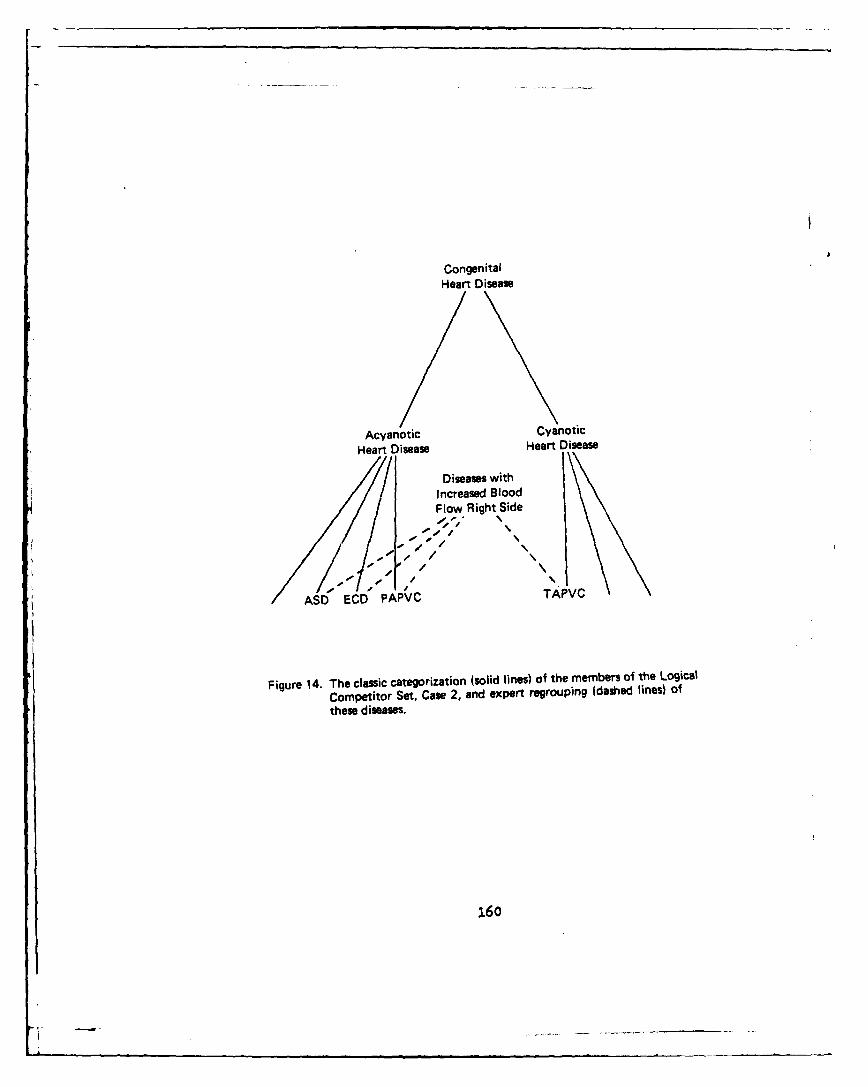
CongenitalHeart Disease
Acyanotic Cyanotic
Heart Disease Heart Disease
Diseases withincreased BloodFlow Right Side
• ,--. \J / \
-- , /\/
,
ASO ECD PAPVC TAPVC
Figure 14. The classic categorization (solid lines) of the members of the Logical
Competitor Set, Case 2, and expert regrouping (dashed lines) of
these diseases.
160

disease." One explanation for these subjects is that
they became "stuck in a chunk;t that is, they were in
the wrong branch of their classic hierarchy and were
not able to benefit from associative hypothesis trig-
gering or 'spreading* activation (e.g., Anderson, 1976).
The two high-level experts, on the other hand, had
created a category for the LCS members that crosses the
classic categorization scheme (See Figure 14). Creation
of this category required them to represent a new dis-
ease feature, the feature of "increased blood flow on
the right side."
Our speculation is that many kinds of logical
and practical groupings exist for the expert, tailored
to different problem contexts and even different phases
of patient data collection. e.g., "the not too sick two-
day-old child" in the very early phases of diagnosis.
The totality of these groupings for the expert need not
be strictly hierarchicali that is, the groupings may
"cross over'" each other in many different ways forming
more a 'latticeo structure than a formal hierarchy
(Pople, 1977).
The pervasiveness of memory groupings in the ex-
pert is a logical extension of the general "perceptual
chunking hypothesis" of Simon and Chase (Simon and
161
Chase, 1973) and all of its ramifications (e.g., Chase
and Chi, 1980). The cognitive mchunks" for an environ-
ment that people create with experience are those that
serve their goals for functioning in that environment
(e.g., see Egan and Schwartz, 1979, for "electronics
trouble shooters") and, in addition to activational
functions cited above, probably serve the problem sol-
ver by focusing problem solving activity as we will now
discuss.
The RelationshiD Between Knowledge and Problem Solving
As problem solving research has moved from seman-
tically "lean" domains, e.g., various toy problems such
as the "Tower of Hanoi" and "cryptarithmetic" (e.g.,
Newell and Simon, 1972), to semantically rich domains
such as physics or Oengineering thermodynamics", the im-
portance to problem solving of domain knowledge has
been increasingly recognized (e.g., Bhaskar and Simon,
1977).
One of the issues we set out to address with
this study was the relationship between domain knowledge
and general'problem solving processes.0 One way to ad-
dress this issue is from a framework for problem solv-
ing processes set out by Newell (Newell, 1969). Newell
proposed a power-generality dimension for problem
162
.r
solving procedures. General procedures are those that
apply widely (the conditions for their applicability
are non-stringent) but offer little guarantee of suc-
cess. Examples are "means-ends" analysis and "hill
climbing." Powerful procedures are those that have
well specified conditions which must be met for their
applicability, hence, are tailored to particular closed
environments. An example is the formula for solving
quadratic equations. Our work and that of others
(e.g., Elstein, et. al., 1978) has shown that the gen-
eral problem solving procedure for diagnosis is one of
hypothetico-deduction and that all subjects, regardless
of experience, share this general approach. However,
the present study has shown that this alone "will not
get one very far". The general process must be backed
up by a rich body of accurate, well organized medical
knowledge.
With regard to procedures for diagnosis, we
speculate that as disease knowledge develops to include
numerous pragmatic disease clusterings, as outlined
earlier, corresponding sets of relatively powerful pro-
cedures, in Newell's sense, are concurrently created.
Hence, we would propose that as the diagnostician es-
tablishes various partitions of the disease space, for
example, the Logical Competitor Sets of various kinds,
163
MOP
he also establishes associated strong *local" proced-
ures for working within those regions of the space. This
would mean, for instance, that the exDerienced diagnos-
tician would have relatively intact or readily assem-
bled 'plans" (Sacerdoti, 1977# Vahnlehn and Brown, 1979)
or *scripts' (Schank and Abelson. 1977) for discrimin-
ating among hypotheses within conceptual disease group-
ings of various kinds and levels of generality.
The worth of any such plan or script depends on
a great deal of specific content of knowledge, including
the best alternative disease candidates, precise clin-
ical data expectations for each alternative disease,
and differential patient data items that can be used to
distinguish among the set of target disease candidates.
Novices in the present study at times showed de-
ficiency both in assembling a good set of alternative
disease candidates, and in the specific knowledge neces-
sary to test a disease or disambiguate among alterna-
tives. What is happening when less experienced subjects
fail to consider a good set of disease candidates or
evaluate data items poorly? One explanation is that
necessary knowledge is stored in memory incorrectly or
not at all (i.e., knowledge avoidso - the person does
not know about Subvalvular Aortic Stenosis). Another
exilanation concerns problems of access subjects simply
1 61

do not retrieve useful knowledge or retrieve it in some
faulty manner.
Post-experimental discussions (conducted after
all cases were completed) with the subjects from this
study indicated that many subjects, when they failed to
generate a good set of candidate hypotheses or inter-
preted items ooorly (e.g., the "click" in Case 1). knew
better" in some sense. Under conditions outside the di-
agnostic task, they could often discuss Aortic Stenosis
variants, e.g., Subvalvular Aortic Stenosis, or the im-
port of the =click" in Valvular Aortic Stenosis, etc.
One subject called the experimenter on the day after his
session, in which he had erroneously diagnosed Case 2 and
never thought of the correct diagnosis, to tell him that
the correct diagnosis had "dawned on him in the shower."
Psychology has long known that the ability to ac-
cess and use knowledge that one "has" is situationally
dependent (e.g., Melton, 1963)1 Tulving and Pearlstone,
1966). For example, knowledge medical subjects might
display on a "paper and pencil" test is not necessarily
what they could display "on-line" in the diagnostic set-
ting. (It was for this reason that the current study,
despite its interest in "knowledge," was conducted in a
diagnostic context rather than in some other manner).
Yet, it in this *task-accessible" knowledge that is
165
I -

I II.
crucial to successful performance. Knowledge must be
utilized appropriately in particular contexts where it
is needed. The present work and other recent in-
vestigations of novice problem solvers (Polson, At-
wood. Jeffries, and Turner. in press), suggest that
deficiencies of situational access to extant knowledge
rival difficulties associated with knowledge error or
absolute lack of knowledge for these individuals.
Imvlications for Training and Assessment of Competence
Some suggestions for helping a new diagnos-
tician develop an apDropriate and, especially, a situa-
tionally useable knowledge base can be proposed. First,
a disease and other diseases likely to be confused with
it in a diagnostic setting should be emphasized to-
gether in instruction and, to the extent possible, in
the clinical experiences of the diagnostician in train-
ing. This encourages the memory unitization of these
diseases in categories or other kinds of connected know-
ledge organizations. Unitization aids access and is a
hedge against oversight since information in a unit has
two modes of *on-line" activation, associations from ex-
ternal events and activations directed by the unit itself
(Anderson, 19601 Cohen, 1966). The competitor set cate-
gory also provides focus for the creation of a plan
166

for disambiguating among the member diseases. Because
real clinical experiences are constrained by the dis-
tribution of patients in the training setting, simu-
lated diagnostic encounters (e.g., McGuire and Soloman,
19711 Swanson. Feltovich, and Johnson, 1977) could pro-
vide a vehicle for augmenting natural experiences and
for packaging pre-specified sets of experiences. for
example, the presentation in close proximity of cases
involving competitor set diseases. Secondly. tutorial
instruction in the diagnostic process itself must at-
tempt to interact with the on-line thought processes of
the learner as he engages in diagnostic-like tasks.
This is to help ensure that what is to be taught will
be connected both to the situational cues and to the
state of active memory likely to exist at some later
time when the new material will be needed during a real
diagnostic encounter (c.f.Flexser'and Tulving, 19781
Tulving., 1976). Expert-based instructional devices
(computer assisted instruction or decision support sys-
tems) that contain expert knowledge and are capable of
performing diagnosis In an expert-like manner, could
provide diagnostic practice exercises in which the de-
vice diagnoses a case in parallel with a *student,*
prompting alternative hypotheses when they are over-
looked, correcting erroneous interpretations, and
*~:~167
offering instruction when this seems necessary
(Brown and Burton, 19751 Clancey, 1979; Clancey, Short-
liffe, and Buchanan, 1980l Johnson, Severance, and
Peltovich. 19791 Swanson, Feltovich and Johnson, 1977).
Finally, it would be advantageous if much of the learn-
ing of medical content for those in training could be
tied as closely as possible to its conditions of ulti-
mate use. The program in medical education at McMaster
University (Barrows and Tamblyn, in press) and the prob-
lem-based program at Michigan State (Elstein, et.al.,
1978), seem prototypic of such an aporoach. Under these
programs, much of the basic medical subject matter (e.g.,
physiology) that a student learns is organized within
representative professional problems, including diag-
noses. The problem directs what is to be learned.
In addition to some suggestions for instruction
and training, the current research carries a suggestion
for the evaluation of clinical competence. While the
recent trend in assessment is toward testing clinical
skill using diagnostic and other problem solving tasks
in addition to more standard oaper and pencil testing,
most of these attemots, for example, the "patient man-
agement problems" of the National Board of Medical Ex-
aminers, concentrate on the process of data collection.
168

They focus on what the testee chooses to "see", that is,
questions asked of the patient and lab tests ordered, etc.
While this issue is surely important, the present study
has shown that it is equally important to consider the
inferences a person makes from the data he does see,
e.g., the interpretations of basic findings, the hy-
potheses considered, and the hypothesis evaluations
made. Evaluation of the quality of interpretation need
not be limited to final outcomes, that is, whether or
not the person gets the problem Oright". We have shown
that careful task analyses of problems can yield inter-
mediate forms, e~g., the handling of Logical Competitor
Sets, to which a person's performance can be compared.
Directions for Future Research
Several directions for future research are sug-
gested by the current work. The first of these is the
problem of knowledge access and knowledge uses Not much
is currently known about the structure of the knowledge
base in memory that facilitates its situational use.
Yet, this is clearly a critical issue in problem solv-
ing within semantically rich domains. A second impor-
tant focus is to investigate the Olocalo plans or
Oscriptso that competent diagnosticians associate with
the various partitions of the Odisease space" that they
169

recognize. These partitions and associated plans might
range in generality from particular disease categories
to highly general definitions of the problem context-
for example, "the healthy-appearing five year old."
This kind of investigation appears to be the most prom-
ising avenue for studying the "procedures" ef diagnosis
which have hitherto been studied only at thpir most
general level, that is, at the level of hypothetico-
deduction. Work of this sort will require a better map-
ping of the partitions good diagnosticians use - where
the current study is only a start. Finally, the current
study can be viewed as one step in a cyclical research
paradigm that involves experimentation and more formal
cognitive simulation. The current Minnesota diagnostic
simulation model (Swanson. 1978; Swanson et. al., 1979)
was originally -designed as a model of the expert and its
initial version was built based on studies similar to the
present one. As a result of the present study, adjust-
ments and additions to the initial expert simulation mod-
el have been made. In addition, as a result of the study.
the framework now exists for the creation of a more novice
simulation. This may enable the formal study of learn-
ing mechanisms (e.g., Anderson, Kline, and Beasley, 1979)
170

responsible for the transition from "noviceness" to5-
expertise. The simulations will also direct a new
cycle of more focused experimentation.
It is hoped that the present study provides
some guidance for the study of problem solving in
semantically rich domains. Such work requires both
task-environment and knowledge-base analysis and the
creation of problem solving environments that make
the interaction between the problem's information
structure and the solver's knowledge structure com-
vrehensible to the observer.
I
171
/'WA

BIBLIOGRAPHY
Akin, 0. Models of architectural knowledge.London: Pion, 1980.
Anderson, J. R. Language, memory, and thought. Hills-dale, N. J.: Lawrence Erlbaum Associates, 1976.
Anderson, J. R. Concepts. propositions. and schemata:What are the cognitive units? Paper presentedat the Nebraska Symposium on Motivation, 1980.To aDpear in the proceedings, 1981.
Anderson, J. R., Kline, P. J., & Beasley, C. M. A gen-eral learning theory and its application toschema abstraction. In G. H. Bower (Ed.), Thepsychology of learning and motivation. New YorksAcademic Press, 1979.
Ausubel, D. P. Some Dsychological aspects of the struc-ture of knowledge. In S. Elam (Ed.), Educationand the structure of knowledge. Chicago: RandMcNally, 1964.
Barnett, J. A., & Bernstein, M. I. Knowledge-basedsystems: A tutorial. Technical Report TM-L-5903/000/00 (NTISs AD/A-044883), Santa Monica,Cal.: System Development Corporation, June, 1977.
Barrows, H. S. Overview of medical problem solving.Paper presented at the Conference on the Role ofProblem Solving in Medicine, Smuggler's Notch,Jeffersonville, Vt., October, 1979.
Barrows, H. S., Feightner, J. W., Neufeld, V. R., &Norman, G. R. Analysis of the clinical methodsof medical students and physicians. Final report,Ontario Department of Health Grants ODH-PR-273and ODH-DM-226, Hamilton, Ontario, Canada:McMaster University, March, 1978.
Barrows, H. S., & Tamblyn, R. M. Problem based learn-ina, An aoproach to medical education. New York.Springer, in press.
172
I.__ __ _ _______ __ i___ l____ II__ Il_

Beach, L. R. Cue probabilism and inference behavior.Psychological Monographs, 1964, 78, 1-20.
Bhaskar, R., & Simon, H. A. Problem solving in seman-tically rich domains: An example from engineeringthermodynamics. Cognitive Science, 1977, 1,193-215.
Bobrow, D. G., & Norman, D. A. Some principles of mem-ory schemata. In D. G. Bobrow & A. Collins(Eds.),Representation and understandina: Studies incognitive science. New Yorks Academic Press,1975.
Brown, J. S., & Burton, R. R. Multiple representationsof knowledge for tutorial reasoning. In D. G.Bobrow & A. Collins (Eds.), Representation andunderstanding Studies in cognitive science.New York# Academic Press, 1975.
Brown, J. S., & Burton, R. R. Diagnostic models for pro-cedural bugs in basic mathematical skills. Cog-nitive Science, 1978, 2, 155-192.
Buchanan, B. G., & Feigenbaum, E. A. DENDRAL and Meta-DENDRAL: Their applications dimensions.Artifiqial Intelligence, 1978, 11.
Carey, S. Cognitive competence. In D. Connolley &J. Bruner (Eds.), The growth of competence.New Yorks Academic Press, 1973.
Charness, N. Components of skill in bridge. CanadianJournal of Psycholoo-v, 1979, 3U, 1-50.
Chase, W. G., & Chi, M. T. H. Cognitive skill: Im-plications for spatial skill in large-scale en-vironments. In J. Harvey (Ed.), Cognition,social behavior, and the environment. Potomac,Md.s Lawrence Erlbaum Associates, 1980.
Chase, W. G., & Simon, H. A. Perception in chess.Cognitive Psycholog-. 1973, !L, 55-81. (a)
Chase, W. G., & Simon, H. A. The mind's eye in chess.In W. G. Chase (Ed.), Visual information proces-sing. New Yorks Academic Press, 1973. (b)
173i

Chi, M. T. H. Knowledge structures and memory develov-ment. In R. S. Siegler (Ed.), Children's Think-inn: What develops? Hillsdale, N.J., LawrenceErlbaum Associates, 1978.
Chi, M. T. H. Exploring a child's knowledge ofdinosaurs: A Case study. Paper presented at theSociety for Research in Child Development,March 1979.
Chi, M. T. H., Feltovich, P. J., & Glaser, R. ReDre-sentation of physics knowledge by exoerts andnovices. Technical Report No. 2, ONR ContractNo. N00014-78-C-0375, NR157-421. Pittsburgh,Pa., Learning Research and Development Center,University of Pittsburgh, 1980.
Chiesi , H. L., Spilich, G. J., & Voss, J. F. Acqui-sition of domain-related information in relationto high and low domain knowledge. Journal ofVerbal Learning and Verbal Behavior, 1979, 18257-274.
Clancey, W. J. Tutoring rules for guiding a casemethod dialogue. International Journal of Man-Machine Studies, 1979, 11, 25-49.
Clancey, W. J. Shortliffe, E. H., & Buchanan, B. G.Intelligent computer-aided instruction for med-ical diagnosis. Proceedings of 3rd AnnualSvmoosium of Computer Application in MedicalCare, 1980, 175-183.
Cohen, B. H. Some or none characteristics of coding be-havior. Journal of Verbal Learning and VerbalBehavior, 1966, , 182-187.
Corsale, K., & Gitomer, D. Developmental and Individ-ual differences in mathematical avtitude. Paperpresented at the 20th annual meeting of thePsychonomic Society, Phoenix, Arizona, 1979.
de Groot, A. D. Thought and choice in chess. New York:Basic Books Inc., 1965.
174

Egan. D. E., & Schwartz, B. J. Chunking in recall ofsymbolic drawings. Memory and Cognition, 1979,Z. 149-158.
Eisenstadt, M., & Kareev, Y. Aspects of human prob-lem solving, The use of internal representations.In D. A. Norman and D. E. Rumelhart (Eds.),Exploration in Cognition, San Francisco: W. H.Freeman & Co., 1975.
Elstein, A. S., Loupe, M. J., & Erdmann, J. G. An ex-perimental study of medical diagnostic thinking.Journal of Structural Learning, 1971. 2, 45-53.
Elstein, A. S., Shulman, L. S., & Sprafka, S. A.Medical problem solving. Cambridge, Mass.: Har-vard University Press, 1978.
Ernst, G. & Newell, A. GPS: A case-study in generalityand problem solving. New Yorks Academic Press,1969.
Feigenbaum, E. A. The art of artificial intelligence,Themes and case studies of knowledge engineering.Proceedings IJCAI-5, 1977, 1014-1029.
Fikes, R. A heuristic pro m-for solving problemsstated as non-deterministic procedures.Ph.D. Thesis, Carnegie-Mellon University, 1969.
Fitts, P. M. & Posner, M. I. Human performance.Belmont, Cal., Brooks Cole, 1967.
Flexser, A. J., & Tulving, E. Retrieval independenceIn recognition and recall. Psychological Re-view, 1978, §J, 153-171.
Frey, P. M., & Adesman, P. Recall memory for visually-presented chess positions. Memory and Cognition,1976, 4, 541-547.
Friedland, P. Knowledge-based hierarchical planning inmolecular genetics. Ph.D. Thesis, ComputerScience Deoartment, Stanford University, 1979.
175
I - -. ' .. ." "' ' " ' - -' " - -r% :. -. . ." . . !

Friedman, W. F.. & Kirkpatrick, S. E. Congenitalaortic stenosis. In A. J. Moss, F. H. Adams,& G. C. Emmanouilides (Eds.), Heart diseasein infants, children, and adolescents. Baltimore,Md.: Williams and Wilkins, 1977.
Glaser, R., & Pellegrino, J. W. Improving the skillsof learning. Intelligence, in Dress.
Goldin, S. E. Effects of orienting tasks on recog-nition of chess positions. American Journal ofPsycholoR, 1978, 91. 659-672.
Green, C. The apnlication of theorem proving to Ques-tion-answerine systems. Ph.D. Thesis, Departmentof Electrical Engineering, Stanford University.1969. Also, Stanford Artificial IntelligenceProject Memo AI-96, June 1969.
Greeno, J. G. Trends in the theory of knowledge forproblem solving. In D. T. Tuma & F. Reif (Eds.),Problem solving and education: Issues in teachingand research. Hillsdale, N.J., Lawrence ErlbaumAssociates, 1980.
Hartmann, A. F., Goldring, D., Strauss, A. W., Hernan-dez, A., McKnighit, R. C., & Weldon, C. S. Coarc-tation of the aorta. In A. J. Moss, F. H. Adams,& G. C. Emmanoulides (Eds.), Heart disease in in-fants, children, and adolescents. Baltimore, Md.,Williams and Wilkins Co., 1977.
Hinsley, D. A., Hayes, J. R., & Simon, H. A. Fromwords to equations Meaning and rebresentation inalgebra word problems. In M. A. Just & P. A. Car-penter (Ids.), Cognitive processes in comprehension.Hillsdale, N.J., Lawrence Erlbaum Associates, 1978.
Johnson, P. E., Feltovich, P. J., Moller, J. H., &Swanson, D.B. ?linical expertise, Theory anddata from the d anoss Co oenitalhg As-SuA. Paper presented at the meeting of theAmerican Educational Research Association, 1979.(a)
Johnson, P. E., Severance, D. G., & Feltovich, P. J. De-sign of decision support systems in medicinesRationale and principles from the analysis of phy-sician expertise. Proceedings of the Twelfth HawaiiInternational Conference on System Sciences. Hawaii
£979 (b)176
Kassirer, J, P., & Gorry, Go A, Clinical problem solv-ing: A behavioral analysis. Annals of InternalMdicine, 89, 245-255.
Lerkin, J. H. Skilled problem solving in physics: A hi-erarchical planning approach. Journal of StructuralfLearning, 1980.,
Lawlis, G. F., & Lu, E. Judgment of counseling process:Reliability, ageement, and error. PsychologicalBulletin, 1972, 28, 17-20.
Lesgold, A. M., Feltovich, P. J., Glaser, Re, & Wang, Y.The accuisition of perpetual diamostic skill infadiologv. Tech. Report No* 1, ONE ContractNo. NQ0014-79-C-0215. Pittsburgh, PA& LRDC, Uni-versity of Pittsburgh, 1981.
Lucas, R. V., & Schmidt, Re BE Anomalous venous con-nections, pulmonary and systemic. In A. J. Moss,F. H. Adams, & Go C. Emmanouilides (Ed.. Heartdisease in inats, children. and adolescents.Baltimore, Mdes Williams and Wilkins, 1977.
Martiniuk, Re GoC Individual differences in motor perform-ance and learning. In J. H. Wilmore (Edo), Aie-ciue and sport sciences review* N.Y.: AcademicPress, 1974
McGuire# C., & Bashook, P. conc frmwr o
sented at the meeting of the American Educational
Research Association, 1978.McGuire, C. H., & Solomon, L. Clinical simulation.
New York: Appleton-Century-Crofte, 1971.
Melton, A. W. Implications of short-term memory for ageneral theory of memory. Journal of VerbalLearning and Verbal Behavior, 1963, Z, 1-21.
Minsky, Me A framework for representing knowledge.In P. Winston (Ed.). The Psychologry of computerviin New York: McGraw Hill Inc., 1975.
Moller, J. H. Ilsentials ofpediatric cardicJoavPhiladelphiat F. A. Davis Company, 1971.M
Moller, J. H., Amplatz, Ke, & Edwards, J. egqnxn4% hert imlgl.Kalama zoo, Mich.t
177
Moss, A. J., Adams, F. H., & Emmanouilides, G. C.Heart disease in infants, children and adoles-cents. Baltimore, Md., Williams and WilkinsCompany, 1977.
Newell, A. Heuristic programming: Ill-structured prob-lems. In J. Aronofsky (Ed.), Progress in oper-ations research, vol. III. New York, Wiley, 1969.
Newell, A. Artificial intelligence and the concept ofmind. In R. C. Shank & K. M. Colby (Eds.), Com-puter models of language and thouwht. San Fran-cisco, W. H. Freeman & Co., 1973.
Newell, A., & Rosenbloom, Paul S. Mechanisms of skillacquisition, the law of practice, and productionsystem architecture. In J. R. Anderson (Ed.),Co-nitive Skills and their acquisition. Hillsdale,N.J.: Lawrence Erlbaum, in press.
Newell, A., & Simon, H. Human problem solving. Engle-wood Cliffs, N.J., Prentice-Hall, 1972.
Norman, D. A., Rumelhart, D. E., & the LNR ResearchGroup. Explorations in cognition. San Francisco,Freeman, 1975.
Ornstein, P. A., & Corsale, K. Organizational factorsin children's memory. In C. Re Puff (Ed .),Memory organization and structure. New York:Academic Press, in press.
Paige, J. M., & Simon, H. A. Cognitive processes insolving algebra work problems. In B. Kleinmuntz(Ed.), Problem solvin . New York, Wiley, 1966.
Pellegrino, J. W., Chi, M. T. H., & Majetic, D. Abil-itv differences and the Processing of quanti-tative information. Paoer presented at the 19thAnnual Meeting of the Psychonomic Society, SanAntonio, Texas, 1978.
Piaget, J. The Psychologv of Intelligence. Totowa,N. J., Littlefield, Adams & Co., 1972.
178

Polson, P., Atwood, M. E., Jeffries, R., & Turner, A.The processes involved in software design. In J.R.Anderson (Ed.), Cognitive skills and their accuisi-tion. Hillsdale, N.J.: LEA, in press.
Pople, H. E. The formation of composite hypothesis indiagnostic Droblem solvings An exercise insynthetic reasoning. Paper presented at the In-ternational Joint Conference on ArtificialIntelligence, 1977.
Quillan, R. The teachable language comprehenders Asimulation program and theory of language.Comm. ACM, 1969, 12, 45 9-476.
Reed, S. K. Category vs. item learning Implicationsfor categorization models. Memory and Cognition,1978, 6, 612-621.
Reitman, J. Skilled perception in GO: Deducing memorystructures from inter-response times. CognitivePsychology, 1976, 8, 336-356.
Rosch, E., & Mervis, C.B. Family resemblances Studiesin the internal structure of categories. Coa-nitive Psychology, 1975, Z, 573-605.
Rosch, E., Mervis, Co B., Gray, W., Johnson, D., &Boyes-Braem, P. Basic objects in natural cate-gories. Cognitive Psychology, 1976, 8, 383-440.
Rubin, A. D. Hyothesis formation and evaluation in med-ical diagnoos8 (MIT-AI Technical Report No. 316),Cambridge,Mass.: MIT, 1975.
Rumelhart, D. E. Analogical processes and proceduralrearesentatio . CHIP Report No. 81, Center forHuman Information Processing, University of Cal-ifornia, San Diego: February 1979.
Rumelhart, D. E., & Norman, D. A. Accretion, tuning andrestructuring, Three modes of learning. InR. Klatsky & J. W. Cotton (Edo.), Sgmantic Fac-torsi.n conliion. Hillsdale, N.J.: Lawrence
ErlbaumAssociates, 1977.
179

Rumelhart, D. E. & Ortony, A. The representation ofknowledge in memory. In R. C. Anderson, R. J.Spiro, & W. E. Montague (Eds.), Schooling and theacquisition of knowledge. Hillsdale, N.J.:Lawrence Erlbaum Associates, 1977.
Sacerdoti, E. D. A structure for plans and behavior.New Yorks Elsevier-North Holland Publishing,1977.
Schank, R. & Abelson, R. Scripts, plans. goals, andunderstanding. Hillsdale, N.J., LawrenceErlbaum Associates, 1977.
Shavelson, R. J. Some aspects of the correspondence be-tween content structure and cognitive structure inphysics instruction. Journal of Educational Psv-chology. 1972, U, 225-234.
Shortliffe, E. H. Computer-based medical consultations:MYCIN. New Yorks American Elsevier, 1976.
Siegler, R. S. Three aspects of cognitive development.Cognitive Psychology, 1976, 8, 481-520.
Siegler, R. S. The origins of scientific reasoning.In R. S. Siegler (Ed.), Children's thinkingWhat develops? Hillsdale, N. J.: Lawrence Erlbaum Associates, 1978.
Simon, D. P., & Simon, H. A. Individual differences insolving physics problems. In R. Siegler (Ed.),Children's thinking: What develops? Hillsdale,N. J., Lawrence Erlbaum Associates, 1978.
Simon, H. A., & Chase, W. G. Skill in chess. AmericanScientist, 1973, 61, 394-403.
Spilich, G. J., Vesonder, G. T., Chiesi, H. L., & Voss,J. F. Text processing of domain-related inform-ation for individuals with high and low domainknowledge. Journal of Verbal Learning and VerbalBehavior, 1979, 14, 506-522.
Stevens, A., Collins, A., & Goldin, S. E. Misconcep-tions in student's understanding. InternationalJournal Man-Machine Studies, 1979, U1, 145-156.
180
Swanson, DB Cmuter simlto of expert problemsolving in medical diaxnosis. Ph.D. Thesis,University of Minnesota, 1978.
Swanson, Do Boo Feltovich, P. J., & Johnson, P. E.Are information Drocessiniz theories relevant toinstructional psychology? Paper presented atthe meeting of the American Educational ResearchAssociation, 1977.
Swanson, Do, Feltovich, P. J., Johnson, P. E., &Moller, J. H. A comiputer simulation study ofclinical ex~ertisea Toward a knowledge-baseddefinition of clinical competence. Paper pre-sented at the meeting of the Ameican EducationalResearch Association, 1979.
Thorndyke, P. W., & Stasz, Cathleen. Individual dif-ferences in procedures for knowledge acquisitionfrom maps. Cognitive Psvcholo, 1980, 12,137-175.
g
Thro, M. P. Relationships between associative andcontent structure of physical concepts.Journal of Educational Psychology, 1978, 2_0,971-978.
Tinsley, H. E. A.. & Weiss, D. J. Interrater reliabil-_ity and agreement of subjective judgments.Journal of Counseliniz Psycholoxv, 1975, 2,
358-376. _
Tulving, E. Ecphoric processes in recall and recog-Inition. In J. Brown (Ed.), Recall and recoiznition. Londons Wiley, 1976.
Tulving, E., & Pearlstone, E. Availability versusaccessibility of information in memory for words.Journal of Verbal Learning and Verbal Behavior,1966, 1, 381-391.
181

VanLehn, K., & Brown, J. S. Planning nets: A rep-resentation for formalizing analogies and seman-tic models of procedural skills. In R. E. Snow,P. A. Federico, & W. E. Montague (Eds.),Aptitude, learning and instruction Cognitiveprocess analyses. Hillsdale, N. J.: LawrenceErlbaum Associates, 1979.
Wason, P. C., & Johnson-Laird, P. N. Psychology ofreasoning: Structure and content. Cambridge,MA: Harvard University Press, 1972.
Wortman, P. M. Medical diagnosiss An informationprocessing approach. Computers and BiomedicalResearch, 1972, 5, 315-325.
Wortman, P. M., & Greenberg, L. D. Coding, recoding,and decoding of hierarchical information inlong-term memory. Journal of Verbal Learningand Verbal Behavior, 1971, 10, 234-243.
182
- w -..-.- -

APPENDIX A
THE INFORMATION STRUCTURE OF A CASE
DESCRIPTION
Age, weight, height, presenting problem
HISTORY
History of pregnancy, birth weight
Neonatal history
Childhood history (if applicable)
Family history
PHYSICAL EXAMINATION
General appearance
Fingers and toes
Respiration
Blood Pressure
Pulses
Chest examination
General appearance
Lungs
Palpation
First heart mounds, systolic murmurs
Second heart sounds, diastolic murmurs
Palpation of abdomen
X-RAY
Formal report
EKG
Formal report
183

APPENDIX B
Patient Files for the FiveCases of the Study
CASE 1
Discrete Membranous Subaortic Stenosis
DESCRIPTION
(1) The patient is an 11 year old boy. He weighs
62 pounds and is 53 inches tall. His presenting
problem is that a physician doing a school phy-
sical, three months ago, heard a murmur.
HISTORY
(2) The child was born following a normal pregnancy
and delivery. He was delivered at full term
with a birthweight of 5 pounds, 6 ounces.
(3) A murmur was heard at birth by the attending
physician, but there has never been a follow-up
of this murmur until the present.
(4) Other than the murmur at birth, there were no
problems in the neonatal period. Specifically,
the child was not cyanotic in the neonatal
period.
(5) The boy led a normal, asymptomatic childhood
until the age of 10 years when, during an exam-
ination for a school physical, a murmur was
noted by the physician doing the exam and he re-
ferred the child to the University of Minnesota
for evaluation.
184
I-i - Il I

(6) His growth and development have been normal when
compared to his siblings and he has done well in
school.
(7) There is no history of cyanosis or chest pains.
He currently participates in baseball, football,
and other sports and has no evidence of fatigue
or dyspnea on exertion.
(8) There have been no unusual childhood illnesses,
and he has had no hospitalization or operations.
(9) The family history is negative for congenital
heart disease.
PHYSICAL EXAM
(10) On physical examination, this is a healthy appear-
ing, 11 year old boy. The facies are normal.
(11) There is no cyanosis and the fingers and toes ap-
pear normal.
(12) There is no evident respiratory distress. Res-
piration rate is 16/minute.
(13) The blood pressure in the left arm is 90/70, in
the right arm 102/60 and in the leg is 120/60
by auscultation.
(14) The pulses are normal and equal in arms and legs.
The pulse rats is 84/minute.
(15) The chest in symmetrical with no bulge. There is
mild asymetry of the back associated with slight185

thoracic scoliosis. The lungs are clear to aus-
cultation.
(16) The apex impulse is in the fif-h intercostal
space in the midclavicular line.
(17) There is a systolic thrill felt below the right
clavicle, along the mid-left sternal border, and
in the suprasternal notch.
(18) The first heart sound is normal and there is no
systolic ejection click.
(19) A grade 4 over 6 systolic ejection murmur is
heard best at the right upper sternal border, but
also heard along the left sternal border, and less
well throughout the precordium. The murmur is also
heard in the neck.
(20) The second heart sound is normal and followed by a
grade 1-2 over 6 early diastolic murmur heard best
at the second left intercostal space.
(21) Neither the liver nor spleen was palpable.
(22) The chest x-ray shows normal cardiac size and con-
tour and normal vascularity, but prominence of the
ascending aorta.
186
L4

AD-A1lS 753 PITTSBURGH UNIV PA LEARNING RESEARCH AND DEVELOPMEN--ETC F/6 6/5KNOWLEDGE BASED C0OMPONEINTS OF EXPERTISE IN MEDICAL DIAGNSIS.cUI
SEP 81 P Jt FELTOVICH N0O1-79-C-0215
U14CLASSIFIED LRDC-81/PDS-2 N.J iiIEEEEEE
mommhhhhhhhhhhlmhhhhhhhhmhhl

(23) The EKG shows a QRS axis of between +30 and
450. The P waves, T waves, and PR interval
are normal. The pattern shows probably left
ventricular hypertrophy.
187

CASE 2
Total Anomalous Pulmonary Venous Connection
DESCR IPTION
(1) The patient is a 5 year old girl. She weighs 33
pounds and is 41 inches tall. Her presenting
problem is a murmur heard by a family physician.
HISTORY
(2) The child was born following a normal pregnancy.
labor, and delivery. She was delivered at full
term with a birthweight of 7 pounds, 4 ounces.
(3) A murmur was heard in the first day of life by
the attending physician. He continued to see the
child periodically and referred the child to the
University of Minnesota hospitals at age 5 years.
(4) Other than the murmur, there were no problems in
the neonatal period. Specifically, the child was
not cyanotic in the neonatal period.
(5) Infancy was unremarkable. However, between the
ages of 2 and 3 years the patient had numerous in-
fections including flu, upper respiratory infec-
tions, and otitis, and required several hospital-
izations for respiratory infections in her home
town.
188
. .. i, uii i i | |RIII |

(6) In the past two years, her health has been good.
There have been no complaints of chest pain,
dyspnea or palpations. She has always pre-
ferred (qiet activities and declines Invitations
by peers to participate in active sports.
(7) The mother, who has two younger children, has
noted that in the last two years, when the child
is cold, her lips turn blue. In the last year,
the mother has noted dusky nail beds but cannot
relate this to any specific conditions.
(8) Development has been normal throughout life but
growth has been slow.
(9) Aside from a paternal aunt who has an asympto-
matic heart murmur, there is no family history of
heart disease. The child's two younger siblings
are asymptomatic.
PHYSIC"LEA
(10) On physical examination, this is an "anxious" ap-
pearing 5 year old. The facies are normal.
(11) She shows minimal circumoral cyanosis. Finger-
nails appear minimally cyanotic with slight watch-
crystal formation of the fingernails.
(12) There is no evident respiratory distress. Res-
piration rate is 20/minute.
189
(13) The blood pressure is 106/60 in the right arm,
108/64 in the left arm and 314/72 in the leg by
ausculfatory method.
(14) The pulses are full and equal in arms and legs.
The pulse rate is .110/minute.
(15) Examination of the chest shows a prominent pro-
cordial bulge. The lungs are clear to auscul-
tation.
(16) The apex impulse is felt lateral to the mid-
clavicular line in the 6th intercostal space.
(17) Auscultation of the heart shows a first heart
sound with a very loud component.. The second
heard sound is widely split all the time and ap-
pears fixed. The pulmonary component is a little
prominent.
(18) A grade 2-3 over 6 systolic ejection type murmur
is present along the upper left sternal border,
the murmur being as lbud or perhaps even louderI
over the left upper back.
(19) A grade 2 over 6 mid to late diastolic murmur is
present along the left sternal border.
(20) The liver and spleen are not palpable.
190

X-RAY
(21) The chest X-ray shows moderate cardiomegaly and
markedly increased pulmonary vasculature. There
is insufficient amount of barium in the esophagus
on the lateral view to adequately evaluate the
size of the left atrium. There is an unusual
shadow seen in the right side representing, very
likely, an anomalously coursing pulmonary vein.
(22) The MMC shows right axis deviation of +1200, a
wandering atrial pacemaker, right atrial enlarge-
ment, and right ventricular hypertrophy. There is
an rsR's' pattern in lead V
191

CASE 3
Patent Ductus Ateriosus
DESCRIPTION
(1) The patient is a 6 month old girl. She weighs 18
pounds, 10 ounces, and her length is 27* inches.
Her presenting problem is a murmur.
HISTORY
(2) The child was born following a pregnancy in which
there was "flu" at 2* months of gestation. Other-
wine, labor and delivery were unremarkable. She
was delivered at full term with a birth weight of
7 pounds 8 ounces.
(3) A cardiac murmur was first heard. at the- age-of.2*
weeks by her physician in Colorado Springs, Colo-
rado. Otherwise, there were no problems in the
neonatal period.
(4) The family then moved to Minnesota and the murmur
was again heard at age 4 months by their pedia-
trician who referred her to the University of
Minnesoea hospitals.
(5) There have been a paucity of cardiac symptoms.
The mother, who has one older child, has noted no
real differences between this child and her other
child.
192
- 4 .

(6) There have boon no difficulties with feeding, that
is, no excessive perspiration or slowness.
(7) On questioning, mother says occasionally the baby
is blue around the mouth during exertion.
(8) There have been no unusual number of respiratory
infections.
(9) The family history is negative for congenital heart
disease.
PHYSICAL EXA
(10) On physical examination, this is a Ochubby=,haalthy
looking baby. The facies are somewhat flat and
show broad nasal bridge but are otherwise normal.
(11) There is no cyanosis ard the fingers and toes are
normal.
(12) There is no evident respiratory distress. The
respiratory rate varies from 40 to 14/minute.
(13) The simultaneous flush blood pressures in the
right arm and leg are 90.
(14) The peripheral pulses seem brisk in both arms and
the legs. The pulse rate varies from 120 to
16O/minute.
(15) The chest is symmetrical. The lungs are clear to
auscultation.
193

(16) The apex impulse is located in the fourth inter-
costal space in the mid-clavicular line.
(17) There are no heaves or thrills.
(18) The first and second heart sounds are normal.
(19) A grade 2 over 6 systolic murmur coupled to a
grade 1 over 6 diastolic murmur is heard in the
left infraclavicular area. The murmur has the
characteristics of a machinery murmur. It
reaches its peak intensity at about the second
heart sound.
(20) The liver is palpable 1 cm. below the right cos-
tal margin and the spleen tip at the left costal
margin.
(21) The X-ray shows normal cardiac size and contour.
The pulmonary vasculature is normal or slightly
increased.
(22) The EKG has voltage criteria for left ventricular
hypertrophy.
m .9' m m m
CASE 4
Pulmonary Atresia 'DES CR IPT ION
(1) The patient is a 4 day old male. His weight is
7 pounds 4i ounces and his length is 20 inches.
His presenting problem is-cyanosis.
HISTORY
(2) The child was born at term following a normal
pregnancy and delivery. The mother had no un-
usual health problems during pregnancy.
(3) The child was cyanotic at birth and because of
cyanosis which persisted in oxygen and also mild
respiratory distress, he was transferred to St.
Paul Children's Hospital.
(4i) On the third day of life, a murmur was initially
heard and because of this he was transferred to
the University of Minnesota Hospitals.
(5) Feeding was started on the first day of life and
went well although the child regurgitated on sev-
eral occasions.
(6) The family history is negative for congenital
heart disease.
MSICL~
() on physical examination, the child is vigorous
195

and has a good cry. The facies are normal.
(8) He in markedly cyanotic even in an atmosphere of
100% oxygen. The cyanosis is generalized.
(9) Respiratory rate varies from 50 to 100/minute.
(10) The blood pressures in the arm and leg are 40 mil-
limeters mercury by simultaneous flush methods.
(11) The peripheral pulses are normal and equal in
arms and legs. The pulse rate is 130/minute.
(12) The chest is symmetrical. On auscultation, the
lungs show no rales or rhonchi.
(13) There are no heaves or thrills. The apex impulse
is in the 4th intercostal space in the mid-clavic-
ular line.
(14) The first heart sound is loud and there is a grade
2 over 6 systolic murmur along the lower left
sternal border. There is little radiation. This
murmur is not heard in the back or head.
(15) The second heart sound is single and perhaps
slightly increased in intensity. There is no gal-
lop or diastolic murmur.
(16) The liver edge is palpable 2 cam. below the right
costal margin. The spleen is not palpable.
(17) The X-ray shows the heart to be somewhat enlarged
with the right atrium also noted to be enlarged.
196
Ko

The pulmonary vasculature is diminished. There is
no evidence of pulmonary infiltrate. The base of
the heart is narrow.
EKG
(18) The EKG has a QRS axis of +50° and shows sinus
tachycardia, right atrial enlargement, and non-
specific ST and T wave changes. In addition, the
EKG shows a small R wave in the right precordium
suggesting or compatible with a hypoplastic right
ventricle.
197
sop_

CASE 5Coarotation of the Aorta and
Bicuspid Aortic Valve and Mitral Insufficiency
DESCR IPT ION
(1) The patient is a 9 month old boy. He weighs 18
pounds, 2* ounces and is 28J inches long. The
K presenting problem is a heart murmur noted by a
private physician three weeks ago on a routine
examination.
HISTORY
(2) The child was born following a normal pregnancy
and delivery. He was delivered, along with a
twin, at full term with a birth weight of 6 pounds
141 ounces.* The twin is &symptomatic. There is a
healthy 14 year old brother.
(3) There wer e no problems at birth or in the neonatal
period. Specifically, there were no murmurs heard
or cyanosis in the neonatal period.
(14) Although the child had been checked periodically,
no murmur was noted until three weeks ago. Cyanos-
is has never been noted.
(5) There have been no problems with feedings, that is,no uuual perspiring, tiring, or slowness,* His
suck in good.
198
(6) There have been no unusual illnesses, in partic-
unar, no recurrent respiratory infections.
(7) His growth and general development have been
normal when compared to his 4 year old brother.
He is active but mother thinks somewhat less active
than his twin or 4 year old brother.
(8) The family history is negative for congenital
heart disease.
PHYSICAL EA
(9) On physical examination, this is a well-developed,
well nourished alert 9 month old child. Facies
are normal.
(10) There is no cyanosis and fingers and toes are
normal.
(11) There is no evident respiratory distress. Res-
piratory rate is 16/minute.
(12) Blood pressures were 140 in the left arm and 80
in the left leg by simultaneous flush method.
Blood pressure in the right arm by auscultation
140/90. Blood pressure in left arm by auscul-
tation 160/90.
(13) The radial pulses are bilaterally sharp and
readily palpable. Femoral pulses are not pal-
pable bilaterally. Pulse rate is 110/minute.
199
I:

(14) The neck is supple with no masses. There is no
thrill in the suprasternal notch.
(15) The chest is symmetrical. The lungs are clear
to auscultation.
(16) The apex impulse is in the fifth intercostal
%pace in the mid clavicular line.
(17) There are no heaves or thrills.
(18) The first heart sound is split with the second
component perhaps representing an apical systol-
ic ejection click. The second heart sound is
split and P2 is slightly accentuated.
(19) There is a grade 3 over 6 soft systolic murmur at
the apex which radiates to the axilla.
(20) There is a grade 1 to 2 over 6 short, harsh sys-
tolic murmur heard best in the aortic area. This
murmur is not well heard in the interscapular
area.
(21) The liver is palpable 2 cm below the right costal
margin and the spleen is not palpable.
X-RAY(22) The X-ray shows moderately enlarged heart and left
atrial enlargement. The pulmonary vasculature is
prominent and perhaps slightly increased.
200
M611-

EKG
(23) The EKG shows biventricular hypertrophy and left
atrial enlargement. The axis is normal.
END
201
PA.... _ _ n lI I I nI _

APPENDIX C
INSTRUCTIONS TO SUBJECTS
This is a study of diagnostic thinking. I will
give you exercises in which you are to reach a diag-
nostic conclusion given a set of data from a patient
case.
The data I will give you are based upon actual
cases. They consist of written statements, developed
by a faculty consultant to the project, summarizing
each of the major categories of patient findings.
History statements were developed by the consultant
based on his reading of the patient chart. Physical
examination statements represent conclusions reached
by a pediatric cardiologist who originally conducted
the examination. X-ray and EKG statements are from
the "formal reports" which are part of the patient
file.
The exercises I will present all represent in-
stances of congenital heart disease or conditions
often confused with congenital heart disease.s@ There-
forel. some data that you would normally find in a
general pediatric work-up have boen omitted.* All
screening of data was done by the faculty consultant.
If some datum of interest to you is not eventually re-
ported# assume that it was found to be normal. The
202

diseases in the study may differ in how common they
are, how complex they are, etc. Hence, you should try
to diagnose each case independently of the others.
For each exercise, I will give you a "patient
file" consisting of typed sheets of patient data.
The data are typed in the order: general patient de-
scription, history, physical examination, X-ray andI
EKG. Within these major categories, data are seg-
mented into small numbered groups.j
When I give you the patient file, I would like
you to open it and read out loud each numbered data
group starting with the first. For each numbered
data group, read its number followed by the data it-
self. Then indicate when you have finished reading
the group by saying "period" or "stop."
After you read a numbered data group, please
think out loud about, its significance toward formu-
lating a diagnosis for the patient. When you have
finished thinking about a data group, go on and read
the next.
Please try to be as thorough as possible in re-
porting your thoughts as they aris, even if they
seem unimportant to you. In particular, try to make
clear when you first think of something, for example,
a possible diagnosis, whe1~her data are consistent or
203
inconsistent with Ohuriches" you have, and when you
eliminate a diagnosis you had been considering.
As you read the patient file please do not
*skip ahead"; it is important that you consider the
data in the order in which they appear.
If, at any time, you want to review any data
group you may do so. If you return to previously con-
sidered data, please read it aloud again so that I
will kniow what data you are considering. After you
have re-read and thought about data please return to
the next new data group.
At four points during each exercise, after his-
tory, physical, X-ray, and EKG I will say "Please tell
me about hunches." At these points, I would like you
to just tell me what diagnoses (if any) you are active-
ly considering for the patient at the time I interrupt.
At these times I do not want you to do a great deal of
additional thinking beyond what you have already done
when I interrupt. The purpose of my "probe" is simply
to get an explicit listing of the hypotheses you are
considering. Report your hypotheses In the manner
that best represent. the way you are thinking about
them. If ou have no hypotheses or *hunches" when I
interrupt, say so and go on.

Throughout the exercise, whenever I judge that
an unusual amount of time has passed without your say-
ing anything, I will say OPlease talk more." This is
just to encourage you to report your thoughts.
At the end of each exercise, I will ask you to
give a primary diagnosis. This is the diagnosis you
think is the best description of the patient's con-
dition. I will also ask you to give secondary diag-
noses. These are diagnoses you feel might apply to
the patient, but about which you are not as confident
as you are about the primary diagnosis. You may give
as many as two secondary diagnosesi you may also give
one or none.
I will give you the Ocatheterization diagnosis"
for each case after you have completed all the cases
of the study.
This in a research project and not a test.
Your participation will be confidential as described
in the consent forms hence, I hope you will be relaxed
in doing the exercise.
Do you have any questions?
205

APPENDIX D
INTERRATER AGREEMENT
A Study of Interrater Agreement in Coding All
Hypotheses from the Protocols
A study was completed to determine the degree to
which three independent raters agreed in their inter-
pretations and subsequent scoring of statements gener-
ated by subjects while diagnosing cases of congenital
heart disease. Subjects were presented with medical
case data in the area of pediatric cardiology and were
asked to speak aloud as they considered the given in-
formation while making a diagnosis. The resulting pro-
tocdls of subjects' performance (e.g., Appendix H), con-
tain the hypotheses, comments, and diagnoses from twelve
subjects, four experts, four residents ("trainees") in
the area of pediatric cardiology, and four medical
students. If high or moderately high interrater agree-
ment could be found for a chosen subset of transcripts,
then the investigator could state with some degree of
confidence that the scoring of the protocol* is consis-
tent across transcripts and lbetween raters.
Tinsley and Weiss (1975), proposed that interrater
agreement reflects the extent to which different judges
tend to make exactly the same judgments about a given
subject response. Interrater agreement rather than
206
'2 _ _ _ _t _ _ _ _ _ _ _ _ _ _ _ _ _ __ _ _ _ _ _ _ _ _ _ _ _ _ _ _ _

interrater reliability was selected as the appropriate
index, as interrater reliability measures the degree to
which th. ratings of different judges are proportional
when expressed as deviations from their means (Tinsley
and Weiss, 1975). In this study, the investigator was
interested in the absolute agreement between raters
rather than the amount of deviation from a mean. The
interrater agreement coefficient considers the percent-
age of agreement between raters and adjusts this per-
centage by removing the percentage of agreement pre-
dicted by chance. This coefficient, Cohen's k, is
represented ass
k .o - Pc1 -PC
where.PC - percentage or proportion of ratings in which the
two judges agreePO = percentage or proportion of ratings for which
agreement is expected by chance
The figures used for chance probability of interrater
agreement were prepared by Tinsley and Weiss based on
formulas appearing in an article by Lawlis and Lu (1972).
The chance probability figures used were based on a zero
point discrepancy between judges.
Six oases (one, VCLAC, was not included in the
present - repn4), were peonted to each of ther
207

twelve subjects previously described. From the seventy-
two resulting protocols, thirteen (18%) were selected
for inclusion in the agreement study. The reports were
chosen so that each subject and case was examined at
least once. An effort was made to include an equal num-
ber of experts, midrange ("trainees") and novices (med-
ical students) in the study. Tables 1-D and 2-D in-
dicate the nature of the cases and subjects selected.
Table 1-DFrequency of Subjects Examined
Within Each Case
Case Freouency of Sublects Examined
TAPVC 3 (2 experts, ljnovice)
Pulmonary Atresia 2 (1 expert, 1 novice)
VCLAC 2 (1 midrange, 1 novice)
Coarctation 2 (2 midrange)
Subaortic Stenosis 1 (l expert)
Patent Ductus 3 (1 expert, 1 midrange,1 novice)
Table 2-DDistribution of Subject Level
of Expertise
Subject Level of Exvertise Freguency
Expert 5
Midrange 4
Novice 4
208
iJ

Independent ratings were made by two of the three
judges for each of the thirteen reports selected. Each
judge listed in order all of the hypotheses used by
subjects that she identified in the examination of the
protocol. A comparison was made of these hypothesis
lists and the proportion of agreement between raters
was determined (Table 3-D). The total number of hy-
potheses for each subject indicates a combined set of
common and unique hypotheses generated by both judges.
The interrator agreement index, Cohen's K, was em-
ployed to adjust for agreement due to chance. The re-
sults are shown in Table 4-D. The results indicate a
high degree of interrater agreement between indepen-
dent judges of the hypotheses generated by the sub-
jects in the protocol. A visual comparison of the pro-
portion of agreement and interrater agreement co-
efficients suggests that the probability of agreement
by chance is small. This is indicated by only a slight
decrease in the interrater agreement coefficients (a
mean decrease of .01).
Although the numbers of subjects within each level
of medical skill is small, the averages in Tables 5-D
and 6-D suggest that the interrater agreements within
skill level and within cases is high.
209

Table 3-DProportion Agreement Between Raters on Cases
ProportionCase Subjects Agreement
TAPVC El 38 of 46 - .83
S4 21 of 28 .75
E2 18 of 25 .72Pulmonary Atresia E4 17 of 21 - .81
S2 24 of 27 .89VCLAC T3 15 of 24 - .63
S1 9 of 15 .60Coarctation T4 21 of 27 .78
T1 10 of 14 .71Subaortic Stenosis E3 20 of 24 - .83Patient Ductus Arteriosus T2 8 of 11 .73
E4 7 of 8 .88
S3 8 of 9 .89
210
I
IIA_ _ _ _ _ _ __ _ _ _ _ _- -

Table 4-DInterrater Agreement Coefficients for Cases
Cases Subjects Cohen's K
TAPVC El K - .83
S4 K -. 74
E2 K -. 71
Pulmonary Atresia E4 K -. 80
S2 K -. 89
VCLAC T3 K -. 61
Si K -. 57
Coarctation T4 K -. 77
T1 K -. 69
Subaortic Stenosis E3 K -. 82
Patient Ductus Arteriosus T2 K - .70
E4 K -. 86S3 K -. 88
211
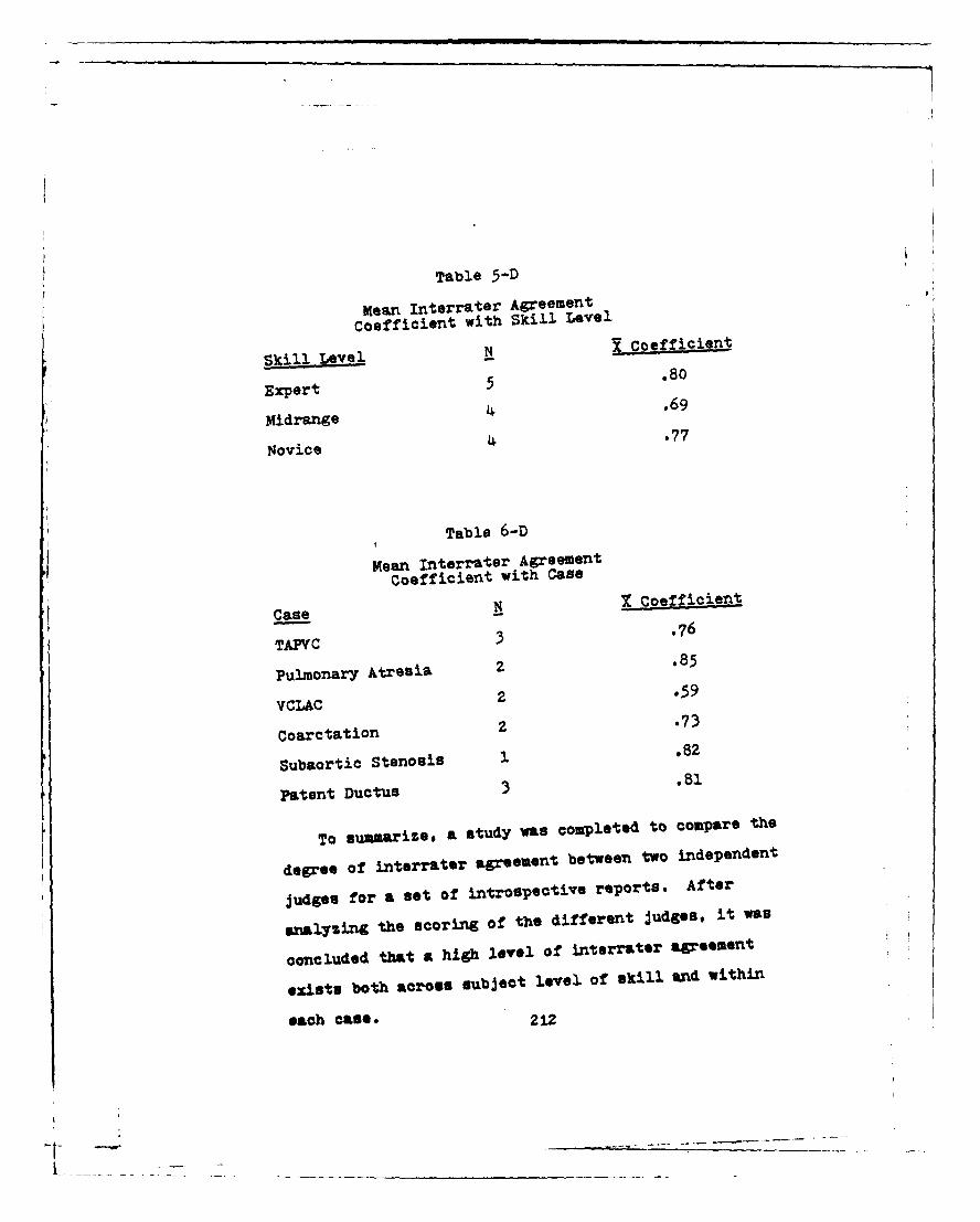
Table 5-D
Mean Interrater AgreementCoefficient with Skill Level
Skill Level N Coefficient
Expert 5 .80
Midrange .69
Novice 4 .77
Table 6-D
Mean Interrater AgreementCoefficient with Case
Case N ! Coefficient
TAPVC 3 .76
Pulmonary Atresia 2 .85
VCLAC 2 .59
Coarctation 2 .73
Subaortic Stenosis 1 .82
patent Ductus 3 .81
To Summarize, a study was completed to compare the
degree of interrater agreement between two independent
judges for a set of introspective reports. After
ialyzing the scoring of the different judges, it was
concluded that a high level of interrater agreement
exists both across subject level of skill and within
each case. 212

APPENDIXC E
SCORING INMTUCTIONS 'CASE I
(General)
All scoring that you do should be as "blind" as
possible as to the identity and level of experience of
the subject being scored. Hence, you should assemble
all subjects' protocols for the case and ask some4
other person to cover up the identifying markings
that appear on each page of each protocol (e.g., S7)
so that these markings are not visible when you score.
A suggestion is to use a small. piece of paper and some
tape so that these covers can be removed later without
damage to the protocol. You should also shuffle the
pile of protocols before scoring so that there will be
no systematic order.
A set of scoring instructions will be provided
for the case. You should first read these instruc-Itions to get a general idea of what you are to bedoing. Then, it is equally important that you read
each protocol once or twice before you do any scoring
to get a general "feel" for what the subject is doing
on the case.
213
1.a

Practice trials and procedure
For all scoring to be done on the case, there
will be a small number (two subjects) of practice scor-
ing trials that will give us an opportunity to compare
our scoring before you score the remainder of cases by
yourself. The procedure to be followed on these prac-
ticestrials is as follows:
1) Read scoring instructions for each type of
scoring you are to do on the case.
2) Score for two subjects whom I will suggest.
Practice subjects are chosen because they
provide good examples of the scoring de-
cisions to be made. For practice scoring
trials it will be necessary for you to know
the number (e.g., S7) of the subject you are
scoring.
3) After scoring, compare your scoring on
practice subjects to mine (which will be
provided) and read my coments on my scor-
ing decisions (also provided) for the prac-
tice subjects. Make notes on discrepancies
Am scorin.
4) When you have done the above for the prac-
tice subjects, call me so that we can
214
I- r_-_____ *,l r "" :-- = ., ",... .. "

discuss any problems you had in scoring and
discrepancies in scoring.
Production Scorina
After the practice period for the case, you
will score all subjects (including trial subjects)
blind and using only the instructions and materials
provided. On the basis of practice scoring trials,
some modifications of scoring instructions may be
made.
21.5

APPENDIX F
SCORING INSTRUCTIONS
Subvalvular Aortic Stenosis,
Use of Aortic Stenosis Variants
There are two objectives of the scoring asso-
ciated with this table. The first is to document the
variants of Aortic Stenosis that are used together by
the subject at the time of presentation of a numbered
data item and at the times when the bubject reports
hunches, and to document the first mentioning (trig-
gering) of each variant by the subject. The second
is to document the evaluative "tone" with which the
subject mentions the variant.
As with other scoring on the Aortic Stenosis
case, the following correspondences hold:
Valvular Aortic Stenosis (ValvAS) - "aortic
stenosis" when it is given without qualifi-
cation as to type (Valvular AS is taken to be
the default value of "aortic stenosis" when
this term is left unqualified), "valvular
aortic stenosis," "aortic valvular stenosis,"
"biscupid aortic valve," and other statements
that clearly place a stenosis at the level of
the aortic valve.
216
- m

Subvalvular Aortic StenosGis (SubAS). "sub-
valvular aortic stenosist "membranous aortic
stenosis,n Onembranoun stenosis," and other
statements that clearly place a stenosis
below the level of the aortic valve.
Suvravalvuwar Aortic Stenosis (SuM.S) - "supra-
valvular aortic stenosis" and other statementsI
that clearly place a stenosis above the level of
4the aortic valve. These are not to be cornfused
with "coarctation of the aorta" and "inter-
rupted aortic arch" which are different diseases.
Scoring is directed at whether or not each of the
three variants of Aortic Stenosis is mentioned by a sub-
ject at the time of presentation of each numbered data
item (and at four points of solicitation of hunches) and,
for each mentioned ether it in mentioned with a posi-
tive (+), negative (-), or neatral (o) valence as a
possible explanation for the patient's condition. The
first mention (triggering) of a variant is a mention like
others and is to be so coded. However, a special symbol
4t) will be used for the first mention of a variant to
signify its special status. Triggerings will also be
coded for evaluative valence ( , -, 0).
217
I- I-

Scoriri Procedure
A table (Table 1) is provided which has three
coding lines for each subject with each line labelled
at the left with either ValvAS, SubAS, or SupAS. Across
the top are codes for the twenty-two data items and the
four places of solicitation of hunches, all arranged in
order from left to right.
The unit of analysis for the scoring is the sub-
ject's entire response after his reading of a numbered
data item. Read each numbered data item 4sEnd "hunches"
at the appropriate points) starting from the beginning
of the case. After reading each data item, then read
the subject's entire response following the data item
and decide which, if any, of the variants of Aortic
Stenosis have been mentioned by the subject. Then, for
each variant mentioned, decide if it is mentioned with a
positive, negative, or neutral evaluative valence as a
possible explanation for the patient's condition and
place a corresponding +, -, o, on the appropriate line
for the variant and under the column heading for the
data item as shown below.(1) (2) . . . . .
ValvAS +S1 SubAS + -
SUpAS
218I
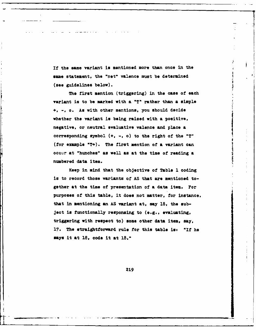
If the same variant is mentioned more than once in the
sme statement, the "net" valence must be determined '(see guidelines below).
The first mention (triggering) in the case of each
variant is to be marked with a "T"n rather than a simple
-o. As with other mentions, you should decide
whether the variant is being raised with a positive,
negative, or neutral evaluative valence and place acorresponding symbol ( a - ) to the right of the "T"
(for example "T+). The first mention of a variant can
occur at "hunches" as well as at the time of reading a
numbered data item.
Keep in mind that the objective of Table 1 coding
is to record those variants of AS that are mentioned to-
gether at the time of presentation of a data item. For
purposes of this table, it does not matter, for instance,
that in mentioning an AS variant at, say 18, the sub-
ject is functionally responsing to (seg. evaluating,
triggering with respect to) some other data item, say,
17. The straightforward rule for this table ist "If he
says it at 18, code it at 18."
219
Guidelines and Cautions
I. "Mention" just means mention
This scoring should be relatively easy since we
are just looking for the variants of AS that are
mentioned in any way in the subject protocols that
follow each data item.
II. Judging , -, 0 in recording the variants mentioned
When a variant of AS is mentioned, it can be used
as a possible explanation for the case with an eval-
uative valence that runs from extreme negative (e.g.,
"That suggests we are surely not dealing with SubASO)
to extreme positive ("Until proved otherwise, now, the
child must have valvular aortic stenosis.") Many
statements fall somewhere in between. When you score
a statement, make every effort to decide if the state-
ment has a basically positive or negative evaluative
loading. The zero (or neutral) convention should be
used s paringly and reserved for those instances where
you simply cannot make a positive-negative discrimin-
ation. Two kinds of statements seem most suited to the
zero convention and examples of each are given below:
Puzzled thoughts and slips of the tongue
(10) On physical exam, this is a healthy ap-
pearing boy. The facies are normal.
220

S. Ok, I think, ah, this is sort of important
again in ruling out certain congenital prob-
lems. I think maybe more helpful, is the
fact that the facies are normal, ah, In the
category of diseases I'm looking at, I believe
it is one form of aortic stenosis, I don't re-
member if it is supra or 8ubvalvular, but ah,
there is a specific facies to ah, that form of
aortic stenosis, and I think this would be a con-
sideration so that this would be helpful in ruling
out that particular form. I believe, I think it
is supravalvular.
Subvalvular Aortic Stenosis is mentioned here for the
first time and must be coded with a T. The evaluative
valence, however, seems impossible to determine and the
zero convention should be used. Supraval1vular is also
mentioned here for the first time, and the valence seems
to be negative. Although the words Naortic stenosis"
appear, it seems clear that the only function of these
words is in conjunction with "supra" or subyalyular"l
hence, Valvular Aortic Stenosim is not coded separate-
ly.
221
L: _ _ __ _. ...__ _ __ __ _ __ _ __ _- _ __ _I___ _I _ __ _

Double evaluations. within the same statement@ that
seem to cancel each other out
(17) There is a systolic thrill felt below
the right clavicle, along the aid-left
sternal border, and in the suprasternal
notoh.
S. Uh, the presence of a thrill in those areas
really suggests that this child has aortic
stenosis. Uh, and (I can't tell from that
description whether it is subvalvular, supra-
valvular, or valvular aortic stenosis), (al-
though the murmur in subvalvular aortic
stenosis is usually located more towards the
aid-left sternal border than the upper right
sternal border).
Subvalvular AS is mentioned here for the first time.
The statement in the first parentheses seems to have
positive valence for SubAS since the subject says the
thrill cannot be used to discriminate among the three
variants. On the other hand, the second statement
(second parentheses) seems somewhat negative for SubAS
since the thrill is not exactly where he would like to
see it. Again, this is an appropriate situation for
222

the zero convention on SubAS9 SupAS and ValvAS are also
mentioned here.
III. Variants mentioned at "hunches" points
Variants that are used at the times when the sub-
ject offers hunches have a special evaluative status.
The fact that they are raised as "hunches" at all
would seem to land some positive valence to their
evaluative dimension - almost no matter how they are
discuseed as hunches. Hence, if a subject says Ox, y,
and z are possible and q is extremely unlikely", al-
though the wording itself for "q" seems negative, the
fact that 'q" is being raised at all at the point
where the subject's job is to report hunches overrides
the wording itself. Short of absolutely negative state-
ments (such as, "It surely cannot be SubAS"), almost
all uses that occur at "hunches points" should be as-
signed a positive valence.
IV. Levels of ambiguity in referencing AS variants
LEM 1 (Little Ambiguity)
These are fairly direct statements of the AS var-
iant (e.g., "aortic stenosis', "SubAS", etc.) and
should cause little problem. The conventions for cor-
respondence, given at the beginning of this set of
223.
~ - F-I-u
rules, should be followed. Remember, "aortic stenosis"
without further qualification, is ValvAS in the
scoring.
.13M 2 (Stenosis mentioned with location discernible):
At times, "stenosis" (or perhaps some variation
like "obstruction") is discussed, the location of the
stenosis is not directly given, but the statement, as
a whole, can be used to discern the location.
Eg. 1 (23) The EKG shows a QRS axis of between +300
and +450. The P waves, T waves, and PR
interval are normal. The pattern shows
probable left ventricular hypertrophy.
So The EKG is compatible with the presence
of a stenotic or an obstruction at the
aortic valve level. It is not diagnostic
by any means. But, it helps to support
the previous concepts of a stenosis at the
aortic level, at aortic valve level, or im-
mediately above it ....
This statement would be scored for both Valvular AS and
Supravalvular AS (from the phrase "immediately above
it").
224
, • "l | |i
Eg. 2 (22) The chest x-ray shows normal cardiac size
and contour and normal vascularity, but
prominence of the ascending aorta.
St OK, that all fits in with, this is prob-
ably post-stenotic aortic dilatation,
which means uh, well, he is twelve years
old, so (it) is probably moderately
stenosed.
The problem is to determine what the referent of "it"
is. The related discussion of "post-stenotic aortic
dilatation" is enough to localize the referant to some
kind of Aortic Stenosis, but is not sufficient to local-
ize beyond that (that is, not to Sub or Supra Valvular),
Hence, by the default conventions, the statement should
coded as ValvAS.
LEVMLI (Referents outside the statement)s
Some subject statements make indirect reference to
something outside of the statement being scored (e.g.,
"That goes along with what I said before"). It has
been decided not to automatically exclude all such
statements as too ambiguous to code. The guideline is
to assign the referent 2n& if you are quite certain as
to its identity. Err on the side of not coding; if
there is any question in your mind, do not code.
225

A rule of thumb might be to assign the referent if you
are sure "beyond a reasonable doubt."
In attempting to assign such a referent, it is
valid to use the case context surrounding the statement
which contains the "outside" reference; however, in
doing so, do not consult farther than one statement
back and one statement forward.
The following is an example of an outside refer-
ence that seems clear enough to code. The outside ref-
erence is in the second statement, 18.
(17) There is a systolic thrill felt below the
right clavicle, along the mid-left sternal
border, and in the suprasternal notch.
S: Ok, I would ah, I guess this would be ah,
somewhat suspicious of aortic stenosis. The
fact that there is a systolic thrill below
the right, yeah, below the right clavicle,
Ok, that is ah, in the aortic area where I
would suspect, where I would expect to find
it if this were aortic stenosis. So I think
this would be a very significant finding,
in helping me to zero in a little bit
stronger on aortic stenosis as my prime
possibility.
226
!I
_ _ _ ___

aa
(18) The first heart sound is normal and there is no
systolic ejection click.
St Ah, the first heart sound is normal. That, I
would expect, ah. The fact that there is no
systolic ejection, ah, would ah. That would go
along too. Im not really expecting ah, a vol-
ume overload, so I guess that would again go
along with what I'm thinking.
(19) A grade 4 over 6 systolic ejection murmur is
heard best at the right upper sternal border
but also heard along the left sternal border
and less well throughout the precordium. The
murmur is also heard in the neck.
St OK, I think this is a very good description of
the murmur of aortic stenosis and this would be
very helpful information for me to ah, point-
ing in on aortic stenosis.
The problem referent is the one referred to by the
phrase "that would go along with what I'm thinking,"
which appears at 18.
At 17, the subject has stated that 17 was a *very
significant finding" that helped him set up Aortic
Stenosis as his "prime possibility". At 19, Aortic
Stenosis is, again, the only disease candidate that ap-
pears. Sandwiched, as it is, between two strong227
assertions for Aortic Stenosis there seems little doubt
that the referent at 18 is the same.
LEVEL 4 (Things too nebulous to code):
Using the "beyond a reasonable doubt" criterion,
there will be statements judged too ambiguous to be
scored as ValvAS, SubAS, or SupAS. This is a judge-
ment call and we must rely on the judgement of the
scorer. The following seems to be an example of such
a statements
(17) There is a systolic thrill felt below the
right clavicle, along the mid-left sternal
border, and in the suprasternal notch.
St Systolic below the right clavicle (pause)
hmm, in the suprasternal notch, that (pause)
a systolic thrill below the right clavicle.
Well, when you have a ductus you can feel a
thrill below the left clavicle, or a coarc.
Maybe this indicates that he has a right
aortic arch and ah, a coarc or a coarc of
his right arch or some other aortic outflow
obstruction. A suprasternal notch thrill,
again, is well known to accompLny ah, left
ventricular outflow obstruction.
228
-r-w -

"Aortic outflow obstruction" and "left ventricular
outflow obstruction" are too ambiguous, here, to oode
as any variant of Aortic Stenosis since the subject
could conceivably mean other things (e.g., various kinds
of interruptions of the aortic arch).
V. Inferences from negations
No inference should be made from the negation of one
AS variant to the assertion of another; that is, "not
valvular aortic stenosis" should not, in itself, be
taken as evidence for the mentioning, by the subject, of
some other variant.
VI. The "non-valvular" convention
When a subject refers to "non-valvular aortic sten-
osis", an attempt should be made to determine which of
the other aortic stenosis variants he means. When you
cannot make this decision with good confidence, leave
the subject statement unscored.
VII . Concatenations of any Aortic Stenosis variant with
Aortic Insufficiency (AI)
Whenever a subject conjoins Aortic Insufficiency
with any one of the Aortic Stenosis variants (a conjoint
hypothesis, ASAI), it is only the Aortic Stenosis part
of the hypothesis that is the target of scoring in
Table l. Use the same rules for scoring this part of
229

the conjoint hypothesis that are to be used for scoring
Aortic Stenosis variants when they occur by themselves.
2
t
230 '
iIS
V

APPEMIX G
CASLE I
Coding by the Second Scorer
for Table 1
Table 1-G gives the final coding for Table 1 in
the body of the report by a second scorer other than
the author. The two scorers, the author and the al-
ternate, recorded a combined total of 168 uses of
the three Aortic Stenosis variants across subjects.
With regard to whether each variant was mentioned
at the time of a particular data point, the two
scorers agreed on 161 (96%) of the coding.. The cells
with a discrepancy are marked with a "check" in
Table 1-G. A check indicates that the author judged
a variant to be used in that cell while the other
scorer did not, or vice-versa. None of these dis-
crepancies would affect any analysis or interpretation
based on Table 1 in the body of the report.
231

X
C4 X X XX XX XXX - XX x l XXXXX
x wx xxxxx
24 X X XX XX X X X XXX
.40 a
x c
ACSx X X X XX
zU
1 x xx x .X X
Cc
x x
N , X XX X
- 0 0 4AWW0 0 0' A O W W O
PAPAM I U2324
'TX X !

APPENDIX H
SCORIN~G DISCUSSIONCase 1 - Table 1
Subject T3
This Appendix gives the complete protocol of sub-
ject T3 from the diagnosis of Case 1. After each
numbered data item from the Case, the subject's re-
sponse is indicated by wT3*o The scoring decisions
of the author in relation to Table 1 in the report
body, are indicated after the subject's response
("DlV etc.), along with the discussions of these
decisions which were presented to the second scorer
as part of the training procedure for scoring (see
Appendix E).
(1) This patient is an 11. year old Caucasian boy.
He weighs 62 pounds and is 53 inches tall. His
presenting problem is that a physician doing a
school physical three months ago, heard a murmur.
22: Now he is well developed, ok, that's normal.
I guess most of these kids when they are referred
to ah, pediatric cardiologist after a murmur is
heard on school physical, most of them turn out
to have a functional murmur. And, ah, and so my
hunch right now is that he's got a functional233
heart murmur. If I were to take a cross-section
of these kids.
2.L There is nothing codable.in this-state-
mentl that in no ValTAS, SubAS, SupAS.
HISTORY
(2) The child was born following a normal pregnancy
and delivery. He was delivered at full term with
a bfrthweight of five pounds, six ounces.
fl, No comment.
D2: Nothing.
(3) A murmur was heard at birth by the attending
physician, but there has never been a follow-up
of this murmur until the present.
t., Again, that may be helpful but ah, again it
could be a fluke. The kid can still have a
functional murmur. It does lend credence to the
fact that probably the kid may have a congenital
heart lesion.
ia, Nothing.(4) Other than the murmur at birth, there were no
problems in the neonatal period. Specifically,
the child was not cyanotic in the neonatal period.
121 No comment.
I " Nothing.
234
~I
() The boy led a normal, asymptomatic childhood
until the age of 10 years when, during an exam-
ination for a school physical, a murmur was
noted by the physician doing the exam and he
referred the child to the University of Minne-
sota for evaluation.
.2s I guess that was the last physical a year
ago and it might take that long to get the kid
plugged into the system.
Rj Nothing.
(6) His growth and development have been normal when
compared to his siblings and he has done well
in school.
fl: No comment.
D6: Nothing.
() There is no history of cyanosis or chest pains.
He currently participates in baseball, football,
and other sports and has no evidence of fatigue
or dyspnea on exertion.
.Us Well, I guess we're being led down that
whatever he's got is probably not significant.
It in not a lesion that is causing him signifi-
cant distress, either cyanosis or congestive
heart failure or cardiac ah, limitation on a
235

cardiac basis. I guess would go along with mild
aortic stenosis or subvalvular aortic stenosis,
coarctation of the aorta, functional heart mur-
mur, anything like that.
_ : "Aortic stenosis" (unqualified as to anatom-
ical type) and "subvalvular aortic stenosis" are
mentioned for the first time and both with a
positive valence. Hence, a "T+ for both ValvAS
and SubAS. This is a good example of why un-
qualified "aortic stenosis" is coded as valvular.
The subject contrasts "aortic stenosis" with
SubAS. When a subject means to refer to a non-
valvular variant, he will do so. This may not
be a perfect heuristic but it is a good one.
(8) There have been no unusual childhood illnesses,
and he has had no hospitalization or operations.
T3: You know this kid still could have acquired
heart disease and ah, I guess that he may have
had subclinical SBE, this happens a lot, that
kids have got a subclinical case of SBE and are
cured and ahow up and have a murmur of ASAI and
then nothing else* The kid could have had a
bicuspid valve at birth and a subclinical case
of SB! I suppose. Whatever it is, it isn't
236
-w--- -z-

causing him much trouble.
D8: Even though it is conjoined with AI, that
is, ASAI, Aortic Stenosis is mentioned here and
not further qualifiedt hence, score ValvAS,
"Bicuspid Aortic Valve" is mentioned here and
we are scoring this as ValvAS. Both mentions of
ValvAS are mildly positivel hence, score +"
for ValvAS.
(9) The family history is negative for congenital
heart disease.
.U: No comment.
Dga Nothing.
(HHX) Hunches after history, please.
TU: My hunch at this point is that the kid has
got minimal congenital heart disease, right now.
And he probably has low-grade aortic stenosis, i
maybe he has subvalvular aortic stenosis which
is a common lesion, he may have coarctation of
the aorta. Those are my hunches following
history.
, Score "+" for both ValvAS and SubAS.
PHYSIC A(10) On plysical examination, this is a healthy ap-
pearing, 11 year old boy. The facies are normal.
T31 Ah. no comment.237

.IOs Nothing.
(11) There is no cyanosis and the fingers and toes
appear normal.
T~s No comment.
Dlls Nothing.
(12) There is no evidence of respiratory distress.
Respiration rate is 16/minute.
T3: No comment.
D12, Nothing.
(13) The blood pressure in the left arm is 90/70, in
the right arm 102/60 and in the leg is 120/60
by auscultation.
TMs Ah, I think in kids that you. can have
ah, ah, blood pressure in your lower extremities
higher than your right, than your upper ex-
tremities. I think that the maximum you are al-
lowed though is ah, the maximum is 15 milli-
meters of mercury which this kid exceeds. I
wonder if he has got multiple peripheral artery
stenosee which we've seen a couple of cases of.
And that would go along again with bicuspid
aortic valve just like coarctation of the aorta.
I think probably that this might be a normal
finding, let's see what his pulses are.
Lug "Bicuspid aortic valve" is the only thing
238

T
here for which we are scoring; hence, "+" for
ValvAS.
(14) The pulses are normal and equal in arms and legs.
The pulse rate is 84/minute.
fut Ah, I'm inclined to believe on the basis of
this, that the kid has got higher blood pressure
in his legs is a normal finding. Especially if
they did it with the wrong size cuffs, we're not
told about that. It might be a normal finding.
D4, Nothing.
(15) The chest is symmetrical with no bulge. There is
mild asymmetry of the back associated with slight
thoracic scoliosis. The lungs are clear to
auscultation.
1t_ Now I assume that this kid didn't have
scoliosis when he was a baby. But, kids with
scoliosis have a variety of heart murmurs which
may or may not be associated with congenital
heart disease. On the other hand kids with con-
genital heart disease frequently have some
scoliosis. That's all.
21 s Nothing.
(16) The apex impulse is in the fifth intercostal
space in the aid-clavicular line.
221 That's normal.
239
T

D16, Nothing.
(17) There is a systolic thrill felt below the right
clavicle, along the id-left sternal border, and
in the suprasternal notch.
T3: That would go along with aortic stenosis, I
think. They don't say this kid has. got any other
kind of vascular disease like Marfans or anything
like that that might present with multiple artery
stenoses, with multiple peripheral artery sten-
oses or diffuse arterial disease. Let me reserve
the right until I see his x-rays and junk in here,
and see the rest of his exam. But I think this
kid has probably got aortic stenosis on the basis
of the suprasternal notch thrill. Ah, okay, ah,
I think I'm at 18, right?
E, Yeah, you're on 18.
IT Unqualified "aortic stenosis" is mentioned
twice, both positively. Hence code "+" for
ValvAS.
(18) The first heart sound is normal and there is no
systolic ejection click.
T2: I guess I'm led to then, to the notion
that this kid has not got ah, valvular aortic
stenosis. FTequently kids with supravalvular
aortio stenosis may have a thrill, so mentioned,
2140

but also these kids may have unusual facies and
they have cupid's bow mouth and saddle nose,
and, no, unusual nose, kind of cutsey-pie
elfin facies. Ah, a lot like the kids with
supravalvular pulmonary artery stenosis. But
you don't need to have that. Subvalvular
aortic stenosis may give the kid a suprasternal
notch, ah, thrill, but ah, I'm not ah, persuaded
by what I've seen so far. Okay, still this kid
could have ah, and still this kid could have ah,
arterial disease, pre-existing arterial disease.
D28: ValvAS, SubAS, and SupAS are all mentioned
here. There is only one mention of Valvular AS
and it is surely negative. "Supravalvular
aortic stenosis" is mentioned for the first time
and should be coded with a "T" at 18 even though
the functional data item for the triggering seemsto be 17. Deciding for the valence for the trig-
gering of SupAS here is difficult. I judged this
to be a case of "multiple evaluations that cancel
each other out':
"Kids with SupAS may have a thrill somentioned"
"but also these kids may have unusual facies"
"but you don't need to have that"
241

Not every such constellation of statements need
be coded "o". Every effort should be made to do-
aide an overall, net valence (+, -). I simply
could not decide either way on SupAS in this
instance and coded it .". "Subvalvular aortic
stenosis" is mentioned and I judged its valence to
be mildly positive.
Note, the alternative scorer judged the eval-
uative valence for SubAS here to be neutral (o).
She took the reference to be one of "multiple
evaluations that cancel",
"SubAS may give a thrill""but I'm not persuaded by what I've
seen so far"
I took the same set of statements to mean "SubAS
is possible but what I've seen so far is not enough
to conclude this." Hence,I saw the statement as
mildly positive at this time.
(19) A grade 4 over 6 systolic ejection murmur is heard
best at the right upper sternal border, but also
heard along the left sternal border, and less well
throughout the precordium. The murmur is also heard
in the neck.
,s Aortic stenosis.
Wt, "Aortic stenosis", hence, "+" on ValvAS.
242
I
I-m l• . , , , l | I -

This particular coding is worth note. It may
well be the case that this particular subject,
and others like him who obviously have know-
ledge of other AS variants and have used them
repeatedly earlier in the case, may in fact
mean more by a general "aortic stenosis"
statement than just Valvular AS; it may well
be a "shorthand" for all the aortic stenoses.
However, across all of these subjects and dif-
fering particular mentions of unqualified
"aortic stenosis", trying to untangle different
meanings for the same words, i.e., "aortic
stenosis", would be too unreliable and tenuous
a process. Hence, we will stay "close to the
belt". Whenever "aortic stenosis" and its cor-
respondents (first sheet of instructions for
scoring Table 1, Appendix F) is given without
qualification as to more specific location
(supra or sub), the only safe, consistent thing
to do is code it as ValvAS. This will be right
most of the time.
(20) The second heart sound is normal and followed by
a grade 1-2 over 6 early diastolic murmur heard
beat at the second left intercostal space.
?3o Aortic insufficiency. So this kid's got
2413

ASAI, no click, he's probably got subvalvular
aortic stenosis.
D20, This one is difficult. OSubvalvular aor-
tic stenosis" is mentioned and the tone is
positives hence, " " for SubAS. The hard part
is what to do with the unqualified "AS" as part
of "ASAI". By the rules, this should be
treated as ValvAS. In keeping with the rules,
I coded a mention of YalvAS and coded the val-
once as "0".
(21) Neither the liver nor the spleen was palpable.
XTf Ah, I think I don't have any comment
about that. And they don' t say anything about
family history.
D21, Nothing.
(HPEx) At the end, do you have hunches at the end of
this?
T3, Yeah, I do. I think this kid has got
subvalvular aortic stenosis probably membran-
ous. The only thing that they don't mention
anything about what the murmur does with exer-
ciao or changing in position. But I think that
this kid has got a history that is most like ah,
membranous subaortic stenosis.
244-

DHPEX: "Subvalvular aortic stenosis",
probably membranous", and "membranous sub-
aortic stenosis" are all input to one "+" on
SubAS.
X-RAY
(22) The chest X-ray shows normal cardiac size and
contour and normal vascularity, but prominence
of the ascending aorta.
T: The ah, this would go along with ah, with
ah, membranous subaortic stenosis, as well.
I'm, okay, it would go along with membranous
subaortic stenosis as well. If he has some
dilatation of the aortic root it could be on
the basis of his ASAI and that ASAI is probably
secondary to the wear and tear on the valve.
Usually, _.says *at mso t ki* with Vaivmla iAS
have, valvular AS have a click and its absence
mitigates against valvular aortic stenosis.
The kid has probably got a subvalvular &em-
brane and Al secondary to that, secondary to
that. And that is probably accounting for his
secondary findings.
245
-n --
D22s The statements regarding SubAS are
clearly positive and the statements regard-
ing "Valvular aortic stenosis" are clearly
negativet "absence (of click) mitigates
against valvular aortic utenosis".
Note, this is a nice example of how a
subject reconciles Al in SubAS, that is, as
resulting from secondary damage to the aortic
valve from blood "pounding" on it over the sub-
aortic stenosis (see Scoring Guidelines for
Table 1, Appendix F).
(MVray) Hunches after X-ray?
1t My hunch at the end of x-ray is the same,
that he has, that he has ah, membranous Sub-
aortic Stenosis with aortic valvular insuf-
ficiency.
DHrayt Positive SubAS Is clear.3KG.
(23) The KG shows a QRS axis of between +300 and
+450 . The P waves, T waves, and PR interval
are normal. The pattern shows probable left Iventricular hypertrophy.
T3s So he's not got strain. He's not got,
if he doesn't have an ectopic T ware axis
he's not got strain, his axis is normal but24.6
I|
t -

in the leftward direction he's got probably
left ventricular hypertrophy, he's probably
got some mild aortic outflow obstruction, he
probably doej have a subvalvular membrane, he
probably does have aortic insufficiency ah,
which is probably moderate, probably not in-
considerable and he probably doesn't have
aortic valvular stenosis. If he did have a
click I would say that this kid probably had
a bicuspid aortic valve, occult secondary ah,
subacute bacterial endocarditis, now resolved.
I suppose that the absence of a click may be,
ah, may be misleading so that I guess that is
probably my secondary diagnosis. Is that
okay? My summary then, do you want?
D23: "He probably does have a subvalvular mem-
brane . . . and he probably doesn't have
aortic valvular stenosis." A plus for SubAS
and a minus for ValvAS seems clear. Note that 4
the deciding issue is really the "no click".
(HEKG) Yeah, why don't you just give me a set of
hunches now and then I'll ask you ah, you know,
the straightforward question.
flt Okay, my hunch is that the kid has got
subvalvular AS with ah . . . subvalvular
24~7
iii

membranous AS with secondary aortic insuf-
ficiency, that's my hunch.
DHE: Positive SubAS
Fs Okay, now we can get to the other thing.
I would like you to, as explicitly as possi-
ble, give me a primary diagnosis.
Tlt My primary diagnosis is subvalvular mem-
branous aortic stenosis with aortic insuf-
ficiency.
Et Okay.
fli And my secondary diagnosis is ah, is ah,
aortic stenosis with insufficiency ah, prob-
ably with bicuspid aortic valve and probably ah,
with a history of occult subacute bacterial
endocarditis.
Comment
Note here that in an obvious reference to Val-
vular Aortic Stenosis, the subject uses the simple term
"aortic stenosiso. ValvAS is surely the "unmarked
case" or "default value" of unqualified "aortic
stenOSis".
iji

APPENDIX I
SCORING INTRUCTIONS
Subvalvular Aortic StenosisInstructions for Scoring Target Data Items
The object of this scoring is to document how the
subject evaluates each of a set of target data items
with respect to the major variants of Aortic Stenosis,
that is, Valvular Aortic Stenosis (or unqualified
"Aortic Stenosis"), Subvalvular Aortic Stenosis, and
Supravalvular Aortic Stenosis. Generally speaking, the
target data items are the main object of this scoring
and the intent is to determine whether the subject in-
terprets each target item as positive, negative, or
neutral evidence for the presence in the patient of
each Aortic Stenosis variant.
There are six target data items. Some items con-
stitute the entire numbered data item presented to the
subject in the case; others are a subpart of a numbered
data item* The six target items are given below. For
those that are a subpart of a numbered data item, the
target subpart is underlined.
NORMAL FACIES,
(10) On physical examination, this is a healthy-
apearing, 11_year old _boy. The facies
249

are normal.
(17) There is a systolic thrill felt below the
right clavicle, along the aid-left sternal
border, and in the suprasternal notch.(ENTMRX)
NO CLICK .
(18) The first heart sound is normal and there is
no systolic ejection click.
THE MURMUR
(19) A grade 4 over 6 systolic ejection murmur isheard best at the right upper sternal border,
but also heard along the left sternal border,
and less well throughout the precordium. The
murmur is also heard in the neck.(XNTIRE)
AORTIC INSUFFICIENCY:
(20) The second heart sound is normal and followed
by a grade 1-2 over 6 early diastolic murmur
heard best at the second left intercostal space.
PROMINENT AORTA:
(22) The chest x-ray shows normal.cardiac.size and
contour and normal vascularity, but p
of the ascending aorta.
As with Table 1 scoring, the target set of diseases
is the not of Aortic Stenosis variantsi Valvular AS,
Subvalyular AS, and Supravalvular AS. The set of
250

correspondences between these three types of AS and
particular subtypes and wordings that are to be in-
cluded within each is the same as for Table 1 scoring
and the first page of Table 1 scoring instructions
should be consulted.
Evaluation of a target item with respect to one or
more of the AS variants need not occur at the time when
a target data item is presented (e.g., a subject may
evaluate 17 at time of presentation of, say 18). The
objective is to score the evaluation wherever it oc-
curs. Preliminary screening has shown that the only
evaluations of the target items in relation to AS var-
iants occur at item 10 and items 17-HEKG. Hence,
scoring will focus on subjects' responses at these
spots which are potential "places of evaluation"
(POE's).
Scoring in Table 2 really involves three kinds of
decisionss
(a) Deciding if any and which Aortic Stenosisvariant(s) is mentioned.
(b) For AS variants mentioned, deciding if any andwhich target item(s) is being evaluated.
(c) Deciding the valence (+, -, o) for the eval-uation.
251
I

I
Scorin Progedure
A table (Table 2) is provided for this scoring.
Across the top are the six target items asid, under each,
codes for each of the three AS variants. On the left
margin, for each subject, are listed the ten potential
places of evaluation (POE'S) which are ordered from top
to botton (10, 17-HEKG).
Read the entire subject response at each POE, taken
in order, starting at 10 and then skipping to 17. At
each POE, decide if any and which AS variants are men-
tioned. For each AS variant mentioned, decide if any
and which target data items are being evaluated with re-
spect to the AS variant. For each AS variant - target
data item pair, decide the valence of the evaluation
(positive, negative, neutral) and place a " ", "-, or
"o" to the right of the POE and in the column corres-
ponding to the AS variant and target data item being
evaluated by the subject.
Guidelines and Cautions
I. Deciding if any and which AS variant is mentioned
This component of the task of scoring Table 2 is fthe sme as the scoring task for Table 1 and the
correspondence conventions (first page, Table 1
252
A
9.

scoring, Appendix F) and scoring guidelines (especially
Table 1, #IV, Appendix F, Levels of ambiguity in refer-
encing AS variants) for that table should be applied.
In particular, the AS variant being evaluated by the
subject in relation to a target data item will not al-
ways be named explicitly. Table 2 scoring is quite im-
portant; hence, every attempt should be made to dis-
ambiguate which AS variant is being evaluated. The
Table 1 guidelines set reasonable boundaries on how
far we can go in attempting disambiguation.
For Table 2, it is not enough that an AS variant be
mentionedi that is only the first step in deciding a
codable item. In addition, the AS Yariant, once men-
tioned, must be evaluated with respect to some target
data item.
II. Deciding if any and which target item(s) is eval-
uated
To constitute something scorable, an AS variant
must be evaluated with respect to some target data
item. Evaluation of an AS variant with respect to a
target item means that a target item is referred to by
the subject, in relation to an AS variant, in a manner
that indicates that the target item bears on the
status of the AS variant as a candidate hypothesis
253
- ,~----...-..--.........---...........---..==-=Nona*=- -

(i.e., the target item is evaluated as evidence, of
whatever kind, for the AS variant).
A difficulty arises because some target data items
are subparts of numbered data items (presented in the
case) and we are only interested in the target subpart
(as discussed earlier in the Appendix). The following
rules should apply.
Tarxet subpart mentioned explicitlys If the target sub-
part of numbered data item is explicitly mentioned (e.g.,
"no click") in an evaluation, there is no problem.
Score the evaluation.
Numbered data item only If only the numbered data item
containing the target subpart is referred to in an eval-
uation - without distinction as to which subpart is
being evaluated - score it as though it were the target
subpart. In this sense, the target subpart is the "de-
fault" for the numbered data item. That is, unless
otherwise indicated, we assume the subject is referring
to the target.
Non-target submart mentioned explicitlys When the non-
target subpart of a numbered data item is referred to
explicitly in an evaluation, the evaluation is not to
be scored,
2541
. .

Eg. (18) The first heart sound is normal and there is
no systolic ejection click.
St (The first heart sound is normal so probably
the two A-V valves are normal). There is no
systolic ejection click as you would expect
with valvular aortic stenosis . . .
The first parenthesized statement, since it explicitly
refers to the non-target part of 18, is not a scorable
statement (even if the subject had evaluated the "nor-
mal first heart sound" with regard to some AS variant).
The remainder of this subject's statement is scorable.
III. Judging +, -, o for the evaluation
Whenever an AS variant is evaluated with respect
to a target data item, the valence of the evaluation
can run from extreme negative ("Well, that rules out
SubAS.") to extreme positive ("Well, that strongly sup-
ports the idea that we are dealing with SubAS in this
patient") with some turning point in between. The gen-
eral guidelines given in the instructions for Table 1,
#II, Appendix F, should be followed here.
Again, it should be emphasized that the zero con-
vention should be used sparingly; use the subject's
whole statement and make every effort to detect a posi-
tive or negative valence. The zero convention should
be reserved for the instances where you simply cannot
255
_T

decide.
"Multiple evaluations that cancel each other outm
is still a good candidate for the zero convention.
(But not necessarily -. only if you cannot decide the
net valence). However, in Table 2 scoring, this would
refer only to multiple evaluations at the same POE.
If a subject makes an additional evaluation of a target
item at a different POE, score the POE's separately.
This is the main reason for including different POE's
in the table.
IV. Scoring at "hunches" points
Remember, that the objective of Table 2 scoring is
to score evaluations of target data items. Hence, when
mention is made of AS variants at "hunches" points,
they are only scorable if they refer to some target
data item. Subject discussions of AS variants at
"hunches" points that do not make use of some target
data item are not of interest here.
V. Target items that happen to be trigger points
It makes no difference, for this table, if a tar-
get item happens to be the item where an AS variant is
first mentioned.* In the sense of this table, trigger-
ing is an evaluation like any other. Just decide the
Positive, negative, or neutral valence of the target
256

item - AS variant relationship.
VI. Target item 20 - Aortic Insufficiency
Data item 20 is a straightforward description of
the murmur of Aortic Insufficiency (AI).and just about
all subjects will interpret it as such. This itself is
not the interpretation of interest in this table. The
question, for this table, is whether the subject then
goes on to interpret either Al or 20 in relation to,
that is, as evidence for, a variant of AS. The follow-
ing guidelines should apply.
(a) If the subject interprets 20 as AI but makes no
evaluation of Al with respect to an AS variant,
the statement is not scorable.
(b) If the subject interprets 20 as Al and evaluates
Al with respect to some AS variant, the state-
ment is scorable.
(a) If the subject does not explicitly interpret 20
as AI, but does evaluate 20 with respect to some
AS variant, score the statemente In this in-
stance, we will assume the evaluation of 20 is
mediated by Al.
AI is a condition that occurs more often with some
variants of AS than with others, hence, AI constitutes
legitimate evidence for deciding which variant of AS is
257
present in the patient. We are only interested in Al
when it is used by the subject for this function.
VII. AI and the "prominent aorta"
In evaluating "prominent aorta" with regard to
Subvalvular AS, a subject may make a statement that,
on the surface of it, seems negative. If, however, he
goes on to say that the "proainent aorta" could be ac-
counted for, under SubAS, when SubAS is accompanied by
AI, then you should try to decide if the subject is
using the accompanying Al to account for the aorta.
If you decide he is, then the real evaluation is more
likely positive. This is a Judgment call and is left
to the scorer. The subject's response to 20 can pro-
vide some guidance by providing evidence of how con-
vinced the subject is that Al is present. The follow-
ing is an example that appears to take Al into account
("Yalvar damage" means AI),
Eg. (22) The chest X-ray shows normal cardiac size
and normal vascularity, but prominence of the
ascending aorta.
St Again, this is consistent with aortic sten-
osis, ah, and this could represent just
post-stenotic dilatation. Ah, again, usually
you don't have, you don't have, post-
stenotic dilatation with SubAs unless you
258
-q

have some valvar damage as well. That is about
all I can say.
This is surely positive for general Aortic Stenosis.
The questionable part is the evaluation with respect
to SubAS. The evaluation should probably be taken as
having a positive valence, especially since the sub-
ject had earlier (at 20) strongly tied SubAS to Al:
(20) _
Ss Now, this is consistent with some aortic
insufficiency and, ah, adds weight to the
possibility of a subaortic membrane ah,
which has a high incidence of a diastolic
murmur associated with it.
VIII. Hypothetical considerations of data
At times, subjects make statements about what
their thoughts might be, had the data been other than
it was. We can only handle data that was presented in
the case and cannot deal with what might have been&Eg. (HEKG) 4
St So my continuing hunch is congenital aortic
stenosis, presumably subaortic if the examiner
is correct in his description of the physical
259
.V £

findings, but if there is an ejection click
present, that was missed by the examiner,
then this is congenital valvular aortic
stenosis with some hypertrophy.
The case, in fact, presented "no click" which is judged
here as positive for SubAS. The subject's proposal of
the presence of a click and its positive loading on
Valvular AS is hypothetical.
; 260
1
r K

APPENDIX J
CASE 1 0
Coding by the Second Scorerfor Table 2
The purpose of coding that produced Table 2 in
the rep.--t body for Case 1, was to document the eval-
uation by subjects of six target data items with re-
spect to the variants of Aortic Stenosis. Subjects
were free to evaluate, at any later time, any data
item they had already seen. Hence, evaluations of a
target item were not restricted to the place in the
case where the item was first presented to the subject.
The objective in coding for Table 2 was to capture all
evaluations of a target item as positive(+), negative
or "neutral" (o) evidence for the existence of one
of the variants of Aortic Stenosis in the patient, no
matter when these evaluations took place (MZerom is
best thought of as an evaluation for which the scorer
could not decide whether the data item was viewed as
positive or negative evidence by the subject - ae
Appendix I for scoring instructions on this point).
When a scorer judged that there was more than one
separate evaluation of a target item with respect to
the same AS variant, the Onetv evaluation was used in
constructing Table 2. Neither scorer judged that there
261
1~ i

were ever more than two separate evaluations of any
target data item with respect to the same variant;
hence, the following rule was used in constructing
Table 2:
First Second Recorded inEValuation Evaluation Table 2
+ 0
No combination involving two uses of "zero" occured for
either scorer.
Table 1-J gives the equivalent of Table 2 produced
from the coding of the second scorer. A non-blank cell
entry in either of these two tables indicates that the
scorer judged a data item - AS variant evaluation (at
least one) to have taken place. There are a combined
total of 88 such entries across the two tables. The
two scorers agreed on 83 of these (94%) that a codable
evaluation had taken place. The cells of disagreement
are marked with a single "check" in Table l-J in-
dicating that the author judged an evaluation to have
occurred while the second scorer did not or vice-versa.
There are 83 data item - AS variant cells in which
both scorers judged an evaluation to have taken place.
In 78 of these (94%), the two scorers assigned the
262
IT
I

6w6
o+ + + + + +
+ + +
, . .. !U C
+ +2
" -s a E + + + .
'. + + +
LUUI I
+E +
>2
+ +U
fb +4. 4
W.5
263
t''-i U, a _ m I I I. . ... !- ..
hill in ii lI 'nn ,n I I I l. . . . .. 0.. 1. . .

identical value (+, -o a) to the evaluation* No dis-
agreements crossed the "zero boundary"I that is, there
is no cell where one scorer assigned a "plus" while the
other assigned a "minus". The cells in which the two
scorers disagreed on the Ovalue" of the evaluation are
marked with a double "check" in Table l-J,
Except for the disagreements on the evaluations of
"NO CLICK" in relation to Valvular Aortic Stenosis, no
other disagreements would in any way alter the analyses
or interpretation based on Table 2 in the body of the
report except that one student (32), based on Table l-J,
would be given "credit" for evaluating all three variants
with respect to one (the same) data item.
The only effect of the "NO CLICK" disagreements
would be to make the discussion of the evaluations of
this finding by some individual subjects somewhat less
*clean" (although it would not be altered in any sub-
stantial way). This being the case, the disagreements
on this finding merit some discussion.
The author judged 32 to have evaluated "NO CLICK"'
as positive for ValvAS while the second scorer judged
no evaluation to have taken place. The problem here is
an indirect referents "The fact that there is no sys-
tolio ejection, that would go along too . . . that
264
,J

4 t
would agin go along with what I' thinking." The
question is whether, a referent can be assigned for what
the subject is "thinking." At the immediately pro-
ceding data item the subject had said that his "prime
possibility" was VaIvular Aortic Stenosis. Scorers
were allowed to assign indirect referents if they were
confident in them (Se Appendix F instructions) and this
referent seemed clear to the author.
Regarding T4, the relevant protocol for the subject 4
is given as an example in Figure 5 of the body of the
report. This is a relatively clear negative evaluation
by the standards of what on* ses in the protocols from
this study, although the author may have benefitted
from having heard the consternation in the tone of the
subject's voice at this point in the case.
The difficulty for both subjects 32 and Z3 has
exactly the same basis which can be seen in 23's proto-
col given in Figure 4 of the body of the report. The
subjects raised the idea that Valvular Aortic Stenosis
of a "very severe degree" night present without a
click. The author took this as a "hypothetical" (i.e.,
"no click" would be expected had the AS been severe) and
judged the basic evaluation to be negative.
265
PITTSBURGH/LESGOLD September 9, 1981 Page 1
Navy Navy
Dr. Ed Aiken 1 Dr. William L. MaloyNavy Personnel R&D Center Principal Civilian Advisor forSan Diego, CA 92152 Education and Training
Naval Training Command, Code OOAMeryl S. Baker Pensacola, FL 32508NPR DCCode P309 1 CAPT Richard L. Martin, USNSan Diego, CA 92152 Prospective Commanding Officer
USS Carl Vinson (CVN-70)Dr. Robert Breaux Newport News Shipbuilding and Drydock CoCode N-711 Newport News, VA 23607NAVTRAEQUIPCENOrlando, FL 32813 1 Dr William Montague
Navy Personnel R&D CenterCDR Mike Curran San Diego, CA 92152Office of Naval Research800 N. Quincy St. 1 Ted M. I. YellenCode 270 Technical Information Office, Code 201Arlington, VA 22217 NAVY PERSONNEL R&D CENTER
SAN DIEGO, CA 92152DR. PAT FEDERICONAVY PERSONNEL R&D CENTER I Library, Code P201LSAN DIEGO, CA 92152 Navy Personnel R&D Center
Son Diego, CA 92152Dr. John FordNavy Personnel R&D Center 1 Technical DirectorSan Diego, CA 92152 Navy Personnel R&D Center
San Diego, CA 92152I LT Steven D. Harris, MSC, USN
Code 6021 6 Commanding OfficerNaval Air Development Center Naval Research LaboratoryWarminster, Pennsylvania 18974 Code 2627
Washington, DC 20390I Dr. Jim Hollan
Code 304 1 PsychologistNavy Personnel R & D Center ONR Branch OfficeSan Diego, CA 92152 Bldg 114, Section D
666 Summer StreetDr. Norman J. Kerr Boston, MA 02210Chief of Naval Technical TrainingNaval Air Station Memphis 75) 1 PsychologistMllington, TN 38054 ONR Branch Office
536 S. Clark StreetMr. Orvin A. Laron Chicago, ML 60605Code 306Navy Personal RD Center 1 Office of Naval ResearchSan Diego, CA 92152 Code 437
800 N. Quincy SStreetArlington, VA 22217
-1
PITTSBURGH/LESGOLD September 9. 1981 Page 2
Navy Navy
5 Personnel & Training Research Programs 1 Dr. Alfred F. Smode(Code 458) Training Analysis & Evaluation Group
Office of Naval Research (TAEG)Arlington. VA 22217 Dept. of the Navy
Orlando, FL 32813I Psychologist
ONR Branch Office 1 Dr. Richard Sorensen1030 East Green Street Navy Personnel R&D CenterPasadena, CA 91101 San Diego, CA 92152
1 Special Asst. for Education and 1 Roger Weissinger-BaylonTraining (OP-O1E) Department of Administrative Sciences
Rm. 2705 Arlington Annex Naval Postgraduate SchoolWashington. DC 20370 Monterey, CA 93940
1 Office of the Chief of Naval Operations 1 Dr. Robert WisherResearch Development & Studies Branch Code 309
(OP-115) Navy Personnel R&D CenterWashington, DC 20350 San Diego, CA 92152
1 LT Frank C. Petho, MSC, USN (Ph.D) 1 Mr John H. WolfeSelection and Training Research Division Code P310Human Performance Sciences Dept. U. S. Navy Personnel Research and
Naval Aerospace Medical Research Laborat Development CenterPensacola, FL 32508 San Diego, CA 92152
1 Dr. Gary PoockOperations Research DepartmentCode 55PKNaval Postgraduate SchoolMonterey, CA 93940
1 Roger W. Remington, Ph.DCode L52NAMRLPensacola, FL 32508
1 Dr. Worth Scanland, DirectorResearch, Development, Test & EvaluationN-5Naval Education and Training CommandNAS, Pensacola, FL 32508
1 Dr. Robert G. 3ithOffice of Chief of Naval OperationsOP-987HWashington. DC 20350
- - r ,P ,-, ,--
PITTSBURGH/LESGOLD September 9, 1981 Page 3
Army Army
Technical Director 1 Dr. Frederick SteinheiserU. S. Army Research Institute for the Dept. of Navy
Behavioral and Social Sciences Chief of Naval Operations5001 Eisenhower Avenue OP-113Alexandria, VA 22333 Washington, DC 20350
Mr. James Baker 1 Dr. Joseph WardSystems Manning Technical Area U.S. Army Research InstituteArmy Research Institute 5001 Eisenhower Avenue5001 Eisenhower Ave. Alexandria, VA 22333Alexandria, VA 22333
1 Dr. Beatrice J. FarrU. S. Army Research Institute5001 Eisenhower Avenue 4Alexandria, VA 22333
Dr. Dexter FletcherU.S. Army Research Institute5001 Eisenhower AvenueAlexandria,VA 22333
DR. FRANK J. HARRISU.S. ARMY RESEARCH INSTITUTE5001 EISENHOWER AVENUEALEXANDRIA. VA 22333
Dr. Michael KaplanU.S. ARMY RESEARCH INSTITUTE5001 EISENHOWER AVENUEALEXANDRIA, VA 22333
Dr. Milton S. KatzTraining Technical AreaU.S. Army Research Institute5001 Eisenhower AvenueAlexandria, VA 22333
Dr. Harold F. O'Neil, Jr.Attn: PERI-OKArmy Research Institute5001 Eisenhower AvenueAlexandria, VA 22333
1 Dr. Robert 3SsmorU. S. Army Research Institute for the
Behavioral and Social Sciences5001 Eisenhower AvenueAlexandria, VA 22333

PITTSBURGH/LESGOLD September 9, 1981 Page 4
Air Force Marines
Dr. Earl A. Alluis 1 H. William GreenupHQ. AFHRL (AFSC) Education Advisor (E031)Brooks AFB, TX 78235 Education Center, MCDEC
Quantico, VA 22134Dr. Genevieve HaddadProgram Manager 1 Special Assistant for MarineLife Sciences Directorate Corps MattersAFOSR Code lOOMBolling AFB, DC 20332 Office of Naval Research
800 N. Quincy St.2 3700 TCHTW/TTGH Stop 32 Arlington, VA 22217
Sheppard AFB, TX 763111 DR. A.L. SLAFKOSKY
SCIENTIFIC ADVISOR (CODE RD-1)
HQ, U.S. MARINE CORPSWASHINGTON, DC 20380

PITTSBURGH/LESGOLD September 9, 1981 Page 5
CoastGuard Other DoD
Chief, Psychological Reserch Branch 12 Defense Technical Information CenterU. S. Coast Guard (G-P-1/2/TP42) Cameron Station, Bldg 5Washington. DC 20593 Alexandria, VA 22314
Attn: TC
1 Military Assistant for Training andPersonnel Technology
Office of the Under Secretary of Defensefor Research & Engineering
Room 3D129, The PentagonWashington, DC 20301
1 DARPA14O0 Wilson Blvd.Arlington, VA 22209
4
~ ~ -

PITTSBURGH/LESGOLD September 9, 1981 Page 6
Civil Govt Non Govt
Dr. Paul G. Chapin 1 Dr. John R. AndersonLinguistics Program Department of PsychologyNational Science Foundation Carnegie Mellon UniversityWashington, DC 20550 Pittsburgh, PA 15213
Dr. Susan Chipman 1 Anderson, Thomas H., Ph.D.Learning and Development Center for the Study of ReadingNational Institute of Education 174 Children's Research Center1200 19th Street NW 51 Gerty DriveWashington, DC 20208 Champiagn, IL 61820
William J. cLaurin 1 Dr. John Annett66610 Howie Court Department of PsychologyCamp Springs, MD 20031 University of Warwick
Coventry CV4 7ALDr. Arthur Melmed ENGLANDNational Intitute of Education1200 19th Street NW 1 DR. MICHAEL ATWOODWashington, DC 20208 SCIENCE APPLICATIONS INSTITUTE
40 DENVER TECH. CENTER WESTDr. Andrew R. Molnar 7935 E. PRENTICE AVENUEScience Education Dev. ENGLEWOOD, CO 80110
and ResearchNational Science Foundation 1 1 psychological research unitWashington, DC 20550 Dept. of Defense (Army Office)
Campbell Park OfficesDr. Joseph Psotka Canberra ACT 2600, AustraliaNational Institute of Education1200 19th St. NW 1 Dr. Alan BaddeleyWashington,DC 20208 Medical Research Council
Applied Psychology UnitDr. Frank Withrow 15 Chaucer RoadU. S. Office of Education Cambridge CB2 2EF400 Maryland Ave. SW ENGLANDWashington, DC 20202
1 Dr. Patricia BaggettDr. Joseph L. Young, Director Department of PsychologyMemory & Cognitive Processes University of ColoradoNational Science Foundation Boulder, CO 80309Washington, DC 20550
Mr Avron BarrDepartment of Computer Science
Stanford UniversityStanford, CA 94305
Liaison ScientistsOffice of Naval Research,Branch Office -, LondonBox 39 FPO New York 09510
1 4l -" - m - " " . . ..

|I
PITTSBURGH/LESGOLD September 9, 1981 Page 7
Non Govt Non Govt
Dr. Lyle Bourne 1 Dr. Allan M. CollinsDepartment of Psychology Bolt Beranek & Newman. Inc.University of Colorado 50 Moulton StreetBoulder, CO 80309 Cambridge, Ma 02138
Dr. John S. Brown 1 Dr. Lynn A. CooperXEROX Palo Alto Research Center LRDC3333 Coyote Road University of PittsburghPalo Alto, CA 94304 3939 O'Hara Street
Pittsburgh, PA 15213Dr. Bruce BuchananDepartment of Computer Science 1 Dr. Meredith P. CrawfordStanford University American Psychological AssociationStanford, CA 94305 1200 17th Street, N.W.
Washington, DC 200361 DR. C. VICTOR BUNDERSON
WICAT INC. 1 Dr. Kenneth B. CrossUNIVERSITY PLAZA, SUITE 10 Anacapa Sciences, Inc.1160 SO. STATE ST. P.O. Drawer QOREM, UT 84057 Santa Barbara, CA 93102
Dr. Pat Carpenter 1 LCOL J. C. EggenbergerDepartment of Psychology DIRECTORATE OF PERSONNEL APPLIED RESEARCCarnegie-Mellon University NATIONAL DEFENCE HQPittsburgh, PA 15213 101 COLONEL BY DRIVE
OTTAWA, CANADA K1A OK2Dr. John B. CarrollPsychometric Lab 1 Dr. Ed FeigenbaumUniv. of No. Carolina Department of Computer ScienceDavie Hall 013A Stanford UniversityChapel Hill, NC 27514 Stanford, CA 94305
Dr. William Chase 1 Dr. Richard L. FergusonDepartment of Psychology The American College Testing ProgramCarnegie Mellon University P.O. Box 168Pittsburgh, PA 15213 Iowa City. IA 52240
Dr. Michelin@ Ci I Mr. Wallace FeurzeigLearning R & D Center Bolt Beranek & Newman, Inc.University of Pittsburgh 50 Moulton St.3939 O'Hara Street Cambridge, MA 02138Pittsburgh, PA 15213
1 Dr. Victor FieldsDr. William Clancey Dept. of PsychologyDepartment of Computer Science Montgomery CollegeStanford University Rockville, MD 20850Stanford, CA 94305
II
U - _ _ __ _
PITTSBURGH/LESGOLD September 9, 1981 Page 8
Non Govt Non Govt
Dr. John R. Frederiksen 1 Dr. Earl Hunt ,Bolt Beranek & Newman Dept. of Psychology50 Moulton Street University of WashingtonCambridge, MA 02138 Seattle, WA 98105
Dr. Alinda Friedman 1 Dr. Ed HutchinsDepartment of Psychology Navy Personnel R&D CenterUniversity of Alberta San Diego, CA 92152Edmonton, AlbertaCANADA T6G 2E9 1 Dr. Walter Kintsch
Department of PsychologyDR. ROBERT GLASER University of ColoradoR1DC
Boulder, CO 80302UNIVERSITY OF PITTSBURGH3939 O'HARA STREET Dr. David KierasPITTSBURGH, PA 15213 Department of Psychology
University of ArizonaDr. Marvin D. Glock Tuscon, AZ 85721217 Stone HallCornell University 1 Dr. Kenneth A. KlivingtonIthaca, NY 14853 Program Officer
Alfred P. Sloan FoundationDR. JAMES G. GREENO 630 Fifth AvenueLRDC New York, NY 10111UNIVERSITY OF PITTSBURGH3939 O'HARA STREET Dr. Stephen KosslynPITTSBURGH, PA 15213 Harvard University
Department of PsychologyDr. Barbara Hayes-Roth 33 Kirkland StreetThe Rand Corporation Cambridge. MA 021381700 Main StreetSanta Monica, CA 90406 1 Dr. Marcy Lansman .,
Department of Psychology, NI 25Dr. Frederick Hayes-Roth University of WashingtonThe Rand Corporation Seattle, WA 981951700 Main StreetSanta Monica, CA 90406 1 Dr. Jill Larkin
Department of PsychologyDr. Kristina Hooper Carnegie Mellon UniversityClark Kerr Hall Pittsburgh, PA 15213University of CaliforniaSanta Cruz, CA 95060 1 Dr. Alan Lesgold
Learning R&D CenterGlenda Greenwald, Ed. University of Pittsburgh"Human Intelligence Newsletter" Pittsburgh, PA 15260P. 0. Box 1163Birmingham, MI 48012
1-L~ _ _ _

PITTSBURGH/LESGOLD September 9, 1981 Page 9
Non Govt Non Govt
Dr. Erik McWilliams 1 Dr. Fred ReifScience Education Dev. and Research SESAME '
National Science Foundation c/o Physics DepartmentWashington, DC 20550 University of California
Berkely, CA 94720Dr. Mark MillerTI Computer Science Lab 1 Mary RileyC/O 2824 Winterplace Circle LRDCPlano, TX 75075 University of Pittsburgh
3939 O'Hara StreetDr. Allen Mi-o Pittsburgh, PA 15213Behavioral Technology Laboratories1845 Elena Ave., Fourth Floor 1 Dr. Andrew M. RoseRedondo Beach, CA 90277 American Institutes for Research
1055 Thomas Jefferson St. NWDr. Donald A Norman Washington, DC 20007Dept. of Psychology C-009Univ. of California, San Diego 1 Dr. Ernst Z. RothkopfLa Jolla, CA 92093 Bell Laboratories
600 Mountain AvenueDr. Seymour A. Papert Murray Hill, NJ 07974Massachusetts Institute of TechnologyArtificial Intelligence Lab 1 Dr. David Rumelhart545 Technology Square Center for Human Information ProcessingCambridge, MA 02139 Univ. of California, San DiegoLa Jolla, CA 92093
Dr. James W. Pellegrino
University of California, 1 Dr. Alan SchoenfeldSanta Barbara Department of Mathematics
Dept. of Psychology Hamilton CollegeSanta Barabara, CA 93106 Clinton, NY 13323
MR. LUIGI PETRULLO 1 DR. ROBERT J. SEIDEL2431 N. EDGEWOOD STREET INSTRUCTIONAL TECHNOLOGY GROUPARLINGTON, VA 22207 HUMRRO
300 N. WASHINGTON ST.DR. PETER POISON ALEXANDRIA, VA 22314DEPT. OF PSYCHOLOGYUNIVERSITY OF COLORADO 1 Committee on Cognitive ResearchBOULDER, CO 80309 % Dr. Lonnie R. Sherrod
Social Science Research CouncilMINRAT M. L. RAUCH 605 Third AvenueP II 4 New York, NY 10016BUNDESMINISTERIUM DER VERTEIDIGUNGPOSTFACH 1328
D-53 BONN 1, GERMANY
* 4.

PITTSBURGH/LESGOLD September 9, 1981 Page 10
Non Govt Non Govt
Robert S. Siegler 1 Dr. Kikumi TatsuakaAssociate Professor Computer Based Education ResearchCarnegie-Mellon University LaboratoryDepartment of Psychology 252 Engineering Research LaboratorySohenley Park University of IllinoisPittsburgh, PA 15213 Urbana, IL 61801
Dr. Edward E. .mith 1 Dr. John ThomasBolt Beranek & Newman, Inc. IBM Thomas J. Watson Research Center50 Moulton Street P.O. Box 218Cambridge, MA 02138 Yorktown Heights, NY 10598
Dr. Robert Smith 1 Dr. Douglas TowneDepartment of Computer Science Univ. of So. CaliforniaRutgers University Behavioral Technology LabsNew Brunswick, NJ 08903 1845 S. Elena Ave.
Redondo Beach, CA 90277Dr. Richard SnowSchool of Education 1 Dr. Benton J. UnderwoodStanford University Dept. of PsychologyStanford, CA 94305 Northwestern University
Evanston, IL 60201Dr. Robert Sternbergept. of Psychology 1 Dr. Phyllis Weaver
Yale University Graduate School of EducationBox 11A, Yale Station Harvard UniversityNew Haven, CT 06520 200 Larsen Hall, Appian Way
Cambridge, MA 02138DR. ALBERT STEVENSBOLT BERANEK & NEWMAN, INC. 1 DR. GERSHON WELTMAN50 MOULTON STREET PERCEPTRONICS INC.CAMBRIDGE, MA 02138 6271 VARIEL AVE.
WOODLAND HILLS, CA 91367 A
David E. Stone, Ph.D.Hazeltine Corporation 1 Dr. Keith T. Wescourt7680 Old Springhouse Road Informeion Sciences Dept.McLean, VA 22102 The Rand Corporation
1700 Main St.DR. PATRICK SUPPES Santa Monica, CA 90406INSTITUTE FOR MATHEMATICAL STUDIES IN
THE SOCIAL SCIENCESSTANFORD UNIVERSITYSTANFORD, CA 94305
LV
DT1C