paf acetylhydrolase assay kit - cayman chemical · cayman’s paf acetylhydrolase assay kit...
TRANSCRIPT
www.caymanchem.comCustomer Service 800.364.9897Technical Support 888.526.53511180 E. Ellsworth Rd · Ann Arbor, MI · USA
PAF Acetylhydrolase Assay Kit
Item No. 760901
3GENERAL INFORMATION
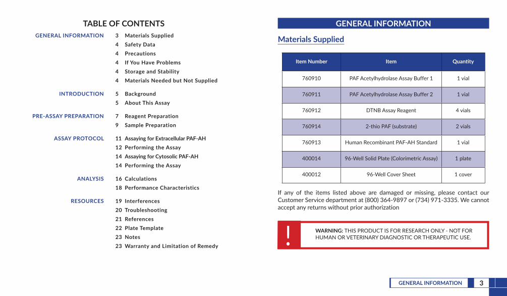
TABLE OF CONTENTS GENERAL INFORMATION 3 Materials Supplied
4 Safety Data4 Precautions4 If You Have Problems4 Storage and Stability4 Materials Needed but Not Supplied
INTRODUCTION 5 Background5 About This Assay
PRE-ASSAY PREPARATION 7 Reagent Preparation9 Sample Preparation
ASSAY PROTOCOL 11 Assaying for Extracellular PAF-AH12 Performing the Assay14 Assaying for Cytosolic PAF-AH14 Performing the Assay
ANALYSIS 16 Calculations18 Performance Characteristics
RESOURCES 19 Interferences20 Troubleshooting21 References22 Plate Template23 Notes23 Warranty and Limitation of Remedy
GENERAL INFORMATION
Materials Supplied
Item Number Item Quantity
760910 PAF Acetylhydrolase Assay Buffer 1 1 vial
760911 PAF Acetylhydrolase Assay Buffer 2 1 vial
760912 DTNB Assay Reagent 4 vials
760914 2-thio PAF (substrate) 2 vials
760913 Human Recombinant PAF-AH Standard 1 vial
400014 96-Well Solid Plate (Colorimetric Assay) 1 plate
400012 96-Well Cover Sheet 1 cover
If any of the items listed above are damaged or missing, please contact our Customer Service department at (800) 364-9897 or (734) 971-3335. We cannot accept any returns without prior authorization
! WARNING: THIS PRODUCT IS FOR RESEARCH ONLY - NOT FORHUMAN OR VETERINARY DIAGNOSTIC OR THERAPEUTIC USE.

5INTRODUCTION4 GENERAL INFORMATION
Safety DataThis material should be considered hazardous until further information becomes available. Do not ingest, inhale, get in eyes, on skin, or on clothing. Wash thoroughly after handling. Before use, the user must review the complete Safety Data Sheet, which has been sent via email to your institution.
PrecautionsPlease read these instructions carefully before beginning this assay.
If You Have ProblemsTechnical Service Contact Information
Phone: 888-526-5351 (USA and Canada only) or 734-975-3888Fax: 734-971-3641E-Mail: [email protected]: M-F 8:00 AM to 5:30 PM EST
In order for our staff to assist you quickly and efficiently, please be ready to supply the lot number of the kit (found on the outside of the box).
Storage and StabilityThis kit will perform as specified if stored at -20°C and used before the expiration date indicated on the outside of the box.
Materials Needed But Not Supplied1. A plate reader capable of measuring absorbance at 405-414 nm2. Adjustable pipettes and a repeating pipettor3. A source of pure water; glass distilled water or HPLC-grade water is
acceptable
INTRODUCTION
BackgroundPlatelet-activating factor (PAF) is a biologically active phospholipid synthesized by a variety of cells upon stimulation. PAF is converted to the biologically inactive lyso-PAF by the enzyme PAF acetylhydrolase (PAF-AH). PAF-AHs are located intra- and extra-cellularly (e.g., cytosolic and plasma). Plasma PAF-AH is highly selective for phospholipids with very short acyl groups at the sn-2 position and is associated with lipoproteins.1
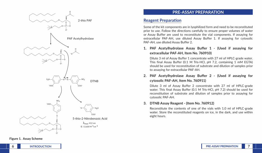
About This AssayCayman’s PAF Acetylhydrolase Assay Kit provides an accurate and convenient method for measurement of PAF-AH activity (both cytosolic and extracellular). The assay uses 2-thio PAF which serves as a substrate for all PAF-AHs.2 Upon hydrolysis of the acetyl thioester bond at the sn-2 position by PAF-AH, free thiols are detected using 5,5’-dithio-bis-(2-nitrobenzoic acid) (DTNB; Ellman’s reagent) (see Figure 1, on page 6).
7PRE-ASSAY PREPARATION6 INTRODUCTION
S
O
OO P
O
O-N+
O
2-thio PAF
S-
O
OO P
O
O-N+
O
-O
O2N
-OOC
S S
COO-
NO2
S
O
OO P
O
O-N+
S
-OOC
NO2
-S
COO-
N2O
5-thio-2-Nitrobenzoic Acid λmax: 412 nm
ε: 13,600 M-1cm-1
PAF Acetylhydrolase
DTNB
Figure 1. Assay Scheme
PRE-ASSAY PREPARATION
Reagent PreparationSome of the kit components are in lyophilized form and need to be reconstituted prior to use. Follow the directions carefully to ensure proper volumes of water or Assay Buffer are used to reconstitute the vial components. If assaying for extracellular PAF-AH, use diluted Assay Buffer 1. If assaying for cytosolic PAF-AH, use diluted Assay Buffer 2.
1. PAF Acetylhydrolase Assay Buffer 1 - (Used if assaying for extracellular PAF-AH, Item No. 760910)Dilute 3 ml of Assay Buffer 1 concentrate with 27 ml of HPLC-grade water. This final Assay Buffer (0.1 M Tris-HCl, pH 7.2, containing 1 mM EGTA) should be used for reconstitution of substrate and dilution of samples prior to assaying for extracellular PAF-AH.
2. PAF Acetylhydrolase Assay Buffer 2 - (Used if assaying for cytosolic PAF-AH, Item No. 760911)Dilute 3 ml of Assay Buffer 2 concentrate with 27 ml of HPLC-grade water. This final Assay Buffer (0.1 M Tris-HCl, pH 7.2) should be used for reconstitution of substrate and dilution of samples prior to assaying for cytosolic PAF-AH.
3. DTNB Assay Reagent - (Item No. 760912)Reconstitute the contents of one of the vials with 1.0 ml of HPLC-grade water. Store the reconstituted reagents on ice, in the dark, and use within eight hours.
8 PRE-ASSAY PREPARATION 9PRE-ASSAY PREPARATION
Sample PreparationIn general, any PAF-AH sample can be measured by this assay. However, cytosolic PAF-AH has to be measured using an end-point assay instead of a continuous assay. Cytosolic PAF-AH is sensitive to DTNB and EGTA.4 The sample must be free of particulates to avoid interference in the absorbance measurement. Thiols, thiol-scavengers, and PAF-AH inhibitors must be removed from the samples before performing the assay (extensive dialysis will eliminate most of the interfering substances of small molecular size). If the samples are too dilute, they can be concentrated using an Amicon centrifuge concentrator with a molecular weight cut-off of 30,000.
Tissue Homogenate1. Prior to dissection, rinse tissue with a PBS (phosphate buffered saline) solution,
pH 7.4, to remove any red blood cells and clots.2. Homogenize the tissue in 5-10 ml of cold buffer (i.e., 0.1 M Tris-HCl, pH 7.2)
per gram tissue.3. Centrifuge at 10,000 x g for 15 minutes at 4°C.4. Remove the supernatant for assay and store on ice. If not assaying on the
same day, freeze the sample at -80°C. The sample will be stable for at least one month.
Cell Lysate1. Collect cells by centrifugation (i.e., 1,000-2,000 x g for 10 minutes at 4°C).
For adherent cells, do not harvest using proteolytic enzymes; rather use a rubber policeman.
2. Homogenize or sonicate cell pellet in 1 ml of cold buffer (i.e., 0.1 M Tris-HCl, pH 7.2).
3. Centrifuge at 10,000 x g for 15 minutes at 4°C.4. Remove the supernatant for assay and store on ice. If not assaying on the
same day, freeze the sample at -80°C. The sample will be stable for at least one month.
4. 2-thio PAF (substrate) - (Item No. 760914)Evaporate the ethanolic solution of 2-thio PAF under a gentle stream of nitrogen. Reconstitute the contents by vortexing with 12 ml of either diluted Assay Buffer 1 or diluted Assay Buffer 2 to achieve a final concentration of 200 µM. Make sure to vortex until the substrate solution becomes clear (high background absorbance may result if the substrate is not completely dissolved). We recommend using the reconstituted substrate within two weeks.3
5. Human Recombinant PAF-AH Standard - (Item No. 760913)A solution of human recombinant PAF-AH is supplied as a positive control. A 10 µl aliquot of the enzyme per well causes an increase of approximately 0.025 absorbance unit/min. when assaying for extracellular PAF-AH.
11ASSAY PROTOCOL10 PRE-ASSAY PREPARATION
Plasma1. Collect blood using an anticoagulant such as heparin or citrate.2. Centrifuge the blood at 700-1,000 x g for 10 minutes at 4°C. Transfer the
plasma (upper layer) to a clean test tube being careful not to disrupt the white buffy layer. Store plasma on ice until assaying or freeze at -80°C. The plasma sample will be stable for at least one week.
Serum1. Collect blood without an anticoagulant.2. Allow blood to clot for 30 minutes at 25°C.3. Centrifuge the blood at 2,000 x g for 15 minutes at 4°C. Transfer the serum
(upper layer) to a clean test tube being careful not to disrupt the white buffy layer. Store serum on ice until assaying or freeze at -80°C. The serum sample will be stable for at least one week.
ASSAY PROTOCOL
Assaying for Extracellular PAF-AH Pipetting Hints
• It is recommended that an adjustable pipette be used to deliver substrate, DTNB, and buffer to the wells. This saves time and helps to maintain more precise times of incubation.
• Use different tips to pipette substrate, DTNB, and sample.• Before pipetting each reagent, equilibrate the pipette tip in that reagent
(i.e., slowly fill the tip and gently expel the contents, repeat several times).
• Do not expose the pipette tip to the reagent(s) already in the well.
General Information• The final volume is 225 µl in all of the wells.• Use diluted Assay Buffer 1, containing EGTA, in the assay.• It is not necessary to use all of the wells on the plate at one time.• If the appropriate enzyme dilution is not known, it may be necessary to
assay at several dilutions.• NOTE: Enzymatic reaction rates are temperature dependent. Be diligent about
maintaining consistency in temperature with all samples measured. When comparing reaction rates to those reported in the literature, be aware of potential differences in rate based on the temperature used.
12 ASSAY PROTOCOL 13ASSAY PROTOCOL
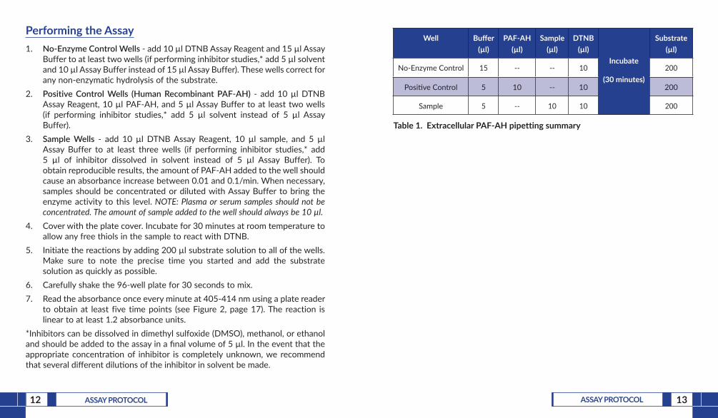
Performing the Assay1. No-Enzyme Control Wells - add 10 µl DTNB Assay Reagent and 15 µl Assay
Buffer to at least two wells (if performing inhibitor studies,* add 5 µl solvent and 10 µl Assay Buffer instead of 15 µl Assay Buffer). These wells correct for any non-enzymatic hydrolysis of the substrate.
2. Positive Control Wells (Human Recombinant PAF-AH) - add 10 µl DTNB Assay Reagent, 10 µl PAF-AH, and 5 µl Assay Buffer to at least two wells (if performing inhibitor studies,* add 5 µl solvent instead of 5 µl Assay Buffer).
3. Sample Wells - add 10 µl DTNB Assay Reagent, 10 µl sample, and 5 µl Assay Buffer to at least three wells (if performing inhibitor studies,* add 5 µl of inhibitor dissolved in solvent instead of 5 µl Assay Buffer). To obtain reproducible results, the amount of PAF-AH added to the well should cause an absorbance increase between 0.01 and 0.1/min. When necessary, samples should be concentrated or diluted with Assay Buffer to bring the enzyme activity to this level. NOTE: Plasma or serum samples should not be concentrated. The amount of sample added to the well should always be 10 µl.
4. Cover with the plate cover. Incubate for 30 minutes at room temperature to allow any free thiols in the sample to react with DTNB.
5. Initiate the reactions by adding 200 µl substrate solution to all of the wells. Make sure to note the precise time you started and add the substrate solution as quickly as possible.
6. Carefully shake the 96-well plate for 30 seconds to mix.7. Read the absorbance once every minute at 405-414 nm using a plate reader
to obtain at least five time points (see Figure 2, page 17). The reaction is linear to at least 1.2 absorbance units.
*Inhibitors can be dissolved in dimethyl sulfoxide (DMSO), methanol, or ethanol and should be added to the assay in a final volume of 5 µl. In the event that the appropriate concentration of inhibitor is completely unknown, we recommend that several different dilutions of the inhibitor in solvent be made.
Well Buffer (µl)
PAF-AH (µl)
Sample (µl)
DTNB (µl)
Incubate
(30 minutes)
Substrate (µl)
No-Enzyme Control 15 -- -- 10 200
Positive Control 5 10 -- 10 200
Sample 5 -- 10 10 200
Table 1. Extracellular PAF-AH pipetting summary
14 ASSAY PROTOCOL 15ASSAY PROTOCOL
Assaying for Cytosolic PAF-AH
General Information• The final volume is 225 µl in all of the wells.• Use diluted Assay Buffer 2 in the assay.• It is not necessary to use all of the wells on the plate at one time.• If the appropriate enzyme dilution is not known, it may be necessary to
assay at several dilutions.
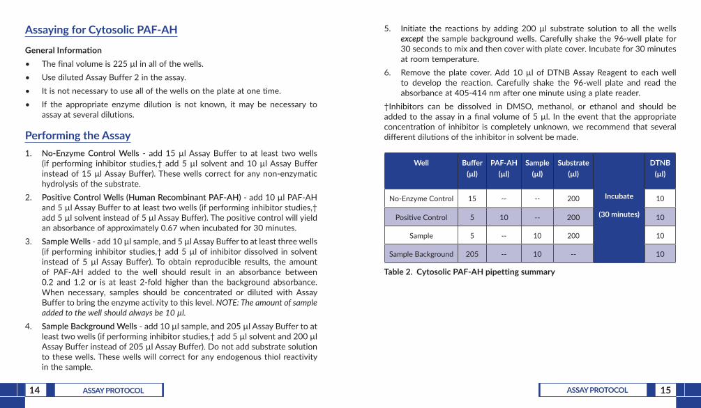
Performing the Assay1. No-Enzyme Control Wells - add 15 µl Assay Buffer to at least two wells
(if performing inhibitor studies,† add 5 µl solvent and 10 µl Assay Buffer instead of 15 µl Assay Buffer). These wells correct for any non-enzymatic hydrolysis of the substrate.
2. Positive Control Wells (Human Recombinant PAF-AH) - add 10 µl PAF-AH and 5 µl Assay Buffer to at least two wells (if performing inhibitor studies,† add 5 µl solvent instead of 5 µl Assay Buffer). The positive control will yield an absorbance of approximately 0.67 when incubated for 30 minutes.
3. Sample Wells - add 10 µl sample, and 5 µl Assay Buffer to at least three wells (if performing inhibitor studies,† add 5 µl of inhibitor dissolved in solvent instead of 5 µl Assay Buffer). To obtain reproducible results, the amount of PAF-AH added to the well should result in an absorbance between 0.2 and 1.2 or is at least 2-fold higher than the background absorbance. When necessary, samples should be concentrated or diluted with Assay Buffer to bring the enzyme activity to this level. NOTE: The amount of sample added to the well should always be 10 µl.
4. Sample Background Wells - add 10 µl sample, and 205 µl Assay Buffer to at least two wells (if performing inhibitor studies,† add 5 µl solvent and 200 µl Assay Buffer instead of 205 µl Assay Buffer). Do not add substrate solution to these wells. These wells will correct for any endogenous thiol reactivity in the sample.
5. Initiate the reactions by adding 200 µl substrate solution to all the wells except the sample background wells. Carefully shake the 96-well plate for 30 seconds to mix and then cover with plate cover. Incubate for 30 minutes at room temperature.
6. Remove the plate cover. Add 10 µl of DTNB Assay Reagent to each well to develop the reaction. Carefully shake the 96-well plate and read the absorbance at 405-414 nm after one minute using a plate reader.
†Inhibitors can be dissolved in DMSO, methanol, or ethanol and should be added to the assay in a final volume of 5 µl. In the event that the appropriate concentration of inhibitor is completely unknown, we recommend that several different dilutions of the inhibitor in solvent be made.
Well Buffer (µl)
PAF-AH (µl)
Sample (µl)
Substrate (µl)
Incubate
(30 minutes)
DTNB (µl)
No-Enzyme Control 15 -- -- 200 10
Positive Control 5 10 -- 200 10
Sample 5 -- 10 200 10
Sample Background 205 -- 10 -- 10
Table 2. Cytosolic PAF-AH pipetting summary
16 ANALYSIS 17ANALYSIS
ANALYSIS
Calculations
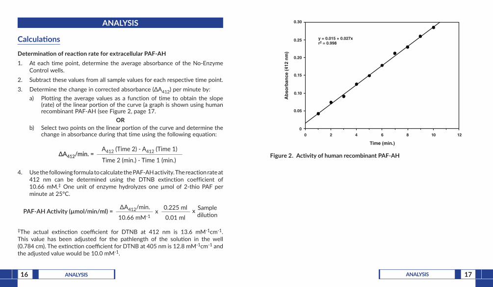
Determination of reaction rate for extracellular PAF-AH1. At each time point, determine the average absorbance of the No-Enzyme
Control wells.2. Subtract these values from all sample values for each respective time point.3. Determine the change in corrected absorbance (ΔA412) per minute by:
a) Plotting the average values as a function of time to obtain the slope (rate) of the linear portion of the curve (a graph is shown using human recombinant PAF-AH (see Figure 2, page 17.
ORb) Select two points on the linear portion of the curve and determine the
change in absorbance during that time using the following equation:
ΔA412/min. = A412 (Time 2) - A412 (Time 1)
Time 2 (min.) - Time 1 (min.)
4. Use the following formula to calculate the PAF-AH activity. The reaction rate at 412 nm can be determined using the DTNB extinction coefficient of 10.66 mM.‡ One unit of enzyme hydrolyzes one µmol of 2-thio PAF per minute at 25°C.
PAF-AH Ac�vity (µmol/min/ml) = ∆A412/min.
10.66 mM-1x 0.225 ml
0.01 mlx Sample
dilu�on
‡The actual extinction coefficient for DTNB at 412 nm is 13.6 mM-1cm-1. This value has been adjusted for the pathlength of the solution in the well (0.784 cm). The extinction coefficient for DTNB at 405 nm is 12.8 mM-1cm-1 and the adjusted value would be 10.0 mM-1.
Time (min.)
0 2 4 6 8 10 12
Ab
sorb
ance
(41
2 n
m)
0
0.05
0.10
0.15
0.20
0.25
0.30
y = 0.015 + 0.027x r2 = 0.998
Figure 2. Activity of human recombinant PAF-AH
19RESOURCES18 ANALYSIS
Determination of cytosolic PAF-AH activity1. Subtract the absorbance of the No-Enzyme Control wells from all other
wells on the plate.2. Use the following equation to calculate the enzymatic change in absorbance
as a function of time.
3. Use the following formula to calculate the PAF-AH activity. The reaction rate at 412 nm can be determined using the DTNB extinction coefficient of 10.66 mM.‡ One unit of enzyme hydrolyzes one µmol of 2-thio PAF per minute at 25°C.
PAF-AH Ac�vity (µmol/min/ml) = ∆A412/min.
10.66 mM-1x 0.225 ml
0.01 mlx Sample
dilu�on
‡The actual extinction coefficient for DTNB at 412 nm is 13.6 mM-1cm-1. This value has been adjusted for the pathlength of the solution in the well (0.784 cm). The extinction coefficient for DTNB at 405 nm is 12.8 mM-1cm-1 and the adjusted value would be 10.0 mM-1.
Performance Characteristics
Sensitivity:The detection range of the assay is from 0.02 to 0.2 µmol/min/ml of PAF acetylhydrolase activity which is equivalent to an absorbance increase of 0.01 to 0.1 per minute.
Precision:When a series of 89 PAF-AH measurements were performed on the same day, the intra-assay coefficient of variation was 3.5%. When a series of 89 PAF-AH measurements were performed on five different days under the same experimental conditions, the inter-assay coefficient of variation was 10%.
RESOURCES
Interferences
1. SolventsMethanol, ethanol, and DMSO have no effect on PAF-AH activity. PAF-AH inhibitors can be dissolved in any of the above solvents and only 5 µl added to the assay.
2. Culture Media and BuffersAll buffers and media should be tested for high background absorbances before doing any experiments. If the initial background absorbances are higher than 0.3 absorbance units then the samples should be diluted in Assay Buffer before performing the assay. Tris, Hepes, and phosphate buffers work in the assay.
3. Thiols and Thiol-ScavengersSamples containing thiols (i.e., glutathione, cysteine, dithiothreitol, or 2-mercaptoethanol) will exhibit high background absorbances and interfere with PAF-AH activity determination. Samples containing thiol-scavengers (i.e., N-ethylmaleimide) will inhibit color development. Extensive dialysis will eliminate most of the interference substances of small molecular size.
∆A412/min. = A412 (sample) - A412 (sample background)
Time (30 min.)
20 RESOURCES 21RESOURCES
Troubleshooting
Problem Possible Causes Recommended Solutions
Erratic values; dispersion of duplicates/triplicates
A. Poor pipetting/technique
B. Bubble in the well(s)
A. Carefully tap the side of the plate with your finger to remove bubbles
B. Be careful not to splash the contents of the wells
No color development
A. DTNB or sample was not added to well(s)
B. The enzymatic activity was too low
A. Make sure to add all components to the wells
B. Standardize the assay with the human PAF-AH standard.
C. Concentrate your sample so that the enzyme activity is in the assay’s detection range.
The color development was too fast
Too much enzyme added to well(s)
Dilute your samples with diluted Assay Buffer and re-assay
High background absorbance(>0.3)
A. Substrate not in solution
B. Thiols present in sample
A. Make sure to vortex the substrate until a clear solution is made
B. Remove thiols or thiol reagents from sample
The reaction rate is not linear at high absorbance
Plate reader not sensitive enough at high absorbance
A. Use only the points at lower concentrations in the linear portion for making the curve
B. Dilute your sample with diluted Assay Buffer and re-assay
References1. Stafforini, D.M., Prescott, S.M., and McIntyre, T.M. Human plasma platelet-
activating factor acetylhydrolase. J. Biol. Chem. 262, 4223-4230 (1987).2. Aarsman, A.J., Neys, F.W., and van den Bosch, H. Catabolism of platelet-
activating factor and its acyl analog. Differentiation of the activities of lysophospholipase and platelet-activating-factor acetylhydrolase. Eur. J. Biochem. 200, 187-193 (1991).
3. Stewart, A.G. and Grigoriadis, G. Structure-activity relationships for platelet-activating factor (PAF) and analogs reveal differences between PAF receptors on platelets and macrophages. J. Lipid Mediators 4, 299-308 (1991).
4. Narahara, H., Nishioka, Y., and Johnston, J.M. Secretion of platelet-activating factor acetylhydrolase by human decidual macrophages. J. Clin. Endocrinol. Metab. 77, 1258-1262 (1993).
22 RESOURCES 23RESOURCES
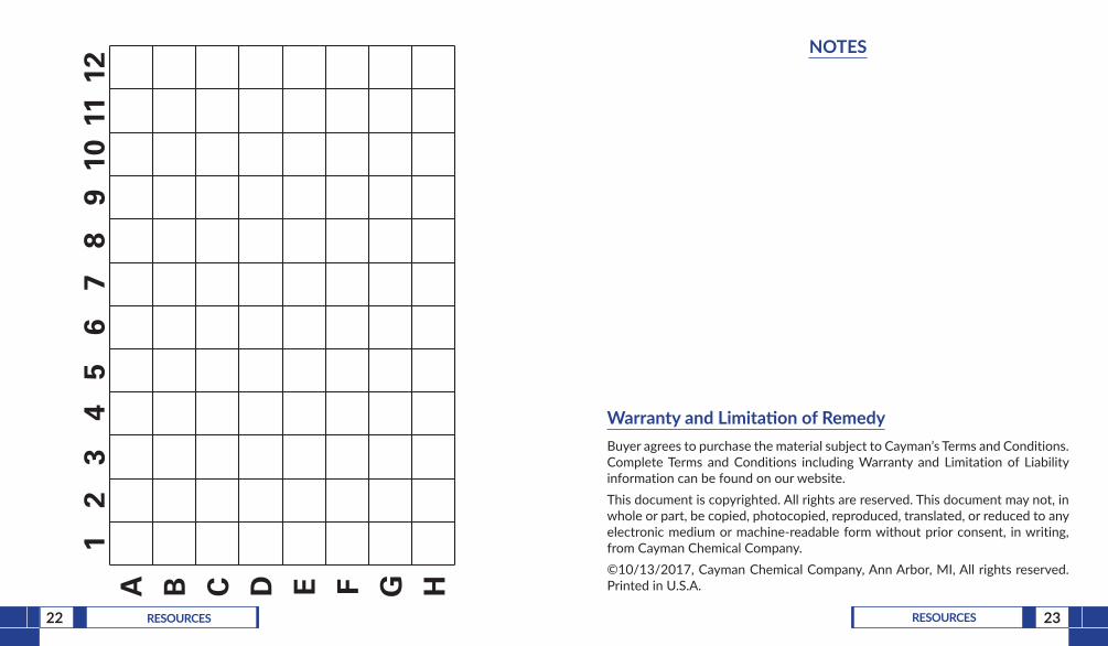
A B C D E F G H
12
34
56
78
910
1112
NOTES
Warranty and Limitation of RemedyBuyer agrees to purchase the material subject to Cayman’s Terms and Conditions.Complete Terms and Conditions including Warranty and Limitation of Liability information can be found on our website.This document is copyrighted. All rights are reserved. This document may not, in whole or part, be copied, photocopied, reproduced, translated, or reduced to any electronic medium or machine-readable form without prior consent, in writing, from Cayman Chemical Company.©10/13/2017, Cayman Chemical Company, Ann Arbor, MI, All rights reserved. Printed in U.S.A.