pages 42 to 46. chemical composition carbon hydrogen oxygen nitrogen sulfur (sometimes) ...
TRANSCRIPT

PROTEINSPages 42 to 46

PROTEINS Chemical composition
CarbonHydrogen OxygenNitrogenSulfur (sometimes)
Monomer/Building BlockAmino Acids (20 different amino acids)

Table 5-1

Fig. 5-UN1
Aminogroup
Carboxylgroup
carbon

Peptidebond
Fig. 5-18
Amino end(N-terminus)
Peptidebond
Side chains
Backbone
Carboxyl end(C-terminus)
(a)
(b)
A peptide bond is the bond joining adjacent
amino acids.
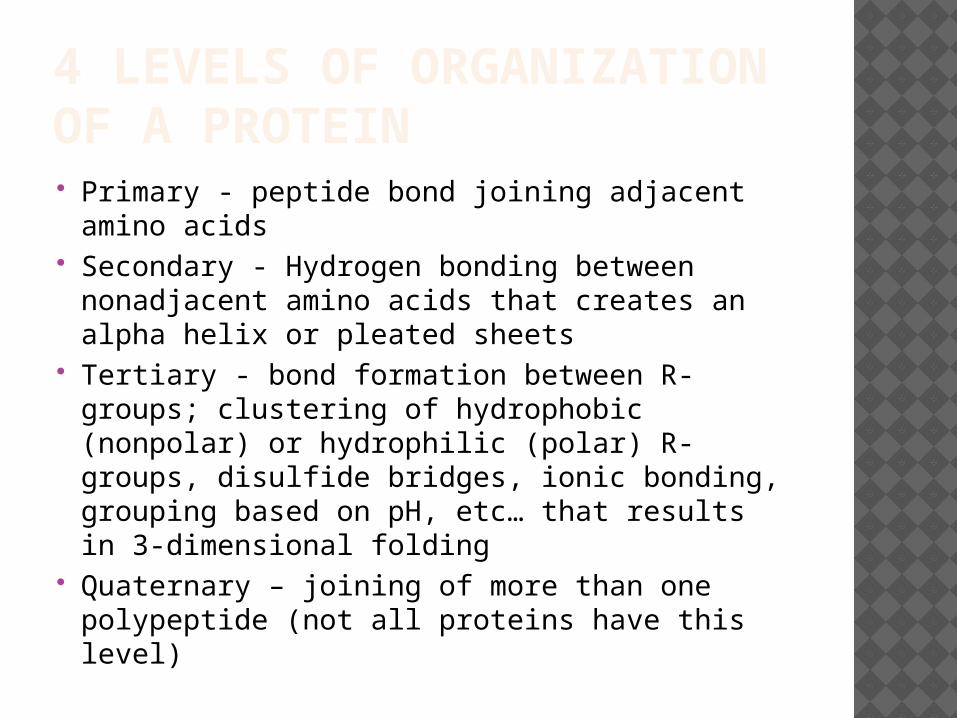
4 LEVELS OF ORGANIZATION OF A PROTEIN Primary - peptide bond joining adjacent
amino acids Secondary - Hydrogen bonding between
nonadjacent amino acids that creates an alpha helix or pleated sheets
Tertiary - bond formation between R-groups; clustering of hydrophobic (nonpolar) or hydrophilic (polar) R-groups, disulfide bridges, ionic bonding, grouping based on pH, etc… that results in 3-dimensional folding
Quaternary – joining of more than one polypeptide (not all proteins have this level)

Fig. 5-21
PrimaryStructure
SecondaryStructure
TertiaryStructure
pleated sheet
Examples ofamino acidsubunits
+H3N Amino end
helix
QuaternaryStructure

Fig. 5-21b
Amino acidsubunits
+H3N Amino end
Carboxyl end125
120
115
110
105
100
95
9085
80
75
20
25
15
10
5
1

Fig. 5-21c
Secondary Structure
pleated sheet
Examples ofamino acidsubunits
helix
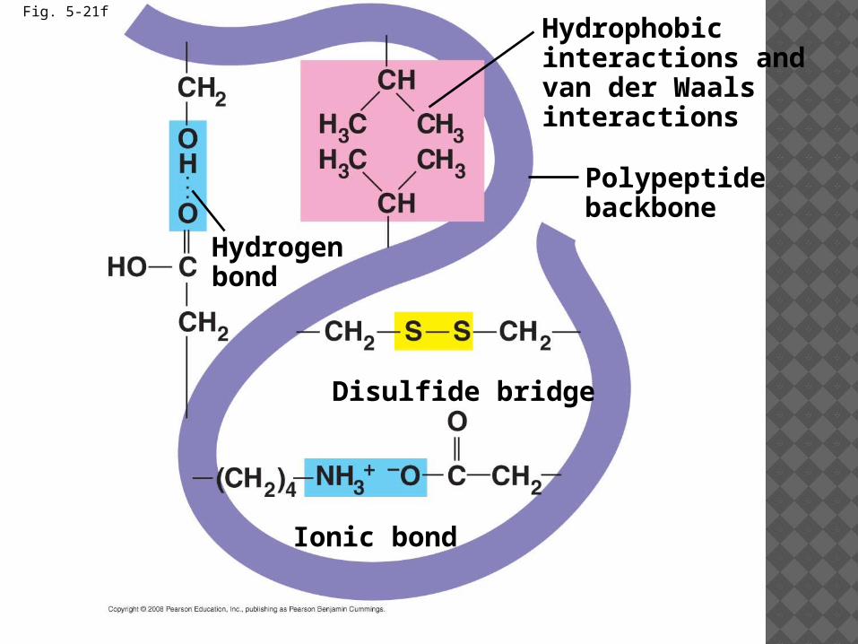
Fig. 5-21f
Polypeptidebackbone
Hydrophobicinteractions andvan der Waalsinteractions
Disulfide bridge
Ionic bond
Hydrogenbond

Fig. 5-21g
Polypeptidechain
Chains
HemeIron
Chains
CollagenHemoglobin
IMPORTANCE OF STRUCTURE A slight change in primary structure can
affect a protein’s structure and ability to function
Sickle-cell disease, an inherited blood disorder, results from a single amino acid substitution in the protein hemoglobin

Fig. 5-22
Primarystructure
Secondaryand tertiarystructures
Quaternarystructure
Normalhemoglobin(top view)
Primarystructure
Secondaryand tertiarystructures
Quaternarystructure
Function Function
subunit
Molecules donot associatewith oneanother; eachcarries oxygen.
Red bloodcell shape
Normal red bloodcells are full ofindividualhemoglobinmoledules, eachcarrying oxygen.
10 µm
Normal hemoglobin
1 2 3 4 5 6 7
ValHisLeuThrProGluGlu
Red bloodcell shape
subunit
Exposedhydrophobicregion
Sickle-cellhemoglobin
Moleculesinteract withone another andcrystallize intoa fiber; capacityto carry oxygenis greatly reduced.
Fibers of abnormalhemoglobin deformred blood cell intosickle shape.
10 µm
Sickle-cell hemoglobin
GluProThrLeuHisVal Val
1 2 3 4 5 6 7