pairing-specific, activity-dependent presynaptic facilitation at ap
TRANSCRIPT

The Journal of Neuroscience, January 1994, 14(l): 366-363
Pairing-specific, Activity-dependent Presynaptic Facilitation at Ap/ysia Sensory-Motor Neuron Synapses in Isolated Cell Culture
Lise S. Eliot, Robert D. Hawkins, Eric R. Kandel, and Samuel Schacher
Center for Neurobiology and Behavior, College of Physicians and Surgeons of Columbia University, New York, New York 10032
Synapses made by Ap/ysia sensory neurons onto motor- and interneuron followers in the intact nervous system exhibit an associative form of synaptic facilitation that is thought to contribute to classical conditioning of the animal’s gill and siphon withdrawal reflex (Hawkins et al., 1983; Walters and Byrne, 1983). Here we demonstrate that a similar associative facilitation can be induced between individual sensory and motor neurons isolated in culture. Pairing tetanic stimulation with either of two facilitatory transmitters, 5-HT or small car- dioactive peptide, considerably prolongs facilitation com- pared to either tetanus or transmitter alone. When corrected for the depression that occurs simply in response to low- frequency testing, the facilitation produced by one pairing trial does not decay for more than 20 min after training. This facilitation requires the temporal pairing (0.5 set forward interstimulus interval) of the two stimuli, tetanus and 5-HT. Delivering the same two stimuli in an unpaired fashion (1 min forward interval) fails to produce the long-lasting effect. Measurements of spontaneous transmitter release during either paired or unpaired training reveal no changes in uni- tary mEPSP or mEPSC (“mini”) amplitude, indicating that the facilitation involves a presynaptic mechanism. While both forms of training dramatically increase the initial frequency of spontaneous release, mini frequency does not remain elevated as long as the evoked EPSP following paired train- ing, nor does paired training specifically enhance sponta- neous release frequency. Pairing-specific facilitation was not blocked by the protein kinase C inhibitor H7. In contrast, the same training procedure produced pairing-specific in- creases of sensory neuron excitability and action potential width, suggesting that CAMP-mediated processes are in- volved in the paired effect. Although Ca2+ influx is necessary for the associative effect (Abrams, 1985), we find that the facilitation does not require influx through L-type voltage- gated Ca*+ channels, since the effect was not blocked by the dihydropyridine antagonist nitrendipine. Together, these findings indicate that the mechanism underlying associative, activity-dependent facilitation is intrinsic to the sensory neu- ron synapse, that it is presynaptically mediated by process-
Received Feb. 26, 1993; revised July 7, 1993; accepted July 13, 1993. We thank Tracey Cohen for technical assistance, and Sarah Mack, Charles Lam,
and Robert Wooley for help in preparing the figures. This work was supported by the Howard Hughes Medical Institute (E.R.K.), Grant MH 262 12 from the NIMH (R.D.H.), GraniGM 32099 from the NIH (S.S.), and an NSF predoctoral fellow- shio (L.S.E.).
doxxespondence should be addressed to Lise Eliot, Division of Neuroscience, Baylor College of Medicine, One Baylor Plaza, Houston, TX 77030.
Copyright 0 1994 Society for Neuroscience 0270-6474/94/140368-16$05.00/O
es unique to evoked synaptic transmission, and that it ap- pears to involve a pairing-specific broadening of the presynaptic action potential, allowing enhanced Ca2+ influx through the dihydropyridine-insensitive channels responsi- ble for release.
[Key words: synaptic plasticity, associative, 5-HT, post- tetanic potentiation, spontaneous release, spike broaden-
h71
Synaptic plasticity is a fundamental means by which environ- mental cues alter the function of the nervous system in both development and learning. Of particular interest are associative forms of synaptic plasticity-changes in synaptic strength trig- gered by the nearly simultaneous occurrence of two cellular stimuli. These types of plasticity seem excellent candidates for cellular mechanisms underlying selective synapse stabilization (Collingridge and Singer, 1990; Cline, 199 I) and associative learning (Byrne, 1987; Abrams and Kandel, 1988; Hawkins et al., 1993). A number of associative forms of synaptic plasticity have been described, all of which require neuronal activity as at least one of the associative cellular stimuli, including activity- dependent enhancement of presynaptic facilitation (Hawkins et al., 1983; Walters and Byrne, 1983) and inhibition (Small et al., 1989) long-term potentiation (Levy and Steward, 1979; Bar- rionuevo and Brown, 1983; Walters and Byrne, 1985), and long- term depression (Ito et al., 1982; Stanton and Sejnowski, 1989).
One of these associative and activity-dependent forms of plas- ticity has been identified in cellular studies of classical condi- tioning in the marine mollusk Aplysia californica: activity-de- pendent enhancement of heterosynaptic facilitation at siphon sensory neuron synapses, the first afferent relay in the gill and siphon withdrawal circuit (Hawkins, 199 1). Pairing a touch to the siphon, which triggers reflexive withdrawal, with aversive shock to the animal’s tail produces a conditioned enhancement of the withdrawal response (Carew et al., 198 1, 1983). By itself, tail shock activates modulatory intemeurons that heterosynapti- tally facilitate transmitter release from sensory neuron terminals (Hawkins et al., 198 1; Hawkins and Schacher, 1989; Mackey et al., 1989). When sensory neuron activity similar to that pro- duced by siphon touch is paired with and immediately precedes this neuromodulatory input, the magnitude and duration of this facilitation are enhanced (Hawkins et al., 1983; Walters and Byrne, 1983; Buonomano and Byrne, 1990).
Pairing-specific facilitation is thought to involve an augmen- tation of the mechanisms underlying heterosynaptic facilitation alone. Three different transmitters contribute to the presynaptic facilitation induced by tail shock: 5-HT, small cardioactive pep- tide (SCP), and the as yet unidentified transmitter released by

The Journal of Neuroscience, January 1994. 74(l) 369
the L29 group of modulatory interneurons (Brunelli et al., 1976; Hawkins et al., 1981; Abrams et al., 1984; Glanzman et al., 1989; Mackey et al., 1989; Mercer et al., 199 1). The most ex- tensively studied of these is 5-HT, which facilitates release from sensory neurons by two different processes. The first involves closure of K+ channels, which broadens the presynaptic action potential, increasing Ca2+ influx at transmitter release sites (Klein et al., 1982; Siegelbaum et al., 1982; Baxter and Byrne, 1990; Hochner and Kandel, 1992; Eliot et al., 1993). This effect is mediated in part by CAMP-dependent protein kinase (Castel- lucci et al., 1982; Shuster et al., 1985; Goldsmith and Abrams, 1992). The second process is independent of changes in the action potential and involves either vesicle mobilization or a direct enhancement of exocytosis (Hochner et al., 1986b). This second process is mediated, at least in part, by protein kinase C, and is reflected by an increased frequency of spontaneous transmitter release in cultured sensory-motor neuron synapses (Braha et al., 1990; Dale and Kandel, 1990; Ghirardi et al., 1992).
How does paired activity enhance facilitation by 5-HT? Haw- kins et al. (1983) found that spike broadening is increased in a pairing-specific manner, and Abrams (1985) demonstrated that this associative effect requires Ca2+ influx during pairing. These findings suggested that Ca2+ influx during the paired activity enhances the first process of 5-HT facilitation, CAMP-dependent action potential broadening. Biochemical studies support this model, indicating that Ca2+ augments CAMP production by stimulating adenylate cyclase in concert with 5-HT (Ocorr et al., 1985; Eliot et al., 1989; Abrams et al., 1991; Yovell and Abrams, 1992). However, several questions remain concerning the molecular mechanism of the associative facilitation. For example, previous findings do not rule out a role for the second process in pairing-specific facilitation. Because the second pro- cess is mediated in large part by protein kinase C, which is also dually activated by 5-HT and intracellular Ca*+ (Sacktor and Schwartz, 1990; Sossin and Schwartz, 1992), this mobilization- like process seems a good candidate for pairing-specific en- hancement. Also, if pairing-specific facilitation requires Ca*+ influx, what is the source of this influx? Aplysia sensory neurons are known to have at least two types of voltage-dependent Ca2+ channels, a dihydropyridine-sensitive, noninactivating channel similar to the vertebrate L-type, and a more rapidly inactivating, dihydropyridine-insensitive channel (Edmonds et al., 1990). Al- though L-type channels are present in regions containing syn- aptic terminals, they play no role in synaptic transmission or its facilitation by 5-HT (Edmonds et al., 1990; Eliot et al., 1993). However, the L-current may be an essential source ofCa2+ influx underlying pairing-specific facilitation, particularly because it is itself substantially enhanced by 5-HT (Braha et al., 1993).
The ability to reconstitute synapses between individual Aply- sia sensory and motor neurons in culture has proven advan- tageous for analyzing the mechanisms underlying various forms of synaptic plasticity. Cultured sensory-motor neuron synapses have been found to retain nearly every nonassociative form of plasticity they exhibit in vivo, including short-term homosynap- tic depression and potentiation, and both short- and long-term heterosynaptic facilitation and inhibition (Montarolo et al., 1986, 1988; Rayport and Schacher, 1986; Schacher et al., 1990; Eliot et al., 1990). Can associative forms of plasticity also be repro- duced in culture? We undertook a study of associative activity- dependent facilitation in culture to determine whether the plas- ticity is inherent to the synapse, and if so, to exploit this simple
system in further studying its underlying mechanisms. These studies in turn addressed several questions: (1) Is the associative facilitation presynaptic? (2) Does it involve an enhancement of the second or spike-independent process of facilitation? (3) Does it require protein kinase C activation? (4) Does it involve an enhancement of the first process in facilitation (spike broad- ening) and enhanced production of CAMP? (5) Does it require Ca2+ influx through L-type CaZ+ channels?
Materials and Methods
Cellculture. Synapses were reconstituted in vitro between Aplysia pleural mechanosensory neurons and one of two classes of motor neurons: either the gill motor neuron, L7, obtained from juvenile (1-5 gm) animals, or LFS siphon motor neurons, obtained from adult (70-I 20 gm) animals. LFS motor neurons were isolated based on their size, pigmentation, and location in the left ventral abdominal ganglion, mostly caudal and a bit lateral to the LE cluster. However, it is likely that a few of the follower neurons were not LFS type, because up to 12 cells were found in a single ganglion onto which sensory neurons made sizable synaptic connections in culture, whereas only seven or eight such cells have been found actually to cause siphon movements (Hickie and Walters, 1990). Al- though the identity ofthe follower neurons is thus somewhat ambiguous, they will be referred to as LFS cells, because it is likely that at least half were properly of this class. All of these cells had similar properties: 50- 100 pm size, unpigmented appearance, two or three major axon branch- es, and a characteristic “notch” in their membrane potential when re- leased from very hyperpolarized levels (- 80 to - 90 mV), probably due to A-type K+ current activation.
Cells were dissociated and cultured as described by Rayport and Schacher (1986). Briefly, abdominal and pleural ganglia were incubated at 34°C for 2.5 hr in 1% protease (type IX, Sigma) dissolved in L15 medium (Flow Laboratories) supplemented to achieve the following final concentrations: 385 mM NaCI, 10 mM KCI, 11 mM CaCI,, 28 mM MgCI,, 27 mM MgSO,, 2.3 mM NaHCO,, 35 mM D-glucose. Ganglia were desheathed and then individual cells removed with a long, Bexihle- tipped glass electrode. Pleural sensory neurons were plated in contact with a motor neuron in a 50 mm poly-L-lysine-coated plastic culture dish containing 2.5 ml of culture medium (50% supplemented L15 + 50% sterile filtered hemolymph). For cultures containing two sensory neurons synapsing on a common motor neuron (Fig. lA), the sensory neurons were plated as far apart from each other on the target motor neuron as possible to minimize electrical coupling between them (Bank and Schacher, 1992). Synaptic connections appeared as early as 24 hr after plating and rest at room temperature. Dishes were transferred to an 18°C incubator after 24 hr.
Synaptic electrophysiology. Experiments were performed on cultures 3-5 d after plating (2-4 d for spontaneous release experiments) in me- dium containing 50% supplemented L15 and 50% artificial seawater (460 mM NaCI, 10 mM KCl, 11 mM C&l,, 55 mM MgCl,, 2 rnr+r NaHCO,, 10 mM Na-HEPES, pH 7.6). All experiments except those measuring spontaneous transmitter release were conducted at 15-l 8°C. Motor neu- rons were impaled with sharp microelectrodes (15-25 MR) filled with 2 M K-acetate, 0.5 M KCl, buffered to pH 7.2 with 10 mM K-HEPES and held at -80 to -90 mV when recordina evoked EPSPs using the bridge circuit on an Axoclamp 2A amplifier-(Axon Instruments,Bur- hngame CA). Sensory neurons were stimulated extracellularly, using unpolished patch pipettes (1-3 MR) filled with extracellular solution. At each trial, the intensity of a 0.1 msec current pulse to the sensory neuron was slowly increased until a single EPSP appeared in the motor neuron; tetanus was elicited by raising the intensity 20% above threshold level of the last trial. Although such a procedure would result in failures if the sensory neuron had been stimulated intracehularly, with extra- cellular stimulation it reliably produced a single action potential in the sensory neuron for every pulse in the train, as judged by the occurrence of EPSPs recorded in the motor neuron at high gain. The few experi- ments in which failures were observed were discarded. Each synapse was tested once shortly after motor neuron impalement, and then rested 15 min before beginning the experiment. In experiments using two presynaptic neurons, both sensory neurons were impaled at the end of the experiment to test the degree of electrical coupling between them. The experiment was discarded if tetanic stimulation of one sensory

370 Eliot et al. * Associative Synaptic Plasticity in Culture
neuron delivered in the presence of 5-HT produced any action potentials in the other sensory neuron.
Measurements of spontaneous release. Spontaneous release was mea- sured exclusively in cultures containing a single sensory neuron in con- tact with a single motor neuron as described previously (Dale and Kan- del, 1990). We used two different methods to record miniature postsynaptic responses in pairing experiments. Initially, we recorded mEPSPs in L7 motor neurons under current clamp using sharp elec- trodes. In later experiments, we switched to whole cell voltage clamp and recorded mEPSCs in the smaller LFS motor neurons using patch pipettes (four of nine paired, and four of nine unpaired experiments). For current-clamp experiments, L7 was impaled with a beveled, low- resistance microelectrode (6-8 MR). Voltage-clamp experiments were performed in the whole cell configuration (Hamill et al., 198 1) using 3- 4 MQ patch pipettes filled with 465 mM Cs-gluconate, 2 mM MgCI,, 1 mM CaCI,, 10 mM K-EGTA, 5 mM Na,ATP, 0.1 mM Tris-GTP, 10 mM reduced glutathione, and 50 mM Cs-HEPES, buffered to pH 7.2 with CsOH. Although mEPSCs were recorded using continuous single elec- trode voltage clamp on the Axoclamp, we switched back to current clamp to measure evoked EPSPs in these experiments.
Measurements of mEPSP and mEPSC frequencies and amplitudes were performed as described by Dale and Kandel (1990). Postsynaptic recordings were made at both high gain (AC coupled) to measure min- iature EPSPs or EPSCs and low aain (DC coupled) to measure evoked EPSPs. High-gain recordings we; filtered (at 300 ‘Hz for mEPSPs and 2-4 kHz for mEPSCs), digitized (4 kHz sampling frequency), and an- alyzed using a PDP-I 1 computer and programs written in Basic-23. The mEPSPs or mEPSCs were detected manually based on fast rise time and amplitude greater than peak-to-peak noise level (see Fig. 6). Peak amplitude and the interval between minis were then determined automatically. The better signal-to-noise of mEPSCs measured under whole-cell voltage clamp made it possible to use automatic thresholding to detect them. Histograms of mini amplitudes were fit with l-3 gaussian distributions determined using the simplex algorithm and the method ofmaximum likelihood (Dale and Kandel, 1990). In most L7 recordings, peak-to-peak noise was about 40 pV, so this was used as a lower cutoff for mEPSP amplitude. Average unitary mEPSP amplitude was 76.3 f 6.6 (*SEM) PV (n = 9). For LFS recordings, the noise cutoff varied more between experiments but was generally set at 6 pA. Average uni- tarv mEPSC amulitude was 17.5 * 3.3 DA (n = 7). Distributions of both mEPSP andmEPSC sizes showed distinct peaks, generally in mul- tiple intervals ofthe first amplitude, suggesting that the lower-end cutoff does not distort the first peak. Noise levels often rose during experiments using either type of recording. Experiments were rejected when the noise exceeded the smallest signals detectable at the beginning of the exper- iment. Because of the difficulty of maintaining low-noise recordings for an extended period of time, the effects of paired or unpaired training on spontaneous release were compared between different synapses al- ternately given either paired or unpaired training.
Drum a&ication. Modulatorv transmitters (5-HT or SCP) were ao- - . . plied as a 5 ~1 bolus in the immediate vicinity‘of the cells, either using a Hamilton syringe as previously described (Schacher et al., 1990) or, for temporal pairing experiments, using a 5 ~1 capillary glass pipette positioned just upstream and aimed at the cells. In the latter case, timing was controhed by a Picospritzer (General Valve). The 5 ~1 of extracelz lular medium contained either 50 UM 5-HT or 50 UM SCPb (Peninsula Laboratories). A constant bath perfusion (approximately 1 mljmin) con- trolled washout from the 2.5 ml culture dish. In some experiments, 0.5% fast green was also included to monitor the location of the puff. Fast green was no longer visible near the cells 30 set after the puff. In some later experiments (see Figs. 7, 9), 5-HT was applied by micro- perfusing a 10 PM solution immediately over the cells beginning at the onset ofthe tetanus, and then switching to control saline microperfusion after 15 sec. Although it is difficult to determine the exact duration of 5-HT exposure, it appeared comparable for both application methods, based on another measure of 5-HT action in sensory neurons, reduced spike-frequency accommodation (Klein et al., 1986). Thus, in parallel experiments, 5-HT produced large increases in the number of sensory neuron action potentials elicited by fixed intracellular depolarizing step commands (“anti-accommodation”), and this effect was completely re- versed in 60-90 set for both methods of application (not shown). Since this duration can be estimated with greater certainty than 5-HT washout itself, we have used it in the figures (see Figs. 4, 5, 7, 9, 13) to indicate 5-HT exposure. Thus, the time indicated by the 5-HT bar (90 set) is an upper estimate of the maximum duration of 5-HT exposure in syn- aptic experiments.
The protein kinase inhibitor H7 (Seikagaku) was dissolved in water (10 mM stock solution) and applied by microperfusion in a final con- centration of 200 or 400 PM in extracehular sohrtion. Nitrendipine (gift of A. Scriabine, Miles Pharmaceutical) was made fresh daily as a 10 mM stock in dimethyl sulfoxide (DMSO) and bath applied in a final concentration of 2 or IO PM.
Isolated sensory clusters. Clusters of pleural mechanosensory neurons were dissected out, treated with 0.5-l .O% protease (type XIV, Sigma) in artificial seawater at 18°C for 30-90 min, and allowed to recover for 2-6 d at 18°C in artificial seawater supplemented with amino acids, vitamins, sugar, penicillin, and streptomycin (Eisenstadt et al., 1973). The pleural sensory neuron cluster is a relatively pure preparation con- taining only sensory neuron cell bodies and neuropil (Walters et al., 1983). The protease treatment is needed to disrupt synaptic transmission from any terminals of neuromodulatory cells that survive following isolation.
Clusters were pinned to the Sylgard floor of a 40 ~1 well continuously perfused at 40 pl/sec with supplemented artificial seawater (without amino acids) containing 100 FM 3-isobutyl-1-methylxanthine (IBMX) diluted from a 0.5 M stock solution in DMSO. IBMX was added to block CAMP breakdown, thereby focusing our measurements on effects due to changes in CAMP production. A single sensory neuron was im- paled with a lo-20 MQ electrode containing 2.5 M KCl. The cell was allowed to rest for 10 min following impalement before an experiment was begun. During training, the entire population of sensory neurons was stimulated by delivering a 20 Hz, 2 set train of I msec, 100 V biphasic shocks (“tetanus”) between two silver wires positioned on op- posite sides of the well (see Fig. 10). Cells in the cluster fire action potentials one-for-one with the shocks. 5-HT (4 or IO UM, with 0.01% fast green) was applied for 30 set beginning 0.5 set (paired) or 1 min (unpaired) after the beginning of the tetanus. Timing of the 5-HT de- livery was controlled by an electronic valve in the perfusion line. The 5-HT concentration reached steady state within a few seconds during washin and washout. Sensory neuron excitability was quantified as the number of action potentials resulting from a 500 msec depolarizing intracellular current pulse whose magnitude was adjusted before training to produce one action potential. Action potential duration was measured from the peak to the point on the falling phase that was at 30% of peak amplitude, using a laboratory interface to an IBM-compatible PC and commercially available software (SPIKE, Hilal Associates, Englewood, NJ). The action potential was measured at the end of a 20 Hz, 1 set train, which inactivates the delayed rectifier K+ current, making the action potential duration more sensitive to changes in S-type K+ current.
Data analysis. Data in the text and figures are presented as mean * SEM, and except for Figure 11 are normalized to pretest magnitude (EPSP amplitude or action potential duration). The results shown in Figure 3 are corrected for homosynaptic depression by dividing nor- malized experimental values by the average normalized control (test alone) amplitude at each time point. In experiments measuring spon- taneous transmitter release (see Fig. 7), data were log transformed and then normalized, so measurements ofboth evoked EPSP amplitude and mini frequency are presented as geometric mean * SEM. Normalized data were analyzed by two-way ANOVA with repeated measures. When a significant main effect of treatment or interaction was found, the time course of the effect was then examined using either one-way ANOVA at each time point followed by Dunnett’s two-tailed f test for multiple groups, or using t tests at each individual time point when only two groups were being compared.
Results Pairing 5-HT or SCP with tetanic stimulation prolongs facilitation compared to either procedure alone Aplysia sensory-motor neuron synapses reconstituted in culture exhibit both homosynaptic facilitation in response to tetanic stimulation and heterosynaptic facilitation in response to neu- romodulator application (Rayport and Schacher, 1986; Schach- er et al., 1990; Eliot et al., 1990). As a first step in assessing whether associative facilitation also occurs in culture, we com- pared the effects of each treatment alone with their effect when delivered in combination. Two groups of cultures were used for these experiments, each containing two sensory neurons syn- apsing onto a single L7 motor neuron. In the first group, one

The Journal of Neuroscience, January 1994, 14(l) 371
BI 5-HT
CONTROL SN
TET. ALONE SN 1111
B2 5-HT
PAIRED SN 11111111111111111111llll
WIT ALONE SN
Cl 5-HT
PAIRED SN 11111111111111111111llll
UNPAfRED SN
c2 5-HT - -
REST SN
UNPAIRED PAIRED
Figure 1. Experimental design. A, Sensory to motor neuron synapses were established in culture using either one or two presynaptic mechanosensory neurons (.S) plated in contact with a sin- gle motor neuron (M), in this case a siphon motor neuron from the LFS group. B, Design of experiments com- paring tetanus alone, 5-HT alone, paired training, and no training (control). Two groups of cultures containing two sen- sory neurons (SN) synapsing on a single L7 motor neuron were used. In one group (B,) one sensory neuron was tet- anized (20 Hz, 2 set), while the other received no training, to serve as a con- trol for homosynaptic depression. In the other group (B,), one sensory neuron was tetanized immediately before ap- plication of 5-HT or SCP (and thus re- ceived paired training), while the other received no stimulation, to measure the effect of transmitter alone. Each syn- apse was tested once, 10 min before training, and then posttested four times as indicated in Figures 2 and 3. C, De- sign of experiments comparing paired and unpaired training. Two groups of cultures containing a single LPS motor neuron and either one or two presyn- aptic sensory neurons were used. In the two sensory neuron design (C,), one sensory neuron received tetanus paired with a 5 ~1 puff of 50 PM 5-HT (0.5 set after onset of tetanus), while the other sensory neuron received tetanus 1 min before the 5-HT. In the one sensory neuron design (C’,), a single presynaptic sensory neuron received either paired or unpaired training in round A, fol- lowed by the opposite form of training in round B. Each synapse was pretested twice, at 5 min intervals, and then test- ed four times after training, as indicated in Results. The rest period was 30 min for synaptic experiments and 15 min for experiments measuring action po- tential width in pleural sensory neuron clusters.
sensory neuron served as control for the effects of normal syn- aptic depression while the other sensory neuron received a tet- anus to assess the magnitude and duration of posttetanic po- tentiation (PTP, Fig. lB,). Similarly, in the second group only one sensory neuron was tetanized, but in these cultures a brief application of 5-HT was delivered immediately after the onset
of the tetanus, so that one synapse was exposed to 5-HT alone while the other received 5-HT paired with tetanus (Fig. 1B2). In another study, we found that PTP does not occur in culture when the presynaptic sensory neuron is impaled with an intra- cellular electrode (Eliot, 1991), so sensory neurons were stim- ulated extracellularly, as described in Materials and Methods.

372 Eliot et al. * Associative Synaptic Plasticity in Culture
Tetanus L7F~~.~~F-..
5-HT
L7
. -. _ _. _ _ Tetanus + 5-HT
L7 i‘--.
-to
B.
I 5 15
Tlme(mln)
I I I 0 I 5
Time(min)
Figure 2. Pairing 5-HT with tetanus prolongs facilitation compared to either treatment alone. A, EPSPs recorded in L7 motor neurons in response to extracellular stimulation of presynaptic sensory neurons. Four different treatments were delivered (at arrow) to two separate groups of cultures, as illustrated in Figure I, B, and B, In the first group of cultures, the sensory neuron receiving test alone undergoes homo- synaptic depression, while the sensory neuron receiving tetanus alone exhibits PTP lasting 5-10 min. In the second group, the sensory neuron exposed to 5-HT alone is facilitated only briefly after transmitter ap- plication, while the sensory neuron receiving tetanus paired with 5-HT remains facilitated up to I5 min after training. B, Group data. Cultures containing two sensory neurons synapsing on a common L7 motor neuron were trained in two groups (n = IO in each): test alone (Cont.) versus PTP (Tel.), and 5-HT versus paired (Tel. + 5-HT). Each synapse was pretested once, received its treatment IO min later (indicated by arrow), and then posttested I, 5, IO, and I5 min after treatment. Error bars indicate SEM. Initial EPSP amplitudes were 12.7 k I .8 mV (Cont.), 13.4 + 2.2 (5-HT), 13.6 + 1.6 (ret.), and 14.1 k 2.6 (Tet. + 5-HT), and were not significantly different by one-way ANOVA.
. .0 Control - -o--5HT - 0 -SCP --fi-Tet. +5HT + Tet. -A -SCP+Tet.
0 5 10 15 20 Time(min)
Figure 3. Either 5-HT or SCP can induce the paired enhancement of facilitation, which appears nondecremental when normalized for homo- synaptic depression. Results are from three groups of cultures, using the experimental design of Figure IB: (I) test alone (Control) versus tetanus alone (n = 5), (2) 5-HT alone versus 5-HT paired with tetanus (n = 6), and (3) SCP alone versus SCP paired with tetanus (n = 6). Synapses were pretested IO min before treatment (at arrow), and then posttested 5, IO, 15, and 20 min after treatment. Responses were normalized to pretest level within individual experiments (results comparable to Fig. 2), and then normalized again to the average amplitude of the control group at each time point, to evaluate the time course of each treatment independently of homosynaptic depression. Initial EPSP amplitudes were II.3 f 2.3 (Control), 16.0 f 4.4 (SHT), 12.5 f 2.4 (XP), 12.0 + 2.6 (Tet.), 14.8 + I.8 (5HT + ret.), and 10.8 f I.5 (SCP + Tef.), and were not significantly different by one-way ANOVA.
Results from both groups of cultures are plotted together in Figure 2. Each of the three experimental treatments facilitated the synapse compared to control, but the treatments differed in the duration of facilitation (overall, F,,36 = 12.52 for treatment and F,,.,,, = 7.96 for treatment x time, p < 0.001; treatment also produced a significant effect at every posttest, p < 0.001 by one-way ANOVA). 5-HT alone produces facilitation that is evident 1 min after application (13 1 + 8%, Dunnett’s two-tailed t = 4.79, p < 0.01 compared to control), but with continuous bath perfusion the facilitation is no longer significant 5 min after application. Tetanic stimulation alone produces a comparable amount of initial facilitation (126 k 7%, t = 3.80, p < 0.01) and the effect lasts somewhat longer (at 5 min, t = 2.53, p < 0.05). Nevertheless, the PTP is clearly decremental, such that 10 min after tetanization the synapse is no longer significantly enhanced (t = 1.96, NS). In contrast to these short-lived effects, pairing 5-HT with the same tetanic stimulation produces con- siderably longer-lasting facilitation, remaining enhanced 15 min after training (1 = 5.73, p < 0.01).
As is evident from the decay of the control group responses

The Journal of Neuroscience, January 1994, 14(l) 373
A PRE TRAINING
PAIRED:
5HT Puff
A
UNPAIRED: TETANUS 1’ BEFORE 5HT
POST
5’ lo’ 1Y
-IL
I 20mV
100msec
I PAIRED
--o- UNPAIRED
-10 -5 0 5
TIME (min)
(Fig. 2) sensory neuron synapses exhibit profound depression, even with very low-frequency stimulation. To minimize this effect, we performed another set of experiments in which we eliminated the test 1 min after training. We also added a third group of cultures to this experiment, to test whether facilitation by another modulatory transmitter, SCP, is similarly enhanced by paired activity. The results with 5-HT, tetanus, 5-HT + tetanus, and control were similar to those shown in Figure 2. To evaluate the facilitatory effects independently of homosy- naptic depression, we normalized EPSP amplitudes by the av- erage amplitude of the “test alone” group at each time point. These data are displayed in Figure 3.
These experiments reveal that either 5-HT or SCP when paired with tetanus produce considerably longer-lasting facilitation than transmitter or tetanus treatment alone. The paired enhancement is virtually indistinguishable for the two transmitters. Although the facilitatory effect of either transmitter alone washes out be- fore the first posttest, the paired group remains substantially facilitated up to 20 min later. Moreover, this facilitation appears nondecremental, especially compared to the facilitation by tet- anus alone. Facilitation was significant overall (F,,Z, = 14.30 for treatment and F4,, ,z = 6.91 for treatment x time, p < 0.001) and at each posttest (by one-way ANOVA, p< 0.001). As in the previous experiment, the tetanus alone group is significantly facilitated for 5 min after training (Dunnett’s t = 3.15, p < 0.05)
Figure 4. Associative facilitation us- ing a two sensory neuron design. A, EPSPs recorded in an LFS motor neu- ron in response to separate extracellular stimulation of two presynaptic sensory neurons using the protocol in Figure 1 C, Each synapse was pretested twice at 5 min intervals (the second pretest ofeach synapse is shown here) and then one synapse received tetanus paired with 5-H? application while the other syn- anse was tetanized I min before 5-HT. Costtests reveal initial facilitation of both synapses, but only the paired syn- apse remains facilitated at later time points. B, Group data from I4 experi- ments. Overall, pairing significantly en- hanced facilitation. The horizontal bar next to 5-HT in this and subsequent figures indicates the onset and outer limit of washout following the puff at t = 0 min (see Materials and Methods). Arrows indicate delivery of extracellu- lar tetanus to the unpaired (v) and paired (P) sensory neurons. Average EPSP amplitude before training was 15.0 + 2.8 mV for the paired synapses and 17.7 f 3.3 mV for the unpaired synapses.
and then decays back toward control (t = 2.29 at 10 min, NS). In contrast, both paired groups are significantly facilitated at every posttest (Dunnett’s t test, p < 0.01). Significant interac- tions in the analysis of variance reveal that pairing tetanus with either SCP or 5-HT enhances facilitation more than would be expected by simply adding PTP and heterosynaptic facilitation (overall, F,,,, = 8.46 for tetanus x 5-HT interaction, p < 0.02; F,,, = 4.25 for tetanus x SCP interaction, p < 0.05, one-tailed). For both transmitters, this interaction influences the duration, more than the initial magnitude of facilitation, since it is sig- nificant at 15 and 20 mitt, but not at 5 and 10 min posttraining (p < 0.05 and p < 0.01 at 15 and 20 min, respectively, for pairing with either transmitter, one-way ANOVA).
These experiments demonstrate that pairing either facilitatory transmitter, SCP or 5-HT, with tetanic stimulation, produces a long-lasting synaptic enhancement. In contrast to the brief fa- cilitation induced by a short transmitter exposure, and the some- what longer-lasting, but still decremental PTP induced by tet- anus alone, the facilitation induced by pairing is nondecremental for at least 20 min when corrected for homosynaptic depression.
Long-lasting facilitation requires the temporal pairing of 5-HT and tetanus
The synergistic facilitation induced by pairing transmitter with tetanus suggests that this form of plasticity depends on temporal

374 Eliot et al. * Associative Synaptic Plasticity in Culture
A ROUND A: UNPAIRED TRAINING
t 30 MIN REST
ROUND B: PAIRED J
PRE 1 5 10 15
(MIN POST)
-I 10mV
100 msec
UP WIT m
-10 -5 0 5 10 15
TIME (min)
Figure 5. Associative facilitation using a one sensory neuron design. A, Pairing-specific facilitation in a synapse given unpaired training in the first round, followed by paired training in the second round, using the protocol in Figure lC, As in the two cell experiments, pairing increased the duration but not the initial amplitude of facilitation. B, Group data from 20 experiments counterbalanced for order of training. Excluding the 1 min posttest, pairing produced an overall enhancement of facilitation. Average EPSP amplitude was 17.3 ? 2.5 mV before paired training and 16.4 + 2.4 mV before unpaired training.
conjunction of the two stimuli, like the facilitation induced by pairing presynaptic activity with facilitatory input in the gan- glion (Hawkins et al., 1983; Walters and Byrne, 1983). However, it is possible that transmitters and tetanus interactively facilitate the synapse without requiring temporal conjunction. To test whether this facilitation requires temporal pairing, we thus com- pared paired training to training in which the two stimuli are delivered unpaired in time.
We performed two sets of experiments to assess the temporal specificity of activity-dependent facilitation in culture. The first
experimental design used cultures containing two sensory neu- rons synapsing on a common siphon motor neuron (Fig. lA,C,). Each synapse was pretested twice, at 5 min intervals, and then a coin flip determined which would be the paired cell. Pairing consisted of a tetanus followed 0.5 set after onset by a 5 ~1 pufl of 50 ELM S-HT. The unpaired cell received the tetanus 1 min prior to 5-HT application.
Figure 4A illustrates the effects ofpaired and unpaired training in one experiment. Both paired and unpaired cells show sub- stantial facilitation 1 min after training. However, the paired synapse remains enhanced many minutes after training, whereas the unpaired synapse does not. Group data are presented in Figure 4B. Overall, pairing produced a significant enhancement of facilitation compared to unpaired training (F,,,, = 7.17, p < 0.02). Individual t tests show the difference between paired and unpaired groups is not significant at the 1 min posttest, but is significant at 5, 10, and 15 min posttests (p < 0.0 1,O.O 1, and 0.02, respectively).
In the second set of experiments, an A/B design was used to assess the effects of pairing within a single synapse (Fig. ICI’,). A single sensory neuron received either paired or unpaired train- ing in a first round, was rested 30 mitt, and then received the alternate form of training in a second round. Figure 5 shows the results ofthese experiments. Again, paired training produced significantly more facilitation than unpaired training. There was no difference between paired and unpaired groups 1 min after training, but from 5 to 15 min posttraining, the paired group was significantly more facilitated than the unpaired group (F,,,, = 5.49, p < 0.05). Individual t tests indicate the paired group was significantly more facilitated than the unpaired group at each of the 5, 10, and 15 min posttests (p < 0.05, one-tailed).
In both sets of experiments, we aligned the time of posttests to the time of 5-HT application to eliminate the contribution of 5-HT washout to the duration of facilitation. As a conse- quence, the unpaired synapse was posttested 1 min later, relative to its tetanus, than the paired cell. This difference probably does not account for the observed pairing effect, however, because in later experiments we aligned the posttests relative to the tetanus-that is, the unpaired cell was tested 1 min earlier than the paired cell -and the pairing effect persisted (data not shown).
These two sets of experiments demonstrate that the persistent facilitation requires the temporal pairing of 5-HT and tetanus. Pairing appeared not to affect the initial magnitude of facilita- tion, although this could be a ceiling effect. The paired effect was more robust using the two-sensory neuron design than the A/B design. This difference may be due to the long-lasting nature of the facilitation, although we did not find any systematic order effect in the A/B experiments.
Analysis of spontaneous release during pairing-speccific facilitation
The ability to study pairing-specific facilitation in culture pro- vides a number of advantages for mechanistic analysis. We exploited the ability to measure spontaneous transmitter re- lease-mEPSPs or mEPSCs-that unambiguously reflect the re- lease process of a single presynaptic neuron in cultures contain- ing just one sensory and one motor neuron. Measurements of mini amplitude provide an assay for changes in quanta1 am- plitude that could implicate a postsynaptic mechanism of pair- ing-specific facilitation. Measurements of mini frequency al- lowed us to test whether pairing-specific facilitation involves

The Journal of Neuroscience, January 1994, 14(l) 375
A PAIRED TRAINING
EPSP: A-‘--- 4 LL
‘MMITELY 5 MN 510 MIN 10MIN 15MIN PRE mEPSCs:
-I 1omv
5om.C
B UNPAIRED
EPSP: -b---
PRE mEPSCs:
TRAINING i -rl--J----
IMMSTELY 5 MIN 510 MIN 10MH 1SMIN
r -I 20mV
5omsac
Figure 6. Effect of paired and unpaired training on evoked and spontaneous release in cultures containing a single sensory neuron synapsing on a single LFS motor neuron. Each synapse was given only one form of training, paired (A) or unpaired (B), in alternate experiments. mEPSCs (indicated by dots) were recorded under whole-cell voltage clamp (- 50 mV holding potential) and evoked EPSPs were recorded in bridge mode, with the motor neuron hyperpolarized to -90 mV. Both paired and unpaired training produce large initial increases in mini frequency, but the enhancement is not pairing specific and does not persist as long as pairing-specific facilitation of evoked release. The actual frequency of spontaneous release is considerably lower in these synapses than is depicted. Sweeps containing mEPSCs were selected to illustrate their amplitude, kinetics, and relative changes in frequency after training.
presynaptic modulation of processes independent of the action potential or excitation-secretion coupling.
Figure 6 illustrates the effects of paired and unpaired training on evoked and spontaneous release in two different LFS cultures. Resting mEPSC frequency is quite low (0.080 + 0.011 see-I in paired cultures, and 0.078 * 0.02 1 set-I in unpaired cultures, n = 9 each). Following both paired and unpaired training, the frequency rises dramatically. This initial increase decays fairly rapidly, such that 5-10 min after training, both paired and un- paired mini frequencies remain only slightly elevated above resting levels. In contrast to the comparable decay of paired and unpaired mEPSC frequency, the evoked EPSP remains facili- tated up to 15 min after paired training. Because the previous set of experiments indicated that there is no pairing effect 1 min
after training, we omitted the 1 min posttest of the evoked EPSP in these experiments to provide an uninterrupted interval for measuring spontaneous release after either paired or unpaired training.
Overall, pairing produced a significant enhancement of evoked EPSP facilitation (F,,,, = 8.49, p < 0.02, three-way ANOVA with motor neuron type as an additional factor; see Materials and Methods), but had no significant effect on spontaneous re- lease frequency (F,,,, = 0.54, NS, three-way ANOVA; Fig. 7). Although mini frequency was significantly elevated for both groups together (p < 0.01 at all intervals up to 5 min posttest, Dunnett’s multiple range test), mini frequencies of paired and unpaired groups were not significantly different from each other at any time point. The time course of facilitation ofthe unpaired

376 Eliot et al. * Associative Synaptic Plasticity in Culture
200 1 5.HT m
-10 -5 0 5 10 15
++ PAIRED
XX UNPAIRED
IO -5 0 5 10 15
TIME (min)
Figure 7. Pairing-specific facilitation is not accompanied by an in- crease in spontaneous release frequency. A, Group data showing pairing- specific facilitation ofevoked release (n = 8 each for paired and unpaired groups; training regiment was alternated between experiments). The data are plotted on a log scale so as to be comparable to spontaneous release data. Average EPSP amplitude before training was 22.7 -C 5.1 mV for the paired synapses and 19.5 + 1.9 mV for the unpaired synapses. B, Spontaneous release was not enhanced in a pairing-specific manner (n = 9; one paired and one unpaired experiment were not included in A because their EPSPs produced spikes in the postsynaptic neuron). Al- though release frequency is substantially elevated immediately after training, it does not remain elevated for the duration of facilitation. In these experiments, 5-HT was applied by microperfusion, as described in Materials and Methods.
evoked EPSP was similar to the time course of spontaneous release frequency. Thus, the unpaired group was significantly facilitated relative to pretest amplitude only at 5 min posttrain- ing (p < 0.0 1, Dunnett’s multiple range test). In contrast, the paired EPSP remained significantly facilitated at each of the 5, 10, and 15 min posttests (p < 0.01 at 5 and 10 min, p < 0.05 at 15 min). While spontaneous release in the paired group tended to be slightly higher than in the unpaired group 10-l 5 min after training, an analysis of covariance indicates that the effect of pairing on the evoked EPSP at that time was significantly greater
A
m PRE
0 POST
B
8 12 16 20 24 28 32 36 40
AMPLITUDE (PA)
u EVOKED
T I SPONTANEOUS
UNPAIRED PAIRED
Figure 8. Pairing-specific facilitation is not accounted for by a change in quanta1 size. A, Distribution of mEPSC amplitudes before (from - 10 to - 1 min) and after (from 0 to + 15 min) paired training in one ex- periment from the group depicted in Figure 7. Both distributions could be fit with three gaussian distributions. The mean and SDS of the peaks before training were 11.2 ? 2.2, 19.8 + 3.4, and 33.1 f 4.6 pA (n = 55), and after training were 11.0 f 2.5, 22.2 + 2.1, and 31.5 f 3.1 pA (n = 118). The mean amplitude of the apparent unitary mEPSCs is thus unchanged by paired training, although the evoked EPSP in this ex- periment was facilitated to 196% of the amplitude of the first EPSP of the tetanus. B, Average facilitation of the evoked EPSP at 5, 10, and 15 min posttests (normalized to the first EPSP of the tetanus) is signif- icant for both paired and unpaired groups (t = 8.35 for paired, p < 0.05; t = 11.42 for unpaired, p < 0.0 1; n = 8 in each group), whereas quanta1 size is not significantly changed in either group (t = 1.36 for paired, t = 0.7 1 for unpaired, NS). Moreover, paired facilitation of the evoked EPSP is greater than unpaired facilitation (t = 2.15, p < 0.05), whereas there is no difference between changes in paired and unpaired quanta1 size (t = 0.43, NS). Unitary mini amplitude was determined for each experiment as in A, by fitting Gaussian curves to the amplitude distribution of spontaneous events in all the intervals before or after training, excluding 1 min windows during which the evoked EPSP was tested.
than would be expected from the difference in spontaneous re- lease frequency (F ,.,, = 6.29, p < 0.05).
Analysis of unitary mEPSP and mEPSC amplitudes before and after training indicates that changes in quanta1 size also cannot account for pairing-specific facilitation. Amplitude dis- tributions of mEPSPs or mEPSCs generally yielded multiple gaussian peaks, with the vast majority of minis fitting in the first, or lowest, amplitude class (Fig. 8A). The mean amplitude of larger events tended to be integral multiples of the smallest mean amplitude, although the small number of such events makes a quantitative comparison impossible. We thus com-
The Journal of Neuroscience, January 1994, 14(l) 377
pared the mean amplitude of the smallest or presumed unitary events before and after the two different training procedures. Averaging all posttests for the paired group, the evoked EPSP increased by 79.0 f 21.4% (normalized to first EPSP of the tetanus), while the unitary amplitudes showed a small increase of 6.0 f 4.4% during the 15 min after training. Similarly, in the unpaired group, the evoked EPSP increased by 27.0 & 1 l.l%, whereas unitary size increased by 3.3 I 4.6% (Fig. 8L?). These small changes in unitary size are not statistically significant, nor is there any difference between paired and unpaired groups. These results suggest that changes in quanta1 size cannot account for pairing-specific facilitation.
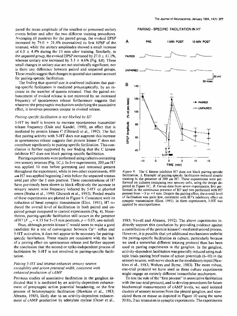
PAIRING - SPECIFIC FACILITATION IN H7
A PRE 1 MN POST 10MIN POST
PAIRED --f---- -r-----r-
UNPAIRED - -f----- ./-z
The finding that quanta1 size is unaltered indicates that pair- ing-specific facilitation is mediated presynaptically, by an in- crease in the number of quanta released. That the paired en- hancement of evoked release is not mirrored by an increased frequency of spontaneous release furthermore suggests that whatever the presynaptic mechanism underlying the associative effect, it involves processes unique to evoked release.
Pairing-.speciJic facilitation is not blocked by H7
5-HT by itself is known to increase spontaneous transmitter release frequency (Dale and Kandel, 1990), an effect that is mediated by protein kinase C (Ghirardi et al., 1992). The fact that pairing activity with 5-HT does not augment this increase in spontaneous release suggests that protein kinase C does not contribute significantly to pairing-specific facilitation. This con- clusion is further supported by our finding that the C kinase inhibitor H7 does not block pairing-specific facilitation.
Pairing experiments were performed using cultures containing two sensory neurons (Fig. 1 C,). In five experiments, 200 PM H7 was applied 10 min before pretesting and remained present throughout the experiment, while in two other experiments, 400 PM H7 was applied beginning 2 min before the unpaired tetanus until just after the 1 min posttest. These concentrations of H7 have previously been shown to block effectively the increase in sensory neuron mini frequency induced by 5-HT or phorbol esters (Braha et al., 1990; Ghirardi et al., 1992). Pooled results of these experiments are plotted in Figure 9. Consistent with its reduction of basal synaptic transmission (Eliot, 199 I), H7 re- duced the overall level of facilitation in both paired and un- paired groups compared to control experiments (Fig. 4). None- theless, pairing-specific facilitation still occurs in the presence ofH7 Vu, = 4.33 for 5-l 5 min posttests, p < 0.05, one-tailed).
Thus, although protein kinase C would seem to make a good candidate for a site of convergence between CaZ+ influx and 5-HT activation, it does not appear to be necessary for pairing- specific facilitation. These results are consistent with the lack of a pairing effect on spontaneous release and further support the conclusion that the second or spike-independent process of facilitation by 5-HT is not involved in pairing-specific facili- tation.
40 J 44 UP
5-HT - 1
-10 -5 0 5 10 15
TIME (min)
Figure 9. The C kinase inhibitor H7 does not block pairing-specific facilitation. A, Example of pairing-specific facilitation induced despite training in the presence of 200 PM H7. These experiments were per- formed on cultures containing two sensory cells, using the design de- picted in Figure 1 C, B, Group data from seven experiments, five per- formed in the continuous presence of H7 and two performed with H7 present from - 3 to + 1 min. Despite the pairing effect, the overall level of facilitation was quite low, consistent with H7’s inhibitory effect on synaptic transmission (Eliot, 1991). In these experiments, 5-HT was applied by microperfusion.
Pairing 5-HT and tetanus enhances sensory neuron excitability and action potential width, consistent with enhanced production of CAMP
1985; Yovell and Abrams, 1992). The above experiments in- directly support this conclusion by providing evidence against a contribution of the protein kinase C-mediated second process. However, it is possible that yet additional mechanisms underlie the pairing-specific facilitation in culture, particularly because we used a somewhat different training protocol than has been used in pairing experiments in the ganglion. In the ganglion, activity-dependent facilitation was generally induced using mul- tiple trials pairing brief trains of action potentials (6-10) in the sensory neuron, with nerve shock as the modulatory input (Haw- kins et al., 1983; Walters and Byrne, 1983). The more robust, one-trial protocol we have used in these culture experiments might engage an entirely different intracellular mechanism.
Previous studies of associative facilitation in the ganglion in- To test the role of the “first process” in associative facilitation dicated that it is mediated by an activity-dependent enhance- with the one-trial protocol, and to develop procedures for future ment of presynaptic action potential broadening, or the first biochemical measurements of CAMP levels, we used isolated process of heterosynaptic facilitation (Hawkins et al., 1983; clusters of sensory neurons from the pleural ganglion, and stim- Abrams, 1985) likely due to an activity-dependent enhance- ulated them en masse as depicted in Figure 10 using the same ment of CAMP production by adenylate cyclase (Ocorr et al., 20 Hz, 2 set tetanus as in synaptic experiments. The experiments

378 Eliot et al. * Associative Synaptic Plasticity in Culture
PERFUSION \ \
SHOCK -
I t I I
5HT /4 I 1 I
SN /
UNPAIRED PAIRED
Figure 10. Physical arrangement for training isolated clusters ofpleural sensory neurons. The pairing protocol for isolated sensory neurons was the same as synaptic experiments (Fig. 1 C). A train of action potentials (tetanus) was- produced in the sensory neurons by extracellular shock either 0.5 set (oaired) or 60 set (unnaired) before a 30 set pulse of 5-HT delivered through the perfusion: An intracellular electrode in one of the cells in the cluster was used to monitor the effects of these training procedures on the biophysical properties of the cell.
were conducted in the presence of the phosphodiesterase inhib- itor IBMX (100 PM, 0.02% DMSO) to rule out confounding effects of CAMP breakdown, so that the results might more directly reflect differences in adenylate cyclase activation.
In a first set of experiments, we measured excitability in in- dividual sensory neurons in response to either the same paired (0.5 set forward) or unpaired (1 min forward) training procedure used in synaptic experiments (Fig. 1C). We used both left and right pleural sensory neuron clusters from a single animal, ran- domly assigning one to receive paired and the other unpaired training. Sensory neurons normally exhibit strong spike accom- modation during a long depolarizing step command. By itself, 5-HT has a robust anti-accommodationeffect (Klein et al., 1986) which appears to be a purely CAMP-mediated phenomenon as well as a selective assay of the S-type K current (Klein et al., 1986; Baxter and Byrne, 1990; Hochner and Kandel, 1992).
As Figure 11 illustrates, pairing tetanus with 5-HT signifi- cantly enhances sensory neuron excitability compared to un- paired training. Pairing produces more anti-accommodation than unpaired training overall on the three trials following training (F,,,, = 6.71, p < 0.05) with significant differences at each of the trials by individual t tests. The effect cannot be accounted for by the different interval between the tetanus and posttests for paired and unpaired groups because the tetanus by itself slightly decreases excitability. If anything, this would tend to decrease excitability of the paired group more than the unpaired group.
This associative enhancement of sensory neuron excitability suggests that the one-trial procedure enhances CAMP production in a pairing-specific manner, consistent with earlier biochemical experiments using a high K+ pairing protocol (Ocorr et al., 1985). The results also suggest that the S-type K+ current is down-modulated in a pairing-specific manner. To address whether this down-modulation might contribute to the synaptic
facilitation, we performed a second set of experiments on iso- lated sensory clusters. In these experiments, we asked whether the same training procedure produces any pairing-specific changes in action potential width that could contribute to an enhance- ment of transmitter release.
Figure 12 illustrates the effect of various training procedures on action potential width. To enhance the contribution of the S-type K+ current, we measured action potential duration at the end ofa 1 set, 20 Hz train, which inactivates the delayed rectifier K+ current. In these experiments, we used an A/B design in which each cluster received two training sessions with a 15 min interval between sessions. Some cells received training with 5-HT alone and 5-HT paired with tetanus, and others received training with tetanus alone and no training (i.e., a 4 min “rest” between the last pretest and the first posttest). Training order was coun- terbalanced. As has been previously demonstrated (Klein and Kandel, 1978), 5-HT alone prolongs action potential repolari- zation (Fig. 12A). This is reflected in a significant overall effect of 5-HT (F,,,, = 11.39, p < 0.01). When 5-HT was paired with a 2 set tetanus, this action potential broadening was significantly enhanced compared to 5-HT alone (p < 0.02 on each posttest by individual t tests). By contrast, tetanic stimulation alone slightly decreased action potential duration compared to the no- training control (p < 0.05 on the first posttest). Thus, the greater broadening produced by paired training appears to be due to an interactive effect between tetanus and 5-HT, greater than the sum of either treatment alone. This result is reflected in a sig- nificant 5-HT x tetanus interaction (F,,,, = 17.71, p < 0.001).
These results agree with earlier studies demonstrating that pairing activity with 5-HT prolongs action potential duration (Hawkins et al., 1983; Abrams, 1985). It seems likely that both this pairing-specific spike broadening and the pairing-specific anti-accommodation effect are mediated by the same CAMP- dependent closure of S-type K channels, which would be ex- pected to contribute to facilitation of the synapse.
Ca2+ influx through L-type channels is not necessary for pairing-specific facilitation
The pairing-specific increases in action potential duration and cellular excitability are consistent with the finding that CAMP concentration is increased in sensory neurons given paired as compared to unpaired exposure to 5-HT and high K+ depolar- ization (Ocorr et al., 1985). This enhancement is likely mediated by dual activation of adenylate cyclase by Cal+ and 5-HT (Eliot et al., 1989; Abrams et al., 199 1; Yovell and Abrams, 1992). Sensory neurons contain at least two different voltage-activated Ca*+ channels (Edmonds et al., 1990) raising the issue of the source of CaZ+ influx that contributes to pairing-specific facili- tation. Pairing-specific facilitation may require Ca*+ influx through one specific type of channel, both channels, or neither of the identified currents. We tested whether the L-type current plays a role in pairing-specific facilitation by repeating the dif- ferential training experiments in culture in the presence of the dihydropyridine antagonist nitrendipine. We found in previous voltage-clamp experiments that 10 HIM nitrendipine blocks about 90% of L-type current in sensory neurons (Eliot et al., 1993). Unlike their action in vertebrate neurons, dihydropyridine an- tagonists do not require depolarization to block Ca2+ currents effectively in Aplysia neurons (Nerbonne and Gurney, 1987; Edmonds et al., 1990).
By itself, nitrendipine slightly increases sensory neuron ex- citability (data not shown), probably due to reduction of IKcCaj.

The Journal of Neuroscience, January 1994, 14(l) 379
J+ L -l---L IlOW UNPAIRED
B
7-
6.
5-
PRE t
0.75 1.75 Figure II. Pairing-specific increase in
POST sensory neuron excitability. A, An in- tracellular depolarizing current pulse was adjusted to produce one spike in the sensory neuron before training (PRE). Paired training (fop) produced a larger and longer-lasting increase in excitability or number of action poten- tials produced by the current pulse than unpaired training of the contralateral cluster from the same animal (bottom). B, Group data (n = I2 per group). Paired
:: 4 Y t=J 3-
PAIRED
2. UNPAIRED
1,= -------------------
4 AL U P
“7 r -1 .o 0 1.0
TIME (MN) 2.0
training produced a significantly greater increase in excitability than unpaired training on the three trials following training. The tetanus occurred at the uvows for paired (P) and unpaired (U) training, and 5-HT was delivered dur- ing the time indicated by the horizontal bar. The intracellular current was not significantly different for the paired and unnaired erouos t 1.4 f 0.2 nA for the paired gr&p and‘ I .3 f 0.2 nA for the unpaired group).
This excitability increase often leads to spontaneous firing when 5-HT is applied to unimpaled sensory neurons, so the number of action potentials during training is not adequately controlled. To counteract this effect, pairing experiments were performed in the presence of l-3 PM tetrodotoxin (TTX), which has no effect on sensory neuron spike amplitude, yet is quite effective in reducing the excitability increase by nitrendipine. In control experiments we found that this amount of TTX, together with the same concentration of DMSO vehicle used to solubilize nitrendipine (0.02 or O.lO%), did not interfere with pairing- specific facilitation (data not shown).
Under these conditions, nitrendipine did not block the oc- currence of pairing-specific facilitation. Neither 2 KM (n = 7) nor IO FM (n = 6) nitrendipine appeared to eliminate pairing- specific facilitation. Because the results with either concentra- tion were similar, data were pooled and are displayed in Figure 13. Overall, there was a marginal effect ofpairing in the presence of nitrendipine (F,,,, = 3.17, p < 0.05, one-tailed) and a sig- nificant pairing-by-trial interaction (F,,,, = 4.20, p < 0.02), indicating that the paired effect differed over time. As previously reported (Edmonds et al., 1990), dihydropyridine blockade has no effect on basal synaptic transmission: EPSPs were normally
depressed, to 79 -t 5% of initial value after perfusing in TTX and DMSO (n = 3 1) versus 84 * 8% after adding TTX, DMSO, and nitrendipine (n = 22). Two observations suggest that the persistence of pairing-specific facilitation is not due to inefficacy of nitrendipine. First, the effect on sensory neuron excitability using either 2 or 10 PM nitrendipine was very reliable, indicating that the drug is not highly labile. Second, we found in voltage- clamp experiments that 2 WM nitrendipine is about 80% as ef- fective as 10 FM at blocking the sustained Ca*+ current (not shown). These results indicate that Ca*+ influx through L-type channels is not necessary for the pairing effect, although the fact that the facilitation was somewhat less robust than in control experiments (Fig. 4) suggests these channels may make a partial contribution to the facilitation. Nonetheless, the failure of ni- trendipine to block the associative effect implicates by elimi- nation the dihydropyridine-insensitive channels as an important source of Ca2+ for pairing-specific facilitation.
Discussion
We have demonstrated that associative, activity-dependent syn- aptic facilitation can be induced in a reconstituted synapse in

380 Eliot et al. * Associative Synaptic Plasticity in Culture
A SHT PAIRED
B
120 T
Figure 12. Pairing-specific increase in frequency-broadened spike width. A, The action potential in a sensory neuron at the end of a 1 set, 20 Hz train before (PRE) and after (POST) training with either 5-HT alone or 5-HT paired with a 2 set, 20 Hz tetanus. Paired training produced a larger increase in action potential duration that 5-HT alone. B, Group data (n = 12 per group). Paired training produced a significantly greater increase in spike width compared to 5-HT alone on the three trials following training. Tetanus alone produced a slight decrease compared to rest (a 3 min pause indicated by the break in the abscissa). Tetanus occurred at the urrow and 5-HT was delivered at the time indicated by the horizontal bar. Spike duration has been normalized to the average value on the three pretests, which was not significantly different for the four groups (7.1 + 0.7 msec for the paired group, 7.3 k 0.7 for 5-HT alone, 8.4 ? 0.8 for tetanus alone, and 8.0 ? 0.7 for rest).
vitro. Pairing tetanus and 5-HT produces longer-lasting facili- tation than either tetanus or 5-HT alone (Fig. 2). When nor- malized for the effect of homosynaptic depression, pairing-spe- cific facilitation showed little decrement over the 20 min we monitored it (Fig. 3).
The relative durations of synaptic enhancement in our ex- periments in culture closely resemble those produced by activity alone (PTP), heterosynaptic modulation alone, and simulta- neous activity + modulation in similar experiments in the gan- glion (Walters and Byrne, 1985). In the animal, simultaneous activation of sensory neurons and their heterosynaptic modu- lators occurs in response to strong cutaneous activation, and the resulting enhanced facilitation likely contributes to behavioral site-specific sensitization (Walters, 1987). Here we took advan- tage of the reduced culture preparation to demonstrate that the enhanced facilitation also occurs when the onset of the tetanus precedes 5-HT by 0.5 set, but does not occur when tetanus is
PAIRING-SPECIFIC FACILITATION IN NITRENDIPINE
A PRE POST:
5’ 10’ 15 20’
-.A-- TEilzi JLJLLAL
20 mV
100msec
-.- PAIRED
- UNPAIRED
-10 -5 0 5 10 15 20
MINUTES
Figure 13. The L-type Ca 2+ channel blocker nitrendipine does not block pairing-specific facilitation. A, EPSPs recorded in an LFS motor neuron in response to extracellular stimulation of paired (top) and un- paired (bottom) sensory neurons in the presence of 10 FM nitrendipine (0.1% DMSO), with 3 PM TTX added to lower excitability. Despite the presence of nitrendipine, paired training significantly enhances facili- tation compared to unpaired training. B, Group data from 13 experi- ments (six as in A, and seven using 2 PM nitrendipine, 0.02% DMSO, 1 PM TTX). Average EPSP amplitude before training was 14.9 + 3.0 mV for paired cells and 14.0 + 2.8 mV for unpaired cells.
delivered 1 min before 5-HT (Figs. 4, 5). This associative prop- erty makes the activity-dependent facilitation a good candidate for a cellular mechanism contributing to classical conditioning (Hawkins et al., 1983: Walters and Byrne, 1983) and thus our results support the idea that classical conditioning and site- specific sensitization may share the same associative synaptic mechanism (Walters, 1987).
The finding that activity-dependent facilitation occurs in cul- ture also demonstrates that it is intrinsic to the sensory-motor synapse. While it does not rule out a contribution ofother circuit elements to associative facilitation in the ganglion, this finding demonstrates that such additional circuit elements are not nec- essary for the basic phenomenon. These results extend the range of synaptic plasticity that can be studied in culture to include not just homosynaptic and heterosynaptic plasticity, but con- junctive forms as well. This study also introduces a simple, one-

The Journal of Neuroscience, January 1994, 14(l) 381
trial protocol with all the technical advantages of cell culture to more easily analyze the mechanisms underlying pairing-specific facilitation.
Pairing-specific facilitation is mediated presynaptically, perhaps by action potential broadening
A major advantage of isolating single synapses in culture is that one can measure spontaneous transmitter release and be certain of its presynaptic source. We measured spontaneous release to look directly for changes in the release process during pairing- specific facilitation. The finding that unitary mEPSP or mEPSC size did not change significantly during pairing-specific facili- tation (Fig. 8) indicates that the plasticity is mediated by a change in amount of transmitter released, rather than a change in postsynaptic responsiveness. Unitary amplitude did increase slightly following both paired and unpaired training, but this effect was not significant, nor is its magnitude sufficient to ac- count for the magnitude ofevoked EPSP facilitation. The finding of a presynaptic locus for pairing-specific facilitation is not sur- prising given that both components of this conjunctive plastic- ity- PTP and heterosynaptic facilitation-have previously been found to be presynaptically mediated in sensory neurons (Cas- tellucci and Kandel, 1976; Dale and Kandel, 1990; Eliot, 199 1).
Two mechanisms have been previously identified to partic- ipate in the presynaptic facilitation by 5-HT alone. The first involves CAMP-dependent broadening ofthe presynaptic action potential (Klein and Kandel, 1978) which enhances Ca2+ influx through voltage-gated channels in terminal regions (Eliot et al., 1993). A change in presynaptic spike width is necessary for facilitation of relatively nondepressed sensory neuron synapses (Hochner et al., 1986a). In contrast, when sensory neurons are depressed by repeated activation, the amount of release is less sensitive to the duration of presynaptic depolarization, but 5-HT remains effective in facilitating the synapse. The exact mecha- nism by which 5-HT induces this spike-independent enhance- ment of release is not known, but it has been termed “mobili- zation” because of the characteristic increase in EPSP slope it produces, as if the release sites are primed with vesicles to be rapidly released (Hochner et al., 1986b). While CAMP may also play a role in this mobilizing effect of 5-HT (Goldsmith and Abrams, 1991) the second process can be distinguished from the first process in that it involves protein kinase C (Braha et al., 1990; Ghirardi et al., 1992) which is also activated by 5-HT in sensory neurons (Sacktor and Schwartz, 1990; Sossin and Schwartz, 1992).
Our findings indicate that only the first of these two mecha- nisms contributes significantly to the one-trial, pairing-specific facilitation in culture. Using the same protocol, we find that sensory neuron spike width is enhanced in a pairing-specific manner (Fig. 12). The finding that sensory neuron excitability is also enhanced with paired training (Fig. 11) suggests that the broadening effect is due to CAMP-dependent closure of S-type K+ channels (Klein et al., 1986; Baxter and Byrne, 1990). While both the excitability and spike-width effects in isolated sensory neurons appear to decay somewhat faster than the synaptic fa- cilitation, the decay is probably accelerated by the frequent post- testing and the long depolarizing test commands (to measure excitability) or action potential trains (to measure spike width) used in these experiments. Sensory neuron impalement may also accelerate the decay of these associative biophysical changes,
ture (Eliot, 199 1). Thus, the true duration of pairing-specific increases in sensory neuron excitability and spike width is likely to be closer to that of the synaptic facilitation in culture, as was found previously for spike broadening and pairing-specific fa- cilitation in the ganglion (Hawkins et al., 1983).
These results thus corroborate and extend earlier evidence that pairing-specific facilitation in the ganglion is due to an increase in action potential duration (Hawkins et al., 1983) and S-current downregulation (Hawkins and Abrams, 1984) and furthermore suggest that despite procedural differences, pairing- specific facilitation in culture and in the ganglion represent the same form of plasticity.
While our evidence supports a role of spike broadening, a number of findings argue against a role of the second process in the associative facilitation. First, the fact that we used pre- dominantly naive or nondepressed synapses in these experi- ments suggests that the second process is unlikely to be involved, since this mechanism is defined by its role in reversing synaptic depression (Hochner et al., 1986b; Ghirardi et al., 1992). A second piece of evidence is our finding that pairing-specific fa- cilitation can be induced as well using the modulatory peptide SCP as it is using 5-HT (Fig. 3). Like 5-HT, SCP increases CAMP levels in sensory neurons, broadens the presynaptic action po- tential, and facilitates sensory-motor synaptic transmission (Abrams et al., 1984). However, unlike 5-HT, SCP does not activate the second process (Schacher et al., 1990). A third piece ofevidence is that the inhibitor H7, which blocks the component of the second process mediated by protein kinase C, did not prevent pairing-specific facilitation (Fig. 9). Like the facilitation of depressed synapses, the enhancement of spontaneous release frequency by 5-HT is mediated by protein kinase C (Braha et al., 1990; Ghirardi et al., 1992). Thus, the last piece of evidence argues particularly strongly against a role ofthe second process- the finding that the associative facilitation is not accompanied by a pairing-specific enhancement of spontaneous release fre- quency (Fig. 7). The fact that spontaneous release frequency does not remain elevated during the sustained period of evoked facilitation tends generally to rule out any facilitatory mecha- nism that is independent of the action potential. However, it was recently found that associative facilitation in the ganglion produces an increase in EPSP slope, which is thought to reflect predominantly the second process (G. A. Clark, R. D. Hawkins, and E. R. Kandel, unpublished observations). Thus, it remains possible that pairing-specific facilitation involves a protein ki- nase C-independent component of mobilization not reflected in spontaneous release.
Conclusion
We have provided evidence that associative synaptic facilitation occurs in isolated synapses in culture and that it is presynapt- ically mediated by mechanisms unique to evoked transmitter release. Our findings indicate that CAMP-dependent kinase, and not protein kinase C, is critically involved in pairing-specific facilitation. While Ca2+ influx is probably necessary to induce the associative effect (Abrams, 1985) influx through dihydro- pyridine-sensitive channels is required neither for its induction nor its expression. These results suggest that a pairing-specific increase in presynaptic action potential width contributes to the associative facilitation by enhancing Ca2+ influx through the dihydropyridine-insensitive channels underlying transmitter re-
since it dramatically inhibits homosynaptic facilitation in cul- lease.

382 Eliot et al. * Associative Synaptic Plasticity in Culture
References Abrams TW (1985) Activity-dependent presynaptic facilitation: an
associative mechanism in Aplysia. Cell Mol Neurobiol 5:123-145. Abrams TW, Kandel ER (1988) Is contiguity detection in classical
conditioning a system or a cellular property? Learning in Aplysia suggests a possible molecular site. Trends Neurosci 11: 128-l 35.
Abrams TW, Castellucci VF, Camardo JS, Kandel ER, Lloyd PE (1984) Two endogenous neuropeptides modulate the gill and siphon with- drawal reflex in Aplysiu by presynaptic facilitation involving CAMP- dependent closure of a serotonin-sensitive potassium channel. Proc Nat1 Acad Sci USA 81:7956-7960.
Abrams TW, Karl KA, Kandel ER (199 1) Biochemical studies of stimulus convergence during classical conditioning in Aplysiu: dual regulation of adenylate cyclase by CaZ+/calmodulin and transmitter. J Neurosci 11:2655-2665.
Bank M, Schacher S (1992) Segregation of presynaptic inputs on an identified target neuron in vitro: structural remodeling visualized over time. J Neurosci 12:2960-2972.
Barrionuevo G, Brown TH (1983) Associative long-term potentiation in hippocampal slices. Proc Nat1 Acad Sci USA 80:7347-735 1.
Baxter DA, Byrne JH (1990) Differential effects of CAMP and sero- tonin on membrane current, action potential duration, and excit- ability in somata ofpleural sensory neurons ofAp/ysia. J Neurophysiol 641978-990.
Braha 0, Dale N, Hochner B, Klein M, Abrams TW, Kandel ER, Klein M (1990) Second messengers involved in the two processes of pre- synaptic facilitation that contribute to sensitization and dishabitua- tion in Ap/.vsiu sensory neurons. Proc Nat1 Acad Sci USA 87:2040- 2044.
Braha 0, Edmonds B, Sacktor T, Kandel ER (1993) The contributions of protein kinase A and protein kinase C to the actions of 5-HT on the L-type Ca*+ current of the sensory neurons in Aplysia. J Neurosci 13:1839-1851.
Brunelli M, Castellucci V, Kandel ER (1976) Synaptic facilitation and behavioral sensitization in Aplysiu: possible role of serotonin and cyclic AMP. Science 194: 1178-l 18 1.
Buonomano DV, Byrne JH (1990) Long-term synaptic changes pro- duced by a cellular analog of classical conditioning in Aplysiu. Science 249:420--123.
Byrne JH (1987) Cellular analysis ofassociative learning. Physiol Rev 671329-439.
Carew TJ, Walters ET, Kandel ER (198 1) Classical conditioning in a simple withdrawal reflex in Aplysiu culifornicu. J Neurosci 1: 1426- 1437.
Carew TJ, Hawkins RD, Kandel ER (1983) Differential classical con- ditioning of a defensive withdrawal reflex in Apjysiu culijbrnicu. Sci- ence 2 19:397400.
Castellucci V, Kandel ER (1976) Presynaptic facilitation as a mech- anism for behavioral sensitization in Aplysiu. Science 194: 1176-l 178.
Castellucci VF, Naim A, Greengard P, Schwartz JH, Kandel ER (1982) Inhibitor of adenosine 3’:5’-monophosphatedependent protein ki- nase blocks presynaptic facilitation in Aplysiu. J Neurosci 2:1673- 1681.
Cline HT (1991) Activity-dependent plasticity in the visual systems of frogs and fish. Trends Neurosci 14: 104-l 11.
Collineridee GL. Sineer W (1990) Excitatory amino acid receptors and syna-ptic”plasticityyTrends Pharmacol Sci -1 1:290-296.
Dale N, Kandel ER (1990) Facilitatory and inhibitory transmitters modulate spontaneous transmitter release at cultured Aplysiu sen- sorimotor synapses. J Physiol (Lond) 42 1:203-222.
Edmonds B, Klein M, Dale N, Kandel ER (1990) Contributions of two types of calcium channels to synaptic transmission and plasticity. Science 25O:l 142-1147.
Eisenstadt M, Goldman JE, Kandel ER, Koike H, Koester J, Schwartz JH (1973) Intrasomatic injection ofradioactive precursors for study- ing transmitter synthesis in identified neurons of Aplysiu culifornicu. Proc Nat1 Acad Sci USA 70:3371-3375.
Eliot LS (199 1) Presynaptic modulation of Aplysiu sensory-to-motor neuron synapses in culture. PhD thesis, Columbia University.
Eliot LS, Dudai Y, Kandel ER, Abrams TW (1989) Ca2+/calmodulin sensitivity may be common to all forms of neural adenylate cyclase. Proc Nat1 Acad Sci USA 86:9564-9568.
Eliot LS, Kandel ER, Hawkins ER (1990) Activity-dependent mod-
ulation of spontaneous transmitter release in single cultured Aplysiu sensorimotor neuron synapses. Sot Neurosci Abstr 16: 10 13.
Eliot LS, Kandel ER, Siegelbaum SA, Blumenfeld H (1993) Imaging terminals of Ap/ysiu sensory neurons demonstrates role of enhanced Ca*+ influx in presynaptic facilitation. Nature 361:634-637.
Ghirardi M, Braha 0, Hochner B, Montarolo PG, Kandel ER, Dale N (1992) Roles of PKA and PKC in facilitation of evoked and spon- taneous transmitter release at depressed and nondepressed synapses in Aplysiu sensory neurons. Neuron 9:470-489.
Glanzman DL. Mackev SL. Hawkins RD. Dvke AM. Llovd PE. Kandel ER (1989) ‘Depletion of serotonin in the-nervous system orAp/ysiu reduces the behavioral enhancement of gill withdrawal as well as the heterosynaptic facilitation produced by tail shock. J Neurosci 9:4200- 4213.
Goldsmith BA, Abrams TW (199 1) Reversal of synaptic depression by serotonin at Aplysia sensory neuron synapses involves activation of adenvlate cvclase. Proc Nat1 Acad Sci USA 88:9021-9025.
Goldsmith BA, Abrams TW (1992) CAMP modulates multiple K+ currents, increasing spike duration and excitability in Aplysiu sensory neurons. Proc Nat1 Acad Sci USA 89: I 148 l-l 1485.
Hamill OP, Marty A, Neher E, Sakmann B, Sigworth FJ (198 1) Im- proved patch-clamp techniques for high-resolution current recording from cells and cell-free membrane patches. Pfluegers Arch 391:85- 100.
Hawkins RD (199 1) Cell biological studies of conditioning in Aplysiu. In: Neurobiology of learning, emotion and affect (Madden J, ed), pp 3-28. New York: Raven.
Hawkins RD, Abrams TW (1984) Evidence that activity-dependent facilitation underlying classical conditioning in Aplysiu involves mod- ulation of the same ionic current as normal presynaptic facilitation. Sot Neurosci Abstr 10:268.
Hawkins RD, Schacher S (1989) Identified facilitator neurons L29 and L28 are excited by cutaneous stimuli used in dishabituation, sensi- tization, and classical conditioning of Aplysiu. J Neurosci 9:4236- 4245.
Hawkins RD, Castellucci VF, Kandel ER (198 1) Intemeurons in- volved in mediation and modulation ofgill-withdrawal reflex in Aply- siu. II. Identified neurons produce heterosynaptic facilitation con- tributing to behavioral sensitization. J Neurophysiol 45:3 15-326.
Hawkins RD, Abrams TW, Carew TJ, Kandel ER (1983) A cellular mechanism of classical conditioning in Aplysiu: activity-dependent amplification of presynaptic facilitation. Science 2 19:400-405.
Hawkins RD, Kandel ER, Siegelbaum SA (1993) Learning to modulate transmitter release: themes and variations in synaptic plasticity. Annu Rev Neurosci 16:625-665.
Hickie C, Walters ET (1990) Identified central motor neurons are necessary for directional siphon responses in Aplysiu. Sot Neurosci Abstr 16:19.
Hochner B, Kandel ER (1992) Modulation of a transient K’ current in the pleural sensory neurons of Aplysiu by serotonin and CAMP: imolications for snike broadening. Proc Nat1 Acad Sci USA 89: 11476 11480.
Hochner B, Klein M, Schacher S, Kandel ER (1986a) Action-potential duration and the modulation of transmitter release from the sensory neurons of Aplysiu in presynaptic facilitation and behavioral sensi- tization. Proc Nat1 Acad Sci USA 83:84 10-8414.
Hochner B. Klein M. Schacher S. Kandel ER (1986b) Additional component in the cellular mechanism of presynaptic facilitation con- tributes to behavioral dishabituation in Aplysiu. Proc Nat1 Acad Sci USA 83:8794-8798.
Ito M, Sakurai M, Tongroach P (1982) Climbing fibre induced de- pression of both mossy fiber responsiveness and glutamate sensitivity of cerebellar Purkinje cells. J Physiol (Lond) 324: 113-l 34.
Klein M, Kandel ER (1978) Presynaptic modulation of voltage-de- pendent Ca*+ current: mechanism for behavioral sensitization in kplysiu culijknicu. Proc Nat1 Acad Sci USA 75:35 12-35 16.
Klein M. Camardo J. Kandel ER (1982) Serotonin modulatesa soecific potassium current in the sensory neurons that show presynaptic fa- cilitation in Aplysiu. Proc Nat1 Acad Sci USA 79:57 13-57 17.
Klein M, Hochner B, Kandel ER (1986) Facilitatory transmitters and CAMP can modulate accommodation as well as transmitter release in Aplysiu sensory neurons: evidence for parallel processing in a single cell. Proc Nat1 Acad Sci USA 83:7994-7998.
Levy WB, Steward 0 (1979) Synapses as associative memory elements in the hippocampal formation. Brain Res 175:233-245.
The Journal of Neuroscience, January 1994, 74(l) 383
Mackey SL, Kandel ER, Hawkins RD (1989) Identified serotonergic neurons LCB 1 and RCB 1 in the cerebral ganglia of Aplysiu produce presynaptic facilitation of siphon sensory neurons. J Neurosci 9:4227- 4235.
Mercer AR, Emptage NJ, Carew TJ (1991) Pharmacological disso- ciation of modulatory effects of serotonin in Aplysiu sensory neurons. Science 254:181 I-1813.
Montarolo PG, Goelet P, Castellucci VF, Morgan J, Kandel ER, Schach- er S (1986) A critical period of macromolecular synthesis in Iong- term heterosynaptic facilitation in Aplysia. Science 234: 1249-l 254.
Montarolo PC, Kandel ER, Schacher S (1988) Long-term heterosyn- aptic inhibition in Aplysia. Nature 333: 17 l-1 74.
Nerbonne JM, Gurney AM (1987) Blockade of Ca*+ and K+ currents in bag cell neurons of Aplysia culijmzica by dihydropyridine Ca2+ antagonists. J Neurosci 7:882-893.
Ocorr KA, Walter ET, Byrne JH (1985) Associative conditioning an- alog selectively increases CAMP levels of tail sensory neurons in kply- sia. Proc Nat1 Acad Sci USA 82:2548-2552.
Rayport SC, Schacher S (1986) Synaptic plasticity in vitro: cell culture of identified Aplysia neurons mediating short-term habituation and sensitization. J Neurosci 6:759-763.
Sacktor TC, Schwartz JH (1990) Sensitizing stimuli cause transloca- tion of protein kinase C in Ap/ysiu sensory neurons. Proc Nat1 Acad Sci USA 87:2036-2039.
Schacher S, Montarolo PG, Kandel ER (1990) Selective short- and long-term effects of serotonin, small cardioactive peptide, and tetanic stimulation on sensorimotor synapses ofAplysiu in culture. J Neurosci 10:3286-3294.
Shuster MJ, Camardo JS, Siegelbaum SA, Kandel ER (1985) Cyclic
AMP-dependent protein kinase closes the serotonin-sensitive K+ channels of Aplysiu sensory neurones in cell-free membrane patches. Nature 3 13:392-395.
Siegelbaum SA, Camardo JS, Kandel ER (I 982) Serotonin and cyclic AMP close single K+ channels in Aplysia sensory neurons. Nature 299:413-417.
Small SA, Kandel ER, Hawkins RD (1989) Activity-dependent en- hancement of presynaptic inhibition in Aplysia sensory neurons. Sci- ence 243:1603-1606.
Sossin WS, Schwartz JH (1992) Selective activation of Ca’+ -activated PKCs in Aplysiu neurons by 5-HT. J Neurosci 12: I 160-l 168.
Stanton PK, Sejnowski TJ (1989) Associative long-term depression in the hippocampus induced by hebbian covariance. Nature 339: 215-218.
Walters ET (1987) Multiple sensory neuronal correlates ofsite-specific sensitization in Aplysiu. J Neurosci 7:4084 17.
Walters ET, Byrne JH (1983) Associative conditioning of single sen- sory neurons suggests a cellular mechanism for learning. Science 2 19: 405408.
Walters ET, Byrne JH (1985) Long-term enhancement produced by activity-dependent modulation of Aplysiu sensory neurons. J Neu- rosci 51662-672.
Walters ET, Byrne JH, Carew TJ, Kandel ER (1983) Mechanoafferent neurons innervating tail of Aplysiu: 1. Response properties and syn- aptic connections. J Neurophysiol 50: 1522-l 542.
Yovell Y, Abrams TW (1992) Temporal asymmetry in activation of Aplysia adenylyl cyclase by calcium and transmitter may explain tem- poral requirements of conditioning. Proc Nat1 Acad Sci USA 89: 6526-6530.