palate presentation
TRANSCRIPT
DR GAURAV CHATURVEDI
SOFT PALATEThe soft palate is a mobile musculomembranous
structure attached anteriorly to the hard palate and
blending laterally with the pharynx.
It separates the nasal cavity from the oral cavity and forms a
partial partition between the nasopharynx above it
and the oropharynx below it.
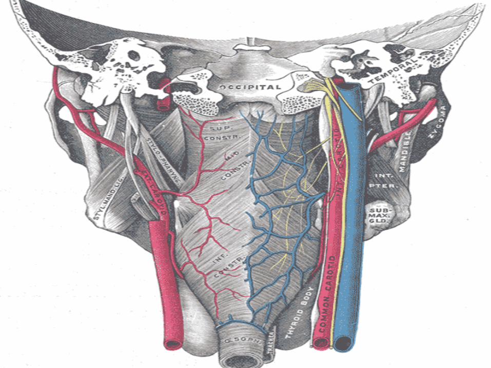
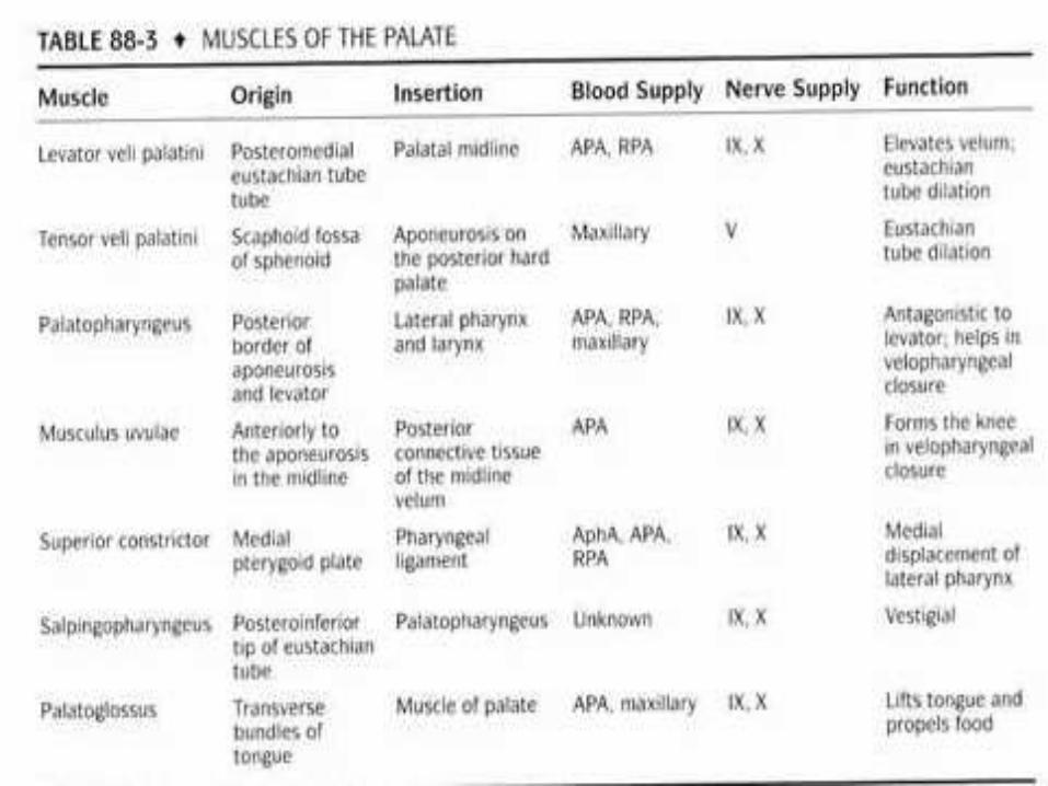
MUSCLES OF SOFT PALATE The velopharyngeal musculature consists of several
muscular slings formed by the following:
Levator veli palatini
Tensor veli palatini
Palatopharyngeus
Palatoglossus
Musculi uvulae
Superior pharyngeal constrictor.
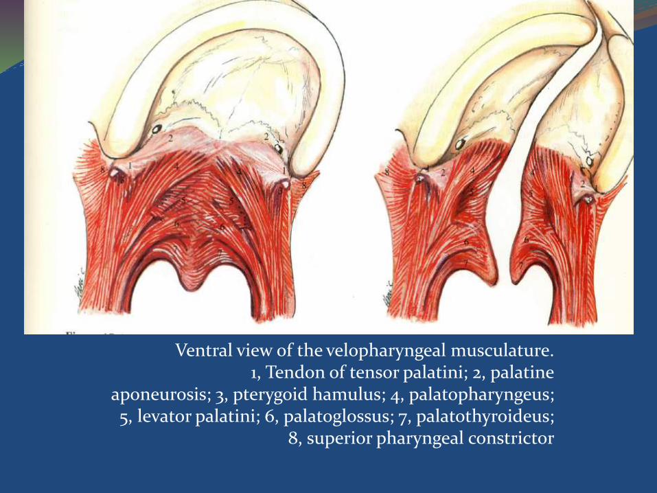
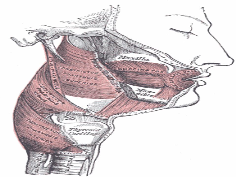
Ventral view of the velopharyngeal musculature.1, Tendon of tensor palatini; 2, palatine
aponeurosis; 3, pterygoid hamulus; 4, palatopharyngeus;5, levator palatini; 6, palatoglossus; 7, palatothyroideus;
8, superior pharyngeal constrictor
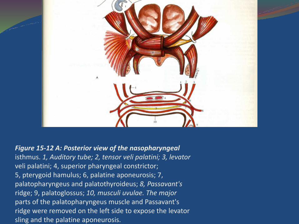
Figure 15-12 A: Posterior view of the nasopharyngealisthmus. 1, Auditory tube; 2, tensor veli palatini; 3, levatorveli palatini; 4, superior pharyngeal constrictor;5, pterygoid hamulus; 6, palatine aponeurosis; 7,palatopharyngeus and palatothyroideus; 8, Passavant'sridge; 9, palatoglossus; 10, musculi uvulae. The majorparts of the palatopharyngeus muscle and Passavant'sridge were removed on the left side to expose the levatorsling and the palatine aponeurosis.
PALATINE APONEUROSIS The palatine aponeurosis is a sheet of fibrous tissue
that provides stability and flexibility to the soft palate and serves as an anchoring point for a number of its muscles.
Along its attachment to the hard palate, the palatine aponeurosis continues with the periosteum and submucosal connective tissue on the oral and nasal surfaces on the hard palate.
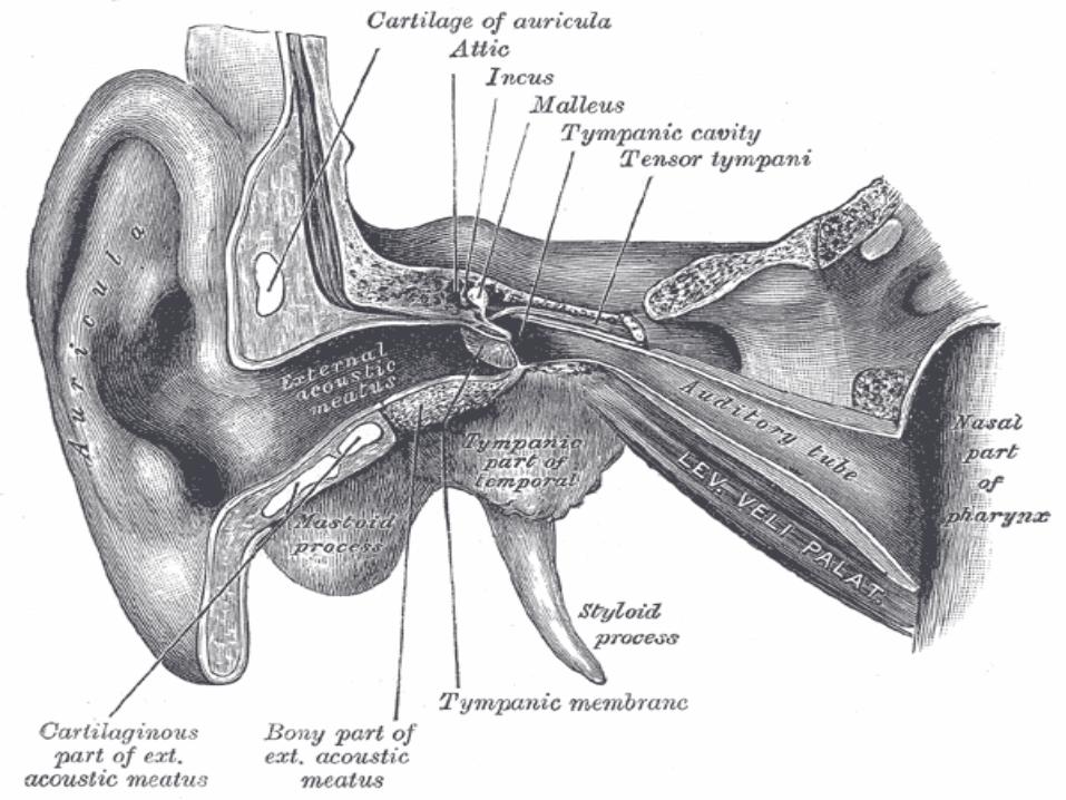
Levator veli palatini The levator arises from:
The rough quadrate area on the inferior surface of the petrous portion of the temporal bone in front of the carotid canal,
The carotid sheath,
Part of the medial surface of the cartilaginous portion of the auditory tube.
Fibers are inserted:
into the nasal surface of the palatine aponeurosis and blend with the muscle fibers of the opposite side, forming the V-shaped levator sling that occupies the middle portion of the soft palate.
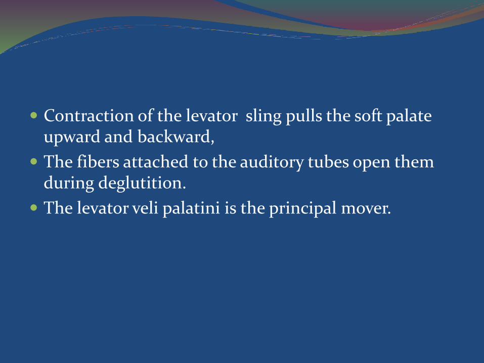
Contraction of the levator sling pulls the soft palate upward and backward,
The fibers attached to the auditory tubes open them during deglutition.
The levator veli palatini is the principal mover.
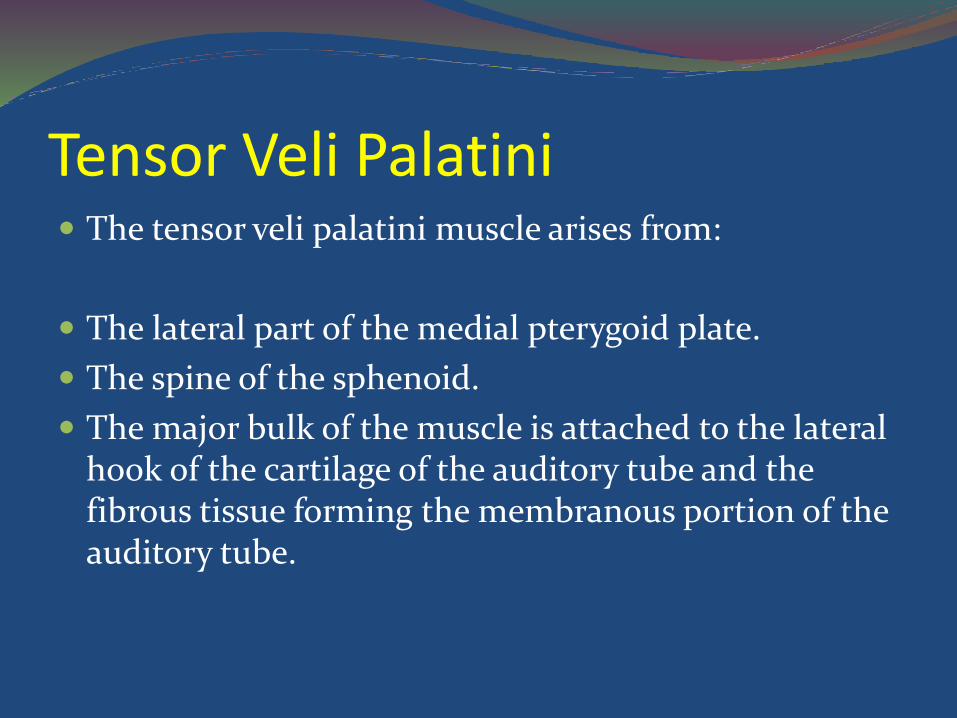
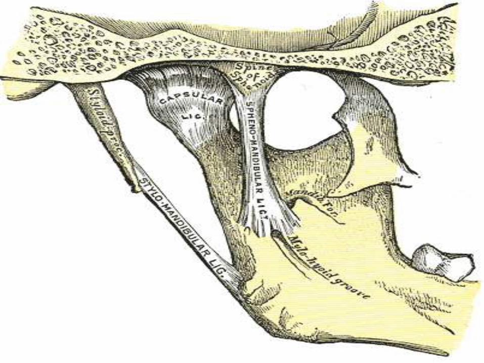
Tensor Veli Palatini The tensor veli palatini muscle arises from:
The lateral part of the medial pterygoid plate.
The spine of the sphenoid.
The major bulk of the muscle is attached to the lateral hook of the cartilage of the auditory tube and the fibrous tissue forming the membranous portion of the auditory tube.
PATIENT PRESENTATION Cleft palate with cleft lip and alveolus –
Alveolar portion of the cleft lies between the maxillary lateral incisor and canine tooth roots.
In comparison to noncleft individuals, patients with unilateral cleft lip and palate have overall reduction in crown size and delay of eruption of permanent dentition.
The nasal septum is deviated and buckled toward the cleft side.
The absence of a portion of the inferior piriformaperture and the hypoplasia of the lateral nasal bony platform at the maxillary wall contribute to the cleft nasal deformity; the nasal base is depressed, the ala collapses, and the floor widens.
In the bilateral complete cleft lip and palate, the pre-maxillary segment containing the central and lateral incisor tooth roots is discontinuous from the alveolar arch.
The lateral segments often collapse inward and lingually, resulting in “locking out” of the premaxilla.
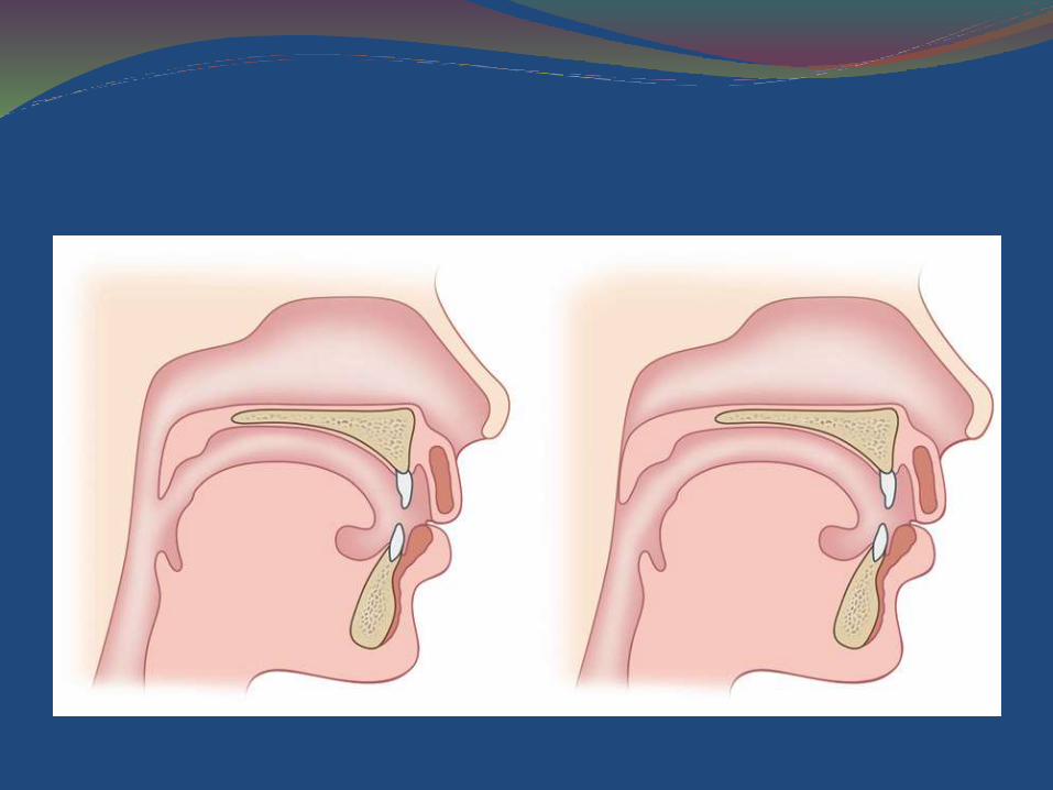
Clefts of the secondary palate
Also called incomplete cleft palate, a cleft of the secondary palate may be variable, from an opening in the posterior soft palate to a cleft extending up to the incisive foramen. There is almost always a separation of the bony shelves of the hard palate.
Most commonly, dentition is normal and symmetric.
Submucous cleft palate
Submucous cleft palate occurs when the palate has mucosal continuity but the underlying levator palatinimuscle is discontinuous across the midline and longitudinally oriented, similar to the muscle anatomy in overt clefts of the palate.
Calnan’s classic triad is present.
Ear pathology
In multicenter studies, incidence of otitis media effusion has been found to be 96–100% in cleft patients, measured by both middle ear effusions on otoscopy and impedance testing and middle ear aspiration.
In the cleft palate, impairment of tubal dilation is thought to occur from complex misalignment of the paratubal musculature.
In addition, intrinsic abnormalities of tubal cartilage framework rendering a Eustachian tube more collapsible have been noted.
In addition, some theorize that constant bathing of the tube orifice with oropharyngeal refluxed material leads to inflammation and obstruction of drainage.
Pierre Robins Syndrome has high risk of hearing loss.
Pierre Robin sequence
Pierre Robin described the triad of micrognathia, glossoptosis, and respiratory distress.
Of the children diagnosed with Pierre Robin sequence, 60–90% have cleft palate4
Palatal cleft is usually isolated to the velum and can be V shaped or, more typically, U-shaped
GROWTH
At birth, the average weight is the same for cleft and unaffected newborns.
Studies observing infants with cleft lip and palate show initial growth retardation by the time they undergo surgical lip repair.
When the same children reach the age for palatoplasty, they have significantly lagged on the growth curve.
Average growth returns to normal compared with unaffected children by the age of 4 years.
Children with orofacial clefting stabilize and continue normal growth to at least 6 years of age, with no statistically significant differences in height and weight when compared to unaffected children.
In later childhood, however, average weight and height of children with cleft appear to diminish compared with those of control subjects.
Duration of puberty and pubertal skeletal growth were prolonged an average of 1 year,
FEEDING Infants with cleft lip can usually be fed by breast or
regular bottle.
For breastfeeding babies, the soft breast tissue often will fill the opening in the lip and/or gum line well, sealing off the mouth and allowing the baby to create adequate suction. Sometimes it is helpful for the mother to hold a finger across the cleft during feeding.
When starting a feeding, hold the baby in a slightly upright position.
Try to keep the nipple in the center of the baby's mouth. Do not let it slip up into the cleft.
Babies with cleft palate usually need special bottles and techniques to feed adequately.
We encourage mothers who planned to breastfeed to express their milk and feed it to their infants using a special bottle.
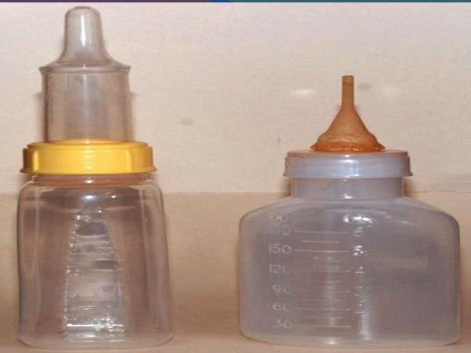
There are three types of bottles that we recommend for feeding babies with clefts –
Mead-Johnson Cleft Palate Nurser.
The Haberman Feeder.
The Pigeon Nipple.
Haberman Feeder The baby can obtain milk by compressing the nipple
against the roof of the mouth, without need for suction as reservoir is present just above the nipple.
Mead- Johnson As the baby begins to suck, squeeze the bottle with a
firm steady pressure to the count of "squ-e-e-ze-two-three," relaxing your squeeze on the two-three count. This provides the baby with an intermittent flow of milk
Pigeon Nipple Pigeon Nipple works by compression only. The nipple
has a firm side that goes toward the roof of the mouth and a softer side that goes on the tongue.
A small notch at the base of the nipple serves as an air vent. This notch should be uppermost under the baby's nose when feeding.
Tightening the nipple and collar slows the flow of milk. Loosening it makes the flow faster.
Timing of palate repair Victor Veau first made the observation of a correlation
between age at repair and speech outcome in 1931.
He noted that children who had undergone repair before 12 months of age were much more likely to have normal speech than those with repair between 2 and 4 years of age.
Despite the absence of hard evidence supporting earlier palate repairs, a growing body of opinion seems to support palate repair around 9–10 months of age for children with apparently normal development.

Maxillary growth
Palatoplasty has been shown to detrimentally affect maxillary growth.
Transverse growth of the maxillary arch is narrowed in comparison with that in noncleft patients, resulting in typical malocclusion traits of crowding, lateral cross-bite, and open bite.
Whether the narrowed arch and maxillary growth inhibition result from surgical scarring or intrinsic maxillary underdevelopment remains a matter of debate; most likely it is a combination of the tw0
Although it might seem preferable to wait until a more advanced age for palate repair, given the growth effects on the maxilla, it is far more difficult to establish normal speech.
Congenital Palatal Insufficiency(CPI)
It has been said that cleft palate is a type of defect that
can be “seen, felt, and heard.
Palatal insufficiency may be caused by the velum being too short and/or by a deficiency in the antero-posterior dimension of the hard palate.