paleopathology of skeletal fluorosis - college of liberal …€¦ · · 2006-12-05paleopathology...
TRANSCRIPT

Paleopathology of Skeletal FluorosisJUDITH LITTLETON*Department of Anthropology, University of Auckland,Auckland, New Zealand
KEY WORDS fluoride; prehistoric Arabia; Bahrain; endemicfluorosis
ABSTRACT Skeletal fluorosis is one of a range of conditions causingexcessive ossification and joint ankylosis in skeletons. It is rarely considered,however, in differential diagnoses of palaeopathological lesions. This paperconsiders the identification of skeletal fluorosis in a skeletal sample from theisland of Bahrain, Arabian Gulf, dating to ca. 250 BC–AD 250.
Approximately 4% of 255 adult skeletons in the sample have hyperostosiclesions resulting in joint ankylosis primarily of the lumbar vertebrae, as wellas the major joints. These lesions most frequently occur among males in the501 age group. Chemical analysis on a small series of bone and dentalsamples confirmed the presence of high levels of fluoride, while staining of theteeth is evidence of dental fluorosis. The level of dental fluorosis is comparablewith a naturally occurring fluoride level in water of between 1–2 ppm. Theprevalence of hyperostosic lesions, however, appears higher than expected,and two possible reasons are suggested: confusion between a diagnosis ofdiffuse idiopathic skeletal hyperostosis and skeletal fluorosis on partial or lessseverely affected skeletons; and the presence of predisposing factors forskeletal fluorosis on the island in the past. Am J Phys Anthropol 109:465–483,1999. r 1999 Wiley-Liss, Inc.
Palaeopathological studies have frequentlydemonstrated the difficulty of diagnosingthe cause of skeletal lesions, particularlybetween hyperostosic conditions (Rogers etal., 1987; Crubezy, 1990). Endemic fluorosisdue to naturally occurring high levels offluoride in water and soil is a major healthproblem in some parts of the world today(Teotia and Teotia, 1984). While moderncases of the disease include those due toindustrial intoxication, airborne fluorides incoal, and deep well bores (Mwainiki et al.,1994; Wang and Huang, 1995), there is noreason to assume it was not also a problemin the past. Recent analyses from NorthIndia (Lukacs et al., 1985) and Pompeii(Torino et al., 1995) have pointed to thepresence of high fluoride levels in someancient populations, including ancient China(Wang and Huang, 1995).
While fluorosis is rarely considered as apossible cause of pathological changes inskeletal samples, in areas with naturallyhigh fluoride levels it obviously needs to betaken into account. The aim of this paperwas, through examination of a skeletal popu-lation from Bahrain, to clarify the identifica-tion of skeletal fluorosis, which is oftenignored as a probable cause of hyperostosiclesions in skeletal remains.
BACKGROUND: SKELETAL FLUOROSIS
Skeletal fluorosis results from the inges-tion of excessive amounts of fluoride. Thereis no fixed toxic level of fluoride, since the
Grant sponsor: Ministry of Information, Bahrain; Grant spon-sor: ANU Australian Postgraduate Research Award scholarship.
*Correspondence to: Judith Littleton, Department of Anthro-pology, University of Auckland, Private Mail Bag 92019, Auck-land, New Zealand. E-mail: [email protected]
Received 18 September 1995; accepted 1 April 1999.
AMERICAN JOURNAL OF PHYSICAL ANTHROPOLOGY 109:465–483 (1999)
r 1999 WILEY-LISS, INC.
development of fluorosis depends upon envi-ronmental factors. Intoxication can resultfrom acute doses, but more commonly thecondition is chronic due to prolonged intakeof high levels of fluoride (above 0.7 ppm insome places; Jolly et al., 1973). The mostcommon natural source of fluoride in thesecases is water, especially well and springwater where the surrounding substrata con-tain fluoride (Teotia and Teotia, 1988).
When excessive amounts of fluoride areingested, a series of biochemical changesoccurs (Mariano-Menez et al., 1990). Fluo-ride is rapidly absorbed into serum throughthe stomach and upper intestine. The ab-sorbed fluoride is carried to the bone, whereit can replace hydroxyl in the bone hydroxy-apatite, creating fluoroapatite. This occursmost rapidly in the trabecular portion of thebone (Teotia and Teotia, 1988). The abnor-mal bone has an increased density. Theabsorbed fluoride also stimulates the forma-tion of new irregular bone at the sites oftendon and ligament insertions, resulting ingradual ossification of soft tissues. In thepresence of adequate dietary calcium, themain picture is of osteosclerosis; however, incases where dietary calcium is inadequate,the absorbed fluoride may result in second-ary hyperparathyroidism, leading to boneloss, so that bone density may include areasof both sclerosis and porosis (Teotia andTeotia, 1988; Mithal et al., 1993). This osteo-porotic type of skeletal fluorosis occurs inchildren and younger adults, particularly inareas with extremely high levels of fluoridein the water (Christie, 1980; Krishnamach-ari, 1986).
While airborne exposure through coal firesand dust has been reported (Haikel et al.,1986; Wang and Huang, 1995), most com-monly the osteosclerotic type of fluorosisonly develops after 15–20 years of continu-ous exposure to water containing high levelsof fluoride (Jolly et al., 1969). The minimumwater level ever reported as causing changesis 0.7 ppm (Jolly et al., 1973), but there is noclear linear relationship between the amountof fluoride in drinking water and the develop-ment of skeletal fluorosis (Chibole, 1987).Studies in temperate, developed countrieshave demonstrated no significant signs ofskeletal fluorosis in communities with water
levels of 4 ppm (McClure et al., 1958; Kamin-sky et al., 1990), yet work in some Indianvillages has demonstrated skeletal fluorosiswhen the water level is much lower (Moudgilet al., 1986; Teotia and Teotia, 1988). Influ-encing factors other than water level appearto be period of exposure, climate, other traceelements in the water, dietary intake offluoride, nutritional status, water storagemethods, work patterns, and tea-drinkinghabits (Jolly et al., 1969; Haikel et al., 1986;Zietsman, 1991).
The major signs of fluorosis are in thedentition and the skeleton. Teeth are af-fected by fluorotic staining and pitting of thesurface enamel (Moller, 1982). Fluorosis inthe skeleton is marked by increased boneproduction, and thickening and coarseningof bone trabeculae, with a correspondingincrease in bone density resulting in a‘‘ground glass’’ appearance on X-rays (Bul-lough and Vigorta, 1984; Wang et al., 1994).The entire skeleton is affected. The craniumbecomes thick and heavy with gradual oblit-eration of the diploe, while ossification ofligaments results in the development ofirregular margins around the foramen mag-num. Postcranial bones become heavy andirregular, with abnormal prominence ofmuscle and tendinous insertions and visibleenthesophytes (Rogers and Waldron, 1995).Ossification of spinal ligaments is common,especially of the ligamenta flava, and intra-transverse and interspinous ligaments. Insevere cases, compression of the spinal cordcan result (Misra et al., 1988). The vertebralbodies appear larger than normal, and even-tually there is complete fusion of the verte-brae by thick osteophytes. These changesare most common in the cervical and lumbarregions. The ribs appear large and haveroughened surfaces due to ligament ossifica-tion. Ankylosis of the costovertebral andcostosternal joints may occur, with conse-quent ‘‘freezing’’ of the chest cavity in anaspirated position (Jolly et al., 1969). Calcifi-cation of ligamentous and tendinous inser-tions is most marked on those areas of theskeleton subject to strain, especially thelinea aspera, the interosseous membranebetween the radius and ulna and the tibiaand fibula, and the sacrotuberous and sacro-spinous ligaments (Moller and Gudjonsson,
466 J. LITTLETON

1967). In advanced cases, ankylosis mayoccur in these locations (Teotia and Teotia,1988). The disease tends to affect olderadults, and men more frequently thanwomen (Jolly et al., 1969).
The condition has been regularly reportedin certain regions of the world where fluo-ride occurs at naturally high levels. Theseinclude North India (Teotia and Teotia, 1988),parts of Africa (Chibole, 1987; Mwainiki etal., 1994), parts of China (Wang et al., 1995),and the Arabian Gulf (the origin of thecurrent sample; FitzGerald-Finch, 1981).
In 1983, a single probable case of skeletalfluorosis was found among Bronze Age hu-man remains from Bahrain Island (Frohlichet al., 1989). Changes characteristic of skel-etal fluorosis were identified, and the diagno-sis was confirmed by chemical analysis ofthe bone, revealing a 1% fluoride level. Atthe time, it was assumed that this was anunusual case and that this individual hadbeen a foreigner to Bahrain, possibly fromNorth India (Frohlich et al., 1989).
Since then, however, both dental and skel-etal changes reminiscent of fluorosis havebeen found in other skeletal samples fromthe island (e.g., Littleton and Frohlich, 1989).In particular, during a more extensive sur-vey of paleopathological patterns amongskeletons from a Hellenistic cemetery on theisland, many individuals with ossification atthe ligament and tendinous insertions andosteophytic growth around the joint marginswere recorded (Littleton, 1993).
While there are several possible causes ofsuch ossification, this paper illustrates theimportance of considering fluorosis. It ar-gues that many of the changes observed inthe Bahrain population are indicative ofskeletal fluorosis. The prevalence of thedisease is explained by considering predis-posing factors for fluorosis, and the possiblesocial impact of the disease is assessed.
THE AREA AND THE SAMPLE
The state of Bahrain is located halfwayalong the Arabian Gulf, 30 km from theeastern shore of Saudi Arabia (Fig. 1). It is asmall group of islands, the main one (Bahr-ain Island) being approximately 30 km wideand 50 km long. Traditionally, the islands’inhabitants relied upon a mixture of agricul-
ture, fishing, and trading. Sweet water isderived from a fluctuating supply from natu-ral aquifers which arise in Saudi Arabia(Larsen, 1983). The aquifer was tapped onthe island by a series of wells extendingdown into the underground basin, as well asa small number of natural springs. Waterlevels have fluctuated over time and settle-ment on the island has been dependent uponthis, contracting towards the north and westof the island when water levels decline. Theisland receives less than 7 cm of rainfall ayear and therefore agriculture was totallydependent upon this underground watersupply prior to the development of desalina-tion.
The island is known to have been occupiedfrom 7000 BP to the present. The skeletalsample described in this current work datesfrom the Hellenistic or Tylos period, ca.2300–1700 BP, which appears to have been atime of relative prosperity and active trad-ing in manufactured goods, in addition tostaples such as dates, wood, and grain(Salles, 1987).
The human remains come from the DS3cemetery, which is part of the al-Malikiyahmound field. This area contained a mixtureof Iron Age and Hellenistic tombs and lies onthe western edge of a Bronze Age mound-field. The site was completely excavated bythe Bahrain Directorate of Archaeology andMuseums between 1984–1986 except for ap-proximately 20% of the cemetery, bulldozedprior to excavation. The remaining areacomprised a minimum of 15 burial mounds.The absolute number of graves is 292, ofwhich 38 contained no skeletal material.
The burial ground was located on non-arable land bordering irrigated land. Aerialsurvey suggests that a settlement site waslocated nearby and that the cemetery prob-ably derived from the inhabitants of a smallvillage in the immediate area (Littleton,1998). Based on population size estimatesfrom the cemetery, the village would havecontained, on average, between 100–250 in-dividuals. Dietary reconstruction on the ba-sis of archaeological as well as dental andskeletal evidence suggests that the peopleburied in the cemetery followed an agricul-tural lifestyle, possibly supplemented byfishing. The maintenance of the cemetery
467SKELETAL FLUOROSIS IN BAHRAIN

over time and relative lack of expenditure onthese graves suggest that the area was usedby local residents (Littleton, 1998).
The total number of individuals from theDS3 cemetery was 1,051. Of these, 631 weresubadults. Many of the skeletons, however,were incomplete. Paleopathological data aretherefore analyzed on two levels: individu-als for whom age and sex could be assigned,and the total number of individual skeletalelements. The sample composition can beseen in Table 1. When left- and right-sidebones were present in one individual, right-side bones were recorded. In general, how-ever, there was little discordance betweensides in severity of lesions.
METHODS
Age and sex were designated through theuse of multiple indicators. Morphologicalfeatures of the pelvis and crania were usedfor the identification of sex (Phenice, 1969;Brothwell, 1981). Where possible, adult ageswere assigned to 10-year age groups (501
was the oldest group). A combination ofpubic symphysis changes (Meindl et al.,1985), sacroauricular surface indicators(Lovejoy et al., 1985), degree of epiphysealclosure (Bass, 1981), and cranial sutureclosure (Meindl and Lovejoy, 1985) wereused for adult age estimation.
Pathological conditions were recorded inseveral categories, none of which, strictlyspeaking, were mutually exclusive. Thesewere: traumatic, resorptive (porotic hyperos-tosis, osteoporosis), proliferative (e.g., perios-titis, osteomyelitis), or sclerosing/hypertro-phic (Powell, 1988; Littleton, 1993). Initiallyit was expected that this final conditionwould include neoplasms. In the entire sam-ple, however, only two cases of probablebenign osteomata were found. Instead it wasobserved that a large number of bonesshowed signs of excessive ossification of theligaments and interosseous membranes, ac-companied by some disorganization of thebone cortex. As a result, each bone wasgraded into one of four categories: 0, normal;
Fig. 1. Location of Bahrain in the Arabian Gulf.
468 J. LITTLETON

1, slight—accentuation of muscle markings,increased robusticity of the bone (Fig. 2); 2,moderate—ossification of ligamentous inser-tions and interosseus membranes (3–10 mm),with protruding masses of bone, and thebeginning of joint ankylosis (Fig. 3); and 3,ankylosis of joints, near complete ligamentossification (Fig. 4).
Some of these increases in rugosity mayhave been due to physical activity and age(Crubezy, 1990) and not due to a pathologi-cal condition. In subsequent analysis, there-fore, only moderate-severe cases were consid-ered (i.e., enthesophytes .3 mm in length orankylosis).
Both each bone and each individual skel-eton were scored according to this scale.Individual skeletons were classified into un-affected, slight, moderate, or severe classeson the basis of condition of the majority ofthe bones.
All permanent teeth were also recorded,using a recording scheme adapted from Dean(1934) and Jolly (Jolly et al., 1969). Fourcategories were identified: 1, normal—trans-lucent enamel, no sign of brownish staining;
2, slight—scattered white opacities on thetooth’s surface; 3, moderate—superficial andminute pitting, brownish staining; and 4,severe—marked pitting with widespreadstaining.
Postmortem staining of teeth in burialdeposits is common. In this instance, stainswere only recorded when it was obvious thatthe mark was formed within the enamel andwas resistant to removal by acetone; inaddition, the determination of fluorosis wasdependent upon the observation of opacitiesand pitting (Thylstrup and Fejerskov, 1978).Individuals were categorized as having mild,moderate, or severe changes on the basis ofthe two most severely affected teeth in theirdentition.
Chemical analysis of four teeth and bonefragments from three individuals was con-ducted. Dental analysis by electron micro-probe scanning was conducted by Dr. G.Suckling of the Dental Research Unit, Medi-cal Research Council of New Zealand. Follow-ing established methodology, scans weretaken through each tooth to determinechanges in fluoride level due to diagenesis
TABLE 1. Sample composition by skeletal element1
Element Male Female 20–29 yr 30–39 yr 40–49 yr 501 yr Total
Occipital 67 57 27 28 1 36 125Parietal 150 156 55 66 38 16 312Orbit 105 116 51 41 26 18 122Frontal 63 63 23 29 19 9 126Face 24 20 9 7 4 5 44Basicranium 26 29 13 9 6 4 55Endocranium 29 44 18 15 7 5 73Temporal 115 126 49 50 28 16 251Mandible 29 34 12 15 10 7 61Cervical vertebrae 60 57 28 31 18 13 119Thoracic vertebrae 67 63 30 34 17 14 132Lumbar vertebrae 68 60 30 36 17 14 130Sacrum 48 38 20 21 15 13 87Pelvis 127 109 55 76 32 33 236Ribs 47 42 21 18 15 11 90Sternum 28 24 13 9 10 7 55Clavicle 111 119 61 57 29 22 235Scapula 79 66 34 32 18 23 145Humerus 134 111 54 54 26 28 146Radius 123 104 48 60 24 24 229Ulna 111 94 41 48 24 24 205Hand 25 22 10 9 7 4 47Femur 139 116 56 63 26 27 255Patella 75 61 41 30 20 8 69Tibia 104 89 40 30 17 19 193Fibula 85 79 38 43 20 18 165Foot 74 65 34 35 18 22 142Dentitions2 74 78 21 19 1651 Right side; left used only when right side not present.2 Two or more teeth present.
469SKELETAL FLUOROSIS IN BAHRAIN

(Suckling et al., 1992). The human bone wastested by atomic absorption spectrometry atthe Department of Chemistry, AustralianNational University, following the methodol-ogy described by Szpunar et al. (1978). Sincefluoride is subject to diagenesis and accumu-lates in bone over time, samples were takenfrom the surface and from the middle of thebone cortex. Soil samples from the graveswere also tested to determine whether thesoil content in Bahrain contained high levelsof fluoride which could affect buried remains.
RESULTSSkeletal material
Table 2 demonstrates the distribution ofall lesions throughout the adult skeletonsfrom DS3. No subadult bones were found tohave any evidence of skeletal hypertrophy.However, adult skeletal elements with exten-sive muscular attachments frequentlyshowed signs of excessive ossification. Thusthe skull tended to be affected very little
with the exception of the basicranial area,the area of the nuchal line and mastoid, andthe gonial angle of the mandibular ramus. Asmall proportion of these bones (,5%)showed signs of overdevelopment of themuscle attachments (stage 1); none, how-ever, showed more definite evidence of liga-ment ossification.
The postcrania in the sample were morefrequently affected. Major effects were seenin the thoracic area (24.7% of ribs and 11.0%of thoracic vertebrae, including all stages).Fusion, or near fusion, of the sternocostaljoints occurred, with progressive ossificationof the anterior ligaments covering the ster-nal body.
In the sample, the probability of boneankylosis and ligamentous ossification wasgreatest in the lower vertebrae, with thelumbo-sacral area being most frequentlyaffected. This ankylosis can be readily distin-guished from osteophytosis, since the firstligaments to show signs of ossification were
Fig. 2. Slight ossification along linea aspera on distal (top) and proximal (middle) and femur moderatechanges to patella.
470 J. LITTLETON

the supra- and interspinous ligaments alongthe neural arch (Fig. 4) (Rogers and Wal-dron, 1995). Osteophytic growth on the ver-tebral bodies tended to be flowing ossifica-tion from the central body, rather thanhorizontal outgrowth (Fig. 4). There were noaccompanying signs of joint degenerationsuch as porosity or eburnation of the articu-lar surfaces.
Figure 5 is an X-ray of a severely affectedspine (from T8 to L4) compared to a normalvertebral column from DS3. While the patho-logical spine was damaged postmortem, sev-eral characteristic features of this pathologi-cal condition are demonstrated. The anteriorspinal ligaments are fused, and thick osteo-phytes protrude from the central anteriorvertebral body. Complete ankylosis has oc-curred on several vertebral bodies, espe-cially L2–L4, with encroachment upon theintervertebral disc space; all intervertebraldisc spaces show signs of marked narrowing.The posterior spine on T8 has a roughenedsurface with exostoses along the lower bor-der, and the apophyseal joints, where vis-ible, are surrounded by osteophytes. Themost obvious difference between the twospines, however, is the degree of radioden-
sity. The pathological case is near opaquewith markedly greater density of bone, in-dicative of osteosclerosis.
In the sample, the pelvis was also fre-quently affected, even more commonly thanthe sacrum. The major ligament insertions,particularly the sacro-tuberous ligament,were visibly ossified in 5.1% of pelves. Insevere cases, joint ankylosis occurred, pri-marily at the sacroauricular joint.
In comparison to the trunk the limbs wereless affected, and arms less than legs. Theclavicles and scapulae only rarely showedany definite signs of joint ankylosis, thoughan increased degree of muscle marking wasobservable. Signs on the humerus, radius,and ulna were more frequent and obvious.Changes to the humeri included increaseddevelopment of the deltoid tuberosity,gradual ossification of the ligaments at thesites of insertion, and ossification of themuscle attachments around the olecranonfossa.
Ossification of the ligament insertions onthe radii and ulnae was generally apparent,particularly on the olecranon of the ulna andaround the radial tuberosity. Ossification ofthe interosseus membrane was also ob-
Fig. 3. Moderate enthesophytes on patella (right) compared to normal (left).
471SKELETAL FLUOROSIS IN BAHRAIN

served, although complete fusion of the twobones was not found. In contrast, the bonesof the hand had no visible enthesophytes.
After the changes to the vertebrae andribs, the most characteristic changes wereseen in the bones of the leg. The foot wasmost frequently and severely affected, with7.7% of feet showing evidence of excessossification, particularly of the talo-calca-neal ligament (Fig. 6). Changes to the femurwere generally nonspecific, consisting of in-creased rugosity in the attachment of thelinea aspera and the insertion of the patellarligament; only 1.2% had measurable enthe-sophytes. Changes in the patella includedthe gradual ossification of the anterior patel-lar ligament so that, in severe cases, spic-
ules of bone protruded vertically from theanterior surface in both inferior and supe-rior directions.
As with the radius and ulna, the tibia andfibula showed ossification of the interosseusligaments, marked ossification of the patel-lar ligament insertion, and ossification ofthe interosseus membrane. In moderate-severe cases, spicules of bone protrudedfrom each of these locations, resulting in oneindividual with ankylosis of the tibia andfibula.
Female skeletons were much less fre-quently affected by these changes (Table 2;16.7% compared to 37.3% considering allstages; P , 0.001), although the pattern ofchanges throughout the skeleton was simi-lar (Spearman’s rank correlation r 5 0.597;P 5 ns). Among males, moderate-severechanges were observed in most types ofskeletal elements. Among females only thepelvis, ribs, sternum, fibula, and foot showedsigns of moderate-severe changes, althoughthe numbers were too small to test thisdifference statistically. This lower frequencyoccurred despite the fact that the pelvic areain females is subject to marked trauma andpossible ligament ossification due to child-birth (Angel, 1971).
This condition is age-related (Table 2). Nobones in the 20–30-year age group wereaffected. In the 30–40-year age group, only
Fig. 4. Severe osteophytosis (joint ankylosis) of lumbarvertebrae (L1–5). Breaks between L1–L2, and L3–L4 post-mortem. Note ankylosis of vertebral arch between L1–L3.
TABLE 2. Prevalence of moderate-severe enthesohytesamong Bahrain adults (%)
ElementAll
adults Males Females
30–40yearsold
40–50yearsold
501yearsold
Cervical vert. 0.0 0.0 0.0 0.0 0.0 0.0Thoracic vert. 1.5 3.0 0.0 0.0 0.0 14.3Lumbar vert. 2.3 4.4 0.0 0.0 0.0 21.4Sacrum 3.4 6.3 0.0 0.0 0.0 15.4Pelvis 5.1 7.6 3.7 1.3 9.4 12.1Ribs 6.7 10.6 2.4 5.6 6.7 27.3Sternum 12.7 21.4 4.2 11.1 0.0 57.1Clavicle 0.4 1.8 0.0 0.0 3.4 0.0Scapula 1.4 0.5 0.0 0.0 5.6 4.3Humerus 4.1 6.0 0.0 3.7 0.0 14.3Radius 2.2 6.6 0.0 0.0 0.0 16.7Ulna 3.9 7.4 0.0 4.2 0.0 25.0Hand 0.0 0.0 0.0 0.0 0.0 0.0Femur 1.2 3.0 0.0 0.0 0.0 7.4Patella 2.9 2.9 0.0 0.0 0.0 12.5Tibia 2.6 4.0 0.0 0.0 0.0 15.8Fibula 3.6 6.5 2.0 2.3 0.0 16.7Foot 7.7 9.8 3.7 0.0 0.0 22.7Individuals 7.1 21.6 11.5 10.0 23.8 15.8
472 J. LITTLETON

Fig. 5. X-ray of affected spine (right) compared to normal spine from DS3 (left).
473SKELETAL FLUOROSIS IN BAHRAIN

the pelvis, ribs, sternum, humerus, ulna,and fibula had any evidence of moderate-severe changes. In the 40–50-year age group,the percentage of affected bone increased,but only a small number of elements wereaffected. A marked increase was observed inthe 501 age group. Cases of extensive jointankylosis were only observed in this oldestage group.
Sample sizes were too small to analyze therelationship of moderate-severe changes withboth age and sex. Very few females belongedto the oldest age group, and very few femaleswere affected overall.
In its severe form, the condition appearsto have been a disease of the oldest membersof the society, though there was obviously aprogression in both prevalence and severityduring adulthood. Female skeletons tend tobe less frequently affected and certainly lessseverely than males. The pathologicalchanges suggest a chronic and steadily devel-oping disease, eventually affecting the major-ity of the population.
Teeth
Due to widespread antemortem tooth lossin the Bahrain populations, particularly in
the oldest age groups (Littleton and Frohlich,1989), fluorotic staining on the teeth couldnot be definitely associated with postcranialchanges in any one individual. Among theentire sample of adults from DS3, 44.8% hadsome degree of dental fluorosis (Table 3).Most of these changes were mild, although5% of the population had severe changes inthe form of marked pitting and widespreadstaining.
Chemical analysis
Samples of bone were analyzed from threeindividuals. Samples were selected fromfragmentary remains, using the midshaft ofthe femur. Of the three individuals selected,only one (S5/38) had any evidence of osteo-
TABLE 3. Prevalence of dental fluorosis amongadult dentitions
NFluorotic
(%)Moderate-severe (%)
Severe(%)
15–20 years old 10 30.0 10.0 0.020–30 years old 21 47.6 23.8 9.530–40 years old 19 21.1 15.8 0.0All adults 165 44.8 18.2 4.9Males 74 48.6 21.6 4.1Females 78 46.2 11.5 6.4
Fig. 6. Ossification of thetalo-calcaneal ligament par-ticularly marked on calca-neus (right).
474 J. LITTLETON

phytosis. The lesions consisted of small osteo-phytic outgrowths around the costal jointsand along the linea aspera of the femur.None of the osteophytes was longer than 3mm. Neither of the remaining two individu-als (70A/13 and 81/14) had any evidence ofexcessive ossification.
Soil samples were matched for two of thesamples. Neither sample contained any de-tectable fluoride which could have been apotential source of contamination (Table 4).As a further check of diagenetic change inthese two individuals, samples were takenfrom both the outer surface of the cortex anda midsection of the femur. In S5/38 therewas a decline in the fluoride level within thesame individual, suggesting some move-ment of fluoride through the bone. However,in 70A/13 there was no detectable fluoride ineither sample, suggesting that while fluo-ride was mobile through the bones, therehad been no uptake of fluoride from thesurrounding soil or from possible groundwa-ter.
Normal fluoride values in bone range from0.019–0.073% (McClure et al., 1958; Zipkinet al., 1958). The fluoride levels detected inthe mid-section of 81/14 and S5/38 werethree times greater and comparable withfluoride water levels of between 1.0–2.6 ppmin a temperate environment (Table 4) (Mc-Clure et al., 1958). The levels were lowerthan those detected in the severely affectedBronze Age individual, BS40 (Table 4)(Frohlich et al., 1989).
Chemical analysis was also conducted onfour permanent teeth and compared to NewZealand data collected by the New ZealandDental Research Unit. The four teeth se-lected all had some evidence of brown stain-ing and opacity, but only one (LM2 W) hadany sign of surface pitting. Scanning byelectron microprobe demonstrated that fluo-ride had been mobile through the cementumand dentine but with little diffusion throughthe enamel, and the ratio of surface to innerfluoride levels was within the same range asmodern teeth (Table 4) (Richards et al.,
TABLE 4. Fluoride levels from adult bone and teeth samples with comparisons
Skeletal fluoride Age Sex Sample source % dry weight
Bahrain
BS40 Adult 401 Male Femur 1.0001
70A/13 Adult femur ? Midsection 0.000Outer surface 0.000
81/38 Adult femur ? Midsection 0.210S5/38 Adult 451 femur Female Midsection 0.210
Outer surface 0.28070a/13 Soil 0.000S5/38 0.000Comparative samples, fluoride ppm,1 ppm Adult ? Iliac crest 0.019–0.0732
0.2 ppm Bone 0.0533
1.0 ppm Bone 0.1383
2.6 ppm Bone 0.2673
4.0 ppm Bone 0.4133
Dental fluoride Age Sex Surface ppm F Inner ppm F
Bahrain
R I1 B Adult ? 2,100–2,500 200R I2 B Adult ? 1,500–2,000 100R I2 W Adult ? 1,000–4,000 200LM 2 W Adult ? 3,000–4,000 200–400Comparative samples4
Low (,1 ppm F) Adult Adult ,1,000 1001 ppm F Adult Adult 1,000–1,500 100–200Severe fluorosis Adult Adult 6,000 5001 Frohlich et al. (1989).2 Zipkin et al. (1958).3 McClure et al. (1958).4 New Zealand Dental Research Unit.F, fluoride
475SKELETAL FLUOROSIS IN BAHRAIN

1989). Three of the teeth demonstrated ele-vated fluoride levels. The inner fluoride lev-els of RI1 B, RI2 W, and LM2 W werecomparable to dental fluoride levels ob-served in the temperate climate of NewZealand at water levels of 1 ppm. The toothwith pitting, LM2 W, had an inner fluoridelevel that ranged between expected valuesat 1 ppm water levels and areas of severefluorosis.
DISCUSSIONFluoride levels In Bahrain
The chemical analysis points to high butvariable levels of fluoride intake in thispopulation. This is not, however, clear evi-dence that the skeletal changes outlinedabove were due to skeletal fluorosis. Fluo-ride is subject to diagenetic change in bone,and the mobility of fluoride in the bone wasdemonstrated in the bone samples by thedifference between the outer and midsectionanalyses (Table 4), which means that abso-lute values need to be treated with caution.
A further difficulty is that, while dentalchanges due to fluorosis are well-defined(Moller, 1982) and present in this sample,there is no definite association within indi-viduals between the presence of dental andskeletal changes. The reason for this istwofold. Firstly, dental fluorosis is observ-able at lower fluoride levels than skeletalfluorosis, and there is no predictive relation-ship between the two conditions (Richardset al., 1989). Secondly, in this populationextensive antemortem tooth loss among oldadults makes it impossible to associate exten-sive osteophytic growth with dental lesions.
Apart from the chemical analysis, thepresence of dental fluorosis also points to thepresence of high levels of fluoride in theBahrain samples. Cases of dental fluorosishave been recorded on the island (Barnes,1981), while modern cases of skeletal fluoro-sis have also been described from nearbyQatar (Azar et al., 1961), coastal SaudiArabia (Walters, 1954), Sharjah, and AbuDhabi (FitzGerald-Finch, 1981), reflectinglocally high levels of naturally occurringfluoride.
Water samples from a series of wells onthe island confirm this. A variety of testshave been made upon spring water in Bahr-
ain, resulting in values between 0.5–1.5ppm (Matter, 1985; Musaiger and Khunji,1990). The optimum fluoride level for retard-ing dental decay without adverse signs forBahrain has been calculated at 0.5 ppm. AsMatter (1985, p. 6) states, ‘‘Irrespective ofwhich set of values are taken the overallindications are of high fluoride content inthe ground water.’’
The variability in water levels betweenlocations tends to be due to local variation inthe substrata plus the depth of water anddegree of sedimentation. It is not possible touse contemporary data to directly predictfluoride levels in the past, since differencesin average temperatures, the amount ofrain, and the water levels of the aquiferscould cause some variation in fluoride levels.However, significant variations in the levelof fluoride in underground water would notbe expected over this period unless massiveclearance and erosion had caused substan-tial leaching of fluoride from surface soil.There is no evidence on Bahrain for suchmassive clearance; nor do the soil samplesfrom the burials indicate high levels of fluo-ride. In any case, the consequences of sucherosion would be expected to result in anincreased level of fluoride in the present andnot explain the past frequency of fluoroticlesions.
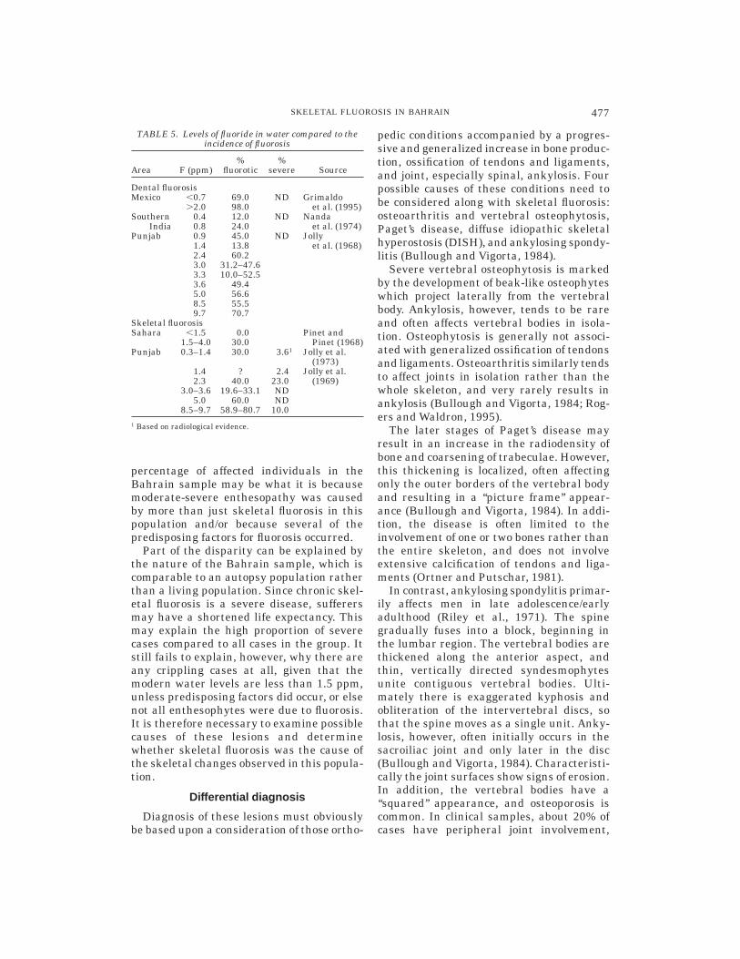
Table 5 lists comparative percentages ofskeletal and dental fluorosis with the compa-rable water levels in a number of differentplaces. There is wide variation in the percent-age of people affected in areas of similarwater levels. The levels of dental fluorosisobserved among historical groups in Bahr-ain are comparable with levels seen at fluo-ride contents of between 1–2 ppm (confirmedby the chemical analysis). The levels ofskeletal fluorosis are less clear. Between1–2.5 ppm, the percentage of individualswith skeletal fluorosis varies from 0–40.4%of the population. In the Bahrain sample,12.7% of adults had moderate-severe enthe-sophytes, and 3.6% joint fusion. While someauthors claim that severe fluorosis does notoccur below 4 ppm (McClure et al., 1958;Kaminsky et al., 1990), others have provedthe presence of severe skeletal fluorosis atwater levels of 1.35–1.5 ppm, given predis-posing factors (Pinet and Pinet, 1968). The
476 J. LITTLETON
percentage of affected individuals in theBahrain sample may be what it is becausemoderate-severe enthesopathy was causedby more than just skeletal fluorosis in thispopulation and/or because several of thepredisposing factors for fluorosis occurred.
Part of the disparity can be explained bythe nature of the Bahrain sample, which iscomparable to an autopsy population ratherthan a living population. Since chronic skel-etal fluorosis is a severe disease, sufferersmay have a shortened life expectancy. Thismay explain the high proportion of severecases compared to all cases in the group. Itstill fails to explain, however, why there areany crippling cases at all, given that themodern water levels are less than 1.5 ppm,unless predisposing factors did occur, or elsenot all enthesophytes were due to fluorosis.It is therefore necessary to examine possiblecauses of these lesions and determinewhether skeletal fluorosis was the cause ofthe skeletal changes observed in this popula-tion.
Differential diagnosis
Diagnosis of these lesions must obviouslybe based upon a consideration of those ortho-
pedic conditions accompanied by a progres-sive and generalized increase in bone produc-tion, ossification of tendons and ligaments,and joint, especially spinal, ankylosis. Fourpossible causes of these conditions need tobe considered along with skeletal fluorosis:osteoarthritis and vertebral osteophytosis,Paget’s disease, diffuse idiopathic skeletalhyperostosis (DISH), and ankylosing spondy-litis (Bullough and Vigorta, 1984).
Severe vertebral osteophytosis is markedby the development of beak-like osteophyteswhich project laterally from the vertebralbody. Ankylosis, however, tends to be rareand often affects vertebral bodies in isola-tion. Osteophytosis is generally not associ-ated with generalized ossification of tendonsand ligaments. Osteoarthritis similarly tendsto affect joints in isolation rather than thewhole skeleton, and very rarely results inankylosis (Bullough and Vigorta, 1984; Rog-ers and Waldron, 1995).
The later stages of Paget’s disease mayresult in an increase in the radiodensity ofbone and coarsening of trabeculae. However,this thickening is localized, often affectingonly the outer borders of the vertebral bodyand resulting in a ‘‘picture frame’’ appear-ance (Bullough and Vigorta, 1984). In addi-tion, the disease is often limited to theinvolvement of one or two bones rather thanthe entire skeleton, and does not involveextensive calcification of tendons and liga-ments (Ortner and Putschar, 1981).
In contrast, ankylosing spondylitis primar-ily affects men in late adolescence/earlyadulthood (Riley et al., 1971). The spinegradually fuses into a block, beginning inthe lumbar region. The vertebral bodies arethickened along the anterior aspect, andthin, vertically directed syndesmophytesunite contiguous vertebral bodies. Ulti-mately there is exaggerated kyphosis andobliteration of the intervertebral discs, sothat the spine moves as a single unit. Anky-losis, however, often initially occurs in thesacroiliac joint and only later in the disc(Bullough and Vigorta, 1984). Characteristi-cally the joint surfaces show signs of erosion.In addition, the vertebral bodies have a‘‘squared’’ appearance, and osteoporosis iscommon. In clinical samples, about 20% ofcases have peripheral joint involvement,
TABLE 5. Levels of fluoride in water compared to theincidence of fluorosis
Area F (ppm)%
fluorotic%
severe Source
Dental fluorosisMexico ,0.7 69.0 ND Grimaldo
.2.0 98.0 et al. (1995)Southern 0.4 12.0 ND Nanda
India 0.8 24.0 et al. (1974)Punjab 0.9 45.0 ND Jolly
1.4 13.8 et al. (1968)2.4 60.23.0 31.2–47.63.3 10.0–52.53.6 49.45.0 56.68.5 55.59.7 70.7
Skeletal fluorosisSahara ,1.5 0.0 Pinet and
1.5–4.0 30.0 Pinet (1968)Punjab 0.3–1.4 30.0 3.61 Jolly et al.
(1973)1.4 ? 2.4 Jolly et al.2.3 40.0 23.0 (1969)
3.0–3.6 19.6–33.1 ND5.0 60.0 ND
8.5–9.7 58.9–80.7 10.01 Based on radiological evidence.
477SKELETAL FLUOROSIS IN BAHRAIN

predominantly in the lower limbs, includingthe metatarsophalangeal joints (Riley et al.,1971; Manchester, 1982). The changes tendto be arthritic in nature, with destruction ofthe joint surfaces.
DISH, or Forrestier’s disease, occurs pri-marily in older individuals, especially men(Rogers, 1982). The condition is diagnosedby the presence of thick bridging osteo-phytes, especially along the right antero-lateral aspect of the thoracic spine. This mayresult in the ankylosis of several contiguousvertebral bodies (Vernon-Roberts et al.,1974). While the cervical and lumbar spinemay be affected, the condition begins in thethoracic area with the calcification and ossi-fication of the paraspinal ligaments. Theposterior longitudinal ligaments may be-come ossified, and there is sometimes anky-losis of the apophyseal joints (El Garf andKhater, 1984). In general, the intervertebraldisc space is preserved without marked nar-rowing (Vernon-Roberts et al., 1974; but seeHarris et al., 1974). There may be involve-ment of other joints apart from the spine.The most commonly affected areas are thepelvis, upper femur, heel, and knee (Uts-inger, 1985). On the pelvis, ‘‘whiskering’’along the ilia is prevalent, and there tends tobe para-articular osteophytosis, with calcifi-cation of ligaments such as the sacrospi-nous, calcaneal, and patellar ligaments. In alarge sample (El Garf and Khater, 1984),skeletal findings were restricted to the spineand lower extremities. The condition rarelyencompasses osteoporosis, and costochron-
dral ossification is similarly rare (Manches-ter, 1982). The prevalence of the disease inan autopsy population was between 6–12%of all individuals (Rogers, 1982). In thissame series, 65% were male and 88% weremore than 50 years of age (Rogers, 1982).
Comparison with the Bahrain lesions
Each of these diseases has similarities tothe condition described at DS3. However,the Bahrain material does not suggest ei-ther osteoarthritis or Paget’s disease, be-cause of the presence of extensive ossifica-tion of tendons and ligaments, and multiplejoint ankylosis. Table 6 lists the chief diag-nostic features of the remaining conditionsas compared to the Bahrain material.
Ankylosing spondylitis (AS) in unlikely,given the age distribution of changes in theBahrain population. While males were morefrequently affected than females in the skel-etal sample, the affected individuals weremuch older than the age group affected byAS. In addition, vertebral bridging in AS isby thin syndesmophytes rather than thethick osteophytes observed in the Bahrainmaterial. Osteophytes in this condition areaccompanied by joint erosion, also not ob-served in the DS3 sample.
Apart from fluorosis, the symptoms ofDISH are most similar to the lesions ob-served in the Bahrain population. Diagnos-tic criteria for DISH were described by Cru-bezy (1990). The age distribution of DISH issimilar to that observed in Bahrain, as aresome of the other signs, such as thick osteo-
TABLE 6. Diagnostic features of possible causes compared to the specimens from Bahrain1
Diffuse idiopathicskeletal hyperostosis Ankylosing spondylitis Fluorosis Bahrain
Age .40 years old ca. 20 years old Older adults (prolongedhabitation except inextremely high Fareas)
.40 years old
Sex M . F M . F M . F M . FVertebral ankylosis Thoracic Late total Lumbar first, later total Lumbar, then totalVertebral bridge Thick osteophytes Thin syndesmophytes Thick osteophytes Thick osteophytesLateral Thoracic right . left Central Central CentralSquaring Absent Present Absent AbsentCostovertebral/
costosternal jointsLigament
calcificationArthritic changes Ligament calcification Ligament
calcificationBone Normal Osteoporotic Osteosclerotic OsteocleroticPeripheral joints Mainly lower Primarily lower Lower and upper AllDental mottling Absent Absent Present Present1 Based on Jolly et al. (1969); Riley et al. (1971); Vernon-Roberts et al. (1974); Manchester (1982); Rogers and Waldron (1995).
478 J. LITTLETON

phytes on the vertebrae. There are, however,significant differences.
Fusion of vertebrae in DISH is generallyalong the anterolateral aspect of the bodyand often unilateral to the right side (Resnickand Niwayama, 1976). In contrast, fusion inthe Bahrain samples occurred in the centralarea of the body and had no lateral prefer-ence. Sometimes in DISH the osteophyteswill fuse; however, Crubezy (1990) notedthat in most studies, the osteophytes haveseparate growths with facets between theupper and lower osteophytes. In the Bahrainmaterial the osteophytes did not form well-defined margins, and fusion was complete.Vertebral ankylosis in DISH does not affectthe intervertebral disc space, but amongfused vertebrae from Bahrain there wasmarked narrowing of this space (Fig. 5).
There are also significant extravertebraldifferences. While enthesophytes did occuron the patella, calcaneus, and olecranon inthe Bahrain sample, they were not re-stricted to these sites. Osteophytic growthwas much more widespread and affected theinterosseous membranes. In addition,costchondral ossification is rare in DISH(Manchester, 1982), as is ankylosis of theapophyseal joints (El Garf and Khater, 1984),yet these were frequent signs among theBahrain material.
The closest correspondence with the le-sions observed in Bahrain is with skeletalfluorosis. The epidemiological picture is con-sistent with a disease affecting older indi-viduals, primarily men. Unlike DISH, verte-bral ankylosis begins in the lumbar regionand has no lateral preference in skeletalfluorosis and in the Bahrain material. Theintervertebral space in fluorosis is nar-rowed, and this was observed in the Bahrainmaterial. All joints subject to wear and tearwere affected in the sample consistent withfluorosis rather than DISH. Finally, fluoro-sis results in osteosclerosis, with a thick-ened cortex—a finding observed in the Bahr-ain material (Fig. 5), while changes in bonedensity do not occur in DISH except foroccasional osteoporosis (Manchester, 1982).
As Crubezy (1990) emphasized, however,diagnosis must rely upon a range of indica-tors, and isolating a single cause on fragmen-tary material may be impossible.
Distinguishing between DISH and skeletalfluorosis
In diffuse idiopathic skeletal hyperostosis,Utsinger (1985) suggests diagnosis be basedupon the following criteria:
1. Continuous ossification along the antero-lateral aspect of at least four contiguousbodies, primarily in the thoraco-lumbarspine (definite DISH);
2. Continuous ossification along the antero-lateral aspect of at least two contiguousvertebral bodies (possible DISH); and
3. symmetrical and peripheral enthesopa-thy of the posterior heel, superior patellaor olecranon, with the new bone having awell-defined cortical margin in the ab-sence of inflammatory joint disease (pos-sible DISH).
In areas with high levels of fluoride, thefirst and second criteria of Utsinger (1985)will not distinguish between skeletal fluoro-sis and DISH in a skeletal sample, althoughunilateral fusion is indicative of DISH ratherthan skeletal fluorosis (Vernon-Roberts etal., 1974). Additional criteria such as bonedensity, width of the intervertebral space,involvement of the apophyseal joints, com-pleteness of fusion, and the extent of extra-vertebral enthesophytes need to be takeninto account. Definite diagnosis between thetwo conditions and other enthesopathiesdepends upon having more than isolatedskeletal elements.
In the Bahrain material, individuals withsevere enthesopathy, accompanied by sclero-sis and joint narrowing, most probably suf-fered from skeletal fluorosis. The possibleconfusion between DISH and fluorosis, how-ever, means that the percentage of individu-als with fluorosis is not necessarily equiva-lent to the percentage of individuals withmoderate-severe enthesopathy, since thismay incorporate some individuals withDISH, particularly moderate cases. Even so,given modern levels of fluoride on the island,it would appear that there were cripplingcases of skeletal fluorosis on the island. Thisraises the questions of why this should be so,and of what predisposing factors may havebeen present.
479SKELETAL FLUOROSIS IN BAHRAIN

Predisposing factors to skeletal fluorosis
Fluorosis is categorized into three stagesin radiographic studies (Jolly et al., 1969;based on Roholm, 1937):
1. Blurring and coarsening of trabeculae;2. Merging of trabeculae, narrowing of the
medullary cavities, early ossification ofligaments;
3. Marbled bone, irregular periosteal thick-ening, and more extensive ossification(based on Roholm, 1937).
These can be compared to the three stagesused for recording the degree of lesionsamongst the Bahrain skeletons; stage 3 ofRoholm (1937) of crippling fluorosis can beseen as roughly equivalent to the stage 3 ofskeletal recording. It was initially claimedthat crippling fluorosis required water lev-els of more than 10 ppm before it occurred(Jolly et al., 1969). More studies, however,have demonstrated that in many popula-tions, crippling occurs above 3 ppm, and canoccur at water levels of 1.35–1.5 ppm (Haikelet al., 1986; Pinet and Pinet, 1968), giventhe presence of predisposing factors.
Temperature is a major factor in fluorosis.In hot, dry climates, water intake is substan-tially greater, so that the same fluoride levelin water will promote more fluorosis in a hotclimate than in a temperate climate (Gala-gan and Lamson, 1953; Zietsman, 1991). Atthe same time, temperature affects evapora-tion rates: the greater the evaporation ofwater, the more concentrated fluoride be-comes. Standing water in jars or pots isparticularly vulnerable. High levels of fluo-ride-rich sediment in water can also resultin a higher fluoride intake (Nanda et al.,1974).
The chemical constituents of water alsohave an effect on the physiological uptake offluoride. Calcium and magnesium in watertend to inhibit fluoride ingestion, while highsodium levels and alkalinity promote fluo-ride ingestion (Pinet and Pinet, 1968). Simi-larly, a low dietary intake of calcium andphosphorus is implicated in elevated levelsof skeletal fluorosis (Mithal et al., 1993).General nutritional status also plays a role,since malnourished individuals appear to be
more prone to develop dental and skeletalfluorosis (Massler and Schour, 1952).
Sources of fluoride other than water alsoplay a role. Plants irrigated with watercontaining fluoride contain low levels offluoride, while dust can be a major contami-nant (Haikel et al, 1986). Smoke from fluo-rine-rich coal has also been identified as acause of fluorosis in China (Wang and Huang,1995). Fish may contain high levels of fluo-ride from sea water, as does sea salt, whichis often used in cooking (Moller, 1982). Oneof the greatest dietary sources of fluoride,however, is tea (both leaf and brick forms),which has been implicated in several studieswhere higher than expected levels of skel-etal fluorosis were found (Jackson andWeidman, 1958; Azar et al., 1961; Wang andHuang, 1995).
Finally, work in India has demonstratedthat manual laborers are more likely todevelop skeletal fluorosis than their seden-tary counterparts (Pandit et al., 1940). Thisis probably because people working outsidetend to drink more water. Moreover, it ap-pears that the development of new fluoroticbone occurs at those sites most subjected tostrain and minor trauma (Jolly et al., 1969;Wang et al., 1994). These factors may allpredispose certain communities towardsfluorosis.
On Bahrain itself, it can be hypothesizedthat some of these factors applied. Firstlyhigh temperatures, particularly in summer,plus the shallowness of the subsurface wa-ter, would all increase the fluoride level inwater. Any storage of water in pots, whichpresumably occurred in the case of house-hold water, would have caused a concentra-tion of fluoride. Since the level of sodium inthe water tends to be high on the island, thiswould also serve to increase fluoride intake(Musaiger and Khunji, 1990).
In addition, sea water in the Arabian Gulfcontains between 3.36–8.72 ppm, resultingin a high fluoride content in fish (Azar et al.,1961). Since fish are traditionally a majorsource of protein on the island, this wouldalso be a reason for elevated levels of skel-etal fluorosis. With regard to nutritionaladequacy it is difficult to draw any conclu-sions, but historically, diets in the MiddleEast region tend to be low in calcium which
480 J. LITTLETON

would predispose the population, particu-larly those doing heavy agricultural labor, tofluorosis (Walters, 1954; El Tannir, 1959).
Finally, this population came from anagricultural village where many men werepresumably involved in manual labor (Little-ton, 1998). This is precisely the group mostlikely to develop skeletal fluorosis, becauseof their greater water intake and harderphysical labour (Jolly et al., 1969).
Therefore, despite only moderate levels offluoride in the groundwater, the number ofpredisposing factors present on Bahrainmeans that skeletal fluorosis was a healthproblem for this community in the past. Thelevels of fluorosis observed are equivalent tothose found in areas of low chronic endemic-ity in India and the Sahara (Teotia andTeotia, 1984; Pinet and Pinet, 1968). It canbe expected that, as in similar areas today(Jolly et al., 1973; Haimanot, 1990), thepresence of skeletal fluorosis would havehad a social impact.
Possible social impact of skeletalfluorosis
In skeletal fluorosis there is only a looselink between the degree of skeletal lesionsand disability, except in extreme cases (Chen-Yueng et al., 1983). There is, however, ageneral progression in the clinical course offluorosis. Early signs are vague pains andarthralgia. This generally progresses to back-ache, pain in the spine, and signs of stiffnessand rigidity as well as constipation. Withincreasing calcification of tendons and liga-ments there is a limitation of joint move-ment and inability to close the fist. The finalstages of the disease are associated withstage 3 and include difficulty in walking,with a generalized attitude of flexion andankylosis until the spine and chest becomefixed and the sufferer is crippled (Teotia andTeotia, 1988). In rare cases, neurologicalcomplications occur due to compression ofthe spinal cords and radiomyelopathology(Misra et al., 1988; Naidu et al., 1988).
Faccini and Teotia (1974) described 10patients with typical signs of moderate tosevere skeletal fluorosis. While three hadflexion deformities of the spine, none werecompletely immobilized despite extensiveossification. In addition, Faccini and Teotia
(1974, p. 47) stated that ‘‘all were able toperform, at least, domestic work.’’ This dis-parity between the physical signs of fluoro-sis and its effects is important to rememberwhen gauging what effect fluorosis has upona population.
A second factor to be accounted for is thatthe degree of disability experienced is oftenrelated to physical strain through life (Pan-dit et al., 1940). It appears that the bonesmost subject to stress are most likely todevelop lesions. Thus in India, male agricul-tural workers tend to develop exostoses inthe lumbar region and lower limbs, whileamong females, changes are most commonin the wrist, shoulder, and neck (Jolly et al.,1969).
Applying these two factors to the skeletalpopulation from Bahrain, the effects of fluo-rosis would not be apparent except amongthose over 40 years of age and, in particular,males. In the DS3 group, 2.6% of adultsshowed evidence of joint ankylosis. Thesewere all men over 50 years of age at the timeof death. There were no signs of quadriple-gia, as was evident in the Bronze Age skel-eton recorded by Frohlich et al. (1989). Nev-ertheless, in terms of heavy labor it isunlikely that, given the extensive vertebralossification, these men would have been fullparticipants in the workforce, though theremay have been little limitation in theirperformance of lighter tasks. In the youngerage groups, apart form increasing stiffness,there may have been little impact of thisdisease.
The importance of skeletal fluorosis isseen in that it may have meant that olderadults were not as economically indepen-dent as younger members of the society. Atleast some would have had difficulty inmoving freely, making their performance ofheavy labor difficult, and in severe casesplacing a burden upon younger members. Itdoes, however, appear that this most ex-treme instance only rarely arose, since lessthan 10% of the population survived beyond50 years. Nevertheless, in a population witha high level of dependents, skeletal fluorosisin its severe form would have been an addi-tional public health problem.
481SKELETAL FLUOROSIS IN BAHRAIN

CONCLUSIONS
In areas where naturally occurring levelsof fluoride are high, skeletal fluorosis needsto be considered as a possible cause of hyper-ostosic conditions in skeletal samples. Distin-guishing between fluorosis and DISH maybe difficult in fragmentary and less severecases. However, the additional criteria ofjoint space narrowing, osteosclerosis, lack oflaterality in vertebral ankylosis, and upperand lower peripheral joint involvement willaid in distinguishing the two conditions.
The prevalence of skeletal fluorosis inskeletal samples will not, however, be di-rectly predictable from water levels of fluo-ride in the past. In Bahrain it appearsprobable that predisposing factors may haveoperated in the past, and these also need tobe considered in any identification of thedisease. Yet in many areas of the worldtoday, skeletal fluorosis is a major publichealth issue (Wang and Huang, 1995), andprobably was in the past.
ACKNOWLEDGMENTS
Access to skeletal material and help dur-ing recording was provided by Shaika NailaAl-Khalifa and the staff of the BahrainNational Museum. I greatly appreciate theirassistance, as well as that of my supervisorsBruno Frohlich and Colin Groves. Dr. GraceSuckling generously undertook analysis ofthe dental samples. Lissant Bolton, PeterDowling, and three anonymous reviewersprovided constructive comments on this pa-per; I thank them for their efforts, and claimresponsibility for any remaining errors.
LITERATURE CITED
Angel JL. 1971. The people of Lerna. Washington, DC:Smithsonian Press. p 159.
Azar H, Nucho C, Bayyuk S, Bayyuk H. 1961. Skeletalsclerosis due to chronic fluoride intoxication. AnnIntern Med 55:193–200.
Barnes D. 1981. Oral health situation analysis, Bahr-ain. Report to the World Health Organisation, Bahr-ain. p 15.
Bass W. 1981. Human osteology. MO: Missouri Archaeo-logical Society. p 327.
Brothwell D. 1981. Digging up bones. London: BritishMuseum of Natural History. p 208.
Bullough P, Vigorta V. 1984. Atlas of orthopaedic pathol-ogy. New York: Gower Medical Publishing. p 259.
Chen-Yueng M, Wong R, Tan F, Enarson D, Schulzar M,Knickerbocker J, Subarao K, Grzybouski S. 1983.Epidemiologic health study of workers in an alumi-num smelter in Kitimat, B.C. II. Effects on musculo-
skeletal and other systems. Arch Environ Health38:34–40.
Chibole O. 1987. Epidemiology of dental fluorosis inKenya. J R Soc Health 107:242–243.
Christie D. 1980. The spectrum of radiographic bonechanges in children with fluorosis. Radiology 136:85–90.
Crubezy E. 1990. Diffuse idiopathic skeletal hyperosto-sis: diagnosis and importance in paleopathology. JPaleopathol 3:107–118.
Dean H. 1934. Classification of mottled enamel diagno-sis. J Am Dent Assoc 21:1421–1426.
El Garf A, Khater R. 1984. Diffuse idiopathic skeletalhyperostosis (DISH). A clinicoradiological study of thedisease pattern in Middle Eastern populations. JRheumatol 11:804–807.
El Tannir M. 1959. Mottling of the enamel in Mecca andthe Arabian Peninsula—‘‘a survey and research studycarried out in Saudi Arabia. Am J Public Health49:45–52.
Faccini J, Teotia S. 1974. Histopathological assessmentof endemic skeletal fluorosis. Calcif Tissue Res 16:45–57.
FitzGerald-Finch O. 1981. Radiology in the Middle East:a review of ten thousand cases. J Trop Med Hyg84:37–40.
Frohlich B, Ortner D, Al Khalifa H. 1989. Humandisease in the ancient Middle East. Dilmun 14:61–73.
Galagan D, Lamson G. 1953. Climate and endemicdental fluorosis. Public Health Rep 68:497–508.
Grimaldo M, Borja-Aburto V, Ramirez A, Ponce M, RososM, Dios-Barrigo F. 1995. Endemic fluorosis in SanLuis Potosi, Mexico. I. Identification of risk factorsassociated with human exposure to fluoride. EnvironRes 68:25–30.
Haikel Y, Voegel J, Frank R. 1986. Fluoride content ofwater, dust, soils and cereals in the endemic fluorosisarea of Kouribga (Morocco). Arch Oral Biol 31:279–286.
Haimanot R. 1990. Neurological complications of en-demic skeletal fluorosis with special emphasis onradiculo-myelopathy. Paraplegia 28:244–251.
Harris J, Carter A, Glick E, Storey G. 1974. Ankylosinghyperostosis 1. Clinical and radiological features. AnnRheum Dis 33: 210–215.
Jackson D, Weidman S. 1958. Fluorine in human bonerelated to age and the water supply of differentregions. J Pathol Bacteriol 86:451–459.
Jolly S, Singh B, Mathur O, Malhorta K. 1968. Epidemio-logical, clinical and biochemical study of endemicdental fluorosis in the Punjab. Br Med J [Clin Res]4:427–429.
Jolly S, Singh B, Mathur O. 1969. Endemic fluorosis inPunjab (India). Am J Med 47:553–563.
Jolly S, Prasad S, Sharma R, Chandler R. 1973. En-demic fluorosis in India. Fluoride 6:4–18.
Kaminsky L, Mahoney M, Leach J, Melius J, Miller M.1990. Fluoride: benefits and risks of exposure. CritRev Oral Biol Med 1:261–281.
Krishnamachari K. 1986. Skeletal fluorosis in humans:a review of recent progress in the understanding ofthe disease. Prog Food Nutr Sci 10:279–314.
Larsen C. 1983. Holocene land use on the BahrainIslands. Chicago: Chicago University Press. p 339.
Littleton J. 1993. Articulating the past: an osteosocialanalysis. Ph.D. thesis, Australian National Univer-sity.
Littleton J. 1998. Skeletons and social composition:Bahrain 300 BC–250 AD. Oxford: Archaeopress. p154.
Littleton J, Frohlich B. 1989. An analysis of dentalpathology from historic Bahrain. Paleorient 15:59–75.
482 J. LITTLETON

Lovejoy O, Meindl R, Barton T, Mensforth R. 1985.Chronological metamorphosis of the auricular surfaceof the ilium: a new method for the determination ofadult skeletal age at death. Am J Phys Anthropol68:15–28.
Lukacs J, Retief D, Jarrige J. 1985. Dental disease inprehistoric Baluchistan. Natl Geogr Res 1:184–197.
Manchester K. 1982. An ossifying diathesis of 1st cen-tury AD date. Proc Eur Meet Paleopathol Assoc 4:267–281.
Mariano-Menez M, Wakley G, Farley S, Baylink D.1990. Fluoride metabolism and the osteoporotic pa-tient. In: Simmons D, editor. Nutrition and bonedevelopment. Oxford: Oxford University Press. p 295–301.
Massler M, Schour I. 1952. Relation of endemic dentalfluorosis to malnutrition. J Am Dent Assoc 44:156–165.
Matter A. 1985. Investigations on fluorides in Bahrain.Marama: Environmental Protection Agency, Bahrain.10 p.
McClure F, McCann H, Leone N. 1958. Excessive fluo-ride in water and bone chemistry. Public Health Rep73:741–746.
Meindl R, Lovejoy C. 1985. Ectocranial suture closure: arevised method for the determination of skeletal ageat death based on the lateral-anterior sutures. Am JPhys Anthropol 44:507–512.
Meindl R, Lovejoy C, Mensforth R, Walker R. 1985. Arevised method of age determination using the ospubis, with a review and tests of accuracy of othercurrent methods of pubic symphyseal aging. Am JPhys Anthropol 68:29–45.
Misra U, Husain M, Newton G, Nag D, Ray P. 1988.Endemic fluorosis presenting as cervical cord depres-sion. Arch Environ Health 43:18–21.
Mithal A, Trivedi N, Gupta S, et al. 1993. Radiologicalspectrum of endemic fluorosis: relationship with cal-cium intake. Skeletal Radiol 22:257–261.
Moller I. 1982. Fluorides and dental fluorosis. Int Dent J32:135–147.
Moller PF, Gudjonsson S. 1967. Massive fluorosis ofbones and ligaments. Clin Orthop 55:5–15.
Moudgil A, Srivastava R, Vasudev A, Bagga A, Gupta A.1986. Fluorosis with crippling skeletal deformities.Indian Pediatr 23:767–773.
Musaiger A, Khunji Z. 1990. Chemical quality of drink-ing water in Bahrain. J R Soc Health 3:104–105.
Mwainiki D, Courtney J, Gaylor J. 1994. Endemicfluorosis: an analysis of needs and possibilities basedon case studies in Kenya. Soc Sci Med 39:807–813.
Naidu M, Dinakar I, Rao K, et al. 1988. Intraosseuousschwannoma of the cervical spine associated withskeletal fluorosis. Clin Neurol Neurosurg 90:257–260.
Nanda R, Zipkin I, Doyle J, Horowitz H. 1974. Factorsaffecting the prevalence of dental fluorosis in Luc-know, India. Arch Oral Biol 19:781–792.
Ortner D, Putschar W. 1981. Identification of pathologi-cal conditions in human skeletal remains. Washing-ton, DC: Smithsonian Press. 488 p.
Pandit C, Raghavachari T, Rao D, Krisnamorli V. 1940.Endemic fluorosis in South India. Indian J Med Res28:533–558.
Phenice T. 1969. A newly developed visual method ofsexing the os pubis. Am J Phys Anthropol 30:297–302.
Pinet A, Pinet F. 1968. Endemic skeletal fluorosis in theSahara. Fluoride 1:86–93.
Powell ML. 1988. Health and status in prehistory.Washington, DC: Smithsonian Press. 242 p.
Resnick D, Niwayama G. 1976. Radiographic and patho-logic features of spinal involvement in diffuse idio-pathic skeletal hyperostosis (DISH). Radiology 119:559–568.
Richards A, Fejerskov O, Baelum V. 1989. Enamelfluoride in relation to severity of human dental fluoro-sis. Adv Dent Res 3:148–153.
Riley M, Ansell B, Bywaters E. 1971. Radiologicalmanifestations of ankylosing spondylitis according toage at onset. Ann Rheum Dis 30:138–145.
Rogers J. 1982. Diffuse idiopathic skeletal hyperostosisin ancient populations. Proc Eur Meet PaleopatholAssoc 4:94–105.
Rogers J, Waldron T. 1995. A field guide to joint diseasein archaeology. Chichester: John Wiley and Sons.138 p.
Rogers J, Watt I, Dieppe P. 1987. Palaeopathology ofspinal osteophytosis, vertebral ankylosis, ankylosingspondylitis, and vertebral hyperostosis. Ann RheumDis 44:113–120.
Roholm K. 1937. Fluorine intoxication. London: Lewis.213 p.
Salles J-F. 1987. The Arab-Persian Gulf under theSeleucids. In: Kuhurt A, Sherwin-White S, editors.Hellenism in the East. London: Duckworth. p 75–184.
Suckling G, Cuttress T. 1992. Proton microprobe deter-mination of profiles of fluoride levels in the enameland dentine of human teeth. J Paleopathol MonogrPubl 2:391–400.
Szpunar C, Lambert J, Buikstra J. 1978. Analysis ofexcavated bone by atomic absorption. Am J PhysAnthropol 48:199–202.
Teotia S, Teotia M. 1984. Endemic fluorosis in India: achallenging national health problem. J Assoc Physi-cians India 32:347–352.
Teotia S, Teotia M. 1988. Endemic skeletal fluorosis:clinical and radiological variants. Fluoride 21:39–44.
Thylstrup A, Fejerskov O. 1978. Clinical appearance ofdental fluorosis in permanent teeth in relation tohistologic changes. Community Dent Oral Epidemiol6:315–328.
Torino M, Rognini M, Fornaciari G. 1995. Dental fluoro-sis in ancient Herculaneum [letter]. Lancet 345:1306.
Utsinger P. 1985. Diffuse idiopathic skeletal hyperosto-sis. Clin Rheumatol Dis 2:325–351.
Vernon-Roberts B, Pirie C, Trentwith V. 1974. Pathologyof the dorsal spine in ankylosing hyperostosis. AnnRheum Dis 33:281–288.
Walters J. 1954. Uncommon endemic diseases of thePersian Gulf area. Trans R Soc Trop Med Hyg 48:385–394.
Wang L, Huang J. 1995. Outline of control practice ofendemic fluorosis in China. Soc Sci Med 41:1191–1195.
Wang Y, Yin Y, Gilula L, Wilson A. 1994. Endemicfluorosis of the skeleton: radiographic features in 127patients. Am J Radiol 162:93–98.
Zietsman S. 1991. Spatial variation of fluorosis andfluoride content of water in an endemic area inBophuthawswana. J Dent Assoc S Afr 46:11–15.
Zipkin I, McClure F, Leone N, Lee W. 1958. Fluoridedeposition in human bones after prolonged ingestionof fluoride in drinking water. Public Health Rep73:732–740.
483SKELETAL FLUOROSIS IN BAHRAIN