palmaris longus muscle variations: clinical signifi cance
TRANSCRIPT

DOI: 10.1515/folmed-2017-0035
289Folia Medica I 2017 I Vol. 59 I No. 3
ORIGINAL ARTICLE, MEDICINE
Palmaris Longus Muscle Variations: Clinical Signifi cance andProposal of New Classifi cationsGeorgi P. Georgiev1, Alexandar A. Iliev2, Iva N. Dimitrova3, Georgi N. Kotov2,Lina G. Malinova2, Boycho V. Landzhov2
1 Department of Orthopaedics and Traumatology, Queen Giovanna University Hospital-ISUL, Medical University of Sofi a, Sofi a, Bulgaria2 Department of Anatomy, Histology and Embryology, Medical University of Sofi a, Sofi a, Bulgaria3 Department of Cardiology, St. Ekaterina University Hospital, Medical University of Sofi a, Sofi a, Bulgaria
Correspondence:Georgi P. Georgiev, Department of Orthopaedics and Traumato-logy, Queen Giovanna University Hospital-ISUL, Medical University of Sofi a, 8 Bialo more St., 1527 Sofi a, BulgariaE-mail: [email protected]: +359 884 493 523
Received: 03 Nov 2016Accepted: 12 Jan 2017Published Online: 19 April 2017Published: 29 Sep 2017
Key words: palmaris longus muscle, variation, classifi cation, clinical signifi cance, human
Citation: Georgiev GP, Iliev AA, Dimitrova IN, Kotov GN, Ma-linova LG, Landzhov BV. Palmaris longus muscle variations: clinical signifi cance and proposal of new classifi cations.
Folia Medica 2017;59(3):289-297.doi: 10.1515/folmed-2017-0035
Background: The palmaris longus muscle is one of the most variable muscles in the human body and there have been numerous variations reported. The diff er-ent palmaris longus variations are interesting not only from an anatomical point of view, but they could also have defi nite clinical signifi cance.Aim: The aim of this study was to examine the diff erent types of variations of pal-maris longus muscle in the Bulgarian population.Materials and methods: Over a period of 15 years, 56 formol–carbol fixed human cadavers were studied to investigate the diff erent variations of palmaris longus muscle (PLM).Results: Various anatomical variations of PLM have been reported: absence (2.68%); reversed palmaris longus coexisting with an additional abductor digiti minimi muscle (0.89%); digastric (0.89%); palmaris longus with intermediate mus-cle belly (1.79%) and duplication (1.79%).Conclusions: To reveal the wide variety of the types of palmaris longus muscle and their importance for clinical practice, we make a brief literature review con-cerning the diff erent types of variations, their role in the median and ulnar neu-ropathy in the wrist or as structures simulating a soft tissue tumour and the appli-cation of palmaris longus tendon in plastic and reconstructive surgery as grafting material. We also present new systematic anatomical and clinical classifi cations of palmaris longus variations by dividing them into two simple groups.
BACKGROUND
The palmaris longus muscle (PLM) is classically described as a slender, fusiform muscle situated medially to the fl exor carpi radialis. It originates from the medial epicondyle of the humerus and from the adjacent intermuscular septa and deep fascia. It prolongs into a long tendon, which passes anteriorly to the fl exor retinaculum. A few fi bres separate from the tendon and interweave with the transverse fi bres of the retinaculum, but the largest portion of the tendon passes distally. As the tendon crosses the retinaculum, it broadens out and turns into a fl at sheath which then becomes incorporated into the palmar aponeurosis. PLM is a weak acces-sory fl exor of the wrist which tenses the palmar aponeurosis.1 PLM could also contribute to thumb
abduction, when a slip extending from it attaches to the superfi cial surface of the abductor pollicis brevis muscle.2
The palmaris longus muscle is one of the most variable muscles in the human body and numerous anatomical variations have been reported.1,3-6 The PLM may be agenetic, double, split, tendinous, digastric and may have various insertions. It may be inserted on the fl exor retinaculum, the fascia of the forearm, the fascia and the muscles of the hypothenar, the short abductor of the thumb, near the metacarpophalangeal joints, the tendon of the fl exor carpi ulnaris muscle, the pisiform bone or the scaphoid bone.7 Due to its limited action in carpal fl exion and the fact that there is no functional loss in the forearm and hand after its removal, it is an
290
G. Georgiev et al
Folia Medica I 2017 I Vol. 59 I No. 3
ideal donor for plastic and reconstructive surgery.8 However, this muscle can also be responsible for me-dian and/or ulnar nerve compression syndromes.9-11 It may also simulate a tumour in the region of the antebrachium.9
In this report we present the occurrence of PLM variations in the Bulgarian population for the fi rst time and also review the existing information on different PLM variations and their possible clinical signifi cance. The new point of our study is presenting simple and systematic anatomical and clinical clas-sifi cations of the many PLM variations by dividing them into two simple groups.
MATERIALS AND METHODS
Over a period of 15 years, 56 formol–carbol fixed human cadavers were studied to investigate the dif-ferent variations of PLM. Of these 56 cadavers, 24 were male in the age range of 53-85 yrs; 32 were female in the age range of 61-88 yrs. Dissections were approved by the Medical Legal Offi ce and Local Ethics Committee.
Skin and superfi cial antebrachial fascia were dissected layer-by-layer and lifted to expose the underlying superfi cial fl exor muscles of the forearm. The subcutaneous veins and nerves were observed. The deep forearm (antebrachial) fascia were dissected and removed. The origin, course and insertion of PLM were observed and the different variations were reported. Photographs were taken to document the observed variations.
RESULTS
Of the 112 dissected and thoroughly observed up-per limbs, we found variations of the PLM only in 9 cases. The percentages of occurrence of these variations were: absence (2.68%); reversed palmaris longus coexisting with an additional abductor digiti minimi muscle (0.89%); digastric (0.89%); palmaris longus with intermediate muscle belly (1.79%) and duplication (1.79%). In the remaining 103 upper limbs we discovered a structure and position of the superfi cial muscles of the forearm conforming to normal morphology, without any anatomical varia-tions. The documented variations were observed both in right and left forearms but only unilaterally in each of the 9 cadavers. There was no evidence of previous injury, surgical interventions or any sort of anomalies and diseases involving the musculo-skeletal system.
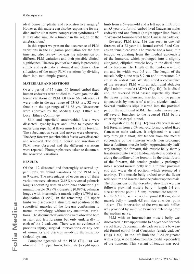
Complete agenesis of the PLM (Fig. 1a) was observed in 3 upper limbs, two male (a right upper
limb from a 69-year-old and a left upper limb from an 85-year-old formol-carbol fi xed Caucasian males cadaver) and one female (a right upper limb from a 77-year-old formol-carbol fi xed Caucasian cadaver).
Reversed PLM (Fig. 1b) was seen in one left forearm of a 73-year-old formol-carbol fi xed Cau-casian female cadaver. The muscle had a long, thin tendon, originating from the medial epicondyle of the humerus, which prolonged into a slightly elongated, elliptical muscle body in the distal third of the forearm. The length of the tendon was 13.6 cm and its width was 0.5 cm. The length of the muscle belly alone was 8.9 cm and it measured 2.6 cm at its widest part. We also noted a coexistence of the reversed PLM with an additional abductor digiti minimi muscle (ADM) (Fig. 1b). In its distal end, the reversed PLM passed superfi cially above the fl exor retinaculum and inserted into the palmar aponeurosis by means of a short, slender tendon. Several tendinous slips inserted into the proximal part of the additional ADM. The median nerve gave off several branches to the reversed PLM before entering the carpal tunnel.
A digastric PLM (Fig. 1c) was observed in one left forearm from a 69-year-old formol-carbol fi xed Caucasian male cadaver. It originated in a usual way through a short, fl at tendon from the medial epicondyle of the humerus which then prolonged into a fusiform muscle belly. Approximately half-way through the forearm, this muscle belly sharply transformed into a wide tendon, situated superfi cially along the midline of the forearm. In the distal fourth of the forearm, this tendon gradually prolonged into a second muscle belly with a thinner proximal end and wider distal portion, which resembled a teardrop. This muscle belly arched over the fl exor retinaculum and inserted into the palmar aponeurosis. The dimensions of the described structures were as follows: proximal muscle belly – length 9.4 cm, size at widest point 1.5 cm; intermediate tendon – length 6.3 cm, size at widest point 0.8 cm; distal muscle belly – length 4.8 cm, size at widest point 1.6 cm. The innervation of the two muscle bellies was provided by multiple branches extending from the median nerve.
PLM with an intermediate muscle belly was discovered in two upper limbs (a 53-year-old formol-carbol fi xed Caucasian male cadaver and a 63-year-old formol-carbol fi xed Caucasian female cadaver)(Figs 1 d,e). In the left limb the PLM originated with a long, wide tendon from the medial epicondyle of the humerus. This variant of tendon was posi-

Palmaris Longus Muscle Variations
291Folia Medica I 2017 I Vol. 59 I No. 3
tioned along the midline of the forearm and gradu-ally prolonged into an elliptical, slightly elongated muscle belly in the middle third of the forearm. Slightly above the level of the wrist, this muscle belly sharply prolonged into a short, thick and wide tendon, which arched above the fl exor retinaculum and inserted into the palmar aponeurosis through a wide insertion. In the middle part of the forearm, the median nerve gave off several branches to the belly of the variant of PLM. The proximal tendon of the PLM had a length of 8.9 cm and measured 0.7 cm at its widest point. The intermediate muscle belly had a length of 6.0 cm and measured 1.3 cm at its widest point. The distal tendon was 3.3 cm long and 0.8 cm at its widest point. In the right forearm, the origin, course, insertion and innerva-tion of the variant PLM were identical. However, the proximal and distal tendons were much thinner and the muscle belly had the shape of a spindle. The dimensions of these structures were as follows: proximal tendon – length 9.1 cm, width 0.3 cm; intermediate muscle belly – length 7.4 cm, size at its widest point 1.0 cm; distal tendon – length 3.6 cm; width 0.5 cm.
Duplication of the PLM was discovered in two upper limbs (63 and 75-year-old formol-carbol fi xed Caucasian female cadavers). After remov-ing the fascia and exposing the superfi cial fl exor muscles of the forearm in the left limb (Fig. 1f), we discovered the presence of two muscles with the characteristic features of the palmaris longus muscle. The fi rst muscle was located on the lateral side and originated from the medial epicondyle of the humerus with a small, thin muscle belly, which prolonged into a long tendon midway through the forearm. This tendon then inserted into the palmar aponeurosis. The length of the muscular belly was 9.7 cm and its size at its widest point measured 1.2 cm. The distal tendon of this PLM was 8.6 cm long and 0.4 cm wide. The second PLM was located medially and was partially covered by the distal tendon of the lateral PLM. It originated with a long, thick tendon from the medial epicondyle of the humerus which prolonged into a thick, spindle-shaped muscle belly, located in the middle third of the forearm. The muscle belly gradually prolonged into the distal tendon, which was short, thick and broad and separated into several bundles. Some of these bundles were attached to the fl exor reti-naculum, while others inserted into the proximal points of origin of the muscles of the thenar and hypothenar. These tendinous slips arched over the
median nerve and the ulnar artery and nerve at the canal of Guyon. The length of the proximal tendon of this medial PLM was 7.8 cm and its width was 0.6 cm; the muscle belly measured 6.5 cm in length and 1.7 cm at its widest point; fi nally, the distal tendon measured 2.4 cm in length and 0.6 cm in width. Both variants of PLM were innervated by short branches from the median nerve. In the right forearm, the two PLM muscles were identical with the previously described, however, they differed in size. The length of the muscular belly of the lateral PLM was 8.8 cm and its size at its widest point measured 0.9 cm. The distal tendon was 7.5 cm long and 0.3 cm wide. As for the medial PLM, the length of its proximal tendon was 6.3 cm and its width was 0.4 cm; the length of the muscular
Figure 1. Photographs of PLM variations: a) absence; b) reversed PLM (asterisk) and ADM (small asterisk) (published in Rom J Morphol Embryol 2009, 50(4):725–727); c) digastric PLM (asterisks); d and e) PLM with intermediate muscle belly (asterisk); f) duplication of PLM (asterisks).

292
G. Georgiev et al
Folia Medica I 2017 I Vol. 59 I No. 3
belly was 6.9 cm and its size at the widest point measured 0.8 cm; the distal tendon was 2.7 cm long and 0.3 cm wide.
DISCUSSION
PLM is a phylogenetically retrogressive muscle whose presence is restricted only to mammals and especially to those that use load to walk, such as the orangutan.12 According to Keith13, the PLM shows a higher degree of degeneration in apes and monkeys than in man and is present only in 25% of gorillas. Furthermore, the functions of the PLM are different among various animals. In some animals it participates in the exposure of their claws. Nu-merous animals that we share a common ancestor with (such as the orangutan) still actively use the muscle to grab things while climbing. Other primate relatives (such as the chimpanzee and gorilla), like humans, do not actively employ the muscle, which is why numerous variants can be observed.14 With time, the thumb apparatus in primates began to evolve (especially the muscles of the thenar) and consequently the PLM became vestigial.15 PLM, as a skeletal muscle, originates from the mesoderm of the myotomes of the somites. Hypaxial myoblasts fuse to form multi-nucleated myotubes, which migrate to the periphery and form the cells of the striated skeletal muscle tissue, which form the appendicular muscles (muscles of the limbs). Although the pre-cursors follow an intrinsic program which allows them to differentiate into muscle cells, this process is controlled by environmental signals. During the early embryogenesis the absence of such signals in the ectoderm leads to premature differentiation of the precursors, which in turn may cause agenesis or incomplete genesis of the respective muscles.16
The PLM is the most variable muscle in the hu-man arm and one of the most variable muscles in the human body.4-6 In the literature there are also reports of an aberrant palmaris longus coexisting with other anatomical variations, such as persistent median artery and fl exor carpi ulnaris muscle.17
Numerous reports of the variations of this muscle could be found in literature (Table 1).
Reimann et al.18 classifi ed the different PLM variations in the following way: complete agenesis, variation in the location and shape of the fl eshly portion, aberrancy in the attachment in either ex-tremity, duplication or triplication, accessory slips, or substitutions of similar shape or position.
As seen from the presented literature review (Table 1), there are numerous data concerning the
different anatomical variations of PLM. In order to present simple and systematic anatomical and clinical classifi cations of these variations we divide them into two simple groups. The anatomical classifi cation includes: variations concerning an additional muscle belly and variations of the PLM tendon or fi bro-tendinous slip and accessory muscles. The clinical classifi cation divides the PLM variations into such related to compression syndromes or simulating a soft tissue mass and variations signifi cant for plastic and reconstructive surgery of the hand.
ANATOMICAL CLASSIFICATION
PLM variations with changes in the normal anatomi-cal position and/or accessory muscle bellies (Fig. 2):1) Reversed PLM (RPL), bifi d or trifi d reversed and RPL coexisting with an additional ADM;2) Digastric PLM;3) PLM with intermediate muscle belly.Variations of the PLM tendon and/or accessory muscles (Fig. 3):1) Absence;2) Duplication of PLM;3) Triplication;4) Accessory slips to the hypothenar muscles;5) PLM profundus.
CLINICAL CLASSIFICATION
PLM variations related to median/ulnar nerve com-pression or simulating a soft tissue mass:1) Reversed PLM (RPL), bifi d or trifi d reversed and RPL coexisting with an additional ADM (possible median/ulnar nerve compression or simulating a soft tissue mass);2) Digastric PLM (possible median nerve compres-sion or simulating a soft tissue mass);3) Accessory slips (possible median/ulnar nerve compression);4) PLM profundus (possible median nerve compres-sion in the carpal tunnel);5) PLM with centrally placed muscle belly (simu-lating a soft tissue mass);PLM variations related to reconstructive surgery:1) Absence (lack of tendon grafts);2) Duplication or triplication of PLM (additional tendon grafts)
Of all these variations, PLM agenesis is the most frequent variation.18,19 Reimann et al.18 reported the absence of PLM in 12.9%. According to other literature data, there is prevalence of the absence of around 15%, although it is well known that there are differences in the occurrence of PLM absence

Palmaris Longus Muscle Variations
293Folia Medica I 2017 I Vol. 59 I No. 3
between different ethnic groups.20 These differences are presented in Table 2. In our study the absence of PLM was reported in only 2.68% of Bulgarians – data which correspond to the aforementioned study of Troha et al.22 in Caucasians. The basis for these racial differences is not yet clear. How-ever, the current predominant hypothesis is that early modern humans have a single origin from East Africa, where the rate of PLM agenesis is the lowest.25 Whether the phylogenetic degeneration of PLM occurred after Homo sapiens migrated out of Africa into new geographic areas is a question still to be fully studied. Another hypothesis associates the low rate of absence of PLM in Africans with the high prevalence of manual labour.26
Literature data contradict with regard to the symmetry of absence of PLM and the gender preva-lence.20,21,24 In our study, 67% of the reported cases of absence were male and 33% female. Most studies point out that the absence of PLM is most com-monly encountered in women and in left arms.18,27 Our fi ndings, though, suggest that in the Bulgarian population, the absence of PLM is most commonly discovered in men and on the right side. It has been proven that the absence of PLM is hereditary and the gene responsible for the regulation of its morphological development is the HOX gene.12,28 Moreover, a possible dominant expression of genes responsible for PLM variations in family members has also been proposed.5
The reported frequency of other variations of this muscle, with the exclusion of its absence, was 9.0%. Among the reported variations are: duplication or bifi d PLM, triple-headed PLM, accessory PLM, palmaris profundus and reversed PLM.29 In our study different PLM variations, with the exclusion of its absence, were 5.36%: RPL coexisting with an additional ADM (0.89%); digastric PLM (0.89%); PLM with an intermediate muscle belly (1.79%) and duplication (1.79%). Most of these aberrations have little clinical signifi cance with the exception of the reversed PLM, which may lead to a symptomatic compression of the median and/or ulnar nerve.30
Compression neuropathies in the wrist are fre-quent and have been well described. They can be provoked by ganglia, neoplastic masses, vascular abnormalities, ligamentous attachments, and also different anomalous muscles.6 Anatomic anomalies of the upper limbs and particularly of the PLM are frequent and may lead to serious complaints in cer-tain professional groups.31 Variations of this muscle could cause nerve compression with slow progressive
symptoms such as carpal tunnel syndrome more frequently than acute nerve compression.32 These symptoms include oedema on the palmar surface of the wrist, weakness and reduction of muscular strength, pain and numbness in the area innervated by the median and/or ulnar nerve. According to Depuydt et al.33 in cases when median nerve symptoms are effort-related or when there is a relapse after an adequate release of the carpal tunnel, the possibil-ity of an anomalous muscle must be considered. There are many reports in literature of median and/or ulnar nerve compression due to the existence of a variant PLM.9,11,32,33 In all cases of entrapment by a variant PLM, the excision of this muscle is curative. Moreover, in clinical practice, the variant PLM could be incidentally found during clinical examination without provoking clinical symptoms and may simulate a soft tissue tumor.9 In cases of nerve and artery compression by anomalous muscles, ultrasound imaging and/or MRI could be used as imaging modalities that could help in determining muscle variations.34
The PLM is often considered to be an ideal donor in reconstructive and plastic surgery.35 This is accounted for by the fact that this muscle has suffi cient length and diameter and could be used as harvest material without donor site morbidity in various reconstruction interventions.35 Knowledge of different PLM variations is important to provide safe and successful surgical procedures.8 The PLM could be used for tendon grafts in the replacement of long fl exors of the fi ngers, the fl exor pollicis longus tendon, dorsal fi nger injuries involving both soft tissue loss and extensor tendon defects.8 It is also utilized as a simple static support in the treatment of facial paralysis, in digital pulley reconstruction, lip augmentation, and in various nerve palsies as tendon transfer.8,23,24
CONCLUSION
PLM variations are described as one of the most common muscular variations in the human body, and, as we have discussed in detail, their pres-ence has specifi c clinical signifi cance. Therefore, the possible presence of PLM variations must be considered by clinicians during clinical examination of the forearm, during surgical interventions in that region, or while searching for an entrapment site of the median and/or ulnar nerve. We believe that the two new anatomical and clinical classifi cations presented will add to the literature data and will give a better insight into PLM variations.

294
G. Georgiev et al
Folia Medica I 2017 I Vol. 59 I No. 3
Table 1. Reported variations of the PL muscle
Variations of the PL Reference
Absence Reimann et al. (1944)18;Troha et al. (1990)22;Ceyhan and Mavt (1997)21;Sebastin et al. (2005)20;Kapoor et al. (2008)27;Kigera and Mukwaya (2011)26;Yammine (2013)25
Duplication or triplication Macalister (1875)3;Reimann et al. (1944)18;Mori (1964)4;Georgiev et al. (2009)17
Digastric muscle (with proximal and distal bellies) Macalister (1875)3
Reversed muscle (muscle belly reaching the palmar aponeurosis, with tendon located proximally)
Macalister (1875)3;Meyer and Pfl aum (1987)10;Giunta et al. (1993)11;Ninkovic et al. (1995)32;Depuydt et al. (1998)33;Georgiev et al. (2009)5
Bifi d or trifi d reversed variation (proximal tendon and distal bifi d/trifi d muscle belly)
Natsis et al. (2007)19
Reversed muscle coexisting with ADM Georgiev and Jelev (2009)6
PLM with intermediate muscle belly Kachlik et al. (2016)23
‘Palmaris accessorius’originating from the fl exor carpi radialis, the biceps brachii, the fl exor carpi ulnaris or the fl exor digitorum superfi cialis
Macalister (1875)3;Mori (1964)4
‘Palmaris accessorius’ inserting into the middle third of the antebrachial fascia, the thenar fascia, the ab-ductor digiti minimi or the carpal bones as separate partitions
Macalister (1875)3;Mori (1964)4
PLM profundus Dyreby et al. (1982)24
Table 2. PLM agenesis among different ethnic groups and populations
Ethnic group or population Occurrence (%)
Asians 4.8% for in vivo studies (Sebastin et al. 2005)20
4.3% for cadaveric studies (Sebastin et al. 2005)20
Native Americans (Red Indian and Amazonian) 7.1% for in vivo studies (Sebastin et al. 2005)20
Africans 11.3% (Sebastin et al. 2005)20
Turkish 64% (Ceyhan and Mavt, 1997)21
North American Caucasians 5.5% (Troha et al. 1990)22

Palmaris Longus Muscle Variations
295Folia Medica I 2017 I Vol. 59 I No. 3
Figure 2. PLM variations with changes in the normal anatomical position and/or accessory muscle bellies:a) RPL; b) bifi d reversed; c) RPL coexisting with ADM; d) digastric PLM; e) PLM with intermediate muscle belly.
Figure 3. Variations of the PLM tendon and/or accessory muscles:a) Absence; b) Duplication of PLM; c) Triplication; d) Accessory slips; e) PLM profundus.
CONFLICT OF INTERESTS
The authors declare no confl ict of interests.
ACKNOWLEDGEMENT
The authors would like to express their most sin-cere gratitude and to pay their respects to all the men and women who donated their bodies for the purpose of scientifi c research. We would also like to thank Mr. Martin Tsekov for his contribution in preparing the fi gures in this manuscript.
REFERENCES
1. Clemente CD. Anatomy of the Human Body. 30th Ed. Philadelphia: Lea and Febiger;1985.
2. Gangata H, Ndou R, Louw G. The contribution of the palmaris longus muscle to the strength of thumb abduction. Clin Anat 2010;23(4):431-6.
3. Macalister A. Additional observations on muscular anomalies in human anatomy (third series), with a catalogue of the principal muscular variations hith-erto published. Trans Roy Irish Acad 1875;25:86-9.

296
G. Georgiev et al
Folia Medica I 2017 I Vol. 59 I No. 3
4. Mori M. Statistics on the musculature of the Japa-nese. Okajimas Folia Anat Jpn 1964;40:252-4.
5. Georgiev GP, Jelev L, Surchev L. Presence of a pal-maris longus related variations in three members of a family. J Hand Surg Eur 2009;34(2):277-8.
6. Georgiev GP, Jelev L. Unusual coexistence of a variant abductor digiti minimi and reversed palmaris longus and their possible relation to median and ul-nar nerves entrapment at the wrist. Rom J Morphol Embryol 2009;50(4):725-7.
7. Lalit M, Singla RK, Piplani S. Bifi d inverted pal-maris longus muscle – a case report. Eur J Anat 2014;18(3):341-3.
8. Zeybek A, Gürünlüoğlu R, Çavdar, et al. A clinical reminder: a palmaris longus muscle variation. Ann Plast Surg 1998;41(2):224-5.
9. Turner MS, Caird DM. Anomalous muscles and ulnar nerve compression at the wrist. Hand 1977;9(2):140-2.
10. Meyer FN, Pfl aum BC. Median nerve compression at the wrist caused by a reversed palmaris longus muscle. J Hand Surg Am 1987;12(3):369-71.
11. Giunta R, Brunner U, Wilhelm K. [Bilateral reversed palmaris longus muscle - a rare cause of peripheral median nerve compression syndrome. Case report.] Unfallchirurg 1993;96(10):538-40 (German).
12. Angelini Júnior LC, Angelini FB, de Oliveira BC, et al. Use of the tendon of the palmaris longus muscle in surgical procedures: study on cadavers. Acta Ortop Bras 2012;20(4):226-9.
13. Keith A. On the chimpanzees and their relationship to the gorilla. Proc Zool Soc Lond 1899;2:296-314.
14. Thejodhar P, Potu BK, Vasavi RG. Unusual palmaris longus muscle. Indian J Plast Surg 2008;41(1):95-6.
15. Maughan H, Masel J, Birky CW Jr, et al. The roles of mutation accumulation and selection in loss of sporulation in experimental populations of Bacillus subtilis. Gen 2007;177(2):937-48.
16. Amthor H, Christ B, Patel K. A molecular mecha-nism enabling continuous embryonic muscle growth - a balance between proliferation and differentiation. Devel 1999;126(5):1041-53.
17. Georgiev GP, Jelev L, Ovtscharoff WA. Unusual combination of muscular and arterial variations in the upper extremity: a case report of a variant pal-maris longus and an additional tendinous portion of the fl exor carpi ulnaris together with a persistent median artery. Anatomy 2009;3:58-61.
18. Reimann AF, Daseler EH, Anson BJ, et al. The pal-maris longus muscle and tendon. A study of 1600 extremities. Anat Rec 1944;89(4):495-505.
19. Natsis K, Levva S, Totlis T, et al. Three-headed reversed palmaris longus muscle and its clinical
signifi cance. Ann Anat 2007;189(1):97-101.20. Sebastin SJ, Puhaindran ME, Lim AYT, et al. The
prevalence of absence of the palmaris longus – a study in a Chinese population and a review of the literature. J Hand Surg Br 2005;30(5):525-7.
21. Ceyhan O, Mavt A. Distribution of agenesis of the palmaris longus muscle in 12-18-year-old age groups. Indian J Med Sci 1997;51(5):156-60.
22. Troha R, Baibak GJ, Kelleher JC. Frequency of the palmaris longus tendon in North American Cauca-sians. Ann Plast Surg 1990;25(6):477-8.
23. Kachlik D, Hajek P, Konarik M, et al. Coincidence of superfi cial brachiomedian artery and bitendinous palmaris longus: a case report. Surg Radiol Anat 2016;38(1):147-51.
24. Dyreby JR, Engber WD. Palmaris profundus - rare anomalous muscle. J Hand Surg Am 1982;7(5):513-4.
25. Yammine K. Clinical prevalence of palmaris longus agenesis: a systematic review and meta-analysis. Clin Anat 2013;26(6):709-18.
26. Kigera JW, Mukwaya S. Frequency of agenesis palmaris longus through clinical examination - an East African study. PLoS One 2011;6(12):e28997.
27. Kapoor SK, Tiwari A, Kumar A, et al. Clinical relevance of palmaris longus agenesis: common anatomical aberration. Anat Sci Int 2008;83(1):45-8.
28. Keese GR, Wongworawat MD, Frykman G. The clinical signifi cance of the palmaris longus tendon in the pathophysiology of carpal tunnel syndrome. J Hand Surg Br 2006;31(6):657-60.
29. Park MJ, Namdari S, Yao J. Anatomic variations of the palmaris longus muscle. Am J Orthop (Belle Mead NJ) 2010;39(2):89-94.
30. Natsis K, Iordache G, Gigis I, et al. Persistent median artery in the carpal tunnel: anatomy, embryology, clinical signifi cance, and review of the literature. Folia Morphol (Warsz) 2009;68(4):193-200.
31. Baldi SV, Hug U, Jandali AR, et al. Painful tendon insertion anomaly of the palmaris longus muscle in a professional cellist. Handchir Mikrochir Plast Chir 2005;37(6):415-7.
32. Ninković M, Hefel L, Öhler K. Acute median nerve compression produced by reversed palmaris longus muscle. Eur J Plast Sur 1995;18:129-30.
33. Depuydt KH, Schuurman AH, Kon M. Reversed pal-maris longus muscle causing effort-related median nerve compression. J Hand Surg Br 1998;23(1):117-9.
34. Zeiss J, Jakab E, Khimji T, et al. The ulnar tunnel at the wrist (Guyon’s canal): normal MR anatomy and variants. AJR Am J Roentgenol 1992;158(5):1081-5.
35. Sebastin SJ, Lim AYT, Bee WH, et al. Does the absence of palmaris longus affect grip and pinch strength? J Hand Surg Br 2005;30(4): 406-8.
Palmaris Longus Muscle Variations
297Folia Medica I 2017 I Vol. 59 I No. 3
Вариации мышцы palmaris longus: клиническое значение ипредложение новой классификации Георги П. Георгиев1, Александр A. Илиев2, Ива Н. Димитрова3, Георги Н. Котов2,Лина Г. Малинова2, Бойчо В. Ланджов2
1 Кафедра ортопедии и травматологии, УМБАЛ „ Царица Йоанна“- ИСУЛ, Медицинский университет - София, София, България2 Кафедра анатомии, гистологии и эмбриологии, Медицинский университет - София, София, Болгария3 Кафедра кардиологии, УМБАЛ „Св. Екатерина“, Медицинский университет- София, София, Болгария
Адрес для корреспонденции: Георги П. Георгиев, Кафедра ортопедии и травматологии, УМБАЛ „ Царица Йоанна“-ИСУЛ, Медицинский университет - София, ул. „Бяло море“ 8, София, БолгарияE-mail: [email protected]л: +359 884 493 523
Дата получения: 03 ноября 2016Дата приемки: 12 января 2017Дата онлайн публикации: 19 апреля 2017Дата публикации: 29 сентября 2017
Ключевые слова: мышца palmaris longus, вариация, классификация, клиническое значение, человеческое существо
Образец цитирования:Georgiev GP, Iliev AA, Dimitrova IN, Kotov GN, Malinova LG, Lan-dzhov BV. Palmaris longus muscle variations: clinical signifi cance and proposal of new classifi ca-tions.
Folia Medica 2017;59(3):289-297.doi: 10.1515/folmed-2017-0035
Введение: Мышца palmaris longus является одной из самых вариабельных мышц в теле человека и сообщается о её многочисленных вариациях. Раз-личные вариации palmaris longus представляют собой интерес не только с анатомической точки зрения, но могут иметь и определённое клиническое значение.
Цель: Целью данной работы является исследование различных вариаций мышцы palmaris longus среди болгарского населения.
Материалы и методы: В течение 15-летнего периода были исследованы 56 человеческих трупов, зафиксированных в формалин-карболе, с целью уста-новления различных вариаций ПЛ.
Результаты: Были установлены различные вариации palmaris longus: отсут-ствие (2.68%); обратный палмарис лонгус, сосуществующий с дополнитель-ным m. abductor digiti minimi (0.89%); двубрюшная мышца (0.89%); palmaris longus с промежуточным мышечным животом (1.79%) и парная (1.79%).
Заключение: С целью установления большого разнообразия разновидно-стей palmaris longus и их значения в клинической практике, нами был состав-лен краткий обзор литературы, связанной с разновидностями вариаций, их ролью в медианной и локтевой невропатии запястья или в качестве структур, симулирующих опухоль мягких тканей, и применение сухожилия palmaris longus в пластической и реконструктивной хирургии в качестве трансплан-тационного материала. Нами также представлены новые системные анатоми-ческие и клинические классификации вариаций palmaris longus, распреде-лённые в две обыкновенные группы.