palsy: a cross-sectional study improves gait
TRANSCRIPT
Page 1/20
Walking in the Hibbot, an innovative walking aidimproves gait characteristics in children with cerebralpalsy: a cross-sectional studyAnn Hallemans ( [email protected] )
Universiteit Antwerpen https://orcid.org/0000-0003-4101-5279Ria Cuppers
Universiteit AntwerpenSteven Truijen
Universiteit AntwerpenFrank Ego
Universiteit AntwerpenMarian Moens
HederDirk Wenmakers
Made for MovementPhilippe Caers
Made for MovementJo Lebeer
Universiteit Antwerpen
Research
Keywords: Walking aids, Gait Rehabilitation, Cerebral palsy
Posted Date: January 31st, 2020
DOI: https://doi.org/10.21203/rs.2.20661/v2
License: This work is licensed under a Creative Commons Attribution 4.0 International License. ReadFull License

Page 2/20
AbstractBackground: Crouch gait is a common gait impairment in children with cerebral palsy (CP). Growing up andgaining weight, lever arm dysfunctions further deteriorate and walking e�ciency progressively worsens overtime with risk of losing ambulant capacity in adolescent life. The purpose of this study is to evaluate whetherwalking in an innovative walking aid can reduce the �exed gait pattern by improving alignment of the lowerextremity joints and improving weight bearing.
Methods: A semi-robotic walking aid called “Hibbot has been designed, which can be used in rehabilitation anddaily-life settings. In a cross-sectional study, �ve children with CP, aged 3-7 years, were familiarized with theHibbot, by using the walking aid for 30 minutes, twice a week during 3 months under the supervision of theirphysiotherapists, who were instructed how to use the Hibbot. Using 3D gait analysis kinematics of the lowerlimb and trunk as well peak vertical ground reaction forces were compared between walking in their habitual(or no) walking aid and walking in the Hibbot, using a paired samples t-test (p < 0.05, spm1d.org).
Results: After the familiarization period of 3 months, a signi�cant improvement in hip extension (meandifference 9.4 ± 1.4°) and knee extension (mean difference 10.1 ± 0.5°) during stance was observed whenwalking in the Hibbot compared to habitual walking. Improvements were also observed in trunk posture.However, differences in peak forces between habitual walking and walking in the Hibbot could not becon�rmed.
Conclusions: These results are promising as to the possibility of the Hibbot walking aid in counteracting a�exed gait pattern. Further research is necessary to investigate the net joint moments and muscle forcesexplaining the higher knee and hip extension and improved trunk posture while walking in the Hibbot.Furthermore research should focus on determining which children with CP would bene�t most, what arecontraindications and what dose effects are to be expected.
Trial Registration: ClinicalTrials.gov, NCT04172324. Registered November 21 st , 2019 - Retrospectivelyregistered. https://clinicaltrials.gov/ct2/show/NCT04172324
BackgroundCerebral palsy is the most common motor impairment in childhood with a worldwide incidence of 2.5/1000.(1)The damage in the central nervous system causes muscle weakness, shortening of muscle-tendon units,muscle spasticity and impaired selective motor control (SMC) (2, 3) which results in gait limitations to avarying degree, as classi�ed by the GMFCS-level.(4)
One of the major gait di�culties is crouch gait. Crouch gait is de�ned as excessive ankle dorsi�exion, knee andhip �exion during the stance phase. This gait disorder is common among patients with cerebral palsy andresults in gait ine�ciency because of additional energy expenditure and increased muscular effort. (5-8)
A signi�cant correlation between scores on the Selective Control Assessment of the Lower Extremities (SCALE)(9) and impaired gait has been found in children with CP.(2, 9)

Page 3/20
These �ndings suggest that a crouch gait pattern may be related to impaired neuromuscular control and thismight play a crucial role in the persistence of a �exed gait pattern.
Past interventions to alleviate crouch gait primarily worked on weakness, spasticity, range of motion orbalance. Yet, improvements in isolated impairments do not necessarily carry over to functional activities i.e.improved walking.(10, 11)
Recent research showed that crucial conditions for effective interventions on activities (i.e. walking) shouldstart at a young age to enhance experience-dependent neuroplasticity, should be task speci�c, highly intensiveand should be integrated into functional daily activities to enhance motor learning.(12-18) Evidence forneuroplastic changes induced by early intervention on upper limb motor activity has been found in recentstudies.(19-21) However, research on neuroplastic changes as a result of intervention on lower limb activity islacking.
The actual practice of gait either over ground or on treadmill ,with or without partial body weight support, hasbeen shown to improve gait speed and endurance in CP patients aged 5 – 25 years.(22, 23) To mimic aphysiotherapist’s manual gait facilitation for optimal extensor muscle activity and postural control, differentrobotic assisted gait training devices have been developed, initially for applicability in adult neurologicconditions. The bene�t of robotic assisted gait training is the potential to reach a higher number of repetitionsand higher intensity than can be produced without a therapist’s manual assistance. However, at present, thelevel of evidence for robotic assisted gait training is still weak and inconsistent in children with CP.(24, 25) Ontop of that, these systems are not adapted for small children, have a high economic cost and poor portability.
In order to potentially �ll in the gap for early intervention taking into account the requirement of task speci�cityand functionality, a new semi-robotic walking device for small children, is designed by a multidisciplinary team,and is called “Hibbot”. Hibbot is an assistive mechanical device, lightweight and portable that can be used insmall children during rehabilitation and in daily life. This study provides proof of concept of the possibility ofthe Hibbot walking aid in counteracting a �exed gait pattern in young children with CP.
MethodsThe manuscript adheres to the Strengthening the Reporting of Observational studies in Epidemiology(STROBE) statement (checklist in supplementary material).
AimThe purpose of this study was to evaluate whether walking in the Hibbot affects gait kinematics anddynamics. The mechanism of the Hibbot manages to control all degrees of freedom (3x rotation and 3xtranslation) at the level of the pelvis and lower trunk. (Figure 1a,1b). The goal is to maximize a child’s ownmuscle activity and postural control while giving as minimal support as needed. We assume that the design ofthe Hibbot facilitates correct activation of the gluteal muscles necessary for postural stability. Furthermore,su�cient hip extension movement is required to walk in the Hibbot. When the legs are �exed, fall protectionbars touch the ground and locomotion is inhibited.

Page 4/20
Our hypothesis is that walking in Hibbot would (a) improve trunk posture and extension in the lower limbs (b)improve weight bearing during the single stance phase. To test these hypotheses, lower limb joint angles andground reaction forces were compared between walking with the Hibbot and habitual walking in a cross-sectional study.
DesignWe performed a cross-sectional study comparing walking in the Hibbot to walking without walking aid/using ahabitual walking aid in 5 children diagnosed with CP. The study was approved by the local ethics committee ofthe Antwerp University Hospital (18/44/509 B300201838159) and retrospective registered as ClinicalTrials.gov Protocol Record NCT04172324.
SettingThe study was performed in collaboration with a licensed care provider and multifunctional centre directed atchildren and adolescents with a disability providing directly accessible care, short stay and respite care inAntwerp, Belgium. Participants were recruited in November 2018. Over a period of 3 months, between January1st and March 31st in 2019, the children were familiarized with the Hibbot by using the walking aid for 30minutes, twice a week, under the supervision of a physiotherapist. Physical examination and gait analysis wereperformed before (December 17 – 21, 2018) and after (April 1 – 5, 2019) the familiarization period, at the localgait lab.
ParticipantsChildren had to meet the following inclusion criteria: con�rmed diagnosis of CP, GMFCS level II-IV, age between2 and 7 years, stature less than 1.25 meters and body weight less than 30 kilograms. Exclusion criteria were:�xed contractures in the lower limbs, hip dysplasia, in�ltration with botulinum toxin 3 months or less prior tothe start of the study, orthopaedic operations 6 months or less prior to the start of the study, insu�cient mentalcapacity to understand instructions, insu�cient motivation to walk and no prior experience in walking with theHibbot.
Treating physicians provided the researchers with the information for possible inclusion. Subsequently, studyinformation was provided to parents. After parents had signed an informed consent, patients were screened foreligibility.
To describe the study population age, gender, height, weight, BMI, GMFCS level(4) and mobility by passiverange of movement measurement (PROM)(26), strength by manual muscle testing (MMT) (27), spasticity bythe Modi�ed Ashworth Scale (MAS)(28) and lower limb selectivity by Selective Control Assessment of theLower Extremity (SCALE )(29) were assessed. The data of the clinical examination was meaningful byinterpretation of the gait parameters i.e. step length, (a)symmetry in, maximal hip and knee extension duringstance phase.

Page 5/20
MeasurementsKinematics and kinetics of gait were recorded by an optical motion capture system (8 camera’s, 120 Hz.,Qualysis Prore�ex, Göteborg, Sweden) surrounding a 10-meter walkway and synchronised with 3 force-platforms (two of 0.5mx0.5m and one of 1.0x0.5m; AMTI Accugait, 1000 Hz., Advanced Medical TechnologyInc., Massachussets, USA). Re�ective markers were attached to the skin over the processus spinosus of the 7th
cervical vertebra (C7), incisura jugulars (IJ), processus xyphoideus (PX) and bilaterally over the clavicular head(CC), major trochanters (TM), lateral epicondyles (LE), lateral malleoli (LM), calcanei (CA) and 2nd metatarsalheads (MT). Marker trajectories were tracked and labelled in QTM (Qualisys Track Manager) software afterwhich kinematic and kinetic data were combined in C3D �le format. In addition, two Sony HD video cameras(Type: HDRCX240E, 50Hz.) were placed to record the sagittal and frontal plane kinematics. Children wereencouraged to walk at self-selected speed over the walkway in Hibbot and using their habitual walking aid (ifnecessary, Table 1) in a randomized order. Patients walked either barefoot or with ankle-foot orthoses, ifnecessary. Prior to gait analysis a standard physical examination was performed by the physiotherapist toassess joint mobility (ranges of motion of hip, knee and ankle towards �exion/extension, ab/adduction andinternal/external rotation) and strength, spasticity and selectivity of lower limb muscles (psoas, adductors,quadriceps, hamstrings, gracilis, gastrocnemius and soleus).
Data analysisGait analysis data in C3D �le format was analysed using visual 3D software (Visual3D Professional v5.01.9, C-motion, Kingston, ON, Canada) and custom models. The body was modelled as an interconnected chain ofrigid segments: CC – TM for trunk, TM – LE for thigh, LE – LM for leg and LM – MT for foot. In addition, thethorax was modelled as a 6 – degrees – of – freedom segment with the segment coordinate systemde�nitions partially adapted from the ISB recommendations.(30) The segment origin coincided with IJ; thevertical axis (Z) was de�ned by the line connecting a virtual point at a �xed distance from PX in the direction ofthe IJ – C7 axis to the midpoint between IJ and C7 (pointing upwards); the medio-lateral axis (X) was de�nedas the line connecting the left to right CC; the antero-posterior axis was the common line perpendicular to Z andX, pointing forward.
Events of heel strike and toe off were determined from force plate recordings, CA and MT marker trajectoriesusing the “Automatic Gait Events” command in Visual 3D software. Automatically assigned events werevisually inspected.
Hip and knee kinematics in the sagittal planeHip �exion and extension were calculated as the planar angle between by CC – TM – LE in the sagittal plane,expressed relative to gait cycle duration (0 – 100%). Knee �exion and extension was characterised as theplanar angle between TM – LE – LM as a function of gait cycle duration. The anatomical position with fullextension of the hip and knee is characterised by angles of 180°. Angles are calculated against clockworkdirection. At the hip, an angle < 180° indicates extension, whereas an angle > 180° indicates �exion. At theknee, an angle < 180° indicates �exion.

Page 6/20
Thorax kinematicsKinematics of the thorax was characterised by Euler/Cardan angles (XYZ) of the thorax coordinate systemrelative to the global reference frame, normalized to 100% of gait cycle duration. Flexion and extension of thethorax were measured around the X-axis (ante�exion positive, retro�exion negative). Lateral �exion of thethorax was measured around the Y-axis (ipsilateral side positive).
Peak vertical ground reaction force (Fz)
The peak vertical ground reaction force was determined as the maximum value of the vertical component ofthe ground reaction force vector during stance.
Step-time parametersUsing the “Temporal Distance Calculations for Gait” command, speed (m/s), normalised speed(statures/second), stride length (m), step length (m), stride width (m), stance time (s), swing time (s) anddouble limb support time (s) were calculated.
Statistical analysisDiscrete outcome variables were analysed using the Statistical Package for social Sciences Software (SPSSversion 24 for Windows, IBM Statistics). Per subject, data were averaged over different gait cycles (3 – 8 gaitcycles per condition). Wilcoxon signed-rank test was performed to evaluate the difference of gait kinematicsand kinetics between the Hibbot- and habitual condition. The signi�cance level was set at p<0.05. Data wereanalysed separately for the before- and after-measurements. Missing data will be treated as missing.
Differences in the time pro�les of hip, knee and thorax kinematics across the entire gait cycle were analysed bystatistical parametric mapping (spm1d.org) using custom MatLab scripts (version R2018a for Windows).Kinematic time pro�les were compared between Hibbot and habitual walking condition, separately for pre- andpost-measurements, by means of spm1D paired samples t-test. If the null hypothesis was true, identical curveswould be observed in both conditions. The null hypothesis was rejected when the t value exceeded the criticaltest statistical value t*. Signi�cance level was set at p<0.05.
Results
ParticipantsIn November 2018 ten potential participants were recruited, of whom both parents signed informed consent.After screening, two participants were excluded because of prior experience with Hibbot. During thefamiliarisation period, 3 additional children were excluded: one because of lack of understanding and lackmotivation to walk, one because of a �xed �exion contracture in the hip joint and one because of in�ltrationwith botulinum toxin in March 2019. Flowchart of participants can be found in �g.3.

Page 7/20
Descriptive dataDemographic characteristics, GMFCS level and results from the physical examination can be found in table 1and table 2.
[INSERT TABLE 1 HERE]
[INSERT TABLE 2 HERE]
Outcome dataPeak vertical ground reaction forces and step-time parameters are shown in table 3. Figure 4 represents thekinematic time pro�les of the hip, knee and thorax as a function of gait cycle duration. Time pro�les arerepresented before and after the familiarization period
[INSERT TABLE 3 HERE]
Main results
Hip and knee kinematics in the sagittal planeBefore the familiarisation with Hibbot (left panel in Figure 4), only a signi�cant increase in hip �exion (Meandifference 7.0 ± 0.02°) during loading response is observed when walking in Hibbot compared to the habitualcondition. After the familiarisation period (right panel in Figure 2), a signi�cant increase in hip extension (Mean difference 9.4 ± 1.4°) is seen from 20 to 75% of the gait cycle (spm1D paired samples t-test, p<0.001)using the Hibbot in comparison with the habitual walking aid/no walking aid.
Before the familiarisation with Hibbot (left panel in �gure 4), a signi�cant increase in knee �exion duringterminal swing (Mean difference 13.8 ± 0.8°) is observed when walking in Hibbot compared to the habitualcondition. After the familiarisation period (right panel in Figure 2), the knee is signi�cantly more extended(Mean difference 10.1 ± 0.5°) from 32 to 74% of the gait cycle (spm1D paired samples t-test, p<0.001) whilefrom 85 to 98% the knee is more �exed (Mean difference 1.9± 0.8°) during terminal swing (spm1D pairedsamples t-test, p=0.0002)
Thorax kinematicsBefore familiarisation (left panel in �gure 4), during stance and early swing (from 0 to 78% of the gait cycle)the thorax is signi�cantly more in retro�exion (Mean difference 7.4 ±1.3°) when walking in Hibbot compared tothe habitual condition (spm1D paired samples t-test, p<0.001). After the familiarisation period, this retro�exedposition of the thorax normalizes (Figure 4, right panel), although the range of motion appears to be limited. Inthe frontal plane, initially lateral �exion of the thorax is constrained which appears to improve after thefamiliarisation period of 3 months.
Page 8/20
Peak vertical ground reaction force (Fz)Differences in peak vertical ground reaction force between Hibbot and habitual walking condition were notsigni�cant for the entire group.
Step-time parametersNo signi�cant differences were found in any of the step-time parameters between walking in Hibbot andwalking in the habitual walking aid (Table 3).
DiscussionThe primary goal of this study was to evaluate our hypothesis that walking in Hibbot would (a) improveposture (i.e. reduce �exion) of the lower limbs and (b) improve weight bearing during single stance phase.While individual effects on walking with the Hibbot were variable, as expected in this heterogeneous patientpopulation, several statistically signi�cant trends were observed across the cohort.
The �ndings support our �rst hypotheses of improved posture when walking with the Hibbot, after afamiliarization period. A signi�cantly higher hip and knee extension in stance phase was found while walkingwith the Hibbot compared to habitual walking as well as improved lateral trunk control. Few studies comparedthe effects of walking devices on gait kinematics and spatiotemporal gait parameters.(31-34) Walking withhandheld walkers (anterior walkers, posterior walker or crutches) in comparison to walking without any deviceresulted in increased knee extension, which corroborates our �ndings. On the other hand, walking withhandheld walkers resulted in an increased anterior pelvic tilt, while walking with the Hibbot resulted inimproved trunk posture in both the sagittal and frontal plane. When comparing an anterior versus posteriorwalker, a posterior walker may enhance posture alignment.(31)
At least on the level of kinematics, walking in the Hibbot thus results in a better hip and knee extension duringstance phase. This is in line with previous research showing that support on the level of the pelvis and lowertrunk by the brace could enhance synergic movements of the legs as well as upper trunk righting. (35)However, it is important to investigate the net joint moments and muscle forces explaining the higher knee andhip extension and improved trunk posture while walking in the Hibbot. Kinetics, revealing the load on upperextremities using a handheld walker have been studied (36, 37). Handheld walkers induce a partial weight lifteffect through the arms. Secondly, the centre of mass is moved anteriorly, moving the onset of the groundreaction force forward in relation to the knees, which will increase externally extending knee moments (31).This compensatory mechanism, especially used in weak patients, compromises postural alignment.
The second hypothesis that walking with the Hibbot resulted in augmented weight bearing during single stancephase could not be con�rmed. Walking speed and step length decreased when walking with the Hibbot incomparison with the habitual walker, although not signi�cant. This might account for a smaller peak verticalground reaction force. (38) Possibly, higher inertial forces were needed to start walking with the Hibbotbecause of its additional weight (7kg) in comparison to the habitual situation (posterior walker or no device)which may partially explain the decreased walking speed. Furthermore, the compensation strategies used by
Page 9/20
the child in his/her habitual gait pattern to generate propulsion and speed may be inhibited or not availablewhile walking in the Hibbot. Potentially the reduction in walking speed can also be considered as anadvantage. Even in typically developing children lower walking speed is related to trunk righting and moreextension in the lower limbs.(39) A decreased walking speed may be an advantage to learn the correct posturalalignment during walking, but further research is needed to con�rm this.
This study has several limitations. First, the intensity of the training period was too low to be considered as areal intervention. A 3-month period at a rate of 2 times a week training for 20 minutes per session must beconsidered a familiarization to the Hibbot instead of an intervention. Evidence for a high intense dose ofintervention has been found regarding e�cient interventions for upper limb activities.(40) A long-term followup study is recommended to judge the impact on gait related parameters as well as transfer to real lifesettings.
The second limitation was the small sample size. The recruitment of 10 patients at only one school and theoccurrence of dropouts, because of lack of cognition/cooperation, �xed contractures and Botox intervention,resulted in a sample of 5 patients. These 5 remaining patients were very heterogeneous both on motor andcognitive function; the 3 patients with GMFCS-level II were 8 years old by post measurements while the 2patients with GMFCS-III were younger than 6 years old and showed weak cognitive function. Finally, althoughthe braces were custom made, some pressure points did result in pain, which could in�uence the gait pattern.Regarding data analysis, the spm1D method has the advantage that the entire time curve can be analysed buta limitation is that changes in range of motion cannot be detected.
Future research should include registration of the muscular activation patterns as well as net joint moments.These outcome measurements are clinical important to interpret the kinematic changes and to determine if theHibbot indeed elicits a different neuromuscular control. Next to investigating neuromuscular parameters, alsoparameters in the domains of activity and participation should be investigated.
Further studies should also be done to specify the indications (inclusion criteria) of usefulness of the Hibbot,as well as specify exclusion criteria. Inclusion of a larger and more homogenous sample size andimplementing adequate concurrent controls is mandatory.
ConclusionThese results are promising as to the possibility of the Hibbot walking aid in counteracting a �exed gaitpattern. The design of an optimal early intervention study based on task speci�c gait training with the Hibbothas to be worked out, based on the knowledge available regarding motor learning of walking and gaitrehabilitation.
Declarations
Ethics approval and consent to participateThe current study was approved by the local ethics committee of the Antwerp University Hospital (18/44/509B300201838159) and retrospective registered as Clinical Trials.gov Protocol Record (NCT04172324).
Page 10/20
The parents/legal representatives of the eligible participants were provided with information letter and writteninformed consent letters were obtained for all the participants prior to their participation in the experiment.
Consent for publicationConsent was obtained for the publication of the pictures in �gure 1.
Availability of data and materials The datasets and analyses used during the current study are available from the corresponding author onreasonable request.
Financial competing interestsR.C., pediatric physiotherapist, is co-developer of the Hibbot and became in 2018 Clinical Advisor for the use ofthe Hibbot in the Norwegian company Made for movement, that is marketing the Hibbot. R.C. is shareholder inthe company Made for movement.
D.W., engineer, is co-developer of the Hibbot and became in 2018 Research and Development engineer in thecompany Made for movement. D.W. is shareholder in the company Made for movement.
P.C. is co-developer of the Hibbot and became in 2018 Product Manager engineer in the company Made formovement. P.C.is shareholder in the company Made for movement.
FundingThe technical development of the Hibbot was partially funded in 2014 by the Agency for Innovation, Scienceand Technology (IWT) Brussels Belgium with project number 135018.
Authors' contributions JL, EF, AH, ST, MM and RC participated in the study design and method process. AH performed the dataextraction and data analysis. RC and AH wrote the manuscript. DW and PC provided the technical descriptionand gave technical assistance during the intervention and gait analysis. JL supervised the study and revisedthe manuscript.
AcknowledgementsThe authors would like to thank the Master students of the University of Antwerp i.e. Celens Eline, WeyenbergLotte, Ceulemans Kiara, Cavens Karolien, Taeymans Laura and Van de Reck Charlotte, who were involved in theliterature review on walking aids as well as data processing. Many thanks to all the co-workers at Heder i.e. the

Page 11/20
physiotherapists that were involved in the intervention with the Hibbot under supervision of Ronny Van Aschefor helping to recruit and select patients. Thanks to the staff of the gait laboratory for their assistance andprofessional work.
Thanks to Kaat Desloovere, from the university of Leuven for giving advice on design and an earlier proof ofconcept.
Abbreviations3D: 3 Dimensional
AFO: Ankle Foot Orthosis
BMI: Body Mass Index
C7: Cervical Vertebra
CA: Calcanei
CC: Clavicular Caput
CP: Cerebral Palsy
F: Female
GMFCS: Gross Motor Functional Classi�cation Scale,
IJ: Incisura Jugularis
ISB: International Society of Biomechanics
L: Left
LE: Lateral Epicondyles
LM: Lateral Malleoli
M: Male
MAS: Modi�ed Ashworth Score
MMT: Manual Muscle Testing
MT: Metatarsal Heads
NT: Not Tested
PROM: Passive Range of Movement

Page 12/20
PX: Processus Xyphoideus
QTM: Qualisys Track Manager
R: Right
ROM: Range of Movement
SCALE: Selective Control Assessment of the Lower Extremity
SMC: Selective Motor Control
SMO: Supra Malleolar Orthosis
TM: Trochanter Major
References1. Oskoui M, Coutinho F, Dykeman J, Jette N, Pringsheim T. An update on the prevalence of cerebral palsy: a
systematic review and meta-analysis. Dev Med Child Neurol. 2013;55(6):509-19.
2. Zhou JY, Lowe E, Cahill-Rowley K, Mahtani GB, Young JL, Rose J. In�uence of impaired selective motorcontrol on gait in children with cerebral palsy. J Child Orthop. 2019;13(1):73-81.
3. Meyns P, Van Gestel L, Bar-On L, Goudriaan M, Wambacq H, Aertbelien E, et al. Children with SpasticCerebral Palsy Experience Di�culties Adjusting Their Gait Pattern to Weight Added to the Waist, WhileTypically Developing Children Do Not. Front Hum Neurosci. 2016;10:657.
4. Palisano R, Rosenbaum P, Walter S, Russell D, Wood E, Galuppi B. Development and reliability of a systemto classify gross motor function in children with cerebral palsy. Dev Med Child Neurol. 1997;39(4):214-23.
5. Rha DW, Cahill-Rowley K, Young J, Torburn L, Stephenson K, Rose J. Biomechanical and Clinical Correlatesof Stance-Phase Knee Flexion in Persons With Spastic Cerebral Palsy. PM R. 2016;8(1):11-8; quiz 8.
�. Ahlin K, Himmelmann K, Nilsson S, Sengpiel V, Jacobsson B. Antecedents of cerebral palsy according toseverity of motor impairment. Acta Obstet Gynecol Scand. 2016;95(7):793-802.
7. Papageorgiou E, Nieuwenhuys A, Vandekerckhove I, Van Campenhout A, Ortibus E, Desloovere K.Systematic review on gait classi�cations in children with cerebral palsy: An update. Gait Posture.2019;69:209-23.
�. Noorkoiv M, Lavelle G, Theis N, Korff T, Kilbride C, Baltzopoulos V, et al. Predictors of Walking E�ciency inChildren With Cerebral Palsy: Lower-Body Joint Angles, Moments, and Power. Phys Ther. 2019;99(6):711-20.
9. Fowler EG, Staudt LA, Greenberg MB, Oppenheim WL. Selective Control Assessment of the Lower Extremity(SCALE): development, validation, and interrater reliability of a clinical tool for patients with cerebral palsy.Dev Med Child Neurol. 2009;51(8):607-14.
10. Schweizer K, Romkes J, Coslovsky M, Brunner R. The in�uence of muscle strength on the gait pro�le score(GPS) across different patients. Gait Posture. 2014;39(1):80-5.

Page 13/20
11. Dodd KJ, Taylor NF, Damiano DL. A systematic review of the effectiveness of strength-training programsfor people with cerebral palsy. Arch Phys Med Rehabil. 2002;83(8):1157-64.
12. Novak I, McIntyre S, Morgan C, Campbell L, Dark L, Morton N, et al. A systematic review of interventions forchildren with cerebral palsy: state of the evidence. Dev Med Child Neurol. 2013;55(10):885-910.
13. Novak I, Morgan C, Adde L, Blackman J, Boyd RN, Brunstrom-Hernandez J, et al. Early, Accurate Diagnosisand Early Intervention in Cerebral Palsy: Advances in Diagnosis and Treatment. JAMA Pediatr.2017;171(9):897-907.
14. Byrne R, Noritz G, Maitre NL, Group NCHED. Implementation of Early Diagnosis and Intervention Guidelinesfor Cerebral Palsy in a High-Risk Infant Follow-Up Clinic. Pediatr Neurol. 2017;76:66-71.
15. Novak I, Honan I. Effectiveness of paediatric occupational therapy for children with disabilities: Asystematic review. Aust Occup Ther J. 2019;66(3):258-73.
1�. Morgan C, Novak I, Dale RC, Badawi N. Optimising motor learning in infants at high risk of cerebral palsy:a pilot study. BMC Pediatr. 2015;15:30.
17. Morgan C, Novak I, Dale RC, Guzzetta A, Badawi N. Single blind randomised controlled trial of GAME(Goals - Activity - Motor Enrichment) in infants at high risk of cerebral palsy. Res Dev Disabil. 2016;55:256-67.
1�. Morgan C, Darrah J, Gordon AM, Harbourne R, Spittle A, Johnson R, et al. Effectiveness of motorinterventions in infants with cerebral palsy: a systematic review. Dev Med Child Neurol. 2016;58(9):900-9.
19. Bleyenheuft Y, Dricot L, Gilis N, Kuo HC, Grandin C, Bleyenheuft C, et al. Capturing neuroplastic changesafter bimanual intensive rehabilitation in children with unilateral spastic cerebral palsy: A combined DTI,TMS and fMRI pilot study. Res Dev Disabil. 2015;43-44:136-49.
20. Eliasson AC, Nordstrand L, Ek L, Lennartsson F, Sjostrand L, Tedroff K, et al. The effectiveness of Baby-CIMT in infants younger than 12 months with clinical signs of unilateral-cerebral palsy; an explorativestudy with randomized design. Res Dev Disabil. 2018;72:191-201.
21. Reid LB, Rose SE, Boyd RN. Rehabilitation and neuroplasticity in children with unilateral cerebral palsy. NatRev Neurol. 2015;11(7):390-400.
22. Moreau NG, Bodkin AW, Bjornson K, Hobbs A, Soileau M, Lahasky K. Effectiveness of RehabilitationInterventions to Improve Gait Speed in Children With Cerebral Palsy: Systematic Review and Meta-analysis. Phys Ther. 2016;96(12):1938-54.
23. Booth ATC, Buizer AI, Meyns P, Oude Lansink ILB, Steenbrink F, van der Krogt MM. The e�cacy offunctional gait training in children and young adults with cerebral palsy: a systematic review and meta-analysis. Dev Med Child Neurol. 2018;60(9):866-83.
24. Lefmann S, Russo R, Hillier S. The effectiveness of robotic-assisted gait training for paediatric gaitdisorders: systematic review. J Neuroeng Rehabil. 2017;14(1):1.
25. Carvalho I, Pinto SM, Chagas DDV, Praxedes Dos Santos JL, de Sousa Oliveira T, Batista LA. Robotic GaitTraining for Individuals With Cerebral Palsy: A Systematic Review and Meta-Analysis. Arch Phys MedRehabil. 2017;98(11):2332-44.
2�. Fosang AL, Galea MP, McCoy AT, Reddihough DS, Story I. Measures of muscle and joint performance inthe lower limb of children with cerebral palsy. Dev Med Child Neurol. 2003;45(10):664-70.

Page 14/20
27. Manikowska F, Chen BP, Jozwiak M, Lebiedowska MK. Validation of Manual Muscle Testing (MMT) inchildren and adolescents with cerebral palsy. NeuroRehabilitation. 2018;42(1):1-7.
2�. Mutlu A, Livanelioglu A, Gunel MK. Reliability of Ashworth and Modi�ed Ashworth scales in children withspastic cerebral palsy. BMC Musculoskelet Disord. 2008;9:44.
29. Balzer J, Marsico P, Mitteregger E, van der Linden ML, Mercer TH, van Hedel HJ. Construct validity andreliability of the Selective Control Assessment of the Lower Extremity in children with cerebral palsy. DevMed Child Neurol. 2016;58(2):167-72.
30. Wu G, van der Helm FC, Veeger HE, Makhsous M, Van Roy P, Anglin C, et al. ISB recommendation onde�nitions of joint coordinate systems of various joints for the reporting of human joint motion--Part II:shoulder, elbow, wrist and hand. J Biomech. 2005;38(5):981-92.
31. Krautwurst BK, Dreher T, Wolf SI. The impact of walking devices on kinematics in patients with spasticbilateral cerebral palsy. Gait Posture. 2016;46:184-7.
32. Poole M, Simkiss D, Rose A, Li FX. Anterior or posterior walkers for children with cerebral palsy? Asystematic review. Disabil Rehabil Assist Technol. 2018;13(4):422-33.
33. Logan L, Byers-Hinkley K, Ciccone CD. Anterior versus posterior walkers: a gait analysis study. Dev MedChild Neurol. 1990;32(12):1044-8.
34. Park ES, Park CI, Kim JY. Comparison of anterior and posterior walkers with respect to gait parameters andenergy expenditure of children with spastic diplegic cerebral palsy. Yonsei Med J. 2001;42(2):180-4.
35. Degelaen M, De Borre L, Buyl R, Kerckhofs E, De Meirleir L, Dan B. Effect of supporting 3D-garment on gaitpostural stability in children with bilateral spastic cerebral palsy. NeuroRehabilitation. 2016;39(2):175-81.
3�. Konop KA, Stri�ing KM, Wang M, Cao K, Eastwood D, Jackson S, et al. [Upper extremity kinetics and energyexpenditure during walker-assisted gait in children with cerebral palsy]. Acta Orthop Traumatol Turc.2009;43(2):156-64.
37. Baker KM, Lu L, Klein JP, Cao K, Reiners K, Johnson C, et al. Kinematic analysis of upper extremity jointmotion in children using posterior walkers. Conf Proc IEEE Eng Med Biol Soc. 2004;7:5100-3.
3�. Moissenet F, Leboeuf F, Armand S. Lower limb sagittal gait kinematics can be predicted based on walkingspeed, gender, age and BMI. Sci Rep. 2019;9(1):9510.
39. Schwartz MH, Rozumalski A, Trost JP. The effect of walking speed on the gait of typically developingchildren. J Biomech. 2008;41(8):1639-50.
40. Sakzewski L, Provan K, Ziviani J, Boyd RN. Comparison of dosage of intensive upper limb therapy forchildren with unilateral cerebral palsy: how big should the therapy pill be? Res Dev Disabil. 2015;37:9-16.
Tables Table 1: Demographic characteristics of the participating children

Page 15/20
Age Gender GMFCS Diagnosis Height(cm)
WEIGHT(kg)
BMI Walking Aid AFO
1 4j 6m F III Spastic bilateral 94 14 15.84 Kaye walker yes 2 5j 9m M III Spastic bilateral 110 16 13.22 Kaye walker yes 3 7j 10m F II Dyskinetic bilateral 120 19.5 13.54 none yes 4 7j 9m F II Ataxic bilateral 114 20 15.39 none no 5 7j 9m M II Dyskinetic bilateral 131 28 16.32 none SMO
Abbreviations: F=Female, M= Male, AFO = Ankle Foot Orthosis, BMI = Body Mass Index, SMO = Supra Malleolar Orthosis
Table 2: Results from the physical examination of the participating children.
SIDE HIPEXTENSION*
KNEEEXTENSION**
KNEEPOPLITEALANGLE **
SPASTICITYPSOAS(MAS)
SPASTICITYHAM
STRINGS(MAS)
STRENGTHHIP
EXTENSORS(MMT)
STRENGTHKNEE
EXTENSORS(MMT)
1 L +10° 0° 40/40 NT NT NT NT R +5° -5° 40/40 NT NT NT NT2 L 0° 0° -90/-70 1 1+ NT NT R -5° 0° -95/-75 1 1+ NT NT3 L 0° 0° -60/-50 1+ 1 3 4+ R -5° -5° -70/-45 1+ 1+ 3 4+4 L 0° 0° -70/-40 1 1+ NT NT R 0° 0° -80/-40 1 1+ NT NT5 L -5° -5° -60/-30 1+ 1 NT 4+ R -5° -5° -50/-30 1 1+ NT 4
Abbreviations: L=Left, R= Right, ROM=Range of Movement, MAS=Manual Ashworth Score, MRT= Manual Muscle Testing, NT = NotTested
* Modi�ed Thomas test: Goniometer scoring of hip range of movement, the angle is determined from the zero or neutral position ofthe hip. A plus (+) angle refers to the ROM of hip extension, a minus (-) angle refers to a limitation in ROM of hip extension.
** ROM measured with a goniometer in supine position, the angle is determined from the zero or neutral position of the knee. A plus(+) angle refers to hyperextension of the knee, a minus (-) angle refers to a limitation in ROM of knee extension.
Table 3:Step-time parameters

Page 16/20
CONDITION HABITUAL WALKING HIBBOT WILCOXON RANK TEST n* Mean stdev mean stdev p-valueSPEED (m/s) Before 3 0.51 0.32 0.47 0.11 0.882
After 5 0.59 0.15 0.39 0.20 0.076STATURES PER SECOND Before 3 0.45 0.27 0.41 0.09 0.882
After 5 0.51 0.10 0.32 0.20 0.076STRIDE_LENGTH (m) Before 3 0.68 0.19 0.55 0.10 0.297
After 5 0.65 0.08 0.51 0.20 0.117STEP_LENGTH (m) Before 3 0.33 0.09 0.26 0.01 0.655
After 5 0.33 0.06 0.24 0.11 0.175STRIDE_WIDTH (m) Before 3 0.09 0.03 0.08 0.04 0.655
After 5 0.11 0.04 0.10 0.04 0.465STANCE_TIME (s) Before 3 1.76 1.73 1.36 1.30 0.807
After 5 0.70 0.19 1.00 0.24 0.117SWING_TIME (s) Before 3 0.39 0.13 0.47 0.22 0.624
After 5 0.39 0.09 0.37 0.12 0.465DOUBLE LIMB SUPPORT TIME
(s)
Before 3 1.18 1.55 0.50 0.31 0.624
After 5 0.35 0.13 0.52 0.16 0.117
* Before the familiarisation period 2 out of 5 participants were not yet able to walk in the Hibbot. After the familiarisation period of 3
months, all 5 participants successfully completed several walking trials.
Figures

Page 17/20
Figure 1
The “Hibbot” a) Different adjustable spring mechanism support the child in 6 degrees of freedom of direction.The 3 rotational directions (frontal, sagittal and vertical axis) are working at the level of the brace. The mobilityof the brace can be adjusted by screws for the rotational and frontal axis and by a spring for the sagittal plane.The range of movement (ROM) in these 3 rotational planes can be set up gradually at the level of the brace.Set at zero ROM the brace is �xed static and gives more stability in that speci�c direction. The 3 translationaldirections (back/forward, up/down and sideways) are controlled by spring mechanism working at the wheelaxes, fall prevention sticks and a reverse break. The compass in the wheel axes controls sideways swaying by

Page 18/20
pulling the child back to the centre. The force that is controlling this lateral sway can gradually scaled. Thereverse brake has an on/off button. When the brake is off, the Hibbot can roll backwards which might happenif the child performs insu�cient hip extension during mid-stance. The fall prevention sticks do not touch theground when walking but catch the child when falling. When the sticks touch the ground, the Hibbot is stuckand the child has to stand up before restart walking. b) The Hibbot or ‘hip-robot’ is an innovative walking aiddesigned to replicate the manual support provided by a therapist at the pelvis and lower trunk. A braceattached to the Hibbot is the communication between the child and the device, so there is no saddle to lean on.A child has his or her hands free and there is no frame in front of the child.
Figure 2
Page 19/20
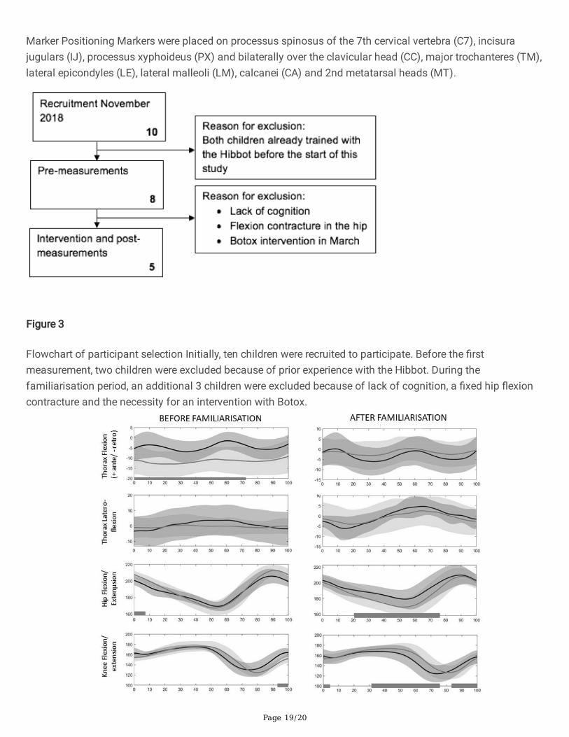
Marker Positioning Markers were placed on processus spinosus of the 7th cervical vertebra (C7), incisurajugulars (IJ), processus xyphoideus (PX) and bilaterally over the clavicular head (CC), major trochanteres (TM),lateral epicondyles (LE), lateral malleoli (LM), calcanei (CA) and 2nd metatarsal heads (MT).
Figure 3
Flowchart of participant selection Initially, ten children were recruited to participate. Before the �rstmeasurement, two children were excluded because of prior experience with the Hibbot. During thefamiliarisation period, an additional 3 children were excluded because of lack of cognition, a �xed hip �exioncontracture and the necessity for an intervention with Botox.

Page 20/20
Figure 4
Flowchart of participant selection Initially, ten children were recruited to participate. Before the �rstmeasurement, two children were excluded because of prior experience with the Hibbot. During thefamiliarisation period, an additional 3 children were excluded because of lack of cognition, a �xed hip �exioncontracture and the necessity for an intervention with Botox.
Supplementary Files
This is a list of supplementary �les associated with this preprint. Click to download.
STROBEchecklistcrosssectional.docx