pancreatic polypeptide - journal of clinical...
TRANSCRIPT
J. clin. Path., 33, Suppl. (Ass. Clin. Path.), 8, 43-50
Pancreatic polypeptideT. E. ADRIAN
From the Department of Medicine, Royal Postgraduate Medical School, Du Cane Road, London W12 OHS
Pancreatic polypeptide (PP) was discovered fortui-tously during the purification of insulin from birds(Kimmel et al., 1968) and later from mammals(Chance et al., 1976). PP has since been extractedand purified from several mammalian species. Itcontains 36 amino-acid residues and is quite distinctfrom other known hormonal peptides (Lin andChance, 1974). The amino-acid sequence of thepeptides extracted from man, pig, dog, sheep, andcow differs only in one or two residues in positions 2,6, or 23 (Lin and Chance, 1974). The biologicalactivity of these mammalian peptides has beenfound to reside in the C terminal hexapeptide (Linand Chance, 1978). Rodent PP, however, appears tobe somewhat different in that it does not cross-reactwith the majority of antisera raised to the bovinepeptide.
Localisation
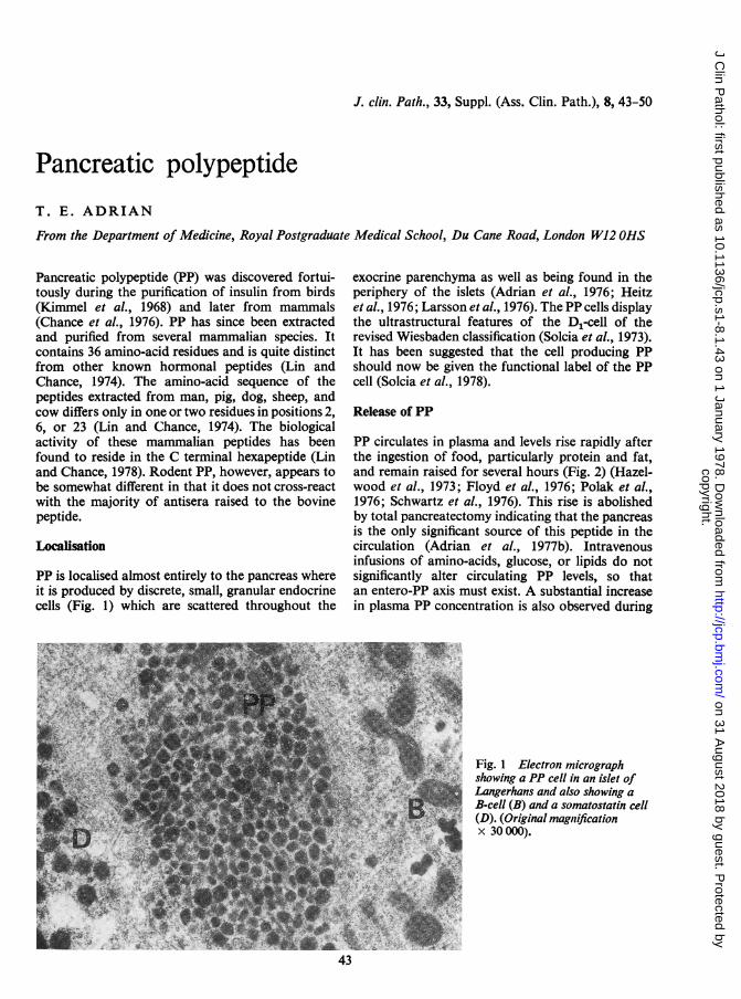
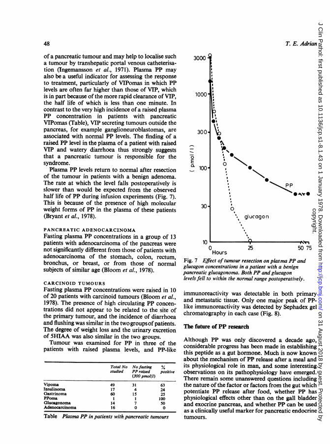
PP is localised almost entirely to the pancreas whereit is produced by discrete, small, granular endocrinecells (Fig. 1) which are scattered throughout the
exocrine parenchyma as well as being found in theperiphery of the islets (Adrian et al., 1976; Heitzet al., 1976; Larsson et al., 1976). The PP cells displaythe ultrastructural features of the D,-cell of therevised Wiesbaden classification (Solcia et al., 1973).It has been suggested that the cell producing PPshould now be given the functional label of the PPcell (Solcia et al., 1978).
Release of PP
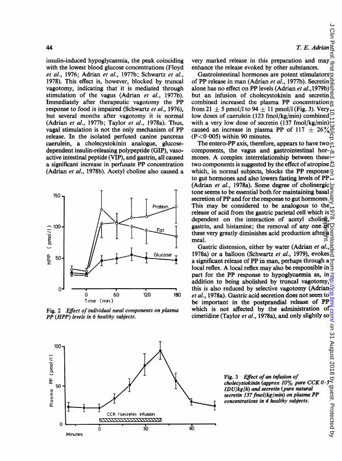
PP circulates in plasma and levels rise rapidly afterthe ingestion of food, particularly protein and fat,and remain raised for several hours (Fig. 2) (Hazel-wood et al., 1973; Floyd et al., 1976; Polak et al.,1976; Schwartz et al., 1976). This rise is abolishedby total pancreatectomy indicating that the pancreasis the only significant source of this peptide in thecirculation (Adrian et al., 1977b). Intravenousinfusions of amino-acids, glucose, or lipids do notsignificantly alter circulating PP levels, so thatan entero-PP axis must exist. A substantial increasein plasma PP concentration is also observed during
Fig. 1 Electron micrographshowing a PP cell in an islet ofLangerhans and also showing aB-cell (B) and a somatostatin cell(D). (Original magnificationx 30 000).
43
copyright. on 31 A
ugust 2018 by guest. Protected by
http://jcp.bmj.com
/J C
lin Pathol: first published as 10.1136/jcp.s1-8.1.43 on 1 January 1978. D
ownloaded from
44
insulin-induced hypoglycwith the lowest blood gluet al., 1976; Adrian et c1978). This effect is, hovagotomy, indicating thstimulation of the vaguImmediately after theraresponse to food is impaibut several months afte(Adrian et al., 1977b; Tvagal stimulation is notrelease. In the isolatedcaerulein, a cholecystodependent insulin-releasiiactive intestinal peptide (a significant increase in 1(Adrian et al., 1978b). A
150-
^ 100
a-a 50I
00Time v(min'
Fig. 2 Effect of individualPP (HPP) levels in 6 health.
T. E. Adrian
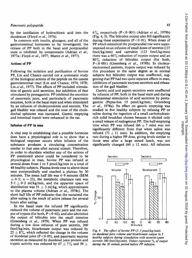
aemia, the peak coinciding very marked release in this preparation and mayicose concentrations (Floyd enhance the release evoked by other substances.71., 1977b; Schwartz et al., Gastrointestinal hormones are potent stimulatorswever, blocked by truncal of PP release in man (Adrian et al., 1977b). Secretintat it is mediated through alone has no effect on PP levels (Adrian et al.,1979b)is (Adrian et al., 1977b). but an infusion of cholecystokinin and secretinLpeutic vagotomy the PP combined increased the plasma PP concentrationired (Schwartz et al., 1976), from 21 ± 5 pmol/l to 94 ± 11 pmol/l (Fig. 3). Veryer vagotomy it is normal low doses of caerulein (123 fmol/kg/min) combined'aylor et al., 1978a). Thus, with a very low dose of secretin (137 fmol/kg/min)the only mechanism of PP caused an increase in plasma PP of 117 ± 26%perfused canine pancreas (P<0 005) within 90 minutes.)kinin analogue, glucose- The entero-PP axis, therefore, appears to have twong polypeptide (GIP), vaso- components, the vagus and gastrointestinal hor-VIP), and gastrin, all caused mones. A complex interrelationship between theseperfusate PP concentration two components is suggested by the effect of atropine,cetyl choline also caused a which, in normal subjects, blocks the PP response
to gut hormones and also lowers fasting levels of PP(Adrian et al., 1978a). Some degree of cholinergic
T tone seems to be essential both for maintaining basalsecretion ofPP and for the response to gut hormones.
Protein This may be considered to be analogous to the
Fat l?-release of acid from the gastric parietal cell which isdependent on the interaction of acetyl choline,
t~~ F t ~gastrin, and histamine; the removal of any one ofthese very greatly diminishes acid production after ameal.
Gastric distension, either by water (Adrian et al.,Glucose 1978a) or a balloon (Schwartz et al., 1979), evokes
a significant release of PP in man, perhaps through alocal reflex. A local reflex may also be responsible inpart for the PP response to hypoglycaemia as, inaddition to being abolished by truncal vagotomy,this is also reduced by selective vagotomy (Adrian
60 120 180 et al., 1978a). Gastric acid secretion does not seem tobe important in the postprandial release of PP
meal components on plasma which is not affected by the administration ofy subjects. cimetidine (Taylor et al., 1978a), and only slightly so
Fig. 3 Effect ofan infusion ofcholecystokinin (approx 10% pure CCK 0.5IDU/kgfh) and secretin (pure naturalsecretin 137 fmol/kg/min) on plasma PPconcentrations in 4 healthy subjects.
CCK Isecretin infusion
6 3 W*
Minutes
100-
ECL
a_50 -
aEa
0 6 .
30 60
copyright. on 31 A
ugust 2018 by guest. Protected by
http://jcp.bmj.com
/J C
lin Pathol: first published as 10.1136/jcp.s1-8.1.43 on 1 January 1978. D
ownloaded from
Pancreatic polypeptide
by the instillation of hydrochloric acid into theduodenum (Floyd et al., 1976).In common with insulin, glucagon, and all of the
gastrointestinal hormones so far investigated, therelease of PP both in the basal and postprandialstate is inhibited by somatostatin (Adrian et al.,1977a; Floyd et al., 1977; Marco et al., 1977).
Actions of PP
Following the isolation and purification of bovinePP, Lin and Chance carried out a systematic studyof the biological actions of the peptide on the caninegastrointestinal tract (Lin and Chance, 1974, 1978;Lin et al., 1977). The effects of PP included stimula-tion of gastric acid secretion, but inhibition of thatstimulated by pentagastrin. PP inhibited the secretionof pancreatic juice, and particularly of pancreaticenzymes, both in the basal state and when stimulatedby an infusion of cholecystokinin and secretin. Thegall bladder was relaxed and the tone of the chole-dochal sphincter was increased. Gastric emptyingand intestinal transit were enhanced in the rat.
Infusion of PP in man
A vital step in establishing that a possible hormonedoes have a physiological role is to show that abiological response occurs when infusion of thesubstance produces a circulating concentrationsimilar to that seen after natural stimuli. Therefore,in order to elucidate whether any of the actions ofPP mentioned above could be considered to bephysiological in man, bovine PP was infused atseveral doses from 1 to 5 pmol/kg/min in a total of48 healthy subjects. Plasma levels rose to above thoseseen postprandially and reached a plateau by 30minutes. The mean half life was 6.9 minutes (SEM± 0.3; n = 23), the metabolic clearance rate was5.1 ± 0'2 ml/kg/min, and the apparent space ofdistribution was 51 ± 3 ml/kg, which approximatesto the plasma volume (Adrian et al., 1978c). Theshort half life of PP indicates that the sustained riseafter eating is the result of active release for severalhours after eating.
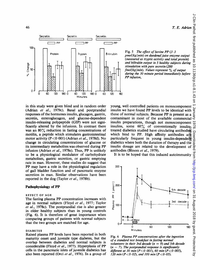
In the basal state the infused PP significantlyreduced the volume of pancreatic juice and the out-put of trypsin (for both, P<0 02), and also abolishedthe output of bilirubin into the small intestine(Greenberg et al., 1979). When PP was infusedduring a low dose infusion of pure secretin, 200fmol/kg/min, bicarbonate output was reduced by25 ± 8 %, which reflected the change in the volumeof duodenal juice (P<0 '05), and pancreatic enzymesecretion as measured by duodenal juice protein andtryptic activity was reduced by 67 ± 7% and 59 ±
45
6%, respectively (P<0*001) (Adrian et al., 1979b)(Fig. 4, 5). The bilirubin output also fell significantlyduring these experiments (P<0 01). When doses ofPP which mimicked the postprandial rise were super-imposed on an infusion of small doses of secretin (137fmol/kg/min) and caerulein (123 fmol/kg/min)there was a 60% reduction of trypsin output and an80% reduction of bilirubin output (for both,P<0 001) (Greenberg et al., 1978b). In cholecy-stectomised patients, trypsin output was reduced bythis procedure to the same degree as in normalsubjects but bilirubin output was unaffected, sug-gesting that PP had two quite separate effects in man,inhibition of pancreatic enzyme secretion and relaxa-tion of the gall bladder.
Gastric acid and pepsin secretion were unaffectedby infusion of PP, both in the basal state and duringsubmaximal stimulation of acid secretion by penta-gastrin (Peptavlon 15 pmol/kg/min; Greenberget al., 1978a). Its effect on gastric emptying wasstudied in five healthy subjects by infusing PP orsaline during the ingestion of a small carbohydrate-rich solid breakfast chosen because it elicited onlya small release of endogenous PP. The half-emptyingtime when PP was infused (66 ± 7 min) was notsignificantly different from that when saline wasinfused (71 ± 11 min). In addition, the emptyingrate during a higher PP dose, giving levels similar tothose seen after a large mixed lunch, was notsignificantly changed (60 + 12 min). All infusions
Secretin1
I PP
100 -
Q)Wa o
0 60
Secretinrb
Bicarbonate .
120 180 0 60 120 180Minutes
Fig. 4 The effect ofbovine PP (3.3 pmol/kg/min)on duodenal juice volume and bicarbonate output in Shealthy subjects during stimulation with secretin (puresecretin 200 fmol/kg/min). Values represent % ofoutputduring the 30 minute period before PP infusion.
copyright. on 31 A
ugust 2018 by guest. Protected by
http://jcp.bmj.com
/J C
lin Pathol: first published as 10.1136/jcp.s1-8.1.43 on 1 January 1978. D
ownloaded from
T. E. Adrian
Secret in Secret in
-ZPP r PP
Protein Trypsin
0 60 120 180 0 60 120 1lMinutes
Fig. 5 The effect ofbovine PP (3.3pmol/kg/min) on duodenal juice enzyme output(measured as tryptic activity and total protein)and bilirubin output in S healthy subjects duringstimulation with pure secretin (200fmollkg/min). Values represent % ofoutputduring the 30 minute period immediately beforePP infusion,
in this study were given blind and in random order(Adrian et al., 1979c). Basal and postprandialresponses of the hormones insulin, glucagon, gastrin,secretin, enteroglucagon, and glucose-dependentinsulin-releasing polypeptide (GIP) were not signi-ficantly altered by the infusions. In contrast therewas an 80% reduction in fasting concentrations ofmotilin, a peptide which stimulates gastrointestinalmotor activity (P<0001) (Adrian et al., 1978d). Nochange in circulating concentrations of glucose orits intermediary metabolites was observed during PPinfusion (Adrian et al., 1978e). Thus, PP is unlikelyto be a physiological modulator of carbohydratemetabolism, gastric secretion, or gastric emptyingrate in man. However, these studies do suggest thatPP may have a role in the physiological regulationof gall bladder function and of pancreatic enzymesecretion in man. Similar observations have beenreported in the dog (Taylor et al., 1978b).
Pathophysiology of PP
EFFECT OF AGE
The fasting plasma PP concentration increases withage in normal subjects (Floyd et al., 1977; Tayloret al., 1978c). The postprandial rise is also greaterin older healthy subjects than in young controls(Fig. 6). It is therefore of great importance whencomparing groups of patients with normal subjectsthat the two groups are matched for age.
DIABETESRaised plasma PP levels have been reported in bothmaturity onset and juvenile type diabetes, but theoverlap between diabetics and normal subjects isconsiderable (Floyd et al., 1977). Hyperplasia of PPcells in the pancreatic islets of juvenile diabetics hasalso been reported (Orci et al., 1976). In a group of
young, well controlled patients on monocomponentinsulin we have found PP levels to be identical withthose of normal subjects. Because PP is present as a
contaminant in most of the available commercialinsulin preparations, though not monocomponentinsulins, some 60% of conventionally insulin-treated diabetics studied have circulating antibodieswhich bind to PP. High affinity antibodies are
particularly frequent in young insulin-dependentdiabetics where both the duration of therapy and theinsulin dosage are related to the development ofantibodies (Bloom et al., 1979).
It is to be hoped that this induced autoimmunity
300 -,
200-Ea.
a-
a-a
Ea 100-a
0
5th decade
i/ **'{....i....{ 3rd decade
M,
a. o..,.oMeal
6Minutes
60 l20 l80
Fig. 6 Plasma PP concentrations after the ingestionofa standard test breakfast in fasting normalvolunteers in their 3rd decade (n = 9) and 5th decade(n = 7). The postprandial response is significantlydifferent at 30 min (P<0.001), 60 min (P<0.OOS),120 min (P<0.02), and 180 min (P<0.OS).
Secretinr P
I IPP
100.
a,
£50U
0
46
copyright. on 31 A
ugust 2018 by guest. Protected by
http://jcp.bmj.com
/J C
lin Pathol: first published as 10.1136/jcp.s1-8.1.43 on 1 January 1978. D
ownloaded from
Pancreatic polypeptide
will be avoided in the future by the use of highlypurified insulins.
OBESITYThe obese hyperglycaemic mouse (ob/ob) is the mostintensively studied animal model of human lateonset diabetes mellitus. Parabiotic animal experi-ments where ob/ob animals maintain normal weightwhen paired with normal but not ob/ob mates haveled to the conclusion that the hyperphagia andresulting weight gain is the result of the lack ofcirculating satiety factor from the gut which innormal animals controls food intake (Coleman,1978). It has been reported that the administrationof PP reverses the hyperphagia and weight gainin these mice, which subsequently become normo-glycaemic (Malaisse-Lagae et al., 1977). Studies onthe release of PP in the obese mouse are awaitedwith interest. We have shown (unpublished observa-tions) that the pancreas of the fat mouse contains anormal amount of extractable PP-like immuno-reactivity (147 ± 14 pmol/g) compared with thatof the lean litter mate (124 + 10 pmol/g). However,a reduction in the number of PP cells has previouslybeen reported (Orci et al., 1976).
In man, PP release by a standard test breakfastin 19 patients with morbid obesity (225 ± 7% idealweight) was not significantly different from that seenin 16 healthy age- and sex-matched normal controls(106 ± 3% ideal weight). In addition the PPresponse was unaffected by ileojejunal bypass in theobese patients (Besterman et al., 1978c).
PEPTIC ULCERBecause of the observed actions of bovine PP onacid secretion in the dog it was of particular interestto investigate PP release in patients with duodenalulcer. Both fasting concentrations of PP (n = 40)and the response to food (n = 21) in duodenal ulcerpatients were similar to those seen in age-matchedcchtrols (Adrian et al., 1977b). It seems unlikely,therefore, that PP is involved in the pathogenesisof this syndrome.
STEATORRHOEAAs PP release appears to be controlled by mechanismssimilar to those which control the secretion of pan-creatic enzymes, it was clearly of interest to investi-gate its release in patients with chronic pancreatitis.The PP response to food is grossly impaired inpatients with chronic pancreatitis and steatorrhoea(Adrian et al., 1977a; Adrian et al., 1979a); therewas a similarly impaired response to the intravenousinjection of secretin1 which normally stimulates PPrelease. In patients with chronic pancreatitis without1 A comparatively impure preparation obtained from Boots.
47
overt exocrine deficiency the release of PP wasnot significantly different from normal. In contrastto the findings in pancreatic steatorrhoea, patientswith steatorrhoea resulting from active coeliacdisease or acute tropical sprue had normal basaland peak levels of PP after the meal. The measure-ment of PP may thus provide confirmatory evidencethat steatorrhoea is of pancreatic origin, but it isunhelpful in assessing chronic pancreatitis withoutexocrine deficiency, where there is a diagnosticproblem.
DIARRHOEAThe infusion of low doses of PP in the dog caused amarked increase in the motility of the gastricantrum, duodenum, ileum, and colon (Lin andChance, 1978), and the substance was patented as apotential veterinary laxative. We therefore investi-gated the postprandial PP response in patients withdiarrhoea resulting from several different causes.Plasma PP levels after our standard test breakfastwere not significantly raised in 12 patients with acuteinfective diarrhoea, in 14 patients with Crohn'sdisease, and in 24 patients with ulcerative colitis(Besterman et al., 1978b). Patients with the irritablebowel syndrome had a moderately increased releaseofPP after the test breakfast. However, this increasedresponse was noted in patients who presented withconstipation as well as those with diarrhoea.
GUT RESECTIONPP release appears to be potentiated by an intestinalfactor. Its source was investigated bystudying plasmaPP release after our standard test breakfast in 18patients with partial ileal resection and nine patientswith partial colonic resection for inflammatory boweldisease, neoplasia, radiation, fibrosis, or trauma;the operations had been performed at least one yearbefore the study.Both basal and postprandial PP levels were
surprisingly raised in these two groups of patients(Besterman et al., 1978a). Further work is requiredto elucidate the cause and effect of this.
PANCREATIC ENDOCRINE TUMOURSOver half of the endocrine tumours of the pancreasand their metastases so far investigated containlarge numbers of PP cells which can be shown byimmunofluorescence (Polak et al., 1976). Hepaticand lymph node metastases sometimes contain evenhigher concentrations of immunoreactive PP thanthe primary pancreatic tumour, indicating that PPis a product of the tumour. Plasma levels are oftengrossly raised in these patients, sometimes exceeding1000 times the normal basal level. The measurementof plasma PP may thus provide additional evidence
copyright. on 31 A
ugust 2018 by guest. Protected by
http://jcp.bmj.com
/J C
lin Pathol: first published as 10.1136/jcp.s1-8.1.43 on 1 January 1978. D
ownloaded from
T. E. Adrian
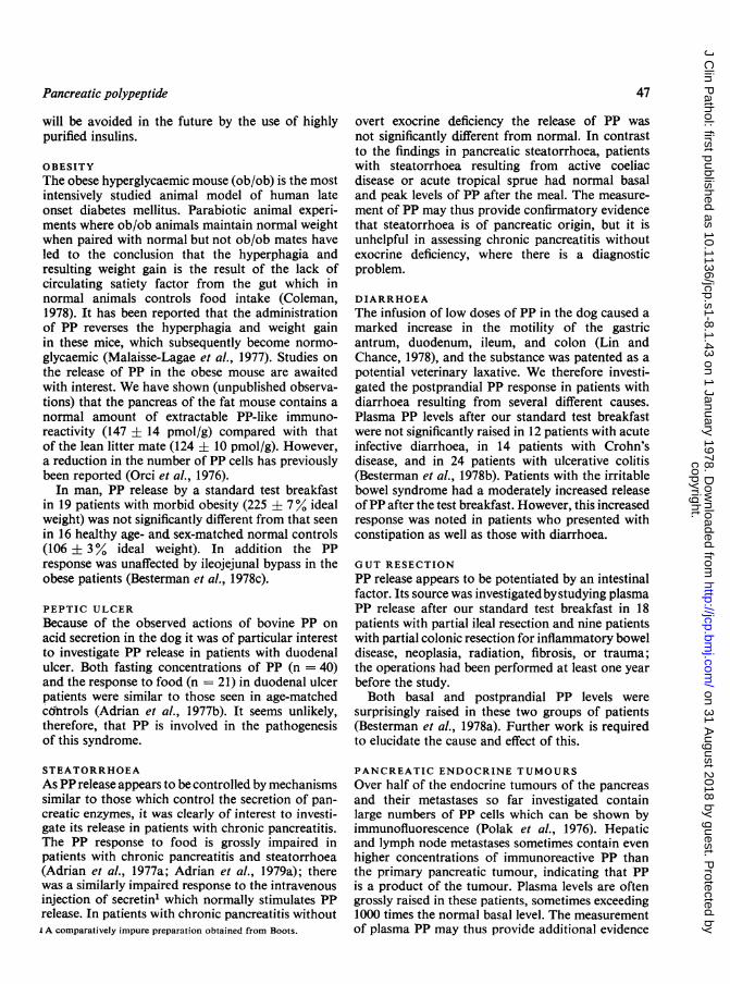
of a pancreatic tumour and may help to localise sucha tumour by transhepatic portal venous catheterisa-tion (Ingemansson et al., 1971). Plasma PP mayalso be a useful indicator for assessing the responseto treatment, particularly of VlPomas in which PPlevels are often far higher than those of VIP, whichis in part because of the more rapid clearance of VIP,the half life of which is less than one minute. Incontrast to the very high incidence of a raised plasmaPP concentration in patients with pancreaticVIPomas (Table), VIP secreting tumours outside thepancreas, for example ganglioneuroblastomas, areassociated with normal PP levels. The finding of araised PP level in the plasma of a patient with raisedVIP and watery diarrhoea thus strongly suggeststhat a pancreatic tumour is responsible for thesyndrome.Plasma PP levels return to normal after resection
of the tumour in patients with a benign adenoma.The rate at which the level falls postoperatively isslower than would be expected from the observedhalf life of PP during infusion experiments (Fig. 7).This is because of the presence of high molecularweight forms of PP in the plasma of these patients(Bryant et al., 1978).
PANCREATIC ADENOCARCINOMAFasting plasma PP concentrations in a group of 13patients with adenocarcinoma of the pancreas werenot significantly different from those of patients withadenocarcinoma of the stomach, colon, rectum,bronchus, or breast, or from those of normalsubjects of similar age (Bloom et al., 1978).
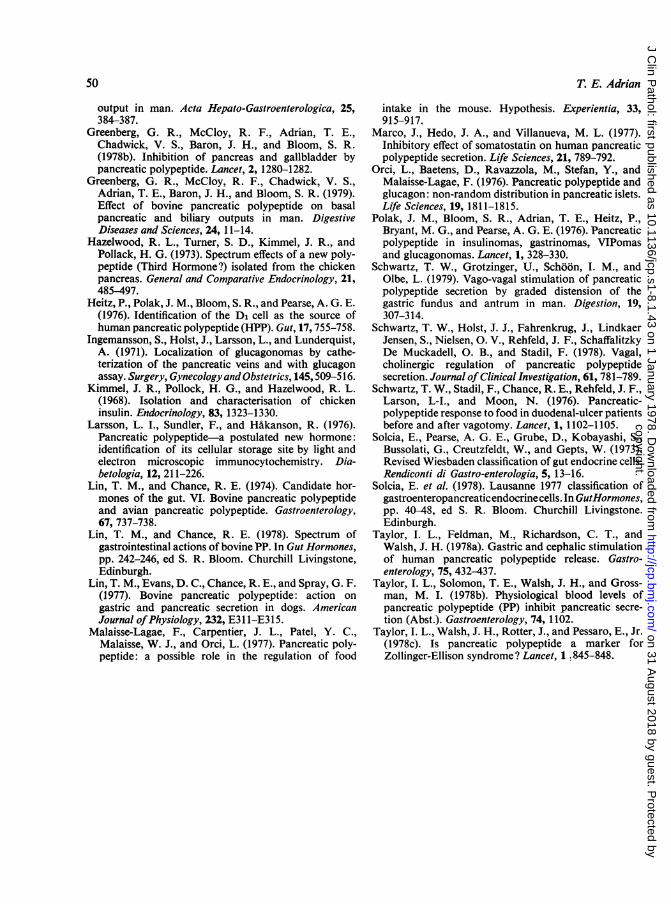
CARCINOID TUMOURSFasting plasma PP concentrations were raised in 10of 20 patients with carcinoid tumours (Bloom et al.,1978). The presence of high circulating PP concen-trations did not appear to be related to the site ofthe primary tumour, and the incidence of diarrhoeaand flushing was similar in the twogroupsof patients.The degree of weight loss and the urinary excretionof 5HIAA was also similar in the two groups.Tumour was examined for PP in three of the
patients with raised plasma levels, and PP-like
Total No No fasting %studied PP raised positive
(300 pmol/l)
Vipoma 49 3 1 63Insulinoma 17 4 24Gastrinoma 60 15 25PPoma 1 1 100Glucagonoma 14 7 50Adenocarcinonma 16 0 0
Table Plasma PP in patients with pancreatic tumours
3000
0. 100 o
:PP
30,\ 0'. glucagon
10 A0 25 50 7Hours
Fig. 7 Efect of tumour resection on plasma PP andglucagon concentrations in a patient with a benignpancreatic glucagonoma. Both PP and glucagonlevels fell to within the normal range postoperatively.
a
75
immunoreactivity was detectable in both primaryand metastatic tissue. Only one major peak of PP-like immunoreactivity was detected by Sephadex gelchromatography in each case (Fig. 8).
The future of PP research
Although PP was only discovered a decade ago,considerable progress has been made in establishingthis peptide as a gut hormone. Much is now knownabout the mechanism of PP release after a meal andits physiological role in man, and some interestingobservations on its pathophysiology have emerged.There remain some unanswered questions includingthe nature of the factor or factors from the gut whichpotentiate PP release after food, whether PP hasphysiological effects other than on the gall bladderand exocrine pancreas, and whether PP can be usedas a clinically useful marker for pancreatic endocrinetumours.
48
copyright. on 31 A
ugust 2018 by guest. Protected by
http://jcp.bmj.com
/J C
lin Pathol: first published as 10.1136/jcp.s1-8.1.43 on 1 January 1978. D
ownloaded from
Pancreatic polypeptide 49
20
10 irO VtL iver 2°'
20-
0 oE
a_ CytochromeX_ Albumin C Glucagon 1251
20 40 60 80Fraction number
Fig. 8 Gel chromatography ofPP immunoreactivityin tumour extracts from 2 metastatic carcinoid tumours.One major peak was seen eluting in the sameposition as pure natural human PP.
References
Adrian, T. E., Besterman, H. S., Christofides, N. D., andBloom, S. R. (1978a). Interaction of gastrointestinalhormones and cholinergic innervation on the release ofpancreatic polypeptide (Abst.). Scandinavian Journalof Gastroenterology, 13, Suppl. 49, 1.
Adrian, T. E., Besterman, H. S., Mallinson, C. N.,Czaykowska, W. M., and Bloom, S. R. (1977a).Studies on the release of pancreatic polypeptide andits relationship to pancreatic exocrine function (Abst.).Journal ofEndocrinology, 75, 35P-36P.
Adrian, T. E., Besterman, H. S., Mallinson, C. N.,Garalotis, C., and Bloom, S. R. (1979a). Impairedpancreatic polypeptide release in chronic pancreatitiswith steatorrhoea. Gut, 20, 98-101.
Adrian, T. E., Besterman, H. S., Mallinson, C. N.,Greenberg, G. R., and Bloom, S. R. (1979b). Inhibitionof secretin-stimulated pancreatic secretion by pan-creatic polypeptide. Gut, 20, 37-40.
Adrian, T. E., Bloom, S. R., Besterman, H. S., Barnes,A. J., Cook, T. J. C., Russell, R. C. G., and Faber,R, G. (1977b). Mechanism of pancreatic polypeptiderelease in man. Lancet, 1, 161-163.
Adrian, T. E., Bloom, S. R., Bryant, M. G., Polak, J. M.,Heitz, P., and Barnes, A. J. (1976). Distribution andrelease of human pancreatic polypeptide. Gut, 17,940-944.
Adrian, T. E., Bloom, S. R., Hermansen, K., andIversen, J. (1978b). Pancreatic polypeptide, glucagonand insulin secretion from the isolated perfused caninepancreas. Diabetologia, 14, 413-417.
Adrian, T. E., Greenberg, G. R., Besterman, H. S., andBloom, S. R. (1978c). Pharmacokinetics of pancreaticpolypeptide in man. Gut, 19, 907-909.
Adrian, T. E., Greenberg, G. R., Besterman, H. S.,Christofides, N. D., and Bloom, S. R. (1978d). PPinfusion in man: pharmacokinetics at 3 dose levels andeffects on gastrointestinal and pancreatic hormones(Abst.). Scandinavian Journal of Gastroenterology, 13,Suppl. 49, 3.
Adrian, T. E., Greenberg, G. R., Besterman, H. S.,McCloy, R. F., Chadwick, V. S., Barnes, A. J.,Mallinson, C. N., Baron, J. H., Alberti, K. G. G. M.,and Bloom, S. R. (1978e). PP infusion in man-summary of initial investigations. In Gut Hormones,pp. 265-267, ed. S. R. Bloom. Churchill Livingstone,Edinburgh.
Adrian, T. E., Greenberg, G. R., McCloy, R. F.,Fitzpatrick, M. L., and Bloom, S. R. (1979c). How toassess the physiological role of a new peptide hormone:pancreatic polypeptide infusion in man. Journal ofEndocrinology (in press).
Besterman, H. S., Bloom, S. R., Adrian, T. E.,Christofides, N. D., Sarson, D. L., Mallinson, C. N.,Pero, A., and Modigliani, R. (1978a). Gut hormoneprofile after gut resection (Abst.). Gut, 19, A972.
Besterman, H. S., Bloom, S. R., Christofides, N. D.,Mallinson, C. N., Pera, A., and Modigliani, R.(1978b). Gut hormone profile in inflammatory boweldisease (Abst.). Gut, 19, A988.
Besterman, H. S., Sarson, D. L., Blackburn, A. M.,Cleary, J., Pilkington, T. R. E., Gazet, J. C., andBloom, S. R. (1978c). Gut hormone profile in morbidobesity and following jejunoileal bypass (Abst.).Gut, 19, A986.
Bloom, S. R., Adrian, T. E., Barnes, A. J., and Polak,J. M. (1979). Autoimmunity in diabetics induced byhormonal contaminants of insulin. Lancet, 1, 14-17.
Bloom, S. R., Adrian, T. E., Pera, A., Polak, J. M.,Besterman, H. S., Modigliani, R., Bryant, M. G.,Modlin, I. M., Barnardo, D. E., and Grahame-Smith,D. G. (1978). PP in patients with the carcinoidsyndrome, pancreatic endocrine tumours and adeno-carcinomas (Abst.). Gastroenterology, 74, 1012.
Bryant, M. G., Bloom, S. R., Adrian, T. E., andMallinson, C. N. (1978). Chromatographic analysis oftumour produced hormones (Abst.). Gut, 19, A445.
Chance, R. E., Root, M. A., and Galloway, J. A. (1976).The immunogenicity of insulin preparations. ActaEndocrinologica, 83, Suppl. 205, 185-196.
Coleman, D. L. (1978). Obese and diabetes: two mutantgenes causing diabetes-obesity syndromes in mice.Diabetologia, 14, 141-148.
Floyd, J. C., Fajans, S. S., and Pek, S. (1976). Regulationin healthy subjects of the secretion ofhuman pancreaticpolypeptide, a newly recognised pancreatic islet poly-peptide. Transactions of the Association of AmericanPhysicians, 89, 146-158.
Floyd, J. C., Fajans, S. S., Pek, S., and Chance, R. E.(1977). A newly recognised pancreatic polypeptide;plasma levels in health and disease. Recent Progress inHormone Research, 33, 519-570.
Greenberg, G. R., McCloy, R. F., Adrian, T. E., Baron,J. H., and Bloom, S. R. (1978a). Effect of bovinepancreatic polypeptide on gastric acid and pepsin
copyright. on 31 A
ugust 2018 by guest. Protected by
http://jcp.bmj.com
/J C
lin Pathol: first published as 10.1136/jcp.s1-8.1.43 on 1 January 1978. D
ownloaded from
so T. E. Adrian
output in man. Acta Hepato-Gastroenterologica, 25,384-387.
Greenberg, G. R., McCloy, R. F., Adrian, T. E.,Chadwick, V. S., Baron, J. H., and Bloom, S. R.(1978b). Inhibition of pancreas and gallbladder bypancreatic polypeptide. Lancet, 2, 1280-1282.
Greenberg, G. R., McCloy, R. F., Chadwick, V. S.,Adrian, T. E., Baron, J. H., and Bloom, S. R. (1979).Effect of bovine pancreatic polypeptide on basalpancreatic and biliary outputs in man. DigestiveDiseases and Sciences, 24, 11-14.
Hazelwood, R. L., Turner, S. D., Kimmel, J. R., andPollack, H. G. (1973). Spectrum effects of a new poly-peptide (Third Hormone?) isolated from the chickenpancreas. General and Comparative Endocrinology, 21,485-497.
Heitz, P., Polak, J. M., Bloom, S. R., and Pearse, A. G. E.(1976). Identification of the Di cell as the source ofhuman pancreatic polypeptide (HPP). Gut, 17,755-758.
Ingemansson, S., Holst, J., Larsson, L., and Lunderquist,A. (1971). Localization of glucagonomas by cathe-terization of the pancreatic veins and with glucagonassay. Surgery, Gynecology and Obstetrics, 145, 509-516.
Kimmel, J. R., Pollock, H. G., and Hazelwood, R. L.(1968). Isolation and characterisation of chickeninsulin. Endocrinology, 83, 1323-1330.
Larsson, L. I., Sundler, F., and HAkanson, R. (1976).Pancreatic polypeptide-a postulated new hormone:identification of its cellular storage site by light andelectron microscopic immunocytochemistry. Dia-betologia, 12, 211-226.
Lin, T. M., and Chance, R. E. (1974). Candidate hor-mones of the gut. VI. Bovine pancreatic polypeptideand avian pancreatic polypeptide. Gastroenterology,67, 737-738.
Lin, T. M., and Chance, R. E. (1978). Spectrum ofgastrointestinal actions of bovine PP. In Gut Hormones,pp. 242-246, ed S. R. Bloom. Churchill Livingstone,Edinburgh.
Lin, T. M., Evans, D. C., Chance, R. E., and Spray, G. F.(1977). Bovine pancreatic polypeptide: action ongastric and pancreatic secretion in dogs. AmericanJournal ofPhysiology, 232, E311-E315.
Malaisse-Lagae, F., Carpentier, J. L., Patel, Y. C.,Malaisse, W. J., and Orci, L. (1977). Pancreatic poly-peptide: a possible role in the regulation of food
intake in the mouse. Hypothesis. Experientia, 33,915-917.
Marco, J., Hedo, J. A., and Villanueva, M. L. (1977).Inhibitory effect of somatostatin on human pancreaticpolypeptide secretion. Life Sciences, 21, 789-792.
Orci, L., Baetens, D., Ravazzola, M., Stefan, Y., andMalaisse-Lagae, F. (1976). Pancreatic polypeptide andglucagon: non-random distribution in pancreatic islets.Life Sciences, 19, 1811-1815.
Polak, J. M., Bloom, S. R., Adrian, T. E., Heitz, P.,Bryant, M. G., and Pearse, A. G. E. (1976). Pancreaticpolypeptide in insulinomas, gastrinomas, VIPomasand glucagonomas. Lancet, 1, 328-330.
Schwartz, T. W., Grotzinger, U., Schoon, I. M., andOlbe, L. (1979). Vago-vagal stimulation of pancreaticpolypeptide secretion by graded distension of thegastric fundus and antrum in man. Digestion, 19,307-314.
Schwartz, T. W., Holst, J. J., Fahrenkrug, J., LindkaerJensen, S., Nielsen, 0. V., Rehfeld, J. F., SchaffalitzkyDe Muckadell, 0. B., and Stadil, F. (1978). Vagal,cholinergic regulation of pancreatic polypeptidesecretion. Journal of Clinical Investigation, 61, 781-789.
Schwartz, T. W., Stadil, F., Chance, R. E., Rehfeld, J. F.,Larson, L-I., and Moon, N. (1976). Pancreatic-polypeptide response to food in duodenal-ulcer patientsbefore and after vagotomy. Lancet, 1, 1102-1105.
Solcia, E., Pearse, A. G. E., Grube, D., Kobayashi, S.,Bussolati, G., Creutzfeldt, W., and Gepts, W. (1973).Revised Wiesbaden classification of gut endocrine cells.Rendiconti di Gastro-enterologia, 5, 13-16.
Solcia, E. et al. (1978). Lausanne 1977 classification ofgastroenteropancreaticendocrine cells. In GutHormones,pp. 40-48, ed S. R. Bloom. Churchill Livingstone.Edinburgh.
Taylor, I. L., Feldman, M., Richardson, C. T., andWalsh, J. H. (1978a). Gastric and cephalic stimulationof human pancreatic polypeptide release. Gastro-enterology, 75, 432-437.
Taylor, I. L., Solomon, T. E., Walsh, J. H., and Gross-man, M. I. (1978b). Physiological blood levels ofpancreatic polypeptide (PP) inhibit pancreatic secre-tion (Abst.). Gastroenterology, 74, 1102.
Taylor, I. L., Walsh, J. H., Rotter, J., and Pessaro, E., Jr.(1978c). Is pancreatic polypeptide a marker forZollinger-Ellison syndrome? Lancet, 1 .845-848.
copyright. on 31 A
ugust 2018 by guest. Protected by
http://jcp.bmj.com
/J C
lin Pathol: first published as 10.1136/jcp.s1-8.1.43 on 1 January 1978. D
ownloaded from