pancreatic tumors med students-2- · • tumor extension outside of pancreas • extensive portal...
TRANSCRIPT

Pancreatic Tumors
Margo Shoup, MD
Associate Professor of Surgery
Loyola University Medical Center

Introduction
• 38,000 cases a year
• Risk factors
– Smoking
– Pancreatitis
• Real risk, but only 5% of
pancreatic cancer patients
Pancreatic Tumors

Genetics
• Tumor suppressor gene p53
• Mitogen activating gene k-ras
• COX-2
• VEGF
Pancreatic Tumors

Definitions
• Most common malignant pancreatic tumor is pancreatic ductal adenocarcinoma
• Difficult at diagnosis to determine etiology
– Periampullary tumor
• Pancreatic –65%
• Distal bile duct
• Ampulla
• Duodenum
• Islet cell
Pancreatic Tumors

Classification of pancreatic
tumors
• Cystic tumors
– Cystadenoma
• Serous
• Mucinous
• Intraductal papillary mucinous
• Solid and Pseudopapillary
Pancreatic Tumors

Surgical Options
• Enucleation
• Distal pancreatectomy with or without
splenectomy
• Central pancreatectomy
• Ampullectomy
• Pancreaticoduodenectomy
Pancreatic Tumors

Classification of pancreatic
tumors
• Malignant
– Adenocarcinoma
• Mucinous
• Adenosquamous
• Anaplastic
• Duodenal/ampullary/distal bile duct
– Cystadenocarcinoma• Mucinous
• Intraductal papillary
– Acinar
• Endocrine
Pancreatic Tumors

Tumor Markers
• CA 19-9
– Most commonly valued marker
– Not specific, high levels seen in benign disease
– Normalization following resection appears to be
associated with improved outcome
– Rising level after resection is a marker of relapse
– Levels > 1500 correlate with unresectable tumors
• Not cost effective for screening
Pancreatic Tumors

Clinical suspicion• Patients with pancreatic cancer commonly present
with advanced disease
– Head tumors – proximity to vascular structures
– Body and Tail – metastatic disease
• Symptoms are nonspecific
– Vague discomfort, dyspepsia, bloating
– Jaundice
– Weight loss, back pain usually a sign of advance disease
– Significant back pain 9% resectability vs minimal back pain 31% resectability
– New onset diabetes in patients over 60 should raise suspicion.
Pancreatic Tumors

Diagnosis
• History
– Weight loss
– Change in urine and stool
– Gastric outlet symptoms
– Back pain
• Physical
– Jaundice
– Cachectic
– Palpable mass
Pancreatic Tumors

Work up• CBC
• Liver function tests
• Hepatitis profile
• Hemolytic profile
• Ultrasound
• CT – identify mass, evaluate vessel involvement
• ERCP – double duct sign for head mass
• EUS – If not sure if pancreatitis vs tumor
Pancreatic Tumors

CT Findings
• Adenocarcinoma
– Irregular border
– Not hypervascular
– Pancreatic ductal dilatation
– Distal pancreatic atrophy
Pancreatic Tumors

Pancreatic adenocarcinoma

Pancreatic adenocarcinoma

CT Findings
• Neuroendocrine
– Well circumscribed
– Hypervascular
– No atrophy
• Cystic
– Appear fluid filled
– Well circumscribed
Pancreatic Tumors

Neuroendocrine Tumor

Intraductal papillary mucinous neoplasm

ERCP
• Not usually necessary
• Often performed if seen by Gastroenterologists
• Necessary if biliary stent is needed
• Double duct sign
– Strictured common bile duct and pancreatic duct
• Biopsy possible, not always needed
Pancreatic Tumors

Treatment Options
• Tissue diagnosis – NOT NECESSARY
– Unless surgery is not planned
• Potentially resectable tumors
– Laparoscopy to rule out metastatic disease
– Head tumors – pancreaticoduodenectomy
• Pancreatic head, distal common bile duct, duodenum, +/-antrum, gallbladder
• Pancreaticogastrostomy or jejunostomy, hepaticojejunostomy, gastrojejunostomy
– Body or Tail tumors – distal pancreatectomy with splenectomy
Pancreatic Tumors

Reconstruction Following Standard
Pancreaticoduodenectomy

Reconstruction Following Pylorus
Preserving Pancreaticoduodenectomy

Prognosis after surgery
• 1-3% perioperative mortality rate in the best
hands (30-day or same admisstion mortality)
– Previously was 20%
• 5 year survival
– Pancreas – 10-15%
– Bile Duct – 15-20%
– Duodenum – 50%
– Ampulla – 35%
– Islet cell – 40%
Pancreatic Tumors
Adjuvant therapy
• Options for chemotherapy and radiotherapy
– Inconclusive evidence that CRT improves survival
– GITSG trial
– 43 patients randomized to CRT vs. no CRT
– CRT had improved survival
• Neoadjuvant therapy
– Clinical trials

Predictors of outcome
• Nodal status
• Size (< 2cm)
• Margin status
Pancreatic Tumors

Complications
• Pancreatic duct leak/fistula
– Drain amylase level more than 3x serum
– 10-20%
• Biliary leak/Gastrojejunostomy leak
– Less common
• Delayed gastric emptying
• Pancreatitis
• Diabetes
• Dumping syndrome – exocrine insufficiency
Pancreatic Tumors

Follow-up
• If patients are asymptomatic follow with physical exam and history
• If patients start to become symptomatic, obtain CT
– Weight loss
– Anorexia
– Weakness
• Someone will order a CT sooner
– Patients peace of mind
• What to do with results if a recurrence is noted?
– Treatment with chemotherapy in the metastatic setting has not been shown to prolong life.
Pancreatic Tumors

Unresectable
• Majority of patients
• Locally advanced, not metastatic – May receive
chemotherapy with radiation.
– A small number of patients will respond enough to
become resectable.
– Median Survival 4-5 months if metastatic
– Median Survival 7-9 months if not metastatic
• Back pain can be palliated with celiac axis
blockade – alcohol injection
Pancreatic Tumors

Unresectable
• Metastatic disease – treatment options limited to
experimental medications and chemotherapy.
• Patients should have biliary stent placed by ERC
(Endoscopic retrograde cholangiogram)
– If unable to place stent due to technical difficulties,
should have operative biliary bypass
– Choledochojejunostomy, Hepaticojejunostomy,
Cholecystojejunostomy
• If considering CRT – need biopsy
Pancreatic Tumors

Unresectable Disease
• Biliary stents
– Plastic stent
• Best if patient considered for surgery
• 3- month longevity
• Easily removed
– Metal “Wallstent”
• Permanent
• Lasts 6 months to a year
• Difficult to remove surgically
Pancreatic Tumors

Defining Non-resectability
• Histologically confirmed hepatic, serosal,
peritoneal or omental metastasis
• Celiac or high portal node involvement
• Tumor extension outside of pancreas
• Extensive portal vein involvement by tumor or
invasion/encasement of celiac axis, hepatic
artery, or superior mesenteric artery.
Laparoscopic Staging
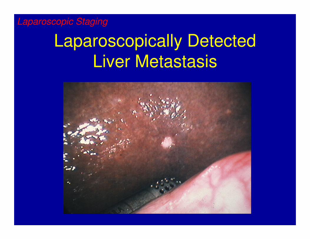
Laparoscopically Detected
Liver Metastasis
Laparoscopic Staging

Locally Advanced Tumors
• Considered candidates for chemoradiation if metastatic disease is not present.
• May be considered for subsequent surgical resection depending on the response to the chemoradiation.
• Patients with pancreatic adenocarcinoma metastatic to the liver or peritoneum are candidates for palliative chemotherapy, but not radiation.
Laparoscopic Staging

Locally Advanced Pancreatic Cancer
• Contemporary imaging modalities failed to
detect metastatic disease in 37% of patients.
• Patients considered for protocols including
radiation for locally advanced pancreatic
cancer should be staged laparoscopically
prior to initiating therapy.
Laparoscopic Staging

End of Life Issues
• Pancreatic cancer
– Almost as many people die each year from the disease as are diagnosed each year
– Pain/Back pain
• Biggest issue
• Control with celiac block, fentanyl patch
• Palliative radiation
– Gastric outlet obstruction – can be palliated by duodenal stent or gastric bypass (gastrojejunostomy)
• Patients with advanced disease should be referred to a hospice situation early
Pancreatic Tumors

End of Life
• Options for treatment vs no treatment
– Chemotherapy disappointing
• 5-FU, Gemcitabine, oxaliplatin
– Quality of Life
• Radiation
– Time consuming
– 5 days a week for 6 weeks
– Benefit not guaranteed
Pancreatic Tumors

End of Life
• Questions from patients –
– How much time do I have?
– Will you still be my doctor?
– How will I die?
– What should I do now?
Pancreatic Tumors

Case 1
• 52 year old man noted to have icteric sclera
and mild jaundice, no pain.
• H&P
• PE
• Labs
• Differential Diagnosis

Case 1
• Ultrasound
– Dilated intra- and extra-hepatic bile ducts, no
stones. Liver normal
– CT – 3 cm mass in head of pancreas. No liver
lesions. Dilated CBD and pancreatic duct
(Double duct sign)
– Now what?

Case 2
• 44 year old woman
• CT – pancreatic head mass
• Multiple liver lesions
• Now what?

Case 3
• 65 year old male had a screening CT scan at
the mall showing a 2 cm mass in the tail of
the pancreas.
• Asymptomatic
• Differential
• Work up
• Treatment

Recommendations for
Pancreatic Cancer• Laparoscopic
– Patients with resectable disease
– No evidence of gastric outlet obstruction
– Have biliary stent, or can receive biliary stent if needed
– Patients with locally advanced tumors, no metastasis on
imaging, considered for local therapy
• Open Exploration
– Failed biliary stent
– Gastric outlet obstruction
Laparoscopic Staging