parallel input makes the brain run faster
TRANSCRIPT
www.elsevier.com/locate/ynimg
NeuroImage 40 (2008) 1792–1797Parallel input makes the brain run faster
Tommi Raij,a,⁎ Jari Karhu,b Dubravko Kičić,c,d,e Pantelis Lioumis,c,e Petro Julkunen,b
Fa-Hsuan Lin,a,f Jyrki Ahveninen,a Risto J. Ilmoniemi,c,d,e Jyrki P. Mäkelä,c,e
Matti Hämäläinen,a Bruce R. Rosen,a and John W. Belliveaua
aMGH/MIT/HMS Athinoula A. Martinos Center for Biomedical Imaging, Bldg 149, 13th St, Charlestown, MA 02129, USAbDepartment of Clinical Neurophysiology, Kuopio University Hospital, FinlandcBioMag Laboratory, HUSLAB — Helsinki University Central Hospital, FinlanddDepartment of Biomedical Engineering and Computational Science, Helsinki University of Technology, FinlandeHelsinki Brain Research Center, FinlandfInstitute of Biomedical Engineering, National Taiwan University, Taiwan
Received 8 October 2007; revised 14 January 2008; accepted 18 January 2008Available online 14 February 2008
In serial sensory processing, information flows from the thalamus viaprimary sensory cortices to higher-order association areas. However,association cortices also receive, albeit weak, direct thalamocorticalsensory inputs of unknown function. For example, while informationproceeds from primary (SI) to secondary (SII) somatosensory cortex ina serial fashion, both areas are known to receive direct thalamocorticalsensory input. The present study examines the potential roles of suchparallel input arrangements. The subjects were presented with mediannerve somatosensory stimuli with the instruction to respond with thecontralateral hand. The locations and time courses of the activated brainareas were first identified with magnetoencephalography (MEG). In asubsequent session, these brain areas were modulated with single-pulsetranscranial magnetic stimulation (TMS) at 15–210 ms after thesomatosensory stimulus while electroencephalography (EEG) wasrecorded. TMS pulses at 15–40 ms post-stimulus significantly speededup reaction times and somatosensory-evoked responses, with largestfacilitatory effects when the TMS pulse was given to contralateral SII atabout 20 ms. To explain the results, we propose that the earlysomatosensory-evoked physiological SII activation exerts an SIIYSIinfluence that facilitates the reciprocal SIYSII pathway – with TMS toSII we apparently amplified thismechanism. The results suggest that thehuman brain may utilize parallel inputs to facilitate long-distancecortico-cortical connections, resulting in accelerated processing andspeeded reaction times. This arrangement could also allow very earlytop–down modulation of the bottom–up stream of sensory information.© 2008 Elsevier Inc. All rights reserved.
Keywords: Brain; Human; Somatosensory; Parallel processing; Bottom–up;Top–down
⁎ Corresponding author. Fax: +1 617 726 7422.E-mail address: [email protected] (T. Raij).Available online on ScienceDirect (www.sciencedirect.com).
1053-8119/$ - see front matter © 2008 Elsevier Inc. All rights reserved.doi:10.1016/j.neuroimage.2008.01.055
Introduction
Serial bottom–up flow of information from sensory thalamicnuclei via primary sensory cortices to higher-order association areashas been well-established (Pons et al., 1987). However, directthalamocortical inputs bypassing the primary sensory cortices alsoexist. In non-human primates, direct input to the secondarysomatosensory cortex SII (Kaas and Garraghty, 1991; Zhang et al.,2001, 1996) and crossmodal inputs to islets in sensory associationcortices (Schroeder et al., 2001) have been reported. In humans,higher-order cortices may become activated even earlier thanprimary sensory cortices (Barba et al., 2002; ffytche et al., 1995;Karhu and Tesche, 1999), which suggests parallel pathwayarrangements. However, the functional roles of parallel sensoryinputs to association cortices are unknown. The current studyexamines the possible advantages of such inputs. Specifically,inspired by the “counter streams” theory of visual processing(Ullman, 1995, 1996), we hypothesized that they facilitate cortico-cortical communications between primary sensory cortex and thehigher-order cortical areas that receive parallel inputs directly fromthe thalamus.
To this aim, we first presented somatosensory median nervestimuli with a reaction time (RT) task while measuring the brainactivations with magnetoencephalography (MEG). This providedthe locations and timings of the activated somatomotor network. Ina subsequent session, the identified brain areas were then mod-ulated with a transcranial magnetic stimulation (TMS) pulse atdifferent latencies after the somatosensory stimulus. The resultingmodulations were detected with simultaneous RT and electro-encephalographic (EEG) recordings. We hypothesized that a TMSpulse given immediately after the somatosensory stimulus wouldspeed up brain processing and RTs. Moreover, we anticipated thatthe RT advantage would be greatest when higher-order cortical
1793T. Raij et al. / NeuroImage 40 (2008) 1792–1797
areas, rather than the primary somatosensory cortex, were stim-ulated with TMS.
Materials and methods
Subjects, stimuli, and task
The subjects were three healthy human males (age 26–41 years,one left-handed). The somatosensory stimuli were 0.2-ms electricalimpulses to the dominant hand median nerve, generating a visiblethumb twitch. To preclude anticipatory effects, the interstimulusinterval was variable (mean 2.3 s, range 1.5–21 s). The experimentwas conducted in 4-min runs, each containing 40 stimuli/responses.The task was to respond to each stimulus with the index fingerof the non-dominant hand (contralateral to the somatosensorystimulus) as quickly as possible while RTwas measured. Outlier RTs(4.3%)were removed based on falling outsidemean±2 SD across allruns.
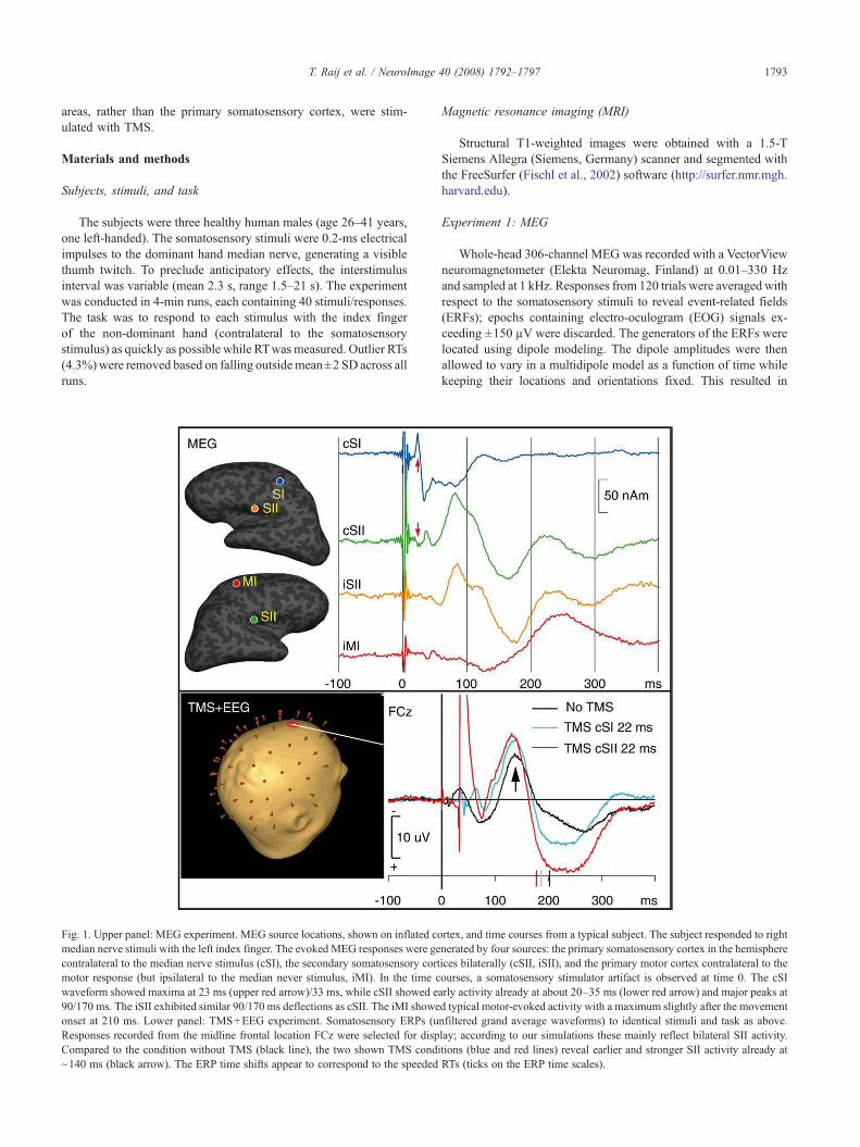
Fig. 1. Upper panel: MEG experiment. MEG source locations, shown on inflated comedian nerve stimuli with the left index finger. The evoked MEG responses were gecontralateral to the median nerve stimulus (cSI), the secondary somatosensory cortmotor response (but ipsilateral to the median never stimulus, iMI). In the time cwaveform showed maxima at 23 ms (upper red arrow)/33 ms, while cSII showed ea90/170 ms. The iSII exhibited similar 90/170 ms deflections as cSII. The iMI showeonset at 210 ms. Lower panel: TMS+EEG experiment. Somatosensory ERPs (uResponses recorded from the midline frontal location FCz were selected for dispCompared to the condition without TMS (black line), the two shown TMS condi~140 ms (black arrow). The ERP time shifts appear to correspond to the speeded
Magnetic resonance imaging (MRI)
Structural T1-weighted images were obtained with a 1.5-TSiemens Allegra (Siemens, Germany) scanner and segmented withthe FreeSurfer (Fischl et al., 2002) software (http://surfer.nmr.mgh.harvard.edu).
Experiment 1: MEG
Whole-head 306-channel MEG was recorded with a VectorViewneuromagnetometer (Elekta Neuromag, Finland) at 0.01–330 Hzand sampled at 1 kHz. Responses from 120 trials were averaged withrespect to the somatosensory stimuli to reveal event-related fields(ERFs); epochs containing electro-oculogram (EOG) signals ex-ceeding ±150 µV were discarded. The generators of the ERFs werelocated using dipole modeling. The dipole amplitudes were thenallowed to vary in a multidipole model as a function of time whilekeeping their locations and orientations fixed. This resulted in
rtex, and time courses from a typical subject. The subject responded to rightnerated by four sources: the primary somatosensory cortex in the hemisphereices bilaterally (cSII, iSII), and the primary motor cortex contralateral to theourses, a somatosensory stimulator artifact is observed at time 0. The cSIrly activity already at about 20–35 ms (lower red arrow) and major peaks atd typical motor-evoked activity with a maximum slightly after the movementnfiltered grand average waveforms) to identical stimuli and task as above.lay; according to our simulations these mainly reflect bilateral SII activity.tions (blue and red lines) reveal earlier and stronger SII activity already atRTs (ticks on the ERP time scales).

1794 T. Raij et al. / NeuroImage 40 (2008) 1792–1797
millisecond-accuracy time courses of the activated brain areas(Hämäläinen and Hari, 2002).
Experiment 2: Navigated TMS and EEG
Single-pulse TMS (Ilmoniemi et al., 1999) was delivered with aMagstim Rapid stimulator (Magstim Company, UK) and figure-of-eight coil (Magstim 9925) navigated with eXimia NBS™ (NexstimLtd., Finland) to target the brain areas identified with MEG. TMSintensity was 120% of the subject-specific motor threshold. A totalof 25–31 runs were recorded per subject (including three withoutTMS pulses) resulting in 1040–1240 trials per subject. To probedifferent stages of processing, the TMS pulse latency across runswas varied 15–210 ms after the somatosensory stimulus, with theTMS latencies tailored for each subject individually based on theirMEG responses. The order of TMS latencies in each brain locationwas randomized. Simultaneous EEG was recorded using a 60-channel TMS-compatible eXimia EEG system (Nexstim), band-pass filtered at 0.1–350 Hz, and sampled at 1.45 kHz at 16-bitdepth (mean reference). The EEG amplifiers were decoupled fromthe electrodes for 9 ms during delivery of the TMS pulse. The EEGresponses were averaged with respect to the somatosensory stimulito reveal event-related potentials (ERPs) separately for each TMSlocation and latency. Epochs contaminated by eye blinks werediscarded using ±100 µV threshold. EEG sensor locations that bestreflected the activity of cSI, cSII, iSII, and iMI were determined byforward modeling the MEG data to simulate corresponding ERPs.Peak ERP latencies were then identified for somatosensory-evokedN20/P45/P75/N140 components separately for each TMS location
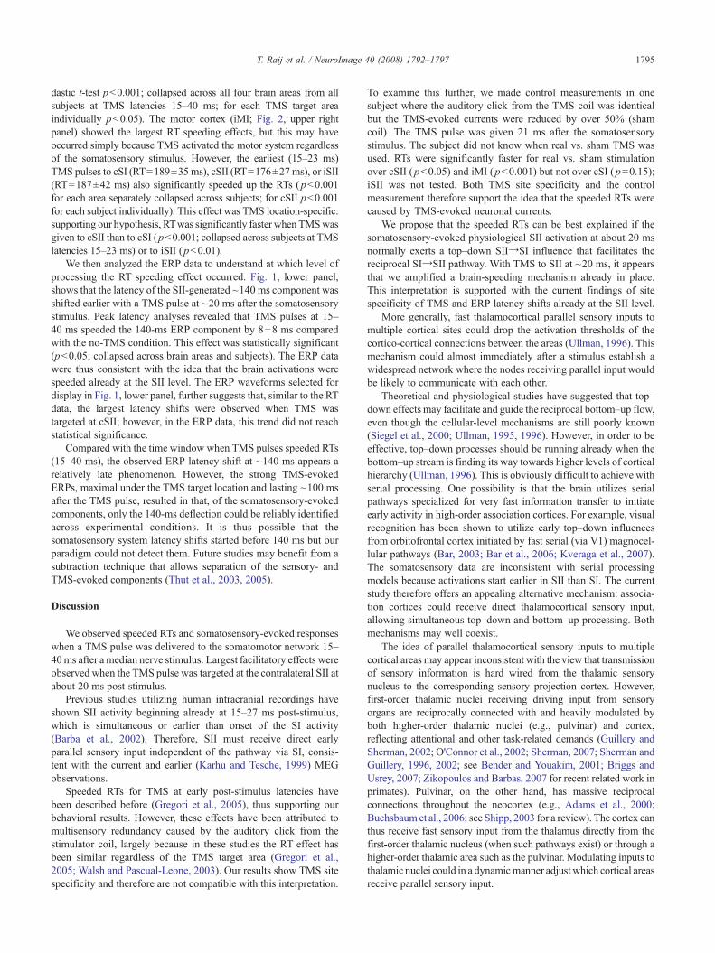
Fig. 2. Reaction time change (0 ms=RTwithout TMS) as a function of TMS pulseeffects separately for the four brain areas that were targeted with TMS. All brain alines), with the strongest linear correlation with TMS of the contralateral SII (significantly faster RTs than without TMS. While this effect was observed in eachrespect to the reaction key hand) and for contralateral SII. Data collapsed across s
and latency (provided that the strong TMS-evoked response did notdistort the somatosensory-evoked component beyond recognition).
Results
First, MEG (Fig. 1, upper panel) revealed expected (Hari andForss, 1999) sources and their activation time courses in thecontralateral primary somatosensory cortex (cSI), SIIs bilaterally(cSII, iSII), and ipsilateral motor cortex (iMI). Simulations suggestedthat the observed cSII activity at about 20–35 ms could not beexplained by volume conduction from unaccounted cSI sources.
Second, in a subsequent session, processing in these four brainareas was modulated with a single TMS pulse at 15–210 ms afterthe somatosensory stimulus while EEG and RT were recorded(Fig. 1, lower panel). Without TMS pulses, RT was 203±29 ms(mean±SD, collapsed across subjects; means of individual subjectshad a range of 197–209 ms).
Fig. 2 shows that the TMS pulse after the somatosensory stimulusclearly effected RTs. In each of the four targeted brain areas, TMSpulse latency was positively correlated with RT. The linear corre-lationwas strongest in cSII (Pearson's correlation r=0.83), somewhatweaker in iSII (r=0.74) and cSI (r=0.74), and weakest in iMI cortex(r=0.54). Lack of correlation between run order and RT suggestedthat fatigue did not play a role (for individual subjects, Pearson's rranged from −0.17 to +0.13).
Prolonged RTs demonstrated that the TMS pulse interfered withthe neuronal processes. As hypothesized, early TMS pulses (15–40 ms after the somatosensory stimulus) were associated with signif-icantly faster RTs than without TMS (Student's 2-tailed heterosce-
latency (0 ms=Median nerve somatosensory stimulus). The panels show thereas showed prolonged RTs with increasing TMS pulse latency (blue trendlower left panel). The earliest TMS pulse latencies were associated withof the four brain areas, it was clearest for ipsilateral MI (contralateral withubjects, mean±SEM error bars. For details, see text.
1795T. Raij et al. / NeuroImage 40 (2008) 1792–1797
dastic t-test pb0.001; collapsed across all four brain areas from allsubjects at TMS latencies 15–40 ms; for each TMS target areaindividually pb0.05). The motor cortex (iMI; Fig. 2, upper rightpanel) showed the largest RT speeding effects, but this may haveoccurred simply because TMS activated the motor system regardlessof the somatosensory stimulus. However, the earliest (15–23 ms)TMS pulses to cSI (RT=189±35ms), cSII (RT=176±27ms), or iSII(RT=187±42 ms) also significantly speeded up the RTs (pb0.001for each area separately collapsed across subjects; for cSII pb0.001for each subject individually). This effect was TMS location-specific:supporting our hypothesis, RTwas significantly faster whenTMSwasgiven to cSII than to cSI (pb0.001; collapsed across subjects at TMSlatencies 15–23 ms) or to iSII (pb0.01).
We then analyzed the ERP data to understand at which level ofprocessing the RT speeding effect occurred. Fig. 1, lower panel,shows that the latency of the SII-generated ~140 ms component wasshifted earlier with a TMS pulse at ~20 ms after the somatosensorystimulus. Peak latency analyses revealed that TMS pulses at 15–40 ms speeded the 140-ms ERP component by 8±8 ms comparedwith the no-TMS condition. This effect was statistically significant(pb0.05; collapsed across brain areas and subjects). The ERP datawere thus consistent with the idea that the brain activations werespeeded already at the SII level. The ERP waveforms selected fordisplay in Fig. 1, lower panel, further suggests that, similar to the RTdata, the largest latency shifts were observed when TMS wastargeted at cSII; however, in the ERP data, this trend did not reachstatistical significance.
Compared with the time window when TMS pulses speeded RTs(15–40 ms), the observed ERP latency shift at ~140 ms appears arelatively late phenomenon. However, the strong TMS-evokedERPs, maximal under the TMS target location and lasting ~100 msafter the TMS pulse, resulted in that, of the somatosensory-evokedcomponents, only the 140-ms deflection could be reliably identifiedacross experimental conditions. It is thus possible that thesomatosensory system latency shifts started before 140 ms but ourparadigm could not detect them. Future studies may benefit from asubtraction technique that allows separation of the sensory- andTMS-evoked components (Thut et al., 2003, 2005).
Discussion
We observed speeded RTs and somatosensory-evoked responseswhen a TMS pulse was delivered to the somatomotor network 15–40ms after a median nerve stimulus. Largest facilitatory effects wereobserved when the TMS pulse was targeted at the contralateral SII atabout 20 ms post-stimulus.
Previous studies utilizing human intracranial recordings haveshown SII activity beginning already at 15–27 ms post-stimulus,which is simultaneous or earlier than onset of the SI activity(Barba et al., 2002). Therefore, SII must receive direct earlyparallel sensory input independent of the pathway via SI, consis-tent with the current and earlier (Karhu and Tesche, 1999) MEGobservations.
Speeded RTs for TMS at early post-stimulus latencies havebeen described before (Gregori et al., 2005), thus supporting ourbehavioral results. However, these effects have been attributed tomultisensory redundancy caused by the auditory click from thestimulator coil, largely because in these studies the RT effect hasbeen similar regardless of the TMS target area (Gregori et al.,2005; Walsh and Pascual-Leone, 2003). Our results show TMS sitespecificity and therefore are not compatible with this interpretation.
To examine this further, we made control measurements in onesubject where the auditory click from the TMS coil was identicalbut the TMS-evoked currents were reduced by over 50% (shamcoil). The TMS pulse was given 21 ms after the somatosensorystimulus. The subject did not know when real vs. sham TMS wasused. RTs were significantly faster for real vs. sham stimulationover cSII (pb0.05) and iMI (pb0.001) but not over cSI (p=0.15);iSII was not tested. Both TMS site specificity and the controlmeasurement therefore support the idea that the speeded RTs werecaused by TMS-evoked neuronal currents.
We propose that the speeded RTs can be best explained if thesomatosensory-evoked physiological SII activation at about 20 msnormally exerts a top–down SIIYSI influence that facilitates thereciprocal SIYSII pathway. With TMS to SII at ~20 ms, it appearsthat we amplified a brain-speeding mechanism already in place.This interpretation is supported with the current findings of sitespecificity of TMS and ERP latency shifts already at the SII level.
More generally, fast thalamocortical parallel sensory inputs tomultiple cortical sites could drop the activation thresholds of thecortico-cortical connections between the areas (Ullman, 1996). Thismechanism could almost immediately after a stimulus establish awidespread network where the nodes receiving parallel input wouldbe likely to communicate with each other.
Theoretical and physiological studies have suggested that top–down effects may facilitate and guide the reciprocal bottom–up flow,even though the cellular-level mechanisms are still poorly known(Siegel et al., 2000; Ullman, 1995, 1996). However, in order to beeffective, top–down processes should be running already when thebottom–up stream is finding its way towards higher levels of corticalhierarchy (Ullman, 1996). This is obviously difficult to achieve withserial processing. One possibility is that the brain utilizes serialpathways specialized for very fast information transfer to initiateearly activity in high-order association cortices. For example, visualrecognition has been shown to utilize early top–down influencesfrom orbitofrontal cortex initiated by fast serial (via V1) magnocel-lular pathways (Bar, 2003; Bar et al., 2006; Kveraga et al., 2007).The somatosensory data are inconsistent with serial processingmodels because activations start earlier in SII than SI. The currentstudy therefore offers an appealing alternative mechanism: associa-tion cortices could receive direct thalamocortical sensory input,allowing simultaneous top–down and bottom–up processing. Bothmechanisms may well coexist.
The idea of parallel thalamocortical sensory inputs to multiplecortical areas may appear inconsistent with the view that transmissionof sensory information is hard wired from the thalamic sensorynucleus to the corresponding sensory projection cortex. However,first-order thalamic nuclei receiving driving input from sensoryorgans are reciprocally connected with and heavily modulated byboth higher-order thalamic nuclei (e.g., pulvinar) and cortex,reflecting attentional and other task-related demands (Guillery andSherman, 2002; O'Connor et al., 2002; Sherman, 2007; Sherman andGuillery, 1996, 2002; see Bender and Youakim, 2001; Briggs andUsrey, 2007; Zikopoulos and Barbas, 2007 for recent related work inprimates). Pulvinar, on the other hand, has massive reciprocalconnections throughout the neocortex (e.g., Adams et al., 2000;Buchsbaum et al., 2006; see Shipp, 2003 for a review). The cortex canthus receive fast sensory input from the thalamus directly from thefirst-order thalamic nucleus (when such pathways exist) or through ahigher-order thalamic area such as the pulvinar. Modulating inputs tothalamic nuclei could in a dynamicmanner adjust which cortical areasreceive parallel sensory input.
1796 T. Raij et al. / NeuroImage 40 (2008) 1792–1797
Given that both association and low-level sensory cortices appearto receive very early parallel crossmodal inputs (Fort et al., 2002,2000; Foxe and Schroeder, 2005; Giard and Peronnet, 1999;Molholm et al., 2002; Murray et al., 2005; Schroeder and Foxe,2002, 2005; Schroeder et al., 2003), some via the pulvinar (Budingeret al., 2006; Hackett et al., 2007), a similar mechanism as suggestedin the current study could also explain why reaction times tomultisensory stimuli are faster than to unisensory stimuli (Raab,1962; Schröger and Widmann, 1998). Early physiological SIIactivations may also serve a protective function due to the roles ofSII in pain processing (Timmermann et al., 2001) and sensorimotorintegration (Forss and Jousmäki, 1998; Huttunen et al., 1996).
It has been suggested that serial processing is more prevalent inhigher primates, and there seems to be an evolutionary shift inmammals where humans have the least amount of parallel sensoryinputs to higher-order areas (Coleman et al., 1999; Kaas andGarraghty, 1991; Zhang et al., 2001, 1996) and therefore increasedserial processing of sensory input. Thus, it appears that in thecourse of evolution humans may have traded some processingspeed for better cognitive control.
From the large number of trials and consistent results acrosssubjects, it follows that the current results are reliable within thestudied population, but due to limited access (the instruments werelocated on different continents), our number of subjects was small.Hence, more studies with larger subject populations are needed toestimate how abundant this mechanism is.
Concluding remarks
The cerebral cortex receives sensory input from the thalamus notonly to primary projection areas but also directly to hierarchicallyhigher-order cortices in a parallel fashion. The current resultssuggest that this facilitates cortico-cortical communication betweenthe areas that receive parallel input, thus making the brain faster.This also allows very early top–down modulation of the bottom–upstream of sensory input. The same mechanism could drop theactivation thresholds between the participating cortical nodes,therefore establishing a distributed neuronal network almostimmediately after a stimulus. Further studies are needed.
Acknowledgments
The authors thank Moshe Bar, Rozalya Bikmullina, DeirdreFoxe, John Foxe, Riitta Hari, Hsiao-Wen Huang, Yu-Hua Huang,Ted Huppert, Iiro Jääskeläinen, G.W. Krauss, Alvaro Pasqual-Leone, Cherif Sahyoun, Dahlia Sharon, and Linda Stenbacka forhelp and comments. This work was supported by US NationalInstitutes of Health Grants R01 NS048279, R01 HD040712, R01NS037462, P41 RR14075, R21 EB007298, National Center forResearch Resources, Sigrid Juselius Foundation, Academy ofFinland, Finnish Cultural Foundation, Instrumentarium ScienceFoundation, Taiwan National Science Council NSC 96-2320-B-002-085, and Taiwan National Health Research Institute 29C97N.
References
Adams, M.M., Hof, P.R., Gattass, R., Webster, M.J., Ungerleider, L.G.,2000. Visual cortical projections and chemoarchitecture of macaquemonkey pulvinar. J. Comp. Neurol. 419, 377–393.
Bar, M., 2003. A cortical mechanism for triggering top–down facilitation invisual object recognition. J. Cogn. Neurosci. 15, 600–609.
Bar,M., Kassam,K.S., Ghuman,A.S., Boshyan, J., Schmid,A.M., Dale, A.M.,Hämäläinen, M.S., Marinkovic, K., Schacter, D.L., Rosen, B.R., Halgren,E., 2006. Top–down facilitation of visual recognition. Proc. Natl. Acad.Sci. U.S.A. 103, 449–454.
Barba, C., Frot, M., Mauguiere, F., 2002. Early secondary somatosensoryarea (SII) SEPs. Data from intracerebral recordings in humans. Clin.Neurophysiol. 113, 1778–1786.
Bender, D.B., Youakim, M., 2001. Effect of attentive fixation in macaquethalamus and cortex. J. Neurophysiol. 85, 219–234.
Briggs, F., Usrey, W.M., 2007. A fast, reciprocal pathway between the lateralgeniculate nucleus and visual cortex in themacaquemonkey. J. Neurosci.27, 5431–5436.
Buchsbaum, M.S., Buchsbaum, B.R., Chokron, S., Tang, C., Wei, T.C.,Byne, W., 2006. Thalamocortical circuits: fMRI assessment of thepulvinar and medial dorsal nucleus in normal volunteers. Neurosci. Lett.404, 282–287.
Budinger, E., Heil, P., Hess, A., Scheich, H., 2006. Multisensory processingvia early cortical stages: connections of the primary auditory corticalfield with other sensory systems. Neuroscience 143, 1065–1083.
Coleman, G.T., Zhang, H.Q., Murray, G.M., Zachariah, M.K., Rowe, M.J.,1999. Organization of somatosensory areas I and II in marsupial cerebralcortex: parallel processing in the possum sensory cortex. J. Neurophy-siol. 81, 2316–2324.
ffytche, D.H., Guy, C.N., Zeki, S., 1995. The parallel visual motion inputsinto areas V1 and V5 of human cerebral cortex. Brain 118, 1375–1394.
Fischl, B., Salat, D.H., Busa, E., Albert, M., Dieterich, M., Haselgrove, C.,van der Kouwe, A., Killiany, R., Kennedy, D., Klaveness, S., Montillo,A., Makris, N., Rosen, B., Dale, A.M., 2002. Whole brain segmentation:automated labeling of neuroanatomical structures in the human brain.Neuron. 33, 341–355.
Forss, N., Jousmäki, V., 1998. Sensorimotor integration in human primaryand secondary somatosensory cortices. Brain Res. 781, 259–267.
Fort, A., Delpuech, C., Pernier, J., Giard, M-H., 2002. Early auditory–visualinteractions in human cortex during nonredundant target identification.Brain Res. Cogn. Brain Res. 14, 20–30.
Foxe, J.J., Schroeder, C.E., 2005. The case for feedforward multisensoryconvergence during early cortical processing. NeuroReport 16,419–423.
Foxe, J.J., Morocz, I.A., Murray, M.M., Higgins, B.A., Javitt, D.C.,Schroeder, C.E., 2000. Multisensory auditory–somatosensory interac-tions in early cortical processing revealed by high-density electricalmapping. Brain Res. Cogn. Brain Res. 10, 77–83.
Giard, M.H., Peronnet, F., 1999. Auditory–visual integration duringmultimodal object recognition in humans: a behavioral and electro-physiological study. J. Cogn. Neurosci. 11, 473–490.
Gregori, B., Curra, A., Dinapoli, L., Bologna, M., Accornero, N., Berardelli,A., 2005. The timing and intensity of transcranial magnetic stimulation,and the scalp site stimulated, as variables influencing motor sequenceperformance in healthy subjects. Exp. Brain Res. 166, 43–55.
Guillery, R.W., Sherman, S.M., 2002. Thalamic relay functions and theirrole in corticocortical communication: generalizations from the visualsystem. Neuron. 33, 163–175.
Hackett, T.A., De La Mothe, L.A., Ulbert, I., Karmos, G., Smiley, J.,Schroeder, C.E., 2007. Multisensory convergence in auditory cortex: II.Thalamocortical connections of the caudal superior temporal plane.J. Comp. Neurol. 502, 924–952.
Hämäläinen, M.S., Hari, R., 2002. Magnetoencephalographic characteriza-tion of dynamic brain activation. Basic principles and methods of datacollection and source analysis. In: Toga, A.W. (Ed.), Brain Mapping:The Methods. Academic Press, New York, pp. 227–253.
Hari, R., Forss, N., 1999. Magnetoencephalography in the study of humansomatosensory cortical processing. Philos. Trans. R. Soc. Lond., B. Biol.Sci. 354, 1145–1154.
Huttunen, J., Wikström, H., Korvenoja, A., Seppäläinen, A.M., Aronen, H.,Ilmoniemi, R.J., 1996. Significance of the second somatosensory cortexin sensorimotor integration: enhancement of sensory responses duringfinger movements. NeuroReport 7, 1009–1012.
1797T. Raij et al. / NeuroImage 40 (2008) 1792–1797
Ilmoniemi, R.J., Ruohonen, J., Karhu, J., 1999. Transcranial magneticstimulation — a new tool for functional imaging of the brain. Crit. Rev.Biomed. Eng. 27, 241–284.
Kaas, J.H., Garraghty, P.E., 1991. Hierarchical, parallel, and serial arrange-ments of sensory cortical areas: connection patterns and functionalaspects. Curr. Opin. Neurobiol. 1, 248–251.
Karhu, J., Tesche, C.D., 1999. Simultaneous early processing of sensoryinput in human primary (SI) and secondary (SII) somatosensory cortices.J. Neurophys. 81, 2017–2025.
Kveraga,K., Boshyan, J., Bar,M., 2007.Magnocellular projections as the triggerof top–down facilitation in recognition. J. Neurosci. 27, 13232–13240.
Molholm, S., Ritter, W., Murray, M.M., Javitt, D.C., Schroeder, C.E., Foxe,J.J., 2002. Multisensory auditory–visual interactions during earlysensory processing in humans: a high-density electrical mappingstudy. Brain Res. Cogn. Brain. Res. 14, 115–128.
Murray, M.M., Molholm, S., Michel, C.M., Heslenfeld, D.J., Ritter, W.,Javitt, D.C., Schroeder, C.E., Foxe, J.J., 2005. Grabbing your ear: rapidauditory–somatosensory multisensory interactions in low-level sensorycortices are not constrained by stimulus alignment. Cereb. Cortex 15,963–974.
O'Connor, D.H., Fukui, M.M., Pinsk, M.A., Kastner, S., 2002. Attentionmodulates responses in the human lateral geniculate nucleus. Nat.Neurosci. 5, 1203–1209.
Pons, T.P., Garraghty, P.E., Friedman, D.P., Mishkin, M., 1987. Physiolo-gical evidence for serial processing in somatosensory cortex. Science237, 417–420.
Raab, D.H., 1962. Statistical facilitation of simple reaction times. Trans. N.Y.Acad. Sci. 24, 574–590.
Schroeder, C.E., Foxe, J.J., 2002. The timing and laminar profile ofconverging inputs to multisensory areas of the macaque neocortex. BrainRes. Cogn. Brain Res. 14, 187–198.
Schroeder, C.E., Foxe, J.J., 2005. Multisensory contributions to low-level,‘unisensory’ processing. Curr. Opin. Neurobiol. 15, 454–458.
Schroeder, C.E., Lindsley, R.W., Specht, C., Marcovici, A., Smiley, J.F.,Javitt, D.C., 2001. Somatosensory input to auditory association cortex inthe macaque monkey. J. Neurophysiol. 85, 1322–1327.
Schroeder, C.E., Smiley, J., Fu, K.G., McGinnis, T., O, O'Connell, M.N.,Hackett, T.A., 2003. Anatomical mechanisms and functional implica-tions of multisensory convergence in early cortical processing. Int. J.Psychophysiol. 50, 5–17.
Schröger, E., Widmann, A., 1998. Speeded responses to audiovisual signalchanges result from bimodal integration. Psychophysiology 35, 755–759.
Sherman, S.M., 2007. The thalamus is more than just a relay. Curr. Opin.Neurobiol. 17, 417–422.
Sherman, S.M., Guillery, R.W., 1996. Functional organization of thalamo-cortical relays. J. Neurophysiol. 76, 1367–1395.
Sherman, S.M., Guillery, R.W., 2002. The role of the thalamus in the flow ofinformation to the cortex. Philos. Trans. R. Soc. Lond., B. Biol. Sci. 357,1695–1708.
Shipp, S., 2003. The functional logic of cortico-pulvinar connections.Philos. Trans. R. Soc. Lond., B. Biol. Sci. 358, 1605–1624.
Siegel,M.,Körding, K.P., König, P., 2000. Integrating top–down andbottom–up sensory processing by somato-dendritic interactions. J. Comput.Neurosci. 8, 161–173.
Thut, G., Northoff, G., Ives, J.R., Kamitani, Y., Pfennig, A., Kampmann, F.,Schomer, D.L., Pascual-Leone, A., 2003. Effects of single-pulsetranscranial magnetic stimulation (TMS) on functional brain activity: acombined event-related TMS and evoked potential study. Clin.Neurophysiol. 114, 2071–2080.
Thut, G., Ives, J.R., Kampmann, F., Pastor, M.A., Pascual-Leone, A., 2005.A new device and protocol for combining TMS and online recordings ofEEG and evoked potentials. J. Neurosci. Methods 141, 207–217.
Timmermann, L., Ploner, M., Haucke, K., Schmitz, F., Baltissen, R.,Schnitzler, A., 2001. Differential coding of pain intensity in the humanprimary and secondary somatosensory cortex. J. Neurophysiol. 86,1499–1503.
Ullman, S., 1995. Sequence seeking and counter streams: a computationalmodel for bidirectional information flow in the visual cortex. Cereb.Cortex 5, 1–11.
Ullman, S., 1996. Sequence seeking and counter streams: a model for visualcortex. In: Ullman, S. (Ed.), High-Level Vision: Object Recognition andVisual Cognition. MIT Press, Cambridge MA.
Walsh, V., Pascual-Leone, A., 2003. Transcranial Magnetic Stimulation —A Neurochronometrics of Mind. MIT Press, Cambridge, MA.
Zhang, H.Q., Murray, G.M., Turman, A.B., Mackie, P.D., Coleman, G.T.,Rowe, M.J., 1996. Parallel processing in cerebral cortex of the marmosetmonkey: effect of reversible SI inactivation on tactile responses in SII.J. Neurophysiol. 76, 3633–3655.
Zhang, H.Q., Murray, G.M., Coleman, G.T., Turman, A.B., Zhang, S.P.,Rowe, M.J., 2001. Functional characteristics of the parallel SI- and SII-projecting neurons of the thalamic ventral posterior nucleus in themarmoset. J. Neurophysiol. 85, 1805–1822.
Zikopoulos, B., Barbas, H., 2007. Parallel driving and modulatory pathwayslink the prefrontal cortex and thalamus. PLoS ONE 2, e848.