parasites: invertebrates. classification 35 phyla of invertebrates half are entirely marine...
TRANSCRIPT
Parasites:Parasites:InvertebratesInvertebrates

Classification

35 phyla of invertebrates
Half are entirely marine
Invertebrates
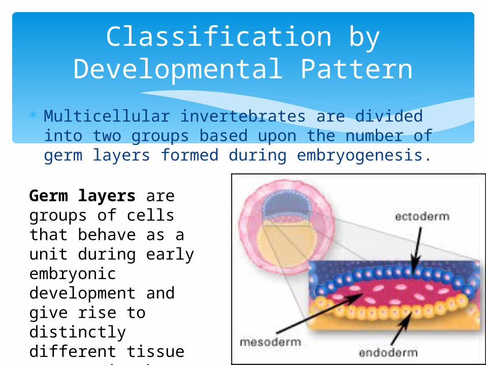
Multicellular invertebrates are divided into two groups based upon the number of germ layers formed during embryogenesis.
Classification by Developmental Pattern
Germ layers are groups of cells that behave as a unit during early embryonic development and give rise to distinctly different tissue systems in the adult.

Diploblastic animals have two germ layers. Ectoderm (outermost layer) Endoderm (innermost layer)
Triploblastic animals have three germ layers
Ectoderm Mesoderm Endoderm
Germ Layers
Fuchsia flatworm
Purple sponge

Triploblastic animals are further classified by their internal body cavity
Triploblastic Animals
•Acoelomate: No internal body cavity•Pseudocoelomate: fluid-filled cavity between endoderm and mesoderm•Coelomate: internal, fluid-filled cavity between endoderm and mesoderm, lined with mesoderm
Coelomate
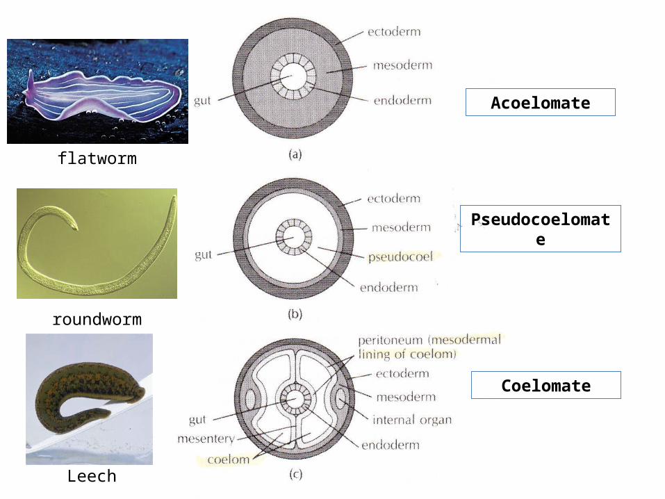
Acoelomate
Pseudocoelomate
Coelomate
roundworm
flatworm
Leech
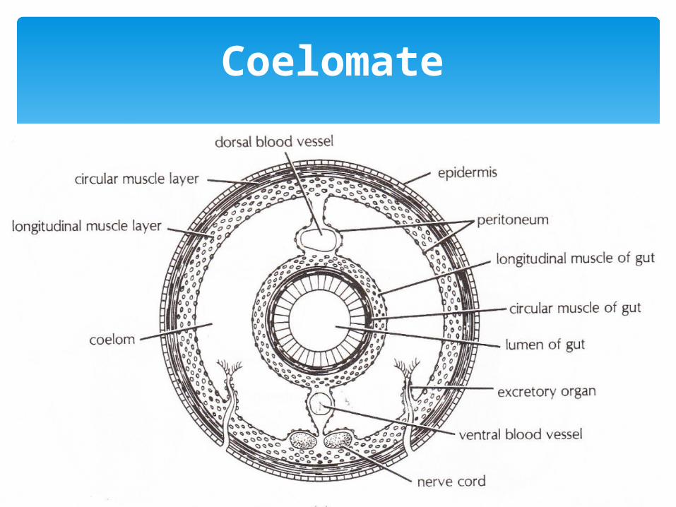
Coelomate

Kingdom AnimaliaSub-kingdom Invertebrata
• I. Phylum Platyhelminthes (flatworms)– Tapeworms, flukes, and non-parasitic flatworms
• II. Phylum Nematoda – Roundworms, hookworms, threadworms
• III. Phylum Annelida – segmented worms
• IV. Phylum Arthropoda – Ticks, mites, lice

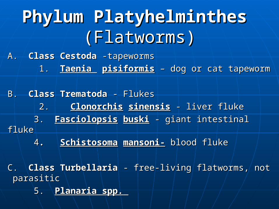
Phylum PlatyhelminthesPhylum Platyhelminthes (Flatworms)(Flatworms)
A. A. Class CestodaClass Cestoda -tapeworms -tapeworms
1. 1. Taenia Taenia pisiformispisiformis – dog or cat tapeworm – dog or cat tapeworm
B. B. Class Trematoda Class Trematoda - Flukes- Flukes
2. 2. ClonorchisClonorchis sinensissinensis - liver fluke - liver fluke
3. 3. FasciolopsisFasciolopsis buskibuski - giant intestinal fluke - giant intestinal fluke
44. . SchistosomaSchistosoma mansoni-mansoni- blood fluke blood fluke
C. C. Class TurbellariaClass Turbellaria - free-living flatworms, not parasitic - free-living flatworms, not parasitic
5. 5. Planaria spp.Planaria spp.
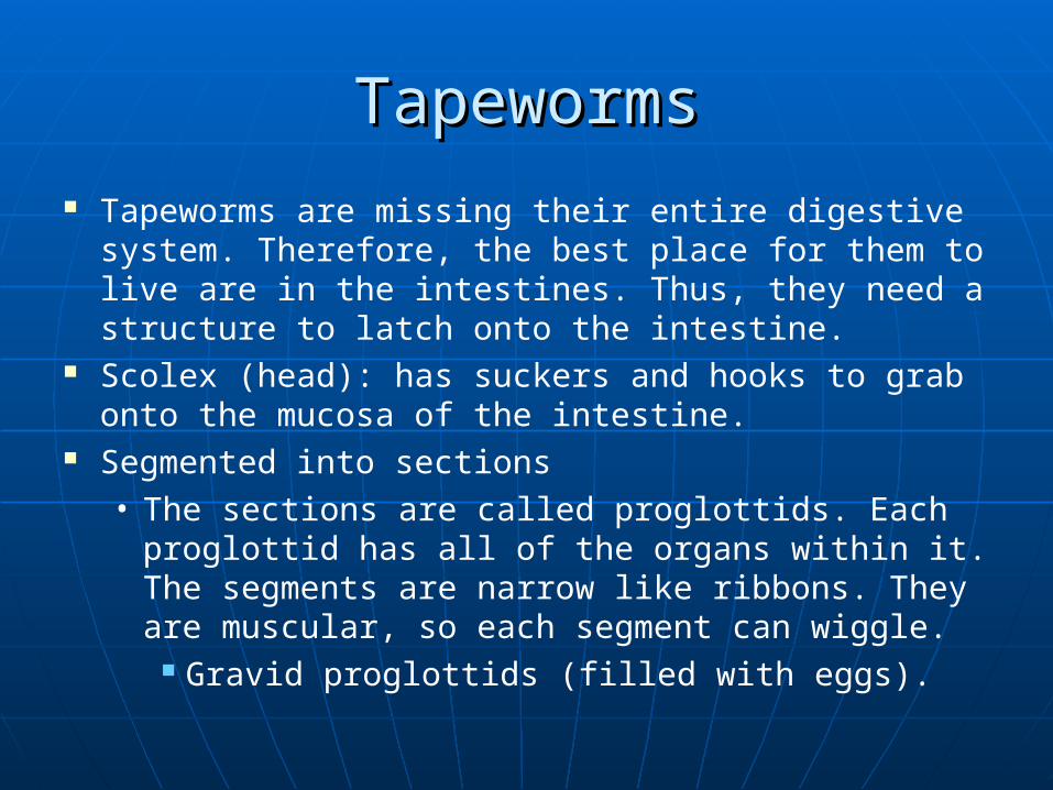
TapewormsTapeworms
Tapeworms are missing their entire digestive system. Therefore, the best place for them to live are in the intestines. Thus, they need a structure to latch onto the intestine.
Scolex (head): has suckers and hooks to grab onto the mucosa of the intestine.
Segmented into sections• The sections are called proglottids. Each
proglottid has all of the organs within it. The segments are narrow like ribbons. They are muscular, so each segment can wiggle.
Gravid proglottids (filled with eggs).

Class CestodaClass Cestoda -tapeworms -tapeworms
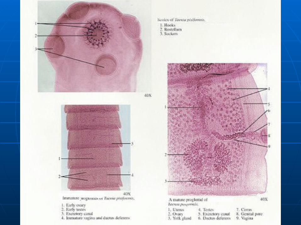
Taenia Taenia pisiformis pisiformis -dog or cat tapeworm-dog or cat tapeworm
Head (scolex)Head (scolex)
SuckersSuckers
HooksHooks
Segments (proglottids)Segments (proglottids)
TestesTestes
OvariesOvaries

Tapeworm Scolex (head) with suckers and hooks

Tapeworm Segments Tapeworm Segments (proglottids)(proglottids)
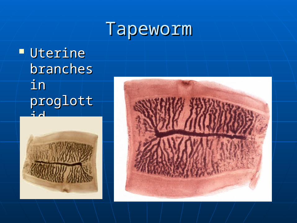
TapewormTapeworm Uterine Uterine
branches branches in in proglottidproglottid

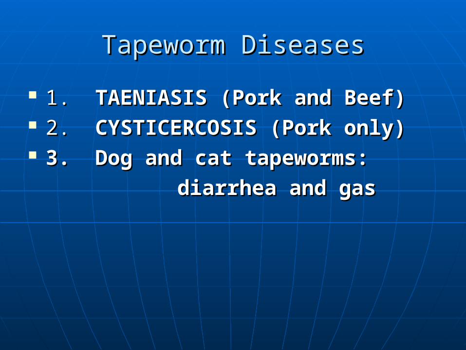
Tapeworm DiseasesTapeworm Diseases
1. 1. TAENIASIS (Pork and Beef)TAENIASIS (Pork and Beef) 2. 2. CYSTICERCOSIS (Pork only)CYSTICERCOSIS (Pork only) 3. Dog and cat tapeworms: 3. Dog and cat tapeworms:
diarrhea and gasdiarrhea and gas

Tapeworm DiseasesTapeworm Diseases TAENIASIS This form of disease can be from either pork or beef intestinal
infections. It is usually asymptomatic, mild GI pain, but you may see the proglottids wiggling in the stool.
CYSTICERCOSIS This is caused by pork tapeworms only. It is much more serious,
even life-threatening. The Tania egg is ingested by the fecal-oral route. Larvae embed in any tissue (esp. muscle, brain, eye). Once ingested, the eggs hatch and invade.
Dog and cat tapeworms: mild symptoms of diarrhea and Dog and cat tapeworms: mild symptoms of diarrhea and gasgas

Tapeworms inside intestinesTapeworms inside intestines


Fluke DiseasesFluke Diseases
Flukes differ from tapeworms in that they are non-segmented.
They live in tiny veins in humans called venuoles. The large number of eggs is what causes the
damage in the human. They cause granulomas (inflammatory lesions). These granulomas can occur in the brain, spinal
cord, liver, and many other organs.

Class Trematoda Class Trematoda - Flukes- Flukes
ClonorchisClonorchis sinensissinensis - liver fluke - liver fluke
Oral suckerOral sucker
PharynxPharynx
Intestines (cecum)Intestines (cecum)
UterusUterus
TestesTestes
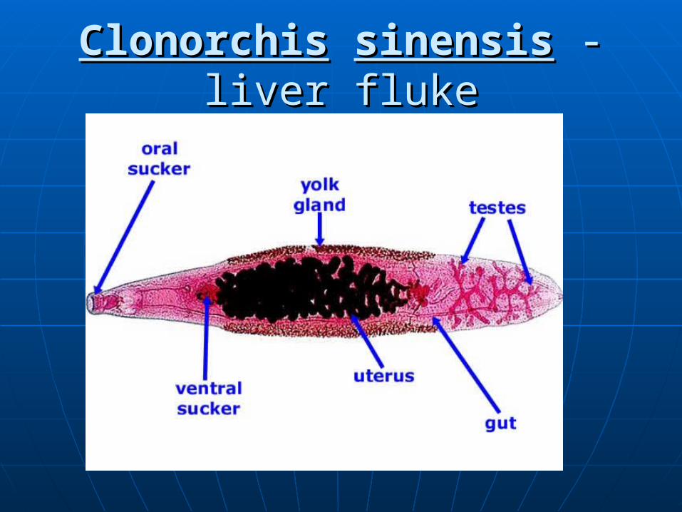
ClonorchisClonorchis sinensissinensis -liver fluke -liver fluke

Class Trematoda Class Trematoda - Flukes- Flukes
FasciolopsisFasciolopsis buskibuski - giant intestinal - giant intestinal flukefluke
Mouth (for feeding)Mouth (for feeding)
Ventral sucker (for attachment)Ventral sucker (for attachment)
Intestines (cecum)Intestines (cecum)
TestesTestes

FasciolopsisFasciolopsis buskibuski giant intestinal flukegiant intestinal fluke

Class Trematoda Class Trematoda - Flukes- Flukes
SchistosomaSchistosoma mansonimansoni
Female blood fluke:Female blood fluke:
Ventral sucker (for attachment)Ventral sucker (for attachment)
OvaryOvary
Mouth (for feeding)Mouth (for feeding)

SchistosomiasisSchistosomiasis
The blood fluke is called Schistostoma. “Schisto” means “split” and “soma” means opening.
The male’s body is split into a gynecohoric canal; this is where the females live. The female is smaller and lives within the male her whole life.
She lays thousands of eggs a day.

SchistosomaSchistosoma
Male
Female
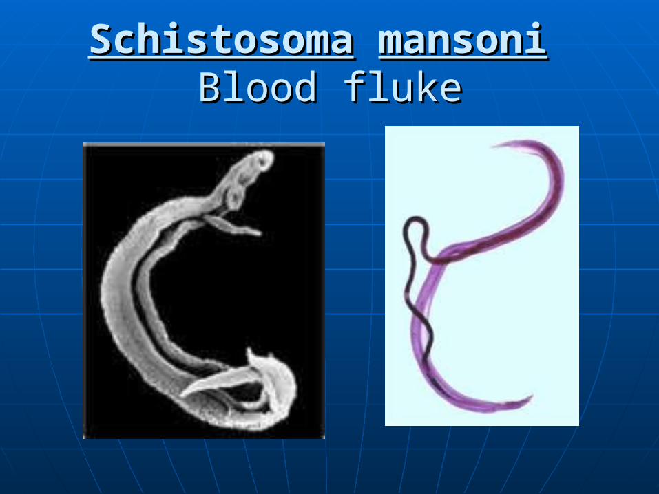
SchistosomaSchistosoma mansonimansoni Blood flukeBlood fluke

SchistosomaSchistosoma mansonimansoni Blood fluke: Female and MaleBlood fluke: Female and Male
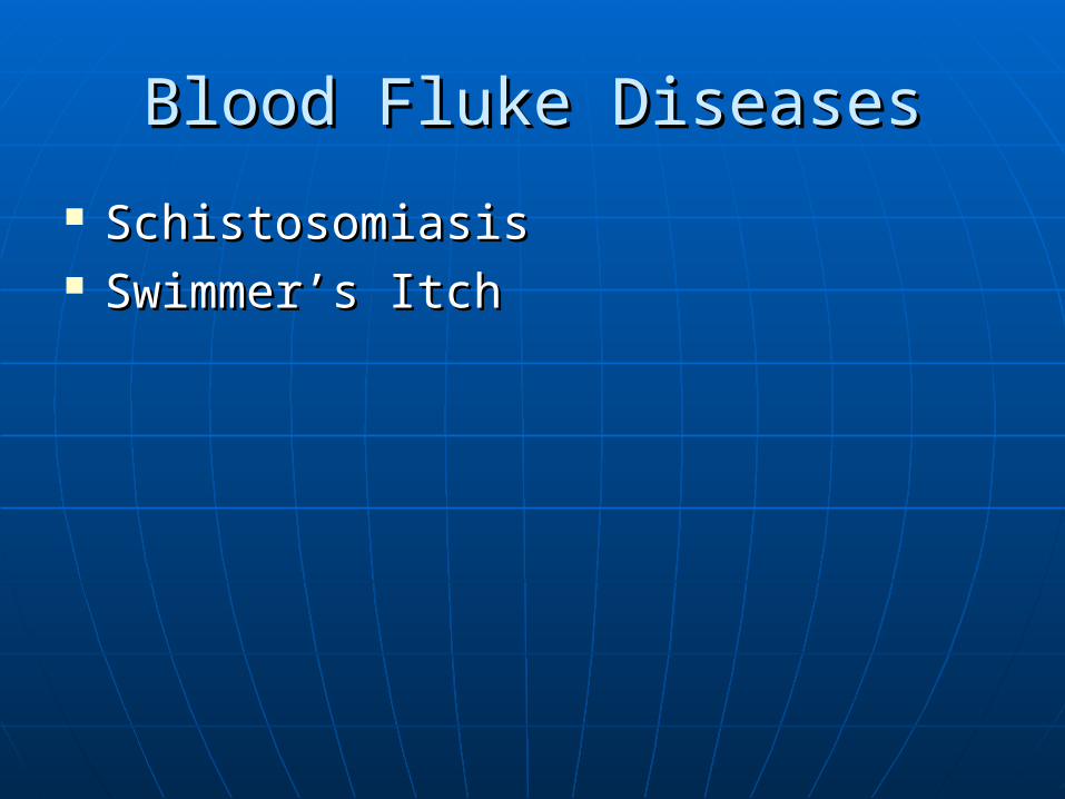
Blood Fluke DiseasesBlood Fluke Diseases
SchistosomiasisSchistosomiasis Swimmer’s ItchSwimmer’s Itch

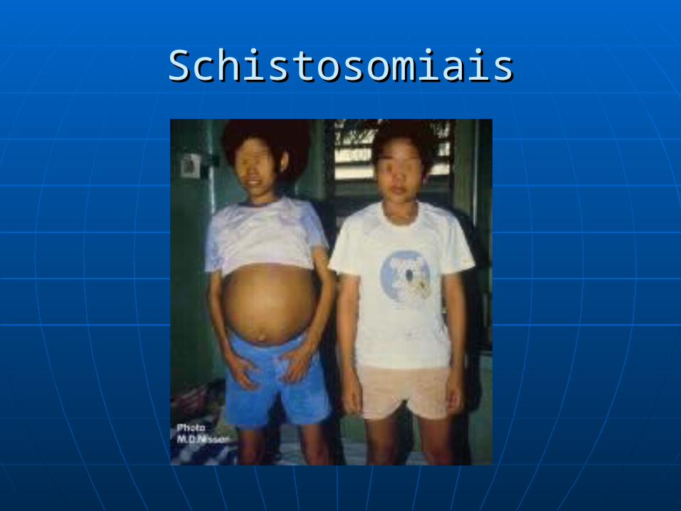
Schistosomiasis Schistosomiasis kills 1-2 million people per year; it’s almost
as bad as malaria. It is a tropical disease. It is also known as “Male menstruation” because there is a bladder fluke which causes hematuria (blood in the urine). When Napoleon invaded Egypt, his men got this disease and called it the Curse of the Pharaoh.
Many people are asymptomatic, but the acute form
(Katayama’s Fever) can occur weeks later, manifesting with fever, coughing, nausea, vomiting, diarrhea, hepatosplenomegaly, and eosinophilia (excess eosinophils, a type of white blood cell that responds to parasitic infections).

SchistosomiasisSchistosomiasis
The schistosomulae migrate to the The schistosomulae migrate to the veins:veins:• The females are smaller and live inside The females are smaller and live inside
the male. the male. • They deposit eggs in the small venules.They deposit eggs in the small venules.• The eggs are moved progressively The eggs are moved progressively
toward the lumen of the intestine and toward the lumen of the intestine and are eliminated with feces or urine.are eliminated with feces or urine.

SchistosomiasisSchistosomiasis
Human contact with water is Human contact with water is necessary for infection by necessary for infection by schistosomes. Skin penetration is schistosomes. Skin penetration is required. required.
Symptoms include: Katayama fever, Symptoms include: Katayama fever, granulomas (occasionally in brain or granulomas (occasionally in brain or spinal cord).spinal cord).
SchistosomiaisSchistosomiais
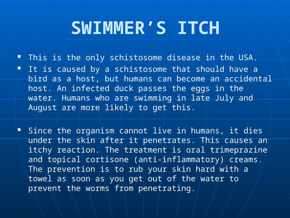
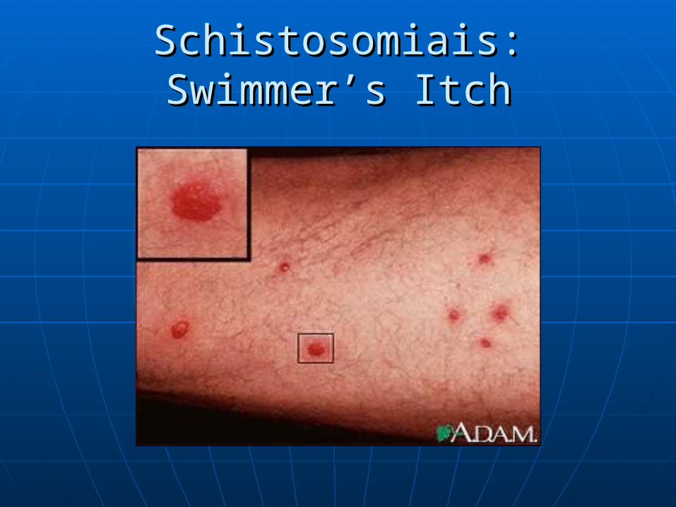
SWIMMER’S ITCH This is the only schistosome disease in the USA. It is caused by a schistosome that should have a bird as a
host, but humans can become an accidental host. An infected duck passes the eggs in the water. Humans who are swimming in late July and August are more likely to get this.
Since the organism cannot live in humans, it dies under the skin after it penetrates. This causes an itchy reaction. The treatment is oral trimeprazine and topical cortisone (anti-inflammatory) creams. The prevention is to rub your skin hard with a towel as soon as you get out of the water to prevent the worms from penetrating.

Swimmer’s ItchSwimmer’s Itch
Schistosomiais: Swimmer’s ItchSchistosomiais: Swimmer’s Itch

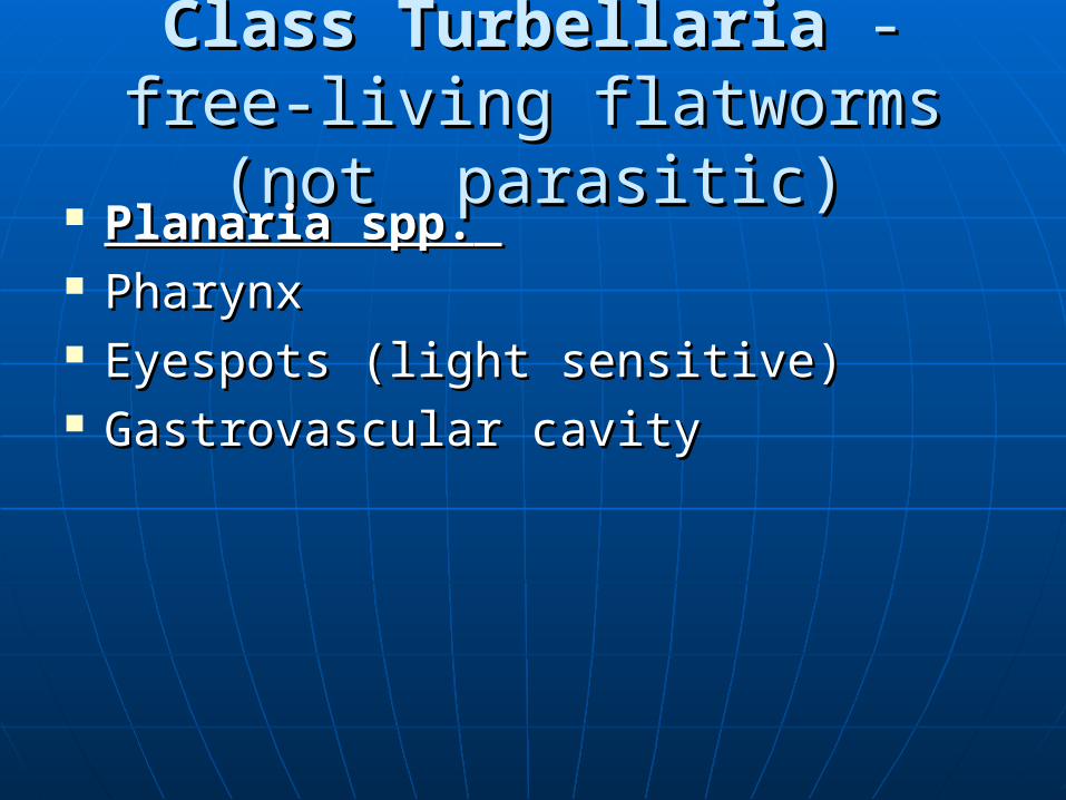
Class TurbellariaClass Turbellaria - free-living - free-living flatworms (not parasitic)flatworms (not parasitic)
Planaria spp.Planaria spp. PharynxPharynx Eyespots (light sensitive)Eyespots (light sensitive) Gastrovascular cavityGastrovascular cavity
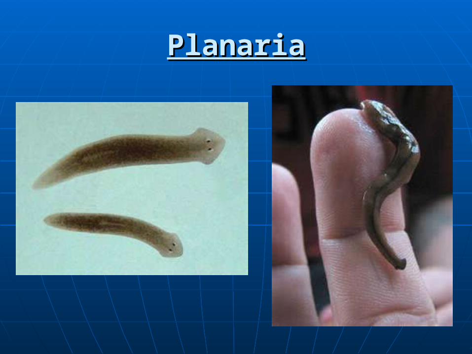
PlanariaPlanaria

PlanariaPlanaria

Kingdom AnimaliaSub-kingdom Invertebrata
• I. Phylum Platyhelminthes (flatworms)– Tapeworms, flukes, and non-parasitic flatworms
• II. Phylum Nematoda – Roundworms, hookworms, threadworms
• III. Phylum Annelida – segmented worms
• IV. Phylum Arthropoda – Ticks, mites, lice

Enterobius vermicularis – pinwormFemale
MouthPharynx

Female
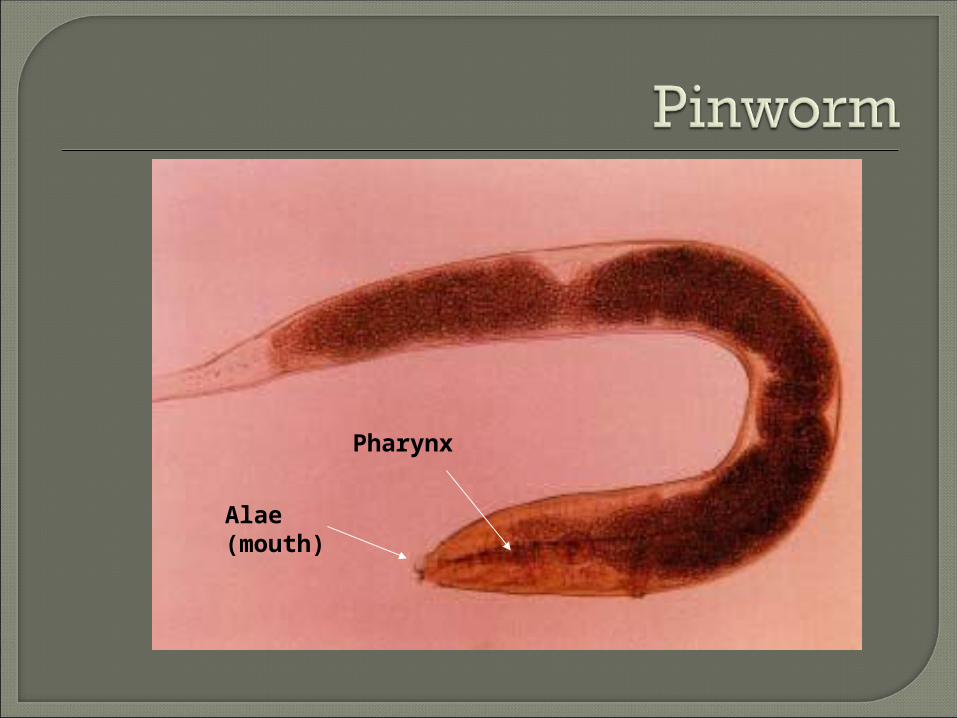
Alae (mouth)
Pharynx
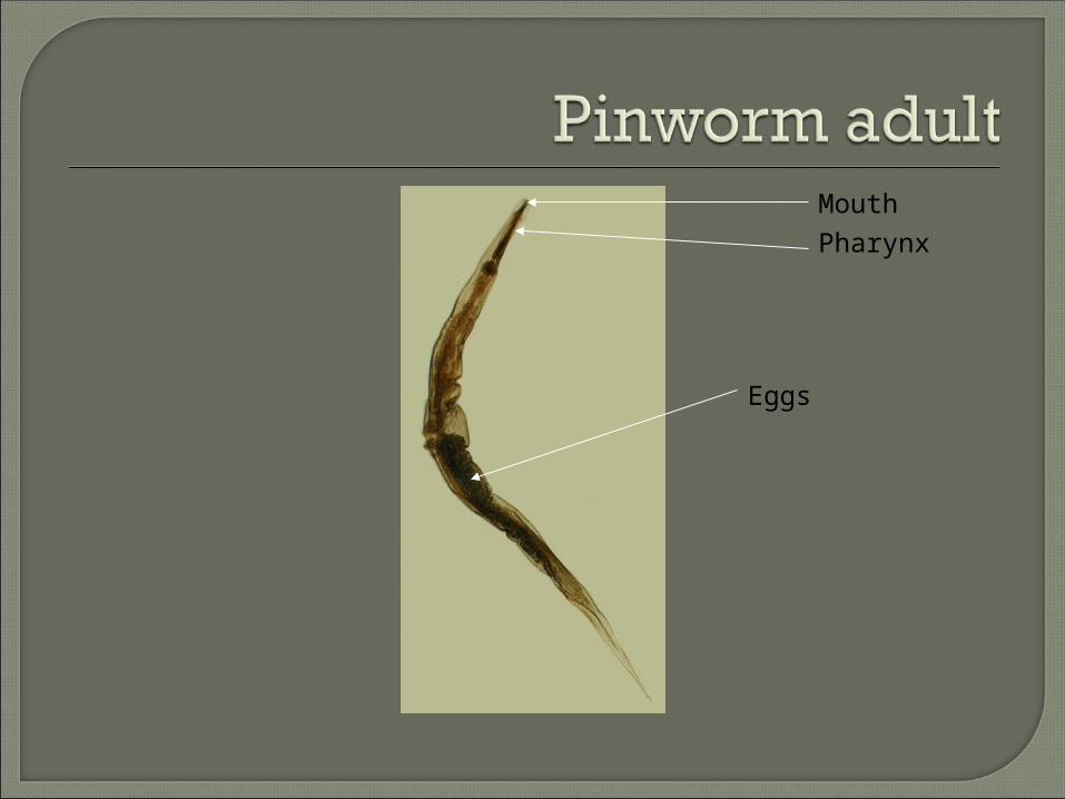
Pharynx
Eggs
Mouth

This is the most common roundworm in the USA, but the best one to have because it produces the least symptoms. It has a simple life cycle; humans are the only host.
The males are smaller than the female. Male tails have specialized ornamentation that are shaped into a curl which allows the male to clasp the female during copulation. The male tail also has spicules to allow attachment to the female.
The female then migrates to the rectum, crawls out onto the skin,
and deposits eggs on the perianal folds. This causes itching, called anal pruritis. Self-infection occurs by transferring infective eggs to the mouth with hands that have scratched the perianal area. This occurs more often in children since they don’t wash their hands as well. The eggs are sticky. Person-to-person transmission can also occur through handling of contaminated clothes or bed linens or contact with contaminated curtains, carpeting, etc.


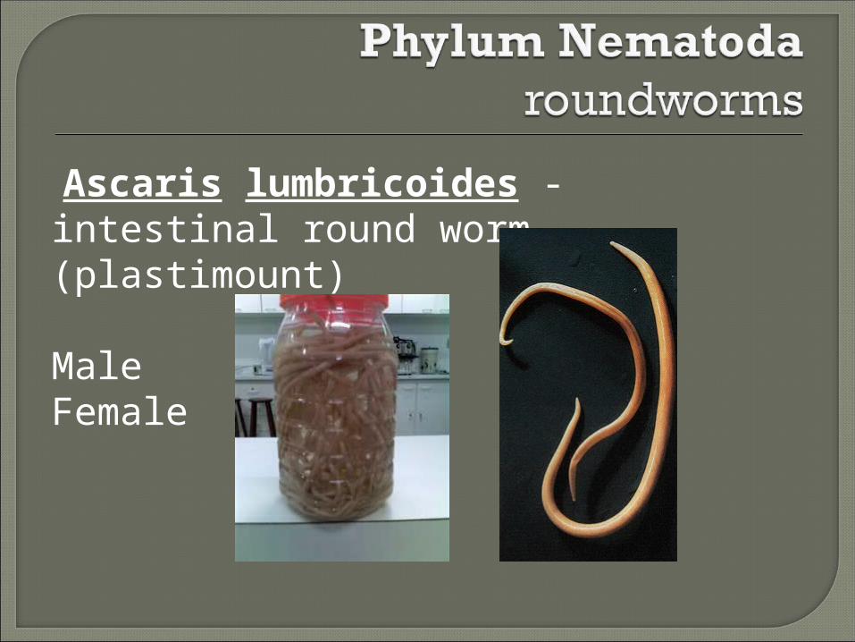
Ascaris lumbricoides - intestinal round worm- (plastimount)
MaleFemale

A female may produce approximately 200,000 eggs per day, which are passed with the feces and are ingested from soil contamination. The larvae are very migratory. They can end up in the lungs and may crawl out the nose. They develop into adults in the intestine. A heavy worm load can lead to intestinal blockage, with a bolus of worms inside; may need surgical removal.
This is the largest worm pathogen, so there are no slides. We do have the entire specimen in a plastic container.
This worm should be considered for the Hall of Fame for two reasons:
• It is the biggest worm.• It causes the most infections of all the Helminthes
world-wide.

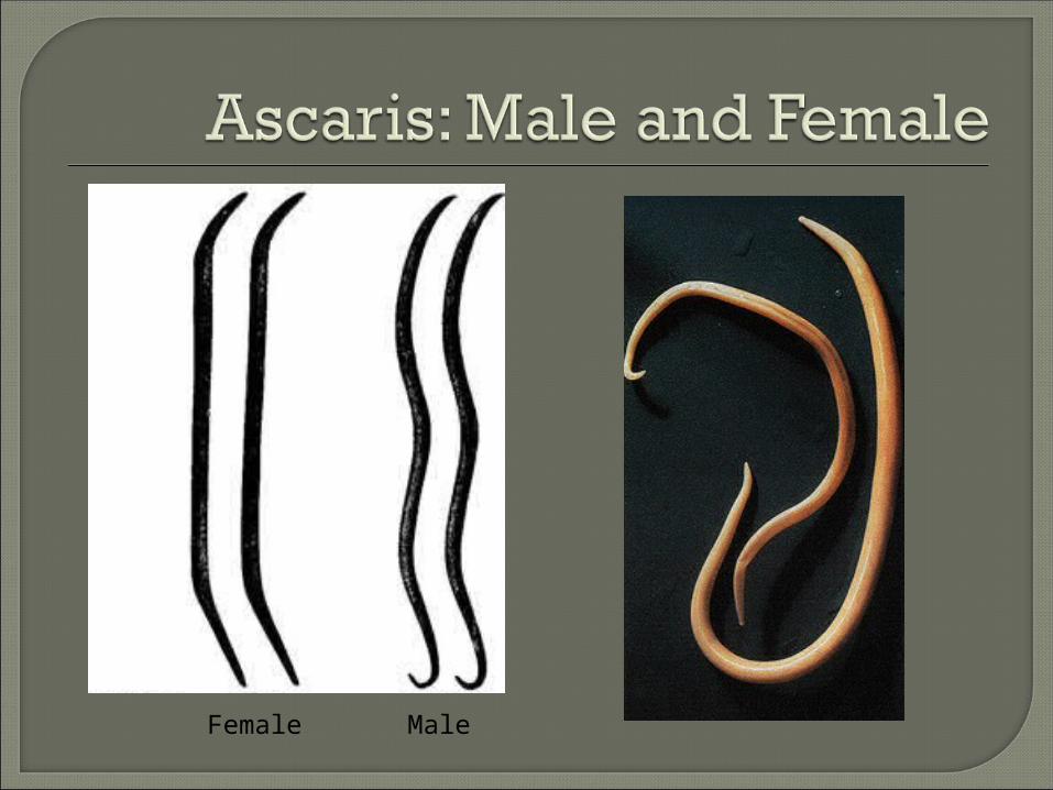
Female Male

Toxocara canis and Toxocara cati are roundworms that infect dogs and cats, but humans can become an accidental host. Preschool children are most often affected, since it is spread by ingestion of contaminated dirt.
They cause a disease in humans called Visceral larva migrans (VLM).

The larvae migrate from the intestines to other tissues, especially the liver.
Most people become only mildly ill. Others can have severe symptoms,
such as brain damage, respiratory failure, cardiac arrhythmias, or eye lesions that resemble cancer or retinal detachment.


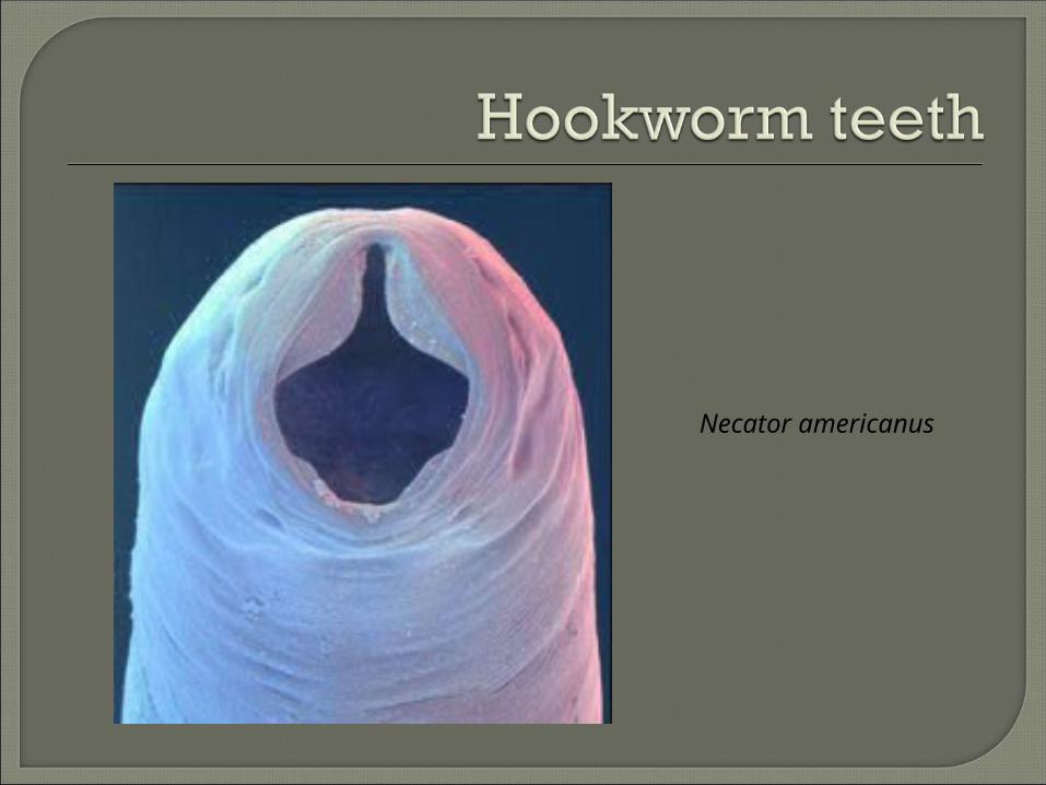
Necator americanus- American hookworm
TeethMuscular pharynx
MalePosterior bursaRays

Necator americanus
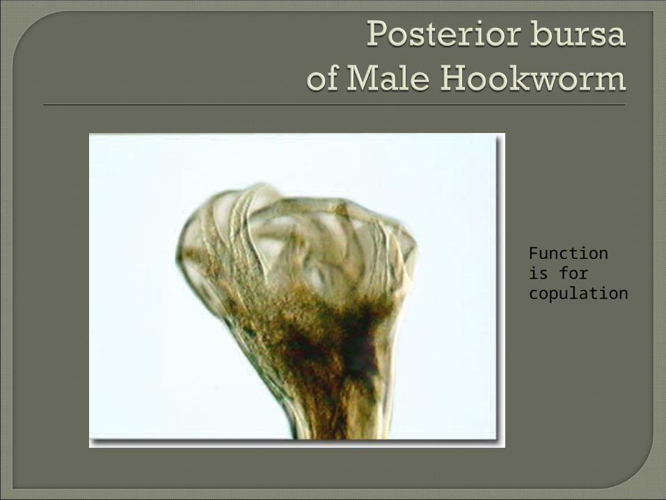
Function is for copulation
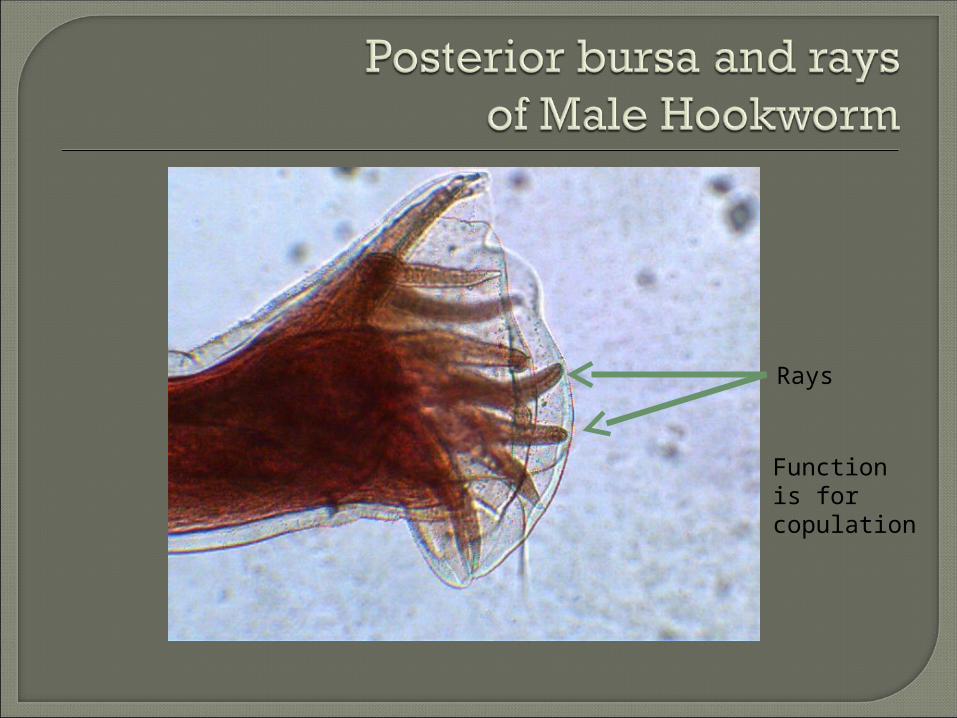
Rays
Function is for copulation

The larvae travel to the heart, then to the lungs, where they penetrate the alveoli, make their way up the trachea, up to the epiglottis, and then they are swallowed. They live in the small intestine lumen. They attach to the mucosa by either teeth or cutting flakes, which causes bleeding and persistent anemia. The patient may also have cardiac symptoms.
In the southern US, there is a condition known as “pica”, which is a desire to eat soil. This is caused from an iron deficiency in the diet, sometimes because of a hookworm infection.
The male has a feathered copulatory bursae,on the tail and the female’s tail is just pointy.

Humans are accidental hosts.The lavae create serpentine tunnels.
Lesions are very red (erythemic) and itchy (pruitic).
If they migrate to the lungs, it is called Loeffler’s syndrome.

This is when a dog or cat hookworm gets into a human by accident. Since we are not the right kind of host, the worm dies under the skin and causes an allergic reaction.
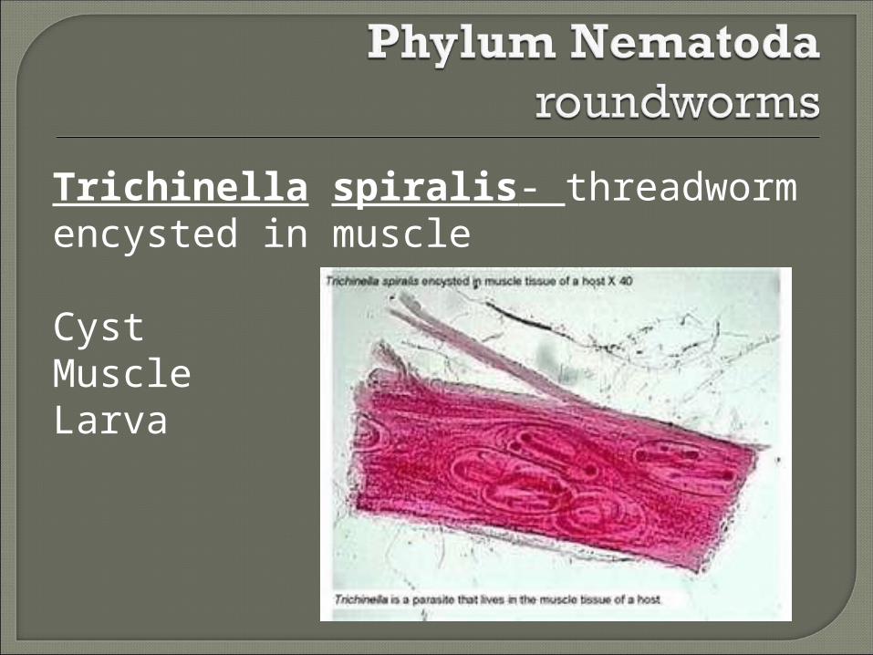
Trichinella spiralis- threadworm encysted in muscle
CystMuscleLarva
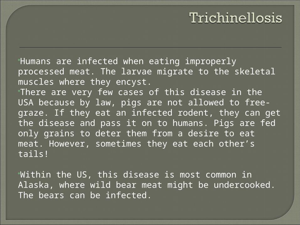
Humans are infected when eating improperly processed meat. The larvae migrate to the skeletal muscles where they encyst.There are very few cases of this disease in the USA because by law, pigs are not allowed to free-graze. If they eat an infected rodent, they can get the disease and pass it on to humans. Pigs are fed only grains to deter them from a desire to eat meat. However, sometimes they eat each other’s tails!
Within the US, this disease is most common in Alaska, where wild bear meat might be undercooked. The bears can be infected.

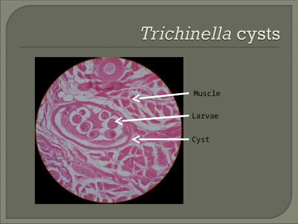
Larvae
Cyst
Muscle

Kingdom AnimaliaSub-kingdom Invertebrata
• I. Phylum Platyhelminthes (flatworms)– Tapeworms, flukes, and non-parasitic flatworms
• II. Phylum Nematoda – Roundworms, hookworms, threadworms
• III. Phylum Annelida – segmented worms
• IV. Phylum Arthropoda – Ticks, mites, lice
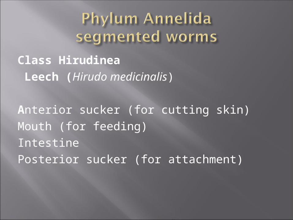
Class Hirudinea Leech (Hirudo medicinalis)
Anterior sucker (for cutting skin)Mouth (for feeding)IntestinePosterior sucker (for attachment)


Leeches have been used as a medicinal agent for centuries.
They were first used in ancient Egypt to bleed people of “bad blood” that they thought was causing illness.
In medieval times, they were used in conjunction with blood letting by incisions.
They are used today for patients with finger amputations, to keep the veins open until the finger can be reattached because their saliva has anesthesia and prevents clotting.

The leech has two suckers: The posterior sucker attaches to the host The anterior sucker makes a slit in the
host’s skin so it can suck the blood. The leech can take in up to 5 times its
weight in blood. Leeches are only used for one person for
medicinal purposes, then they are destroyed to prevent disease transmission.

Kingdom AnimaliaSub-kingdom Invertebrata
• I. Phylum Platyhelminthes (flatworms)– Tapeworms, flukes, and non-parasitic flatworms
• II. Phylum Nematoda – Roundworms, hookworms, threadworms
• III. Phylum Annelida – segmented worms
• IV. Phylum Arthropoda – Ticks, mites, lice

Phylum Arthropoda(Jointed feet)
• Class Arachnia (ticks and mites)
• Class Insecta (fleas and lice)
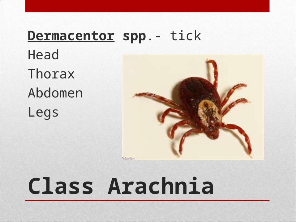
Class Arachnia
Dermacentor spp.- tick
Head
Thorax
Abdomen
Legs

• Hard Ticks• Have a hard body
• Mouth parts are visible from the top surface
• Feed for days
• Soft Ticks• Have a soft, leathery body
• Mouth parts are on the bottom surface
• Feed from minutes to hours

Diseases Transmitted by Hard Ticks
• Diseases Transmitted by Hard Ticks• Dermacenter
• Ehrlichiosis (Ehrlichia spp)
• Rocky Mountain Spotted Fever (Rickettsia rickettsii)
• Ixodes• Lyme Disease (Borrelia burgdorferi)
• Babbesiosis (Babesia microti; USA protozoa)
• Amblyomma• Ehrlickiosis

• Ehrlichiosis • Fever, headache, fatigue, and muscle aches
• Rocky Mountain Spotted Fever• Fever, headache, abdominal pain, vomiting, and muscle pain. A
rash may also develop, but is often absent in the first few days, and in some patients, never develops. Rocky Mountain spotted fever can be a severe or even fatal illness if not treated in the first few days of symptoms.
• Lyme Disease• Fever, headache, fatigue, and a characteristic skin rash
called erythema migrans. If left untreated, infection can spread to joints, the heart, and the nervous system.
• Babesiosis • Fever, chills, sweating, myalgias (muscle aches), fatigue,
hepatosplenomegaly (enlargement of the liver and spleen) and hemolytic anemia (anemia due to break-up of red cells). Symptoms typically occur after an incubation period of 1 to 4 weeks and can last several weeks. The disease is more severe in patients who are immunosuppressed, splenectomized (lack their spleen), or elderly. It can cause death.

Diseases Transmitted by Soft Ticks
• Diseases Transmitted by Soft Ticks
• Ornithidoros
• Endemic relapsing fever (Borrelia hermsii)• High fever, rigors, severe headache, muscle pains,
weakness, anorexia, weight loss, and cough. Systemic complications can include nausea, vomiting, upper abdominal pain due to liver and spleen involvement, and a dry cough. Other manifestations include splenomegaly, hepatomegaly, jaundice, rash, respiratory symptoms, and central nervous system involvement.
Tick Diseases
• Tick paralysis
• More common in children than adults.
• It is an ascending flaccid paralysis, accompanied by fever and toxemia (toxins in their saliva) that can lead to respiratory compromise and death.

Class Arachnia
Sarcoptes scabiei - itch mite
Mouth
Legs
Bristles on rear legs
Bristles on abdomen

Scabies
• Scabies is an itchy, highly contagious skin disease.
• Mites are small eight-legged parasites (in contrast to insects, which have six legs).
• They are tiny, just 1/3 millimeter long. The mite is harmless, but causes an allergic reaction when it burrows into the skin to produce intense itching (pruitis), which tends to be worse at night.
• They prefer thin skin like the areas between the fingers, and the bends of the elbows, wrists, and knees. They can move one inch a minute.
• They are sometimes transmitted by contact with contaminated gym equipment.

Itch Mite

Class Arachnia
Dermatophagoides spp.- dust mite
Thorax
Legs
Abdomen
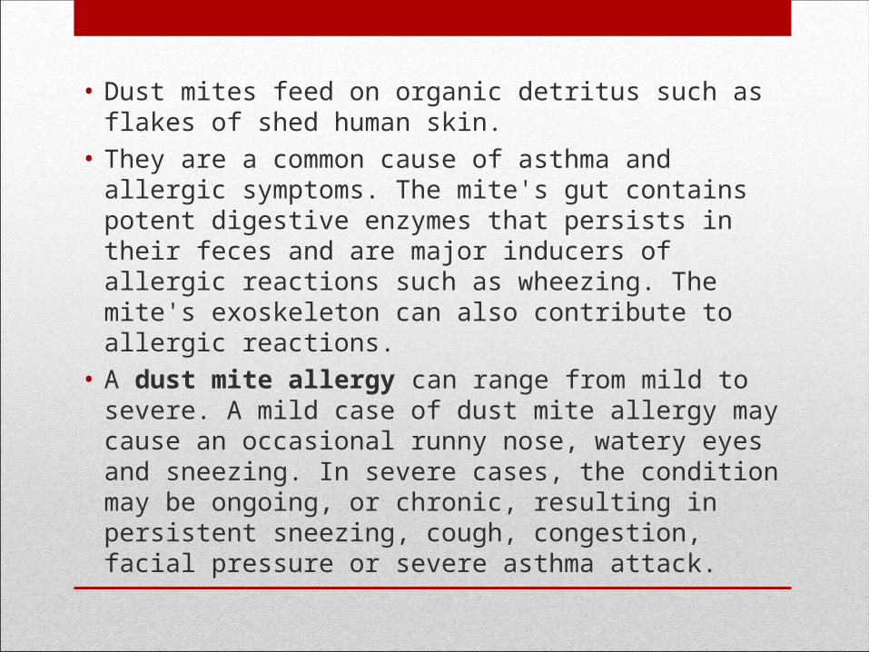
• Dust mites feed on organic detritus such as flakes of shed human skin.
• They are a common cause of asthma and allergic symptoms. The mite's gut contains potent digestive enzymes that persists in their feces and are major inducers of allergic reactions such as wheezing. The mite's exoskeleton can also contribute to allergic reactions.
• A dust mite allergy can range from mild to severe. A mild case of dust mite allergy may cause an occasional runny nose, watery eyes and sneezing. In severe cases, the condition may be ongoing, or chronic, resulting in persistent sneezing, cough, congestion, facial pressure or severe asthma attack.
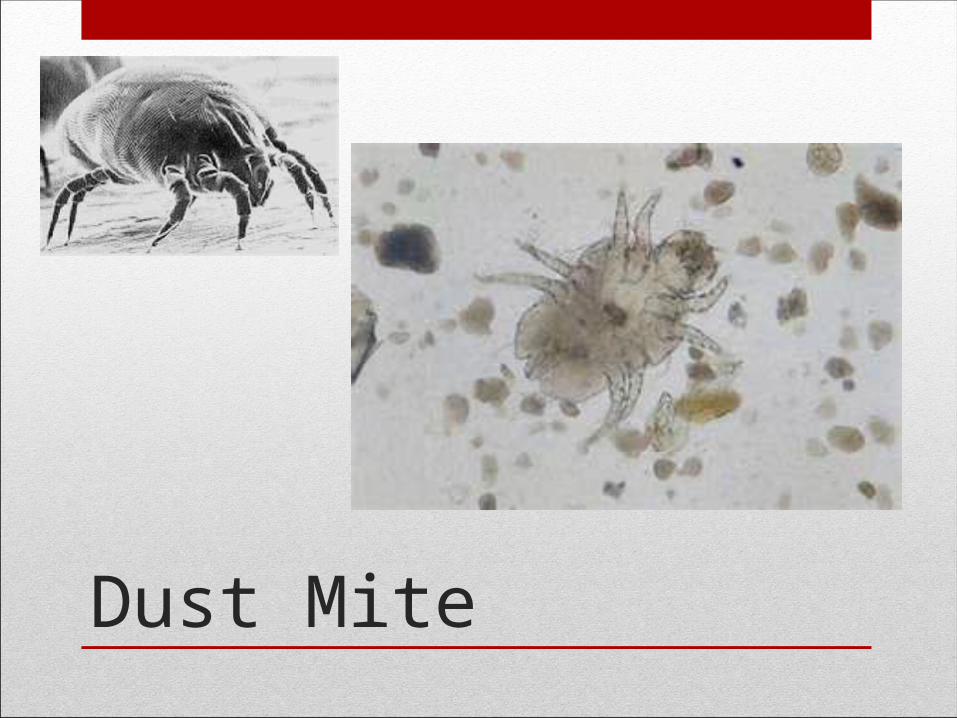
Dust Mite
Phylum Arthropoda
• Class Arachnia (ticks and mites)
• Class Insecta (fleas and lice)

Flea
HeadThoraxAbdomenLegs
CLASS INSECTA
Fleas do not live on their hosts. They take their blood meal and return to their nest (carpet, etc).
Bubonic plague (Yersinia pestis) Wiped out almost 1/3 of Europe Still have outbreaks from rodent population on West Coast
(Squirrels in San Bernardino)Endemic typhus (Rickettsia typhi)
Rodents in Texas
The fleas that are vectors for the above diseases prefer to use a rodent for a host.
Tapeworms in pets When the dog or cat ingests the flea, it ingests the eggs and
larvae inside the flea.
FLEAS ARE VECTORS FOR DISEASES

Pediculus humanis corporus - human body louse
AbdomenThoraxHeadAntennae (for feeling)EyesPlates (protection)Legs
CLASS INSECTA

HUMAN BODY LOUSE
Nests in the seams of clothing, and lays eggs there. It comes out for a blood meal.They are associated with crowded, unsanitary conditions,
such as during war, famines, and natural disasters.Diseases Transmitted by Body Lice
Epidemic Typhus (Rickettsia prowasekii) Relapsing Fever (Borrelia recurrentis and B. hermsii)
TreatmentClothing must be treated with insecticide or dry cleaning. In the old days, they would boil the clothes.The host also is also treated with Lindane solution and
washed off 4-24 hours later.
HUMAN BODY LOUSE
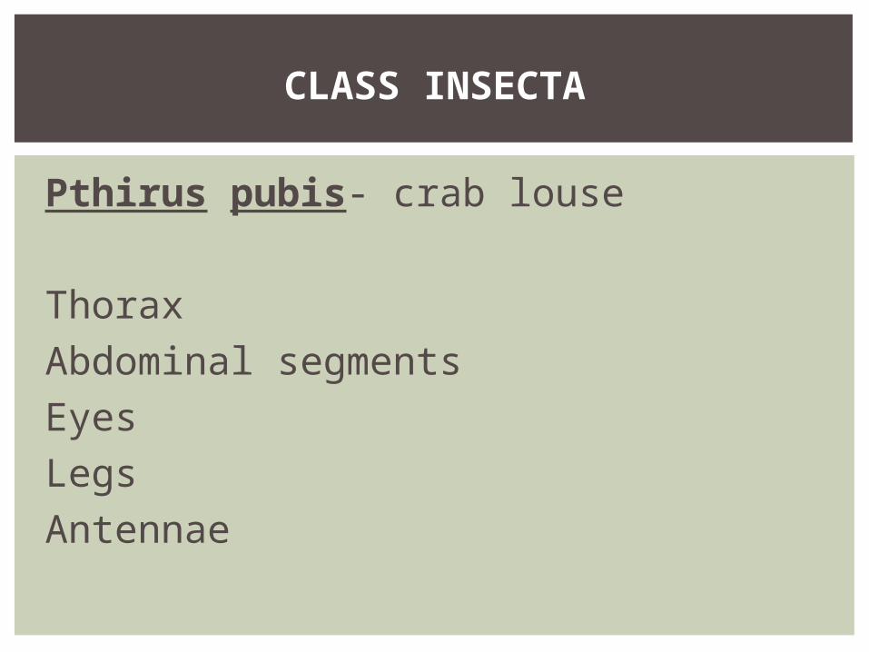
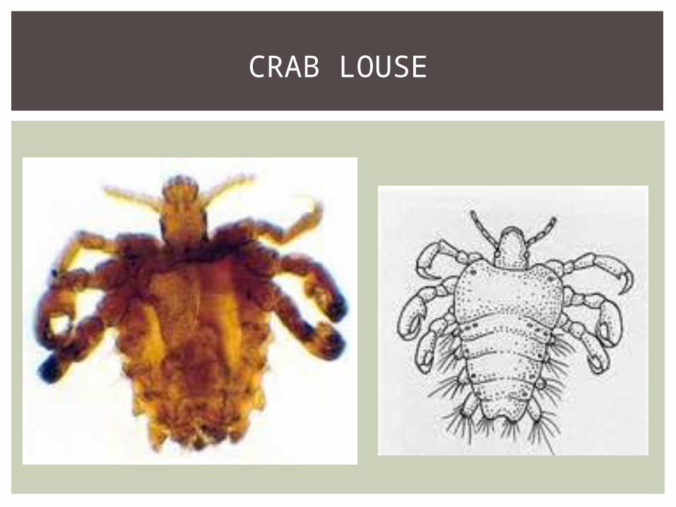
Pthirus pubis- crab louse
ThoraxAbdominal segmentsEyesLegsAntennae
CLASS INSECTA
CRAB LOUSE

Crab lice tend to infest human pubic hair. The species may also live on other areas with hair, including the eyelashes.
They feed exclusively on blood. Humans are the only known hosts.The main symptom is itching, usually in the pubic-hair area,
resulting from hypersensitivity to louse saliva, which can become stronger over two or more weeks following initial infestation.
Pubic lice usually infect a new host only by close contact between individuals, usually through sexual intercourse because they primarily spread through sweat, body fluids. Parent-to-child infestations are more likely to occur through routes of shared towels, clothing, beds or closets. Adults are more frequently infested than children.
CRAB LOUSE