part 3: adult basic and advanced life support
TRANSCRIPT

October 20, 2020 Circulation. 2020;142(suppl 2):S366–S468. DOI: 10.1161/CIR.0000000000000916S366
Key Words: AHA Scientific Statements ◼ apnea ◼ cardiopulmonary resuscitation ◼ defibrillators ◼ delivery of health care ◼ electric countershock ◼ heart arrest ◼ life support care
Ashish R. Panchal, MD, PhD, Chair
Jason A. Bartos, MD, PhDJosé G. Cabañas, MD,
MPHMichael W. Donnino, MDIan R. Drennan, ACP,
PhD(C)Karen G. Hirsch, MDPeter J. Kudenchuk, MDMichael C. Kurz, MD, MSEric J. Lavonas, MD, MSPeter T. Morley, MBBSBrian J. O’Neil, MDMary Ann Peberdy, MDJon C. Rittenberger, MD,
MSAmber J. Rodriguez, PhDKelly N. Sawyer, MD, MSKatherine M. Berg, MD,
Vice ChairOn behalf of the Adult
Basic and Advanced Life Support Writing Group
© 2020 American Heart Association, Inc.
Part 3: Adult Basic and Advanced Life Support2020 American Heart Association Guidelines for Cardiopulmonary Resuscitation and Emergency Cardiovascular Care
Circulation
https://www.ahajournals.org/journal/circ
TOP 10 TAKE-HOME MESSAGES FOR ADULT CARDIOVASCULAR LIFE SUPPORT
1. On recognition of a cardiac arrest event, a layperson should simultaneously and promptly activate the emergency response system and initiate cardiopul-monary resuscitation (CPR).
2. Performance of high-quality CPR includes adequate compression depth and rate while minimizing pauses in compressions,
3. Early defibrillation with concurrent high-quality CPR is critical to survival when sudden cardiac arrest is caused by ventricular fibrillation or pulseless ventricular tachycardia.
4. Administration of epinephrine with concurrent high-quality CPR improves survival, particularly in patients with nonshockable rhythms.
5. Recognition that all cardiac arrest events are not identical is critical for opti-mal patient outcome, and specialized management is necessary for many conditions (eg, electrolyte abnormalities, pregnancy, after cardiac surgery).
6. The opioid epidemic has resulted in an increase in opioid-associated out-of-hospital cardiac arrest, with the mainstay of care remaining the activation of the emergency response systems and performance of high-quality CPR.
7. Post–cardiac arrest care is a critical component of the Chain of Survival and demands a comprehensive, structured, multidisciplinary system that requires consistent implementation for optimal patient outcomes.
8. Prompt initiation of targeted temperature management is necessary for all patients who do not follow commands after return of spontaneous circula-tion to ensure optimal functional and neurological outcome.
9. Accurate neurological prognostication in brain-injured cardiac arrest survivors is critically important to ensure that patients with significant potential for recovery are not destined for certain poor outcomes due to care withdrawal.
10. Recovery expectations and survivorship plans that address treatment, surveil-lance, and rehabilitation need to be provided to cardiac arrest survivors and their caregivers at hospital discharge to optimize transitions of care to home and to the outpatient setting.
PREAMBLEIn 2015, approximately 350 000 adults in the United States experienced non-traumatic out-of-hospital cardiac arrest (OHCA) attended by emergency medical services (EMS) personnel.1 Approximately 10.4% of patients with OHCA survive their initial hospitalization, and 8.2% survive with good functional status. The key drivers of successful resuscitation from OHCA are lay rescuer cardiopulmonary
Dow
nloaded from http://ahajournals.org by on N
ovember 6, 2020

Panchal et al Adult Basic and Advanced Life Support: 2020 AHA Guidelines for CPR and ECC
Circulation. 2020;142(suppl 2):S366–S468. DOI: 10.1161/CIR.0000000000000916 October 20, 2020 S367
resuscitation (CPR) and public use of an automated external defibrillator (AED). Despite recent gains, only 39.2% of adults receive layperson-initiated CPR, and the general public applied an AED in only 11.9% of cases.1 Survival rates from OHCA vary dramatically be-tween US regions and EMS agencies.2,3 After significant improvements, survival from OHCA has plateaued since 2012.
Approximately 1.2% of adults admitted to US hos-pitals suffer in-hospital cardiac arrest (IHCA).1 Of these patients, 25.8% were discharged from the hospital alive, and 82% of survivors have good functional sta-tus at the time of discharge. Despite steady improve-ment in the rate of survival from IHCA, much oppor-tunity remains.
The International Liaison Committee on Resusci-tation (ILCOR) Formula for Survival emphasizes 3 es-sential components for good resuscitation outcomes: guidelines based on sound resuscitation science, ef-fective education of the lay public and resuscitation providers, and implementation of a well-functioning Chain of Survival.4
These guidelines contain recommendations for ba-sic life support (BLS) and advanced life support (ALS) for adult patients and are based on the best available resuscitation science. The Chain of Survival, introduced in Major Concepts, is now expanded to emphasize the important component of survivorship during recovery from cardiac arrest, requires coordinated efforts from medical professionals in a variety of disciplines and, in the case of OHCA, from lay rescuers, emergency dis-patchers, and first responders. In addition, specific rec-ommendations about the training of resuscitation pro-viders are provided in “Part 6: Resuscitation Education Science,” and recommendations about systems of care are provided in “Part 7: Systems of Care.”
INTRODUCTIONScope of the GuidelinesThese guidelines are designed primarily for North Amer-ican healthcare providers who are looking for an up-to-date summary for BLS and ALS for adults as well as for those who are seeking more in-depth information on resuscitation science and gaps in current knowledge. The BLS care of adolescents follows adult guidelines. This Part of the 2020 American Heart Association (AHA) Guidelines for CPR and Emergency Cardiovascular Care includes recommendations for clinical care of adults with cardiac arrest, including those with life-threaten-ing conditions in whom cardiac arrest is imminent, and after successful resuscitation from cardiac arrest.
Some recommendations are directly relevant to lay rescuers who may or may not have received CPR train-ing and who have little or no access to resuscitation
equipment. Other recommendations are relevant to persons with more advanced resuscitation training, functioning either with or without access to resuscita-tion drugs and devices, working either within or outside of a hospital. Some treatment recommendations in-volve medical care and decision-making after return of spontaneous circulation (ROSC) or when resuscitation has been unsuccessful. Importantly, recommendations are provided related to team debriefing and systematic feedback to increase future resuscitation success.
Organization of the Writing GroupThe Adult Cardiovascular Life Support Writing Group included a diverse group of experts with backgrounds in emergency medicine, critical care, cardiology, toxicol-ogy, neurology, EMS, education, research, and public health, along with content experts, AHA staff, and the AHA senior science editors. Each recommendation was developed and formally approved by the writing group.
The AHA has rigorous conflict of interest policies and procedures to minimize the risk of bias or improp-er influence during the development of guidelines. Be-fore appointment, writing group members disclosed all commercial relationships and other potential (in-cluding intellectual) conflicts. These procedures are described more fully in “Part 2: Evidence Evaluation and Guidelines Development.” Disclosure information for writing group members is listed in Appendix 1.
Methodology and Evidence ReviewThese guidelines are based on the extensive evidence evaluation performed in conjunction with the ILCOR and affiliated ILCOR member councils. Three different types of evidence reviews (systematic reviews, scoping reviews, and evidence updates) were used in the 2020 process. Each of these resulted in a description of the literature that facilitated guideline development. A more compre-hensive description of these methods is provided in “Part 2: Evidence Evaluation and Guidelines Development.”
Class of Recommendation and Level of EvidenceAs with all AHA guidelines, each 2020 recommendation is assigned a Class of Recommendation (COR) based on the strength and consistency of the evidence, alterna-tive treatment options, and the impact on patients and society (Table 1). The Level of Evidence (LOE) is based on the quality, quantity, relevance, and consistency of the available evidence. For each recommendation, the writ-ing group discussed and approved specific recommen-dation wording and the COR and LOE assignments. In determining the COR, the writing group considered the LOE and other factors, including systems issues,
Dow
nloaded from http://ahajournals.org by on N
ovember 6, 2020

October 20, 2020 Circulation. 2020;142(suppl 2):S366–S468. DOI: 10.1161/CIR.0000000000000916S368
Panchal et al Adult Basic and Advanced Life Support: 2020 AHA Guidelines for CPR and ECC
economic factors, and ethical factors such as equity, ac-ceptability, and feasibility. These evidence-review meth-ods, including specific criteria used to determine COR and LOE, are described more fully in “Part 2: Evidence Evaluation and Guidelines Development.” The Adult Basic and Advanced Life Support Writing Group mem-bers had final authority over and formally approved these recommendations.
Unfortunately, despite improvements in the design and funding support for resuscitation research, the overall certainty of the evidence base for resuscita-tion science is low. Of the 250 recommendations in these guidelines, only 2 recommendations are sup-ported by Level A evidence (high-quality evidence from more than 1 randomized controlled trial [RCT],
or 1 or more RCT corroborated by high-quality registry studies.) Thirty-seven recommendations are supported by Level B-Randomized Evidence (moderate evidence from 1 or more RCTs) and 57 by Level B-Nonrandom-ized evidence. The majority of recommendations are based on Level C evidence, including those based on limited data (123 recommendations) and expert opin-ion (31 recommendations). Accordingly, the strength of recommendations is weaker than optimal: 78 Class 1 (strong) recommendations, 57 Class 2a (moderate) recommendations, and 89 Class 2b (weak) recommen-dations are included in these guidelines. In addition, 15 recommendations are designated Class 3: No Benefit, and 11 recommendations are Class 3: Harm. Clinical trials in resuscitation are sorely needed.
Table 1. Applying Class of Recommendation and Level of Evidence to Clinical Strategies, Interventions, Treatments, or Diagnostic Testing in Patient Care (Updated May 2019)*
This table defines the Classes of Recommendation (COR) and Levels of Evidence (LOE). COR indicates the strength the writing group assigns the recommendation, and the LOE is assigned based on the quality of the scientific evidence. The outcome or result of the intervention should be specified (an improved clinical outcome or increased diagnostic accuracy or incremental prognostic information).Classes of RecommendationCOR designations include Class 1, a strong recommendation for which the potential benefit greatly outweighs the risk; Class 2a, a moderate recommendation for which benefit most likely outweighs the risk; Class 2b, a weak recommendation for which it’s unknown whether benefit will outweigh the risk; Class 3: No Benefit, a moderate recommendation signifying that there is equal likelihood of benefit and risk; and Class 3: Harm, a strong recommendation for which the risk outweighs the potential benefit. Suggested phrases for writing Class 1 recommendations include • Is recommended• Is indicated/useful/effective/beneficial• Should be performed/administered/otherComparative-effectiveness phrases include treatment/strategy A is recommended/indicated in preference to treatment B, and treatment A should be chosen over treatment B.Suggested phrases for writing Class 2a recommendations include• Is reasonable• Can be useful/effective/beneficialComparative-effectiveness phrases include treatment/strategy A is probably recommended/indicated in preference to treatment B, and it is reasonable to choose treatment A over treatment B.For comparative-effectiveness recommendations (COR 1 and 2a; LOE A and B only), studies that support the use of comparator verbs should involve direct comparisons of the treatments or strategies being evaluated.Suggested phrases for writing Class 2b recommendations include• May/might be reasonable• May/might be considered• Usefulness/effectiveness is unknown/unclear/uncertain or not well-establishedSuggested phrases for writing Class 3: No Benefit recommendations (generally, LOE A or B use only) include• Is not recommended• Is not indicated/useful/effective/beneficial• Should not be performed/administered/otherSuggested phrases for writing Class 3: Harm recommendations include• Potentially harmful• Causes harm• Associated with excess morbidity/mortality• Should not be performed/administered/otherLevels of EvidenceFor LOEs, the method of assessing quality is evolving, including the application of standardized, widely-used, and preferably validated evidence grading tools; and for systematic reviews, the incorporation of an Evidence Review Committee. LOE designations include Level A, Level B-R, Level B-NR, Level C-LD, and Level C-EO. Those categorized as Level A are derived from• High-quality evidence from more than 1 randomized clinical trial, or RCT• Meta-analyses of high-quality RCTs• One or more RCTs corroborated by high-quality registry studiesThose categorized as Level B-R (randomized) are derived from• Moderate-quality evidence from 1 or more RCTs• Meta-analyses of moderate-quality RCTsThose categorized as Level B-NR (nonrandomized) are derived from• Moderate-quality evidence from 1 or more well-designed, well-executed nonrandomized studies, observational studies, or registry studies• Meta-analyses of such studiesThose categorized as Level C-LD (limited data) are derived from• Randomized or nonrandomized observational or registry studies with limitations of design or execution• Meta-analyses of such studies• Physiological or mechanistic studies in human subjectsThose categorized as Level C-EO (expert opinion) are derived from• Consensus of expert opinion based on clinical experienceCOR and LOE are determined independently (any COR may be paired with any LOE).A recommendation with LOE C does not imply that the recommendation is weak. Many important clinical questions addressed in guidelines do not lend themselves to clinical trials. Although RCTs are unavailable, there may be a very clear clinical consensus that a particular test or therapy is useful or effective.
Dow
nloaded from http://ahajournals.org by on N
ovember 6, 2020

Panchal et al Adult Basic and Advanced Life Support: 2020 AHA Guidelines for CPR and ECC
Circulation. 2020;142(suppl 2):S366–S468. DOI: 10.1161/CIR.0000000000000916 October 20, 2020 S369
Guideline StructureThe 2020 Guidelines are organized into knowledge chunks, grouped into discrete modules of information on specific topics or management issues.5 Each modular knowledge chunk includes a table of recommendations that uses standard AHA nomenclature of COR and LOE. A brief introduction or short synopsis is provided to put the recommendations into context with important background information and overarching management or treatment concepts. Recommendation-specific text clarifies the rationale and key study data supporting the recommendations. When appropriate, flow diagrams or additional tables are included. Hyperlinked refer-ences are provided to facilitate quick access and review.
Document Review and ApprovalEach of the 2020 Guidelines documents was submitted for blinded peer review to 5 subject-matter experts nominated by the AHA. Before appointment, all peer reviewers were required to disclose relationships with industry and any other conflicts of interest, and all dis-closures were reviewed by AHA staff. Peer reviewer feedback was provided for guidelines in draft format and again in final format. All guidelines were reviewed and approved for publication by the AHA Science Advi-sory and Coordinating Committee and the AHA Execu-tive Committee. Disclosure information for peer review-ers is listed in Appendix 2.
REFERENCES 1. Virani SS, Alonso A, Benjamin EJ, Bittencourt MS, Callaway CW,
Carson AP, Chamberlain AM, Chang AR, Cheng S, Delling FN, et al: on behalf of the American Heart Association Council on Epidemiology and Prevention Statistics Committee and Stroke Statistics Subcommit-tee. Heart disease and stroke statistics—2020 update: a report from the American Heart Association. Circulation. 2020;141:e139–e596. doi: 10.1161/CIR.0000000000000757
2. Okubo M, Schmicker RH, Wallace DJ, Idris AH, Nichol G, Austin MA, Grunau B, Wittwer LK, Richmond N, Morrison LJ, Kurz MC, Cheskes S, Kudenchuk PJ, Zive DM, Aufderheide TP, Wang HE, Herren H, Vaillancourt C, Davis DP, Vilke GM, Scheuermeyer FX, Weisfeldt ML, Elmer J, Colella R, Callaway CW; Resuscitation Outcomes Consortium In-vestigators. Variation in Survival After Out-of-Hospital Cardiac Arrest Be-tween Emergency Medical Services Agencies. JAMA Cardiol. 2018;3:989–999. doi: 10.1001/jamacardio.2018.3037
3. Zive DM, Schmicker R, Daya M, Kudenchuk P, Nichol G, Rittenberger JC, Aufderheide T, Vilke GM, Christenson J, Buick JE, Kaila K, May S, Rea T, Morrison LJ; ROC Investigators. Survival and variability over time from out of hospital cardiac arrest across large geographi-cally diverse communities participating in the Resuscitation Out-comes Consortium. Resuscitation. 2018;131:74–82. doi: 10.1016/j. resuscitation.2018.07.023
4. Søreide E, Morrison L, Hillman K, Monsieurs K, Sunde K, Zideman D, Eisenberg M, Sterz F, Nadkarni VM, Soar J, Nolan JP; Utstein Formula for Survival Collaborators. The formula for survival in resuscitation. Resuscitation. 2013;84:1487–1493. doi: 10.1016/j. resuscitation.2013.07.020
5. Levine GN, O’Gara PT, Beckman JA, Al-Khatib SM, Birtcher KK, Cigarroa JE, de Las Fuentes L, Deswal A, Fleisher LA, Gentile F, Goldberger ZD, Hlatky MA, Joglar JA, Piano MR, Wijeysundera DN. Recent Innovations, Modifications, and Evolution of ACC/AHA Clinical Practice Guidelines: An Update for
Our Constituencies: A Report of the American College of Cardiology/American Heart Association Task Force on Clinical Practice Guidelines. Cir-culation. 2019;139:e879–e886. doi: 10.1161/CIR.0000000000000651
ACD active compression-decompression
ACLS advanced cardiovascular life support
ADC apparent diffusion coefficient
AED automated external defibrillator
AHA American Heart Association
ALS advanced life support
aOR adjusted odds ratio
AV atrioventricular
BLS basic life support
COR Class of Recommendation
CoSTR International Consensus on Cardiopulmonary Resuscitation and Emergency Cardiovascular Care Science With Treatment Recommendations
CPR cardiopulmonary resuscitation
CT computed tomography
DWI diffusion-weighted imaging
ECG electrocardiogram
ECPR extracorporeal cardiopulmonary resuscitation
EEG electroencephalogram
EMS emergency medical services
ETCO2 (partial pressure of) end-tidal carbon dioxide
ETI endotracheal intubation
GWR gray-white ratio
ICU intensive care unit
IHCA in-hospital cardiac arrest
ILCOR International Liaison Committee on Resuscitation
IO intraosseous
ITD impedance threshold device
IV intravenous
LAST local anesthetic systemic toxicity
LOE Level of Evidence
MAP mean arterial pressure
MRI magnetic resonance imaging
NSE neuron-specific enolase
OHCA out-of-hospital cardiac arrest
Paco2 arterial partial pressure of carbon dioxide
PCI percutaneous coronary intervention
PE pulmonary embolism
PMCD perimortem cesarean delivery
pVT pulseless ventricular tachycardia
RCT randomized controlled trial
ROSC return of spontaneous circulation
S100B S100 calcium binding protein
SGA supraglottic airway
Abbreviations
(Continued )
Dow
nloaded from http://ahajournals.org by on N
ovember 6, 2020
Panchal et al Adult Basic and Advanced Life Support: 2020 AHA Guidelines for CPR and ECC
October 20, 2020 Circulation. 2020;142(suppl 2):S366–S468. DOI: 10.1161/CIR.0000000000000916S370
MAJOR CONCEPTSOverview Concepts of Adult Cardiac ArrestSurvival and recovery from adult cardiac arrest depend on a complex system working together to secure the best outcome for the victim. The main focus in adult cardiac arrest events includes rapid recognition, prompt provision of CPR, defibrillation of malignant shockable rhythms, and post-ROSC supportive care and treat-ment of underlying causes. This approach recognizes that most sudden cardiac arrest in adults is of cardiac cause, particularly myocardial infarction and electric disturbances. Arrests without a primary cardiac origin (eg, from respiratory failure, toxic ingestion, pulmonary embolism [PE], or drowning) are also common, how-ever, and in such cases, treatment for reversible under-lying causes is important for the rescuer to consider.1 Some noncardiac etiologies may be particularly com-mon in the in-hospital setting. Others, such as opioid
overdose, are sharply on the rise in the out-of-hospital setting.2 For any cardiac arrest, rescuers are instructed to call for help, perform CPR to restore coronary and cerebral blood flow, and apply an AED to directly treat ventricular fibrillation (VF) or ventricular tachycardia (VT), if present. Although the majority of resuscitation success is achieved by provision of high-quality CPR and defibrillation, other specific treatments for likely under-lying causes may be helpful in some cases.
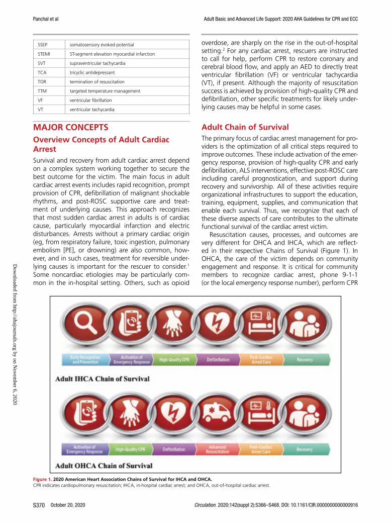
Adult Chain of SurvivalThe primary focus of cardiac arrest management for pro-viders is the optimization of all critical steps required to improve outcomes. These include activation of the emer-gency response, provision of high-quality CPR and early defibrillation, ALS interventions, effective post-ROSC care including careful prognostication, and support during recovery and survivorship. All of these activities require organizational infrastructures to support the education, training, equipment, supplies, and communication that enable each survival. Thus, we recognize that each of these diverse aspects of care contributes to the ultimate functional survival of the cardiac arrest victim.
Resuscitation causes, processes, and outcomes are very different for OHCA and IHCA, which are reflect-ed in their respective Chains of Survival (Figure 1). In OHCA, the care of the victim depends on community engagement and response. It is critical for community members to recognize cardiac arrest, phone 9-1-1 (or the local emergency response number), perform CPR
SSEP somatosensory evoked potential
STEMI ST-segment elevation myocardial infarction
SVT supraventricular tachycardia
TCA tricyclic antidepressant
TOR termination of resuscitation
TTM targeted temperature management
VF ventricular fibrillation
VT ventricular tachycardia
Figure 1. 2020 American Heart Association Chains of Survival for IHCA and OHCA.CPR indicates cardiopulmonary resuscitation; IHCA, in-hospital cardiac arrest; and OHCA, out-of-hospital cardiac arrest.
2020 AHA Chains of Survival for IHCA and OHCA. (2; IHCA, OHCA)
2 horizontal chains for adults, 1 for In-Hospital Cardiac Arrest and 1 for Out-of-Hospital Cardiac Arrest. On each chain, 6 links show icons for actions to help an adult in cardiac arrest.
Dow
nloaded from http://ahajournals.org by on N
ovember 6, 2020

Panchal et al Adult Basic and Advanced Life Support: 2020 AHA Guidelines for CPR and ECC
Circulation. 2020;142(suppl 2):S366–S468. DOI: 10.1161/CIR.0000000000000916 October 20, 2020 S371
(including, for untrained lay rescuers, compression-only CPR), and use an AED.3,4 Emergency medical person-nel are then called to the scene, continue resuscitation, and transport the patient for stabilization and definitive management. In comparison, surveillance and preven-tion are critical aspects of IHCA. When an arrest occurs in the hospital, a strong multidisciplinary approach in-cludes teams of medical professionals who respond, provide CPR, promptly defibrillate, begin ALS measures, and continue post-ROSC care. Outcomes from IHCA are overall superior to those from OHCA,5 likely because of reduced delays in initiation of effective resuscitation.
The Adult OHCA and IHCA Chains of Survival have been updated to better highlight the evolution of sys-tems of care and the critical role of recovery and survi-vorship with the addition of a new link. This Recovery link highlights the enormous recovery and survivorship journey, from the end of acute treatment for critical ill-ness through multimodal rehabilitation (both short- and long-term), for both survivors and families after cardiac arrest. This new link acknowledges the need for the sys-tem of care to support recovery, discuss expectations, and provide plans that address treatment, surveillance, and rehabilitation for cardiac arrest survivors and their caregivers as they transition care from the hospital to home and return to role and social function.
REFERENCES 1. Lavonas EJ, Drennan IR, Gabrielli A, Heffner AC, Hoyte CO,
Orkin AM, Sawyer KN, Donnino MW. Part 10: special circumstanc-es of resuscitation: 2015 American Heart Association Guidelines Update for Cardiopulmonary Resuscitation and Emergency Car-diovascular Care. Circulation. 2015;132(suppl 2):S501–S518. doi: 10.1161/CIR.0000000000000264
2. Dezfulian C, Orkin AM, Maron BA, Elmer J, Girota S, Gladwin MT, Merchant RM, Panchal AR, Perman SM, Starks M, van Diepen S, Lavonas EJ; on behalf of the American Heart Association Council on Cardiopulmonary, Critical Care, Perioperative and Resuscitation; Council on Arteriosclerosis, Thrombosis and Vascular Biology; Council on Cardiovascular and Stroke Nursing; and Council on Clinical Cardiology. Opioid-associated out-of-hospital cardiac arrest: distinctive clinical features and implications for healthcare and public responses: a scientific statement from the American Heart Association. Circulation. In press.
3. Sayre MR, Berg RA, Cave DM, Page RL, Potts J, White RD; Ameri-can Heart Association Emergency Cardiovascular Care Commit-tee. Hands-only (compression-only) cardiopulmonary resuscitation: a call to action for bystander response to adults who experience out-of-hospital sudden cardiac arrest: a science advisory for the public from the American Heart Association Emergency Cardio-vascular Care Committee. Circulation. 2008;117:2162–2167. doi: 10.1161/CIRCULATIONAHA.107.189380
4. Kleinman ME, Brennan EE, Goldberger ZD, Swor RA, Terry M, Bobrow BJ, Gazmuri RJ, Travers AH, Rea T. Part 5: adult basic life support and car-diopulmonary resuscitation quality: 2015 American Heart Association Guidelines Update for Cardiopulmonary Resuscitation and Emergency Cardiovascular Care. Circulation. 2015;132(suppl 2):S414–S435. doi: 10.1161/CIR.0000000000000259
5. Virani SS, Alonso A, Benjamin EJ, Bittencourt MS, Callaway CW, Carson AP, Chamberlain AM, Chang AR, Cheng S, Delling FN, et al: on behalf of the American Heart Association Council on Epidemiology and Prevention Statistics Committee and Stroke Statistics Subcommit-tee. Heart disease and stroke statistics—2020 update: a report from the American Heart Association. Circulation. 2020;141:e139–e596. doi: 10.1161/CIR.0000000000000757
SEQUENCE OF RESUSCITATIONRecognition of Cardiac Arrest
SynopsisLay rescuer CPR improves survival from cardiac arrest by 2- to 3-fold.1 The benefit of providing CPR to a patient in cardiac arrest outweighs any potential risk of providing chest compressions to someone who is unconscious but not in cardiac arrest. It has been shown that the risk of injury from CPR is low in these patients.2
It has been shown previously that all rescuers may have difficulty detecting a pulse, leading to delays in CPR, or in some cases CPR not being performed at all for patients in cardiac arrest.3 Recognition of car-diac arrest by lay rescuers, therefore, is determined on the basis of level of consciousness and the respira-tory effort of the victim. Recognition of cardiac arrest by healthcare providers includes a pulse check, but the importance of not prolonging efforts to detect a pulse is emphasized.
Recommendation-Specific Supportive Text1. Agonal breathing is characterized by slow,
irregular gasping respirations that are inef-fective for ventilation. Agonal breathing is described by lay rescuers with a variety of terms including, abnormal breathing, snoring respirations, and gasping.4 Agonal breath-ing is common, reported as being present in up to 40% to 60% of victims of OHCA.5 The presence of agonal breathing is cited as a common reason for lay rescuers to misdiag-nose a patient as not being in cardiac arrest.6 In patients who are unresponsive, with absent or abnormal breathing, lay rescuers should assume the patient is in cardiac arrest, call for help, and promptly initiate CPR. These 2 crite-ria (patient responsiveness and assessment of breathing) have been shown to rapidly identify a significant proportion of patients who are in cardiac arrest, allowing for immediate initiation of lay rescuer CPR. Further, initiation of chest compressions in patients who are unconscious
Recommendations for Recognition of Cardiac Arrest
COR LOE Recommendations
1 C-LD
1. If a victim is unconscious/unresponsive, with absent or abnormal breathing (ie, only gasping), the lay rescuer should assume the victim is in cardiac arrest.
1 C-LD
2. If a victim is unconscious/unresponsive, with absent or abnormal breathing (ie, only gasping), the healthcare provider should check for a pulse for no more than 10 s and, if no definite pulse is felt, should assume the victim is in cardiac arrest.
Dow
nloaded from http://ahajournals.org by on N
ovember 6, 2020

Panchal et al Adult Basic and Advanced Life Support: 2020 AHA Guidelines for CPR and ECC
October 20, 2020 Circulation. 2020;142(suppl 2):S366–S468. DOI: 10.1161/CIR.0000000000000916S372
but not in cardiac arrest is associated with low rates of significant adverse events.2 The adverse events noted included pain in the area of chest compressions (8.7%), bone fracture (ribs and clavicle) (1.7%), and rhabdomyolysis (0.3%), with no visceral injuries described.2
2. Protracted delays in CPR can occur when check-ing for a pulse at the outset of resuscitation efforts as well as between successive cycles of CPR. Healthcare providers often take too long to check for a pulse7,8 and have difficulty determining if a pulse is present or absent.7–9 There is no evidence, however, that checking for breathing, coughing, or movement is supe-rior to a pulse check for detection of circula-tion.10 Thus, healthcare providers are directed to quickly check for a pulse and to promptly start compressions when a pulse is not defini-tively palpated.9,11
This topic last received formal evidence review in 2010.3
REFERENCES 1. Sasson C, Rogers MA, Dahl J, Kellermann AL. Predictors of sur-
vival from out-of-hospital cardiac arrest: a systematic review and meta-analysis. Circ Cardiovasc Qual Outcomes. 2010;3:63–81. doi: 10.1161/CIRCOUTCOMES.109.889576
2. Olasveengen TM, Mancini ME, Perkins GD, Avis S, Brooks S, Castrén M, Chung SP, Considine J, Couper K, Escalante R, et al; on behalf of the Adult Basic Life Support Collaborators. Adult basic life support: 2020 Interna-tional Consensus on Cardiopulmonary Resuscitation and Emergency Car-diovascular Care Science With Treatment Recommendations. Circulation. 2020;142(suppl 1):S41–S91. doi: 10.1161/CIR.0000000000000892
3. Berg RA, Hemphill R, Abella BS, Aufderheide TP, Cave DM, Hazinski MF, Lerner EB, Rea TD, Sayre MR, Swor RA. Part 5: adult basic life support: 2010 American Heart Association Guidelines for Cardiopulmonary Resus-citation and Emergency Cardiovascular Care. Circulation. 2010;122(suppl 3):S685–S705. doi: 10.1161/CIRCULATIONAHA.110.970939
4. Riou M, Ball S, Williams TA, Whiteside A, Cameron P, Fatovich DM, Perkins GD, Smith K, Bray J, Inoue M, O’Halloran KL, Bailey P, Brink D, Finn J. ‘She’s sort of breathing’: What linguistic fac-tors determine call-taker recognition of agonal breathing in emergency calls for cardiac arrest? Resuscitation. 2018;122:92–98. doi: 10.1016/j. resuscitation.2017.11.058
5. Fukushima H, Imanishi M, Iwami T, Seki T, Kawai Y, Norimoto K, Urisono Y, Hata M, Nishio K, Saeki K, Kurumatani N, Okuchi K. Abnormal breath-ing of sudden cardiac arrest victims described by laypersons and its as-sociation with emergency medical service dispatcher-assisted cardiopul-monary resuscitation instruction. Emerg Med J. 2015;32:314–317. doi: 10.1136/emermed-2013-203112
6. Brinkrolf P, Metelmann B, Scharte C, Zarbock A, Hahnenkamp K, Bohn A. Bystander-witnessed cardiac arrest is associated with reported agonal breathing and leads to less frequent bystander CPR. Resuscitation. 2018;127:114–118. doi: 10.1016/j.resuscitation.2018.04.017
7. Eberle B, Dick WF, Schneider T, Wisser G, Doetsch S, Tzanova I. Check-ing the carotid pulse check: diagnostic accuracy of first responders in pa-tients with and without a pulse. Resuscitation. 1996;33:107–116. doi: 10.1016/s0300-9572(96)01016-7
8. Moule P. Checking the carotid pulse: diagnostic accuracy in students of the healthcare professions. Resuscitation. 2000;44:195–201. doi: 10.1016/s0300-9572(00)00139-8
9. Ochoa FJ, Ramalle-Gómara E, Carpintero JM, García A, Saralegui I. Com-petence of health professionals to check the carotid pulse. Resuscitation. 1998;37:173–175. doi: 10.1016/s0300-9572(98)00055-0
10. Perkins GD, Stephenson B, Hulme J, Monsieurs KG. Birmingham assess-ment of breathing study (BABS). Resuscitation. 2005;64:109–113. doi: 10.1016/j.resuscitation.2004.09.007
11. Mather C, O’Kelly S. The palpation of pulses. Anaesthesia. 1996;51:189–191. doi: 10.1111/j.1365-2044.1996.tb07713.x
Initiation of Resuscitation
Recommendations for Initiation of Resuscitation: Lay Rescuer (Untrained or Trained)
COR LOE Recommendations
1 B-NR1. All lay rescuers should, at minimum,
provide chest compressions for victims of cardiac arrest.
1 C-LD
2. After identifying a cardiac arrest, a lone responder should activate the emergency response system first and immediately begin CPR.
1 C-LD
3. We recommend that laypersons initiate CPR for presumed cardiac arrest, because the risk of harm to the patient is low if the patient is not in cardiac arrest.
2a C-LD
4. For lay rescuers trained in CPR using chest compressions and ventilation (rescue breaths), it is reasonable to provide ventilation (rescue breaths) in addition to chest compressions for the adult in OHCA.
SynopsisAfter cardiac arrest is recognized, the Chain of Survival continues with activation of the emergency response sys-tem and initiation of CPR. The prompt initiation of CPR is perhaps the most important intervention to improve survival and neurological outcomes. Ideally, activation of the emergency response system and initiation of CPR oc-cur simultaneously. In the current era of widespread mo-bile device usage and accessibility, a lone responder can activate the emergency response system simultaneously with starting CPR by dialing for help, placing the phone on speaker mode to continue communication, and im-mediately commencing CPR. In the rare situation when a lone rescuer must leave the victim to dial EMS, the pri-ority should be on prompt EMS activation followed by immediate return to the victim to initiate CPR.
Existing evidence suggests that the potential harm from CPR in a patient who has been incorrectly identi-fied as having cardiac arrest is low.1 Overall, the ben-efits of initiation of CPR in cardiac arrest outweigh the relatively low risk of injury for patients not in cardiac arrest. The initial phases of resuscitation once cardiac arrest is recognized are similar between lay responders and healthcare providers, with early CPR representing the priority. Lay rescuers may provide chest compres-sion–only CPR to simplify the process and encourage CPR initiation, whereas healthcare providers may pro-vide chest compressions and ventilation (Figures 2–4).
Recommendation-Specific Supportive Text1. CPR is the single-most important intervention for
a patient in cardiac arrest, and chest compressions should be provided promptly. Chest compressions are the most critical component of CPR, and a chest
Dow
nloaded from http://ahajournals.org by on N
ovember 6, 2020

Panchal et al Adult Basic and Advanced Life Support: 2020 AHA Guidelines for CPR and ECC
Circulation. 2020;142(suppl 2):S366–S468. DOI: 10.1161/CIR.0000000000000916 October 20, 2020 S373
Figure 2. Adult BLS Algorithm for Healthcare Providers.AED indicates automated external defibrillator; ALS, advanced life support; BLS, basic life support; and CPR, cardiopulmonary resuscitation.
Text in cascading boxes describes the actions that a provider should perform in sequence during an adult cardiac arrest. Arrows guide providers from one box to the next as they perform the actions. Some boxes have 2 arrows that lead outward, each to a different box depending on the outcome of the most recent action taken. Pathways are linked.Box 1 Verify scene safety.Box 2Check for responsiveness. Shout for nearby help. Activate the emergency response system via mobile device (if appropriate). Get an AED and emergency equipment (or send someone to do so).Box 3Look for no breathing or only gasping and check pulse (simultaneously). Is a pulse definitely felt within 10 seconds?If the person is breathing normally and has a pulse, proceed to Box 3a.If the person is not breathing normally but has a pulse, proceed to Box 3b.If the person is not breathing or is only gasping and no pulse is felt, proceed to Box 4.Box 3aMonitor the person until emergency responders arrive. Box 3bProvide rescue breathing, 1 breath every 6 seconds or 10 breaths per minute.Check pulse every 2 minutes; if no pulse, start CPR.If it is a possible opioid overdose, administer naloxone if available per protocol.By this time in all scenarios, emergency response system or backup is activated, and AED and emergency equipment are retrieved or someone is retrieving them.Box 4Start CPR• Perform cycles of 30 compressions and 2 breaths. • Use the AED as soon as it is available.Box 5The AED arrives.Box 6The AED checks the rhythm. Is the rhythm shockable?If Yes, the rhythm is shockable, proceed to Box 7.If No, the rhythm is nonshockable, proceed to Box 8.Box 7• Give 1 shock. Resume CPR immediately for 2 minutes (until prompted by the AED to allow rhythm check). • Continue until advanced life support providers take over or the victim starts to move.Box 8• Resume CPR immediately for 2 minutes (until prompted by the AED to allow rhythm check). • Continue until advanced life support providers take over or the victim starts to move.
Dow
nloaded from http://ahajournals.org by on N
ovember 6, 2020

October 20, 2020 Circulation. 2020;142(suppl 2):S366–S468. DOI: 10.1161/CIR.0000000000000916S374
Panchal et al Adult Basic and Advanced Life Support: 2020 AHA Guidelines for CPR and ECC
Figure 3. Adult Cardiac Arrest Algorithm.CPR indicates cardiopulmonary resuscitation; ET, endotracheal; IO, intraosseous; IV, intravenous; PEA, pulseless electrical activity; pVT, pulseless ventricular tachycar-dia; and VF, ventricular fibrillation.
Cascading numbered boxes correspond to actions the provider should perform in sequence. Each box is separated by an arrow that signifies the pathway the provider should take. Some boxes are separated by 2 arrows that lead to different boxes, meaning that the provider should take a different pathway depending on the outcome of the previous action. Pathways are hyperlinked. Box 1 Start CPR• Give oxygen• Attach monitor/defibrillatorRhythm shockable?Yes, proceed to Box 2 for VF/pVT.No, proceed to Box 9 for Asystole/PEA.Box 2VF/pVTBox 3Deliver shock.Box 4CPR 2 minutes• IV/IO accessIs rhythm shockable?If Yes, proceed to Box 5.If No, proceed to Box 12.Box 5Deliver shock.Box 6CPR 2 minutes• Epinephrine every 3 to 5 minutes.• Consider advanced airway, capnography.Is rhythm shockable?If Yes, proceed to Box 7.If No, proceed to Box 12.Box 7Deliver shock.Box 8CPR 2 minutes• Amiodarone or lidocaine.• Treat reversible causes.Box 9Asystole/PEA.Give Epinephrine ASAP.Box 10CPR 2 minutes• IV/IO access.• Epinephrine every 3 to 5 minutes.• Consider advanced airway, capnography.Is rhythm shockable?If Yes, proceed to Box 5 or Box 7.If No, proceed to Box 11.Box 11CPR 2 minutes.• Treat reversible causes.Is rhythm shockable?If Yes, proceed to Box 5 or Box 7.If No, proceed to Box 12.Box 12• If no signs of return of spontaneous circulation (ROSC), go to Box 10 or Box 11• If ROSC, go to Post–Cardiac Arrest Care• Consider appropriateness of continued resuscitationSidebarCPR Quality• Push hard (at least 2 inches [5 cm]) and fast (100-120/min) and allow complete chest recoil.• Minimize interruptions in compressions.• Avoid excessive ventilation.• Change compressor every 2 minutes, or sooner if fatigued.• If no advanced airway, 30 to 2 compression-ventilation ratio.• Quantitative waveform capnography- If PETCO2 is low or decreasing, reassess CPR quality.Shock Energy for Defibrillation• Biphasic: Manufacturer recommendation (eg, initial dose of 120-200 Joules); if unknown, use maximum available. Second and subsequent doses should be equivalent, and higher doses may be considered.• Monophasic: 360 JoulesDrug Therapy• Epinephrine IV/IO dose: 1 milligram every 3 to 5 minutes• Amiodarone IV/IO dose: First dose: 300 milligram bolus. Second dose: 150 milligram.orLidocaine IV/IO dose: First dose: 1-1.5 milligrams per kilogram. Second dose: 0.5-0.75 milligrams per kilogram.Advanced Airway• Endotracheal intubation or supraglottic advanced airway• Waveform capnography or capnometry to confirm and monitor ET tube placement• Once advanced airway in place, give 1 breath every 6 seconds (10 breaths per minute) with continuous chest compressionsReturn of Spontaneous Circulation (ROSC)• Pulse and blood pressure• Abrupt sustained increase in PETCO2 (typically greater than or equal to 40 millimeters of mercury)• Spontaneous arterial pressure waves with intra-arterial monitoringReversible Causes• Hypovolemia• Hypoxia• Hydrogen ion (acidosis)• Hypo-/hyperkalemia• Hypothermia• Tension pneumothorax• Tamponade, cardiac• Toxins• Thrombosis, pulmonary• Thrombosis, coronary
Dow
nloaded from http://ahajournals.org by on N
ovember 6, 2020

Panchal et al Adult Basic and Advanced Life Support: 2020 AHA Guidelines for CPR and ECC
Circulation. 2020;142(suppl 2):S366–S468. DOI: 10.1161/CIR.0000000000000916 October 20, 2020 S375
Figure 4. Adult Cardiac Arrest Circular Algorithm.CPR indicates cardiopulmonary resuscitation; ET, endotracheal; IO, intraosseous; IV, intravenous; pVT, pulseless ventricular tachycardia; and VF, ventricular fibrillation.
Cascading numbered boxes and a circular pattern correspond to actions the provider should perform in sequence.Box 1Start CPR• Give oxygen.• Attach monitor/defibrillator.Box 2• Check rhythm. This box starts a repetitive pattern, represented by the outside of a circle.If VF/pVT, deliver shock, followed by 2 minutes of:- Continuous CPR- Monitor CPR Quality- Continuous CPR• After 2 minutes, check rhythm again and repeat this cycle until Return of Spontaneous Circulation (ROSC), then initiate post-cardiac arrest care.Inside the circle are listed things to perform as necessary during the resuscitation effort:Drug Therapy• IV/IO access• Epinephrine every 3 to 5 minutes• Amiodarone or lidocaine for refractory VF/pVTConsider Advanced Airway• Quantitative waveform capnographyTreat Reversible CausesSidebarCPR Quality• Push hard (at least 2 inches [5 cm]) and fast (100-120/min) and allow complete chest recoil.• Minimize interruptions in compressions.• Avoid excessive ventilation.• Change compressor every 2 minutes, or sooner if fatigued.• If no advanced airway, 30 to 2 compression-ventilation ratio.• Quantitative waveform capnography- If PETCO2 is low or decreasing, reassess CPR quality.Shock Energy for Defibrillation• Biphasic: Manufacturer recommendation (eg, initial dose of 120 to 200 Joules); if unknown, use maximum available. Second and subsequent doses should be equivalent, and higher doses may be considered.• Monophasic: 360 JoulesDrug Therapy• Epinephrine IV/IO dose: 1 milligram every 3 to 5 minutes• Amiodarone IV/IO dose: First dose: 300 milligram bolus. Second dose: 150 milligrams.orLidocaine IV/IO dose: First dose: 1-1.5 milligrams per kilogram. Second dose: 0.5-0.75 milligrams per kilogram.Advanced Airway• Endotracheal intubation or supraglottic advanced airway• Waveform capnography or capnometry to confirm and monitorET tube placement• Once advanced airway in place, give 1 breath every 6 seconds (10 breaths per minute) with continuous chest compressionsReturn of Spontaneous Circulation (ROSC)• Pulse and blood pressure• Abrupt sustained increase in PETCO2 (typically greater than or equal to 40 millimeters of mercury)• Spontaneous arterial pressure waves with intra-arterial monitoringReversible Causes• Hypovolemia• Hypoxia• Hydrogen ion (acidosis)• Hypo-/hyperkalemia• Hypothermia• Tension pneumothorax• Tamponade, cardiac• Toxins• Thrombosis, pulmonary• Thrombosis, coronary
compression–only approach is appropriate if lay rescuers are untrained or unwilling to provide respi-rations. Beginning the CPR sequence with compres-sions minimized time to first chest compression.2–4 Nationwide dissemination of chest compression–only CPR for lay rescuers was associated with an increase in the incidence of survival with favorable neurological outcome after OHCAs in Japan, likely due to an increase in lay rescuers providing CPR.5 Chest compressions should be provided as soon as possible, without the need to remove the vic-tim’s clothing first.
2. The optimal timing of CPR initiation and emer-gency response system activation was evalu-ated by an ILCOR systematic review in 2020.1 An observational study of over 17 000 OHCA events reported similar results from either a “call-first” strategy or a “CPR-first” strategy.6 In the current era of ubiquitous mobile devices, ideally both the call to activate EMS and the initiation of CPR can occur simultaneously.
3. Four observational studies7–10 reported outcomes from patients who were not in cardiac arrest and received CPR by lay rescuers. No serious harm from
Dow
nloaded from http://ahajournals.org by on N
ovember 6, 2020

Panchal et al Adult Basic and Advanced Life Support: 2020 AHA Guidelines for CPR and ECC
October 20, 2020 Circulation. 2020;142(suppl 2):S366–S468. DOI: 10.1161/CIR.0000000000000916S376
CPR was found in patients when they were later determined not to have been in cardiac arrest.1 This is in contrast to the significant risk of with-holding CPR when a patient is in cardiac arrest, making the risk:benefit ratio strongly in favor of providing CPR for presumed cardiac arrest.
4. In some observational studies, improved outcomes have been noted in victims of cardiac arrest who received conventional CPR (compressions and ventila-tion) compared with those who received chest com-pressions only.5,11,12 Other studies have reported no difference in outcomes for patients receiving conven-tional versus compression-only CPR.11,13–21 Given the potential benefit of conventional CPR, if lay rescuers are appropriately trained, they should be encouraged to concurrently deliver ventilation with compres-sions. A thorough review of the data concerning the ratio of compressions to ventilation when perform-ing conventional CPR is discussed in Ventilation and Compression-to-Ventilation Ratio.
These recommendations are supported by the 2020 ILCOR Consensus on CPR and Emergency Cardiovascular Care Science With Treatment Recommendations (CoSTR).1
Recommdendations for Initiation of Resuscitation: Healthcare Provider
COR LOE Recommendations
1 C-LD1. A lone healthcare provider should
commence with chest compressions rather than with ventilation.
2a C-LD
2. It is reasonable for healthcare providers to perform chest compressions and ventilation for all adult patients in cardiac arrest from either a cardiac or noncardiac cause.
Recommendation-Specific Supportive Text1. The 2010 Guidelines for CPR and Emergency
Cardiovascular Care included a major change for trained rescuers, who were instructed to begin the CPR sequence with chest compres-sions rather than with breaths (circulation, air-way, and breathing versus airway, breathing, and circulation) to minimize the time to initia-tion of chest compressions. This approach is resupported by new literature, summarized in a 2020 ILCOR systematic review (Table 2).1–4 In the recommended sequence, once chest compres-sions have been started, a single trained rescuer delivers rescue breaths by mouth to mask or by bag-mask device to provide oxygenation and ventilation. Manikin studies demonstrate that starting with chest compressions rather than with ventilation is associated with faster times to chest compressions,3,23 rescue breaths,4 and completion of the first CPR cycle.4
2. Healthcare providers are trained to deliver both compressions and ventilation. Delivery of chest compressions without assisted ventilation for
prolonged periods could be less effective than conventional CPR (compressions plus ventila-tion) because arterial oxygen content decreases as CPR duration increases. This concern is espe-cially pertinent in the setting of asphyxial cardiac arrest.11 Healthcare providers, with their training and understanding, can realistically tailor the sequence of subsequent rescue actions to the most likely cause of arrest.
These recommendations are supported by the 2020 CoSTR for BLS.1
Table 2. Adult BLS Sequence22
StepLay Rescuer Not
Trained Lay Rescuer TrainedHealthcare Provider
1 Ensure scene safety. Ensure scene safety. Ensure scene safety.
2 Check for response. Check for response. Check for response.
3 Shout for nearby help. Phone or ask someone to phone 9-1-1 (the phone or caller with the phone remains at the victim’s side, with the phone on speaker mode).
Shout for nearby help and activate the emergency response system (9-1-1, emergency response). If someone responds, ensure that the phone is at the side of the victim if at all possible.
Shout for nearby help/activate the resuscitation team; the provider can activate the resuscitation team at this time or after checking for breathing and pulse.
4 Follow the telecommunicator’s* instructions.
Check for no breathing or only gasping; if none, begin CPR with compressions.
Check for no breathing or only gasping and check pulse (ideally simultaneously). Activation and retrieval of the AED/emergency equipment by the lone healthcare provider or by the second person sent by the rescuer must occur no later than immediately after the check for no normal breathing and no pulse identifies cardiac arrest.
5 Look for no breathing or only gasping, at the direction of the telecommunicator.
Answer the telecommunicator’s questions, and follow the telecommunicator’s instructions.
Immediately begin CPR, and use the AED/defibrillator when available.
6 Follow the telecommunicator’s instructions.
Send the second person to retrieve an AED, if one is available.
When the second rescuer arrives, provide 2-rescuer CPR and use the AED/defibrillator.
AED indicates automated external defibrillator; BLS, basic life support; and CPR, cardiopulmonary resuscitation.
*Telecommunicator and dispatcher are terms often used interchangeably.
Dow
nloaded from http://ahajournals.org by on N
ovember 6, 2020

Panchal et al Adult Basic and Advanced Life Support: 2020 AHA Guidelines for CPR and ECC
Circulation. 2020;142(suppl 2):S366–S468. DOI: 10.1161/CIR.0000000000000916 October 20, 2020 S377
REFERENCES 1. Olasveengen TM, Mancini ME, Perkins GD, Avis S, Brooks S, Castrén M,
Chung SP, Considine J, Couper K, Escalante R, et al; on behalf of the Adult Basic Life Support Collaborators. Adult basic life support: 2020 Interna-tional Consensus on Cardiopulmonary Resuscitation and Emergency Car-diovascular Care Science With Treatment Recommendations. Circulation. 2020;142(suppl 1):S41–S91. doi: 10.1161/CIR.0000000000000892
2. Lubrano R, Cecchetti C, Bellelli E, Gentile I, Loayza Levano H, Orsini F, Bertazzoni G, Messi G, Rugolotto S, Pirozzi N, Elli M. Comparison of times of intervention during pediatric CPR maneuvers using ABC and CAB se-quences: a randomized trial. Resuscitation. 2012;83:1473–1477. doi: 10.1016/j.resuscitation.2012.04.011
3. Sekiguchi H, Kondo Y, Kukita I. Verification of changes in the time taken to initiate chest compressions according to modified basic life support guidelines. Am J Emerg Med. 2013;31:1248–1250. doi: 10.1016/j.ajem.2013.02.047
4. Marsch S, Tschan F, Semmer NK, Zobrist R, Hunziker PR, Hunziker S. ABC versus CAB for cardiopulmonary resuscitation: a prospective, ran-domized simulator-based trial. Swiss Med Wkly. 2013;143:w13856. doi: 10.4414/smw.2013.13856
5. Iwami T, Kitamura T, Kiyohara K, Kawamura T. Dissemination of Chest Compression-Only Cardiopulmonary Resuscitation and Survival After Out-of-Hospital Cardiac Arrest. Circulation. 2015;132:415–422. doi: 10.1161/CIRCULATIONAHA.114.014905
6. Kamikura T, Iwasaki H, Myojo Y, Sakagami S, Takei Y, Inaba H. Advantage of CPR-first over call-first actions for out-of-hospital cardiac arrests in non-elderly patients and of noncardiac aetiology. Resuscitation. 2015;96:37–45. doi: 10.1016/j.resuscitation.2015.06.027
7. White L, Rogers J, Bloomingdale M, Fahrenbruch C, Culley L, Subido C, Eisenberg M, Rea T. Dispatcher-assisted cardiopulmonary resuscitation: risks for patients not in cardiac arrest. Circulation. 2010;121:91–97. doi: 10.1161/CIRCULATIONAHA.109.872366
8. Haley KB, Lerner EB, Pirrallo RG, Croft H, Johnson A, Uihlein M. The fre-quency and consequences of cardiopulmonary resuscitation performed by bystanders on patients who are not in cardiac arrest. Prehosp Emerg Care. 2011;15:282–287. doi: 10.3109/10903127.2010.541981
9. Moriwaki Y, Sugiyama M, Tahara Y, Iwashita M, Kosuge T, Harunari N, Arata S, Suzuki N. Complications of bystander cardiopulmonary resuscita-tion for unconscious patients without cardiopulmonary arrest. J Emerg Trauma Shock. 2012;5:3–6. doi: 10.4103/0974-2700.93094
10. Tanaka Y, Nishi T, Takase K, Yoshita Y, Wato Y, Taniguchi J, Hamada Y, Inaba H. Survey of a protocol to increase appropriate implementation of dispatcher-assisted cardiopulmonary resuscitation for out-of-hos-pital cardiac arrest. Circulation. 2014;129:1751–1760. doi: 10.1161/ CIRCULATIONAHA.113.004409
11. Kitamura T, Iwami T, Kawamura T, Nagao K, Tanaka H, Hiraide A; Im-plementation Working Group for All-Japan Utstein Registry of the Fire and Disaster Management Agency. Bystander-initiated rescue breath-ing for out-of-hospital cardiac arrests of noncardiac origin. Circulation. 2010;122:293–299. doi: 10.1161/CIRCULATIONAHA.109.926816
12. Ogawa T, Akahane M, Koike S, Tanabe S, Mizoguchi T, Imamura T. Out-comes of chest compression only CPR versus conventional CPR conducted by lay people in patients with out of hospital cardiopulmonary arrest
witnessed by bystanders: nationwide population based observational study. BMJ. 2011;342:c7106. doi: 10.1136/bmj.c7106
13. Svensson L, Bohm K, Castrèn M, Pettersson H, Engerström L, Herlitz J, Rosenqvist M. Compression-only CPR or standard CPR in out-of-hos-pital cardiac arrest. N Engl J Med. 2010;363:434–442. doi: 10.1056/ NEJMoa0908991
14. Rea TD, Fahrenbruch C, Culley L, Donohoe RT, Hambly C, Innes J, Bloomingdale M, Subido C, Romines S, Eisenberg MS. CPR with chest compression alone or with rescue breathing. N Engl J Med. 2010;363:423–433. doi: 10.1056/NEJMoa0908993
15. Iwami T, Kawamura T, Hiraide A, Berg RA, Hayashi Y, Nishiuchi T, Kajino K, Yonemoto N, Yukioka H, Sugimoto H, Kakuchi H, Sase K, Yokoyama H, Nonogi H. Effectiveness of bystander-initiated cardiac-only resuscitation for patients with out-of-hospital cardiac arrest. Circulation. 2007;116:2900–2907. doi: 10.1161/CIRCULATIONAHA.107.723411
16. Kitamura T, Iwami T, Kawamura T, Nagao K, Tanaka H, Berg RA, Hiraide A; Implementation Working Group for All-Japan Utstein Registry of the Fire and Disaster Management Agency. Time-dependent effectiveness of chest compression-only and conventional cardiopulmonary resuscitation for out-of-hospital cardiac arrest of cardiac origin. Resuscitation. 2011;82:3–9. doi: 10.1016/j.resuscitation.2010.09.468
17. Ong ME, Ng FS, Anushia P, Tham LP, Leong BS, Ong VY, Tiah L, Lim SH, Anantharaman V. Comparison of chest compression only and standard car-diopulmonary resuscitation for out-of-hospital cardiac arrest in Singapore. Resuscitation. 2008;78:119–126. doi: 10.1016/j.resuscitation.2008.03.012
18. SOS-KANTO Study Group. Cardiopulmonary resuscitation by bystanders with chest compression only (SOS-KANTO): an observational study. Lan-cet. 2007;369:920–926. doi: 10.1016/S0140-6736(07)60451–6
19. Bobrow BJ, Spaite DW, Berg RA, Stolz U, Sanders AB, Kern KB, Vadeboncoeur TF, Clark LL, Gallagher JV, Stapczynski JS, LoVecchio F, Mullins TJ, Humble WO, Ewy GA. Chest compression-only CPR by lay rescu-ers and survival from out-of-hospital cardiac arrest. JAMA. 2010;304:1447–1454. doi: 10.1001/jama.2010.1392
20. Olasveengen TM, Wik L, Steen PA. Standard basic life support vs. continuous chest compressions only in out-of-hospital car-diac arrest. Acta Anaesthesiol Scand. 2008;52:914–919. doi: 10.1111/j.1399-6576.2008.01723.x
21. Panchal AR, Bobrow BJ, Spaite DW, Berg RA, Stolz U, Vadeboncoeur TF, Sanders AB, Kern KB, Ewy GA. Chest compression-only cardiopulmonary resuscitation performed by lay rescuers for adult out-of-hospital cardiac arrest due to non-cardiac aetiologies. Resuscitation. 2013;84:435–439. doi: 10.1016/j.resuscitation.2012.07.038
22. Kleinman ME, Brennan EE, Goldberger ZD, Swor RA, Terry M, Bobrow BJ, Gazmuri RJ, Travers AH, Rea T. Part 5: adult basic life support and cardio-pulmonary resuscitation quality: 2015 American Heart Association Guide-lines Update for Cardiopulmonary Resuscitation and Emergency Cardio-vascular Care. Circulation. 2015;132(suppl 2):S414–S435. doi: 10.1161/CIR.0000000000000259
23. Kobayashi M, Fujiwara A, Morita H, Nishimoto Y, Mishima T, Nitta M, Hayashi T, Hotta T, Hayashi Y, Hachisuka E, Sato K. A manikin-based observa-tional study on cardiopulmonary resuscitation skills at the Osaka Senri med-ical rally. Resuscitation. 2008;78:333–339. doi: 10.1016/j.resuscitation. 2008.03.230
Dow
nloaded from http://ahajournals.org by on N
ovember 6, 2020

Panchal et al Adult Basic and Advanced Life Support: 2020 AHA Guidelines for CPR and ECC
October 20, 2020 Circulation. 2020;142(suppl 2):S366–S468. DOI: 10.1161/CIR.0000000000000916S378
Opening the Airway
IntroductionA patent airway is essential to facilitate proper ventila-tion and oxygenation. Although there is no high-quality evidence favoring one technique over another for es-tablishment and maintenance of a patient’s airway, res-cuers should be aware of the advantages and disadvan-tages and maintain proficiency in the skills required for each technique. Rescuers should recognize that mul-tiple approaches may be required to establish an ad-equate airway. Patients should be monitored constantly to verify airway patency and adequate ventilation and oxygenation. There are no studies comparing different strategies of opening the airway in cardiac arrest pa-tients. Much of the evidence examining the effective-ness of airway strategies comes from radiographic and cadaver studies.
Recommendations for Opening the Airway
COR LOE Recommendations
1 C-EO
1. A healthcare provider should use the head tilt–chin lift maneuver to open the airway of a patient when no cervical spine injury is suspected.
1 C-EO
2. The trained lay rescuer who feels confident in performing both compressions and ventilation should open the airway using a head tilt–chin lift maneuver when no cervical spine injury is suspected.
2b C-EO
3. The use of an airway adjunct (eg, oropharyngeal and/or nasopharyngeal airway) may be reasonable in unconscious (unresponsive) patients with no cough or gag reflex to facilitate delivery of ventilation with a bag-mask device.
2a C-EO
4. In the presence of known or suspected basal skull fracture or severe coagulopathy, an oral airway is preferred compared with a nasopharyngeal airway.
3: No Benefit
C-LD5. The routine use of cricoid pressure in
adult cardiac arrest is not recommended.
Recommendation-Specific Supportive Text1 and 2. The head tilt–chin lift has been shown to be
effective in establishing an airway in noncardiac arrest and radiological studies.2–5 No studies have compared head tilt–chin lift with other airway maneuvers to establish an airway during cardiac arrest.
3. Although there is no evidence examining the effec-tiveness of their use during cardiac arrest, oropha-ryngeal and nasopharyngeal airways can be used to maintain a patent airway and facilitate appropriate ventilation by preventing the tongue from occlud-ing the airway. Incorrect placement, however, can cause an airway obstruction by displacing the tongue to the back of the oropharynx.6,7
4. The benefit of an oropharyngeal compared with a nasopharyngeal airway in the presence of a known or suspected basilar skull fracture or severe coagulopathy has not been assessed in clinical trials. However, an oral airway is pre-ferred because of the risk of trauma with a nasopharyngeal airway. Multiple case reports have observed intracranial placement of naso-pharyngeal airways in patients with basilar skull fractures.8,9
5. There is no evidence that cricoid pressure facili-tates ventilation or reduces the risk of aspiration in cardiac arrest patients. There is some evidence that in non–cardiac arrest patients, cricoid pres-sure may protect against aspiration and gastric insufflation during bag-mask ventilation.10–13 However, cricoid pressure may also impede venti-lation and the placement of a supraglottic airway (SGA) or intubation,14–20 and increase the risk of airway trauma during intubation.21
This topic last received formal evidence review in 2010.22
Recommendations for Opening the Airway After Head and Neck Trauma
COR LOE Recommendations
1 C-EO
1. In cases of suspected cervical spine injury, healthcare providers should open the airway by using a jaw thrust without head extension.
1 C-EO
2. In the setting of head and neck trauma, a head tilt–chin lift maneuver should be performed if the airway cannot be opened with a jaw thrust and airway adjunct insertion.
3: Harm C-LD
3. In the setting of head and neck trauma, lay rescuers should not use immobilization devices because their use by untrained rescuers may be harmful.
Recommendation-Specific Supportive Text1. Healthcare providers should consider the possibil-
ity of a spinal injury before opening the airway. If a spinal injury is suspected or cannot be ruled out, providers should open the airway by using a jaw thrust instead of head tilt–chin lift.2
2. Maintaining a patent airway and providing ade-quate ventilation and oxygenation are priori-ties during CPR. If a jaw thrust and/or insertion of an airway adjunct are ineffective in opening the airway and allowing ventilation to occur, a head tilt–chin lift may be the only way to open the airway. In these cases, this maneuver should be used even in cases of potential spinal injury because the need to open the airway outweighs the risk of further spinal damage in the cardiac arrest patient.
3. When spinal injury is suspected or cannot be ruled out, rescuers should maintain manual spi-nal motion restriction and not use immobilization
Dow
nloaded from http://ahajournals.org by on N
ovember 6, 2020

Panchal et al Adult Basic and Advanced Life Support: 2020 AHA Guidelines for CPR and ECC
Circulation. 2020;142(suppl 2):S366–S468. DOI: 10.1161/CIR.0000000000000916 October 20, 2020 S379
devices. Manual stabilization can decrease movement of the cervical spine during patient care while allowing for proper ventilation and airway control.23,24 Spinal immobilization devices may make it more difficult to maintain airway patency25,26 and provide adequate ventilation.
This topic last received formal evidence review in 2010.22
REFERENCES 1. Deleted in proof. 2. Elam JO, Greene DG, Schneider MA, Ruben HM, Gordon AS,
Hustead RF, Benson DW, Clements JA, Ruben A. Head-tilt method of oral resuscitation. JAMA. 1960;172:812–815. doi: 10.1001/jama.1960. 03020080042011
3. Guildner CW. Resuscitation—opening the airway: a comparative study of techniques for opening an airway obstructed by the tongue. JACEP. 1976;5:588–590. doi: 10.1016/s0361-1124(76)80217-1
4. Greene DG, Elam JO, Dobkin AB, Studley CL. Cinefluorographic study of hyperextension of the neck and upper airway patency. JAMA. 1961;176:570–573. doi: 10.1001/jama.1961.03040200006002
5. Ruben HM, Elam JO, Ruben AM, Greene DG. Investigation of upper airway problems in resuscitation. 1. Studies of pharyngeal x-rays and performance by laymen. Anesthesiology. 1961;22:271–279. doi: 10.1097/00000542-196103000-00017
6. Kim HJ, Kim SH, Min JY, Park WK. Determination of the appropri-ate oropharyngeal airway size in adults: Assessment using ventilation and an endoscopic view. Am J Emerg Med. 2017;35:1430–1434. doi: 10.1016/j.ajem.2017.04.029
7. Kim HJ, Kim SH, Min NH, Park WK. Determination of the appropriate sizes of oropharyngeal airways in adults: correlation with external fa-cial measurements: A randomised crossover study. Eur J Anaesthesiol. 2016;33:936–942. doi: 10.1097/EJA.0000000000000439
8. Schade K, Borzotta A, Michaels A. Intracranial malposition of nasopha-ryngeal airway. J Trauma. 2000;49:967–968. doi: 10.1097/00005373- 200011000-00032
9. Muzzi DA, Losasso TJ, Cucchiara RF. Complication from a nasopharyn-geal airway in a patient with a basilar skull fracture. Anesthesiology. 1991;74:366–368. doi: 10.1097/00000542-199102000-00026
10. Salem MR, Wong AY, Mani M, Sellick BA. Efficacy of cricoid pressure in preventing gastric inflation during bag-mask ventilation in pediatric patients. Anesthesiology. 1974;40:96–98. doi: 10.1097/00000542- 197401000-00026
11. Lawes EG, Campbell I, Mercer D. Inflation pressure, gastric insufflation and rapid sequence induction. Br J Anaesth. 1987;59:315–318. doi: 10.1093/bja/59.3.315
12. Petito SP, Russell WJ. The prevention of gastric inflation–a neglected ben-efit of cricoid pressure. Anaesth Intensive Care. 1988;16:139–143. doi: 10.1177/0310057X8801600202
13. Moynihan RJ, Brock-Utne JG, Archer JH, Feld LH, Kreitzman TR. The ef-fect of cricoid pressure on preventing gastric insufflation in infants and children. Anesthesiology. 1993;78:652–656. doi: 10.1097/00000542- 199304000-00007
14. Brimacombe J, White A, Berry A. Effect of cricoid pressure on ease of insertion of the laryngeal mask airway. Br J Anaesth. 1993;71:800–802. doi: 10.1093/bja/71.6.800
15. Allman KG. The effect of cricoid pressure application on airway patency. J Clin Anesth. 1995;7:197–199. doi: 10.1016/0952-8180(94)00048-9
16. Hartsilver EL, Vanner RG. Airway obstruction with cricoid pressure. Anaes-thesia. 2000;55:208–211. doi: 10.1046/j.1365-2044.2000.01205.x
17. Hocking G, Roberts FL, Thew ME. Airway obstruction with cri-coid pressure and lateral tilt. Anaesthesia. 2001;56:825–828. doi: 10.1046/j.1365-2044.2001.02133.x
18. Turgeon AF, Nicole PC, Trépanier CA, Marcoux S, Lessard MR. Cricoid pres-sure does not increase the rate of failed intubation by direct laryngoscopy in adults. Anesthesiology. 2005;102:315–319. doi: 10.1097/00000542- 200502000-00012
19. Asai T, Goy RW, Liu EH. Cricoid pressure prevents placement of the laryn-geal tube and laryngeal tube-suction II. Br J Anaesth. 2007;99:282–285. doi: 10.1093/bja/aem159
20. McNelis U, Syndercombe A, Harper I, Duggan J. The effect of cricoid pres-sure on intubation facilitated by the gum elastic bougie. Anaesthesia. 2007;62:456–459. doi: 10.1111/j.1365-2044.2007.05019.x
21. Carauna E, Chevret S, Pirracchio R. Effect of cricoid pressure on la-ryngeal view during prehospital tracheal intubation: a propensity-based analysis. Emerg Med J. 2017:132–137. doi: doi: 10.1136/emermed-2016–205715
22. Berg RA, Hemphill R, Abella BS, Aufderheide TP, Cave DM, Hazinski MF, Lerner EB, Rea TD, Sayre MR, Swor RA. Part 5: adult ba-sic life support: 2010 American Heart Association Guidelines for Cardiopulmonary Resuscitation and Emergency Cardiovascular Care. Circulation. 2010;122(suppl 3):S685–S705. doi: 10.1161/ CIRCULATIONAHA.110.970939
23. Majernick TG, Bieniek R, Houston JB, Hughes HG. Cervical spine move-ment during orotracheal intubation. Ann Emerg Med. 1986;15:417–420. doi: 10.1016/s0196-0644(86)80178-0
24. Lennarson PJ, Smith DW, Sawin PD, Todd MM, Sato Y, Traynelis VC. Cervi-cal spinal motion during intubation: efficacy of stabilization maneuvers in the setting of complete segmental instability. J Neurosurg. 2001;94(sup-pl):265–270. doi: 10.3171/spi.2001.94.2.0265
25. Hastings RH, Wood PR. Head extension and laryngeal view during la-ryngoscopy with cervical spine stabilization maneuvers. Anesthesiology. 1994;80:825–831. doi: 10.1097/00000542-199404000-00015
26. Gerling MC, Davis DP, Hamilton RS, Morris GF, Vilke GM, Garfin SR, Hayden SR. Effects of cervical spine immobilization technique and laryn-goscope blade selection on an unstable cervical spine in a cadaver mod-el of intubation. Ann Emerg Med. 2000;36:293–300. doi: 10.1067/ mem.2000.109442
Metrics for High-Quality CPRIntroductionHigh-quality CPR is, along with defibrillation for those with shockable rhythms, the most important lifesav-ing intervention for a patient in cardiac arrest. The evidence for what constitutes optimal CPR contin-ues to evolve as research emerges. A number of key components have been defined for high-quality CPR, including minimizing interruptions in chest compres-sions, providing compressions of adequate rate and depth, avoiding leaning on the chest between com-pressions, and avoiding excessive ventilation.1 How-ever, controlled studies are relatively lacking, and ob-servational evidence is at times conflicting. The effect of individual CPR quality metrics or interventions is difficult to evaluate because so many happen concur-rently and may interact with each other in their effect. Compression rate and compression depth, for exam-ple, have both been associated with better outcomes, yet these variables have been found to be inversely correlated with each other so that improving one may worsen the other.1–3 CPR quality interventions are of-ten applied in “bundles,” making the benefit of any one specific measure difficult to ascertain. As more and more centers and EMS systems are using feed-back devices and collecting data on CPR measures such as compression depth and chest compression fraction, these data will enable ongoing updates to these recommendations.
Dow
nloaded from http://ahajournals.org by on N
ovember 6, 2020

Panchal et al Adult Basic and Advanced Life Support: 2020 AHA Guidelines for CPR and ECC
October 20, 2020 Circulation. 2020;142(suppl 2):S366–S468. DOI: 10.1161/CIR.0000000000000916S380
Recommendations for Positioning and Location for CPR
COR LOE Recommendations
1 C-LD
1. When providing chest compressions, the rescuer should place the heel of one hand on the center (middle) of the victim’s chest (the lower half of the sternum) and the heel of the other hand on top of the first so that the hands are overlapped.
1 C-EO
2. Resuscitation should generally be conducted where the victim is found, as long as high-quality CPR can be administered safely and effectively in that location.
2a C-LD3. It is preferred to perform CPR on a firm
surface and with the victim in the supine position, when feasible.
2b C-LD
4. When the victim cannot be placed in the supine position, it may be reasonable for rescuers to provide CPR with the victim in the prone position, particularly in hospitalized patients with an advanced airway in place.
Recommendation-Specific Supportive Text1. A 2020 ILCOR systematic review identified 3 stud-
ies involving 57 total patients that investigated the effect of hand positioning on resuscitation process and outcomes.4 Although no difference in resusci-tation outcomes was noted, 2 studies found better physiological parameters (peak arterial pressure, mean arterial pressure [MAP], end-tidal carbon dioxide [ETCO2]) when compression was per-formed over the lower third of the sternum com-pared with the middle of the sternum.5,6 A third study found no difference.7 Radiographic studies show the left ventricle is typically located inferior to the internipple line, corresponding with the lower half of the sternum.8 However, hand place-ment inferior to the internipple line may result in compression over the xiphoid.9 Although data from manikin studies conflict, it does not appear to matter whether the dominant or nondominant hand is placed in contact with the sternum.10,11
2. The primary considerations when determining if a victim needs to be moved before starting resuscitation are feasibility and safety of provid-ing high-quality CPR in the location and position in which the victim is found. This is a separate question from the decision of if or when to trans-port a patient to the hospital with resuscitation ongoing.
3. The effectiveness of CPR appears to be maxi-mized with the victim in a supine position and the rescuer kneeling beside the victim’s chest (eg, out-of-hospital) or standing beside the bed (eg, in-hospital).12 It is thought that optimal chest compressions are best delivered with the victim
on a firm surface.13,14 Manikin studies show gen-erally acceptable thoracic compression with CPR performed on a hospital mattress.
4. An older systematic review identified 22 case reports of CPR being performed in the prone posi-tion (21 in the operating room, 1 in the intensive care unit [ICU]), with 10/22 patients surviving.15 In a small case series of 6 patients with refractory IHCA, prone positioning with the use of a board with sandbag to compress the sternum improved hemodynamics during CPR but did not result in ROSC.16 The efficacy of CPR in the prone position is not established, but the very limited evidence suggests it may be better than providing no CPR, when a patient cannot be placed in supine posi-tion, or until this can be done safely.
Recommendations 1, 2, and 3 are supported by the 2020 CoSTR for BLS.4 Recommendation 4 last received formal evidence review in 2010.17
Recommendations for Compression Fraction and Pauses
COR LOE Recommendations
1 C-LD1. In adult cardiac arrest, total preshock and
postshock pauses in chest compressions should be as short as possible.
1 C-LD
2. The healthcare provider should minimize the time taken to check for a pulse (no more than 10 s) during a rhythm check, and if the rescuer does not definitely feel a pulse, chest compressions should be resumed.
2a B-R
3. When 2 or more rescuers are available, it is reasonable to switch chest compressors approximately every 2 min (or after about 5 cycles of compressions and ventilation at a ratio of 30:2) to prevent decreases in the quality of compressions.
2a B-R4. It is reasonable to immediately resume
chest compressions after shock delivery for adults in cardiac arrest in any setting.
2a C-LD
5. For adults in cardiac arrest receiving CPR without an advanced airway, it is reasonable to pause compressions to deliver 2 breaths, each given over 1 s.
2b C-LD6. In adult cardiac arrest, it may be
reasonable to perform CPR with a chest compression fraction of at least 60%.
Recommendation-Specific Supportive Text1. Observational evidence suggests improved out-
comes with increased chest compression frac-tion in patients with shockable rhythms.18,19 Specifically, studies have also reported increased ROSC with shorter perishock pauses.20–22
2. This recommendation is based on the overall principle of minimizing interruptions to CPR and maintaining a chest compression fraction of at least 60%, which studies have reported to be associated with better outcome.18,19,23
Dow
nloaded from http://ahajournals.org by on N
ovember 6, 2020

Panchal et al Adult Basic and Advanced Life Support: 2020 AHA Guidelines for CPR and ECC
Circulation. 2020;142(suppl 2):S366–S468. DOI: 10.1161/CIR.0000000000000916 October 20, 2020 S381
3. Chest compression depth begins to decrease after 90 to 120 seconds of CPR, although com-pression rates do not decrease significantly over that time window.24 A randomized trial using manikins found no difference in the percent-age of high-quality compressions when rotating every 1 minute compared with every 2 minutes.25 Rotating the designated chest compressor every 2 minutes is sensible because this approach main-tains chest compression quality and takes advan-tage of when CPR would ordinarily be paused for rhythm analysis.
4. Two RCTs enrolling more than 1000 patients did not find any increase in survival when pausing CPR to analyze rhythm after defibrillation.26,27 Observational studies show decreased ROSC when chest compressions are not resumed imme-diately after shock.28,29
5. Because chest compression fraction of at least 60% is associated with better resuscitation out-comes, compression pauses for ventilation should be as short as possible.18,19,23
6. A 2015 systematic review reported significant heterogeneity among studies, with some studies, but not all, reporting better rates of survival to hospital discharge associated with higher chest compression fractions.18,19,23 In 2 studies, higher chest compression fraction was associated with lower odds of survival.2,30 Compression rate and depth and cointerventions such as defibrillation, airway management, and medications, are also important and may interact with chest compres-sion fraction. High-performing EMS systems tar-get at least 60%, with 80% or higher being a frequent goal.
Recommendations 1 and 4 are supported by the 2020 CoSTR for BLS.4 Recommendations 2, 3, 5, and 6 last received formal evidence review in 2015.31
Recommendations for Compression Depth and Rate
COR LOE Recommendations
1 B-NR
1. During manual CPR, rescuers should perform chest compressions to a depth of at least 2 inches, or 5 cm, for an average adult while avoiding excessive chest compression depths (greater than 2.4 inches, or 6 cm).
2a B-NR2. In adult victims of cardiac arrest, it is
reasonable for rescuers to perform chest compressions at a rate of 100 to 120/min.
2a C-LD
3. It can be beneficial for rescuers to avoid leaning on the chest between compressions to allow complete chest wall recoil for adults in cardiac arrest.
2b C-EO
4. It may be reasonable to perform chest compressions so that chest compression and recoil/relaxation times are approximately equal.
Recommendation-Specific Supportive Text1. A 2020 ILCOR scoping review32 identified 12
studies, including over 12 500 patients, looking at chest compression components. Several studies found better outcomes, including survival to hos-pital discharge and defibrillation success, when compression depth was at least 5 cm compared with less than 4 cm.3,20,33,34
2. The same review32 identified 13 studies, involv-ing 15 000 patients, that looked at compression rate. Results were somewhat inconsistent across studies, with only 3 observational studies in adults showing an association between higher compres-sion rate and outcomes.1,35,36 The only RCT identi-fied included 292 patients and compared a rate of 100 to a rate of 120, finding no difference in outcomes.37 There is no evidence to suggest altering the suggested compression rate of 100 to 120/min in adults. Three studies have reported that depth decreases as rate increases, highlight-ing the pitfalls of evaluating a single CPR quality metric in isolation.1–3
3. The ILCOR review32 identified 2 observational studies that provided inconsistent results on the association between chest compression release velocity and survival, with 1 study finding no asso-ciation and the other finding that faster release velocity was associated with increased survival.38,39 Not allowing complete chest wall recoil has been associated with increased intrathoracic pressure and decreased coronary perfusion.40,41
4. CPR duty cycle refers to the proportion of time spent in compression relative to the total time of the compression plus decompression cycle. The 2010 Guidelines recommended a 50% duty cycle, in which the time spent in compression and decompression was equal, mainly on the basis of its perceived ease of being achieved in practice. Notably, in a clinical study in adults with out-of-hospital VF arrest (of whom 43% survived to hospital discharge), the mean duty cycle observed during resuscitation was 39%.42 A study in chil-dren also found the mean duty cycle was 40%, suggesting that shorter duty cycles may be the norm in clinical practice.43 Although many animal studies have observed higher blood flows and better outcomes when the duty cycle was less than 50%, the optimal duty cycle is not known. Currently, there is insufficient evidence to war-rant a change from the existing recommendation, which remains a knowledge gap that requires fur-ther investigation.
Recommendations 1, 2, and 3 are supported by the 2020 CoSTR for BLS.4 Recommendation 4 last received formal evidence review in 2010.44
Dow
nloaded from http://ahajournals.org by on N
ovember 6, 2020

Panchal et al Adult Basic and Advanced Life Support: 2020 AHA Guidelines for CPR and ECC
October 20, 2020 Circulation. 2020;142(suppl 2):S366–S468. DOI: 10.1161/CIR.0000000000000916S382
Recommendations for CPR Feedback and Monitoring
COR LOE Recommendations
2b B-R1. It may be reasonable to use audiovisual
feedback devices during CPR for real-time optimization of CPR performance.
2b C-LD
2. It may be reasonable to use physiological parameters such as arterial blood pressure or end-tidal CO2 when feasible to monitor and optimize CPR quality.
Recommendation-Specific Supportive Text1. A 2020 ILCOR systematic review found that most
studies did not find a significant association between real-time feedback and improved patient out-comes.4 However, no studies identified significant harm, and some demonstrated clinically important improvement in survival. One recent RCT reported a 25.6% increase in survival to hospital discharge from IHCA with audio feedback on compression depth and recoil (54% versus 28.4%; P<0.001).45
2. An analysis of data from the AHA’s Get With The Guidelines-Resuscitation registry showed higher likelihood of ROSC (odds ratio, 1.22; 95% CI, 1.04–1.34; P=0.017) when CPR quality was moni-tored using either ETCO2 or diastolic blood pres-sure.46 An observational study in adult patients (IHCA and OHCA) reported that for every 10 mm compression depth increase, ETCO2 increased 1.4 mm Hg.47 A 2018 systematic review of ETCO2 as a prognostic indicator for ROSC48 found variability in cutoff values, but less than 10 mm Hg was generally associated with poor outcome and greater than 20 mm Hg had a stronger association with ROSC than a value of greater than 10 mm Hg. The combination of the association of higher ETCO2 with ROSC and the finding that increased chest compression depth can increase ETCO2 suggests that targeting com-pressions to a value of at least 10 mm Hg, and ide-ally 20 mm Hg or greater, may be useful. The validity and reliability of ETCO2 in nonintubated patients is not well established. When available, invasive arte-rial blood pressure monitoring may also help assess and guide CPR efforts. The use of diastolic blood pressure monitoring during cardiac arrest was asso-ciated with higher ROSC,46 but there are inadequate human data to suggest any specific pressure.
These recommendations are supported by the 2020 CoSTRs for BLS and ALS.4,49
REFERENCES 1. Idris AH, Guffey D, Pepe PE, Brown SP, Brooks SC, Callaway CW,
Christenson J, Davis DP, Daya MR, Gray R, Kudenchuk PJ, Larsen J, Lin S, Menegazzi JJ, Sheehan K, Sopko G, Stiell I, Nichol G, Aufderheide TP; Resuscitation Outcomes Consortium Investigators. Chest compression rates and survival following out-of-hospital cardiac arrest. Crit Care Med. 2015;43:840–848. doi: 10.1097/CCM.0000000000000824
2. Vadeboncoeur T, Stolz U, Panchal A, Silver A, Venuti M, Tobin J, Smith G, Nunez M, Karamooz M, Spaite D, Bobrow B. Chest compression depth
and survival in out-of-hospital cardiac arrest. Resuscitation. 2014;85:182–188. doi: 10.1016/j.resuscitation.2013.10.002
3. Stiell IG, Brown SP, Christenson J, Cheskes S, Nichol G, Powell J, Bigham B, Morrison LJ, Larsen J, Hess E, Vaillancourt C, Davis DP, Callaway CW; Resusci-tation Outcomes Consortium (ROC) Investigators. What is the role of chest compression depth during out-of-hospital cardiac arrest resuscitation? Crit Care Med. 2012;40:1192–1198. doi: 10.1097/CCM.0b013e31823bc8bb
4. Olasveengen TM, Mancini ME, Perkins GD, Avis S, Brooks S, Castrén M, Chung SP, Considine J, Couper K, Escalante R, et al; on behalf of the Adult Basic Life Support Collaborators. Adult basic life support: 2020 Interna-tional Consensus on Cardiopulmonary Resuscitation and Emergency Car-diovascular Care Science With Treatment Recommendations. Circulation. 2020;142(suppl 1):S41–S91. doi: 10.1161/CIR.0000000000000892
5. Cha KC, Kim HJ, Shin HJ, Kim H, Lee KH, Hwang SO. Hemodynamic effect of external chest compressions at the lower end of the ster-num in cardiac arrest patients. J Emerg Med. 2013;44:691–697. doi: 10.1016/j.jemermed.2012.09.026
6. Orlowski JP. Optimum position for external cardiac compression in in-fants and young children. Ann Emerg Med. 1986;15:667–673. doi: 10.1016/s0196-0644(86)80423-1
7. Qvigstad E, Kramer-Johansen J, Tømte Ø, Skålhegg T, Sørensen Ø, Sunde K, Olasveengen TM. Clinical pilot study of different hand positions during manual chest compressions monitored with capnography. Resusci-tation. 2013;84:1203–1207. doi: 10.1016/j.resuscitation.2013.03.010
8. Shin J, Rhee JE, Kim K. Is the inter-nipple line the correct hand position for effective chest compression in adult cardiopulmonary resuscitation? Re-suscitation. 2007;75:305–310. doi: 10.1016/j.resuscitation.2007.05.003
9. Kusunoki S, Tanigawa K, Kondo T, Kawamoto M, Yuge O. Safety of the inter-nipple line hand position landmark for chest compression. Resuscita-tion. 2009;80:1175–1180. doi: 10.1016/j.resuscitation.2009.06.030
10. Nikandish R, Shahbazi S, Golabi S, Beygi N. Role of dominant versus non-dominant hand position during uninterrupted chest compression CPR by novice rescuers: a randomized double-blind crossover study. Resuscita-tion. 2008;76:256–260. doi: 10.1016/j.resuscitation.2007.07.032
11. Kundra P, Dey S, Ravishankar M. Role of dominant hand position dur-ing external cardiac compression. Br J Anaesth. 2000;84:491–493. doi: 10.1093/oxfordjournals.bja.a013475
12. Handley AJ, Handley JA. Performing chest compressions in a confined space. Resuscitation. 2004;61:55–61. doi: 10.1016/j.resuscitation.2003.11.012
13. Nishisaki A, Nysaether J, Sutton R, Maltese M, Niles D, Donoghue A, Bishnoi R, Helfaer M, Perkins GD, Berg R, Arbogast K, Nadkarni V. Ef-fect of mattress deflection on CPR quality assessment for older chil-dren and adolescents. Resuscitation. 2009;80:540–545. doi: 10.1016/j. resuscitation.2009.02.006
14. Noordergraaf GJ, Paulussen IW, Venema A, van Berkom PF, Woerlee PH, Scheffer GJ, Noordergraaf A. The impact of compliant surfaces on in-hospi-tal chest compressions: effects of common mattresses and a backboard. Re-suscitation. 2009;80:546–552. doi: 10.1016/j.resuscitation.2009.03.023
15. Brown J, Rogers J, Soar J. Cardiac arrest during surgery and ventilation in the prone position: a case report and systematic review. Resuscitation. 2001;50:233–238. doi: 10.1016/s0300-9572(01)00362-8
16. Mazer SP, Weisfeldt M, Bai D, Cardinale C, Arora R, Ma C, Sciacca RR, Chong D, Rabbani LE. Reverse CPR: a pilot study of CPR in the prone position. Resuscitation. 2003;57:279–285. doi: 10.1016/s0300-9572(03)00037-6
17. Cave DM, Gazmuri RJ, Otto CW, Nadkarni VM, Cheng A, Brooks SC, Daya M, Sutton RM, Branson R, Hazinski MF. Part 7: CPR techniques and devices: 2010 American Heart Association Guidelines for Cardiopul-monary Resuscitation and Emergency Cardiovascular Care. Circulation. 2010;122:S720–728. doi: 10.1161/CIRCULATIONAHA.110.970970
18. Talikowska M, Tohira H, Finn J. Cardiopulmonary resuscitation qual-ity and patient survival outcome in cardiac arrest: A systematic review and meta-analysis. Resuscitation. 2015;96:66–77. doi: 10.1016/j. resuscitation.2015.07.036
19. Christenson J, Andrusiek D, Everson-Stewart S, Kudenchuk P, Hostler D, Powell J, Callaway CW, Bishop D, Vaillancourt C, Davis D, Aufderheide TP, Idris A, Stouffer JA, Stiell I, Berg R; Resuscitation Outcomes Consortium Investigators. Chest compression fraction determines survival in patients with out-of-hospital ventricular fibrillation. Circulation. 2009;120:1241–1247. doi: 10.1161/CIRCULATIONAHA.109.852202
20. Edelson DP, Abella BS, Kramer-Johansen J, Wik L, Myklebust H, Barry AM, Merchant RM, Hoek TL, Steen PA, Becker LB. Effects of compression depth and pre-shock pauses predict defibrillation failure during cardiac arrest. Re-suscitation. 2006;71:137–145. doi: 10.1016/j.resuscitation.2006.04.008
Dow
nloaded from http://ahajournals.org by on N
ovember 6, 2020

Panchal et al Adult Basic and Advanced Life Support: 2020 AHA Guidelines for CPR and ECC
Circulation. 2020;142(suppl 2):S366–S468. DOI: 10.1161/CIR.0000000000000916 October 20, 2020 S383
21. Eftestøl T, Sunde K, Steen PA. Effects of interrupting precordial com-pressions on the calculated probability of defibrillation success during out-of-hospital cardiac arrest. Circulation. 2002;105:2270–2273. doi: 10.1161/01.cir.0000016362.42586.fe
22. Cheskes S, Schmicker RH, Christenson J, Salcido DD, Rea T, Powell J, Edelson DP, Sell R, May S, Menegazzi JJ, Van Ottingham L, Olsufka M, Pennington S, Simonini J, Berg RA, Stiell I, Idris A, Bigham B, Morrison L; Resuscitation Outcomes Consortium (ROC) Investigators. Perishock pause: an independent predictor of survival from out-of-hospital shockable cardiac arrest. Circulation. 2011;124:58–66. doi: 10.1161/CIRCULATIONAHA.110.010736
23. Vaillancourt C, Everson-Stewart S, Christenson J, Andrusiek D, Powell J, Nichol G, Cheskes S, Aufderheide TP, Berg R, Stiell IG; Resuscitation Out-comes Consortium Investigators. The impact of increased chest com-pression fraction on return of spontaneous circulation for out-of-hos-pital cardiac arrest patients not in ventricular fibrillation. Resuscitation. 2011;82:1501–1507. doi: 10.1016/j.resuscitation.2011.07.011
24. Sugerman NT, Edelson DP, Leary M, Weidman EK, Herzberg DL, Vanden Hoek TL, Becker LB, Abella BS. Rescuer fatigue during actual in-hospital cardiopulmonary resuscitation with audiovisual feedback: a prospective multicenter study. Resuscitation. 2009;80:981–984. doi: 10.1016/j.resuscitation.2009.06.002
25. Manders S, Geijsel FE. Alternating providers during continuous chest com-pressions for cardiac arrest: every minute or every two minutes? Resuscita-tion. 2009;80:1015–1018. doi: 10.1016/j.resuscitation.2009.05.014
26. Jost D, Degrange H, Verret C, Hersan O, Banville IL, Chapman FW, Lank P, Petit JL, Fuilla C, Migliani R, et al; and the DEFI 2005 Work Group. DEFI 2005: a randomized controlled trial of the effect of automated external defibrillator cardiopulmonary resuscitation protocol on outcome from out-of-hospital cardiac arrest. Circulation. 2010;121:1614–1622. doi: 10.1161/CIRCULATIONAHA.109.878389
27. Beesems SG, Berdowski J, Hulleman M, Blom MT, Tijssen JG, Koster RW. Minimizing pre- and post-shock pauses during the use of an automatic ex-ternal defibrillator by two different voice prompt protocols. A randomized controlled trial of a bundle of measures. Resuscitation. 2016;106:1–6. doi: 10.1016/j.resuscitation.2016.06.009
28. Rea TD, Helbock M, Perry S, Garcia M, Cloyd D, Becker L, Eisenberg M. In-creasing use of cardiopulmonary resuscitation during out-of-hospital ven-tricular fibrillation arrest: survival implications of guideline changes. Circula-tion. 2006;114:2760–2765. doi: 10.1161/CIRCULATIONAHA.106.654715
29. Bobrow BJ, Clark LL, Ewy GA, Chikani V, Sanders AB, Berg RA, Richman PB, Kern KB. Minimally interrupted cardiac resuscitation by emergency medi-cal services for out-of-hospital cardiac arrest. JAMA. 2008;299:1158–1165. doi: 10.1001/jama.299.10.1158
30. Cheskes S, Schmicker RH, Rea T, Powell J, Drennan IR, Kudenchuk P, Vaillancourt C, Conway W, Stiell I, Stub D, Davis D, Alexander N, Christenson J; Resuscitation Outcomes Consortium investigators. Chest compression fraction: A time dependent variable of survival in shockable out-of-hospital cardiac arrest. Resuscitation. 2015;97:129–135. doi: 10.1016/j. resuscitation.2015.07.003
31. Kleinman ME, Brennan EE, Goldberger ZD, Swor RA, Terry M, Bobrow BJ, Gazmuri RJ, Travers AH, Rea T. Part 5: adult basic life support and cardiopulmonary resuscitation quality: 2015 American Heart Association Guidelines Update for Cardiopulmonary Resuscitation and Emergency Cardiovascular Care. Circulation. 2015;132(suppl 2):S414–S435. doi: 10.1161/CIR.0000000000000259
32. Considine J, Gazmuri RJ, Perkins GD, Kudenchuk PJ, Olasveengen TM, Vaillancourt C, Nishiyama C, Hatanaka T, Mancini ME, Chung SP, Escalante-Kanashiro R, Morley P. Chest compression components (rate, depth, chest wall recoil and leaning): A scoping review. Resuscita-tion. 2020;146:188–202. doi: 10.1016/j.resuscitation.2019.08.042
33. Stiell IG, Brown SP, Nichol G, Cheskes S, Vaillancourt C, Callaway CW, Morrison LJ, Christenson J, Aufderheide TP, Davis DP, Free C, Hostler D, Stouffer JA, Idris AH; Resuscitation Outcomes Consortium Investigators. What is the optimal chest compression depth during out-of-hospital car-diac arrest resuscitation of adult patients? Circulation. 2014;130:1962–1970. doi: 10.1161/CIRCULATIONAHA.114.008671
34. Babbs CF, Kemeny AE, Quan W, Freeman G. A new paradigm for hu-man resuscitation research using intelligent devices. Resuscitation. 2008;77:306–315. doi: 10.1016/j.resuscitation.2007.12.018
35. Kilgannon JH, Kirchhoff M, Pierce L, Aunchman N, Trzeciak S, Roberts BW. Association between chest compression rates and clinical outcomes
following in-hospital cardiac arrest at an academic tertiary hospital. Resus-citation. 2017;110:154–161. doi: 10.1016/j.resuscitation.2016.09.015
36. Abella BS, Sandbo N, Vassilatos P, Alvarado JP, O’Hearn N, Wigder HN, Hoffman P, Tynus K, Vanden Hoek TL, Becker LB. Chest compression rates during cardiopulmonary resuscitation are suboptimal: a prospective study during in-hospital cardiac arrest. Circulation. 2005;111:428–434. doi: 10.1161/01.CIR.0000153811.84257.59
37. Hwang SO, Cha KC, Kim K, Jo YH, Chung SP, You JS, Shin J, Lee HJ, Park YS, Kim S, et al. A randomized controlled trial of compression rates during cardiopulmonary resuscitation. J Korean Med Sci. 2016;31:1491–1498. doi: 10.3346/jkms.2016.31.9.1491
38. Cheskes S, Common MR, Byers AP, Zhan C, Silver A, Morrison LJ. The association between chest compression release velocity and outcomes from out-of-hospital cardiac arrest. Resuscitation. 2015;86:38–43. doi: 10.1016/j.resuscitation.2014.10.020
39. Kovacs A, Vadeboncoeur TF, Stolz U, Spaite DW, Irisawa T, Silver A, Bobrow BJ. Chest compression release velocity: Association with survival and favorable neurologic outcome after out-of-hospi-tal cardiac arrest. Resuscitation. 2015;92:107–114. doi: 10.1016/j. resuscitation.2015.04.026
40. Yannopoulos D, McKnite S, Aufderheide TP, Sigurdsson G, Pirrallo RG, Benditt D, Lurie KG. Effects of incomplete chest wall decompression during cardiopulmonary resuscitation on coronary and cerebral per-fusion pressures in a porcine model of cardiac arrest. Resuscitation. 2005;64:363–372. doi: 10.1016/j.resuscitation.2004.10.009
41. Zuercher M, Hilwig RW, Ranger-Moore J, Nysaether J, Nadkarni VM, Berg MD, Kern KB, Sutton R, Berg RA. Leaning during chest compressions impairs cardiac output and left ventricular myocardial blood flow in pig-let cardiac arrest. Crit Care Med. 2010;38:1141–1146. doi: 10.1097/ CCM.0b013e3181ce1fe2
42. Johnson BV, Johnson B, Coult J, Fahrenbruch C, Blackwood J, Sherman L, Kudenchuk P, Sayre M, Rea T. Cardiopulmonary resuscitation duty cycle in out-of-hospital cardiac arrest. Resuscitation. 2015;87:86–90. doi: 10.1016/j.resuscitation.2014.11.008
43. Wolfe H, Morgan RW, Donoghue A, Niles DE, Kudenchuk P, Berg RA, Nadkarni VM, Sutton RM. Quantitative analysis of duty cycle in pediatric and adolescent in-hospital cardiac arrest. Resuscitation. 2016;106:65–69. doi: 10.1016/j.resuscitation.2016.06.003
44. Berg RA, Hemphill R, Abella BS, Aufderheide TP, Cave DM, Hazinski MF, Lerner EB, Rea TD, Sayre MR, Swor RA. Part 5: adult basic life support: 2010 American Heart Association Guidelines for Cardiopulmonary Resus-citation and Emergency Cardiovascular Care. Circulation. 2010;122(suppl 3):S685–S705. doi: 10.1161/CIRCULATIONAHA.110.970939
45. Goharani R, Vahedian-Azimi A, Farzanegan B, Bashar FR, Hajiesmaeili M, Shojaei S, Madani SJ, Gohari-Moghaddam K, Hatamian S, Mosavinasab SMM, Khoshfetrat M, Khabiri Khatir MA, Miller AC; MORZAK Collabora-tive. Real-time compression feedback for patients with in-hospital cardiac arrest: a multi-center randomized controlled clinical trial. J Intensive Care. 2019;7:5. doi: 10.1186/s40560-019-0357-5
46. Sutton RM, French B, Meaney PA, Topjian AA, Parshuram CS, Edelson DP, Schexnayder S, Abella BS, Merchant RM, Bembea M, Berg RA, Nadkarni VM; American Heart Association’s Get With The Guidelines–Resuscitation Investigators. Physiologic monitoring of CPR quality during adult cardiac arrest: A propensity-matched cohort study. Resuscitation. 2016;106:76–82. doi: 10.1016/j.resuscitation.2016.06.018
47. Sheak KR, Wiebe DJ, Leary M, Babaeizadeh S, Yuen TC, Zive D, Owens PC, Edelson DP, Daya MR, Idris AH, Abella BS. Quantitative relationship be-tween end-tidal carbon dioxide and CPR quality during both in-hospital and out-of-hospital cardiac arrest. Resuscitation. 2015;89:149–154. doi: 10.1016/j.resuscitation.2015.01.026
48. Paiva EF, Paxton JH, O’Neil BJ. The use of end-tidal carbon dioxide (ETCO2) measurement to guide management of cardiac arrest: A systematic review. Resuscitation. 2018;123:1–7. doi: 10.1016/j.resuscitation.2017.12.003
49. Berg KM, Soar J, Andersen LW, Böttiger BW, Cacciola S, Callaway CW, Couper K, Cronberg T, D’Arrigo S, Deakin CD, et al; on behalf of the Adult Advanced Life Support Collaborators. Adult advanced life sup-port: 2020 International Consensus on Cardiopulmonary Resuscitation and Emergency Cardiovascular Care Science With Treatment Recom-mendations. Circulation. 2020;142(suppl 1):S92–S139. doi: 10.1161/CIR.0000000000000893
Dow
nloaded from http://ahajournals.org by on N
ovember 6, 2020

Panchal et al Adult Basic and Advanced Life Support: 2020 AHA Guidelines for CPR and ECC
October 20, 2020 Circulation. 2020;142(suppl 2):S366–S468. DOI: 10.1161/CIR.0000000000000916S384
Ventilation and Compression-to-Ventilation RatioIntroductionThe provision of rescue breaths for apneic patients with a pulse is essential. The relative contribution of assisted ven-tilation for patients in cardiac arrest is more controversial.
There is concern that delivery of chest compressions without assisted ventilation for prolonged periods could be less effective than conventional CPR (compressions plus breaths) because the arterial oxygen content will decrease as CPR duration increases. This concern is espe-cially pertinent in the setting of asphyxial cardiac arrest. Much of the published research involves patients whose arrests were presumed to be of cardiac origin and in set-tings with short EMS response times. It is likely that a time threshold exists beyond which the absence of ventilation may be harmful, and the generalizability of the findings to all settings must be considered with caution.1
Once an advanced airway has been placed, delivering continuous chest compressions increases the compression fraction but makes it more difficult to deliver adequate ventilation. Simultaneous compressions and ventilation should be avoided,2 but delivery of chest compressions without pausing for ventilation seems a reasonable op-tion.3 The use of SGAs adds to this complexity because ef-ficiency of ventilation during cardiac arrest may be worse than when using an endotracheal tube, though this has not been borne out in recently published RCTs.4,5
Recommendations for Fundamentals of Ventilation During Cardiac Arrest
COR LOE Recommendations
2a C-LD
1. For adults in cardiac arrest receiving ventilation, tidal volumes of approximately 500 to 600 mL, or enough to produce visible chest rise, are reasonable.
2a C-EO2. In patients without an advanced airway, it
is reasonable to deliver breaths either by mouth or by using bag-mask ventilation.
2b C-EO
3. When providing rescue breaths, it may be reasonable to give 1 breath over 1 s, take a “regular” (not deep) breath, and give a second rescue breath over 1 s.
3: Harm C-LD4. Rescuers should avoid excessive
ventilation (too many breaths or too large a volume) during CPR.
Recommendation-Specific Supportive Text1. Studies have reported that enough tidal volume to
cause visible chest rise, or approximately 500 to 600 mL, provides adequate ventilation while minimizing the risk of overdistension or gastric insufflation.6–9
2. Both mouth-to-mouth rescue breathing and bag-mask ventilation provide oxygen and ventilation to the victim.10 To provide mouth-to-mouth res-cue breaths, open the victim’s airway, pinch the victim’s nose, create an airtight mouth-to-mouth seal, and provide a breath.
3. Taking a regular rather than a deep breath pre-vents the rescuer from getting dizzy or light-headed and prevents overinflation of the victim’s lungs. The most common cause of ventilation dif-ficulty is an improperly opened airway,11 so if the victim’s chest does not rise with the first rescue breath, reposition the head by performing the head tilt–chin lift again and then give the second rescue breath. The recommendation for 1 second is to keep the pauses in CPR as brief as possible.
4. Excessive ventilation is unnecessary and can cause gastric inflation, regurgitation, and aspira-tion.12,14 Excessive ventilation can also be harmful by increasing intrathoracic pressure, decreasing venous return to the heart, and diminishing car-diac output and survival.14
This topic last received formal evidence review in 2010.15
Recommendations for Ventilation During Cardiac Arrest: Special Situations
COR LOE Recommendations
2a C-LD
1. It is reasonable for a rescuer to use mouth-to-nose ventilation if ventilation through the victim’s mouth is impossible or impractical.
2b C-EO
2. For a victim with a tracheal stoma who requires rescue breathing, either mouth-to-stoma or face mask (pediatric preferred)–to–stoma ventilation may be reasonable.
Recommendation-Specific Supportive Text1. Mouth-to-nose ventilation may be necessary if
ventilation through the victim’s mouth is impos-sible because of trauma, positioning, or diffi-culty obtaining a seal. A case series suggests that mouth-to-nose ventilation in adults is feasible, safe, and effective.16
2. Effective ventilation of the patient with a tracheal stoma may require ventilation through the stoma, either by using mouth-to-stoma rescue breaths or by use of a bag-mask technique that creates a tight seal over the stoma with a round, pediat-ric face mask. There is no published evidence on the safety, effectiveness, or feasibility of mouth-to-stoma ventilation. One study of patients with laryngectomies showed that a pediatric face mask created a better peristomal seal than a standard ventilation mask.17
This topic last received formal evidence review in 2010.15
Recommendation for Ventilation in Patients With Spontaneous Circulation (Respiratory Arrest)
COR LOE Recommendation
2b C-LD
1. If an adult victim with spontaneous circulation (ie, strong and easily palpable pulses) requires support of ventilation, it may be reasonable for the healthcare provider to give rescue breaths at a rate of about 1 breath every 6 s, or about 10 breaths per minute.
Dow
nloaded from http://ahajournals.org by on N
ovember 6, 2020

Panchal et al Adult Basic and Advanced Life Support: 2020 AHA Guidelines for CPR and ECC
Circulation. 2020;142(suppl 2):S366–S468. DOI: 10.1161/CIR.0000000000000916 October 20, 2020 S385
Recommendation-Specific Supportive Text1. Since the last review in 2010 of rescue breathing
in adult patients, there has been no evidence to support a change in previous recommendations. A study in critically ill patients who required venti-latory support found that bag-mask ventilation at a rate of 10 breaths per minute decreased hypoxic events before intubation.18
This topic last received formal evidence review in 2010.15
Recommendations for Compression-to-Ventilation Ratio: ALS
COR LOE Recommendations
2a B-R
1. Before placement of an advanced airway (supraglottic airway or tracheal tube), it is reasonable for healthcare providers to perform CPR with cycles of 30 compressions and 2 breaths.
2b B-R
2. It may be reasonable for EMS providers to use a rate of 10 breaths per minute (1 breath every 6 s) to provide asynchronous ventilation during continuous chest compressions before placement of an advanced airway.
2b C-LD
3. If an advanced airway is in place, it may be reasonable for the provider to deliver 1 breath every 6 s (10 breaths/min) while continuous chest compressions are being performed.
2b C-LD
4. It may be reasonable to initially use minimally interrupted chest compressions (ie, delayed ventilation) for witnessed shockable OHCA as part of a bundle of care.
Recommendation-Specific Supportive Text1. A 2017 ILCOR systematic review found that a
ratio of 30 compressions to 2 breaths was asso-ciated with better survival than alternate ratios, a recommendation that was reaffirmed by the AHA in 2018.19,20 Most of these studies examined “bundles” of cardiac arrest care, making it impos-sible to know if the improvement was due to the compression-to-ventilation ratio itself. This ratio is supported by a large OHCA RCT in which the use of 30:2 (with a pause in compressions of less than 5 seconds) was at least as good as continuous chest compressions.21
2. In a large trial, survival and survival with favorable neurological outcome were similar in a group of patients with OHCA treated with ventilations at a rate of 10/min without pausing compressions, compared with a 30:2 ratio before intubation.21
3. A 2017 systematic review identified 1 obser-vational human study and 10 animal stud-ies comparing different ventilation rates after advanced airway placement.22 No clear benefit from a rate of 10 was identified, but no other rate was found to be superior. A 2017 ILCOR systematic review did not identify any new evi-dence to alter this recommendation, which was
reiterated in the “2017 AHA Focused Update on Adult BLS and CPR Quality: An Update to the AHA Guidelines for CPR and Emergency Cardiovascular Care.”19,20
4. A 2017 ILCOR systematic review concluded that although the evidence from observational studies supporting the use of bundles of care including minimally interrupted chest compressions was of very low certainty (primarily unadjusted results), systems already using such an approach may con-tinue to do so.19
These recommendations are supported by the 2017 fo-cused update on adult BLS and CPR quality guidelines. 20
REFERENCES 1. Kleinman ME, Brennan EE, Goldberger ZD, Swor RA, Terry M, Bobrow BJ,
Gazmuri RJ, Travers AH, Rea T. Part 5: adult basic life support and car-diopulmonary resuscitation quality: 2015 American Heart Association Guidelines Update for Cardiopulmonary Resuscitation and Emergency Cardiovascular Care. Circulation. 2015;132(suppl 2):S414–S435. doi: 10.1161/CIR.0000000000000259
2. Krischer JP, Fine EG, Weisfeldt ML, Guerci AD, Nagel E, Chandra N. Comparison of prehospital conventional and simultaneous compression-ventilation cardiopulmonary resuscitation. Crit Care Med. 1989;17:1263–1269. doi: 10.1097/00003246-198912000-00005
3. Jabre P, Penaloza A, Pinero D, Duchateau FX, Borron SW, Javaudin F, Richard O, de Longueville D, Bouilleau G, Devaud ML, Heidet M, Lejeune C, Fauroux S, Greingor JL, Manara A, Hubert JC, Guihard B, Vermylen O, Lievens P, Auffret Y, Maisondieu C, Huet S, Claessens B, Lapostolle F, Javaud N, Reuter PG, Baker E, Vicaut E, Adnet F. Effect of Bag-Mask Ven-tilation vs Endotracheal Intubation During Cardiopulmonary Resuscita-tion on Neurological Outcome After Out-of-Hospital Cardiorespiratory Arrest: A Randomized Clinical Trial. JAMA. 2018;319:779–787. doi: 10.1001/jama.2018.0156
4. Benger JR, Kirby K, Black S, Brett SJ, Clout M, Lazaroo MJ, Nolan JP, Reeves BC, Robinson M, Scott LJ, Smartt H, South A, Stokes EA, Taylor J, Thomas M, Voss S, Wordsworth S, Rogers CA. Effect of a Strategy of a Supraglottic Airway Device vs Tracheal Intubation During Out-of-Hospital Cardiac Arrest on Functional Outcome: The AIRWAYS-2 Ran-domized Clinical Trial. JAMA. 2018;320:779–791. doi: 10.1001/jama. 2018.11597
5. Wang HE, Schmicker RH, Daya MR, Stephens SW, Idris AH, Carlson JN, Colella MR, Herren H, Hansen M, Richmond NJ, Puyana JCJ, Aufderheide TP, Gray RE, Gray PC, Verkest M, Owens PC, Brienza AM, Sternig KJ, May SJ, Sopko GR, Weisfeldt ML, Nichol G. Effect of a Strategy of Initial Laryngeal Tube Insertion vs Endotracheal Intubation on 72-Hour Survival in Adults With Out-of-Hospital Cardiac Arrest: A Randomized Clinical Trial. JAMA. 2018;320:769–778. doi: 10.1001/jama.2018.7044
6. Wenzel V, Keller C, Idris AH, Dörges V, Lindner KH, Brimacombe JR. Ef-fects of smaller tidal volumes during basic life support ventilation in pa-tients with respiratory arrest: good ventilation, less risk? Resuscitation. 1999;43:25–29. doi: 10.1016/s0300-9572(99)00118-5
7. Baskett P, Nolan J, Parr M. Tidal volumes which are perceived to be adequate for resuscitation. Resuscitation. 1996;31:231–234. doi: 10.1016/0300-9572(96)00994-x
8. Dörges V, Ocker H, Hagelberg S, Wenzel V, Idris AH, Schmucker P. Smaller tidal volumes with room-air are not sufficient to ensure adequate oxygen-ation during bag-valve-mask ventilation. Resuscitation. 2000;44:37–41. doi: 10.1016/s0300-9572(99)00161-6
9. Dörges V, Ocker H, Hagelberg S, Wenzel V, Schmucker P. Optimisation of tidal volumes given with self-inflatable bags without additional oxygen. Resuscitation. 2000;43:195–199. doi: 10.1016/s0300-9572(99)00148-3
10. Wenzel V, Idris AH, Banner MJ, Fuerst RS, Tucker KJ. The composi-tion of gas given by mouth-to-mouth ventilation during CPR. Chest. 1994;106:1806–1810. doi: 10.1378/chest.106.6.1806
11. Safar P, Escarraga LA, Chang F. Upper airway obstruction in the unconscious patient. J Appl Physiol. 1959;14:760–764. doi: 10.1152/jappl.1959.14.5.760
Dow
nloaded from http://ahajournals.org by on N
ovember 6, 2020

Panchal et al Adult Basic and Advanced Life Support: 2020 AHA Guidelines for CPR and ECC
October 20, 2020 Circulation. 2020;142(suppl 2):S366–S468. DOI: 10.1161/CIR.0000000000000916S386
12. Berg MD, Idris AH, Berg RA. Severe ventilatory compromise due to gastric distention during pediatric cardiopulmonary resuscitation. Resuscitation. 1998;36:71–73. doi: 10.1016/s0300-9572(97)00077-4
13. Deleted in proof. 14. Aufderheide TP, Sigurdsson G, Pirrallo RG, Yannopoulos D, McKnite S,
von Briesen C, Sparks CW, Conrad CJ, Provo TA, Lurie KG. Hyperventila-tion-induced hypotension during cardiopulmonary resuscitation. Circula-tion. 2004;109:1960–1965. doi: 10.1161/01.CIR.0000126594.79136.61
15. Berg RA, Hemphill R, Abella BS, Aufderheide TP, Cave DM, Hazinski MF, Lerner EB, Rea TD, Sayre MR, Swor RA. Part 5: adult basic life support: 2010 American Heart Association Guidelines for Cardiopulmonary Resus-citation and Emergency Cardiovascular Care. Circulation. 2010;122(suppl 3):S685–S705. doi: 10.1161/CIRCULATIONAHA.110.970939
16. Ruben H. The immediate treatment of respiratory failure. Br J Anaesth. 1964;36:542–549. doi: 10.1093/bja/36.9.542
17. Bhalla RK, Corrigan A, Roland NJ. Comparison of two face masks used to deliver early ventilation to laryngectomized patients. Ear Nose Throat J. 2004;83:414, 416.
18. Casey JD, Janz DR, Russell DW, Vonderhaar DJ, Joffe AM, Dischert KM, Brown RM, Zouk AN, Gulati S, Heideman BE, et al; and the PreVent In-vestigators and the Pragmatic Critical Care Research Group. Bag-mask ventilation during tracheal intubation of critically ill adults. N Engl J Med. 2019;380:811–821. doi: 10.1056/NEJMoa1812405
19. Ashoor HM, Lillie E, Zarin W, Pham B, Khan PA, Nincic V, Yazdi F, Ghassemi M, Ivory J, Cardoso R, Perkins GD, de Caen AR, Tricco AC; ILCOR Basic Life Support Task Force. Effectiveness of different compression-to-ventilation methods for cardiopulmonary resuscitation: A systematic review. Resusci-tation. 2017;118:112–125. doi: 10.1016/j.resuscitation.2017.05.032
20. Kleinman ME, Goldberger ZD, Rea T, Swor RA, Bobrow BJ, Brennan EE, Terry M, Hemphill R, Gazmuri RJ, Hazinski MF, Travers AH. 2017 Ameri-can Heart Association Focused Update on Adult Basic Life Support and Cardiopulmonary Resuscitation Quality: An Update to the American Heart Association Guidelines for Cardiopulmonary Resuscitation and Emergency Cardiovascular Care. Circulation. 2018;137:e7–e13. doi: 10.1161/CIR.0000000000000539
21. Nichol G, Leroux B, Wang H, Callaway CW, Sopko G, Weisfeldt M, Stiell I, Morrison LJ, Aufderheide TP, Cheskes S, Christenson J, Kudenchuk P, Vaillancourt C, Rea TD, Idris AH, Colella R, Isaacs M, Straight R, Stephens S, Richardson J, Condle J, Schmicker RH, Egan D, May S, Ornato JP; ROC In-vestigators. Trial of Continuous or Interrupted Chest Compressions during CPR. N Engl J Med. 2015;373:2203–2214. doi: 10.1056/NEJMoa1509139
22. Vissers G, Soar J, Monsieurs KG. Ventilation rate in adults with a tracheal tube during cardiopulmonary resuscitation: A systematic review. Resusci-tation. 2017;119:5–12. doi: 10.1016/j.resuscitation.2017.07.018
DefibrillationIntroductionAlong with CPR, early defibrillation is critical to survival when sudden cardiac arrest is caused by VF or pulseless VT (pVT).1,2 Defibrillation is most successful when ad-ministered as soon as possible after onset of VF/VT and a reasonable immediate treatment when the interval from onset to shock is very brief. Conversely, when VF/VT is more protracted, depletion of the heart’s energy reserves can compromise the efficacy of defibrillation unless replenished by a prescribed period of CPR before the rhythm analysis. Minimizing disruptions in CPR sur-rounding shock administration is also a high priority.
Currently marketed defibrillators use proprietary shock waveforms that differ in their electric character-istics. These deliver different peak currents even at the same programmed energy setting, making compari-sons of shock efficacy between devices challenging. En-ergy setting specifications for cardioversion also differ
between defibrillators. Refer to the device manufac-turer’s recommended energy for a particular waveform.
Technologies are now in development to diagnose the underlying cardiac rhythm during ongoing CPR and to derive prognostic information from the ventricular waveform that can help guide patient management. These still require further testing and validation before routine use.
Recommendations for Defibrillation Indication, Type, and Energy
COR LOE Recommendations
1 B-NR
1. Defibrillators (using biphasic or monophasic waveforms) are recommended to treat tachyarrhythmias requiring a shock.
2a B-R
2. Based on their greater success in arrhythmia termination, defibrillators using biphasic waveforms are preferred over monophasic defibrillators for treatment of tachyarrhythmias.
2a B-NR
3. A single shock strategy is reasonable in preference to stacked shocks for defibrillation in the setting of unmonitored cardiac arrest.
2a C-LD
4. It is reasonable that selection of fixed versus escalating energy levels for subsequent shocks for presumed shock-refractory arrhythmias be based on the specific manufacturer’s instructions for that waveform. If this is not known, defibrillation at the maximal dose may be considered.
2b B-R
5. If using a defibrillator capable of escalating energies, higher energy for second and subsequent shocks may be considered for presumed shock-refractory arrhythmias.
2b C-LD
6. In the absence of conclusive evidence that one biphasic waveform is superior to another in termination of VF, it is reasonable to use the manufacturer’s recommended energy dose for the first shock. If this is not known, defibrillation at the maximal dose may be considered.
Recommendation-Specific Supportive Text1. Emergent electric cardioversion and defibrillation
are highly effective at terminating VF/VT and other tachyarrhythmias. No shock waveform has distin-guished itself as achieving a consistently higher rate of ROSC or survival. Biphasic and monopha-sic shock waveforms are likely equivalent in their clinical outcome efficacy.3
2. No shock waveform has proved to be superior in improving the rate of ROSC or survival. However, biphasic waveform defibrillators (which deliver pulses of opposite polarity) expose patients to a much lower peak electric current with equivalent or greater efficacy for terminating atrial4 and ven-tricular tachyarrhythmias than monophasic (single polarity) defibrillators do.5–10,13 These potential differences in safety and efficacy favor preferen-tial use of a biphasic defibrillator, when available.
Dow
nloaded from http://ahajournals.org by on N
ovember 6, 2020

Panchal et al Adult Basic and Advanced Life Support: 2020 AHA Guidelines for CPR and ECC
Circulation. 2020;142(suppl 2):S366–S468. DOI: 10.1161/CIR.0000000000000916 October 20, 2020 S387
Biphasic defibrillators have largely replaced mono-phasic shock defibrillators, which are no longer manufactured.
3. The rationale for a single shock strategy, in which CPR is immediately resumed after the first shock rather than after serial “stacked” shocks (if required) is based on a number of considerations. These include the high success rate of the first shock with biphasic waveforms (lessening the need for successive shocks), the declining success of imme-diate second and third serial shocks when the first shock has failed,14 and the protracted interruption in CPR required for a series of stacked shocks. A single shock strategy results in shorter interrup-tions in CPR and a significantly improved survival to hospital admission and discharge (although not 1-year survival) compared with serial “stacked” shocks.15–17 It is unknown whether stacked shocks or single shocks are more effective in settings of a monitored witnessed arrest (for example, see the section on Cardiac Arrest After Cardiac Surgery).
4. Regardless of waveform, successful defibril-lation requires that a shock be of sufficient energy to terminate VF/VT. In cases where the initial shock fails to terminate VF/VT, subse-quent shocks may be effective when repeated at the same or an escalating energy setting.18,19 An optimal energy setting for first or subse-quent biphasic defibrillation, whether fixed or escalating, has not been identified, and its selection can be based on the defibrillator’s manufacturer specification.
5. There is no conclusive evidence of superiority of one biphasic shock waveform over another for defibrillation.20 Given the variability in elec-tric characteristics between proprietary biphasic waveforms, it is reasonable to use the energy set-tings specified by the manufacturer for that spe-cific device. If a manufacturer’s specified energy setting for defibrillation is not known at the time of intended use, the maximum dose setting for that device may be considered.
6. Commercially available defibrillators either provide fixed energy settings or allow for escalating energy settings; both approaches are highly effective in terminating VF/VT.18 An optimal energy setting for first or subsequent biphasic defibrillation, whether fixed or escalating, has not been identified and is best deferred to the defibrillator’s manufacturer. A randomized trial comparing fixed 150 J bipha-sic defibrillation with escalating higher shock energies (200–300–360 J) observed similar rates of successful defibrillation and conversion to an organized rhythm after the first shock. However, among patients who required multiple shocks, escalating shock energy resulted in a significantly
higher rate of conversion to an organized rhythm, although overall survival did not differ between the 2 treatment groups.19 When VF/VT is refrac-tory to the first shock, an equivalent or higher energy setting than the first shock may be con-sidered. As yet, there is no conclusive evidence of superiority of one biphasic shock waveform over another for defibrillation.20 It is reasonable to use the energy settings specified by the manufacturer for that specific device. If a manufacturer’s speci-fied energy setting for defibrillation is not known at the time of intended use, the maximum dose setting for that device may be considered.
Recommendations 1, 2, and 6 last received formal evi-dence review in 2015.21 Recommendations 3, 4, and 5 are supported by the 2020 CoSTR for BLS.22
Recommendation for Pads for Defibrillation
COR LOE Recommendation
2a C-LD
1. It is reasonable to place defibrillation paddles or pads on the exposed chest in an anterolateral or anteroposterior position, and to use a paddle or pad electrode diameter more than 8 cm in adults.
Recommendation-Specific Supportive Text1. Anterolateral, anteroposterior, anterior-left infra-
scapular, and anterior-right infrascapular elec-trode placements are comparably effective for treating supraventricular and ventricular arrhyth-mias.24–28 A larger pad/paddle size (within the lim-its of 8–12 cm in diameter) lowers transthoracic impedance.29,30 Self-adhesive pads have largely replaced defibrillation paddles in clinical practice. Before pad placement, remove all clothing and jewelry from the chest.
This recommendation is supported by a 2020 ILCOR scoping review, which found no new information to update the 2010 recommendations.22,31
Recommendation for Automatic- Versus Manual-Mode Defibrillation
COR LOE Recommendation
2b C-LD
1. It may be reasonable to use a defibrillator in manual mode as compared with automatic mode depending on the skill set of the operator.
Recommendation-Specific Supportive Text1. AEDs are highly accurate in their detection of
shockable arrhythmias but require a pause in CPR for automated rhythm analysis.32,33 Manual defibrillation can result in a shorter hands-off period for rhythm confirmation in operators with a sufficient skill for rapid and reliable rhythm interpretation.34,35
This recommendation is supported by a 2020 ILCOR scoping review,22 which found no new information to update the 2010 recommendations.31
Dow
nloaded from http://ahajournals.org by on N
ovember 6, 2020

Panchal et al Adult Basic and Advanced Life Support: 2020 AHA Guidelines for CPR and ECC
October 20, 2020 Circulation. 2020;142(suppl 2):S366–S468. DOI: 10.1161/CIR.0000000000000916S388
Recommendations for CPR Before Defibrillation
COR LOE Recommendations
1 C-LD1. CPR is recommended until a defibrillator
or AED is applied.
2a B-R
2. In unmonitored cardiac arrest, it is reasonable to provide a brief prescribed period of CPR while a defibrillator is being obtained and readied for use before initial rhythm analysis and possible defibrillation.
2a C-LD
3. Immediate defibrillation is reasonable for provider-witnessed or monitored VF/pVT of short duration when a defibrillator is already applied or immediately available.
Recommendation-Specific Supportive Text1. CPR is the single-most important intervention for
a patient in cardiac arrest and should be provided until a defibrillator is applied to minimize inter-ruptions in compressions.
2. When VF/VT has been present for more than a few minutes, myocardial reserves of oxygen and other energy substrates are rapidly depleted. If replen-ished by a period of CPR before shock, defibrilla-tion success improves significantly.1,2,36,37 Because no differences in outcome were seen in studies comparing short (typically approximately about 30 seconds) with prolonged (up to 3 minutes) peri-ods of CPR preceding the initial rhythm analysis, a brief period of CPR while the defibrillator is readied for use may be sufficient in unmonitored cardiac arrest.38–40 Even in monitored arrests, it can take time to attach pads, power on a defibrillator, and charge the capacitor before shock delivery, during which there is good reason to administer CPR.
3. Early defibrillation improves outcome from car-diac arrest.41–43 When VF is of short duration, myocardial reserves of oxygen and other energy substrates are likely to remain intact. During this early electric phase, the rhythm is most respon-sive to defibrillation.44,45 Thus, if the onset of VF is monitored or witnessed with a defibrillator that is already applied, or to which there is immediate access, it is reasonable to administer a shock as soon as possible. Interim CPR should be provided if there is any delay in obtaining or readying the defibrillator for use.
Recommendations 1 and 2 are supported by the 2020 CoSTR for BLS.22 Recommendation 3 last received for-mal evidence review in 2010.46
Recommendation for Anticipatory Defibrillator Charging
COR LOE Recommendation
2b C-EO
1. It may be reasonable to charge a manual defibrillator during chest compressions either before or after a scheduled rhythm analysis.
Recommendation-Specific Supportive Text1. There are differing approaches to charging a
manual defibrillator during resuscitation. It is not uncommon for chest compressions to be paused for rhythm detection and continue to be with-held while the defibrillator is charged and pre-pared for shock delivery. This approach results in a protracted hands-off period before shock. Precharging the defibrillator during ongoing chest compressions shortens the hands-off chest time surrounding defibrillation, without evidence of harm.47 Although no study has directly evalu-ated the effect of precharging itself on cardiac arrest outcome, shorter perishock pauses (which could result from such a strategy) are associated with improved survival from VF arrest.48 Two approaches are reasonable: either charging the defibrillator before a rhythm check or resuming compressions briefly after a rhythm check while the defibrillator charges. Either approach may reduce no-flow time.49,50
This recommendation is supported by the 2020 CoSTR for ALS.51
Recommendation for Postshock Rhythm Check
COR LOE Recommendation
2b C-LD
1. It may be reasonable to immediately resume chest compressions after shock administration rather than pause CPR to perform a postshock rhythm check in cardiac arrest patients.
Recommendation-Specific Supportive Text1. Immediate resumption of chest compressions
after shock results in a shorter perishock pause and improves the overall hands-on time (chest compression fraction) during resuscitation, which is associated with improved survival from VF arrest.16,48 Even when successful, defibrilla-tion is often followed by a variable (and some-times protracted) period of asystole or pulseless electrical activity, during which providing CPR while awaiting a return of rhythm and pulse is advisable. Whether resumption of CPR immedi-ately after shock might reinduce VF/VT is con-troversial.52–54 This potential concern has not been borne out by any evidence of worsened survival from such a strategy. Should there be physiological evidence of return of circulation such as an arterial waveform or abrupt rise in ETCO2 after shock, a pause of chest compres-sions briefly for confirmatory rhythm analysis may be warranted.
This recommendation is supported by the 2020 CoSTR for BLS.22
Dow
nloaded from http://ahajournals.org by on N
ovember 6, 2020

Panchal et al Adult Basic and Advanced Life Support: 2020 AHA Guidelines for CPR and ECC
Circulation. 2020;142(suppl 2):S366–S468. DOI: 10.1161/CIR.0000000000000916 October 20, 2020 S389
Recommendations for Ancillary Defibrillator Technologies
COR LOE Recommendations
2b C- LD
1. The value of artifact-filtering algorithms for analysis of electrocardiogram (ECG) rhythms during chest compressions has not been established.
2b C- LD
2. The value of VF waveform analysis to guide the acute management of adults with cardiac arrest has not been established.
Recommendation-Specific Supportive Text1. CPR obscures interpretation of the underlying
rhythm because of the artifact created by chest compressions on the ECG. This makes it difficult to plan the next step of care and can potentially delay or even misdirect drug therapies if given empirically (blindly) based on the patient’s pre-sumed, but not actual, underlying rhythm. Time taken for rhythm analysis also disrupts CPR. Artifact-filtering and other innovative techniques to disclose the underlying rhythm beneath ongo-ing CPR can surmount these challenges and minimize interruptions in chest compressions while offering a diagnostic advantage to bet-ter direct therapies.55–60 Despite the theoretical advantages, no study has evaluated these tech-nologies in a real-time clinical setting or validated their clinical effectiveness compared to current resuscitation strategies. At present, filtering algorithms are strictly used for visual (manual) rhythm interpretation and not for automated VF/VT rhythm detection in AEDs during ongoing CPR. This added potential application remains untested. Recognizing the need for further clini-cal research, a 2020 ILCOR systematic review recommended against adopting artifact-filtering algorithms for rhythm analysis during CPR at the present time.51 The writing group also endorses the need for further investigation and clinical validation before these technologies are adopted into clinical practice.
2. The electric characteristics of the VF waveform are known to change over time.61 VF waveform anal-ysis may be of value in predicting the success of defibrillation or other therapies during the course of resuscitation.62–64 The prospect of basing thera-pies on a prognostic analysis of the VF waveform in real-time is an exciting and developing avenue of new research. However, the validity, reliabil-ity, and clinical effectiveness of an approach that prompts or withholds shock or other therapies on the basis of predictive analyses is currently uncer-tain. The only prospective clinical trial comparing a standard shock-first protocol with a waveform analysis-guided shock algorithm observed no differences in outcome.65 The consensus of the
writing group is that there is currently insufficient evidence to support the routine use of waveform analysis to guide resuscitation care, but it is an area in which further research with clinical valida-tion is needed and encouraged.
Recommendation 1 is supported by the 2020 CoSTR for ALS.51 Recommendation 2 is supported by a 2020 ILCOR evidence update,51 which found no new infor-mation to update the 2010 recommendations.66
Recommendation for Double Sequential Defibrillation
COR LOE Recommendation
2b C-LD1. The usefulness of double sequential
defibrillation for refractory shockable rhythm has not been established.
Recommendation-Specific Supportive Text1. There is limited evidence examining double
sequential defibrillation in clinical practice. A num-ber of case reports have shown good outcomes in patients who received double sequential defibril-lation. However, these case reports are subject to publication bias and should not be used to sup-port its effectiveness.67 A handful of observational studies demonstrated no difference in outcomes (ROSC, survival, neurological outcome) with the use of double sequential defibrillation compared with standard defibrillation.68–71 These studies should also be interpreted with caution, because the use of double sequential defibrillation was not protocolized and was often used late in the resus-citation after standard resuscitation was unsuc-cessful. Published reports also do not distinguish the application of double sequential defibrillation for truly shock-refractory (incessant) VF versus VF that recurs during the period of CPR after a suc-cessful shock, which is the more common clini-cal scenario.3,7 A 2020 ILCOR systematic review found no evidence to support double sequential defibrillation and recommended against its rou-tine use compared with standard defibrillation.51 A recent pilot RCT (not included in the system-atic review) of 152 patients who remained in VF after at least 3 shocks found higher rates of VF termination and ROSC with double sequential defibrillation or alternative defibrillator pad place-ment compared with standard defibrillation but was not powered for these outcomes and did not report patient survival.72 A number of unan-swered questions remain about double sequen-tial defibrillation, including intershock timing, pad positioning, technique, and the possibility of harm with increased energy and defibrillator damage.73,74 It is premature for double sequential defibrillation to be incorporated into routine clini-cal practice given the lack of evidence. Its useful-ness should be explored in the context of clinical
Dow
nloaded from http://ahajournals.org by on N
ovember 6, 2020

Panchal et al Adult Basic and Advanced Life Support: 2020 AHA Guidelines for CPR and ECC
October 20, 2020 Circulation. 2020;142(suppl 2):S366–S468. DOI: 10.1161/CIR.0000000000000916S390
trials. An ongoing RCT (NCT04080986) may pro-vide answers to some of these questions.
This recommendation is supported by the 2020 CoSTR for ALS.51
REFERENCES 1. Larsen MP, Eisenberg MS, Cummins RO, Hallstrom AP. Predicting survival
from out-of-hospital cardiac arrest: a graphic model. Ann Emerg Med. 1993;22:1652–1658. doi: 10.1016/s0196-0644(05)81302-2
2. Swor RA, Jackson RE, Cynar M, Sadler E, Basse E, Boji B, Rivera-Rivera EJ, Maher A, Grubb W, Jacobson R. Bystander CPR, ventricular fibrillation, and survival in witnessed, unmonitored out-of-hospital cardiac arrest. Ann Emerg Med. 1995;25:780–784. doi: 10.1016/s0196-0644(95)70207-5
3. Kudenchuk PJ, Cobb LA, Copass MK, Olsufka M, Maynard C, Nichol G. Transthoracic incremental monophasic versus biphasic defibrillation by emergency responders (TIMBER): a randomized comparison of monophasic with biphasic waveform ascending energy defibrillation for the resuscita-tion of out-of-hospital cardiac arrest due to ventricular fibrillation. Circula-tion. 2006;114:2010–2018. doi: 10.1161/CIRCULATIONAHA.106.636506
4. Inácio JF, da Rosa Mdos S, Shah J, Rosário J, Vissoci JR, Manica AL, Rodrigues CG. Monophasic and biphasic shock for transthoracic conver-sion of atrial fibrillation: systematic review and network meta-analysis. Resuscitation. 2016;100:66–75. doi: 10.1016/j.resuscitation.2015.12.009
5. Higgins SL, O’Grady SG, Banville I, Chapman FW, Schmitt PW, Lank P, Walker RG, Ilina M. Efficacy of lower-energy biphasic shocks for trans-thoracic defibrillation: a follow-up clinical study. Prehosp Emerg Care. 2004;8:262–267. doi: 10.1016/j.prehos.2004.02.002
6. Didon JP, Fontaine G, White RD, Jekova I, Schmid JJ, Cansell A. Clini-cal experience with a low-energy pulsed biphasic waveform in out-of-hospital cardiac arrest. Resuscitation. 2008;76:350–353. doi: 10.1016/j. resuscitation.2007.08.010
7. van Alem AP, Chapman FW, Lank P, Hart AA, Koster RW. A prospective, randomised and blinded comparison of first shock success of monophasic and biphasic waveforms in out-of-hospital cardiac arrest. Resuscitation. 2003;58:17–24. doi: 10.1016/s0300-9572(03)00106-0
8. Morrison LJ, Dorian P, Long J, Vermeulen M, Schwartz B, Sawadsky B, Frank J, Cameron B, Burgess R, Shield J, Bagley P, Mausz V, Brewer JE, Lerman BB; Steering Committee, Central Validation Committee, Safety and Efficacy Committee. Out-of-hospital cardiac arrest rectilinear biphasic to monophasic damped sine defibrillation waveforms with advanced life support intervention trial (ORBIT). Resuscitation. 2005;66:149–157. doi: 10.1016/j.resuscitation.2004.11.031
9. Schneider T, Martens PR, Paschen H, Kuisma M, Wolcke B, Gliner BE, Russell JK, Weaver WD, Bossaert L, Chamberlain D. Multicenter, random-ized, controlled trial of 150-J biphasic shocks compared with 200- to 360-J monophasic shocks in the resuscitation of out-of-hospital cardiac arrest victims. Optimized Response to Cardiac Arrest (ORCA) Investigators. Cir-culation. 2000;102:1780–1787. doi: 10.1161/01.cir.102.15.1780
10. White RD, Hankins DG, Bugliosi TF. Seven years’ experience with early defi-brillation by police and paramedics in an emergency medical services system. Resuscitation. 1998;39:145–151. doi: 10.1016/s0300-9572(98)00135-x
11. Deleted in proof. 12. Deleted in proof. 13. Leng CT, Paradis NA, Calkins H, Berger RD, Lardo AC, Rent KC,
Halperin HR. Resuscitation after prolonged ventricular fibrillation with use of monophasic and biphasic waveform pulses for external defibrillation. Circulation. 2000;101:2968–2974. doi: 10.1161/01.cir.101.25.2968
14. Koster RW, Walker RG, Chapman FW. Recurrent ventricular fibrillation dur-ing advanced life support care of patients with prehospital cardiac arrest. Re-suscitation. 2008;78:252–257. doi: 10.1016/j.resuscitation.2008.03.231
15. Bobrow BJ, Clark LL, Ewy GA, Chikani V, Sanders AB, Berg RA, Richman PB, Kern KB. Minimally interrupted cardiac resuscitation by emergency medi-cal services for out-of-hospital cardiac arrest. JAMA. 2008;299:1158–1165. doi: 10.1001/jama.299.10.1158
16. Rea TD, Helbock M, Perry S, Garcia M, Cloyd D, Becker L, Eisenberg M. In-creasing use of cardiopulmonary resuscitation during out-of-hospital ven-tricular fibrillation arrest: survival implications of guideline changes. Circula-tion. 2006;114:2760–2765. doi: 10.1161/CIRCULATIONAHA.106.654715
17. Jost D, Degrange H, Verret C, Hersan O, Banville IL, Chapman FW, Lank P, Petit JL, Fuilla C, Migliani R, et al; and the DEFI 2005 Work Group. DEFI 2005: a randomized controlled trial of the effect of automated external
defibrillator cardiopulmonary resuscitation protocol on outcome from out-of-hospital cardiac arrest. Circulation. 2010;121:1614–1622. doi: 10.1161/CIRCULATIONAHA.109.878389
18. Hess EP, Russell JK, Liu PY, White RD. A high peak current 150-J fixed-energy defibrillation protocol treats recurrent ventricular fibrillation (VF) as effectively as initial VF. Resuscitation. 2008;79:28–33. doi: 10.1016/j.resuscitation.2008.04.028
19. Stiell IG, Walker RG, Nesbitt LP, Chapman FW, Cousineau D, Christenson J, Bradford P, Sookram S, Berringer R, Lank P, Wells GA. BIPHASIC Trial: a randomized comparison of fixed lower versus escalating higher en-ergy levels for defibrillation in out-of-hospital cardiac arrest. Circulation. 2007;115:1511–1517. doi: 10.1161/CIRCULATIONAHA.106.648204
20. Morrison LJ, Henry RM, Ku V, Nolan JP, Morley P, Deakin CD. Single-shock defibrillation success in adult cardiac arrest: a systematic review. Resuscita-tion. 2013;84:1480–1486. doi: 10.1016/j.resuscitation.2013.07.008
21. Link MS, Berkow LC, Kudenchuk PJ, Halperin HR, Hess EP, Moitra VK, Neumar RW, O’Neil BJ, Paxton JH, Silvers SM, et al. Part 7: adult ad-vanced cardiovascular life support: 2015 American Heart Association Guidelines Update for Cardiopulmonary Resuscitation and Emergency Cardiovascular Care. Circulation. 2015;132(suppl 2):S444–S464. doi: 10.1161/CIR.0000000000000261
22. Olasveengen TM, Mancini ME, Perkins GD, Avis S, Brooks S, Castrén M, Chung SP, Considine J, Couper K, Escalante R, et al; on behalf of the Adult Basic Life Support Collaborators. Adult basic life support: 2020 Interna-tional Consensus on Cardiopulmonary Resuscitation and Emergency Car-diovascular Care Science With Treatment Recommendations. Circulation. 2020;142(suppl 1):S41–S91. doi: 10.1161/CIR.0000000000000892
23. Deleted in proof. 24. Boodhoo L, Mitchell AR, Bordoli G, Lloyd G, Patel N, Sulke N. DC cardio-
version of persistent atrial fibrillation: a comparison of two protocols. Int J Cardiol. 2007;114:16–21. doi: 10.1016/j.ijcard.2005.11.108
25. Brazdzionyte J, Babarskiene RM, Stanaitiene G. Anterior-posterior versus anterior-lateral electrode position for biphasic cardioversion of atrial fibril-lation. Medicina (Kaunas). 2006;42:994–998.
26. Chen CJ, Guo GB. External cardioversion in patients with persistent atrial fibrillation: a reappraisal of the effects of electrode pad position and trans-thoracic impedance on cardioversion success. Jpn Heart J. 2003;44:921–932. doi: 10.1536/jhj.44.921
27. Stanaitiene G, Babarskiene RM. [Impact of electrical shock waveform and paddle positions on efficacy of direct current cardioversion for atrial fibril-lation]. Medicina (Kaunas). 2008;44:665–672.
28. Krasteva V, Matveev M, Mudrov N, Prokopova R. Transthoracic imped-ance study with large self-adhesive electrodes in two conventional positions for defibrillation. Physiol Meas. 2006;27:1009–1022. doi: 10.1088/0967-3334/27/10/007
29. Kerber RE, Grayzel J, Hoyt R, Marcus M, Kennedy J. Transthoracic re-sistance in human defibrillation. Influence of body weight, chest size, serial shocks, paddle size and paddle contact pressure. Circulation. 1981;63:676–682. doi: 10.1161/01.cir.63.3.676
30. Connell PN, Ewy GA, Dahl CF, Ewy MD. Transthoracic impedance to defibril-lator discharge. Effect of electrode size and electrode-chest wall interface. J Electrocardiol. 1973;6:313–31M. doi: 10.1016/s0022-0736(73)80053-6
31. Jacobs I, Sunde K, Deakin CD, Hazinski MF, Kerber RE, Koster RW, Morrison LJ, Nolan JP, Sayre MR, Defibrillation Chapter C. Part 6: Defi-brillation: 2010 International Consensus on Cardiopulmonary Resus-citation and Emergency Cardiovascular Care Science With Treatment Recommendations. Circulation. 2010;122 (Suppl 2):S325–337. doi: 10.1161/CIRCULATIONAHA.110.971010
32. Loma-Osorio P, Nunez M, Aboal J, Bosch D, Batlle P, Ruiz de Morales E, Ramos R, Brugada J, Onaga H, Morales A, et al. The Gi-rona Territori Cardioprotegit Project: performance evaluation of public defibrillators. Rev Esp Cardiol (Engl Ed). 2018;71:79–85. doi: 10.1016/j.rec.2017.04.011
33. Zijlstra JA, Bekkers LE, Hulleman M, Beesems SG, Koster RW. Auto-mated external defibrillator and operator performance in out-of-hos-pital cardiac arrest. Resuscitation. 2017;118:140–146. doi: 10.1016/j. resuscitation.2017.05.017
34. Kramer-Johansen J, Edelson DP, Abella BS, Becker LB, Wik L, Steen PA. Pauses in chest compression and inappropriate shocks: a comparison of manual and semi-automatic defibrillation attempts. Resuscitation. 2007;73:212–220. doi: 10.1016/j.resuscitation.2006.09.006
35. Cheskes S, Hillier M, Byers A, Verbeek PR, Drennan IR, Zhan C, Morrison LJ. The association between manual mode defibrillation, pre-shock pause duration and appropriate shock delivery when employed by basic life
Dow
nloaded from http://ahajournals.org by on N
ovember 6, 2020

Panchal et al Adult Basic and Advanced Life Support: 2020 AHA Guidelines for CPR and ECC
Circulation. 2020;142(suppl 2):S366–S468. DOI: 10.1161/CIR.0000000000000916 October 20, 2020 S391
support paramedics during out-of-hospital cardiac arrest. Resuscitation. 2015;90:61–66. doi: 10.1016/j.resuscitation.2015.02.022
36. Eftestøl T, Wik L, Sunde K, Steen PA. Effects of cardiopulmonary resus-citation on predictors of ventricular fibrillation defibrillation success during out-of-hospital cardiac arrest. Circulation. 2004;110:10–15. doi: 10.1161/01.CIR.0000133323.15565.75
37. Holmberg M, Holmberg S, Herlitz J. Incidence, duration and survival of ventricular fibrillation in out-of-hospital cardiac arrest patients in sweden. Resuscitation. 2000;44:7–17. doi: 10.1016/s0300-9572(99)00155-0
38. Baker PW, Conway J, Cotton C, Ashby DT, Smyth J, Woodman RJ, Grantham H; Clinical Investigators. Defibrillation or cardiopulmonary re-suscitation first for patients with out-of-hospital cardiac arrests found by paramedics to be in ventricular fibrillation? A randomised control trial. Re-suscitation. 2008;79:424–431. doi: 10.1016/j.resuscitation.2008.07.017
39. Jacobs IG, Finn JC, Oxer HF, Jelinek GA. CPR before defibrillation in out-of-hospital cardiac arrest: a randomized trial. Emerg Med Australas. 2005;17:39–45. doi: 10.1111/j.1742-6723.2005.00694.x
40. Stiell IG, Nichol G, Leroux BG, Rea TD, Ornato JP, Powell J, Christenson J, Callaway CW, Kudenchuk PJ, Aufderheide TP, Idris AH, Daya MR, Wang HE, Morrison LJ, Davis D, Andrusiek D, Stephens S, Cheskes S, Schmicker RH, Fowler R, Vaillancourt C, Hostler D, Zive D, Pirrallo RG, Vilke GM, Sopko G, Weisfeldt M; ROC Investigators. Early versus later rhythm analysis in pa-tients with out-of-hospital cardiac arrest. N Engl J Med. 2011;365:787–797. doi: 10.1056/NEJMoa1010076
41. Bircher NG, Chan PS, Xu Y; American Heart Association’s Get With The Guidelines–Resuscitation Investigators. Delays in Cardiopulmonary Re-suscitation, Defibrillation, and Epinephrine Administration All Decrease Survival in In-hospital Cardiac Arrest. Anesthesiology. 2019;130:414–422. doi: 10.1097/ALN.0000000000002563
42. Valenzuela TD, Roe DJ, Nichol G, Clark LL, Spaite DW, Hardman RG. Outcomes of rapid defibrillation by security officers after cardiac arrest in casinos. N Engl J Med. 2000;343:1206–1209. doi: 10.1056/ NEJM200010263431701
43. White RD, Asplin BR, Bugliosi TF, Hankins DG. High discharge survival rate after out-of-hospital ventricular fibrillation with rapid defibrillation by police and paramedics. Ann Emerg Med. 1996;28:480–485. doi: 10.1016/s0196-0644(96)70109-9
44. Weisfeldt ML, Becker LB. Resuscitation after cardiac arrest: a 3-phase time-sensitive model. JAMA. 2002;288:3035–3038. doi: 10.1001/jama. 288.23.3035
45. Kern KB, Garewal HS, Sanders AB, Janas W, Nelson J, Sloan D, Tacker WA, Ewy GA. Depletion of myocardial adenosine triphosphate during pro-longed untreated ventricular fibrillation: effect on defibrillation success. Resuscitation. 1990;20:221–229. doi: 10.1016/0300-9572(90)90005-y
46. Link MS, Atkins DL, Passman RS, Halperin HR, Samson RA, White RD, Cudnik MT, Berg MD, Kudenchuk PJ, Kerber RE. Part 6: electrical therapies: automated external defibrillators, defibrillation, cardioversion, and pacing: 2010 American Heart Association Guidelines for Cardiopulmonary Resus-citation and Emergency Cardiovascular Care. Circulation. 2010;122(suppl 3):S706–S719. doi: 10.1161/CIRCULATIONAHA.110.970954
47. Edelson DP, Robertson-Dick BJ, Yuen TC, Eilevstjønn J, Walsh D, Bareis CJ, Vanden Hoek TL, Abella BS. Safety and efficacy of defibrillator charging during ongoing chest compressions: a multi-center study. Resuscitation. 2010;81:1521–1526. doi: 10.1016/j.resuscitation.2010.07.014
48. Cheskes S, Schmicker RH, Christenson J, Salcido DD, Rea T, Powell J, Edelson DP, Sell R, May S, Menegazzi JJ, Van Ottingham L, Olsufka M, Pennington S, Simonini J, Berg RA, Stiell I, Idris A, Bigham B, Morrison L; Resuscitation Outcomes Consortium (ROC) Investigators. Perishock pause: an independent predictor of survival from out-of-hospital shockable cardiac arrest. Circulation. 2011;124:58–66. doi: 10.1161/CIRCULATIONAHA.110.010736
49. Hansen LK, Folkestad L, Brabrand M. Defibrillator charging before rhythm analysis significantly reduces hands-off time during resuscita-tion: a simulation study. Am J Emerg Med. 2013;31:395–400. doi: 10.1016/j.ajem.2012.08.029
50. Kemper M, Zech A, Lazarovici M, Zwissler B, Prückner S, Meyer O. Defibril-lator charging before rhythm analysis causes peri-shock pauses exceeding guideline recommended maximum 5 s: A randomized simulation trial. Anaesthesist. 2019;68:546–554. doi: 10.1007/s00101-019-0623-x
51. Berg KM, Soar J, Andersen LW, Böttiger BW, Cacciola S, Callaway CW, Couper K, Cronberg T, D’Arrigo S, Deakin CD, et al; on behalf of the Adult Advanced Life Support Collaborators. Adult advanced life support: 2020 International Consensus on Cardiopulmonary Resus-citation and Emergency Cardiovascular Care Science With Treatment
Recommendations. Circulation. 2020;142(suppl 1):S92–S139. doi: 10.1161/CIR.0000000000000893
52. Berdowski J, ten Haaf M, Tijssen JG, Chapman FW, Koster RW. Time in recurrent ventricular fibrillation and survival after out-of-hospi-tal cardiac arrest. Circulation. 2010;122:1101–1108. doi: 10.1161/ CIRCULATIONAHA.110.958173
53. Hess EP, White RD. Ventricular fibrillation is not provoked by chest compres-sion during post-shock organized rhythms in out-of-hospital cardiac arrest. Resuscitation. 2005;66:7–11. doi: 10.1016/j.resuscitation.2005.01.011
54. Berdowski J, Tijssen JG, Koster RW. Chest compressions cause recurrence of ventricular fibrillation after the first successful conversion by defibril-lation in out-of-hospital cardiac arrest. Circ Arrhythm Electrophysiol. 2010;3:72–78. doi: 10.1161/CIRCEP.109.902114
55. Li Y, Bisera J, Tang W, Weil MH. Automated detection of ventricular fi-brillation to guide cardiopulmonary resuscitation. Crit Pathw Cardiol. 2007;6:131–134. doi: 10.1097/HPC.0b013e31813429b0
56. Tan Q, Freeman GA, Geheb F, Bisera J. Electrocardiographic analysis during uninterrupted cardiopulmonary resuscitation. Crit Care Med. 2008;36(11 Suppl):S409–S412. doi: 10.1097/ccm.0b013e31818a7fbf
57. Li Y, Bisera J, Weil MH, Tang W. An algorithm used for ventricular fibrilla-tion detection without interrupting chest compression. IEEE Trans Biomed Eng. 2012;59:78–86. doi: 10.1109/TBME.2011.2118755
58. Babaeizadeh S, Firoozabadi R, Han C, Helfenbein ED. Analyzing car-diac rhythm in the presence of chest compression artifact for au-tomated shock advisory. J Electrocardiol. 2014;47:798–803. doi: 10.1016/j.jelectrocard.2014.07.021
59. Fumagalli F, Silver AE, Tan Q, Zaidi N, Ristagno G. Cardiac rhythm analysis during ongoing cardiopulmonary resuscitation using the Analysis Dur-ing Compressions with Fast Reconfirmation technology. Heart Rhythm. 2018;15:248–255. doi: 10.1016/j.hrthm.2017.09.003
60. Hu Y, Tang H, Liu C, Jing D, Zhu H, Zhang Y, Yu X, Zhang G, Xu J. The performance of a new shock advisory algorithm to reduce interruptions during CPR. Resuscitation. 2019;143:1–9. doi: 10.1016/j. resuscitation.2019.07.026
61. Asano Y, Davidenko JM, Baxter WT, Gray RA, Jalife J. Optical map-ping of drug-induced polymorphic arrhythmias and torsade de pointes in the isolated rabbit heart. J Am Coll Cardiol. 1997;29:831–842. doi: 10.1016/s0735-1097(96)00588-8
62. Callaway CW, Sherman LD, Mosesso VN Jr, Dietrich TJ, Holt E, Clarkson MC. Scaling exponent predicts defibrillation success for out-of-hospital ventricular fibrillation cardiac arrest. Circulation. 2001;103:1656–1661. doi: 10.1161/01.cir.103.12.1656
63. Coult J, Blackwood J, Sherman L, Rea TD, Kudenchuk PJ, Kwok H. Ventricular Fibrillation Waveform Analysis During Chest Compressions to Predict Survival From Cardiac Arrest. Circ Arrhythm Electrophysiol. 2019;12:e006924. doi: 10.1161/CIRCEP.118.006924
64. Coult J, Kwok H, Sherman L, Blackwood J, Kudenchuk PJ, Rea TD. Ven-tricular fibrillation waveform measures combined with prior shock out-come predict defibrillation success during cardiopulmonary resuscitation. J Electrocardiol. 2018;51:99–106. doi: 10.1016/j.jelectrocard.2017.07.016
65. Freese JP, Jorgenson DB, Liu PY, Innes J, Matallana L, Nammi K, Donohoe RT, Whitbread M, Silverman RA, Prezant DJ. Waveform analy-sis-guided treatment versus a standard shock-first protocol for the treat-ment of out-of-hospital cardiac arrest presenting in ventricular fibrilla-tion: results of an international randomized, controlled trial. Circulation. 2013;128:995–1002. doi: 10.1161/CIRCULATIONAHA.113.003273
66. Neumar RW, Otto CW, Link MS, Kronick SL, Shuster M, Callaway CW, Kudenchuk PJ, Ornato JP, McNally B, Silvers SM, et al. Part 8: adult ad-vanced cardiovascular life support: 2010 American Heart Association Guidelines for Cardiopulmonary Resuscitation and Emergency Car-diovascular Care. Circulation. 2010;122:S729–S767. doi: 10.1161/ CIRCULATIONAHA.110.970988
67. Clemency BM, Pastwik B, Gillen D. Double sequential defibrillation and the tyranny of the case study. Am J Emerg Med. 2019;37:792–793. doi: 10.1016/j.ajem.2018.09.002
68. Beck LR, Ostermayer DG, Ponce JN, Srinivasan S, Wang HE. Effectiveness of Prehospital Dual Sequential Defibrillation for Refractory Ventricular Fi-brillation and Ventricular Tachycardia Cardiac Arrest. Prehosp Emerg Care. 2019;23:597–602. doi: 10.1080/10903127.2019.1584256
69. Mapp JG, Hans AJ, Darrington AM, Ross EM, Ho CC, Miramontes DA, Harper SA, Wampler DA; Prehospital Research and Innovation in Military and Expeditionary Environments (PRIME) Research Group. Prehospital Double Sequential Defibrillation: A Matched Case-Control Study. Acad Emerg Med. 2019;26:994–1001. doi: 10.1111/acem.13672
Dow
nloaded from http://ahajournals.org by on N
ovember 6, 2020

Panchal et al Adult Basic and Advanced Life Support: 2020 AHA Guidelines for CPR and ECC
October 20, 2020 Circulation. 2020;142(suppl 2):S366–S468. DOI: 10.1161/CIR.0000000000000916S392
70. Ross EM, Redman TT, Harper SA, Mapp JG, Wampler DA, Miramontes DA. Dual defibrillation in out-of-hospital cardiac arrest: A retrospec-tive cohort analysis. Resuscitation. 2016;106:14–17. doi: 10.1016/j. resuscitation.2016.06.011
71. Emmerson AC, Whitbread M, Fothergill RT. Double sequential defibrillation therapy for out-of-hospital cardiac arrests: The London experience. Resus-citation. 2017;117:97–101. doi: 10.1016/j.resuscitation.2017.06.011
72. Cheskes S, Dorian P, Feldman M, McLeod S, Scales DC, Pinto R, Turner L, Morrison LJ, Drennan IR, Verbeek PR. Double sequential ex-ternal defibrillation for refractory ventricular fibrillation: the DOSE VF pilot randomized controlled trial. Resuscitation. 2020;150:178–184. doi: 10.1016/j.resuscitation.2020.02.010
73. Gerstein NS, McLean AR, Stecker EC, Schulman PM. External De-fibrillator Damage Associated With Attempted Synchronized Du-al-Dose Cardioversion. Ann Emerg Med. 2018;71:109–112. doi: 10.1016/j.annemergmed.2017.04.005
74. Kudenchuk PJ. Shocking insights on double defibrillation: How, when and why not? Resuscitation. 2019;140:209–210. doi: 10.1016/j.resuscitation. 2019.05.022
Other Electric or Pseudo-Electric Therapies for Cardiac ArrestIntroductionIn addition to defibrillation, several alternative electric and pseudoelectrical therapies have been explored as possible treatment options during cardiac arrest. Trans-cutaneous pacing has been studied during cardiac ar-rest with bradyasystolic cardiac rhythm. The theory is that the heart will respond to electric stimuli by pro-ducing myocardial contraction and generating forward movement of blood, but clinical trials have not shown pacing to improve patient outcomes.
Other pseudoelectrical therapies, such as cough CPR, fist or percussion pacing, and precordial thump have all been described as temporizing measures in select pa-tients who are either periarrest or in the initial seconds of witnessed cardiac arrest (before losing consciousness in the case of cough CPR) when definitive therapy is not readily available. Precordial thump is a single, sharp, high-velocity impact (or “punch”) to the middle sternum by the ulnar aspect of a tightly clenched fist. The force from a precordial thump is intended to transmit electric energy to the heart, similar to a low-energy shock, in hope of terminating the underlying tachyarrhythmia.
Fist (or percussion) pacing is the delivery of a se-rial, rhythmic, relatively low-velocity impact to the sternum by a closed fist.1 Fist pacing is administered in an attempt to stimulate an electric impulse suf-ficient to cause myocardial depolarization. Cough CPR is described as repeated deep breaths followed immediately by a cough every few seconds in an at-tempt to increase aortic and intracardiac pressures, providing transient hemodynamic support before a loss of consciousness.
Recommendation for Electric Pacing
COR LOE Recommendation
3: No Benefit
B-R1. Electric pacing is not recommended for
routine use in established cardiac arrest.
Recommendation-Specific Supportive Text1. Existing evidence, including observational and
quasi-RCT data, suggests that pacing by a trans-cutaneous, transvenous, or transmyocardial approach in cardiac arrest does not improve the likelihood of ROSC or survival, regardless of the timing of pacing administration in established asystole, location of arrest (in-hospital or out-of-hospital), or primary cardiac rhythm (asystole, pulseless electrical activity).2–6 Protracted interrup-tions in chest compressions while the success of pacing is assessed can also be detrimental to sur-vival. It is not known whether the timing of pac-ing initiation may influence pacing success such that pacing may be useful in the initial seconds of select cases of witnessed, monitored cardiac arrest (see the section on Cardiac Arrest After Cardiac Surgery). If pacing is attempted during cardiac arrest related to the special circumstances described above, providers are cautioned against its performance at the expense of high-quality CPR, particularly when assessing electric and mechanical capture.
This topic last underwent formal evidence review in 2010.7
Recommendations for Precordial Thump
COR LOE Recommendations
2b B-NR
1. The precordial thump may be considered at the onset of a rescuer-witnessed, monitored, unstable ventricular tachyarrhythmia when a defibrillator is not immediately ready for use and is performed without delaying CPR or shock delivery.
3: No Benefit
C-LD2. The precordial thump should not be used
routinely for established cardiac arrest.
Recommendation-Specific Supportive Text1 and 2. The intent of precordial thump is to transmit
the mechanical force of the “thump” to the heart as electric energy analogous to a pacing stimulus or very low-energy shock (depending on its force) and is referred to as electromechanical transduction.1 There is no evidence that the use of precordial thump during routine cardiac arrest care in the out-of-hospital or in-hospital settings improves rates of ROSC or survival to hospital discharge.8–12 It may be beneficial only at the very early onset of VT when the arrhythmia is most vulnerable to lower-energy termination such as in responder-witnessed, moni-tored events, or in a controlled laboratory envi-ronment, but even then it is rarely effective.13 Although there are case reports of success without evidence of harm from a precordial thump,9,14,15 if fortuitously administered on the electrically vul-nerable portion of an organized rhythm (T wave), the thump (like an unsynchronized shock) risks
Dow
nloaded from http://ahajournals.org by on N
ovember 6, 2020

Panchal et al Adult Basic and Advanced Life Support: 2020 AHA Guidelines for CPR and ECC
Circulation. 2020;142(suppl 2):S366–S468. DOI: 10.1161/CIR.0000000000000916 October 20, 2020 S393
acceleration or conversion of the rhythm to VF,16–19 analogous to commotio cordis.20 Thus, although the thump may be useful as a single brief inter-vention under specific circumstances (ie, when a cardiac arrest is witnessed by the responder and monitor-confirmed to be due to VF/VT and a defi-brillator is not readily available for use), it should not delay CPR or deployment of a defibrillator.
These recommendations are supported by the 2020 CoSTR for BLS.21
Recommendation for Fist/Percussion Pacing
COR LOE Recommendation
2b C-LD
1. Fist (percussion) pacing may be considered as a temporizing measure in exceptional circumstances such as witnessed, monitored in-hospital arrest (eg, cardiac catheterization laboratory) for bradyasystole before a loss of consciousness and if performed without delaying definitive therapy.
Recommendation-Specific Supportive Text1. Fist, or percussion, pacing is administered with
the goal of stimulating an electric impulse suffi-cient to cause depolarization and contraction of the myocardium, resulting in a pulse. There are a number of case reports and case series that examined the use of fist pacing during asystolic or “life-threatening bradycardic” events1,22–25 show-ing favorable outcomes of survival22 and ROSC.23 None of these studies, however, were controlled or comparative, and it is not known if the use of fist pacing itself improves rates of ROSC or sur-vival compared with standard therapy. There is no role for fist pacing in patients in cardiac arrest.
This recommendation is supported by the 2020 CoSTR for BLS.21
Recommendation for Cough CPR
COR LOE Recommendation
2b C-LD
1. “Cough” CPR may be considered as a temporizing measure for the witnessed, monitored onset of a hemodynamically significant tachyarrhythmia or bradyarrhythmia before a loss of consciousness without delaying definitive therapy.
Recommendation-Specific Supportive Text1. It is important to underscore that while cough CPR
by definition cannot be used for an unconscious patient, it can be harmful in any setting if diverting time, effort, and attention from performing high-quality CPR. Cough CPR is described as a repeti-tive deep inspiration followed by a cough every few seconds before the loss of consciousness. It is feasible only at the onset of a hemodynamically significant arrhythmia in a cooperative, conscious patient who has ideally been previously instructed
on its performance, and as a bridge to definitive care. There are no studies comparing cough CPR to standard resuscitation care. Limited evidence from case reports and case series demonstrates transient increases in aortic and intracardiac pres-sure with the use of cough CPR at the onset of tachyarrhythmias or bradyarrhythmias in conscious patients.10,26–28 These studies suffer from consider-able selection bias and lack of comparison groups, and do not control for the confounding effect of other treatments, making them hard to interpret.
This recommendation is supported by the 2020 CoSTR for BLS.21
REFERENCES 1. Tucker KJ, Shaburihvili TS, Gedevanishvili AT. Manual external (fist) pacing
during high-degree atrioventricular block: a lifesaving intervention. Am J Emerg Med. 1995;13:53–54. doi: 10.1016/0735-6757(95)90243-0
2. Sherbino J, Verbeek PR, MacDonald RD, Sawadsky BV, McDonald AC, Morrison LJ. Prehospital transcutaneous cardiac pacing for symptomatic bradycardia or bradyasystolic cardiac arrest: a sys-tematic review. Resuscitation. 2006;70:193–200. doi: 10.1016/j. resuscitation.2005.11.019
3. White JD, Brown CG. Immediate transthoracic pacing for cardiac asystole in an emergency department setting. Am J Emerg Med. 1985;3:125–128. doi: 10.1016/0735-6757(85)90034–8
4. Hedges JR, Syverud SA, Dalsey WC, Feero S, Easter R, Shultz B. Pre-hospital trial of emergency transcutaneous cardiac pacing. Circulation. 1987;76:1337–1343. doi: 10.1161/01.cir.76.6.1337
5. Barthell E, Troiano P, Olson D, Stueven HA, Hendley G. Prehospital external cardiac pacing: a prospective, controlled clinical trial. Ann Emerg Med. 1988;17:1221–1226. doi: 10.1016/s0196-0644(88)80074-x
6. Cummins RO, Graves JR, Larsen MP, Hallstrom AP, Hearne TR, Ciliberti J, Nicola RM, Horan S. Out-of-hospital transcutaneous pacing by emergency medical technicians in patients with asystolic cardiac arrest. N Engl J Med. 1993;328:1377–1382. doi: 10.1056/NEJM199305133281903
7. Neumar RW, Otto CW, Link MS, Kronick SL, Shuster M, Callaway CW, Kudenchuk PJ, Ornato JP, McNally B, Silvers SM, et al. Part 8: adult ad-vanced cardiovascular life support: 2010 American Heart Association Guidelines for Cardiopulmonary Resuscitation and Emergency Car-diovascular Care. Circulation. 2010;122:S729–S767. doi: 10.1161/ CIRCULATIONAHA.110.970988
8. Nehme Z, Andrew E, Bernard SA, Smith K. Treatment of monitored out-of-hospital ventricular fibrillation and pulseless ventricular tachycardia utilising the precordial thump. Resuscitation. 2013;84:1691–1696. doi: 10.1016/j.resuscitation.2013.08.011
9. Pellis T, Kette F, Lovisa D, Franceschino E, Magagnin L, Mercante WP, Kohl P. Utility of pre-cordial thump for treatment of out of hospital cardiac arrest: a prospective study. Resuscitation. 2009;80:17–23. doi: 10.1016/j.resuscitation.2008.10.018
10. Caldwell G, Millar G, Quinn E, Vincent R, Chamberlain DA. Simple mechanical methods for cardioversion: defence of the precordial thump and cough version. BMJ. (Clin Res Ed). 1985;291:627–630. doi: 10.1136/bmj.291.6496.627
11. Gertsch M, Hottinger S, Hess T. Serial chest thumps for the treatment of ventricular tachycardia in patients with coronary artery disease. Clin Car-diol. 1992;15:181–188. doi: 10.1002/clc.4960150309
12. Rajagopalan RS, Appu KS, Sultan SK, Jagannadhan TG, Nityanandan K, Sethuraman S. Precordial thump in ventricular tachycardia. J Assoc Physi-cians India. 1971;19:725–729.
13. Haman L, Parizek P, Vojacek J. Precordial thump efficacy in termination of induced ventricular arrhythmias. Resuscitation. 2009;80:14–16. doi: 10.1016/j.resuscitation.2008.07.022
14. Befeler B. Mechanical stimulation of the heart: its therapeutic value in tachyarrhythmias. Chest. 1978;73:832–838. doi: 10.1378/chest.73.6.832
15. Volkmann H, Klumbies A, Kühnert H, Paliege R, Dannberg G, Siegert K. [Terminating ventricular tachycardias by mechanical heart stimulation with precordial thumps]. Z Kardiol. 1990;79:717–724.
Dow
nloaded from http://ahajournals.org by on N
ovember 6, 2020

Panchal et al Adult Basic and Advanced Life Support: 2020 AHA Guidelines for CPR and ECC
October 20, 2020 Circulation. 2020;142(suppl 2):S366–S468. DOI: 10.1161/CIR.0000000000000916S394
16. Morgera T, Baldi N, Chersevani D, Medugno G, Camerini F. Chest thump and ventricular tachycardia. Pacing Clin Electrophysiol. 1979;2:69–75. doi: 10.1111/j.1540-8159.1979.tb05178.x
17. Krijne R. Rate acceleration of ventricular tachycardia after a pre-cordial chest thump. Am J Cardiol. 1984;53:964–965. doi: 10.1016/0002-9149(84)90539-3
18. Sclarovsky S, Kracoff OH, Agmon J. Acceleration of ventricular tachy-cardia induced by a chest thump. Chest. 1981;80:596–599. doi: 10.1378/chest.80.5.596
19. Yakaitis RW, Redding JS. Precordial thumping during cardiac resuscitation. Crit Care Med. 1973;1:22–26. doi: 10.1097/00003246-197301000-00004
20. Link MS, Maron BJ, Wang PJ, VanderBrink BA, Zhu W, Estes NA III. Upper and lower limits of vulnerability to sudden arrhythmic death with chest-wall impact (commotio cordis). J Am Coll Cardiol. 2003;41:99–104. doi: 10.1016/s0735-1097(02)02669-4
21. Olasveengen TM, Mancini ME, Perkins GD, Avis S, Brooks S, Castrén M, Chung SP, Considine J, Couper K, Escalante R, et al; on behalf of the Adult Basic Life Support Collaborators. Adult basic life support: 2020 Interna-tional Consensus on Cardiopulmonary Resuscitation and Emergency Car-diovascular Care Science With Treatment Recommendations. Circulation. 2020;142(suppl 1):S41–S91. doi: 10.1161/CIR.0000000000000892
22. Klumbies A, Paliege R, Volkmann H. [Mechanical emergency stimulation in asystole and extreme bradycardia]. Z Gesamte Inn Med. 1988;43:348–352.
23. Iseri LT, Allen BJ, Baron K, Brodsky MA. Fist pacing, a forgotten procedure in bradyasystolic cardiac arrest. Am Heart J. 1987;113:1545–1550. doi: 10.1016/0002-8703(87)90697-1
24. Paliege R, Volkmann H, Klumbies A. The fist as a pacemaker for the heart—investigations about the mechanical stimulation of the heart in case of emergency. Deutsche Gesundheitswesen Zeitschrift für Klinische Medizin. 1982;37:1094–1100.
25. Scherf D, Bornemann C. Thumping of the precordium in ventricular stand-still. Am J Cardiol. 1960;5:30–40. doi: 10.1016/0002-9149(60)90006-0
26. Petelenz T, Iwiński J, Chlebowczyk J, Czyz Z, Flak Z, Fiutowski L, Zaorski K, Petelenz T, Zeman S. Self–administered cough cardiopulmonary resuscita-tion (c-CPR) in patients threatened by MAS events of cardiovascular origin. Wiad Lek. 1998;51:326–336.
27. Niemann JT, Rosborough J, Hausknecht M, Brown D, Criley JM. Cough-CPR: documentation of systemic perfusion in man and in an experimental model: a “window” to the mechanism of blood flow in external CPR. Crit Care Med. 1980;8:141–146. doi: 10.1097/00003246-198003000-00011
28. Marozsán I, Albared JL, Szatmáry LJ. Life-threatening arrhythmias stopped by cough. Cor Vasa. 1990;32:401–408.
Vascular Access
Recommendations for Vascular Access in Cardiac Arrest Management
COR LOE Recommendations
2a B-NR1. It is reasonable for providers to first
attempt establishing intravenous access for drug administration in cardiac arrest.
2b B-NR2. Intraosseous access may be considered
if attempts at intravenous access are unsuccessful or not feasible.
2b C-LD
3. In appropriately trained providers, central venous access may be considered if attempts to establish intravenous and intraosseous access are unsuccessful or not feasible.
2b C-LD4. Endotracheal drug administration may be
considered when other access routes are not available.
SynopsisThe traditional approach for giving emergency pharma-cotherapy is by the peripheral IV route. However, ob-taining IV access under emergent conditions can prove to be challenging based on patient characteristics and
operator experience leading to delay in pharmacologi-cal treatments.
Alternatives to IV access for acute drug administra-tion include IO, central venous, intracardiac, and en-dotracheal routes. Intracardiac drug administration was discouraged in the 2000 AHA Guidelines for CPR and Emergency Cardiovascular Care given its highly special-ized skill set, potential morbidity, and other available options for access.1,2 Endotracheal drug administration results in low blood concentrations and unpredictable pharmacological effect and has also largely fallen into disuse given other access options. Central venous ac-cess is primarily used in the hospital setting because it requires appropriate training to acquire and maintain the needed skill set.
IO access has grown in popularity given the relative ease and speed with which it can be achieved, a higher successful placement rate compared with IV cannula-tion, and the relatively low procedural risk. However, the efficacy of IV versus IO drug administration in car-diac arrest remains to be elucidated.
Recommendation-Specific Supportive Text1. The peripheral IV route has been the traditional
approach to vascular access for emergency drug and fluid administration during resuscitation. The pharmacokinetic properties, acute effects, and clinical efficacy of emergency drugs have primar-ily been described when given intravenously.3–6 The IV route has precedence, is usually accessible, and affords a potentially more predictable drug response, making it a reasonable initial approach for vascular access.
2. The paucity of information on the efficacy of IO drug administration during CPR was acknowl-edged in 2010, but since then the IO route has grown in popularity. IO access is increasingly implemented as a first-line approach for emergent vascular access. A 2020 ILCOR systematic review7 comparing IV versus IO (principally pretibial place-ment) drug administration during cardiac arrest found the IV route was associated with better clin-ical outcomes compared with IO in 5 retrospec-tive studies.8–12 There were significant concerns for bias, particularly due to the fact that need for IO placement may indicate patient or arrest characteristics that are also risk factors for poor outcome. Subgroup analyses of IV versus IO route from 2 RCTs were also included in this systematic review. In these, no statistically significant effect modification by route of administration was iden-tified. Point estimates favored IV access except for the outcome of ROSC in the PARAMEDIC2 trial, where the effect of epinephrine was similar regardless of route.13,14 Site specificity may also be an issue with IO administration, because IO access
Dow
nloaded from http://ahajournals.org by on N
ovember 6, 2020

Panchal et al Adult Basic and Advanced Life Support: 2020 AHA Guidelines for CPR and ECC
Circulation. 2020;142(suppl 2):S366–S468. DOI: 10.1161/CIR.0000000000000916 October 20, 2020 S395
was nearly always pretibial in these studies. On the basis of these results, the writing group con-cluded that establishing a peripheral IV remains a reasonable initial approach, but IO access may be considered when an IV is not successful or feasible. Further research is needed to assess the efficacy of drugs delivered intravenously as com-pared with intraosseously (tibial and humeral).
3. Drug administration by central venous access (by internal jugular or subclavian vein) achieves higher peak concentrations and more rapid circulation times than drugs administered by peripheral IV do,15–17 but there are currently no data comparing clinical outcomes between these access routes. Central access is associated with higher morbid-ity, takes time to perform, and may also require interruption of CPR. Current use of this approach is largely in the hospital and may be considered by skilled providers when IV and IO access are not successful or feasible.
4. Endotracheal drug administration is regarded as the least-preferred route of drug administration because it is associated with unpredictable (but generally low) drug concentrations18–20 and lower rates of ROSC and survival.21
Recommendations 1 and 2 are supported by the 2020 CoSTR for ALS.22 Recommendations 3 and 4 last re-ceived formal evidence review in 2010.20
REFERENCES 1. The American Heart Association in collaboration with the International
Liaison Committee on Resuscitation. Guidelines 2000 for Cardiopulmo-nary Resuscitation and Emergency Cardiovascular Care. Part 6: advanced cardiovascular life support: section 6: pharmacology II: agents to optimize cardiac output and blood pressure. Circulation. 2000;102(suppl):I129–I135.
2. Aitkenhead AR. Drug administration during CPR: what route? Resuscita-tion. 1991;22:191–195. doi: 10.1016/0300-9572(91)90011-m
3. Collinsworth KA, Kalman SM, Harrison DC. The clinical pharmacology of lidocaine as an antiarrhythymic drug. Circulation. 1974;50:1217–1230. doi: 10.1161/01.cir.50.6.1217
4. Greenblatt DJ, Bolognini V, Koch-Weser J, Harmatz JS. Pharmacoki-netic approach to the clinical use of lidocaine intravenously. JAMA. 1976;236:273–277.
5. Riva E, Gerna M, Latini R, Giani P, Volpi A, Maggioni A. Pharmacokinetics of amiodarone in man. J Cardiovasc Pharmacol. 1982;4:264–269. doi: 10.1097/00005344-198203000-00015
6. Orlowski JP, Porembka DT, Gallagher JM, Lockrem JD, VanLente F. Com-parison study of intraosseous, central intravenous, and peripheral intrave-nous infusions of emergency drugs. Am J Dis Child. 1990;144:112–117. doi: 10.1001/archpedi.1990.02150250124049
7. Granfeldt A, Avis SR, Lind PC, Holmberg MJ, Kleinman M, Maconochie I, Hsu CH, Fernanda de Almeida M, Wang TL, Neumar RW, Andersen LW. Intravenous vs. intraosseous administration of drugs during cardiac arrest: A systematic review. Resuscitation. 2020;149:150–157. doi: 10.1016/j. resuscitation.2020.02.025
8. Feinstein BA, Stubbs BA, Rea T, Kudenchuk PJ. Intraosseous compared to intravenous drug resuscitation in out-of-hospital cardiac arrest. Resuscita-tion. 2017;117:91–96. doi: 10.1016/j.resuscitation.2017.06.014
9. Kawano T, Grunau B, Scheuermeyer FX, Gibo K, Fordyce CB, Lin S, Stenstrom R, Schlamp R, Jenneson S, Christenson J. Intraosseous Vas-cular Access Is Associated With Lower Survival and Neurologic Recovery Among Patients With Out-of-Hospital Cardiac Arrest. Ann Emerg Med. 2018;71:588–596. doi: 10.1016/j.annemergmed.2017.11.015
10. Clemency B, Tanaka K, May P, Innes J, Zagroba S, Blaszak J, Hostler D, Cooney D, McGee K, Lindstrom H. Intravenous vs. intraosseous access and return of spontaneous circulation during out of hospital cardiac arrest. Am J Emerg Med. 2017;35:222–226. doi: 10.1016/j.ajem.2016.10.052
11. Nguyen L, Suarez S, Daniels J, Sanchez C, Landry K, Redfield C. Effect of Intravenous Versus Intraosseous Access in Prehospital Cardiac Arrest. Air Med J. 2019;38:147–149. doi: 10.1016/j.amj.2019.02.005
12. Mody P, Brown SP, Kudenchuk PJ, Chan PS, Khera R, Ayers C, Pandey A, Kern KB, de Lemos JA, Link MS, Idris AH. Intraosseous versus intravenous access in patients with out-of-hospital cardiac arrest: Insights from the resuscitation outcomes consortium continuous chest compression trial. Resuscitation. 2019;134:69–75. doi: 10.1016/j.resuscitation.2018.10.031
13. Daya MR, Leroux BG, Dorian P, Rea TD, Newgard CD, Morrison LJ, Lupton JR, Menegazzi JJ, Ornato JP, Sopko G, Christenson J, Idris A, Mody P, Vilke GM, Herdeman C, Barbic D, Kudenchuk PJ; Resuscitation Outcomes Consortium Investigators. Survival After Intravenous Ver-sus Intraosseous Amiodarone, Lidocaine, or Placebo in Out-of-Hospital Shock-Refractory Cardiac Arrest. Circulation. 2020;141:188–198. doi: 10.1161/CIRCULATIONAHA.119.042240
14. Nolan JP, Deakin CD, Ji C, Gates S, Rosser A, Lall R, Perkins GD. Intraos-seous versus intravenous administration of adrenaline in patients with out-of-hospital cardiac arrest: a secondary analysis of the PARAMEDIC2 placebo-controlled trial [published online January 30, 2020]. Intensive Care Med. 2020:Epub ahead of print. doi: 10.1007/s00134-019-05920-7
15. Barsan WG, Levy RC, Weir H. Lidocaine levels during CPR: differences after peripheral venous, central venous, and intracardiac injections. Ann Emerg Med. 1981;10:73–78. doi: 10.1016/s0196-0644(81)80339-3
16. Kuhn GJ, White BC, Swetnam RE, Mumey JF, Rydesky MF, Tintinalli JE, Krome RL, Hoehner PJ. Peripheral vs central circulation times during CPR: a pilot study. Ann Emerg Med. 1981;10:417–419. doi: 10.1016/s0196- 0644(81)80308-3
17. Emerman CL, Pinchak AC, Hancock D, Hagen JF. Effect of injection site on circulation times during cardiac arrest. Crit Care Med. 1988;16:1138–1141. doi: 10.1097/00003246-198811000-00011
18. Schüttler J, Bartsch A, Ebeling BJ, Hörnchen U, Kulka P, Sühling B, Stoeckel H. [Endobronchial administration of adrenaline in preclinical car-diopulmonary resuscitation]. Anasth Intensivther Notfallmed. 1987;22:63–68.
19. Hörnchen U, Schüttler J, Stoeckel H, Eichelkraut W, Hahn N. Endo-bronchial instillation of epinephrine during cardiopulmonary resusci-tation. Crit Care Med. 1987;15:1037–1039. doi: 10.1097/00003246- 198711000-00009
20. Neumar RW, Otto CW, Link MS, Kronick SL, Shuster M, Callaway CW, Kudenchuk PJ, Ornato JP, McNally B, Silvers SM, et al. Part 8: adult ad-vanced cardiovascular life support: 2010 American Heart Association Guidelines for Cardiopulmonary Resuscitation and Emergency Car-diovascular Care. Circulation. 2010;122:S729–S767. doi: 10.1161/ CIRCULATIONAHA.110.970988
21. Niemann JT, Stratton SJ, Cruz B, Lewis RJ. Endotracheal drug administra-tion during out-of-hospital resuscitation: where are the survivors? Resus-citation. 2002;53:153–157. doi: 10.1016/s0300-9572(02)00004-7
22. Berg KM, Soar J, Andersen LW, Böttiger BW, Cacciola S, Callaway CW, Couper K, Cronberg T, D’Arrigo S, Deakin CD, et al; on behalf of the Adult Advanced Life Support Collaborators. Adult advanced life sup-port: 2020 International Consensus on Cardiopulmonary Resuscitation and Emergency Cardiovascular Care Science With Treatment Recom-mendations. Circulation. 2020;142(suppl 1):S92–S139. doi: 10.1161/CIR.0000000000000893
Dow
nloaded from http://ahajournals.org by on N
ovember 6, 2020

Panchal et al Adult Basic and Advanced Life Support: 2020 AHA Guidelines for CPR and ECC
October 20, 2020 Circulation. 2020;142(suppl 2):S366–S468. DOI: 10.1161/CIR.0000000000000916S396
Vasopressor Medications During Cardiac Arrest
Recommendations for Vasopressor Management in Cardiac Arrest
COR LOE Recommendations
1 B-R1. We recommend that epinephrine be
administered for patients in cardiac arrest.
2a B-R
2. Based on the protocols used in clinical trials, it is reasonable to administer epinephrine 1 mg every 3 to 5 min for cardiac arrest.
2a C-LD
3. With respect to timing, for cardiac arrest with a nonshockable rhythm, it is reasonable to administer epinephrine as soon as feasible.
2b C-LD
4. With respect to timing, for cardiac arrest with a shockable rhythm, it may be reasonable to administer epinephrine after initial defibrillation attempts have failed.
2b C-LD
5. Vasopressin alone or vasopressin in combination with epinephrine may be considered in cardiac arrest but offers no advantage as a substitute for epinephrine in cardiac arrest.
3: No Benefit
B-R6. High-dose epinephrine is not
recommended for routine use in cardiac arrest.
SynopsisEpinephrine has been hypothesized to have beneficial effects during cardiac arrest primarily because of its α-adrenergic effects, leading to increased coronary and cerebral perfusion pressure during CPR. Conversely, the β-adrenergic effects may increase myocardial oxygen demand, reduce subendocardial perfusion, and may be proarrhythmic. Two randomized, placebo-controlled trials, enrolling over 8500 patients, evaluated the effi-cacy of epinephrine for OHCA.1,2 A systematic review and meta-analysis of these and other studies3 con-cluded that epinephrine significantly increased ROSC and survival to hospital discharge. Epinephrine did not lead to increased survival with favorable or unfavorable neurological outcome at 3 months, although both of these outcomes occurred slightly more frequently in the epinephrine group.2 Observational data suggest better outcomes when epinephrine is given sooner, and the low survival with favorable neurological outcome in the available trials may be due in part to the median time of 21 minutes from arrest to receipt of epinephrine. This time delay is a consistent issue in OHCA trials. Time to drug in IHCA is generally much shorter, and the effect of epinephrine on outcomes in the IHCA population may therefore be different. No trials to date have found any benefit of either higher-dose epinephrine or other va-sopressors over standard-dose epinephrine during CPR.
Recommendation-Specific Supportive Text1. The suggestion to administer epinephrine was
strengthened to a recommendation based on
a systematic review and meta-analysis,3 which included results of 2 randomized trials of epineph-rine for OHCA, 1 of which included over 8000 patients,1,2 showing that epinephrine increased ROSC and survival. At 3 months, the time point felt to be most meaningful for neurological recovery, there was a nonsignificant increase in survivors with both favorable and unfavorable neurological outcome in the epinephrine group.2 Any drug that increases the rate of ROSC and survival, but is given after several minutes of downtime, will likely increase both favorable and unfavorable neurological outcome. Determining the likelihood of favorable or unfavorable neuro-logical outcome at the time of arrest is currently not feasible. Therefore, continuing to use a drug that has been shown to increase survival, while focusing our broader efforts on shortening time to drug for all patients so that more survivors will have a favorable neurological outcome, seems the most beneficial approach.
2. The existing trials have used a protocol of 1 mg every 3 to 5 minutes. Operationally, administering epinephrine every second cycle of CPR, after the initial dose, may also be reasonable.
3. Of 16 observational studies on timing in the recent systematic review, all found an associa-tion between earlier epinephrine and ROSC for patients with nonshockable rhythms, although improvements in survival were not universally seen.3
4. For shockable rhythms, trial protocols have directed that epinephrine be given after the third shock. The literature supports prioritizing defi-brillation and CPR initially and giving epinephrine if initial attempts with CPR and defibrillation are not successful.3
5. The recent systematic review3 found no difference in outcomes in trials comparing vasopressin alone or vasopressin combined with epinephrine to epi-nephrine alone for cardiac arrest, although these studies were underpowered.
6. Multiple RCTs have compared high-dose with standard-dose epinephrine, and although some have shown higher rates of ROSC with high-dose epinephrine, none have shown improvement in survival to discharge or any longer-term outcomes.4–11
These recommendations are supported by the “2019 AHA Focused Update on Advanced Cardiovascular Life Support: Use of Advanced Airways, Vasopressors, and Extracorporeal CPR During Cardiac Arrest: An Update to the AHA Guidelines for CPR and Emergency Cardio-vascular Care.”12
Dow
nloaded from http://ahajournals.org by on N
ovember 6, 2020

Panchal et al Adult Basic and Advanced Life Support: 2020 AHA Guidelines for CPR and ECC
Circulation. 2020;142(suppl 2):S366–S468. DOI: 10.1161/CIR.0000000000000916 October 20, 2020 S397
REFERENCES 1. Jacobs IG, Finn JC, Jelinek GA, Oxer HF, Thompson PL. Effect of adrena-
line on survival in out-of-hospital cardiac arrest: a randomised double-blind placebo-controlled trial. Resuscitation. 2011;82:1138–1143. doi: 10.1016/j.resuscitation.2011.06.029
2. Perkins GD, Ji C, Deakin CD, Quinn T, Nolan JP, Scomparin C, Regan S, Long J, Slowther A, Pocock H, Black JJM, Moore F, Fothergill RT, Rees N, O’Shea L, Docherty M, Gunson I, Han K, Charlton K, Finn J, Petrou S, Stallard N, Gates S, Lall R; PARAMEDIC2 Collaborators. A Randomized Trial of Epinephrine in Out-of-Hospital Cardiac Arrest. N Engl J Med. 2018;379:711–721. doi: 10.1056/NEJMoa1806842
3. Holmberg MJ, Issa MS, Moskowitz A, Morley P, Welsford M, Neumar RW, Paiva EF, Coker A, Hansen CK, Andersen LW, Donnino MW, Berg KM; International Liaison Committee on Resuscitation Advanced Life Support Task Force Collaborators. Vasopressors during adult cardiac arrest: A sys-tematic review and meta-analysis. Resuscitation. 2019;139:106–121. doi: 10.1016/j.resuscitation.2019.04.008
4. Brown CG, Martin DR, Pepe PE, Stueven H, Cummins RO, Gonzalez E, Jastremski M. A comparison of standard-dose and high-dose epineph-rine in cardiac arrest outside the hospital. The Multicenter High-Dose Epinephrine Study Group. N Engl J Med. 1992;327:1051–1055. doi: 10.1056/NEJM199210083271503
5. Choux C, Gueugniaud PY, Barbieux A, Pham E, Lae C, Dubien PY, Petit P. Standard doses versus repeated high doses of epinephrine in cardiac ar-rest outside the hospital. Resuscitation. 1995;29:3–9. doi: 10.1016/0300- 9572(94)00810-3
6. Gueugniaud PY, Mols P, Goldstein P, Pham E, Dubien PY, Deweerdt C, Vergnion M, Petit P, Carli P. A comparison of repeated high doses and repeated standard doses of epinephrine for cardiac arrest outside the hos-pital. European Epinephrine Study Group. N Engl J Med. 1998;339:1595–1601. doi: 10.1056/NEJM199811263392204
7. Lindner KH, Ahnefeld FW, Prengel AW. Comparison of standard and high-dose adrenaline in the resuscitation of asystole and electrome-chanical dissociation. Acta Anaesthesiol Scand. 1991;35:253–256. doi: 10.1111/j.1399-6576.1991.tb03283.x
8. Lipman J, Wilson W, Kobilski S, Scribante J, Lee C, Kraus P, Cooper J, Barr J, Moyes D. High-dose adrenaline in adult in-hospital asystolic cardiopulmonary resuscitation: a double-blind randomised trial. Anaesth Intensive Care. 1993;21:192–196. doi: 10.1177/ 0310057X9302100210
9. Sherman BW, Munger MA, Foulke GE, Rutherford WF, Panacek EA. High-dose versus standard-dose epinephrine treatment of cardiac arrest after failure of standard therapy. Pharmacotherapy. 1997; 17:242–247.
10. Stiell IG, Hebert PC, Weitzman BN, Wells GA, Raman S, Stark RM, Higginson LA, Ahuja J, Dickinson GE. High-dose epinephrine in adult cardiac arrest. N Engl J Med. 1992;327:1045–1050. doi: 10.1056/ NEJM199210083271502
11. Callaham M, Madsen CD, Barton CW, Saunders CE, Pointer J. A ran-domized clinical trial of high-dose epinephrine and norepinephrine vs standard-dose epinephrine in prehospital cardiac arrest. JAMA. 1992;268:2667–2672.
12. Panchal AR, Berg KM, Hirsch KG, Kudenchuk PJ, Del Rios M, Cabañas JG, Link MS, Kurz MC, Chan PS, Morley PT, et al. 2019 American Heart Association focused update on advanced cardiovascular life support: use of advanced airways, vasopressors, and extracorporeal cardiopul-monary resuscitation during cardiac arrest: an update to the American Heart Association guidelines for cardiopulmonary resuscitation and emergency cardiovascular care. Circulation. 2019;140:e881–e894. doi: 10.1161/CIR.0000000000000732
Nonvasopressor Medications During Cardiac Arrest
Recommendations for Nonvasopressor Medications
COR LOE Recommendations
2b B-R1. Amiodarone or lidocaine may
be considered for VF/pVT that is unresponsive to defibrillation.
2b C-LD2. For patients with OHCA, use of steroids
during CPR is of uncertain benefit.
3: No Benefit
B-NR3. Routine administration of calcium
for treatment of cardiac arrest is not recommended.
3: No Benefit
B-R4. Routine use of sodium bicarbonate is not
recommended for patients in cardiac arrest.
3: No Benefit
B-R5. The routine use of magnesium for cardiac
arrest is not recommended.
SynopsisPharmacological treatment of cardiac arrest is typically deployed when CPR with or without attempted defibril-lation fails to achieve ROSC. This may include vasopres-sor agents such as epinephrine (discussed in Vasopres-sor Medications During Cardiac Arrest) as well as drugs without direct hemodynamic effects (“nonpressors”) such as antiarrhythmic medications, magnesium, so-dium bicarbonate, calcium, or steroids (discussed here). Although theoretically attractive and of some proven benefit in animal studies, none of the latter therapies has been definitively proved to improve overall survival after cardiac arrest, although some may have possible benefit in selected populations and/or special circum-stances.
Recommendations for the treatment of cardiac ar-rest due to hyperkalemia, including the use of calcium and sodium bicarbonate, are presented in Electrolyte Abnormalities. Recommendations for management of torsades de pointes are also presented in Torsades de Pointes.
Recommendation-Specific Supportive Text1. Administration of amiodarone or lidocaine to
patients with OHCA was last formally reviewed in 20181 and demonstrated improved survival to hospital admission but did not improve over-all survival to hospital discharge or survival with good neurological outcome.1,2 However, amiodarone and lidocaine each significantly improved survival to hospital discharge in a pre-specified subgroup of patients with bystander-witnessed arrest, potentially arguing for a
Dow
nloaded from http://ahajournals.org by on N
ovember 6, 2020

Panchal et al Adult Basic and Advanced Life Support: 2020 AHA Guidelines for CPR and ECC
October 20, 2020 Circulation. 2020;142(suppl 2):S366–S468. DOI: 10.1161/CIR.0000000000000916S398
time-dependent benefit and a group for whom these drugs may be more useful. Other antiar-rhythmic agents were not specifically addressed in the most recent evidence review and merit further evaluation. These include bretylium tosylate, which was recently reintroduced in the United States for treatment of immediately life-threatening ventricular arrhythmias but with-out any new information on its effectiveness or safety.3 Sotalol requires administration as a slow infusion, rendering it impractical to use in cardiac arrest.4 Similar limitations also apply to procainamide, although it has been given by rapid infusion as a second-line agent in cardiac arrest, with uncertain benefit.5 The efficacy of antiarrhythmic drugs when given in combina-tion for cardiac arrest has not been systemati-cally addressed and remains a knowledge gap. The role of prophylactic antiarrhythmic medi-cations on ROSC after successful defibrillation is also uncertain. Though not associated with improved survival to hospital discharge, lido-caine decreased the recurrence of VF/pVT when administered prophylactically after success-ful defibrillation and ROSC.6 The “2018 AHA Focused Update on Advanced Cardiovascular Life Support Use of Antiarrhythmic Drugs During and Immediately After Cardiac Arrest: An Update to the AHA Guidelines for CPR and Emergency Cardiovascular Care”1 concluded that lidocaine use could be considered in spe-cific circumstances (such as during EMS trans-port) when treatment of recurrent VF/pVT might be compromised. There is no evidence address-ing the use of other antiarrhythmic drugs for this specific indication.
2. Two randomized trials from the same center reported improved survival and neurological outcome when steroids were bundled in com-bination with vasopressin and epinephrine dur-ing cardiac arrest and also administered after successful resuscitation from cardiac arrest.7,8 However, nonrandomized studies of strictly intra-arrest corticosteroid administration, in addition to standard resuscitation, show mixed outcomes.9,10 Due to the only studies suggesting benefit being from a single center with a bundled intervention, and observational data having conflicting results, whether steroids are beneficial during cardiac arrest remains unclear. At least 1 trial attempting to validate the findings of Mentzelopoulos et al is ongoing (NCT03640949).
3. Since last addressed by the 2010 Guidelines, a 2013 systematic review found little evidence to support the routine use of calcium in undifferentiated car-diac arrest, though the evidence is very weak due
to lack of clinical trials and the tendency to use calcium as a “last resort” medication in refractory cardiac arrest.11 Administration of calcium in spe-cial circumstances such as hyperkalemia and cal-cium blocker overdose is addressed in Electrolyte Abnormalities and in Toxicity: β-Adrenergic Blockers and Calcium Channel Blockers.
4. Clinical trials and observational studies since the 2010 Guidelines have yielded no new evidence that routine administration of sodium bicarbon-ate improves outcomes from undifferentiated cardiac arrest and evidence suggests that it may worsen survival and neurological recovery.12–14 Use of sodium bicarbonate in special circum-stances such as hyperkalemia and drug overdose is addressed in Electrolyte Abnormalities and in Toxicity: Sodium Channel Blockers, Including Tricyclic Antidepressants.
5. Magnesium’s role as an antiarrhythmic agent was last addressed by the 2018 focused update on advanced cardiovascular life support (ACLS) guidelines.1 RCTs have not found it to improve ROSC, survival, or neurological outcome regard-less of the presenting cardiac arrest rhythm,15–18 nor useful for monomorphic VT.19 There are anec-dotal reports and small case series attesting to magnesium’s efficacy in the treatment of torsades de pointes (See Torsades de Pointes).
Recommendations 1 and 5 are supported by the 2018 focused update on ACLS guidelines.1 Recommendation 2 last received formal evidence review in 2015.20 Rec-ommendations 3 and 4 last received formal evidence review in 2010.21
REFERENCES 1. Panchal AR, Berg KM, Kudenchuk PJ, Del Rios M, Hirsch KG, Link MS,
Kurz MC, Chan PS, Cabañas JG, Morley PT, Hazinski MF, Donnino MW. 2018 American Heart Association Focused Update on Advanced Cardiovascular Life Support Use of Antiarrhythmic Drugs During and Immediately After Cardiac Arrest: An Update to the American Heart Association Guidelines for Cardiopulmonary Resuscitation and Emergency Cardiovascular Care. Circulation. 2018;138:e740–e749. doi: 10.1161/CIR.0000000000000613
2. Kudenchuk PJ, Brown SP, Daya M, Rea T, Nichol G, Morrison LJ, Leroux B, Vaillancourt C, Wittwer L, Callaway CW, Christenson J, Egan D, Ornato JP, Weisfeldt ML, Stiell IG, Idris AH, Aufderheide TP, Dunford JV, Colella MR, Vilke GM, Brienza AM, Desvigne-Nickens P, Gray PC, Gray R, Seals N, Straight R, Dorian P; Resuscitation Outcomes Consortium Investigators. Amiodarone, Lidocaine, or Placebo in Out-of-Hospital Cardiac Arrest. N Engl J Med. 2016;374:1711–1722. doi: 10.1056/NEJMoa1514204
3. Chowdhury A, Fernandes B, Melhuish TM, White LD. Antiarrhythmics in Cardiac Arrest: A Systematic Review and Meta-Analysis. Heart Lung Circ. 2018;27:280–290. doi: 10.1016/j.hlc.2017.07.004
4. Batul SA, Gopinathannair R. Intravenous Sotalol - Reintroducing a Forgot-ten Agent to the Electrophysiology Therapeutic Arsenal. J Atr Fibrillation. 2017;9:1499. doi: 10.4022/jafib.1499
5. Markel DT, Gold LS, Allen J, Fahrenbruch CE, Rea TD, Eisenberg MS, Kudenchuk PJ. Procainamide and survival in ventricular fibrillation out-of-hospital cardiac arrest. Acad Emerg Med. 2010;17:617–623. doi: 10.1111/j.1553-2712.2010.00763.x
6. Kudenchuk PJ, Newell C, White L, Fahrenbruch C, Rea T, Eisenberg M. Prophylactic lidocaine for post resuscitation care of patients
Dow
nloaded from http://ahajournals.org by on N
ovember 6, 2020

Panchal et al Adult Basic and Advanced Life Support: 2020 AHA Guidelines for CPR and ECC
Circulation. 2020;142(suppl 2):S366–S468. DOI: 10.1161/CIR.0000000000000916 October 20, 2020 S399
with out-of-hospital ventricular fibrillation cardiac arrest. Resuscitation. 2013;84:1512–1518. doi: 10.1016/j.resuscitation.2013.05.022
7. Mentzelopoulos SD, Zakynthinos SG, Tzoufi M, Katsios N, Papastylianou A, Gkisioti S, Stathopoulos A, Kollintza A, Stamataki E, Roussos C. Vasopressin, epinephrine, and corticosteroids for in-hos-pital cardiac arrest. Arch Intern Med. 2009;169:15–24. doi: 10.1001/ archinternmed.2008.509
8. Mentzelopoulos SD, Malachias S, Chamos C, Konstantopoulos D, Ntaidou T, Papastylianou A, Kolliantzaki I, Theodoridi M, Ischaki H, Makris D, Zakynthinos E, Zintzaras E, Sourlas S, Aloizos S, Zakynthinos SG. Vasopres-sin, steroids, and epinephrine and neurologically favorable survival after in-hospital cardiac arrest: a randomized clinical trial. JAMA. 2013;310:270–279. doi: 10.1001/jama.2013.7832
9. Tsai MS, Chuang PY, Yu PH, Huang CH, Tang CH, Chang WT, Chen WJ. Glucocorticoid use during cardiopulmonary resuscitation may be benefi-cial for cardiac arrest. Int J Cardiol. 2016;222:629–635. doi: 10.1016/j. ijcard.2016.08.017
10. Tsai MS, Huang CH, Chang WT, Chen WJ, Hsu CY, Hsieh CC, Yang CW, Chiang WC, Ma MH, Chen SC. The effect of hydrocortisone on the out-come of out-of-hospital cardiac arrest patients: a pilot study. Am J Emerg Med. 2007;25:318–325. doi: 10.1016/j.ajem.2006.12.007
11. Kette F, Ghuman J, Parr M. Calcium administration during cardiac arrest: a systematic review. Eur J Emerg Med. 2013;20:72–78. doi: 10.1097/MEJ. 0b013e328358e336
12. Vukmir RB, Katz L; Sodium Bicarbonate Study Group. Sodium bicarbonate improves outcome in prolonged prehospital cardiac arrest. Am J Emerg Med. 2006;24:156–161. doi: 10.1016/j.ajem.2005.08.016
13. Ahn S, Kim YJ, Sohn CH, Seo DW, Lim KS, Donnino MW, Kim WY. Sodium bicarbonate on severe metabolic acidosis during prolonged cardiopulmonary resuscitation: a double-blind, randomized, place-bo-controlled pilot study. J Thorac Dis. 2018;10:2295–2302. doi: 10.21037/jtd.2018.03.124
14. Kawano T, Grunau B, Scheuermeyer FX, Gibo K, Dick W, Fordyce CB, Dorian P, Stenstrom R, Straight R, Christenson J. Prehospital sodium bicar-bonate use could worsen long term survival with favorable neurological recovery among patients with out-of-hospital cardiac arrest. Resuscita-tion. 2017;119:63–69. doi: 10.1016/j.resuscitation.2017.08.008
15. Fatovich DM, Prentice DA, Dobb GJ. Magnesium in cardiac arrest (the magic trial). Resuscitation. 1997;35:237–241. doi: 10.1016/s0300- 9572(97)00062-2
16. Allegra J, Lavery R, Cody R, Birnbaum G, Brennan J, Hartman A, Horowitz M, Nashed A, Yablonski M. Magnesium sulfate in the treatment of refractory ventricular fibrillation in the prehospital setting. Resuscita-tion. 2001;49:245–249. doi: 10.1016/s0300-9572(00)00375-0
17. Hassan TB, Jagger C, Barnett DB. A randomised trial to investigate the ef-ficacy of magnesium sulphate for refractory ventricular fibrillation. Emerg Med J. 2002;19:57–62.
18. Thel MC, Armstrong AL, McNulty SE, Califf RM, O’Connor CM. Ran-domised trial of magnesium in in-hospital cardiac arrest. Duke Internal Medicine Housestaff. Lancet. 1997;350:1272–1276. doi: 10.1016/s0140- 6736(97)05048-4
19. Manz M, Jung W, Lüderitz B. Effect of magnesium on sustained ven-tricular tachycardia [in German]. Herz. 1997;22(suppl 1):51–55. doi: 10.1007/bf03042655
20. Link MS, Berkow LC, Kudenchuk PJ, Halperin HR, Hess EP, Moitra VK, Neumar RW, O’Neil BJ, Paxton JH, Silvers SM, et al. Part 7: adult ad-vanced cardiovascular life support: 2015 American Heart Association Guidelines Update for Cardiopulmonary Resuscitation and Emergency Cardiovascular Care. Circulation. 2015;132(suppl 2):S444–S464. doi: 10.1161/CIR.0000000000000261
21. Neumar RW, Otto CW, Link MS, Kronick SL, Shuster M, Callaway CW, Kudenchuk PJ, Ornato JP, McNally B, Silvers SM, et al. Part 8: adult ad-vanced cardiovascular life support: 2010 American Heart Association Guidelines for Cardiopulmonary Resuscitation and Emergency Car-diovascular Care. Circulation. 2010;122:S729–S767. doi: 10.1161/ CIRCULATIONAHA.110.970988
Adjuncts to CPR
Recommendations for Adjuncts to CPR
COR LOE Recommendations
2b C-LD
1. If an experienced sonographer is present and use of ultrasound does not interfere with the standard cardiac arrest treatment protocol, then ultrasound may be considered as an adjunct to standard patient evaluation, although its usefulness has not been well established.
2b C-LD
2. When supplemental oxygen is available, it may be reasonable to use the maximal feasible inspired oxygen concentration during CPR.
2b C-LD
3. An abrupt increase in end-tidal CO2 may be used to detect ROSC during compressions or when a rhythm check reveals an organized rhythm.
2b C-EO4. Routine measurement of arterial blood
gases during CPR has uncertain value.
2b C-EO
5. Arterial pressure monitoring by arterial line may be used to detect ROSC during chest compressions or when a rhythm check reveals an organized rhythm.
SynopsisAlthough the vast majority of cardiac arrest trials have been conducted in OHCA, IHCA comprises almost half of the arrests that occur in the United States annu-ally, and many OHCA resuscitations continue into the emergency department. IHCA patients often have invasive monitoring devices in place such as central venous or arterial lines, and personnel to perform ad-vanced procedures such as arterial blood gas analysis or point-of-care ultrasound are often present. Ad-vanced monitoring such as ETCO2 monitoring is being increasingly used. Determining the utility of such physi-ological monitoring or diagnostic procedures is impor-tant. High-quality CPR, defibrillation when appropri-ate, vasopressors and/or antiarrhythmics, and airway management remain the cornerstones of cardiac arrest resuscitation, but some emerging data suggest that incorporating patient-specific imaging and physiologi-cal data into our approach to resuscitation holds some promise. See Metrics for High-Quality CPR for recom-mendations on physiological monitoring during CPR. More research in this area is clearly needed.
Recommendation-Specific Supportive Text1. Point-of-care cardiac ultrasound can identify
cardiac tamponade or other potentially revers-ible causes of cardiac arrest and identify cardiac motion in pulseless electrical activity.1,2 However,
Dow
nloaded from http://ahajournals.org by on N
ovember 6, 2020

Panchal et al Adult Basic and Advanced Life Support: 2020 AHA Guidelines for CPR and ECC
October 20, 2020 Circulation. 2020;142(suppl 2):S366–S468. DOI: 10.1161/CIR.0000000000000916S400
cardiac ultrasound is also associated with longer interruptions in chest compressions.3 A single small RCT found no improvement in outcomes with the use of cardiac ultrasound during CPR.4
2. No adult human studies directly compare levels of inspired oxygen concentration during CPR. A small number of studies has shown that higher Pao2 dur-ing CPR is associated with ROSC, but this is likely due to differences in patients or resuscitation quality.5–7
3. Observational studies have found that increases in ETCO2 of more than 10 mm Hg may indicate ROSC, although no specific cutoff value indicative of ROSC has been identified.8
4. Arterial Po2 and Pco2 values are dependent on cardiac output and ventilation and therefore will depend on both patient characteristics and CPR quality. One small study found wide discrepancies in blood gases between mixed venous and arte-rial samples during CPR and concluded that arterial samples are not accurate during resuscitation.9
5. If an arterial line is in place, an abrupt increase in diastolic pressure or the presence of an arte-rial waveform during a rhythm check showing an organized rhythm may indicate ROSC.
Recommendations 1, 3, and 5 last received formal evi-dence review in 2015.10 . Recommendation 2 last re-ceived formal evidence review in 2015,10 with an evi-dence update completed in 2020.11 Recommendation 4 last received formal evidence review in 2010.12
REFERENCES 1. Breitkreutz R, Price S, Steiger HV, Seeger FH, Ilper H, Ackermann H,
Rudolph M, Uddin S, Weigand MA, Müller E, Walcher F; Emergency Ultra-sound Working Group of the Johann Wolfgang Goethe-University Hospital, Frankfurt am Main. Focused echocardiographic evaluation in life support and peri-resuscitation of emergency patients: a prospective trial. Resusci-tation. 2010;81:1527–1533. doi: 10.1016/j.resuscitation.2010.07.013
2. Gaspari R, Weekes A, Adhikari S, Noble VE, Nomura JT, Theodoro D, Woo M, Atkinson P, Blehar D, Brown SM, Caffery T, Douglass E, Fraser J, Haines C, Lam S, Lanspa M, Lewis M, Liebmann O, Limkakeng A, Lopez F, Platz E, Mendoza M, Minnigan H, Moore C, Novik J, Rang L, Scruggs W, Raio C. Emergency department point-of-care ultrasound in out-of-hospital and in-ED cardiac arrest. Resuscitation. 2016;109:33–39. doi: 10.1016/j. resuscitation.2016.09.018
3. Clattenburg EJ, Wroe P, Brown S, Gardner K, Losonczy L, Singh A, Nagdev A. Point-of-care ultrasound use in patients with cardiac arrest is associated prolonged cardiopulmonary resuscitation pauses: A pro-spective cohort study. Resuscitation. 2018;122:65–68. doi: 10.1016/j. resuscitation.2017.11.056
4. Chardoli M, Heidari F, Rabiee H, Sharif-Alhoseini M, Shokoohi H, Rahimi-Movaghar V. Echocardiography integrated ACLS protocol versus conventional cardiopulmonary resuscitation in patients with pulseless electrical activity cardiac arrest. Chin J Traumatol. 2012;15:284–287.
5. Spindelboeck W, Schindler O, Moser A, Hausler F, Wallner S, Strasser C, Haas J, Gemes G, Prause G. Increasing arterial oxygen par-tial pressure during cardiopulmonary resuscitation is associated with im-proved rates of hospital admission. Resuscitation. 2013;84:770–775. doi: 10.1016/j.resuscitation.2013.01.012
6. Spindelboeck W, Gemes G, Strasser C, Toescher K, Kores B, Metnitz P, Haas J, Prause G. Arterial blood gases during and their dynamic changes after cardiopulmonary resuscitation: A prospective clinical study. Resusci-tation. 2016;106:24–29. doi: 10.1016/j.resuscitation.2016.06.013
7. Patel JK, Schoenfeld E, Parikh PB, Parnia S. Association of Arterial Oxygen Tension During In-Hospital Cardiac Arrest With Return of Spontaneous Circulation and Survival. J Intensive Care Med. 2018;33:407–414. doi: 10.1177/0885066616658420
8. Sandroni C, De Santis P, D’Arrigo S. Capnography during cardiac arrest. Resuscitation. 2018;132:73–77. doi: 10.1016/j.resuscitation.2018.08.018
9. Weil MH, Rackow EC, Trevino R, Grundler W, Falk JL, Griffel MI. Differ-ence in acid-base state between venous and arterial blood during car-diopulmonary resuscitation. N Engl J Med. 1986;315:153–156. doi: 10.1056/NEJM198607173150303
10. Link MS, Berkow LC, Kudenchuk PJ, Halperin HR, Hess EP, Moitra VK, Neumar RW, O’Neil BJ, Paxton JH, Silvers SM, et al. Part 7: adult ad-vanced cardiovascular life support: 2015 American Heart Association Guidelines Update for Cardiopulmonary Resuscitation and Emergency Cardiovascular Care. Circulation. 2015;132(suppl 2):S444–S464. doi: 10.1161/CIR.0000000000000261
11. Berg KM, Soar J, Andersen LW, Böttiger BW, Cacciola S, Callaway CW, Couper K, Cronberg T, D’Arrigo S, Deakin CD, et al; on behalf of the Adult Advanced Life Support Collaborators. Adult advanced life sup-port: 2020 International Consensus on Cardiopulmonary Resuscitation and Emergency Cardiovascular Care Science With Treatment Recom-mendations. Circulation. 2020;142(suppl 1):S92–S139. doi: 10.1161/CIR.0000000000000893
12. Neumar RW, Otto CW, Link MS, Kronick SL, Shuster M, Callaway CW, Kudenchuk PJ, Ornato JP, McNally B, Silvers SM, et al. Part 8: adult ad-vanced cardiovascular life support: 2010 American Heart Association Guidelines for Cardiopulmonary Resuscitation and Emergency Car-diovascular Care. Circulation. 2010;122:S729–S767. doi: 10.1161/ CIRCULATIONAHA.110.970988
Termination of Resuscitation
Recommendations for Termination of Resuscitation
COR LOE Recommendations
1 B-NR
1. If termination of resuscitation (TOR) is being considered, BLS EMS providers should use the BLS termination of resuscitation rule where ALS is not available or may be significantly delayed.
2a B-NR
2. It is reasonable for prehospital ALS providers to use the adult ALS TOR rule to terminate resuscitation efforts in the field for adult victims of OHCA.
2a B-NR
3. In a tiered ALS- and BLS-provider system, the use of the BLS TOR rule can avoid confusion at the scene of a cardiac arrest without compromising diagnostic accuracy.
2b C-LD
4. In intubated patients, failure to achieve an end-tidal CO2 of greater than 10 mm Hg by waveform capnography after 20 min of ALS resuscitation may be considered as a component of a multimodal approach to decide when to end resuscitative efforts, but it should not be used in isolation.
3: No Benefit
C-LD5. We suggest against the use of point-
of-care ultrasound for prognostication during CPR.
3: Harm C-EO
6. In nonintubated patients, a specific end-tidal CO2 cutoff value at any time during CPR should not be used as an indication to end resuscitative efforts.
SynopsisOHCA is a resource-intensive condition most often as-sociated with low rates of survival. It is important for EMS providers to be able to differentiate patients in
Dow
nloaded from http://ahajournals.org by on N
ovember 6, 2020

Panchal et al Adult Basic and Advanced Life Support: 2020 AHA Guidelines for CPR and ECC
Circulation. 2020;142(suppl 2):S366–S468. DOI: 10.1161/CIR.0000000000000916 October 20, 2020 S401
whom continued resuscitation is futile from patients with a chance of survival who should receive contin-ued resuscitation and transportation to hospital. This will aid in both resource utilization and optimizing a patient’s chance for survival. Using a validated TOR rule will help ensure accuracy in determining futile patients (Figures 5 and 6). Futility is often defined as less than 1% chance of survival,1 suggesting that for a TOR rule to be valid it should demonstrate high accuracy for pre-dicting futility with the lower confidence limit greater than 99% on external validation.
Recommendation-Specific Supportive Text1. The BLS TOR rule recommends TOR when all of
the following criteria apply before moving to the ambulance for transport: (1) arrest was not wit-nessed by EMS providers or first responder; (2) no ROSC obtained; and (3) no shocks were delivered. In a recent meta-analysis of 7 published studies (33 795 patients), only 0.13% (95% CI, 0.03%–0.58%) of patients who fulfilled the BLS termina-tion criteria survived to hospital discharge.3
2. The ALS TOR rule recommends TOR when all of the following criteria apply before moving to the ambu-lance for transport: (1) arrest was not witnessed; (2) no bystander CPR was provided; (3) no ROSC after full ALS care in the field; and (4) no AED shocks were delivered. In a recent meta-analysis of 2 pub-lished studies (10 178 patients), only 0.01% (95% CI, 0.00%–0.07%) of patients who fulfilled the ALS termination criteria survived to hospital discharge.3
3. The BLS TOR rule, otherwise known as the uni-versal TOR rule (arrest not witnessed by EMS pro-viders; no shock delivered; no ROSC), has been prospectively validated in combined BLS and ALS systems.4 Although the rule did not have
adequate specificity after 6 minutes of resuscita-tion (false-positive rate: 2.1%) it did achieve bet-ter than 99% specificity after approximately 15 minutes of attempted resuscitation, while still reducing transportation by half. A retrospective analysis found that application of the universal TOR at 20 minutes of resuscitation was able to predict futility, identifying over 99% of survivors and patients with good neurological outcome.5
4. In intubated patients, an ETCO2 measurement less than 10 mm Hg indicates low to no blood flow. Several small studies provide evidence showing that an ETCO2 less than 10 mm Hg after 20 minutes of ALS resuscitation is strongly but not perfectly pre-dictive of futility.6–9 These small observational stud-ies suffer from high risk of bias. Alternative ETCO2 thresholds and timepoints have been proposed. The use of ETCO2 alone to predict patient outcome needs to be validated in a large prospective study.
5. A recent systematic review found that no sono-graphic finding had consistently high sensitivity for clinical outcomes to be used as the sole criterion to terminate cardiac arrest resuscitation.10 Although some findings demonstrated higher ranges of sen-sitivity and/or specificity, studies examining the use of point-of-care ultrasound during cardiac arrest demonstrate varying results and are hindered by significant bias. There is considerable heterogeneity between studies in terms of timing and application of point-of-care ultrasound as well as inconsistent definitions and terminology in terms of cardiac motion. Further there is little research examining the interrater reliability of ultrasound findings dur-ing cardiac arrest.11,12 In addition, see Adjuncts to CPR for ultrasound as an adjunct to CPR.
Figure 5. Adult basic life support termination of resuscitation rule.2
AED indicates automated external defibrillator; and BLS, basic life support.
Text in cascading boxes describes the actions that a rescuer should perform in sequence to determine whether or not to terminate resuscitation. Arrows guide the rescuers from one box to the next as they perform the actions.
Box 1
Arrest not witnessed by emergency medical services personnel.
No return of spontaneous circulation (before transport).
No AED shock was delivered (before transport).
Box 2
If all criteria are present, consider termination of resuscitation.
Box 3
If any criteria are missing, continue resuscitation and transport.
Dow
nloaded from http://ahajournals.org by on N
ovember 6, 2020

October 20, 2020 Circulation. 2020;142(suppl 2):S366–S468. DOI: 10.1161/CIR.0000000000000916S402
Panchal et al Adult Basic and Advanced Life Support: 2020 AHA Guidelines for CPR and ECC
6. No studies were found that specifically examined the use of ETCO2 in cardiac arrest patients with-out an advanced airway. It is not known whether ETCO2 values during bag-mask ventilation are as reliable as those with an advanced airway in place. Because of the lack of evidence, there is nothing to support using any cutoff value of ETCO2 for decisions about TOR in a nonintubated patient.
Recommendations 1, 2, 3, and 5 are supported by the 2020 CoSTRs for BLS and ALS.13,14 Recommendations 4 and 6 last received formal evidence review in 2015.15
REFERENCES 1. Schneiderman LJ. Defining Medical Futility and Improving Medical Care. J
Bioeth Inq. 2011;8:123–131. doi: 10.1007/s11673-011-9293-3 2. Morrison LJ, Kierzek G, Diekema DS, Sayre MR, Silvers SM, Idris AH,
Mancini ME. Part 3: ethics: 2010 American Heart Association Guide-lines for Cardiopulmonary Resuscitation and Emergency Cardiovas-cular Care. Circulation. 2010;122(suppl 3):S665–S675. doi: 10.1161/ CIRCULATIONAHA.110.970905
3. Ebell MH, Vellinga A, Masterson S, Yun P. Meta-analysis of the accuracy of termination of resuscitation rules for out-of-hospital cardiac arrest. Emerg Med J. 2019;36:479–484. doi: 10.1136/emermed-2018-207833
4. Grunau B, Taylor J, Scheuermeyer FX, Stenstrom R, Dick W, Kawano T, Barbic D, Drennan I, Christenson J. External Validation of the Universal Termination of Resuscitation Rule for Out-of-Hospital Cardiac Arrest in British Columbia. Ann Emerg Med. 2017;70:374–381.e1. doi: 10.1016/j. annemergmed.2017.01.030
5. Drennan IR, Case E, Verbeek PR, Reynolds JC, Goldberger ZD, Jasti J, Charleston M, Herren H, Idris AH, Leslie PR, Austin MA, Xiong Y, Schmicker RH, Morrison LJ; Resuscitation Outcomes Consortium Inves-tigators. A comparison of the universal TOR Guideline to the absence of prehospital ROSC and duration of resuscitation in predicting futility from out-of-hospital cardiac arrest. Resuscitation. 2017;111:96–102. doi: 10.1016/j.resuscitation.2016.11.021
6. Ahrens T, Schallom L, Bettorf K, Ellner S, Hurt G, O’Mara V, Ludwig J, George W, Marino T, Shannon W. End-tidal carbon dioxide measurements as a prognostic indicator of outcome in cardiac arrest. Am J Crit Care. 2001;10:391–398.
7. Levine RL, Wayne MA, Miller CC. End-tidal carbon dioxide and outcome of out-of-hospital cardiac arrest. N Engl J Med. 1997;337:301–306. doi: 10.1056/NEJM199707313370503
8. Wayne MA, Levine RL, Miller CC. Use of end-tidal carbon dioxide to pre-dict outcome in prehospital cardiac arrest. Ann Emerg Med. 1995;25:762–767. doi: 10.1016/s0196-0644(95)70204-0
9. Akinci E, Ramadan H, Yuzbasioglu Y, Coskun F. Comparison of end-tidal carbon dioxide levels with cardiopulmonary resuscitation success present-ed to emergency department with cardiopulmonary arrest. Pak J Med Sci. 2014;30:16–21. doi: 10.12669/pjms.301.4024
10. Reynolds JC, Mahmoud SI, Nicholson T, Drennan IR, Berg K, O’Neil BJ, Welsford M; on behalf of the Advanced Life Support Task Force of the International Liaison Committee on Resuscitation. Prognostication with point-of-care echocardiography during cardiac arrest: a systematic review. Resuscitation. 2020:In press.
11. Flato UA, Paiva EF, Carballo MT, Buehler AM, Marco R, Timerman A. Echo-cardiography for prognostication during the resuscitation of intensive care unit patients with non-shockable rhythm cardiac arrest. Resuscitation. 2015;92:1–6. doi: 10.1016/j.resuscitation.2015.03.024
12. Gaspari R, Weekes A, Adhikari S, Noble VE, Nomura JT, Theodoro D, Woo M, Atkinson P, Blehar D, Brown SM, Caffery T, Douglass E, Fraser J, Haines C, Lam S, Lanspa M, Lewis M, Liebmann O, Limkakeng A, Lopez F, Platz E, Mendoza M, Minnigan H, Moore C, Novik J, Rang L, Scruggs W, Raio C. Emergency department point-of-care ultrasound in out-of-hospital and in-ED cardiac arrest. Resuscitation. 2016;109:33–39. doi: 10.1016/j. resuscitation.2016.09.018
13. Olasveengen TM, Mancini ME, Perkins GD, Avis S, Brooks S, Castrén M, Chung SP, Considine J, Couper K, Escalante R, et al; on behalf of the Adult Basic Life Support Collaborators. Adult basic life support: 2020 Interna-tional Consensus on Cardiopulmonary Resuscitation and Emergency Car-diovascular Care Science With Treatment Recommendations. Circulation. 2020;142(suppl 1):S41–S91. doi: 10.1161/CIR.0000000000000892
14. Berg KM, Soar J, Andersen LW, Böttiger BW, Cacciola S, Callaway CW, Couper K, Cronberg T, D’Arrigo S, Deakin CD, et al; on behalf of the Adult Advanced Life Support Collaborators. Adult advanced life sup-port: 2020 International Consensus on Cardiopulmonary Resuscitation and Emergency Cardiovascular Care Science With Treatment Recom-mendations. Circulation. 2020;142(suppl 1):S92–S139. doi: 10.1161/CIR.0000000000000893
15. Link MS, Berkow LC, Kudenchuk PJ, Halperin HR, Hess EP, Moitra VK, Neumar RW, O’Neil BJ, Paxton JH, Silvers SM, et al. Part 7: adult ad-vanced cardiovascular life support: 2015 American Heart Association Guidelines Update for Cardiopulmonary Resuscitation and Emergency Cardiovascular Care. Circulation. 2015;132(suppl 2):S444–S464. doi: 10.1161/CIR.0000000000000261
Figure 6. Adult advanced life support termination of resuscitation rule.2
ACLS indicates advanced cardiovascular life support; and CPR, cardiopulmonary resuscitation.
Text in cascading boxes describes the actions that a rescuer should perform in sequence to determine whether or not to terminate resuscitation. Arrows guide the rescuers from one box to the next as they perform the actions.
Box 1
Arrest not witnessed.
No bystander CPR.
No return of spontaneous circulation (before transport).
No shock was delivered (before transport).
Box 2
If all criteria are present, consider termination of resuscitation.
Box 3
If any criteria are missing, continue resuscitation and transport.
Dow
nloaded from http://ahajournals.org by on N
ovember 6, 2020

Panchal et al Adult Basic and Advanced Life Support: 2020 AHA Guidelines for CPR and ECC
Circulation. 2020;142(suppl 2):S366–S468. DOI: 10.1161/CIR.0000000000000916 October 20, 2020 S403
ADVANCED TECHNIQUES AND DEVICES FOR RESUSCITATIONAdvanced Airway PlacementIntroductionAirway management during cardiac arrest usually com-mences with a basic strategy such as bag-mask venti-lation. In addition, it may be helpful for providers to master an advanced airway strategy as well as a second (backup) strategy for use if they are unable to establish the first-choice airway adjunct. Because placement of an advanced airway may result in interruption of chest compressions, a malpositioned device, or undesirable
hyperventilation, providers should carefully weigh these risks against the potential benefits of an advanced air-way. The 2019 focused update on ACLS guidelines addressed the use of advanced airways in cardiac ar-rest and noted that either bag-mask ventilation or an advanced airway strategy may be considered during CPR for adult cardiac arrest in any setting.1 Outcomes from advanced airway and bag-mask ventilation inter-ventions are highly dependent on the skill set and ex-perience of the provider (Figure 7). Thus, the ultimate decision of the use, type, and timing of an advanced airway will require consideration of a host of patient and provider characteristics that are not easily defined
Figure 7. Schematic representation of ALS recommendations for use of advanced airways during CPR.ALS indicates advanced life support; CPR, cardiopulmonary resuscitation; and EMS, emergency medical services.
Text in cascading boxes describes the actions that a rescuer should perform in sequence for use of an advanced airway during CPR. Arrows guide the rescuers from one box to the next as they perform the actions. Some boxes have 2 arrows that lead outward, each to a different pathway depending on the outcome of the most recent action taken. Pathways are hyperlinked.
Box 1
Either bag-mask ventilation or an advanced airway strategy may be considered during adult CPR in any setting.
Box 2
If advanced airway is needed and it is an out-of-hospital setting, proceed to Box 3 or Box 4.
If advanced airway is needed and it is an in-hospital setting, proceed to Box 6.
Box 3
Low tracheal intubation success rate or minimal training opportunities for endotracheal tube placement: supraglottic airway can be used. Afterward, proceed to Box 5.
Box 4
High tracheal intubation success rate and/or optimal training opportunities for endotracheal tube placement: either supraglottic airway or endotracheal tube can be used. Note: Frequent experience or frequent retraining is recommended for providers who perform endotracheal intubation.
Afterward, proceed to Box 5.
Box 5
EMS systems performing prehospital intubation should provide program of ongoing quality improvement to minimize complications and track overall supraglottic airway and endotracheal tube placement success rates.
Box 6
Expert providers trained in advanced airway procedures: either supraglottic airway or endotracheal tube can be used. Note: Frequent experience or frequent retraining is recommended for providers who perform endotracheal intubation.
Dow
nloaded from http://ahajournals.org by on N
ovember 6, 2020

Panchal et al Adult Basic and Advanced Life Support: 2020 AHA Guidelines for CPR and ECC
October 20, 2020 Circulation. 2020;142(suppl 2):S366–S468. DOI: 10.1161/CIR.0000000000000916S404
in a global recommendation. Important considerations for determining airway management strategies is pro-vider airway management skill and experience, frequent retraining for providers, and ongoing quality improve-ment to minimize airway management complications.
Recommendation for Advanced Airway Interventions During Cardiac Arrest
COR LOE Recommendation
2b B-R
1. Either bag-mask ventilation or an advanced airway strategy may be considered during CPR for adult cardiac arrest in any setting depending on the situation and skill set of the provider.
Recommendation-Specific Supportive Text1. One large RCT in OHCA comparing bag-mask
ventilation with endotracheal intubation (ETI) in a physician-based EMS system showed no sig-nificant benefit for either technique for 28-day survival or survival with favorable neurological outcome.2 The success rate of ETI in this study was 98%, suggesting a relatively optimal setting for the potential success of ETI as an intervention. Further research is required to determine equiva-lence or superiority between the 2 approaches for acute airway management.
These recommendations are supported by the 2019 fo-cused update on ACLS guidelines.1
Recommendations for Choice of Advanced Airway Device: Endotracheal Intubation Versus Supraglottic Airway
COR LOE Recommendations
2a B-R
1. If an advanced airway is used, a supraglottic airway can be used for adults with OHCA in settings with low tracheal intubation success rates or minimal training opportunities for endotracheal tube placement.
2a B-R
2. If an advanced airway is used, either a supraglottic airway or endotracheal intubation can be used for adults with OHCA in settings with high tracheal intubation success rates or optimal training opportunities for endotracheal tube placement.
2a B-R
3. If an advanced airway is used in the in-hospital setting by expert providers trained in these procedures, either a supraglottic airway or an endotracheal tube placement can be used.
Recommendation-Specific Supportive Text1, 2, and 3. One RCT in OHCA comparing SGA
(with iGel) to ETI in a non–physician-based EMS system (ETI success, 69%) found no difference in survival or survival with favorable neurologi-cal outcome at hospital discharge.3 A second RCT in OHCA comparing SGA (with laryngeal tube) with ETI in a non–physician-based EMS system (ETI success, 52%) found both better survival to hospital discharge and better survival
to hospital discharge with good neurological outcome in the patients managed with SGA.4 These results are challenging to contextualize because they both allowed for provider devia-tion from protocol based on clinical judgment. Additionally, precise thresholds for high or low tracheal intubation success rates have not been identified, though guidance can be taken from the existing clinical trials. Thus, it is difficult to understand the potential benefit (or harm), per individual, that drove the decision to place the specific advanced airway device. The decision on placement of an advanced airway requires an understanding of patient and provider char-acteristics that are not easily defined in a global recommendation. Because of a paucity of stud-ies on advanced airway management for IHCA, the IHCA recommendations are extrapolated from OHCA data. Based on these issues, there is a need for further research specifically on the interface between patient factors and the expe-rience, training, tools, and skills of the provider. Given these reasons, a recommendation for SGA in preference to ETI would be premature.
These recommendations are supported by the 2019 fo-cused update on ACLS guidelines.1
Recommendations for Advanced Airway Placement Considerations
COR LOE Recommendations
1 B-NR1. Frequent experience or frequent
retraining is recommended for providers who perform endotracheal intubation.
1 C-LD
2. If advanced airway placement will interrupt chest compressions, providers may consider deferring insertion of the airway until the patient fails to respond to initial CPR and defibrillation attempts or obtains ROSC.
1 C-LD
3. Continuous waveform capnography is recommended in addition to clinical assessment as the most reliable method of confirming and monitoring correct placement of an endotracheal tube.
1 C-EO
4. EMS systems that perform prehospital intubation should provide a program of ongoing quality improvement to minimize complications and track overall supraglottic airway and endotracheal tube placement success rates.
Recommendation-Specific Supportive Text1. To maintain provider skills from initial training,
frequent retraining is important.5,6 However, future research will need to address the specific type, amount, and duration between training experiences.
2. Although an advanced airway can be placed without interrupting chest compressions,7 unfor-tunately, such interruptions still occur. Therefore,
Dow
nloaded from http://ahajournals.org by on N
ovember 6, 2020

Panchal et al Adult Basic and Advanced Life Support: 2020 AHA Guidelines for CPR and ECC
Circulation. 2020;142(suppl 2):S366–S468. DOI: 10.1161/CIR.0000000000000916 October 20, 2020 S405
providers should weigh the potential benefits of an advanced airway with the benefits of main-taining a high chest compression fraction.8–10
3. In a small clinical trial and several observational studies, waveform capnography was 100% spe-cific for confirming endotracheal tube position during cardiac arrest.11–13The sensitivity of wave-form capnography decreases after a prolonged cardiac arrest.11–13 The use of waveform capnog-raphy to assess the placement of other advanced airways (eg, Combitube, laryngeal mask airway) has not been studied.
4. The rationale for tracking the overall success rate for systems performing ETI is to make informed decisions as to whether practice should allow for ETI, move toward SGA, or simply use bag-mask ventilation for patients in cardiac arrest; recom-mendations will vary depending on the overall success rate in a given system.
These recommendations are supported by the 2019 fo-cused update on ACLS guidelines.1
REFERENCES 1. Panchal AR, Berg KM, Hirsch KG, Kudenchuk PJ, Del Rios M, Cabañas JG,
Link MS, Kurz MC, Chan PS, Morley PT, et al. 2019 American Heart Association focused update on advanced cardiovascular life support: use of advanced airways, vasopressors, and extracorporeal cardiopul-monary resuscitation during cardiac arrest: an update to the American Heart Association guidelines for cardiopulmonary resuscitation and emergency cardiovascular care. Circulation. 2019;140:e881–e894. doi: 10.1161/CIR.0000000000000732
2. Jabre P, Penaloza A, Pinero D, Duchateau FX, Borron SW, Javaudin F, Richard O, de Longueville D, Bouilleau G, Devaud ML, Heidet M, Lejeune C, Fauroux S, Greingor JL, Manara A, Hubert JC, Guihard B, Vermylen O, Lievens P, Auffret Y, Maisondieu C, Huet S, Claessens B, Lapostolle F, Javaud N, Reuter PG, Baker E, Vicaut E, Adnet F. Effect of Bag-Mask Ven-tilation vs Endotracheal Intubation During Cardiopulmonary Resuscita-tion on Neurological Outcome After Out-of-Hospital Cardiorespiratory Arrest: A Randomized Clinical Trial. JAMA. 2018;319:779–787. doi: 10.1001/jama.2018.0156
3. Benger JR, Kirby K, Black S, Brett SJ, Clout M, Lazaroo MJ, Nolan JP, Reeves BC, Robinson M, Scott LJ, Smartt H, South A, Stokes EA, Taylor J, Thomas M, Voss S, Wordsworth S, Rogers CA. Effect of a Strategy of a Supraglottic Airway Device vs Tracheal Intubation During Out-of-Hospital Cardiac Arrest on Functional Outcome: The AIRWAYS-2 Randomized Clin-ical Trial. JAMA. 2018;320:779–791. doi: 10.1001/jama.2018.11597
4. Wang HE, Schmicker RH, Daya MR, Stephens SW, Idris AH, Carlson JN, Colella MR, Herren H, Hansen M, Richmond NJ, Puyana JCJ, Aufderheide TP, Gray RE, Gray PC, Verkest M, Owens PC, Brienza AM, Sternig KJ, May SJ, Sopko GR, Weisfeldt ML, Nichol G. Effect of a Strategy of Initial Laryngeal Tube Insertion vs Endotracheal Intubation on 72-Hour Survival in Adults With Out-of-Hospital Cardiac Arrest: A Randomized Clinical Trial. JAMA. 2018;320:769–778. doi: 10.1001/jama.2018.7044
5. Wong ML, Carey S, Mader TJ, Wang HE; American Heart Association National Registry of Cardiopulmonary Resuscitation Investigators. Time to invasive airway placement and resuscitation outcomes after inhos-pital cardiopulmonary arrest. Resuscitation. 2010;81:182–186. doi: 10.1016/j.resuscitation.2009.10.027
6. Warner KJ, Carlbom D, Cooke CR, Bulger EM, Copass MK, Sharar SR. Paramedic training for proficient prehospital endotracheal intubation. Pre-hosp Emerg Care. 2010;14:103–108. doi: 10.3109/10903120903144858
7. Gatward JJ, Thomas MJ, Nolan JP, Cook TM. Effect of chest compressions on the time taken to insert airway devices in a manikin. Br J Anaesth. 2008;100:351–356. doi: 10.1093/bja/aem364
8. Talikowska M, Tohira H, Finn J. Cardiopulmonary resuscitation qual-ity and patient survival outcome in cardiac arrest: A systematic review
and meta-analysis. Resuscitation. 2015;96:66–77. doi: 10.1016/j. resuscitation.2015.07.036
9. Vaillancourt C, Everson-Stewart S, Christenson J, Andrusiek D, Powell J, Nichol G, Cheskes S, Aufderheide TP, Berg R, Stiell IG; Resus-citation Outcomes Consortium Investigators. The impact of increased chest compression fraction on return of spontaneous circulation for out-of-hospital cardiac arrest patients not in ventricular fibrillation. Resuscitation. 2011;82:1501–1507. doi: 10.1016/j.resuscitation. 2011.07.011
10. Christenson J, Andrusiek D, Everson-Stewart S, Kudenchuk P, Hostler D, Powell J, Callaway CW, Bishop D, Vaillancourt C, Davis D, Aufderheide TP, Idris A, Stouffer JA, Stiell I, Berg R; Resuscitation Outcomes Consortium Investigators. Chest compression fraction determines survival in patients with out-of-hospital ventricular fibrillation. Circulation. 2009;120:1241–1247. doi: 10.1161/CIRCULATIONAHA.109.852202
11. Grmec S. Comparison of three different methods to confirm tracheal tube placement in emergency intubation. Intensive Care Med. 2002;28:701–704. doi: 10.1007/s00134-002-1290-x
12. Takeda T, Tanigawa K, Tanaka H, Hayashi Y, Goto E, Tanaka K. The assess-ment of three methods to verify tracheal tube placement in the emer-gency setting. Resuscitation. 2003;56:153–157. doi: 10.1016/s0300- 9572(02)00345-3
13. Tanigawa K, Takeda T, Goto E, Tanaka K. Accuracy and reliability of the self-inflating bulb to verify tracheal intubation in out-of-hospital cardiac arrest patients. Anesthesiology. 2000;93:1432–1436. doi: 10.1097/00000542-200012000-00015
Alternative CPR Techniques and Devices
IntroductionMany alternatives and adjuncts to conventional CPR have been developed. These include mechanical CPR, impedance threshold devices (ITD), active com-pression-decompression (ACD) CPR, and interposed abdominal compression CPR. Many of these tech-niques and devices require specialized equipment and training.
Mechanical CPR devices deliver automated chest compressions, thereby eliminating the need for man-ual chest compressions. There are 2 different types of mechanical CPR devices: a load-distributing compres-sion band that compresses the entire thorax circum-ferentially and a pneumatic piston device that com-presses the chest in an anteroposterior direction. A recent systematic review of 11 RCTs (overall moderate to low certainty of evidence) found no evidence of im-proved survival with good neurological outcome with mechanical CPR compared with manual CPR in either OHCA or IHCA.1 Given the perceived logistic advan-tages related to limited personnel and safety during patient transport, mechanical CPR remains popular among some providers and systems.
ACD-CPR is performed by using a handheld device with a suction cup applied to the midsternum, actively lifting up the chest during decompressions, thereby en-hancing the negative intrathoracic pressure generated by chest recoil and increasing venous return and cardiac output during the next chest compression. The ITD is a pressure-sensitive valve attached to an advanced airway or face mask that limits air entry into the lungs during the decompression phase of CPR, enhancing the nega-tive intrathoracic pressure generated during chest wall
Dow
nloaded from http://ahajournals.org by on N
ovember 6, 2020

Panchal et al Adult Basic and Advanced Life Support: 2020 AHA Guidelines for CPR and ECC
October 20, 2020 Circulation. 2020;142(suppl 2):S366–S468. DOI: 10.1161/CIR.0000000000000916S406
recoil and improving venous return and cardiac output during CPR.
There are many alternative CPR techniques being used, and many are unproven. As an example, there is insufficient evidence concerning the cardiac arrest bundle of care with the inclusion of “heads-up” CPR to provide a recommendation concerning its use.2 Further investigation in this and other alternative CPR tech-niques is best explored in the context of formal con-trolled clinical research.
Recommendations for Mechanical CPR Devices
COR LOE Recommendations
2b C-LD
1. The use of mechanical CPR devices may be considered in specific settings where the delivery of high-quality manual compressions may be challenging or dangerous for the provider, as long as rescuers strictly limit interruptions in CPR during deployment and removal of the device.
3: No Benefit
B-R2. The routine use of mechanical CPR
devices is not recommended.
Recommendation-Specific Supportive Text1 and 2. Studies of mechanical CPR devices have not
demonstrated a benefit when compared with man-ual CPR, with a suggestion of worse neurological outcome in some studies. In the ASPIRE trial (1071 patients), use of the load-distributing band device was associated with similar odds of survival to hos-pital discharge (adjusted odds ratio [aOR], 0.56; CI, 0.31–1.00; P=0.06), and worse survival with good neurological outcome (3.1% versus 7.5%; P=0.006), compared with manual CPR.3 In the CIRC trial (n=4231), use of load-distributing band–CPR resulted in statistically equivalent rates of survival to hospital discharge (aOR, 1.06; CI, 0.83–1.37) and survival with good neurological outcome (aOR, 0.80; CI, 0.47–1.37).4 In the PARAMEDIC trial (n=4470), use of a mechanical piston device produced similar rates of 30-day survival (aOR, 0.86; CI, 0.64–1.15), and worse survival with good neurological outcome (aOR, 0.72; CI, 0.52–0.99), compared with manual CPR.5 In the LINC trial (n=2589), survival with good neurological outcome was similar in both groups (8.3% versus 7.8%; risk difference, 0.55%; 95% CI, –1.5% to 2.6%).6
Acknowledging these data, the use of mechani-cal CPR devices by trained personnel may be bene-ficial in settings where reliable, high-quality manu-al compressions are not possible or may cause risk to personnel (ie, limited personnel, moving ambu-lance, angiography suite, prolonged resuscitation, or with concerns for infectious disease exposure).
This topic last received formal evidence review in 2015.7
Recommendations for Active Compression-Decompression CPR and Impedance Threshold Devices
COR LOE Recommendations
2b B-NR
1. The effectiveness of active compression-decompression CPR is uncertain. Active compression-decompression CPR might be considered for use when providers are adequately trained and monitored.
2b C-LD
2. The combination of active compression-decompression CPR and impedance threshold device may be reasonable in settings with available equipment and properly trained personnel.
3: No Benefit
A3. The routine use of the impedance
threshold device as an adjunct during conventional CPR is not recommended.
Recommendation-Specific Supportive Text1. A 2013 Cochrane review of 10 trials compar-
ing ACD-CPR with standard CPR found no dif-ferences in mortality and neurological function in adults with OHCA or IHCA.8 An important added consideration with this modality is that of increased rescuer fatigue, which could impair the overall quality of CPR.
2. ACD-CPR and ITD may act synergistically to enhance venous return during chest decom-pression and improve blood flow to vital organs during CPR. The ResQTrial demon-strated that ACD plus ITD was associated with improved survival to hospital discharge with favorable neurological function for OHCA compared with standard CPR, though this study was limited by a lack of blinding, differ-ent CPR feedback elements between the study arms (ie, cointervention), lack of CPR quality assessment, and early TOR.9,10 The 2015 AHA Guidelines Update for CPR and Emergency Cardiovascular Care7 evaluated this topic and noted that though a large RCT of low-quality demonstrated benefit of its use, additional tri-als were needed to confirm the results because of study limitations noted. Thus, ACD-CPR plus ITD was not recommended in previous versions of the AHA Guidelines. However, in settings where the equipment and trained personnel are available, ACD-CPR plus ITD could be an alternative to standard CPR.
3. In the PRIMED study (n=8178), the use of the ITD (compared with a sham device) did not sig-nificantly improve survival to hospital discharge or survival with good neurological function in patients with OHCA.11 Despite the addition of a post hoc analysis of the PRIMED trial for ITD,12 the routine use of the ITD as an adjunct during conventional CPR is not recommended.
Dow
nloaded from http://ahajournals.org by on N
ovember 6, 2020

Panchal et al Adult Basic and Advanced Life Support: 2020 AHA Guidelines for CPR and ECC
Circulation. 2020;142(suppl 2):S366–S468. DOI: 10.1161/CIR.0000000000000916 October 20, 2020 S407
This topic last received formal evidence review in 2015.7
Recommendation for Alternative CPR Techniques
COR LOE Recommendation
2b B-NR
1. Interposed abdominal compression CPR may be considered during in-hospital resuscitation when sufficient personnel trained in its use are available.
Recommendation-Specific Supportive Text1. Interposed abdominal compression CPR is a
3-rescuer technique that includes conventional chest compressions combined with alternating abdominal compressions. The dedicated rescuer who provides manual abdominal compressions will compress the abdomen midway between the xiphoid and the umbilicus during the relaxation phase of chest compression. This topic was last reviewed in 2010 and identified 2 randomized tri-als, interposed abdominal compression CPR per-formed by trained rescuers improved short-term survival13 and survival to hospital discharge,14 compared with conventional CPR for adult IHCA. One RCT of adult OHCA15 did not show any survival advantage to interposed abdominal compression CPR. More evaluation is needed to further define the routine use of this technique.
This topic last received formal evidence review in 2010.16
REFERENCES 1. Wang PL, Brooks SC. Mechanical versus manual chest compressions
for cardiac arrest. Cochrane Database Syst Rev. 2018;8:CD007260. doi: 10.1002/14651858.CD007260.pub4
2. Pepe PE, Scheppke KA, Antevy PM, Crowe RP, Millstone D, Coyle C, Prusansky C, Garay S, Ellis R, Fowler RL, Moore JC. Confirming the Clini-cal Safety and Feasibility of a Bundled Methodology to Improve Car-diopulmonary Resuscitation Involving a Head-Up/Torso-Up Chest Compression Technique. Crit Care Med. 2019;47:449–455. doi: 10.1097/CCM.0000000000003608
3. Hallstrom A, Rea TD, Sayre MR, Christenson J, Anton AR, Mosesso VN Jr, Van Ottingham L, Olsufka M, Pennington S, White LJ, Yahn S, Husar J, Morris MF, Cobb LA. Manual chest compression vs use of an automated chest compression device during resuscitation following out-of-hospital cardiac arrest: a randomized trial. JAMA. 2006;295:2620–2628. doi: 10.1001/jama.295.22.2620
4. Wik L, Olsen JA, Persse D, Sterz F, Lozano M Jr, Brouwer MA, Westfall M, Souders CM, Malzer R, van Grunsven PM, Travis DT, Whitehead A, Herken UR, Lerner EB. Manual vs. integrated automatic load-distributing band CPR with equal survival after out of hospital cardiac arrest. The randomized CIRC trial. Resuscitation. 2014;85:741–748. doi: 10.1016/j. resuscitation.2014.03.005
5. Perkins GD, Lall R, Quinn T, Deakin CD, Cooke MW, Horton J, Lamb SE, Slowther AM, Woollard M, Carson A, Smyth M, Whitfield R, Williams A, Pocock H, Black JJ, Wright J, Han K, Gates S; PARAMEDIC trial collabo-rators. Mechanical versus manual chest compression for out-of-hospital cardiac arrest (PARAMEDIC): a pragmatic, cluster randomised controlled trial. Lancet. 2015;385:947–955. doi: 10.1016/S0140-6736(14)61886-9
6. Rubertsson S, Lindgren E, Smekal D, Östlund O, Silfverstolpe J, Lichtveld RA, Boomars R, Ahlstedt B, Skoog G, Kastberg R, et al. Mechanical chest com-pressions and simultaneous defibrillation vs conventional cardiopulmo-nary resuscitation in out-of-hospital cardiac arrest: the LINC randomized trial. JAMA. 2014;311:53–61. doi: 10.1001/jama.2013.282538
7. Brooks SC, Anderson ML, Bruder E, Daya MR, Gaffney A, Otto CW, Singer AJ, Thiagarajan RR, Travers AH. Part 6: alternative techniques and ancillary devices for cardiopulmonary resuscitation: 2015 American Heart
Association Guidelines Update for Cardiopulmonary Resuscitation and Emergency Cardiovascular Care. Circulation. 2015;132(suppl 2):S436–S443. doi: 10.1161/CIR.0000000000000260
8. Lafuente-Lafuente C, Melero-Bascones M. Active chest compression-de-compression for cardiopulmonary resuscitation. Cochrane Database Syst Rev. 2013:CD002751. doi: 10.1002/14651858.CD002751.pub3
9. Aufderheide TP, Frascone RJ, Wayne MA, Mahoney BD, Swor RA, Domeier RM, Olinger ML, Holcomb RG, Tupper DE, Yannopoulos D, Lurie KG. Standard cardiopulmonary resuscitation versus active compression-de-compression cardiopulmonary resuscitation with augmentation of nega-tive intrathoracic pressure for out-of-hospital cardiac arrest: a randomised trial. Lancet. 2011;377:301–311. doi: 10.1016/S0140-6736(10)62103-4
10. Frascone RJ, Wayne MA, Swor RA, Mahoney BD, Domeier RM, Olinger ML, Tupper DE, Setum CM, Burkhart N, Klann L, Salzman JG, Wewerka SS, Yannopoulos D, Lurie KG, O’Neil BJ, Holcomb RG, Aufderheide TP. Treat-ment of non-traumatic out-of-hospital cardiac arrest with active compres-sion decompression cardiopulmonary resuscitation plus an impedance threshold device. Resuscitation. 2013;84:1214–1222. doi: 10.1016/j. resuscitation.2013.05.002
11. Aufderheide TP, Nichol G, Rea TD, Brown SP, Leroux BG, Pepe PE, Kudenchuk PJ, Christenson J, Daya MR, Dorian P, Callaway CW, Idris AH, Andrusiek D, Stephens SW, Hostler D, Davis DP, Dunford JV, Pirrallo RG, Stiell IG, Clement CM, Craig A, Van Ottingham L, Schmidt TA, Wang HE, Weisfeldt ML, Ornato JP, Sopko G; Resuscitation Outcomes Consor-tium (ROC) Investigators. A trial of an impedance threshold device in out-of-hospital cardiac arrest. N Engl J Med. 2011;365:798–806. doi: 10.1056/NEJMoa1010821
12. Sugiyama A, Duval S, Nakamura Y, Yoshihara K, Yannopoulos D. Imped-ance Threshold Device Combined With High-Quality Cardiopulmonary Re-suscitation Improves Survival With Favorable Neurological Function After Witnessed Out-of-Hospital Cardiac Arrest. Circ J. 2016;80:2124–2132. doi: 10.1253/circj.CJ-16-0449
13. Sack JB, Kesselbrenner MB, Jarrad A. Interposed abdominal compression-cardiopulmonary resuscitation and resuscitation outcome during asystole and electromechanical dissociation. Circulation. 1992;86:1692–1700. doi: 10.1161/01.cir.86.6.1692
14. Sack JB, Kesselbrenner MB, Bregman D. Survival from in-hospital cardiac arrest with interposed abdominal counterpulsation during cardiopulmo-nary resuscitation. JAMA. 1992;267:379–385.
15. Mateer JR, Stueven HA, Thompson BM, Aprahamian C, Darin JC. Pre-hospi-tal IAC-CPR versus standard CPR: paramedic resuscitation of cardiac arrests. Am J Emerg Med. 1985;3:143–146. doi: 10.1016/0735-6757(85)90038-5
16. Cave DM, Gazmuri RJ, Otto CW, Nadkarni VM, Cheng A, Brooks SC, Daya M, Sutton RM, Branson R, Hazinski MF. Part 7: CPR techniques and devices: 2010 American Heart Association Guidelines for Cardiopul-monary Resuscitation and Emergency Cardiovascular Care. Circulation. 2010;122:S720–728. doi: 10.1161/CIRCULATIONAHA.110.970970
Extracorporeal CPR
Recommendation for Extracorporeal CPR
COR LOE Recommendation
2b C-LD
1. There is insufficient evidence to recommend the routine use of extracorporeal CPR (ECPR) for patients with cardiac arrest. ECPR may be considered for select cardiac arrest patients for whom the suspected cause of the cardiac arrest is potentially reversible during a limited period of mechanical cardiorespiratory support.
SynopsisECPR refers to the initiation of cardiopulmonary bypass during the resuscitation of a patient in cardiac arrest. This involves the cannulation of a large vein and artery and ini-tiation of venoarterial extracorporeal circulation and mem-brane oxygenation (ECMO) (Figure 8). The goal of ECPR is to support end organ perfusion while potentially reversible
Dow
nloaded from http://ahajournals.org by on N
ovember 6, 2020

October 20, 2020 Circulation. 2020;142(suppl 2):S366–S468. DOI: 10.1161/CIR.0000000000000916S408
Panchal et al Adult Basic and Advanced Life Support: 2020 AHA Guidelines for CPR and ECC
conditions are addressed. ECPR is a complex intervention that requires a highly trained team, specialized equipment, and multidisciplinary support within a healthcare system. The 2019 focused update on ACLS guidelines1 addressed the use of ECPR for cardiac arrest and noted that there is insufficient evidence to recommend the routine use of ECPR in cardiac arrest. However, ECPR may be considered if there is a potentially reversible cause of an arrest that would benefit from temporary cardiorespiratory support. One important consideration is the selection of patients for ECPR and further research is needed to define patients who would most benefit from the intervention. Further-more, the resource intensity required to begin and main-tain an ECPR program should be considered in the con-text of strengthening other links in the Chain of Survival. Additional investigations are necessary to evaluate cost-effectiveness, resource allocation, and ethics surrounding the routine use of ECPR in resuscitation.
Recommendation-Specific Supportive Text1. There are no RCTs on the use of ECPR for OHCA or
IHCA. Fifteen observational studies were identified for OHCA that varied in inclusion criteria, ECPR set-tings, and study design, with the majority of studies reporting improved neurological outcome associ-ated with ECPR.2 For ECPR use in the in-hospital setting, all studies were assessed as having very seri-ous risk of bias (primarily due to confounding) and the overall certainty of evidence was rated as very low for all outcomes.2 In 3 studies, ECPR was not associated with beneficial effects for short- or long-term neurological outcomes,3–5 while 1 study6 did report associated short- and long-term neurological outcome benefit. Despite many studies reporting favorable outcomes with the use of ECPR, the vast majority of the studies are from single centers with varying inclusion criteria and settings, with decisions to perform ECPR made on a case-by-case basis.
While there is currently no evidence to clearly define what should constitute “selected patients,” most of the studies analyzed included younger patients with fewer comorbidities. More data are clearly needed from studies of higher methodologic quality, includ-ing randomized trials.
These recommendations are supported by the 2019 fo-cused update on ACLS guidelines.1
REFERENCES 1. Panchal AR, Berg KM, Hirsch KG, Kudenchuk PJ, Del Rios M, Cabañas JG,
Link MS, Kurz MC, Chan PS, Morley PT, et al. 2019 American Heart Association focused update on advanced cardiovascular life support: use of advanced airways, vasopressors, and extracorporeal cardiopul-monary resuscitation during cardiac arrest: an update to the American Heart Association guidelines for cardiopulmonary resuscitation and emergency cardiovascular care. Circulation. 2019;140:e881–e894. doi: 10.1161/CIR.0000000000000732
2. Holmberg MJ, Geri G, Wiberg S, Guerguerian AM, Donnino MW, Nolan JP, Deakin CD, Andersen LW; International Liaison Committee on Resuscita-tion’s (ILCOR) Advanced Life Support and Pediatric Task Forces. Extracorpore-al cardiopulmonary resuscitation for cardiac arrest: A systematic review. Re-suscitation. 2018;131:91–100. doi: 10.1016/j.resuscitation.2018.07.029
3. Blumenstein J, Leick J, Liebetrau C, Kempfert J, Gaede L, Groß S, Krug M, Berkowitsch A, Nef H, Rolf A, Arlt M, Walther T, Hamm CW, Möllmann H. Extracorporeal life support in cardiovascular patients with observed re-fractory in-hospital cardiac arrest is associated with favourable short and long-term outcomes: A propensity-matched analysis. Eur Heart J Acute Cardiovasc Care. 2016;5:13–22. doi: 10.1177/2048872615612454
4. Chen YS, Lin JW, Yu HY, Ko WJ, Jerng JS, Chang WT, Chen WJ, Huang SC, Chi NH, Wang CH, Chen LC, Tsai PR, Wang SS, Hwang JJ, Lin FY. Car-diopulmonary resuscitation with assisted extracorporeal life-support ver-sus conventional cardiopulmonary resuscitation in adults with in-hospital cardiac arrest: an observational study and propensity analysis. Lancet. 2008;372:554–561. doi: 10.1016/S0140-6736(08)60958-7
5. Lin JW, Wang MJ, Yu HY, Wang CH, Chang WT, Jerng JS, Huang SC, Chou NK, Chi NH, Ko WJ, Wang YC, Wang SS, Hwang JJ, Lin FY, Chen YS. Comparing the survival between extracorporeal rescue and conventional resuscitation in adult in-hospital cardiac arrests: pro-pensity analysis of three-year data. Resuscitation. 2010;81:796–803. doi: 10.1016/j.resuscitation.2010.03.002
6. Shin TG, Choi JH, Jo IJ, Sim MS, Song HG, Jeong YK, Song YB, Hahn JY, Choi SH, Gwon HC, Jeon ES, Sung K, Kim WS, Lee YT. Extracorporeal cardiopulmonary resuscitation in patients with inhospital cardiac arrest: A comparison with conventional cardiopulmonary resuscitation. Crit Care Med. 2011;39:1–7. doi: 10.1097/CCM.0b013e3181feb339
Figure 8. Schematic depiction of components of extracorporeal membrane oxygenator circuit as used for ECPR.Components include venous cannula, a pump, an oxygenator, and an arterial cannula. ECPR indicates extracorporeal cardiopulmonary resuscitation.
Graphic depicts Graphic depicts venous blood venous blood traveling out traveling out of the patient of the patient through a through a pump and pump and oxygenator, oxygenator, and arterial and arterial blood returning blood returning to the patient.to the patient.
Dow
nloaded from http://ahajournals.org by on N
ovember 6, 2020

Panchal et al Adult Basic and Advanced Life Support: 2020 AHA Guidelines for CPR and ECC
Circulation. 2020;142(suppl 2):S366–S468. DOI: 10.1161/CIR.0000000000000916 October 20, 2020 S409
SPECIFIC ARRHYTHMIA MANAGEMENTWide-Complex Tachycardia
Recommendations for Pharmacological Management of Hemodynamically Stable Wide-Complex Tachycardia
COR LOE Recommendations
2b B-NR
1. In hemodynamically stable patients, IV adenosine may be considered for treatment and aiding rhythm diagnosis when the cause of the regular, monomorphic rhythm cannot be determined.
2b B-R
2. Administration of IV amiodarone, procainamide, or sotalol may be considered for the treatment of wide-complex tachycardia.
3: Harm B-NR
3. Verapamil should not be administered for any wide-complex tachycardia unless known to be of supraventricular origin and not being conducted by an accessory pathway.
3: Harm C-LD
4. Adenosine should not be administered for hemodynamically unstable, irregularly irregular, or polymorphic wide-complex tachycardias.
SynopsisA wide-complex tachycardia is defined as a rapid rhythm (generally 150 beats/min or more when attrib-utable to an arrhythmia) with a QRS duration of 0.12 seconds or more. It can represent any aberrantly con-ducted supraventricular tachycardia (SVT), including paroxysmal SVT caused by atrioventricular (AV) reentry, aberrantly conducted atrial fibrillation, atrial flutter, or ectopic atrial tachycardia. A wide-complex tachycardia can also be caused by any of these supraventricular ar-rhythmias when conducted by an accessory pathway (called pre-excited arrhythmias). Conversely, a wide-complex tachycardia can also be due to VT or a rapid ventricular paced rhythm in patients with a pacemaker.
Initial management of wide-complex tachycardia re-quires a rapid assessment of the patient’s hemodynamic stability. Unstable patients require immediate electric cardioversion. If hemodynamically stable, a presumptive rhythm diagnosis should be attempted by obtaining a 12-lead ECG to evaluate the tachycardia’s features. This includes identifying P waves and their relationship to QRS complexes and (in the case of patients with a pace-maker) pacing spikes preceding QRS complexes.
A wide-complex tachycardia can be regular or ir-regularly irregular and have uniform (monomorphic) or differing (polymorphic) QRS complexes from beat to beat. Each of these features can also be useful in making a presumptive rhythm diagnosis. An irregularly irregular wide-complex tachycardia with monomorphic QRS complexes suggests atrial fibrillation with aber-rancy, whereas pre-excited atrial fibrillation or poly-morphic VT are likely when QRS complexes change
in their configuration from beat to beat. Conversely, a regular wide-complex tachycardia could represent monomorphic VT or an aberrantly conducted reentrant paroxysmal SVT, ectopic atrial tachycardia, or atrial flut-ter. Distinguishing between these rhythm etiologies is the key to proper drug selection for treatment. While hemodynamically stable rhythms afford an opportu-nity for evaluation and pharmacological treatment, the need for prompt electric cardioversion should be antici-pated in the event the arrhythmia proves unresponsive to these measures or rapid decompensation occurs. A more detailed approach to rhythm management is found elsewhere.1–3
Recommendation-Specific Supportive Text1. Before embarking on empirical drug therapy,
obtaining a 12-lead ECG and/or seeking expert consultation for diagnosis is encouraged, if avail-able. If a regular wide-complex tachycardia is sus-pected to be paroxysmal SVT, vagal maneuvers can be considered before initiating pharmaco-logical therapies (see Regular Narrow-Complex Tachycardia). Adenosine is an ultra–short-acting drug that is effective in terminating regular tachy-cardias when caused by AV reentry. Adenosine will not typically terminate atrial arrhythmias (such as atrial flutter or atrial tachycardia) but will transiently slow the ventricular rate by block-ing conduction of P waves through the AV node, afford their recognition, and help establish the rhythm diagnosis. While ineffective in terminat-ing ventricular arrhythmias, adenosine’s relatively short-lived effect on blood pressure makes it less likely to destabilize monomorphic VT in an other-wise hemodynamically stable patient. These fea-tures make adenosine relatively safe for treating a hemodynamically stable, regular, monomorphic wide-complex tachycardia of unknown type4 and as an aid in rhythm diagnosis, although its use is not completely without risk.5,6
2. IV antiarrhythmic medications may be considered in stable patients with wide-complex tachycardia, particularly if suspected to be VT or having failed adenosine. Because of their longer duration of action, antiarrhythmic agents may also be useful to prevent recurrences of wide-complex tachy-cardia. Lidocaine is not included as a treatment option for undifferentiated wide-complex tachy-cardia because it is a relatively “narrow-spec-trum” drug that is ineffective for SVT, probably because its kinetic properties are less effective for VT at hemodynamically tolerated rates than amio-darone, procainamide, or sotalol are.7–10 In con-trast, amiodarone, procainamide, and sotalol are “broader-spectrum” antiarrhythmics than lido-caine and can treat both SVT and VT, but they can
Dow
nloaded from http://ahajournals.org by on N
ovember 6, 2020

Panchal et al Adult Basic and Advanced Life Support: 2020 AHA Guidelines for CPR and ECC
October 20, 2020 Circulation. 2020;142(suppl 2):S366–S468. DOI: 10.1161/CIR.0000000000000916S410
cause hypotension. Since the 2010 Guidelines, a new branded bioequivalent formulation of amio-darone has become available for IV infusion with less hypotensive effects than the older generic formulation.11 There are few direct comparisons of efficacy between amiodarone, procainamide, and sotalol themselves,12 which the writing group felt were insufficient to favor one of these drugs over another, apart from cautioning about their use in patients with long QT, amiodarone in sus-pected pre-excited arrhythmias, or giving these drugs in combination without prior expert con-sultation. Any of these drugs can also worsen wide-complex tachycardia, converting it to an arrhythmia that is more rapid, less hemodynami-cally stable, or more malignant, such that avail-ability of a defibrillator is encouraged when these drugs are administered.13
3. Verapamil is a calcium channel blocking agent that slows AV node conduction, shortens the refractory period of accessory pathways, and acts as a negative inotrope and vasodilator. Its effects are mediated by a different mechanism and are longer lasting than adenosine. Though effective for treating a wide-complex tachycardia known to be of supraventricular origin and not involv-ing accessory pathway conduction, verapamil’s negative inotropic and hypotensive effects can destabilize VT14 and accelerate pre-excited atrial fibrillation and flutter.15 Similar concerns may also apply to other drugs commonly used to treat SVTs, such as diltiazem and β-adrenergic blockers, which are not addressed in this recommendation and require evidence review.
4. The combination of adenosine’s short-lived slowing of AV node conduction, shortening of refractoriness in the myocardium and acces-sory pathways, and hypotensive effects make it unsuitable in hemodynamically unstable patients and for treating irregularly irregular and polymor-phic wide-complex tachycardias. Adenosine only transiently slows irregularly irregular rhythms, such as atrial fibrillation, rendering it unsuitable for their management. The drug’s hypotensive and tissue refractoriness–shortening effects can accelerate ventricular rates in polymorphic VT and, when atrial fibrillation or flutter are con-ducted by an accessory pathway, risk degenera-tion to VF.16 Thus, the drug is not recommended in hemodynamically unstable patients or for treat-ing irregularly irregular or polymorphic wide-com-plex tachycardias.
This topic last received formal evidence review in 2010.17
Recommendation for Electric Management of Hemodynamically Stable Wide-Complex Tachycardia
COR LOE Recommendation
2a C-LD
1. If pharmacological therapy is unsuccessful for the treatment of a hemodynamically stable wide-complex tachycardia, cardioversion or seeking urgent expert consultation is reasonable.
Recommendation-Specific Supportive Text1. When available, expert consultation can be help-
ful to assist in the diagnosis and management of treatment-refractory wide-complex tachycardia. Electric cardioversion can be useful either as first-line treatment or for drug-refractory wide-com-plex tachycardia due to reentry rhythms (such as atrial fibrillation, atrial flutter, AV reentry, and VT). However, electric cardioversion may not be effec-tive for automatic tachycardias (such as ectopic atrial tachycardias), entails risks associated with sedation, and does not prevent recurrences of the wide-complex tachycardia. Notably, when the QRS complex is of uniform morphology, shock synchronized to the QRS is encouraged because this minimizes the risk of provoking VF by a mis-timed shock during the vulnerable period of the cardiac cycle (T wave).18 In contrast, polymorphic wide-complex tachycardias cannot be synchro-nized reliably because of the differing characteris-tics of each QRS complex, and require high-energy defibrillation.19
This topic last received formal evidence review in 2010.17
REFERENCES 1. Al-Khatib SM, Stevenson WG, Ackerman MJ, Bryant WJ, Callans DJ,
Curtis AB, Deal BJ, Dickfeld T, Field ME, Fonarow GC, et al. 2017 AHA/ACC/HRS guideline for management of patients with ventricular ar-rhythmias and the prevention of sudden cardiac death: A report of the American College of Cardiology/American Heart Association Task Force on Clinical Practice Guidelines and the Heart Rhythm Society. Circulation. 2018;138:e272–e391. doi: 10.1161/CIR.0000000000000549
2. Page RL, Joglar JA, Caldwell MA, Calkins H, Conti JB, Deal BJ, Estes NA III, Field ME, Goldberger ZD, Hammill SC, Indik JH, Lindsay BD, Olshansky B, Russo AM, Shen WK, Tracy CM, Al-Khatib SM; Evidence Review Com-mittee Chair‡. 2015 ACC/AHA/HRS Guideline for the Management of Adult Patients With Supraventricular Tachycardia: A Report of the Ameri-can College of Cardiology/American Heart Association Task Force on Clinical Practice Guidelines and the Heart Rhythm Society. Circulation. 2016;133:e506–e574. doi: 10.1161/CIR.0000000000000311
3. January CT, Wann LS, Calkins H, Chen LY, Cigarroa JE, Cleveland JC Jr, Ellinor PT, Ezekowitz MD, Field ME, Furie KL, Heidenreich PA, Murray KT, Shea JB, Tracy CM, Yancy CW. 2019 AHA/ACC/HRS Focused Update of the 2014 AHA/ACC/HRS Guideline for the Management of Patients With Atri-al Fibrillation: A Report of the American College of Cardiology/American Heart Association Task Force on Clinical Practice Guidelines and the Heart Rhythm Society in Collaboration With the Society of Thoracic Surgeons. Circulation. 2019;140:e125–e151. doi: 10.1161/CIR.0000000000000665
4. Marill KA, Wolfram S, Desouza IS, Nishijima DK, Kay D, Setnik GS, Stair TO, Ellinor PT. Adenosine for wide-complex tachycardia: efficacy and safety. Crit Care Med. 2009;37:2512–2518. doi: 10.1097/CCM.0b013e3181a93661
Dow
nloaded from http://ahajournals.org by on N
ovember 6, 2020
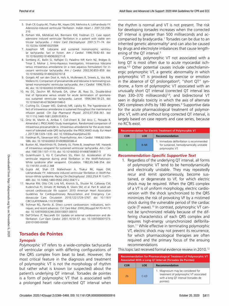
Panchal et al Adult Basic and Advanced Life Support: 2020 AHA Guidelines for CPR and ECC
Circulation. 2020;142(suppl 2):S366–S468. DOI: 10.1161/CIR.0000000000000916 October 20, 2020 S411
5. Shah CP, Gupta AK, Thakur RK, Hayes OW, Mehrotra A, Lokhandwala YY. Adenosine-induced ventricular fibrillation. Indian Heart J. 2001;53:208–210.
6. Parham WA, Mehdirad AA, Biermann KM, Fredman CS. Case report: adenosine induced ventricular fibrillation in a patient with stable ven-tricular tachycardia. J Interv Card Electrophysiol. 2001;5:71–74. doi: 10.1023/a:1009810025584
7. Josephson ME. Lidocaine and sustained monomorphic ventricu-lar tachycardia: fact or fiction. Am J Cardiol. 1996;78:82–83. doi: 10.1016/s0002-9149(96)00271-8
8. Somberg JC, Bailin SJ, Haffajee CI, Paladino WP, Kerin NZ, Bridges D, Timar S, Molnar J; Amio-Aqueous Investigators. Intravenous lidocaine versus intravenous amiodarone (in a new aqueous formulation) for in-cessant ventricular tachycardia. Am J Cardiol. 2002;90:853–859. doi: 10.1016/s0002-9149(02)02707-8
9. Gorgels AP, van den Dool A, Hofs A, Mulleneers R, Smeets JL, Vos MA, Wellens HJ. Comparison of procainamide and lidocaine in terminating sus-tained monomorphic ventricular tachycardia. Am J Cardiol. 1996;78:43–46. doi: 10.1016/s0002-9149(96)00224-x
10. Ho DS, Zecchin RP, Richards DA, Uther JB, Ross DL. Double-blind trial of lignocaine versus sotalol for acute termination of spontane-ous sustained ventricular tachycardia. Lancet. 1994;344:18–23. doi: 10.1016/s0140-6736(94)91048-0
11. Cushing DJ, Cooper WD, Gralinski MR, Lipicky RJ. The hypotensive ef-fect of intravenous amiodarone is sustained throughout the maintenance infusion period. Clin Exp Pharmacol Physiol. 2010;37:358–361. doi: 10.1111/j.1440-1681.2009.05303.x
12. Ortiz M, Martín A, Arribas F, Coll-Vinent B, Del Arco C, Peinado R, Almendral J; PROCAMIO Study Investigators. Randomized comparison of intravenous procainamide vs. intravenous amiodarone for the acute treat-ment of tolerated wide QRS tachycardia: the PROCAMIO study. Eur Heart J. 2017;38:1329–1335. doi: 10.1093/eurheartj/ehw230
13. Friedman PL, Stevenson WG. Proarrhythmia. Am J Cardiol. 1998;82:50N–58N. doi: 10.1016/s0002-9149(98)00586-4
14. Buxton AE, Marchlinski FE, Doherty JU, Flores B, Josephson ME. Hazards of intravenous verapamil for sustained ventricular tachycardia. Am J Car-diol. 1987;59:1107–1110. doi: 10.1016/0002-9149(87)90857-5
15. Gulamhusein S, Ko P, Carruthers SG, Klein GJ. Acceleration of the ventricular response during atrial fibrillation in the Wolff-Parkinson-White syndrome after verapamil. Circulation. 1982;65:348–354. doi: 10.1161/01.cir.65.2.348
16. Gupta AK, Shah CP, Maheshwari A, Thakur RK, Hayes OW, Lokhandwala YY. Adenosine induced ventricular fibrillation in Wolff-Par-kinson-White syndrome. Pacing Clin Electrophysiol. 2002;25(4 Pt 1):477–480. doi: 10.1046/j.1460-9592.2002.00477.x
17. Neumar RW, Otto CW, Link MS, Kronick SL, Shuster M, Callaway CW, Kudenchuk PJ, Ornato JP, McNally B, Silvers SM, et al. Part 8: adult ad-vanced cardiovascular life support: 2010 American Heart Association Guidelines for Cardiopulmonary Resuscitation and Emergency Car-diovascular Care. Circulation. 2010;122:S729–S767. doi: 10.1161/ CIRCULATIONAHA.110.970988
18. Trohman RG, Parrillo JE. Direct current cardioversion: indications, tech-niques, and recent advances. Crit Care Med. 2000;28(suppl):N170–N173. doi: 10.1097/00003246-200010001-00010
19. Dell’Orfano JT, Naccarelli GV. Update on external cardioversion and de-fibrillation. Curr Opin Cardiol. 2001;16:54–57. doi: 10.1097/00001573- 200101000-00008
Torsades de PointesSynopsisPolymorphic VT refers to a wide-complex tachycardia of ventricular origin with differing configurations of the QRS complex from beat to beat. However, the most critical feature in the diagnosis and treatment of polymorphic VT is not the morphology of rhythm but rather what is known (or suspected) about the patient’s underlying QT interval. Torsades de pointes is a form of polymorphic VT that is associated with a prolonged heart rate–corrected QT interval when
the rhythm is normal and VT is not present. The risk for developing torsades increases when the corrected QT interval is greater than 500 milliseconds and ac-companied by bradycardia.1 Torsades can be due to an inherited genetic abnormality2 and can also be caused by drugs and electrolyte imbalances that cause length-ening of the QT interval.3
Conversely, polymorphic VT not associated with a long QT is most often due to acute myocardial isch-emia.4,5 Other potential causes include catecholamin-ergic polymorphic VT, a genetic abnormality in which polymorphic VT is provoked by exercise or emotion in the absence of QT prolongation6; “short QT” syn-drome, a form of polymorphic VT associated with an unusually short QT interval (corrected QT interval less than 330–370 milliseconds)7,8; and bidirectional VT seen in digitalis toxicity in which the axis of alternate QRS complexes shifts by 180 degrees.9 Supportive data for the acute pharmacological treatment of polymor-phic VT, with and without long corrected QT interval, is largely based on case reports and case series, because no RCTs exist.
Recommendation for Electric Treatment of Polymorphic VT
COR LOE Recommendation
1 B-NR1. Immediate defibrillation is recommended
for sustained, hemodynamically unstable polymorphic VT.
Recommendation-Specific Supportive Text1. Regardless of the underlying QT interval, all forms
of polymorphic VT tend to be hemodynamically and electrically unstable. They may repeatedly recur and remit spontaneously, become sus-tained, or degenerate to VF, for which electric shock may be required. When the QRS complex of a VT is of uniform morphology, electric cardio-version with the shock synchronized to the QRS minimizes the risk of provoking VF by a mistimed shock during the vulnerable period of the cardiac cycle (T wave).10 In contrast, polymorphic VT can-not be synchronized reliably because of the dif-fering characteristics of each QRS complex and requires high-energy unsynchronized defibrilla-tion.11 While effective in terminating polymorphic VT, electric shock may not prevent its recurrence, for which pharmacological therapies are often required and the primary focus of the ensuing recommendations
This topic last received formal evidence review in 2010.12
Recommendation for Pharmacological Treatment of Polymorphic VT Associated With a Long QT Interval (Torsades De Pointes)
COR LOE Recommendation
2b C-LD
1. Magnesium may be considered for treatment of polymorphic VT associated with a long QT interval (torsades de pointes).
Dow
nloaded from http://ahajournals.org by on N
ovember 6, 2020

Panchal et al Adult Basic and Advanced Life Support: 2020 AHA Guidelines for CPR and ECC
October 20, 2020 Circulation. 2020;142(suppl 2):S366–S468. DOI: 10.1161/CIR.0000000000000916S412
Recommendation-Specific Supportive Text1. Torsades de pointes typically presents in a recur-
ring pattern of self-terminating, hemodynamically unstable polymorphic VT in context of a known or suspected long QT abnormality, often with an associated bradycardia. Immediate defibrilla-tion is the treatment of choice when torsades is sustained or degenerates to VF. However, termi-nation of torsades by shock does not prevent its recurrence, which requires additional measures. In small case series, IV magnesium has been effec-tive in suppressing and preventing recurrences of torsades.13–16 Magnesium is believed to suppress early afterdepolarizations, which are fluctuations in the myocardial action potential that can trigger the salvos of VT seen in torsades.17 Correcting any electrolyte abnormalities, particularly hypokalemia, is also advisable. Torsades is not treatable with anti-arrhythmic medications, which can themselves pro-long the QT interval and promote the arrhythmia. When given acutely, β-adrenergic blockers can also precipitate torsades by causing or worsening brady-cardia. In patients with bradycardia or pause-precip-itated torsades, expert consultation is best sought for additional measures such as overdrive pacing or isoproterenol,18–20 if needed. The use of magne-sium in torsades de pointes was addressed by the 2010 Guidelines and updated in a 2018 focused update on ACLS guidelines,21 with an interim evi-dence review that identified no new information that would modify previous recommendations.
This topic last received formal evidence review in 2010.12
Recommendations for Pharmacological Treatment of Polymorphic VT Not Associated With a Long QT Interval
COR LOE Recommendations
2b C-LD
1. IV lidocaine, amiodarone, and measures to treat myocardial ischemia may be considered to treat polymorphic VT in the absence of a prolonged QT interval.
3: No Benefit
C-LD2. We do not recommend routine use
of magnesium for the treatment of polymorphic VT with a normal QT interval.
Recommendation-Specific Supportive Text1. Polymorphic VT that is not associated with QT pro-
longation is often triggered by acute myocardial ischemia and infarction,4,5 often rapidly degen-erates into VF, and is treated similarly to other ventricular arrhythmias (VT and VF). However, termination of polymorphic VT with defibrilla-tion may not prevent its recurrence, which often requires additional measures. No RCTs have been performed to determine the best practice for pharmacological management of polymorphic VT. However measures to treat myocardial isch-emia (eg, β-adrenergic blockers or emergent
coronary intervention) as well as lidocaine and amiodarone may be effective22–29 in concert with defibrillation when the arrhythmia is sustained. β-Adrenergic blockers have also been shown to reduce the incidence of ventricular arrhythmias in acute coronary syndromes.30,31 Expert consulta-tion is advisable when other causes of polymor-phic VT are suspected, for which β-adrenergic blockers and antiarrhythmics may also have effi-cacy.6,32 This topic was last addressed by the 2010 Guidelines, with an interim evidence update that identified no new information that would mod-ify previous recommendations. Newer defined diagnostic entities causing polymorphic VT merit future evidence evaluation.
2. In the absence of long QT, magnesium has not been shown to be effective in the treat-ment of polymorphic VT 13 or to afford benefit in the acute management of other ventricular tachyarrhythmias.16
These recommendations are supported by the 2018 fo-cused update on ACLS guidelines.21
REFERENCES 1. Chan A, Isbister GK, Kirkpatrick CM, Dufful SB. Drug-induced QT pro-
longation and torsades de pointes: evaluation of a QT nomogram. QJM. 2007;100:609–615. doi: 10.1093/qjmed/hcm072
2. Saprungruang A, Khongphatthanayothin A, Mauleekoonphairoj J, Wandee P, Kanjanauthai S, Bhuiyan ZA, Wilde AAM, Poovorawan Y. Gen-otype and clinical characteristics of congenital long QT syndrome in Thai-land. Indian Pacing Electrophysiol J. 2018;18:165–171. doi: 10.1016/j.ipej. 2018.07.007
3. Drew BJ, Ackerman MJ, Funk M, Gibler WB, Kligfield P, Menon V, Philippides GJ, Roden DM, Zareba W; American Heart Association Acute Cardiac Care Committee of the Council on Clinical Cardiology; Council on Cardiovascular Nursing; American College of Cardiology Foundation. Prevention of torsade de pointes in hospital settings: a scientific state-ment from the American Heart Association and the American College of Cardiology Foundation. J Am Coll Cardiol. 2010;55:934–947. doi: 10.1016/j.jacc.2010.01.001
4. Pogwizd SM, Corr PB. Electrophysiologic mechanisms underlying ar-rhythmias due to reperfusion of ischemic myocardium. Circulation. 1987;76:404–426. doi: 10.1161/01.cir.76.2.404
5. Wolfe CL, Nibley C, Bhandari A, Chatterjee K, Scheinman M. Polymor-phous ventricular tachycardia associated with acute myocardial infarction. Circulation. 1991;84:1543–1551. doi: 10.1161/01.cir.84.4.1543
6. Liu N, Ruan Y, Priori SG. Catecholaminergic polymorphic ventricular tachycardia. Prog Cardiovasc Dis. 2008;51:23–30. doi: 10.1016/j.pcad. 2007.10.005
7. Cross B, Homoud M, Link M, Foote C, Garlitski AC, Weinstock J, Estes NA III. The short QT syndrome. J Interv Card Electrophysiol. 2011;31:25–31. doi: 10.1007/s10840-011-9566-0
8. Gollob MH, Redpath CJ, Roberts JD. The short QT syndrome: pro-posed diagnostic criteria. J Am Coll Cardiol. 2011;57:802–812. doi: 10.1016/j.jacc.2010.09.048
9. Chapman M, Hargreaves M, Schneider H, Royle M. Bidirectional ven-tricular tachycardia associated with digoxin toxicity and with normal digoxin levels. Heart Rhythm. 2014;11:1222–1225. doi: 10.1016/j. hrthm.2014.03.050
10. Trohman RG, Parrillo JE. Direct current cardioversion: indications, tech-niques, and recent advances. Crit Care Med. 2000;28(suppl):N170–N173. doi: 10.1097/00003246-200010001-00010
11. Dell’Orfano JT, Naccarelli GV. Update on external cardioversion and de-fibrillation. Curr Opin Cardiol. 2001;16:54–57. doi: 10.1097/00001573- 200101000-00008
Dow
nloaded from http://ahajournals.org by on N
ovember 6, 2020

Panchal et al Adult Basic and Advanced Life Support: 2020 AHA Guidelines for CPR and ECC
Circulation. 2020;142(suppl 2):S366–S468. DOI: 10.1161/CIR.0000000000000916 October 20, 2020 S413
12. Neumar RW, Otto CW, Link MS, Kronick SL, Shuster M, Callaway CW, Kudenchuk PJ, Ornato JP, McNally B, Silvers SM, et al. Part 8: adult ad-vanced cardiovascular life support: 2010 American Heart Association Guidelines for Cardiopulmonary Resuscitation and Emergency Car-diovascular Care. Circulation. 2010;122:S729–S767. doi: 10.1161/ CIRCULATIONAHA.110.970988
13. Tzivoni D, Banai S, Schuger C, Benhorin J, Keren A, Gottlieb S, Stern S. Treatment of torsade de pointes with magnesium sulfate. Circulation. 1988;77:392–397. doi: 10.1161/01.cir.77.2.392
14. Tzivoni D, Keren A, Cohen AM, Loebel H, Zahavi I, Chenzbraun A, Stern S. Magnesium therapy for torsades de pointes. Am J Cardiol. 1984;53:528–530. doi: 10.1016/0002-9149(84)90025-0
15. Hoshino K, Ogawa K, Hishitani T, Isobe T, Etoh Y. Successful uses of mag-nesium sulfate for torsades de pointes in children with long QT syndrome. Pediatr Int. 2006;48:112–117. doi: 10.1111/j.1442-200X.2006.02177.x
16. Manz M, Jung W, Lüderitz B. Effect of magnesium on sustained ven-tricular tachycardia [in German]. Herz. 1997;22(suppl 1):51–55. doi: 10.1007/bf03042655
17. Baker WL. Treating arrhythmias with adjunctive magnesium: identify-ing future research directions. Eur Heart J Cardiovasc Pharmacother. 2017;3:108–117. doi: 10.1093/ehjcvp/pvw028
18. DiSegni E, Klein HO, David D, Libhaber C, Kaplinsky E. Overdrive pacing in quinidine syncope and other long QT-interval syndromes. Arch Intern Med. 1980;140:1036–1040.
19. Damiano BP, Rosen MR. Effects of pacing on triggered activity induced by early afterdepolarizations. Circulation. 1984;69:1013–1025. doi: 10.1161/01.cir.69.5.1013
20. Suarez K, Mack R, Hardegree EL, Chiles C, Banchs JE, Gonzalez MD. Isoproterenol suppresses recurrent torsades de pointes in a patient with long QT syndrome type 2. HeartRhythm Case Rep. 2018;4:576–579. doi: 10.1016/j.hrcr.2018.08.013
21. Panchal AR, Berg KM, Kudenchuk PJ, Del Rios M, Hirsch KG, Link MS, Kurz MC, Chan PS, Cabañas JG, Morley PT, Hazinski MF, Donnino MW. 2018 American Heart Association Focused Update on Advanced Car-diovascular Life Support Use of Antiarrhythmic Drugs During and Immediately After Cardiac Arrest: An Update to the American Heart Association Guidelines for Cardiopulmonary Resuscitation and Emer-gency Cardiovascular Care. Circulation. 2018;138:e740–e749. doi: 10.1161/CIR.0000000000000613
22. Vrana M, Pokorny J, Marcian P, Fejfar Z. Class I and III antiarrhythmic drugs for prevention of sudden cardiac death and management of postmyocar-dial infarction arrhythmias. A review. Biomed Pap Med Fac Univ Palacky Olomouc Czech Repub. 2013;157:114–124. doi: 10.5507/bp.2013.030
23. Nalliah CJ, Zaman S, Narayan A, Sullivan J, Kovoor P. Coronary artery re-perfusion for ST elevation myocardial infarction is associated with shorter cycle length ventricular tachycardia and fewer spontaneous arrhythmias. Europace. 2014;16:1053–1060. doi: 10.1093/europace/eut307
24. Brady W, Meldon S, DeBehnke D. Comparison of prehospital mono-morphic and polymorphic ventricular tachycardia: prevalence, response to therapy, and outcome. Ann Emerg Med. 1995;25:64–70. doi: 10.1016/s0196-0644(95)70357-8
25. Brady WJ, DeBehnke DJ, Laundrie D. Prevalence, therapeutic response, and outcome of ventricular tachycardia in the out-of-hospital setting: a comparison of monomorphic ventricular tachycardia, polymorphic ventricular tachycardia, and torsades de pointes. Acad Emerg Med. 1999;6:609–617. doi: 10.1111/j.1553-2712.1999.tb00414.x
26. Luqman N, Sung RJ, Wang CL, Kuo CT. Myocardial ischemia and ven-tricular fibrillation: pathophysiology and clinical implications. Int J Cardiol. 2007;119:283–290. doi: 10.1016/j.ijcard.2006.09.016
27. Gorenek B, Lundqvist CB, Terradellas JB, Camm AJ, Hindricks G, Huber K, Kirchhof P, Kuck KH, Kudaiberdieva G, Lin T, Raviele A, Santini M, Tilz RR, Valgimigli M, Vos MA, Vrints C, Zeymer U. Cardiac arrhythmias in acute coronary syndromes: position paper from the joint EHRA, ACCA, and EAPCI task force. Eur Heart J Acute Cardiovasc Care. 2015;4:386. doi: 10.1177/2048872614550583
28. Carmeliet E. Cardiac ionic currents and acute ischemia: from channels to arrhythmias. Physiol Rev. 1999;79:917–1017. doi: 10.1152/physrev. 1999.79.3.917
29. Steg PG, James SK, Atar D, Badano LP, Blömstrom-Lundqvist C, Borger MA, Di Mario C, Dickstein K, Ducrocq G, Fernandez-Aviles F, et al; and the Task Force on the management of ST-segment elevation acute myocardial infarction of the European Society of Cardiology. ESC Guide-lines for the management of acute myocardial infarction in patients
presenting with ST-segment elevation. Eur Heart J. 2012;33:2569–2619. doi: 10.1093/eurheartj/ehs215
30. Al-Khatib SM, Stevenson WG, Ackerman MJ, Bryant WJ, Callans DJ, Curtis AB, Deal BJ, Dickfeld T, Field ME, Fonarow GC, et al. 2017 AHA/ACC/HRS guideline for management of patients with ventricular ar-rhythmias and the prevention of sudden cardiac death: A report of the American College of Cardiology/American Heart Association Task Force on Clinical Practice Guidelines and the Heart Rhythm Society. Circulation. 2018;138:e272–e391. doi: 10.1161/CIR.0000000000000549
31. Chatterjee S, Chaudhuri D, Vedanthan R, Fuster V, Ibanez B, Bangalore S, Mukherjee D. Early intravenous beta-blockers in patients with acute coronary syndrome—a meta-analysis of randomized trials. Int J Cardiol. 2013;168:915–921. doi: 10.1016/j.ijcard.2012.10.050
32. Van Houzen NE, Alsheikh-Ali AA, Garlitski AC, Homoud MK, Weinstock J, Link MS, Estes NA III. Short QT syndrome review. J Interv Card Electro-physiol. 2008;23:1–5. doi: 10.1007/s10840-008-9201-x
Regular Narrow-Complex TachycardiaIntroductionManagement of SVTs is the subject of a recent joint treatment guideline from the AHA, the American Col-lege of Cardiology, and the Heart Rhythm Society.1
Narrow-complex tachycardia represents a range of tachyarrhythmias originating from a circuit or focus involving the atria or the AV node. Clinicians must determine if the tachycardia is narrow-complex or wide-complex tachycardia and if it has a regular or irregular rhythm. For patients with a sinus tachy-cardia (heart rate greater than 100/min, P waves), no specific drug treatment is needed, and clinicians should focus on identification and treatment of the underlying cause of the tachycardia (fever, dehy-dration, pain). If the patient presents with SVT, the primary goal of treatment is to quickly identify and treat patients who are hemodynamically unstable (ischemic chest pain, altered mental status, shock, hypotension, acute heart failure) or symptomatic due to the arrhythmia. Synchronized cardioversion or drugs or both may be used to control unstable or symptomatic regular narrow-complex tachycar-dia. The available evidence suggests no appreciable differences in success or major adverse event rates between calcium channel blockers and adenosine.2
In patients with narrow-complex tachycardia who are refractory to the measures described, this may indi-cate a more complicated rhythm abnormality for which expert consultation may be advisable.
Recommendations for Electric Therapies for Regular Narrow-Complex Tachycardia
COR LOE Recommendations
1 B-NR
1. Synchronized cardioversion is recommended for acute treatment in patients with hemodynamically unstable SVT.
1 B-NR
2. Synchronized cardioversion is recommended for acute treatment in patients with hemodynamically stable SVT when vagal maneuvers and pharmacological therapy is ineffective or contraindicated.
Dow
nloaded from http://ahajournals.org by on N
ovember 6, 2020
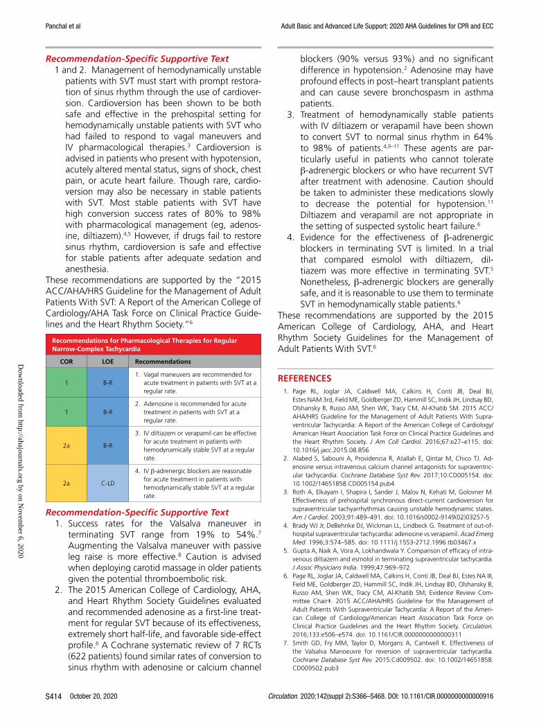
Panchal et al Adult Basic and Advanced Life Support: 2020 AHA Guidelines for CPR and ECC
October 20, 2020 Circulation. 2020;142(suppl 2):S366–S468. DOI: 10.1161/CIR.0000000000000916S414
Recommendation-Specific Supportive Text1 and 2. Management of hemodynamically unstable
patients with SVT must start with prompt restora-tion of sinus rhythm through the use of cardiover-sion. Cardioversion has been shown to be both safe and effective in the prehospital setting for hemodynamically unstable patients with SVT who had failed to respond to vagal maneuvers and IV pharmacological therapies.3 Cardioversion is advised in patients who present with hypotension, acutely altered mental status, signs of shock, chest pain, or acute heart failure. Though rare, cardio-version may also be necessary in stable patients with SVT. Most stable patients with SVT have high conversion success rates of 80% to 98% with pharmacological management (eg, adenos-ine, diltiazem).4,5 However, if drugs fail to restore sinus rhythm, cardioversion is safe and effective for stable patients after adequate sedation and anesthesia.
These recommendations are supported by the “2015 ACC/AHA/HRS Guideline for the Management of Adult Patients With SVT: A Report of the American College of Cardiology/AHA Task Force on Clinical Practice Guide-lines and the Heart Rhythm Society.”6
Recommendations for Pharmacological Therapies for Regular Narrow-Complex Tachycardia
COR LOE Recommendations
1 B-R1. Vagal maneuvers are recommended for
acute treatment in patients with SVT at a regular rate.
1 B-R2. Adenosine is recommended for acute
treatment in patients with SVT at a regular rate.
2a B-R
3. IV diltiazem or verapamil can be effective for acute treatment in patients with hemodynamically stable SVT at a regular rate.
2a C-LD
4. IV β-adrenergic blockers are reasonable for acute treatment in patients with hemodynamically stable SVT at a regular rate.
Recommendation-Specific Supportive Text1. Success rates for the Valsalva maneuver in
terminating SVT range from 19% to 54%.7 Augmenting the Valsalva maneuver with passive leg raise is more effective.8 Caution is advised when deploying carotid massage in older patients given the potential thromboembolic risk.
2. The 2015 American College of Cardiology, AHA, and Heart Rhythm Society Guidelines evaluated and recommended adenosine as a first-line treat-ment for regular SVT because of its effectiveness, extremely short half-life, and favorable side-effect profile.6 A Cochrane systematic review of 7 RCTs (622 patients) found similar rates of conversion to sinus rhythm with adenosine or calcium channel
blockers (90% versus 93%) and no significant difference in hypotension.2 Adenosine may have profound effects in post–heart transplant patients and can cause severe bronchospasm in asthma patients.
3. Treatment of hemodynamically stable patients with IV diltiazem or verapamil have been shown to convert SVT to normal sinus rhythm in 64% to 98% of patients.4,9–11 These agents are par-ticularly useful in patients who cannot tolerate β-adrenergic blockers or who have recurrent SVT after treatment with adenosine. Caution should be taken to administer these medications slowly to decrease the potential for hypotension.11 Diltiazem and verapamil are not appropriate in the setting of suspected systolic heart failure.6
4. Evidence for the effectiveness of β-adrenergic blockers in terminating SVT is limited. In a trial that compared esmolol with diltiazem, dil-tiazem was more effective in terminating SVT.5 Nonetheless, β-adrenergic blockers are generally safe, and it is reasonable to use them to terminate SVT in hemodynamically stable patients.6
These recommendations are supported by the 2015 American College of Cardiology, AHA, and Heart Rhythm Society Guidelines for the Management of Adult Patients With SVT.6
REFERENCES 1. Page RL, Joglar JA, Caldwell MA, Calkins H, Conti JB, Deal BJ,
Estes NAM 3rd, Field ME, Goldberger ZD, Hammill SC, Indik JH, Lindsay BD, Olshansky B, Russo AM, Shen WK, Tracy CM, Al-Khatib SM. 2015 ACC/AHA/HRS Guideline for the Management of Adult Patients With Supra-ventricular Tachycardia: A Report of the American College of Cardiology/American Heart Association Task Force on Clinical Practice Guidelines and the Heart Rhythm Society. J Am Coll Cardiol. 2016;67:e27–e115. doi: 10.1016/j.jacc.2015.08.856
2. Alabed S, Sabouni A, Providencia R, Atallah E, Qintar M, Chico TJ. Ad-enosine versus intravenous calcium channel antagonists for supraventric-ular tachycardia. Cochrane Database Syst Rev. 2017;10:CD005154. doi: 10.1002/14651858.CD005154.pub4
3. Roth A, Elkayam I, Shapira I, Sander J, Malov N, Kehati M, Golovner M. Effectiveness of prehospital synchronous direct-current cardioversion for supraventricular tachyarrhythmias causing unstable hemodynamic states. Am J Cardiol. 2003;91:489–491. doi: 10.1016/s0002-9149(02)03257-5
4. Brady WJ Jr, DeBehnke DJ, Wickman LL, Lindbeck G. Treatment of out-of-hospital supraventricular tachycardia: adenosine vs verapamil. Acad Emerg Med. 1996;3:574–585. doi: 10.1111/j.1553-2712.1996.tb03467.x
5. Gupta A, Naik A, Vora A, Lokhandwala Y. Comparison of efficacy of intra-venous diltiazem and esmolol in terminating supraventricular tachycardia. J Assoc Physicians India. 1999;47:969–972.
6. Page RL, Joglar JA, Caldwell MA, Calkins H, Conti JB, Deal BJ, Estes NA III, Field ME, Goldberger ZD, Hammill SC, Indik JH, Lindsay BD, Olshansky B, Russo AM, Shen WK, Tracy CM, Al-Khatib SM; Evidence Review Com-mittee Chair‡. 2015 ACC/AHA/HRS Guideline for the Management of Adult Patients With Supraventricular Tachycardia: A Report of the Ameri-can College of Cardiology/American Heart Association Task Force on Clinical Practice Guidelines and the Heart Rhythm Society. Circulation. 2016;133:e506–e574. doi: 10.1161/CIR.0000000000000311
7. Smith GD, Fry MM, Taylor D, Morgans A, Cantwell K. Effectiveness of the Valsalva Manoeuvre for reversion of supraventricular tachycardia. Cochrane Database Syst Rev. 2015:Cd009502. doi: 10.1002/14651858. CD009502.pub3
Dow
nloaded from http://ahajournals.org by on N
ovember 6, 2020

Panchal et al Adult Basic and Advanced Life Support: 2020 AHA Guidelines for CPR and ECC
Circulation. 2020;142(suppl 2):S366–S468. DOI: 10.1161/CIR.0000000000000916 October 20, 2020 S415
8. Appelboam A, Reuben A, Mann C, Gagg J, Ewings P, Barton A, Lobban T, Dayer M, Vickery J, Benger J; REVERT trial collaborators. Postural modifi-cation to the standard Valsalva manoeuvre for emergency treatment of supraventricular tachycardias (REVERT): a randomised controlled trial. Lan-cet. 2015;386:1747–1753. doi: 10.1016/S0140-6736(15)61485-4
9. Lim SH, Anantharaman V, Teo WS, Chan YH. Slow infusion of calcium channel blockers compared with intravenous adenosine in the emergency treatment of supraventricular tachycardia. Resuscitation. 2009;80:523–528. doi: 10.1016/j.resuscitation.2009.01.017
10. Madsen CD, Pointer JE, Lynch TG. A comparison of adenosine and vera-pamil for the treatment of supraventricular tachycardia in the prehos-pital setting. Ann Emerg Med. 1995;25:649–655. doi: 10.1016/s0196- 0644(95)70179-6
11. Lim SH, Anantharaman V, Teo WS. Slow-infusion of calcium chan-nel blockers in the emergency management of supraventricular tachycardia. Resuscitation. 2002;52:167–174. doi: 10.1016/s0300- 9572(01)00459-2
Atrial Fibrillation or Flutter With Rapid Ventricular ResponseIntroductionAtrial fibrillation is an SVT consisting of disorganized atrial electric activation and uncoordinated atrial con-traction. Atrial flutter is an SVT with a macroreentrant circuit resulting in rapid atrial activation but intermit-tent ventricular response. These arrhythmias are com-mon and often coexist, and their treatment recommen-dations are similar.
Treatment of atrial fibrillation/flutter depends on the hemodynamic stability of the patient as well as prior history of arrhythmia, comorbidities, and respon-siveness to medication. Hemodynamically unstable patients and those with rate-related ischemia should receive urgent electric cardioversion. Hemodynamically stable patients can be treated with a rate-control or rhythm-control strategy. Rate control is more common in the emergency setting, using IV administration of a nondihydropyridine calcium channel antagonist (eg, diltiazem, verapamil) or a β-adrenergic blocker (eg, metoprolol, esmolol). While amiodarone is typically considered a rhythm-control agent, it can effectively re-duce ventricular rate with potential use in patients with congestive heart failure where β-adrenergic blockers may not be tolerated and nondihydropyridine calcium channel antagonists are contraindicated. Long-term anticoagulation may be necessary for patients at risk for thromboembolic events based on their CHA2DS2-VASc score. The choice of anticoagulation is beyond the scope of these guidelines.
The rhythm-control strategy (sometimes called chemical cardioversion) includes antiarrhythmic medi-cations given to convert the rhythm to sinus and/or pre-vent recurrent atrial fibrillation/flutter (Table 3). Patient selection, evaluation, timing, drug selection, and anti-coagulation for patients undergoing rhythm control are beyond the scope of these guidelines and are presented elsewhere.1,2
The management of patients with preexcitation syn-dromes (aka Wolff-Parkinson-White) is covered in the Wide-Complex Tachycardia section.
Recommendations for Electric Therapies for Atrial Fibrillation/Flutter
COR LOE Recommendations
1 C-LD
1. Hemodynamically unstable patients with atrial fibrillation or atrial flutter with rapid ventricular response should receive electric cardioversion.
1 C-LD
2. Urgent direct-current cardioversion of new-onset atrial fibrillation in the setting of acute coronary syndrome is recommended for patients with hemodynamic compromise, ongoing ischemia, or inadequate rate control.
2a C-LD
3. For synchronized cardioversion of atrial fibrillation using biphasic energy, an initial energy of 120 to 200 J is reasonable, depending on the specific biphasic defibrillator being used.
2b C-LD
4. For synchronized cardioversion of atrial flutter using biphasic energy, an initial energy of 50 to 100 J may be reasonable, depending on the specific biphasic defibrillator being used.
Recommendation-Specific Supportive Text1 and 2. Uncontrolled tachycardia may impair ven-
tricular filling, cardiac output, and coronary perfu-sion while increasing myocardial oxygen demand. While an expeditious trial of medications and/or fluids may be appropriate in some cases, unstable patients or patients with ongoing cardiac ischemia with atrial fibrillation or atrial flutter need to be car-dioverted promptly.1–3 When making the decision for cardioversion, one should also consider whether the arrhythmia is the cause of the tachycardia. Potential exacerbation of rapid ventricular response by secondary causes (eg, sepsis) should be consid-ered and may inform initial attempts at hemody-namic stabilization with pharmacotherapy. There are few data addressing these strategies in hemo-dynamically unstable patients. However, studies demonstrating hemodynamic benefits of success-ful cardioversion have been published.4,5 In addi-tion, risks of hypotension and hypoperfusion with use of negative inotropes have been demonstrated even in normotensive patients.6–8 Hemodynamically unstable patients and those with ongoing cardiac ischemia are likely to benefit from the improved hemodynamic status associated with restoration of sinus rhythm and avoidance of hypotension caused by the alternative pharmacological therapies. Depending on the clinical scenario, patients cardio-verted from atrial fibrillation or atrial flutter of 48 hours’ duration or longer are candidates for antico-agulation. Details about anticoagulation selection can be found elsewhere.2
Dow
nloaded from http://ahajournals.org by on N
ovember 6, 2020

October 20, 2020 Circulation. 2020;142(suppl 2):S366–S468. DOI: 10.1161/CIR.0000000000000916S416
Panchal et al Adult Basic and Advanced Life Support: 2020 AHA Guidelines for CPR and ECC
3 and 4. The electric energy required to successfully cardiovert a patient from atrial fibrillation or atrial flutter to sinus rhythm varies and is gener-ally less in patients with new-onset arrhythmia, thin body habitus, and when biphasic wave-form shocks are delivered.9–15 Obese patients may require greater energy.16 If initial cardio-version is unsuccessful, energy is increased in subsequent attempts. Less energy is generally required for atrial flutter than for atrial fibril-lation.11 Higher energies of 200 J or more are associated with improved first shock success and decreased total energy delivery. In addi-tion, a retrospective analysis found that lower energy shocks were associated with higher risk of cardioversion-induced VF.17 Previous guide-lines included a comparison of monophasic and biphasic waveforms. This recommendation now focuses primarily on biphasic waveforms. Recommended energy levels vary with differ-ent devices, reducing the validity of generalized recommendations. This topic requires further study with a comprehensive systematic review to better understand the optimal electric doses with current devices. The writing group assess-ment of the LOE as C-LD is consistent with the limited evidence using modern devices and energy waveforms.
These recommendations are supported by the “2014 AHA/ACC/HRS Guideline for the Management of Pa-tients With Atrial Fibrillation: A Report of the Ameri-can College of Cardiology/AHA Task Force on Practice Guidelines and the Heart Rhythm Society”18 as well as the focused update of those guidelines published in 2019.2
Recommendations for Medical Therapies for Atrial Fibrillation/Flutter
COR LOE Recommendations
1 B-NR
1. IV administration of a β-adrenergic blocker or nondihydropyridine calcium channel antagonist is recommended to slow the ventricular heart rate in the acute setting in patients with atrial fibrillation or atrial flutter with rapid ventricular response without preexcitation.
2a B-NR
2. IV amiodarone can be useful for rate control in critically ill patients with atrial fibrillation with rapid ventricular response without preexcitation.
3: Harm C-LD
3. In patients with atrial fibrillation and atrial flutter in the setting of preexcitation, digoxin, nondihydropyridine calcium channel antagonists, β-adrenergic blockers, and IV amiodarone should not be administered because they may increase the ventricular response and result in VF.
3: Harm C-EO
4. Nondihydropyridine calcium channel antagonists and IV β-adrenergic blockers should not be used in patients with left ventricular systolic dysfunction and decompensated heart failure because these may lead to further hemodynamic compromise.
Recommendation-Specific Supportive Text1 and 2. Clinical trial evidence shows that nondihydro-
pyridine calcium channel antagonists (eg, diltiazem, verapamil), β-adrenergic blockers (eg, esmolol, pro-pranolol), amiodarone, and digoxin are all effective for rate control in patients with atrial fibrillation/flutter.6–8,19–23 Calcium channel blockers may be more effective than amiodarone, and cause more hypotension.6 Digoxin is rarely used in the acute setting because of slow onset of effect.1,2
Table 3. IV Medications Commonly Used for Acute Rate Control in Atrial Fibrillation and Atrial Flutter18
Medication Bolus Dose Infusion Rate Notes
Nondihydropyridine Calcium Channel Blockers
Diltiazem 0.25 mg/kg IV bolus over 2 min 5–10 mg/h Avoid in hypotension, heart failure, cardiomyopathy, and acute coronary syndromes
Verapamil 0.075–0.15 mg/kg IV bolus over 2 min; may give an additional dose after 30 min if no response
0.005 mg/kg per min Avoid in hypotension, heart failure, cardiomyopathy, acute and coronary syndromes
β-Adrenergic Blockers
Metoprolol 2.5–5 mg over 2 min, up to 3 doses Avoid in decompensated heart failure
Esmolol 500 μg/kg IV over 1 min 50–300 μg/kg per min Short duration of action; avoid in decompensated heart failure
Propranolol 1 mg IV over 1 min, up to 3 doses Avoid in decompensated heart failure
Other Medications
Amiodarone 300 mg IV over 1 h 10–50 mg/h over 24 h Multiple dosing schemes exist for amiodarone
Digoxin 0.25 mg IV, repeated to maximum dose 1.5 mg over 24 h
Typically used as adjunctive therapy with another option from above; caution in patients with renal impairment
IV indicates intravenous.
Dow
nloaded from http://ahajournals.org by on N
ovember 6, 2020

Panchal et al Adult Basic and Advanced Life Support: 2020 AHA Guidelines for CPR and ECC
Circulation. 2020;142(suppl 2):S366–S468. DOI: 10.1161/CIR.0000000000000916 October 20, 2020 S417
3. Based on limited case reports and small case series, there is concern that patients with con-comitant preexcitation and atrial fibrillation or atrial flutter may develop VF in response to accel-erated ventricular response after the administra-tion of AV nodal blocking agents such as digoxin, nondihydropyridine calcium channel antagonists, β-adrenergic blockers, or IV amiodarone.24–27 In this setting, cardioversion is recommended as the most appropriate management.
4. Because of their negative inotropic effect, non-dihydropyridine calcium channel antagonists (eg, diltiazem, verapamil) may further decompensate patients with left ventricular systolic dysfunction and symptomatic heart failure. They may be used in patients with heart failure with preserved ejec-tion fraction. β-Adrenergic blockers may be used in compensated patients with cardiomyopathy; however, they should be used with caution or avoided altogether in patients with decompen-sated heart failure. This recommendation is based on expert consensus and pathophysiologic ratio-nale.2,18,28 β-Adrenergic blockers may be used in patients with chronic obstructive pulmonary dis-ease because multiple studies have shown no negative effects.29
These recommendations are supported by 2014 AHA, American College of Cardiology, and Heart Rhythm So-ciety Guideline for the Management of Patients With Atrial Fibrillation18 as well as the focused update of those guidelines published in 2019.2
REFERENCES 1. January CT, Wann LS, Alpert JS, Calkins H, Cigarroa JE, Cleveland JC Jr,
Conti JB, Ellinor PT, Ezekowitz MD, Field ME, Murray KT, Sacco RL, Stevenson WG, Tchou PJ, Tracy CM, Yancy CW; ACC/AHA Task Force Members. 2014 AHA/ACC/HRS guideline for the management of patients with atrial fibrillation: executive summary: a report of the American College of Cardiology/American Heart Association Task Force on practice guidelines and the Heart Rhythm Society. Circulation. 2014;130:2071–2104. doi: 10.1161/CIR.0000000000000040
2. January CT, Wann LS, Calkins H, Chen LY, Cigarroa JE, Cleveland JC Jr, Ellinor PT, Ezekowitz MD, Field ME, Furie KL, Heidenreich PA, Murray KT, Shea JB, Tracy CM, Yancy CW. 2019 AHA/ACC/HRS Focused Update of the 2014 AHA/ACC/HRS Guideline for the Management of Pa-tients With Atrial Fibrillation: A Report of the American College of Cardiology/American Heart Association Task Force on Clinical Practice Guidelines and the Heart Rhythm Society in Collaboration With the Society of Thoracic Surgeons. Circulation. 2019;140:e125–e151. doi: 10.1161/CIR.0000000000000665
3. McMurray J, Køber L, Robertson M, Dargie H, Colucci W, Lopez-Sendon J, Remme W, Sharpe DN, Ford I. Antiarrhythmic effect of carvedilol after acute myocardial infarction: results of the Carvedilol Post-Infarct Survival Control in Left Ventricular Dysfunction (CAPRICORN) trial. J Am Coll Car-diol. 2005;45:525–530. doi: 10.1016/j.jacc.2004.09.076
4. DeMaria AN, Lies JE, King JF, Miller RR, Amsterdam EA, Mason DT. Echo-graphic assessment of atrial transport, mitral movement, and ventricular performance following electroversion of supraventricular arrhythmias. Cir-culation. 1975;51:273–282. doi: 10.1161/01.cir.51.2.273
5. Raymond RJ, Lee AJ, Messineo FC, Manning WJ, Silverman DI. Cardiac performance early after cardioversion from atrial fibrillation. Am Heart J. 1998;136:435–442. doi: 10.1016/s0002-8703(98)70217-0
6. Delle Karth G, Geppert A, Neunteufl T, Priglinger U, Haumer M, Gschwandtner M, Siostrzonek P, Heinz G. Amiodarone versus diltiazem for rate control in critically ill patients with atrial tachyarrhythmias. Crit Care Med. 2001;29:1149–1153. doi: 10.1097/00003246-200106000-00011
7. Platia EV, Michelson EL, Porterfield JK, Das G. Esmolol versus verapamil in the acute treatment of atrial fibrillation or atrial flutter. Am J Cardiol. 1989;63:925–929. doi: 10.1016/0002-9149(89)90141-0
8. Ellenbogen KA, Dias VC, Plumb VJ, Heywood JT, Mirvis DM. A placebo-con-trolled trial of continuous intravenous diltiazem infusion for 24-hour heart rate control during atrial fibrillation and atrial flutter: a multicenter study. J Am Coll Cardiol. 1991;18:891–897. doi: 10.1016/0735-1097(91)90743-s
9. Glover BM, Walsh SJ, McCann CJ, Moore MJ, Manoharan G, Dalzell GW, McAllister A, McClements B, McEneaney DJ, Trouton TG, Mathew TP, Adgey AA. Biphasic energy selection for transthoracic cardio-version of atrial fibrillation. The BEST AF Trial. Heart. 2008;94:884–887. doi: 10.1136/hrt.2007.120782
10. Inácio JF, da Rosa Mdos S, Shah J, Rosário J, Vissoci JR, Manica AL, Rodrigues CG. Monophasic and biphasic shock for transthoracic conver-sion of atrial fibrillation: systematic review and network meta-analysis. Resuscitation. 2016;100:66–75. doi: 10.1016/j.resuscitation.2015.12.009
11. Gallagher MM, Guo XH, Poloniecki JD, Guan Yap Y, Ward D, Camm AJ. Initial energy setting, outcome and efficiency in direct current cardiover-sion of atrial fibrillation and flutter. J Am Coll Cardiol. 2001;38:1498–1504. doi: 10.1016/s0735-1097(01)01540-6
12. Scholten M, Szili-Torok T, Klootwijk P, Jordaens L. Comparison of mono-phasic and biphasic shocks for transthoracic cardioversion of atrial fibrilla-tion. Heart. 2003;89:1032–1034. doi: 10.1136/heart.89.9.1032
13. Page RL, Kerber RE, Russell JK, Trouton T, Waktare J, Gallik D, Olgin JE, Ricard P, Dalzell GW, Reddy R, Lazzara R, Lee K, Carlson M, Halperin B, Bardy GH; BiCard Investigators. Biphasic versus monophasic shock wave-form for conversion of atrial fibrillation: the results of an international ran-domized, double-blind multicenter trial. J Am Coll Cardiol. 2002;39:1956–1963. doi: 10.1016/s0735-1097(02)01898-3
14. Reisinger J, Gstrein C, Winter T, Zeindlhofer E, Höllinger K, Mori M, Schiller A, Winter A, Geiger H, Siostrzonek P. Optimization of initial en-ergy for cardioversion of atrial tachyarrhythmias with biphasic shocks. Am J Emerg Med. 2010;28:159–165. doi: 10.1016/j.ajem.2008.10.028
15. Alatawi F, Gurevitz O, White RD, Ammash NM, Malouf JF, Bruce CJ, Moon BS, Rosales AG, Hodge D, Hammill SC, Gersh BJ, Friedman PA. Prospective, randomized comparison of two biphasic waveforms for the efficacy and safety of transthoracic biphasic cardioversion of atrial fibrilla-tion. Heart Rhythm. 2005;2:382–387. doi: 10.1016/j.hrthm.2004.12.024
16. Voskoboinik A, Moskovitch J, Plunkett G, Bloom J, Wong G, Nalliah C, Prabhu S, Sugumar H, Paramasweran R, McLellan A, et al. Cardioversion of atrial fibrillation in obese patients: Results from the Cardioversion-BMI randomized controlled trial. J Cardiovasc Electrophysiol. 2019;30:155–161. doi: 10.1111/jce.13786
17. Gallagher MM, Yap YG, Padula M, Ward DE, Rowland E, Camm AJ. Ar-rhythmic complications of electrical cardioversion: relationship to shock en-ergy. Int J Cardiol. 2008;123:307–312. doi: 10.1016/j.ijcard.2006.12.014
18. January CT, Wann LS, Alpert JS, Calkins H, Cigarroa JE, Cleveland JC Jr, Conti JB, Ellinor PT, Ezekowitz MD, Field ME, et al. 2014 AHA/ACC/HRS guideline for the management of patients with atrial fibrillation: a report of the American College of Cardiology/American Heart Association Task Force on practice guidelines and the Heart Rhythm Society. Circulation. 2014;130:e199–e267. doi: 10.1161/CIR.0000000000000041
19. Abrams J, Allen J, Allin D, Anderson J, Anderson S, Blanski L, Chadda K, DiBianco R, Favrot L, Gonzalez J. Efficacy and safety of esmolol vs pro-pranolol in the treatment of supraventricular tachyarrhythmias: a mul-ticenter double-blind clinical trial. Am Heart J. 1985;110:913–922. doi: 10.1016/0002-8703(85)90185-1
20. Siu CW, Lau CP, Lee WL, Lam KF, Tse HF. Intravenous diltiazem is superior to intravenous amiodarone or digoxin for achieving ventricular rate con-trol in patients with acute uncomplicated atrial fibrillation. Crit Care Med. 2009;37:2174–9; quiz 2180. doi: 10.1097/CCM.0b013e3181a02f56
21. Clemo HF, Wood MA, Gilligan DM, Ellenbogen KA. Intravenous amiodarone for acute heart rate control in the critically ill patient with atrial tachyarrhythmias. Am J Cardiol. 1998;81:594–598. doi: 10.1016/s0002-9149(97)00962-4
22. Hou ZY, Chang MS, Chen CY, Tu MS, Lin SL, Chiang HT, Woosley RL. Acute treatment of recent-onset atrial fibrillation and flutter with a tailored dosing regimen of intravenous amiodarone. A randomized, digoxin-controlled study. Eur Heart J. 1995;16:521–528. doi: 10.1093/ oxfordjournals.eurheartj.a060945
Dow
nloaded from http://ahajournals.org by on N
ovember 6, 2020

Panchal et al Adult Basic and Advanced Life Support: 2020 AHA Guidelines for CPR and ECC
October 20, 2020 Circulation. 2020;142(suppl 2):S366–S468. DOI: 10.1161/CIR.0000000000000916S418
23. Salerno DM, Dias VC, Kleiger RE, Tschida VH, Sung RJ, Sami M, Giorgi LV. Efficacy and safety of intravenous diltiazem for treatment of atrial fibrilla-tion and atrial flutter. The Diltiazem-Atrial Fibrillation/Flutter Study Group. Am J Cardiol. 1989;63:1046–1051. doi: 10.1016/0002-9149(89)90076-3
24. Gulamhusein S, Ko P, Carruthers SG, Klein GJ. Acceleration of the ventricular response during atrial fibrillation in the Wolff-Parkinson-White syndrome after verapamil. Circulation. 1982;65:348–354. doi: 10.1161/01.cir.65.2.348
25. Jacob AS, Nielsen DH, Gianelly RE. Fatal ventricular fibrillation following verapamil in Wolff-Parkinson-White syndrome with atrial fibrillation. Ann Emerg Med. 1985;14:159–160. doi: 10.1016/s0196-0644(85)81080-5
26. Boriani G, Biffi M, Frabetti L, Azzolini U, Sabbatani P, Bronzetti G, Capucci A, Magnani B. Ventricular fibrillation after intravenous amioda-rone in Wolff-Parkinson-White syndrome with atrial fibrillation. Am Heart J. 1996;131:1214–1216. doi: 10.1016/s0002-8703(96)90098-8
27. Kim RJ, Gerling BR, Kono AT, Greenberg ML. Precipitation of ventricular fibrillation by intravenous diltiazem and metoprolol in a young patient with occult Wolff-Parkinson-White syndrome. Pacing Clin Electrophysiol. 2008;31:776–779. doi: 10.1111/j.1540-8159.2008.01086.x
28. Yancy CW, Jessup M, Bozkurt B, Butler J, Casey DE Jr, Drazner MH, Fonarow GC, Geraci SA, Horwich T, Januzzi JL, et al; on behalf of the American College of Cardiology Foundation/American Heart Association Task Force on Practice Guidelines. 2013 ACCF/AHA guideline for the man-agement of heart failure: a report of the American College of Cardiology Foundation/American Heart Association Task Force on practice guidelines. Circulation. 2013;128:e240–e327. doi: 10.1161/CIR.0b013e31829e8776
29. Salpeter S, Ormiston T, Salpeter E. Cardioselective beta-blockers for chronic obstructive pulmonary disease. Cochrane Database Syst Rev. 2005:CD003566. doi: 10.1002/14651858.CD003566.pub2
BradycardiaIntroductionBradycardia is generally defined as a heart rate less than 60/min. Bradycardia can be a normal finding, es-pecially for athletes or during sleep. When bradycar-dia occurs secondary to a pathological cause, it can lead to decreased cardiac output with resultant hypo-tension and tissue hypoperfusion. The clinical mani-festations of bradycardia can range from an absence of symptoms to symptomatic bradycardia (bradycar-dia associated with acutely altered mental status, ischemic chest discomfort, acute heart failure, hypo-tension, or other signs of shock that persist despite adequate airway and breathing). The cause of the bradycardia may dictate the severity of the presenta-tion. For example, patients with severe hypoxia and impending respiratory failure may suddenly develop a profound bradycardia that leads to cardiac arrest if not addressed immediately. In contrast, a patient who develops third-degree heart block but is other-wise well compensated might experience relatively low blood pressure but otherwise be stable. There-fore, the management of bradycardia will depend on both the underlying cause and severity of the clinical presentation. In 2018, the AHA, American College of Cardiology, and Heart Rhythm Society published an extensive guideline on the evaluation and manage-ment of stable and unstable bradycardia.2 This guide-line focuses exclusively on symptomatic bradycardia in the ACLS setting and maintains consistency with the 2018 guideline.
Recommendations for Initial Management of Bradycardia
COR LOE Recommendations
1 C-EO
1. In patients presenting with acute symptomatic bradycardia, evaluation and treatment of reversible causes is recommended.
2a B-NR
2. In patients with acute bradycardia associated with hemodynamic compromise, administration of atropine is reasonable to increase heart rate.
2b C-LD
3. If bradycardia is unresponsive to atropine, IV adrenergic agonists with rate-accelerating effects (eg, epinephrine) or transcutaneous pacing may be effective while the patient is prepared for emergent transvenous temporary pacing if required.
2b C-EO4. Immediate pacing might be considered
in unstable patients with high-degree AV block when IV/IO access is not available.
Recommendation-Specific Supportive Text1. Symptomatic bradycardia may be caused by a num-
ber of potentially reversible or treatable causes, including structural heart disease, increased vagal tone, hypoxemia, myocardial ischemia, or medica-tions.2 Bradycardia may be difficult to resolve until the underlying cause is treated, making evaluation of underlying cause imperative, simultaneous with emergent treatments for stabilization.
2. Atropine has been shown to be effective for the treatment of symptomatic bradycardia in both observational studies and in 1 limited RCT.3–7
3. If atropine is ineffective, either alternative agents to increase heart rate and blood pressure or trans-cutaneous pacing are reasonable next steps. For medical management of a periarrest patient, epinephrine has gained popularity, including IV infusion and utilization of “push-dose” admin-istration for acute bradycardia and hypotension. Studies on push-dose epinephrine for bradycardia specifically are lacking, although limited data sup-port its use for hypotension.8 Use of push-dose vasopressor requires careful attention to correct dosing. Medication errors leading to adverse effects have been reported.9 Dopamine infusion can also increase heart rate.10 There are limited studies comparing medications to transcutane-ous pacing for the treatment of bradycardia. A randomized feasibility study in patients failing atropine compared dopamine to transcutaneous pacing and found no difference in survival to dis-charge.10 Whether to trial transcutaneous pacing, epinephrine, dopamine, or other vasoactive agent will likely therefore depend on clinician experi-ence and resources available.
4. For severe symptomatic bradycardia causing shock, if no IV or IO access is available, immedi-ate transcutaneous pacing while access is being
Dow
nloaded from http://ahajournals.org by on N
ovember 6, 2020

Panchal et al Adult Basic and Advanced Life Support: 2020 AHA Guidelines for CPR and ECC
Circulation. 2020;142(suppl 2):S366–S468. DOI: 10.1161/CIR.0000000000000916 October 20, 2020 S419
pursued may be undertaken. A 2006 systematic review involving 7 studies of transcutaneous pac-ing for symptomatic bradycardia and bradyasys-tolic cardiac arrest in the prehospital setting did not find a benefit from pacing compared with standard ACLS, although a subgroup analy-sis from 1 trial suggested a possible benefit in patients with symptomatic bradycardia.11
These recommendations are supported by the “2018 ACC/AHA/HRS Guideline on the Evaluation and Man-agement of Patients With Bradycardia and Cardiac Conduction Delay: A Report of the American College of Cardiology/AHA Task Force on Clinical Practice Guide-lines and the Heart Rhythm Society.”2
Recommendation for Transvenous Pacing for Bradycardia
COR LOE Recommendation
2a C-LD
1. In patients with persistent hemodynamically unstable bradycardia refractory to medical therapy, temporary transvenous pacing is reasonable to increase heart rate and improve symptoms.
Recommendation-Specific Supportive Text1. When bradycardia is refractory to medical man-
agement and results in severe symptoms, the reasonable next step is placement of a temporary pacing catheter for transvenous pacing. Limited evidence for this intervention consists largely of observational studies, many of which have focused on indications and the relatively high complication rate (including bloodstream infec-tions and pneumothorax, among others).12–14 However, when the heart rate does not improve with medications and shock persists, transvenous pacing can improve the heart rate and symptoms until more definitive treatment (correction of underlying cause or permanent pacemaker place-ment) can be implemented.
These recommendations are supported by the 2018 American College of Cardiology, AHA, and Heart Rhythm Society guideline on the evaluation and man-agement of patients with bradycardia and cardiac con-duction delay.2
REFERENCES 1. Deleted in proof. 2. Kusumoto FM, Schoenfeld MH, Barrett C, Edgerton JR, Ellenbogen KA,
Gold MR, Goldschlager NF, Hamilton RM, Joglar JA, Kim RJ, Lee R, Marine JE, McLeod CJ, Oken KR, Patton KK, Pellegrini CN, Selzman KA, Thompson A, Varosy PD. 2018 ACC/AHA/HRS Guideline on the Evaluation and Manage-ment of Patients With Bradycardia and Cardiac Conduction Delay: A Re-port of the American College of Cardiology/American Heart Association Task Force on Clinical Practice Guidelines and the Heart Rhythm Society. Circulation. 2019;140:e382–e482. doi: 10.1161/CIR.0000000000000628
3. Smith I, Monk TG, White PF. Comparison of transesophageal atrial pac-ing with anticholinergic drugs for the treatment of intraoperative bra-dycardia. Anesth Analg. 1994;78:245–252. doi: 10.1213/00000539- 199402000-00009
4. Brady WJ, Swart G, DeBehnke DJ, Ma OJ, Aufderheide TP. The efficacy of at-ropine in the treatment of hemodynamically unstable bradycardia and atrio-ventricular block: prehospital and emergency department considerations. Resuscitation. 1999;41:47–55. doi: 10.1016/s0300-9572(99)00032-5
5. Chadda KD, Lichstein E, Gupta PK, Kourtesis P. Effects of atropine in pa-tients with bradyarrhythmia complicating myocardial infarction. Useful-ness of an optimum dose for overdrive. Am J Med. 1977;63:503–510. doi: 10.1016/0002-9343(77)90194-2
6. Swart G, Brady WJ Jr, DeBehnke DJ, MA OJ, Aufderheide TP. Acute myo-cardial infarction complicated by hemodynamically unstable bradyar-rhythmia: prehospital and ED treatment with atropine. Am J Emerg Med. 1999;17:647–652. doi: 10.1016/s0735-6757(99)90151-1
7. Chadda KD, Lichstein E, Gupta PK, Choy R. Bradycardia-hypotension syndrome in acute myocardial infarction. Reappraisal of the overdrive effects of atropine. Am J Med. 1975;59:158–164. doi: 10.1016/0002- 9343(75)90349-6
8. Nawrocki PS, Poremba M, Lawner BJ. Push Dose Epinephrine Use in the Management of Hypotension During Critical Care Transport. Prehosp Emerg Care. 2020;24:188–195. doi: 10.1080/10903127.2019.1588443
9. Cole JB, Knack SK, Karl ER, Horton GB, Satpathy R, Driver BE. Human Er-rors and Adverse Hemodynamic Events Related to “Push Dose Pressors” in the Emergency Department. J Med Toxicol. 2019;15:276–286. doi: 10.1007/s13181-019-00716-z
10. Morrison LJ, Long J, Vermeulen M, Schwartz B, Sawadsky B, Frank J, Cameron B, Burgess R, Shield J, Bagley P, Mausz V, Brewer JE, Dorian P. A randomized controlled feasibility trial comparing safety and effectiveness of prehospital pacing versus conventional treatment: ‘PrePACE’. Resusci-tation. 2008;76:341–349. doi: 10.1016/j.resuscitation.2007.08.008
11. Sherbino J, Verbeek PR, MacDonald RD, Sawadsky BV, McDonald AC, Morrison LJ. Prehospital transcutaneous cardiac pacing for symptomatic bradycardia or bradyasystolic cardiac arrest: a systematic review. Resusci-tation. 2006;70:193–200. doi: 10.1016/j.resuscitation.2005.11.019
12. Ferguson JD, Banning AP, Bashir Y. Randomised trial of temporary cardiac pacing with semirigid and balloon-flotation electrode catheters. Lancet. 1997;349:1883. doi: 10.1016/S0140-6736(97)24026-2
13. McCann P. A review of temporary cardiac pacing wires. Indian Pacing Elec-trophysiol J. 2007;7:40–49.
14. Jou YL, Hsu HP, Tuan TC, Wang KL, Lin YJ, Lo LW, Hu YF, Kong CW, Chang SL, Chen SA. Trends of temporary pacemaker implant and under-lying disease substrate. Pacing Clin Electrophysiol. 2010;33:1475–1484. doi: 10.1111/j.1540-8159.2010.02893.x
Care After ROSC
Postresuscitation CareIntroductionPost–cardiac arrest care is a critical component of the Chain of Survival. What defines optimal hospital care for patients with ROSC after cardiac arrest is not com-pletely known, but there is increasing interest in identi-fying and optimizing practices that are likely to improve outcomes. The systemic impact of the ischemia-reper-fusion injury caused by cardiac arrest and subsequent resuscitation requires post–cardiac arrest care to simul-taneously support the multiple organ systems that are affected. After initial stabilization, care of critically ill postarrest patients hinges on hemodynamic support, mechanical ventilation, temperature management, di-agnosis and treatment of underlying causes, diagno-sis and treatment of seizures, vigilance for and treat-ment of infection, and management of the critically ill state of the patient. Many cardiac arrest patients who survive the initial event will eventually die because of withdrawal of life-sustaining treatment in the setting of neurological injury. This cause of death is especially
Dow
nloaded from http://ahajournals.org by on N
ovember 6, 2020

Panchal et al Adult Basic and Advanced Life Support: 2020 AHA Guidelines for CPR and ECC
October 20, 2020 Circulation. 2020;142(suppl 2):S366–S468. DOI: 10.1161/CIR.0000000000000916S420
prominent in those with OHCA but is also frequent af-ter IHCA.1,2 Thus, much of postarrest care focuses on mitigating injury to the brain. Possible contributors to this goal include optimization of cerebral perfusion pressure, management of oxygen and carbon dioxide levels, control of core body temperature, and detection and treatment of seizures (Figure 9). Cardiac arrest re-sults in heterogeneous injury; thus, death can also re-sult from multiorgan dysfunction or shock. In light of the complexity of postarrest patients, a multidisciplinary team with expertise in cardiac arrest care is preferred, and the development of multidisciplinary protocols is critical to optimize survival and neurological outcome.
Key topics in postresuscitation care that are not cov-ered in this section, but are discussed later, are targeted temperature management (TTM) (Targeted Tempera-ture Management), percutaneous coronary interven-tion (PCI) in cardiac arrest (PCI After Cardiac Arrest), neuroprognostication (Neuroprognostication), and re-covery (Recovery).
Recommendations for Considerations in the Early Postresuscitation Period
COR LOE Recommendations
1 B-NR
1. A comprehensive, structured, multidisciplinary system of care should be implemented in a consistent manner for the treatment of post–cardiac arrest patients.
1 B-NR
2. A 12-lead ECG should be obtained as soon as feasible after ROSC to determine whether acute ST-segment elevation is present.
2a C-EO
3. To avoid hypoxia in adults with ROSC in the immediate postarrest period, it is reasonable to use the highest available oxygen concentration until the arterial oxyhemoglobin saturation or the partial pressure of arterial oxygen can be measured reliably.
Recommendation-Specific Supportive Text1. Observational studies evaluating the utility of
cardiac receiving centers suggest that a strong system of care may represent a logical clini-cal link between successful resuscitation and ultimate survival.3 Although data are limited, taken together with experience from regional-ized approaches to other emergencies such as trauma, stroke, and ST-segment elevation acute myocardial infarction, consistent implementa-tion of a system of care to manage cardiac arrest patients may improve outcomes.
2. Patients with 12-lead identification of ST-segment elevation myocardial infarction (STEMI) should have coronary angiography for possible PCI, highlighting the importance of obtaining an ECG for diagnostic purposes.4 However, multiple stud-ies have reported that absence of ST-segment
elevations does not rule out an intervenable coro-nary lesion.5–7
3. Several RCTs have compared a titrated approach to oxygen administration with an approach of administering 100% oxygen in the first 1 to 2 hours after ROSC.8–10 All of these were conducted in the prehospital setting. However, these trials only titrated oxygen once an oxygen saturation could be measured with a pulse oximeter. No studies have investigated titration of oxygen in patients for whom oxygen saturation (by pulse oximeter) or partial pressure of oxygen in the blood (by arterial blood gas) cannot be measured. The recommendation to administer 100% oxy-gen until measurement of this vital sign is possible is therefore based on physiology and the expert opinion that hypoxia could worsen end-organ damage and should be avoided.
Recommendation 1 is supported by the 2019 focused update on ACLS guidelines.3 Recommendation 2 last received formal evidence review in 2015.4 Recommen-dation 3 is supported by the 2020 CoSTR for ALS.11
Recommendation for Blood Pressure Management After ROSC
COR LOE Recommendation
2a B-NR
1. It is preferable to avoid hypotension by maintaining a systolic blood pressure of at least 90 mm Hg and a mean arterial pressure of at least 65 mm Hg in the postresuscitation period.
Recommendation-Specific Supportive Text1. Hypotension may worsen brain and other organ
injury after cardiac arrest by decreasing oxy-gen delivery to tissues. The optimal MAP tar-get after ROSC, however, is not clear. This topic was previously reviewed by ILCOR in 2015,12 and a detailed evidence update was conducted by the Australia and New Zealand Council of Resuscitation on behalf of ILCOR for 2020.11 Several observational studies have found that postresuscitation hypotension is associated with worse survival and neurological outcome.13–19 One study found no association between higher MAP during TTM treatment and outcome, although shock at admission was associated with poor outcome.20 Definitions of hypotension vary between studies, with systolic blood pressure of 90 mm Hg and MAP of 65 mm Hg being com-mon cutoffs used. Two RCTs conducted since 2015 compared a lower blood pressure target (standard care or MAP greater than 65 mm Hg in one study and MAP 65–75 mm Hg in the other) with a higher target (MAP 85–100 in one study and MAP 80–100 mm Hg in the other).21,22 Both studies failed to detect any difference in survival or survival with favorable neurological
Dow
nloaded from http://ahajournals.org by on N
ovember 6, 2020

Panchal et al Adult Basic and Advanced Life Support: 2020 AHA Guidelines for CPR and ECC
Circulation. 2020;142(suppl 2):S366–S468. DOI: 10.1161/CIR.0000000000000916 October 20, 2020 S421
Figure 9. Adult Post–Cardiac Arrest Care Algorithm.CT indicates computed tomography; ROSC, return of spontaneous circulation; and STEMI, ST-segment elevation myocardial infarction.
Cascading numbered boxes correspond to actions the provider should perform in sequence. Each box is separated by an arrow that signifies the pathway the provider should take. Some boxes are separated by 2 arrows that lead to different boxes, meaning that the provider should take a different pathway depending on the outcome of the previous action. Pathways are hyperlinked. Boxes 1 through 3 show the Initial Stabilization Phase. Boxes 4 through 8 show Continued Management and Additional Emergent Activities.Box 1ROSC obtainedBox 2Manage airwayEarly placement of endotracheal tubethenManage respiratory parametersStart 10 breaths per minuteSPO2 92% to 98%PaCO2 35 to 45 millimeters of mercurythenManage hemodynamic parametersSystolic blood pressure greater than 90 millimeters of mercuryMean arterial pressure greater than 65 millimeters of mercuryBox 3Obtain 12-lead ECGBox 4Consider for emergent cardiac intervention if• STEMI present• Unstable cardiogenic shock• Mechanical circulatory support requiredBox 5Follows commands?If Yes, proceed to Box 7.If No, proceed to Box 6.Box 6Comatose• TTM• Obtain brain CT• EEG monitoring• Other critical care managementProceed to Box 8.Box 7AwakeOther critical care managementProceed to Box 8.Box 8Evaluate and treat rapidly reversible etiologiesInvolve expert consultation for continued managementInitial Stabilization PhaseSidebarInitial Stabilization PhaseResuscitation is ongoing during the post-ROSC phase, and many of these activities can occur concurrently. However, if prioritization is necessary, follow these steps:• Airway management: Waveform capnography or capnometry to confirm and monitor endotracheal tube placement• Manage respiratory parameters: Titrate FIO2 for Spo2 92% to 98%; start at 10 breaths per minute; titrate to PaCO2 of 35 to 45 millimeters of mercury• Manage hemodynamic parameters: Administer crystalloid and/or vasopressor or inotrope for goal systolic blood pressure greater than 90 millimeters of mercury or mean arterial pressure greater than 65 millimeters of mercuryContinued Management and Additional Emergent ActivitiesThese evaluations should be done concurrently so that decisions on targeted temperature management (TTM) receive high priority as cardiac interventions.• Emergent cardiac intervention: Early evaluation of 12-lead electrocardiogram (ECG); consider hemodynamics for decision on cardiac intervention• TTM: If patient is not following commands, start TTM as soon as possible; begin at 32 to 36 degrees Celsius for 24 hours by using a cooling device with feedback loop• Other critical care management- Continuously monitor core temperature (esophageal, rectal, bladder)- Maintain normoxia, normocapnia, euglycemia- Provide continuous or intermittent electroencephalogram (EEG) monitoring- Provide lung-protective ventilationH’s and T’sHypovolemiaHypoxiaHydrogen ion (acidosis)Hypokalemia/hyperkalemiaHypothermiaTension pneumothoraxTamponade, cardiacToxinsThrombosis, pulmonaryThrombosis, coronary
Dow
nloaded from http://ahajournals.org by on N
ovember 6, 2020
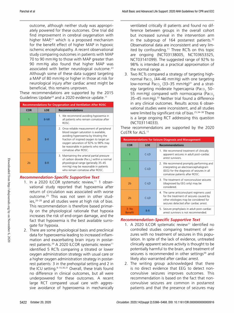
Panchal et al Adult Basic and Advanced Life Support: 2020 AHA Guidelines for CPR and ECC
October 20, 2020 Circulation. 2020;142(suppl 2):S366–S468. DOI: 10.1161/CIR.0000000000000916S422
outcome, although neither study was appropri-ately powered for these outcomes. One trial did find improvement in cerebral oxygenation with higher MAP,21 which is a proposed mechanism for the benefit effect of higher MAP in hypoxic ischemic encephalopathy. A recent observational study comparing outcomes in patients with MAP 70 to 90 mm Hg to those with MAP greater than 90 mm Hg also found that higher MAP was associated with better neurological outcome.23 Although some of these data suggest targeting a MAP of 80 mm Hg or higher in those at risk for neurological injury after cardiac arrest might be beneficial, this remains unproven.
These recommendations are supported by the 2015 Guidelines Update24 and a 2020 evidence update.11
Recommendations for Oxygenation and Ventilation After ROSC
COR LOE Recommendations
1 B-NR1. We recommend avoiding hypoxemia in
all patients who remain comatose after ROSC.
2b B-R
2. Once reliable measurement of peripheral blood oxygen saturation is available, avoiding hyperoxemia by titrating the fraction of inspired oxygen to target an oxygen saturation of 92% to 98% may be reasonable in patients who remain comatose after ROSC.
2b B-R
3. Maintaining the arterial partial pressure of carbon dioxide (Paco2) within a normal physiological range (generally 35–45 mm Hg) may be reasonable in patients who remain comatose after ROSC.
Recommendation-Specific Supportive Text1. In a 2020 ILCOR systematic review,11 1 obser-
vational study reported that hypoxemia after return of circulation was associated with worse outcome.25 This was not seen in other stud-ies,26–28 and all studies were at high risk of bias. This recommendation is therefore based primar-ily on the physiological rationale that hypoxia increases the risk of end-organ damage, and the fact that hypoxemia is the best available surro-gate for hypoxia.
2. There are some physiological basis and preclinical data for hyperoxemia leading to increased inflam-mation and exacerbating brain injury in postar-rest patients.29 A 2020 ILCOR systematic review11 identified 5 RCTs comparing a titrated or lower oxygen administration strategy with usual care or a higher oxygen administration strategy in postar-rest patients: 3 in the prehospital setting and 2 in the ICU setting.8–10,30,31 Overall, these trials found no difference in clinical outcomes, but all were underpowered for these outcomes. A recent large RCT compared usual care with aggres-sive avoidance of hyperoxemia in mechanically
ventilated critically ill patients and found no dif-ference between groups in the overall cohort but increased survival in the intervention arm in the subgroup of 164 postarrest patients.32 Observational data are inconsistent and very lim-ited by confounding.11 Three RCTs on this topic are ongoing (NCT03138005, NCT03653325, NCT03141099). The suggested range of 92% to 98% is intended as a practical approximation of the normal range.
3. Two RCTs compared a strategy of targeting high-normal Paco2 (44–46 mm Hg) with one targeting low-normal Paco2 (33–35 mm Hg)31 and a strat-egy targeting moderate hypercapnia (Paco2 50–55 mm Hg) compared with normocapnia (Paco2 35–45 mm Hg).33 Neither trial found a difference in any clinical outcomes. Results across 6 obser-vational studies were inconsistent, and all studies were limited by significant risk of bias.25,34–38 There is a large ongoing RCT addressing this question (NCT03114033).
These recommendations are supported by the 2020 CoSTR for ALS.11
Recommendations for Seizure Diagnosis and Management
COR LOE Recommendations
1 C-LD1. We recommend treatment of clinically
apparent seizures in adult post–cardiac arrest survivors.
1 C-LD
2. We recommend promptly performing and interpreting an electroencephalogram (EEG) for the diagnosis of seizures in all comatose patients after ROSC.
2b C-LD3. The treatment of nonconvulsive seizures
(diagnosed by EEG only) may be considered.
2b C-LD
4. The same anticonvulsant regimens used for the treatment of seizures caused by other etiologies may be considered for seizures detected after cardiac arrest.
3: No Benefit
B-R5. Seizure prophylaxis in adult post–cardiac
arrest survivors is not recommended.
Recommendation-Specific Supportive Text1. A 2020 ILCOR systematic review11 identified no
controlled studies comparing treatment of sei-zures with no treatment of seizures in this popu-lation. In spite of the lack of evidence, untreated clinically apparent seizure activity is thought to be potentially harmful to the brain, and treatment of seizures is recommended in other settings39 and likely also warranted after cardiac arrest.
2. The writing group acknowledged that there is no direct evidence that EEG to detect non-convulsive seizures improves outcomes. This recommendation is based on the fact that non-convulsive seizures are common in postarrest patients and that the presence of seizures may
Dow
nloaded from http://ahajournals.org by on N
ovember 6, 2020

Panchal et al Adult Basic and Advanced Life Support: 2020 AHA Guidelines for CPR and ECC
Circulation. 2020;142(suppl 2):S366–S468. DOI: 10.1161/CIR.0000000000000916 October 20, 2020 S423
be important prognostically, although whether treatment of nonconvulsive seizures affects outcome in this setting remains uncertain. An ILCOR systematic review done for 2020 did not specifically address the timing and method of obtaining EEGs in postarrest patients who remain unresponsive. Data on the relative benefit of continuous versus intermittent EEG are limited. One study found no difference in survival with good neurological outcome at 3 months in patients monitored with routine (one to two 20-minute EEGs over 24 hours) versus continuous (for 18–24 hours) EEG.40
3. Nonconvulsive seizures are common after cardiac arrest. Whether treatment of seizure activity on EEG that is not associated with clinically evident seizures affects outcome is currently unknown. A randomized trial investigating this question is ongoing (NCT02056236).
4. The 2020 CoSTR recommends that seizures be treated when diagnosed in postarrest patients.11 No specific agent was recommended. However, the CoSTR described 2 retrospective studies sug-gesting valproate, levetiracetam, and fosphe-nytoin may all be effective, with fosphenytoin found to be associated with more hypotension in 1 study.41,42 Common sedatives such as pro-pofol and midazolam have also been found to be effective in suppressing seizure activity after cardiac arrest.43–45
5. A 2020 ILCOR systematic review11 identified 2 RCTs comparing seizure prophylaxis with no seizure prophylaxis in comatose postarrest patients.46,47 Neither study found any difference in occurrence of seizures or survival with favor-able neurological outcome between groups.
These recommendations are supported by the 2020 CoSTR for ALS.11
Recommendations for Other Postresuscitation Care
COR LOE Recommendations
2b B-R1. The benefit of any specific target range
of glucose management is uncertain in adults with ROSC after cardiac arrest.
2b B-R2. The routine use of prophylactic antibiotics in
postarrest patients is of uncertain benefit.
2b B-R3. The effectiveness of agents to mitigate
neurological injury in patients who remain comatose after ROSC is uncertain.
2b B-R4. The routine use of steroids for patients with
shock after ROSC is of uncertain value.
Recommendation-Specific Supportive Text1. One small RCT from 2007,48 found no difference
in survival between strict and moderate glucose control. In the absence of other evidence specific to cardiac arrest, it seems reasonable to manage
blood glucose levels in postarrest patients with the same approach used for the general critically ill population, namely using insulin therapy when needed to maintain a blood glucose of 150 to 180 mg/dL.49
2. A 2020 ILCOR systematic review found 2 RCTs and a small number of observational studies evaluating the effect of prophylactic antibiotics on outcomes in postarrest patients.11,50 The RCTs found no difference in survival or neurological outcome.51,52 One RCT51 did find lower incidence of early pneumonia in those who received pro-phylactic antibiotics, but this did not translate to a difference in other outcomes. When data from the 2 RCTs were pooled, there was no overall dif-ference in infections.51,52
3. The topic of neuroprotective agents was last reviewed in detail in 2010. Multiple agents, including magnesium, coenzyme Q10 (ubiqui-nol), exanatide, xenon gas, methylphenidate, and amantadine, have been considered as possible agents to either mitigate neurological injury or facilitate patient awakening. This work has been largely observational,53–57 although randomized trials have been conducted on coenzyme Q10, xenon gas, and exanatide.58–60 A small trial on the effect of coenzyme Q10 reported better survival in those receiving coenzyme Q10, but there was no significant difference in favorable neurological outcome and these findings have yet to be vali-dated.58 One additional coenzyme Q10 trial was recently completed but results are not yet avail-able (NCT02934555). None of the other studies identified have been able to show a difference in any clinical outcomes with use of any of the agents studied.
4. Since this topic was last updated in detail in 2015, at least 2 randomized trials have been completed on the effect of steroids on shock and other out-comes after ROSC, only 1 of which has been pub-lished to date.61 In this study, shock reversal and other outcomes did not differ between groups. A large retrospective observational study did find that steroid use after cardiac arrest was associ-ated with survival.62 Steroid use for septic shock has been evaluated extensively, with a recent trial of over 1200 patients finding improved survival in those treated with steroids.63 A trial enrolling 3800 patients did not find a mortality benefit, although time to discharge from ICU and time to shock reversal were both shorter in the steroid group.64 Taken together, there is no definitive evidence of benefit from steroids after ROSC. However, the data in sepsis suggest that some patients with severe shock may benefit from steroids and that
Dow
nloaded from http://ahajournals.org by on N
ovember 6, 2020

Panchal et al Adult Basic and Advanced Life Support: 2020 AHA Guidelines for CPR and ECC
October 20, 2020 Circulation. 2020;142(suppl 2):S366–S468. DOI: 10.1161/CIR.0000000000000916S424
the co-occurrence of sepsis and cardiac arrest is important to consider.
Recommendation 1 last received formal evidence re-view in 2010 and is supported by the “Guidelines for the Use of an Insulin Infusion for the Management of Hyperglycemia in Critically Ill Patients” from the Society for Critical Care Medicine.49 Recommendation 2 is sup-ported by the 2020 CoSTR for ALS.11 Recommendations 3 and 4 last received formal evidence review in 2015.24
REFERENCES 1. Witten L, Gardner R, Holmberg MJ, Wiberg S, Moskowitz A,
Mehta S, Grossestreuer AV, Yankama T, Donnino MW, Berg KM. Rea-sons for death in patients successfully resuscitated from out-of-hospital and in-hospital cardiac arrest. Resuscitation. 2019;136:93–99. doi: 10.1016/j.resuscitation.2019.01.031
2. Laver S, Farrow C, Turner D, Nolan J. Mode of death after admission to an intensive care unit following cardiac arrest. Intensive Care Med. 2004;30:2126–2128. doi: 10.1007/s00134-004-2425-z
3. Panchal AR, Berg KM, Cabanas JG, Kurz MC, Link MS, Del Rios M, Hirsch KG, Chan PS, Hazinski MF, Morley PT, et al. 2019 American Heart Association focused update on systems of care: dispatcher-assisted car-diopulmonary resuscitation and cardiac arrest centers: an update to the American Heart Association Guidelines for Cardiopulmonary Resuscita-tion and Emergency Cardiovascular Care. Circulation. 2019;140:e895–e903. doi: 10.1161/CIR.0000000000000733
4. Levine GN, Bates ER, Blankenship JC, Bailey SR, Bittl JA, Cercek B, Chambers CE, Ellis SG, Guyton RA, Hollenberg SM, Khot UN, Lange RA, Mauri L, Mehran R, Moussa ID, Mukherjee D, Ting HH, O’Gara PT, Kushner FG, Ascheim DD, Brindis RG, Casey DE Jr, Chung MK, de Lemos JA, Diercks DB, Fang JC, Franklin BA, Granger CB, Krumholz HM, Linderbaum JA, Morrow DA, Newby LK, Ornato JP, Ou N, Radford MJ, Tamis-Holland JE, Tommaso CL, Tracy CM, Woo YJ, Zhao DX. 2015 ACC/AHA/SCAI Focused Update on Primary Percutaneous Coronary Interven-tion for Patients With ST-Elevation Myocardial Infarction: An Update of the 2011 ACCF/AHA/SCAI Guideline for Percutaneous Coronary Inter-vention and the 2013 ACCF/AHA Guideline for the Management of ST-Elevation Myocardial Infarction. Circulation. 2016;133:1135–1147. doi: 10.1161/CIR.0000000000000336
5. Stær-Jensen H, Nakstad ER, Fossum E, Mangschau A, Eritsland J, Draegni T, Jacobsen D, Sunde K, Andersen GO. Post-resuscitation ECG for selection of patients for immediate coronary angiography in out-of-hospital cardiac arrest. Circ Cardiovasc Interv. 2015;8 doi: 10.1161/ CIRCINTERVENTIONS.115.002784
6. Zanuttini D, Armellini I, Nucifora G, Grillo MT, Morocutti G, Carchietti E, Trillò G, Spedicato L, Bernardi G, Proclemer A. Predictive value of electro-cardiogram in diagnosing acute coronary artery lesions among patients with out-of-hospital-cardiac-arrest. Resuscitation. 2013;84:1250–1254. doi: 10.1016/j.resuscitation.2013.04.023
7. Sideris G, Voicu S, Dillinger JG, Stratiev V, Logeart D, Broche C, Vivien B, Brun PY, Deye N, Capan D, Aout M, Megarbane B, Baud FJ, Henry P. Value of post-resuscitation electrocardiogram in the diagnosis of acute myocardial infarction in out-of-hospital cardiac arrest patients. Resuscita-tion. 2011;82:1148–1153. doi: 10.1016/j.resuscitation.2011.04.023
8. Kuisma M, Boyd J, Voipio V, Alaspää A, Roine RO, Rosenberg P. Com-parison of 30 and the 100% inspired oxygen concentrations during early post-resuscitation period: a randomised controlled pilot study. Resuscita-tion. 2006;69:199–206. doi: 10.1016/j.resuscitation.2005.08.010
9. Bray JE, Hein C, Smith K, Stephenson M, Grantham H, Finn J, Stub D, Cameron P, Bernard S; EXACT Investigators. Oxygen titration after resus-citation from out-of-hospital cardiac arrest: A multi-centre, randomised controlled pilot study (the EXACT pilot trial). Resuscitation. 2018;128:211–215. doi: 10.1016/j.resuscitation.2018.04.019
10. Thomas M, Voss S, Benger J, Kirby K, Nolan JP. Cluster randomised com-parison of the effectiveness of 100% oxygen versus titrated oxygen in pa-tients with a sustained return of spontaneous circulation following out of hospital cardiac arrest: a feasibility study. PROXY: post ROSC OXYgenation study. BMC Emerg Med. 2019;19:16. doi: 10.1186/s12873-018-0214-1
11. Berg KM, Soar J, Andersen LW, Böttiger BW, Cacciola S, Callaway CW, Couper K, Cronberg T, D’Arrigo S, Deakin CD, et al; on behalf of the Adult Advanced Life Support Collaborators. Adult advanced life sup-port: 2020 International Consensus on Cardiopulmonary Resuscitation and Emergency Cardiovascular Care Science With Treatment Recom-mendations. Circulation. 2020;142(suppl 1):S92–S139. doi: 10.1161/CIR.0000000000000893
12. Soar J, Nolan JP, Böttiger BW, Perkins GD, Lott C, Carli P, Pellis T, Sandroni C, Skrifvars MB, Smith GB, Sunde K, Deakin CD; Adult advanced life support section Collaborators. European Resuscitation Council Guidelines for Re-suscitation 2015: Section 3. Adult advanced life support. Resuscitation. 2015;95:100–147. doi: 10.1016/j.resuscitation.2015.07.016
13. Trzeciak S, Jones AE, Kilgannon JH, Milcarek B, Hunter K, Shapiro NI, Hollenberg SM, Dellinger P, Parrillo JE. Significance of arterial hypotension after resuscitation from cardiac arrest. Crit Care Med. 2009;37:2895–903; quiz 2904. doi: 10.1097/ccm.0b013e3181b01d8c
14. Chiu YK, Lui CT, Tsui KL. Impact of hypotension after return of spontane-ous circulation on survival in patients of out-of-hospital cardiac arrest. Am J Emerg Med. 2018;36:79–83. doi: 10.1016/j.ajem.2017.07.019
15. Bray JE, Bernard S, Cantwell K, Stephenson M, Smith K; and the VA-CAR Steering Committee. The association between systolic blood pres-sure on arrival at hospital and outcome in adults surviving from out-of-hospital cardiac arrests of presumed cardiac aetiology. Resuscitation. 2014;85:509–515. doi: 10.1016/j.resuscitation.2013.12.005
16. Russo JJ, Di Santo P, Simard T, James TE, Hibbert B, Couture E, Marbach J, Osborne C, Ramirez FD, Wells GA, Labinaz M, Le May MR; from the CAPITAL study group. Optimal mean arterial pressure in coma-tose survivors of out-of-hospital cardiac arrest: An analysis of area be-low blood pressure thresholds. Resuscitation. 2018;128:175–180. doi: 10.1016/j.resuscitation.2018.04.028
17. Laurikkala J, Wilkman E, Pettilä V, Kurola J, Reinikainen M, Hoppu S, Ala-Kokko T, Tallgren M, Tiainen M, Vaahersalo J, Varpula T, Skrifvars MB; FINNRESUSCI Study Group. Mean arterial pressure and va-sopressor load after out-of-hospital cardiac arrest: Associations with one-year neurologic outcome. Resuscitation. 2016;105:116–122. doi: 10.1016/j.resuscitation.2016.05.026
18. Annoni F, Dell’Anna AM, Franchi F, Creteur J, Scolletta S, Vincent JL, Taccone FS. The impact of diastolic blood pressure values on the neurolog-ical outcome of cardiac arrest patients. Resuscitation. 2018;130:167–173. doi: 10.1016/j.resuscitation.2018.07.017
19. Janiczek JA, Winger DG, Coppler P, Sabedra AR, Murray H, Pinsky MR, Rittenberger JC, Reynolds JC, Dezfulian C. Hemodynamic Resuscitation Characteristics Associated with Improved Survival and Shock Resolu-tion After Cardiac Arrest. Shock. 2016;45:613–619. doi: 10.1097/SHK. 0000000000000554
20. Young MN, Hollenbeck RD, Pollock JS, Giuseffi JL, Wang L, Harrell FE, McPherson JA. Higher achieved mean arterial pressure during therapeu-tic hypothermia is not associated with neurologically intact survival fol-lowing cardiac arrest. Resuscitation. 2015;88:158–164. doi: 10.1016/j. resuscitation.2014.12.008
21. Ameloot K, De Deyne C, Eertmans W, Ferdinande B, Dupont M, Palmers PJ, Petit T, Nuyens P, Maeremans J, Vundelinckx J, Vanhaverbeke M, Belmans A, Peeters R, Demaerel P, Lemmens R, Dens J, Janssens S. Early goal-directed haemodynamic optimization of cerebral oxygenation in co-matose survivors after cardiac arrest: the Neuroprotect post-cardiac arrest trial. Eur Heart J. 2019;40:1804–1814. doi: 10.1093/eurheartj/ehz120
22. Jakkula P, Pettilä V, Skrifvars MB, Hästbacka J, Loisa P, Tiainen M, Wilkman E, Toppila J, Koskue T, Bendel S, Birkelund T, Laru-Sompa R, Valkonen M, Reinikainen M; COMACARE study group. Targeting low-nor-mal or high-normal mean arterial pressure after cardiac arrest and resusci-tation: a randomised pilot trial. Intensive Care Med. 2018;44:2091–2101. doi: 10.1007/s00134-018-5446-8
23. Roberts BW, Kilgannon JH, Hunter BR, Puskarich MA, Shea L, Donnino M, Jones C, Fuller BM, Kline JA, Jones AE, Shapiro NI, Abella BS, Trzeciak S. Association Between Elevated Mean Arterial Blood Pres-sure and Neurologic Outcome After Resuscitation From Cardiac Arrest: Results From a Multicenter Prospective Cohort Study. Crit Care Med. 2019;47:93–100. doi: 10.1097/CCM.0000000000003474
24. Callaway CW, Donnino MW, Fink EL, Geocadin RG, Golan E, Kern KB, Leary M, Meurer WJ, Peberdy MA, Thompson TM, et al. Part 8: post–cardiac arrest care: 2015 American Heart Association Guidelines Update for Car-diopulmonary Resuscitation and Emergency Cardiovascular Care. Circula-tion. 2015;132(suppl 2):S465–482. doi: 10.1161/cir.0000000000000262
Dow
nloaded from http://ahajournals.org by on N
ovember 6, 2020

Panchal et al Adult Basic and Advanced Life Support: 2020 AHA Guidelines for CPR and ECC
Circulation. 2020;142(suppl 2):S366–S468. DOI: 10.1161/CIR.0000000000000916 October 20, 2020 S425
25. Wang HE, Prince DK, Drennan IR, Grunau B, Carlbom DJ, Johnson N, Hansen M, Elmer J, Christenson J, Kudenchuk P, Aufderheide T, Weisfeldt M, Idris A, Trzeciak S, Kurz M, Rittenberger JC, Griffiths D, Jasti J, May S; Resuscitation Outcomes Consortium (ROC) Investigators. Post-resuscita-tion arterial oxygen and carbon dioxide and outcomes after out-of-hos-pital cardiac arrest. Resuscitation. 2017;120:113–118. doi: 10.1016/j. resuscitation.2017.08.244
26. Ebner F, Ullén S, Åneman A, Cronberg T, Mattsson N, Friberg H, Hassager C, Kjærgaard J, Kuiper M, Pelosi P, Undén J, Wise MP, Wetterslev J, Nielsen N. Associations between partial pressure of oxygen and neurological out-come in out-of-hospital cardiac arrest patients: an explorative analysis of a randomized trial. Crit Care. 2019;23:30. doi: 10.1186/s13054-019-2322-z
27. Humaloja J, Litonius E, Efendijev I, Folger D, Raj R, Pekkarinen PT, Skrifvars MB. Early hyperoxemia is not associated with cardiac arrest outcome. Resusci-tation. 2019;140:185–193. doi: 10.1016/j.resuscitation.2019.04.035
28. Johnson NJ, Dodampahala K, Rosselot B, Perman SM, Mikkelsen ME, Goyal M, Gaieski DF, Grossestreuer AV. The Association Between Arte-rial Oxygen Tension and Neurological Outcome After Cardiac Arrest. Ther Hypothermia Temp Manag. 2017;7:36–41. doi: 10.1089/ther.2016.0015
29. Pilcher J, Weatherall M, Shirtcliffe P, Bellomo R, Young P, Beasley R. The effect of hyperoxia following cardiac arrest - A systematic review and meta-analysis of animal trials. Resuscitation. 2012;83:417–422. doi: 10.1016/j.resuscitation.2011.12.021
30. Young P, Bailey M, Bellomo R, Bernard S, Dicker B, Freebairn R, Henderson S, Mackle D, McArthur C, McGuinness S, Smith T, Swain A, Weatherall M, Beasley R. HyperOxic Therapy OR NormOxic Therapy after out-of-hospital cardiac arrest (HOT OR NOT): a randomised controlled feasibility trial. Resus-citation. 2014;85:1686–1691. doi: 10.1016/j.resuscitation.2014.09.011
31. Jakkula P, Reinikainen M, Hästbacka J, Loisa P, Tiainen M, Pettilä V, Toppila J, Lähde M, Bäcklund M, Okkonen M, et al; and the COMACARE study group. Targeting two different levels of both arterial carbon dioxide and arterial oxygen after cardiac arrest and resuscitation: a randomised pilot trial. Intensive Care Med. 2018;44:2112–2121. doi: 10.1007/s00134-018-5453-9
32. Mackle D, Bellomo R, Bailey M, Beasley R, Deane A, Eastwood G, Finfer S, Freebairn R, King V, Linke N, et al; and the ICU-ROX Investigators the Australian New Zealand Intensive Care Society Clinical Trials Group. Con-servative oxygen therapy during mechanical ventilation in the ICU. N Engl J Med. 2020;382:989–998. doi: 10.1056/NEJMoa1903297
33. Eastwood GM, Schneider AG, Suzuki S, Peck L, Young H, Tanaka A, Mårtensson J, Warrillow S, McGuinness S, Parke R, Gilder E, Mccarthy L, Galt P, Taori G, Eliott S, Lamac T, Bailey M, Harley N, Barge D, Hodgson CL, Morganti-Kossmann MC, Pébay A, Conquest A, Archer JS, Bernard S, Stub D, Hart GK, Bellomo R. Targeted therapeutic mild hypercapnia after cardiac arrest: A phase II multi-centre randomised controlled trial (the CCC trial). Resuscitation. 2016;104:83–90. doi: 10.1016/j.resuscitation.2016.03.023
34. Vaahersalo J, Bendel S, Reinikainen M, Kurola J, Tiainen M, Raj R, Pettilä V, Varpula T, Skrifvars MB; FINNRESUSCI Study Group. Arterial blood gas ten-sions after resuscitation from out-of-hospital cardiac arrest: associations with long-term neurologic outcome. Crit Care Med. 2014;42:1463–1470. doi: 10.1097/CCM.0000000000000228
35. Hope Kilgannon J, Hunter BR, Puskarich MA, Shea L, Fuller BM, Jones C, Donnino M, Kline JA, Jones AE, Shapiro NI, Abella BS, Trzeciak S, Roberts BW. Partial pressure of arterial carbon dioxide after resuscitation from cardiac arrest and neurological outcome: A prospective multi-center protocol-directed cohort study. Resuscitation. 2019;135:212–220. doi: 10.1016/j.resuscitation.2018.11.015
36. Roberts BW, Kilgannon JH, Chansky ME, Mittal N, Wooden J, Trzeciak S. Association between postresuscitation partial pressure of arterial carbon dioxide and neurological outcome in patients with post-cardiac arrest syndrome. Circulation. 2013;127:2107–2113. doi: 10.1161/ CIRCULATIONAHA.112.000168
37. von Auenmueller KI, Christ M, Sasko BM, Trappe HJ. The Value of Arterial Blood Gas Parameters for Prediction of Mortality in Survivors of Out-of-hospital Cardiac Arrest. J Emerg Trauma Shock. 2017;10:134–139. doi: 10.4103/JETS.JETS_146_16
38. Ebner F, Harmon MBA, Aneman A, Cronberg T, Friberg H, Hassager C, Juffermans N, Kjærgaard J, Kuiper M, Mattsson N, Pelosi P, Ullén S, Undén J, Wise MP, Nielsen N. Carbon dioxide dynamics in relation to neurologi-cal outcome in resuscitated out-of-hospital cardiac arrest patients: an exploratory Target Temperature Management Trial substudy. Crit Care. 2018;22:196. doi: 10.1186/s13054-018-2119-5
39. Glauser T, Shinnar S, Gloss D, Alldredge B, Arya R, Bainbridge J, Bare M, Bleck T, Dodson WE, Garrity L, Jagoda A, Lowenstein D, Pellock J, Riviello J,
Sloan E, Treiman DM. Evidence-Based Guideline: Treatment of Convulsive Status Epilepticus in Children and Adults: Report of the Guideline Com-mittee of the American Epilepsy Society. Epilepsy Curr. 2016;16:48–61. doi: 10.5698/1535-7597-16.1.48
40. Fatuzzo D, Beuchat I, Alvarez V, Novy J, Oddo M, Rossetti AO. Does con-tinuous EEG influence prognosis in patients after cardiac arrest? Resuscita-tion. 2018;132:29–32. doi: 10.1016/j.resuscitation.2018.08.023
41. Solanki P, Coppler PJ, Kvaløy JT, Baldwin MA, Callaway CW, Elmer J; Pittsburgh Post-Cardiac Arrest Service. Association of antiepileptic drugs with resolution of epileptiform activity after cardiac arrest. Resuscitation. 2019;142:82–90. doi: 10.1016/j.resuscitation.2019.07.007
42. Kapur J, Elm J, Chamberlain JM, Barsan W, Cloyd J, Lowenstein D, Shinnar S, Conwit R, Meinzer C, Cock H, Fountain N, Connor JT, Silbergleit R; NETT and PECARN Investigators. Randomized Trial of Three Anticonvulsant Medications for Status Epilepticus. N Engl J Med. 2019;381:2103–2113. doi: 10.1056/NEJMoa1905795
43. Thömke F, Weilemann SL. Poor prognosis despite successful treatment of postanoxic generalized myoclonus. Neurology. 2010;74:1392–1394. doi: 10.1212/WNL.0b013e3181dad5b9
44. Aicua RI, Rapun I, Novy J, Solari D, Oddo M, Rossetti AO. Early Lance-Adams syndrome after cardiac arrest: prevalence, time to return to aware-ness, and outcome in a large cohort. Resuscitation. 2017;115:169–172. doi: 10.1016/j.resuscitation.2017.03.020
45. Koutroumanidis M, Sakellariou D. Low frequency nonevolving generalized periodic epileptiform discharges and the borderland of hypoxic noncon-vulsive status epilepticus in comatose patients after cardiac arrest. Epilepsy Behav. 2015;49:255–262. doi: 10.1016/j.yebeh.2015.04.060
46. Brain Resuscitation Clinical Trial I Study Group. Randomized clinical study of thiopental loading in comatose survivors of cardiac arrest. N Engl J Med. 1986;314:397–403. doi: 10.1056/nejm198602133140701
47. Longstreth WT Jr, Fahrenbruch CE, Olsufka M, Walsh TR, Copass MK, Cobb LA. Randomized clinical trial of magnesium, diazepam, or both after out-of-hospital cardiac arrest. Neurology. 2002;59:506–514. doi: 10.1212/wnl.59.4.506
48. Oksanen T, Skrifvars MB, Varpula T, Kuitunen A, Pettilä V, Nurmi J, Castrén M. Strict versus moderate glucose control after resuscitation from ventricular fibrillation. Intensive Care Med. 2007;33:2093–2100. doi: 10.1007/s00134-007-0876-8
49. Jacobi J, Bircher N, Krinsley J, Agus M, Braithwaite SS, Deutschman C, Freire AX, Geehan D, Kohl B, Nasraway SA, Rigby M, Sands K, Schallom L, Taylor B, Umpierrez G, Mazuski J, Schunemann H. Guidelines for the use of an insulin infusion for the management of hyperglycemia in critically ill patients. Crit Care Med. 2012;40:3251–3276. doi: 10.1097/CCM. 0b013e3182653269
50. Couper K, Laloo R, Field R, Perkins GD, Thomas M, Yeung J. Prophy-lactic antibiotic use following cardiac arrest: A systematic review and meta-analysis. Resuscitation. 2019;141:166–173. doi: 10.1016/j. resuscitation.2019.04.047
51. François B, Cariou A, Clere-Jehl R, Dequin PF, Renon-Carron F, Daix T, Guitton C, Deye N, Legriel S, Plantefève G, Quenot JP, Desachy A, Kamel T, Bedon-Carte S, Diehl JL, Chudeau N, Karam E, Durand-Zaleski I, Giraudeau B, Vignon P, Le Gouge A; CRICS-TRIGGERSEP Network and the ANTHARTIC Study Group. Prevention of Early Ventilator-Associated Pneumonia after Cardiac Arrest. N Engl J Med. 2019;381:1831–1842. doi: 10.1056/NEJMoa1812379
52. Ribaric SF, Turel M, Knafelj R, Gorjup V, Stanic R, Gradisek P, Cerovic O, Mirkovic T, Noc M. Prophylactic versus clinically-driven antibiotics in coma-tose survivors of out-of-hospital cardiac arrest-A randomized pilot study. Re-suscitation. 2017;111:103–109. doi: 10.1016/j.resuscitation.2016.11.025
53. Pearce A, Lockwood C, van den Heuvel C, Pearce J. The use of thera-peutic magnesium for neuroprotection during global cerebral ischemia associated with cardiac arrest and cardiac surgery in adults: a systematic review. JBI Database System Rev Implement Rep. 2017;15:86–118. doi: 10.11124/JBISRIR-2016-003236
54. Perucki WH, Hiendlmayr B, O’Sullivan DM, Gunaseelan AC, Fayas F, Fernandez AB. Magnesium Levels and Neurologic Outcomes in Patients Undergoing Therapeutic Hypothermia After Cardiac Arrest. Ther Hypo-thermia Temp Manag. 2018;8:14–17. doi: 10.1089/ther.2017.0016
55. Suzuki M, Hatakeyama T, Nakamura R, Saiki T, Kamisasanuki T, Sugiki D, Matsushima H. Serum Magnesium Levels and Neurological Outcomes in Patients Undergoing Targeted Temperature Management After Cardiac Arrest. J Emerg Nurs. 2020;46:59–65. doi: 10.1016/j.jen.2019.10.006
56. Cocchi MN, Giberson B, Berg K, Salciccioli JD, Naini A, Buettner C, Akuthota P, Gautam S, Donnino MW. Coenzyme Q10 levels are low and
Dow
nloaded from http://ahajournals.org by on N
ovember 6, 2020

Panchal et al Adult Basic and Advanced Life Support: 2020 AHA Guidelines for CPR and ECC
October 20, 2020 Circulation. 2020;142(suppl 2):S366–S468. DOI: 10.1161/CIR.0000000000000916S426
associated with increased mortality in post-cardiac arrest patients. Resus-citation. 2012;83:991–995. doi: 10.1016/j.resuscitation.2012.03.023
57. Reynolds JC, Rittenberger JC, Callaway CW. Methylphenidate and amantadine to stimulate reawakening in comatose patients resus-citated from cardiac arrest. Resuscitation. 2013;84:818–824. doi: 10.1016/j.resuscitation.2012.11.014
58. Damian MS, Ellenberg D, Gildemeister R, Lauermann J, Simonis G, Sauter W, Georgi C. Coenzyme Q10 combined with mild hypothermia af-ter cardiac arrest: a preliminary study. Circulation. 2004;110:3011–3016. doi: 10.1161/01.CIR.0000146894.45533.C2
59. Laitio R, Hynninen M, Arola O, Virtanen S, Parkkola R, Saunavaara J, Roine RO, Grönlund J, Ylikoski E, Wennervirta J, Bäcklund M, Silvasti P, Nukarinen E, Tiainen M, Saraste A, Pietilä M, Airaksinen J, Valanne L, Martola J, Silvennoinen H, Scheinin H, Harjola VP, Niiranen J, Korpi K, Varpula M, Inkinen O, Olkkola KT, Maze M, Vahlberg T, Laitio T. Effect of Inhaled Xenon on Cerebral White Matter Damage in Comatose Survivors of Out-of-Hospital Cardiac Arrest: A Randomized Clinical Trial. JAMA. 2016;315:1120–1128. doi: 10.1001/jama.2016.1933
60. Wiberg S, Hassager C, Schmidt H, Thomsen JH, Frydland M, Lindholm MG, Høfsten DE, Engstrøm T, Køber L, Møller JE, Kjaergaard J. Neuroprotec-tive Effects of the Glucagon-Like Peptide-1 Analog Exenatide After Out-of-Hospital Cardiac Arrest: A Randomized Controlled Trial. Circulation. 2016;134:2115–2124. doi: 10.1161/CIRCULATIONAHA.116.024088
61. Donnino MW, Andersen LW, Berg KM, Chase M, Sherwin R, Smithline H, Carney E, Ngo L, Patel PV, Liu X, Cutlip D, Zimetbaum P, Cocchi MN; Col-laborating Authors from the Beth Israel Deaconess Medical Center’s Center for Resuscitation Science Research Group. Corticosteroid therapy in refrac-tory shock following cardiac arrest: a randomized, double-blind, placebo-controlled, trial. Crit Care. 2016;20:82. doi: 10.1186/s13054-016-1257-x
62. Tsai MS, Chuang PY, Huang CH, Tang CH, Yu PH, Chang WT, Chen WJ. Postarrest Steroid Use May Improve Outcomes of Cardiac Arrest Survivors. Crit Care Med. 2019;47:167–175. doi: 10.1097/CCM.0000000000003468
63. Annane D, Renault A, Brun-Buisson C, Megarbane B, Quenot JP, Siami S, Cariou A, Forceville X, Schwebel C, Martin C, Timsit JF, Misset B, Ali Benali M, Colin G, Souweine B, Asehnoune K, Mercier E, Chimot L, Charpentier C, François B, Boulain T, Petitpas F, Constantin JM, Dhonneur G, Baudin F, Combes A, Bohé J, Loriferne JF, Amathieu R, Cook F, Slama M, Leroy O, Capellier G, Dargent A, Hissem T, Maxime V, Bellissant E; CRICS-TRIGGERSEP Network. Hydrocortisone plus Fludrocor-tisone for Adults with Septic Shock. N Engl J Med. 2018;378:809–818. doi: 10.1056/NEJMoa1705716
64. Venkatesh B, Finfer S, Cohen J, Rajbhandari D, Arabi Y, Bellomo R, Billot L, Correa M, Glass P, Harward M, et al; on behalf of the AD-RENAL Trial Investigators and the Australian–New Zealand Intensive Care Society Clinical Trials Group. Adjunctive glucocorticoid therapy in patients with septic shock. N Engl J Med. 2018;378:797–808. doi: 10.1056/NEJMoa1705835
Targeted Temperature ManagementIntroductionTTM between 32°C and 36°C for at least 24 hours is currently recommended for all cardiac rhythms in both OHCA and IHCA. Multiple randomized trials have been performed in various domains of TTM and were sum-marized in a systematic review published in 2015.1 Sub-sequent to the 2015 recommendations, additional ran-domized trials have evaluated TTM for nonshockable rhythms as well as TTM duration. Many of these were reviewed in an evidence update provided in the 2020 COSTR for ALS.2 Many uncertainties within the topic of TTM remain, including whether temperature should vary on the basis of patient characteristics, how long TTM should be maintained, and how quickly it should be started. An updated systematic review on several
aspects of this important topic is needed once currently ongoing clinical trials have been completed.
Recommendations for Indications for TTM
COR LOE Recommendations
1 B-R1. We recommend TTM for adults who do
not follow commands after ROSC from OHCA with any initial rhythm.
1 B-R2. We recommend TTM for adults who do
not follow commands after ROSC from IHCA with initial nonshockable rhythm.
1 B-NR3. We recommend TTM for adults who do
not follow commands after ROSC from IHCA with initial shockable rhythm.
Recommendation-Specific Supportive Text1. Two RCTs of patients with OHCA with an initially
shockable rhythm published in 2002 reported benefit from mild hypothermia when compared with no temperature management.1,3,4 A more recent trial comparing a target temperature of 33°C to 37°C in patients (IHCA and OHCA) with initial nonshockable rhythm also found better outcomes in those treated with a tem-perature of 33°C.5 A large trial is currently under-way testing TTM compared with normothermia (NCT03114033).
2. An RCT published in 2019 compared TTM at 33°C to 37°C for patients who were not follow-ing commands after ROSC from cardiac arrest with initial nonshockable rhythm. Survival with a favorable neurological outcome (Cerebral Performance Category 1–2) was higher in the group treated with 33°C.5 This trial included both OHCA and IHCA and is the first random-ized trial on TTM after cardiac arrest to include IHCA patients. In a subgroup analysis, the ben-efit of TTM did not appear to differ significantly by IHCA/OHCA subgroups.
3. No RCTs of TTM have included IHCA patients with an initial shockable rhythm, and this recommen-dation is therefore based largely on extrapolation from OHCA studies and the study of patients with initially nonshockable rhythms that included IHCA patients. Observational studies on TTM for IHCA with any initial rhythm have reported mixed results. Two studies that included patients enrolled in the AHA Get With The Guidelines-Resuscitation registry reported either no benefit or worse outcome from TTM.6,7 Both were limited by very low overall usage of TTM in the registry and lack of data on presence of coma, making it difficult to determine if TTM was indicated for a given IHCA patient.
This topic last received formal evidence review in 2015,8 with an evidence update conducted for the 2020 CoSTR for ALS.2
Dow
nloaded from http://ahajournals.org by on N
ovember 6, 2020

Panchal et al Adult Basic and Advanced Life Support: 2020 AHA Guidelines for CPR and ECC
Circulation. 2020;142(suppl 2):S366–S468. DOI: 10.1161/CIR.0000000000000916 October 20, 2020 S427
Recommendations for Performance of TTM
COR LOE Recommendations
1 B-R1. We recommend selecting and
maintaining a constant temperature between 32°C and 36°C during TTM.
2a B-NR2. It is reasonable that TTM be maintained
for at least 24 h after achieving target temperature.
2b C-LD3. It may be reasonable to actively prevent
fever in comatose patients after TTM.
3: No Benefit
A
4. We do not recommend the routine use of rapid infusion of cold IV fluids for prehospital cooling of patients after ROSC.
Recommendation-Specific Supportive Text1. In 2013, a trial of over 900 patients compared
TTM at 33°C to 36°C for patients with OHCA and any initial rhythm, excluding unwitnessed asystole, and found that 33°C was not superior to 36°C.9 A more recent trial compared 33°C to 37°C for patients with ROSC after initial non-shockable rhythm and found improved survival with favorable neurological outcome in the group treated with 33°C.5 There have been reports of decreasing utilization of TTM in recent years, with one hypothesis being that some clinicians inter-pret the inclusion of 36°C as a target tempera-ture as being equivalent to normothermia, or no strict temperature control.10 An updated system-atic review is needed on the question of which target temperature is most beneficial. Based on the available evidence, however, TTM at a temp between 32°C and 36°C remains a Class 1 recommendation.
2. One RCT including 355 patients found no dif-ference in outcome between TTM for 24 and 48 hours.11 This study may have been underpow-ered to detect differences in clinical outcomes. The initial 2002 trials cooled patients for 123 and 24 hours4 while the 2013 trial used 28 hours.9 A larger, adaptive clinical trial is currently under-way investigating multiple different durations of hypothermia ranging from 6 to 72 hours, using a target temperature of 33°C for all patients enrolled (NCT04217551). There is no clear best approach to rewarming after TTM, although a protocol of 0.5°C per hour was followed in the 2013 trial.9 The optimal rate of rewarming, and specifically whether slower rates are beneficial, is a knowledge gap, and at least 1 trial is ongoing (NCT02555254).
3. Fever after ROSC is associated with poor neu-rological outcome in patients not treated with TTM, although this finding is reported less con-sistently in patients treated with TTM.12–20 It has not been established whether treatment of fever
is associated with an improvement in outcome, but treatment or prevention of fever appears to be a reasonable approach.
4. A 2015 systematic review found that prehospi-tal cooling with the specific method of the rapid infusion of cold IV fluids was associated with more pulmonary edema and a higher risk of rear-rest.1 Since this review, a number of RCTs on pre-hospital cooling have been conducted. One trial compared the prehospital induction of hypother-mia with any method (including ice packs and cold IV fluids) with no prehospital cooling, and found higher receipt of in-hospital TTM in those who had prehospital initiation. That trial found no increased adverse events in those treated with prehospital cooling.21 Other methods of prehospi-tal cooling, such as esophageal or nasal devices, have also been investigated; whether these affect outcomes is a knowledge gap.
This topic last received formal evidence review in 2015,8 with an evidence update conducted for the 2020 CoSTR for ALS.2
REFERENCES 1. Donnino MW, Andersen LW, Berg KM, Reynolds JC, Nolan JP, Morley PT,
Lang E, Cocchi MN, Xanthos T, Callaway CW, Soar J; ILCOR ALS Task Force. Temperature Management After Cardiac Arrest: An Advisory State-ment by the Advanced Life Support Task Force of the International Li-aison Committee on Resuscitation and the American Heart Association Emergency Cardiovascular Care Committee and the Council on Cardio-pulmonary, Critical Care, Perioperative and Resuscitation. Circulation. 2015;132:2448–2456. doi: 10.1161/CIR.0000000000000313
2. Berg KM, Soar J, Andersen LW, Böttiger BW, Cacciola S, Callaway CW, Couper K, Cronberg T, D’Arrigo S, Deakin CD, et al; on behalf of the Adult Advanced Life Support Collaborators. Adult advanced life sup-port: 2020 International Consensus on Cardiopulmonary Resuscitation and Emergency Cardiovascular Care Science With Treatment Recom-mendations. Circulation. 2020;142(suppl 1):S92–S139. doi: 10.1161/CIR.0000000000000893
3. Bernard SA, Gray TW, Buist MD, Jones BM, Silvester W, Gutteridge G, Smith K. Treatment of comatose survivors of out-of-hospital cardiac ar-rest with induced hypothermia. N Engl J Med. 2002;346:557–563. doi: 10.1056/NEJMoa003289
4. Hypothermia after Cardiac Arrest Study Group. Mild therapeutic hypo-thermia to improve the neurologic outcome after cardiac arrest. N Engl J Med. 2002;346:549–556. doi: 10.1056/NEJMoa012689
5. Lascarrou JB, Merdji H, Le Gouge A, Colin G, Grillet G, Girardie P, Coupez E, Dequin PF, Cariou A, Boulain T, Brule N, Frat JP, Asfar P, Pichon N, Landais M, Plantefeve G, Quenot JP, Chakarian JC, Sirodot M, Legriel S, Letheulle J, Thevenin D, Desachy A, Delahaye A, Botoc V, Vimeux S, Martino F, Giraudeau B, Reignier J; CRICS-TRIG-GERSEP Group. Targeted Temperature Management for Cardiac Arrest with Nonshockable Rhythm. N Engl J Med. 2019;381:2327–2337. doi: 10.1056/NEJMoa1906661
6. Nichol G, Huszti E, Kim F, Fly D, Parnia S, Donnino M, Sorenson T, Callaway CW; American Heart Association Get With the Guideline-Resus-citation Investigators. Does induction of hypothermia improve outcomes after in-hospital cardiac arrest? Resuscitation. 2013;84:620–625. doi: 10.1016/j.resuscitation.2012.12.009
7. Chan PS, Berg RA, Tang Y, Curtis LH, Spertus JA; American Heart Association’s Get With the Guidelines–Resuscitation Investigators. Association Between Therapeutic Hypothermia and Survival Af-ter In-Hospital Cardiac Arrest. JAMA. 2016;316:1375–1382. doi: 10.1001/jama.2016.14380
Dow
nloaded from http://ahajournals.org by on N
ovember 6, 2020

Panchal et al Adult Basic and Advanced Life Support: 2020 AHA Guidelines for CPR and ECC
October 20, 2020 Circulation. 2020;142(suppl 2):S366–S468. DOI: 10.1161/CIR.0000000000000916S428
8. Callaway CW, Donnino MW, Fink EL, Geocadin RG, Golan E, Kern KB, Leary M, Meurer WJ, Peberdy MA, Thompson TM, et al. Part 8: post–cardiac arrest care: 2015 American Heart Association Guidelines Update for Car-diopulmonary Resuscitation and Emergency Cardiovascular Care. Circula-tion. 2015;132(suppl 2):S465–482. doi: 10.1161/cir.0000000000000262
9. Nielsen N, Wetterslev J, Cronberg T, Erlinge D, Gasche Y, Hassager C, Horn J, Hovdenes J, Kjaergaard J, Kuiper M, Pellis T, Stammet P, Wanscher M, Wise MP, Åneman A, Al-Subaie N, Boesgaard S, Bro-Jeppesen J, Brunetti I, Bugge JF, Hingston CD, Juffermans NP, Koopmans M, Køber L, Langørgen J, Lilja G, Møller JE, Rundgren M, Rylander C, Smid O, Werer C, Winkel P, Friberg H; TTM Trial Investigators. Targeted temperature management at 33°C versus 36°C after cardiac arrest. N Engl J Med. 2013;369:2197–2206. doi: 10.1056/NEJMoa1310519
10. Khera R, Humbert A, Leroux B, Nichol G, Kudenchuk P, Scales D, Baker A, Austin M, Newgard CD, Radecki R, Vilke GM, Sawyer KN, Sopko G, Idris AH, Wang H, Chan PS, Kurz MC. Hospital Variation in the Utilization and Im-plementation of Targeted Temperature Management in Out-of-Hospital Cardiac Arrest. Circ Cardiovasc Qual Outcomes. 2018;11:e004829. doi: 10.1161/CIRCOUTCOMES.118.004829
11. Kirkegaard H, Søreide E, de Haas I, Pettilä V, Taccone FS, Arus U, Storm C, Hassager C, Nielsen JF, Sørensen CA, Ilkjær S, Jeppesen AN, Grejs AM, Duez CHV, Hjort J, Larsen AI, Toome V, Tiainen M, Hästbacka J, Laitio T, Skrifvars MB. Targeted Temperature Management for 48 vs 24 Hours and Neurologic Outcome After Out-of-Hospital Cardiac Arrest: A Randomized Clinical Trial. JAMA. 2017;318:341–350. doi: 10.1001/jama.2017.8978
12. Nolan JP, Laver SR, Welch CA, Harrison DA, Gupta V, Rowan K. Outcome following admission to UK intensive care units after cardiac arrest: a sec-ondary analysis of the ICNARC Case Mix Programme Database. Anaesthe-sia. 2007;62:1207–1216. doi: 10.1111/j.1365-2044.2007.05232.x
13. Langhelle A, Tyvold SS, Lexow K, Hapnes SA, Sunde K, Steen PA. In-hos-pital factors associated with improved outcome after out-of-hospital car-diac arrest. A comparison between four regions in Norway. Resuscitation. 2003;56:247–263. doi: 10.1016/s0300-9572(02)00409-4
14. Suffoletto B, Peberdy MA, van der Hoek T, Callaway C. Body temperature changes are associated with outcomes following in-hospital cardiac ar-rest and return of spontaneous circulation. Resuscitation. 2009;80:1365–1370. doi: 10.1016/j.resuscitation.2009.08.020
15. Gebhardt K, Guyette FX, Doshi AA, Callaway CW, Rittenberger JC; Post Cardiac Arrest Service. Prevalence and effect of fever on outcome follow-ing resuscitation from cardiac arrest. Resuscitation. 2013;84:1062–1067. doi: 10.1016/j.resuscitation.2013.03.038
16. Benz-Woerner J, Delodder F, Benz R, Cueni-Villoz N, Feihl F, Rossetti AO, Liaudet L, Oddo M. Body temperature regulation and outcome after cardi-ac arrest and therapeutic hypothermia. Resuscitation. 2012;83:338–342. doi: 10.1016/j.resuscitation.2011.10.026
17. Leary M, Grossestreuer AV, Iannacone S, Gonzalez M, Shofer FS, Povey C, Wendell G, Archer SE, Gaieski DF, Abella BS. Pyrexia and neurologic out-comes after therapeutic hypothermia for cardiac arrest. Resuscitation. 2013;84:1056–1061. doi: 10.1016/j.resuscitation.2012.11.003
18. Cocchi MN, Boone MD, Giberson B, Giberson T, Farrell E, Salciccioli JD, Talmor D, Williams D, Donnino MW. Fever after rewarming: incidence of pyrexia in postcardiac arrest patients who have undergone mild therapeutic hypothermia. J Intensive Care Med. 2014;29:365–369. doi: 10.1177/0885066613491932
19. Bro-Jeppesen J, Hassager C, Wanscher M, Søholm H, Thomsen JH, Lippert FK, Møller JE, Køber L, Kjaergaard J. Post-hypothermia fever is associated with increased mortality after out-of-hospital cardiac ar-rest. Resuscitation. 2013;84:1734–1740. doi: 10.1016/j.resuscitation. 2013.07.023
20. Winters SA, Wolf KH, Kettinger SA, Seif EK, Jones JS, Bacon-Baguley T. Assessment of risk factors for post-rewarming “rebound hyperthermia” in cardiac arrest patients undergoing therapeutic hypothermia. Resuscita-tion. 2013;84:1245–1249. doi: 10.1016/j.resuscitation.2013.03.027
21. Scales DC, Cheskes S, Verbeek PR, Pinto R, Austin D, Brooks SC, Dainty KN, Goncharenko K, Mamdani M, Thorpe KE, Morrison LJ; Strat-egies for Post-Arrest Care SPARC Network. Prehospital cooling to im-prove successful targeted temperature management after cardiac arrest: A randomized controlled trial. Resuscitation. 2017;121:187–194. doi: 10.1016/j.resuscitation.2017.10.002
PCI After Cardiac Arrest
Recommendations for PCI After Cardiac Arrest
COR LOE Recommendations
1 B-NR
1. Coronary angiography should be performed emergently for all cardiac arrest patients with suspected cardiac cause of arrest and ST-segment elevation on ECG.
2a B-NR
2. Emergent coronary angiography is reasonable for select (eg, electrically or hemodynamically unstable) adult patients who are comatose after OHCA of suspected cardiac origin but without ST-segment elevation on ECG.
2a C-LD
3. Independent of a patient’s mental status, coronary angiography is reasonable in all post–cardiac arrest patients for whom coronary angiography is otherwise indicated.
SynopsisCoronary artery disease (CAD) is prevalent in the set-ting of cardiac arrest.1–4 Patients with cardiac arrest due to shockable rhythms have demonstrated particularly high rates of severe CAD: up to 96% of patients with STEMI on their postresuscitation ECG,2,5 up to 42% for patients without ST-segment elevation,2,5–7 and 85% of refractory out-of-hospital VF/VT arrest patients have se-vere CAD.8 The role of CAD in cardiac arrest with non-shockable rhythms is unknown.
When significant CAD is observed during post-ROSC coronary angiography, revascularization can be achieved safely in most cases.5,7,9 Further, successful PCI is associated with improved survival in multiple observa-tional studies.2,6,7,10,11 Additional benefits of evaluation in the cardiac catheterization laboratory include discov-ery of anomalous coronary anatomy, the opportunity to assess left ventricular function and hemodynamic status, and the potential for insertion of temporary me-chanical circulatory support devices.
The 2015 Guidelines Update recommended emer-gent coronary angiography for patients with ST-seg-ment elevation on the post-ROSC ECG. Emergent coronary angiography and PCI have also been also associated with improved neurological outcomes in patients without STEMI on their post-ROSC resus-citation ECG.4,12 However, a large randomized trial found no improvement in survival in patients resus-citated from OHCA with an initial shockable rhythm in whom no ST-segment elevations or signs of shock were present.13 Multiple RCTs are underway. It re-mains to be tested whether patients with signs of shock benefit from emergent coronary angiography and PCI.
Dow
nloaded from http://ahajournals.org by on N
ovember 6, 2020

Panchal et al Adult Basic and Advanced Life Support: 2020 AHA Guidelines for CPR and ECC
Circulation. 2020;142(suppl 2):S366–S468. DOI: 10.1161/CIR.0000000000000916 October 20, 2020 S429
Recommendation-Specific Supportive Text1. Several observational studies have demonstrated
improved neurologically favorable survival when early coronary angiography is performed followed by PCI in patients with cardiac arrest who have a STEMI.5,14–17 This led to a Class 1 recommendation in the 2015 Guidelines Update that has not been contradicted by any other recent studies. This recommendation is consistent with global recom-mendations for all patients with STEMI.
2. Multiple observational studies have shown an associ-ation between emergent coronary angiography and PCI and improved neurological outcomes in patients without ST-segment elevation.5,7,14,15,18 A meta-analysis also supported the use of early coronary angiography in patients without ST-segment eleva-tion.19 However, a large randomized trial found no improvement in survival in patients resuscitated from OHCA with an initial shockable rhythm in whom no ST-segment elevation or signs of shock were pres-ent.20 In addition, while coronary artery disease was found in 65% of patients who underwent coro-nary angiography, only 5% of patients had acute thrombotic coronary occlusions. Multiple RCTs are underway, but the role of emergent coronary angi-ography and PCI in patients without ST-elevation but with signs of shock remains to be tested. The use of emergent coronary angiography in patients with hemodynamic or electric instability is consis-tent with guidelines for non-STEMI patients.21–23 The optimal treatment of hemodynamically and electri-cally stable patients without ST-segment elevation remains unclear. This area was last reviewed system-atically in 2015 and requires additional systematic review after the completion of currently active trials (NCT03119571, NCT02309151, NCT02387398, NCT02641626, NCT02750462, NCT02876458).
3. Evidence suggests that patients who are comatose after ROSC benefit from invasive angiography, when indicated, as do patients who are awake.4,14,18 Therefore, invasive coronary angiography is reason-able independent of neurological status.
This topic last received formal evidence review in 2015.24
REFERENCES 1. Spaulding CM, Joly LM, Rosenberg A, Monchi M, Weber SN,
Dhainaut JF, Carli P. Immediate coronary angiography in survivors of out-of-hospital cardiac arrest. N Engl J Med. 1997;336:1629–1633. doi: 10.1056/NEJM199706053362302
2. Dumas F, Cariou A, Manzo-Silberman S, Grimaldi D, Vivien B, Rosencher J, Empana JP, Carli P, Mira JP, Jouven X, Spaulding C. Immediate percutane-ous coronary intervention is associated with better survival after out-of-hospital cardiac arrest: insights from the PROCAT (Parisian Region Out of hospital Cardiac ArresT) registry. Circ Cardiovasc Interv. 2010;3:200–207. doi: 10.1161/CIRCINTERVENTIONS.109.913665
3. Davies MJ. Anatomic features in victims of sudden coronary death. Coro-nary artery pathology. Circulation. 1992;85(1 Suppl):I19–I24.
4. Yannopoulos D, Bartos JA, Aufderheide TP, Callaway CW, Deo R, Garcia S, Halperin HR, Kern KB, Kudenchuk PJ, Neumar RW, Raveendran G; Ameri-can Heart Association Emergency Cardiovascular Care Committee. The Evolving Role of the Cardiac Catheterization Laboratory in the Manage-ment of Patients With Out-of-Hospital Cardiac Arrest: A Scientific State-ment From the American Heart Association. Circulation. 2019;139:e530–e552. doi: 10.1161/CIR.0000000000000630
5. Kern KB, Lotun K, Patel N, Mooney MR, Hollenbeck RD, McPherson JA, McMullan PW, Unger B, Hsu CH, Seder DB; INTCAR-Cardiology Registry. Outcomes of Comatose Cardiac Arrest Survivors With and Without ST-Segment Elevation Myocardial Infarction: Importance of Coronary Angi-ography. J AM COLL CARDIOL. Cardiovasc Interv. 2015;8:1031–1040. doi: 10.1016/j.jcin.2015.02.021
6. Dumas F, Bougouin W, Geri G, Lamhaut L, Rosencher J, Pène F, Chiche JD, Varenne O, Carli P, Jouven X, Mira JP, Spaulding C, Cariou A. Emergency Percutaneous Coronary Intervention in Post-Cardiac Arrest Patients With-out ST-Segment Elevation Pattern: Insights From the PROCAT II Regis-try. J AM COLL CARDIOL. Cardiovasc Interv. 2016;9:1011–1018. doi: 10.1016/j.jcin.2016.02.001
7. Garcia S, Drexel T, Bekwelem W, Raveendran G, Caldwell E, Hodgson L, Wang Q, Adabag S, Mahoney B, Frascone R, et al. Early access to the cardiac catheterization laboratory for patients resuscitated from cardiac arrest due to a shockable rhythm: the Minnesota Resuscitation Consor-tium Twin Cities Unified Protocol. J Am Heart Assoc. 2016;5:e002670. doi: 10.1161/JAHA.115.002670
8. Yannopoulos D, Bartos JA, Raveendran G, Conterato M, Frascone RJ, Trembley A, John R, Connett J, Benditt DG, Lurie KG, Wilson RF, Aufderheide TP. Coronary Artery Disease in Patients With Out-of-Hospital Refractory Ventricular Fibrillation Cardiac Arrest. J Am Coll Cardiol. 2017;70: 1109–1117. doi: 10.1016/j.jacc.2017.06.059
9. Sideris G, Voicu S, Yannopoulos D, Dillinger JG, Adjedj J, Deye N, Gueye P, Manzo-Silberman S, Malissin I, Logeart D, Magkoutis N, Capan DD, Makhloufi S, Megarbane B, Vivien B, Cohen-Solal A, Payen D, Baud FJ, Henry P. Favourable 5-year postdischarge survival of comatose patients resuscitated from out-of-hospital cardiac arrest, managed with immediate coronary angiogram on admission. Eur Heart J Acute Cardiovasc Care. 2014;3:183–191. doi: 10.1177/2048872614523348
10. Geri G, Dumas F, Bougouin W, Varenne O, Daviaud F, Pene F, Lamhaut L, Chiche JD, Spaulding C, Mira JP, et al. Immediate percutaneous coronary intervention is associated with improved short- and long-term survival after out-of-hospital cardiac arrest. Circ Cardiovasc Interv. 2015;8 doi: 10.1161/circinterventions.114.002303
11. Zanuttini D, Armellini I, Nucifora G, Carchietti E, Trillò G, Spedicato L, Bernardi G, Proclemer A. Impact of emergency coronary angiography on in-hospital outcome of unconscious survivors after out-of-hospital car-diac arrest. Am J Cardiol. 2012;110:1723–1728. doi: 10.1016/j.amjcard. 2012.08.006
12. Patel N, Patel NJ, Macon CJ, Thakkar B, Desai M, Rengifo-Moreno P, Alfonso CE, Myerburg RJ, Bhatt DL, Cohen MG. Trends and Outcomes of Coronary Angiography and Percutaneous Coronary Intervention After Out-of-Hospital Cardiac Arrest Associated With Ventricular Fibrillation or Pulseless Ventricular Tachycardia. JAMA Cardiol. 2016;1:890–899. doi: 10.1001/jamacardio.2016.2860
13. Lemkes JS, Janssens GN, van der Hoeven NW, Jewbali LSD, Dubois EA, Meuwissen M, Rijpstra TA, Bosker HA, Blans MJ, Bleeker GB, Baak R, Vlachojannis GJ, Eikemans BJW, van der Harst P, van der Horst ICC, Voskuil M, van der Heijden JJ, Beishuizen A, Stoel M, Camaro C, van der Hoeven H, Henriques JP, Vlaar APJ, Vink MA, van den Bogaard B, Heestermans TACM, de Ruijter W, Delnoij TSR, Crijns HJGM, Jessurun GAJ, Oemrawsingh PV, Gosselink MTM, Plomp K, Magro M, Elbers PWG, van de Ven PM, Oudemans-van Straaten HM, van Royen N. Coronary Angiography after Cardiac Arrest without ST-Segment Elevation. N Engl J Med. 2019;380:1397–1407. doi: 10.1056/NEJMoa1816897
14. Bro-Jeppesen J, Kjaergaard J, Wanscher M, Pedersen F, Holmvang L, Lippert FK, Møller JE, Køber L, Hassager C. Emergency coronary angiog-raphy in comatose cardiac arrest patients: do real-life experiences support the guidelines? Eur Heart J Acute Cardiovasc Care. 2012;1:291–301. doi: 10.1177/2048872612465588
15. Vyas A, Chan PS, Cram P, Nallamothu BK, McNally B, Girotra S. Early cor-onary angiography and survival after out-of-hospital cardiac arrest. Circ Cardiovasc Interv. 2015;8:e002321. doi: 10.1161/CIRCINTERVENTIONS. 114.002321
16. Waldo SW, Armstrong EJ, Kulkarni A, Hoffmayer K, Kinlay S, Hsue P, Ganz P, McCabe JM. Comparison of clinical characteristics and outcomes
Dow
nloaded from http://ahajournals.org by on N
ovember 6, 2020

Panchal et al Adult Basic and Advanced Life Support: 2020 AHA Guidelines for CPR and ECC
October 20, 2020 Circulation. 2020;142(suppl 2):S366–S468. DOI: 10.1161/CIR.0000000000000916S430
of cardiac arrest survivors having versus not having coronary angiography. Am J Cardiol. 2013;111:1253–1258. doi: 10.1016/j.amjcard.2013.01.267
17. Hosmane VR, Mustafa NG, Reddy VK, Reese CL IV, DiSabatino A, Kolm P, Hopkins JT, Weintraub WS, Rahman E. Survival and neurologic recov-ery in patients with ST-segment elevation myocardial infarction resus-citated from cardiac arrest. J Am Coll Cardiol. 2009;53:409–415. doi: 10.1016/j.jacc.2008.08.076
18. Hollenbeck RD, McPherson JA, Mooney MR, Unger BT, Patel NC, McMullan PW Jr, Hsu CH, Seder DB, Kern KB. Early cardiac catheterization is associated with improved survival in comatose survivors of cardiac arrest without STEMI. Resuscitation. 2014;85:88–95. doi: 10.1016/j.resuscitation. 2013.07.027
19. Khan MS, Shah SMM, Mubashir A, Khan AR, Fatima K, Schenone AL, Khosa F, Samady H, Menon V. Early coronary angiography in patients resus-citated from out of hospital cardiac arrest without ST-segment elevation: A systematic review and meta-analysis. Resuscitation. 2017;121:127–134. doi: 10.1016/j.resuscitation.2017.10.019
20. Lemkes JS, Janssens GN, van Royen N. Coronary Angiography af-ter Cardiac Arrest without ST-Segment Elevation. Reply. N Engl J Med. 2019;381:189–190. doi: 10.1056/NEJMc1906523
21. Amsterdam EA, Wenger NK, Brindis RG, Casey DE Jr, Ganiats TG, Holmes DR Jr, Jaffe AS, Jneid H, Kelly RF, Kontos MC, Levine GN, Liebson PR, Mukherjee D, Peterson ED, Sabatine MS, Smalling RW, Zieman SJ; ACC/AHA Task Force Members; Society for Cardiovascular Angiography and In-terventions and the Society of Thoracic Surgeons. 2014 AHA/ACC guide-line for the management of patients with non-ST-elevation acute coronary syndromes: executive summary: a report of the American College of Car-diology/American Heart Association Task Force on Practice Guidelines. Cir-culation. 2014;130:2354–2394. doi: 10.1161/CIR.0000000000000133
22. Lee L, Bates ER, Pitt B, Walton JA, Laufer N, O’Neill WW. Percutaneous transluminal coronary angioplasty improves survival in acute myocardial infarction complicated by cardiogenic shock. Circulation. 1988;78:1345–1351. doi: 10.1161/01.cir.78.6.1345
23. Hochman JS, Sleeper LA, Webb JG, Sanborn TA, White HD, Talley JD, Buller CE, Jacobs AK, Slater JN, Col J, McKinlay SM, LeJemtel TH. Early re-vascularization in acute myocardial infarction complicated by cardiogenic shock. SHOCK Investigators. Should We Emergently Revascularize Occlud-ed Coronaries for Cardiogenic Shock. N Engl J Med. 1999;341:625–634. doi: 10.1056/NEJM199908263410901
24. Callaway CW, Donnino MW, Fink EL, Geocadin RG, Golan E, Kern KB, Leary M, Meurer WJ, Peberdy MA, Thompson TM, et al. Part 8: post–cardiac arrest care: 2015 American Heart Association Guide-lines Update for Cardiopulmonary Resuscitation and Emergency Car-diovascular Care. Circulation. 2015;132(suppl 2):S465–482. doi: 10.1161/cir.0000000000000262
NeuroprognosticationGeneral Considerations for NeuroprognosticationIntroductionHypoxic-ischemic brain injury is the leading cause of morbidity and mortality in survivors of OHCA and ac-counts for a smaller but significant portion of poor outcomes after resuscitation from IHCA.1,2 Most deaths attributable to postarrest brain injury are due to active
Figure 10. Recommended approach to multimodal neuroprognostication.Neurologic prognostication incorporates multiple diagnostic tests that are synthesized into a comprehensive multimodal assessment at least 72 hours after return to normothermia and with sedation and analgesia limited as possible. Awareness and incorporation of the potential sources of error in the individual diagnostic tests is important. The suggested timing of the multimodal diagnostics is shown here. CT indicates computed tomography; EEG, electroencephalogram; MRI, magnetic resonance imaging; NSE, neuron-specific enolase; ROSC, return of spontaneous circulation; SSEP, somatosensory evoked potential; and TTM, targeted temperature management.
Schematic overview of a multimodal approach to neuroprognostication.Diagram consisting of an X and Y axis. The X axis represents time after Return of Spontaneous Circulation (ROSC). The Y axis represents decision-making modalities. For each modality on the Y axis, specific interventions are delineated within the time specified on the X axis after ROSC.Modality: Clinical Management• At 0 to 30 hours: Targeted Temperature Management (TTM) should begin as soon as possible.• At 30 to 52 hours: Rewarming• At 52 to 72 plus hours: Limit sedation and analgesia as possible; controlled normothermiaModality: Imaging• At 0 to 24 hours: Head CT• At 24 to 72 plus hours: MRIModality: Electrophysiology• At 24 to 72 plus hours: N20 SSEP• At 72 plus hours:- Burst suppression- Persistent status epilepticusModality: Clinical Examination• At 24 to 72 hours: Status myoclonus (record EEG)• At 72 plus hours:- Pupillary light reflex- Quantitative pupillometry- Corneal reflexSerum Blockers• At 24 hours to 72 hours: Serum NSEThroughout Imaging, Electrophysiology, Clinical Examination, and Serum Biomarkers modalities, incorporate diagnostic tests for multimodal prognostication at least 72 hours after normothermia.
Dow
nloaded from http://ahajournals.org by on N
ovember 6, 2020

Panchal et al Adult Basic and Advanced Life Support: 2020 AHA Guidelines for CPR and ECC
Circulation. 2020;142(suppl 2):S366–S468. DOI: 10.1161/CIR.0000000000000916 October 20, 2020 S431
withdrawal of life-sustaining treatment based on a pre-dicted poor neurological outcome. Accurate neurologi-cal prognostication is important to avoid inappropriate withdrawal of life-sustaining treatment in patients who may otherwise achieve meaningful neurological recov-ery and also to avoid ineffective treatment when poor outcome is inevitable (Figure 10).3
Recommendations for General Considerations for Neuroprognostication
COR LOE Recommendations
1 B-NR
1. In patients who remain comatose after cardiac arrest, we recommend that neuroprognostication involve a multimodal approach and not be based on any single finding.
1 B-NR
2. In patients who remain comatose after cardiac arrest, we recommend that neuroprognostication be delayed until adequate time has passed to ensure avoidance of confounding by medication effect or a transiently poor examination in the early postinjury period.
1 C-EO
3. We recommend that teams caring for comatose cardiac arrest survivors have regular and transparent multidisciplinary discussions with surrogates about the anticipated time course for and uncertainties around neuroprognostication.
2a B-NR
4. In patients who remain comatose after cardiac arrest, it is reasonable to perform multimodal neuroprognostication at a minimum of 72 h after normothermia, though individual prognostic tests may be obtained earlier than this.
SynopsisNeuroprognostication relies on interpreting the re-sults of diagnostic tests and correlating those re-sults with outcome. Given that a false-positive test for poor neurological outcome could lead to inap-propriate withdrawal of life support from a patient who otherwise would have recovered, the most im-portant test characteristic is specificity. Many of the tests considered are subject to error because of the effects of medications, organ dysfunction, and tem-perature. Furthermore, many research studies have methodological limitations including small sample sizes, single-center design, lack of blinding, the po-tential for self-fulfilling prophecies, and the use of outcome at hospital discharge rather than a time
point associated with maximal recovery (typically 3–6 months after arrest).3
Because any single method of neuroprognostica-tion has an intrinsic error rate and may be subject to confounding, multiple modalities should be used to im-prove decision-making accuracy.
Recommendation-Specific Supportive Text1. The overall certainty in the evidence of neuro-
logical prognostication studies is low because of biases that limit the internal validity of the studies as well as issues of generalizability that limit their external validity. Thus, the confidence in the prog-nostication of the diagnostic tests studied is also low. Neuroprognostication that uses multimodal testing is felt to be better at predicting outcomes than is relying on the results of a single test to predict poor prognosis.3,4
2. Sedatives and neuromuscular blockers may be metabolized more slowly in post–cardiac arrest patients, and injured brains may be more sensitive to the depressant effects of various medications. Residual sedation or paralysis can confound the accuracy of clinical examinations.5
3. Prognostication of neurological recovery is com-plex and limited by uncertainty in most cases. Discordance in goals of care between clinicians and families/surrogates has been reported in more than 25% of critically ill patients.6 Lack of adequate communication is one important factor, and regular multidisciplinary conversations may help mitigate this.
4. Operationally, the timing for prognostication is typically at least 5 days after ROSC for patients treated with TTM (which is about 72 hours after normothermia) and should be conducted under conditions that minimize the confounding effects of sedating medications. Individual test modalities may be obtained earlier and the results integrated into the multimodality assessment synthesized at least 72 hours after normothermia. In some instances, prognostication and withdrawal of life support may appropriately occur earlier because of nonneurologic disease, brain herniation, patient’s goals and wishes, or clearly nonsurviv-able situations.
These recommendations are supported by the 2020 CoSTR for ALS,4 which supplements the last compre-hensive review of this topic conducted in 2015.7
Dow
nloaded from http://ahajournals.org by on N
ovember 6, 2020

Panchal et al Adult Basic and Advanced Life Support: 2020 AHA Guidelines for CPR and ECC
October 20, 2020 Circulation. 2020;142(suppl 2):S366–S468. DOI: 10.1161/CIR.0000000000000916S432
Use of the Clinical Examination in Neuroprognostication
Recommendations for Clinical Examination for Neuroprognostication
COR LOE Recommendations
2b B-NR
1. When performed with other prognostic tests, it may be reasonable to consider bilaterally absent pupillary light reflex at 72 h or more after cardiac arrest to support the prognosis of poor neurological outcome in patients who remain comatose.
2b B-NR
2. When performed with other prognostic tests, it may be reasonable to consider quantitative pupillometry at 72 h or more after cardiac arrest to support the prognosis of poor neurological outcome in patients who remain comatose.
2b B-NR
3. When performed with other prognostic tests, it may be reasonable to consider bilaterally absent corneal reflexes at 72 h or more after cardiac arrest to support the prognosis of poor neurological outcome in patients who remain comatose.
2b B-NR
4. When performed with other prognostic tests, it may be reasonable to consider status myoclonus that occurs within 72 h after cardiac arrest to support the prognosis of poor neurological outcome.
2b B-NR5. We suggest recording EEG in the
presence of myoclonus to determine if there is an associated cerebral correlate.
3: Harm B-NR
6. The presence of undifferentiated myoclonic movements after cardiac arrest should not be used to support a poor neurological prognosis.
3: Harm B-NR
7. We recommend that the findings of a best motor response in the upper extremities being either absent or extensor movements not be used alone for predicting a poor neurological outcome in patients who remain comatose after cardiac arrest.
SynopsisClinical examination findings correlate with poor out-come but are also subject to confounding by TTM and medications, and prior studies have methodological limitations. In addition to assessing level of conscious-ness and performing basic neurological examination, clinical examination elements may include the pupillary light reflex, pupillometry, corneal reflex, myoclonus, and status myoclonus when assessed within 1 week after cardiac arrest. The ILCOR systematic review in-cluded studies regardless of TTM status, and findings were correlated with neurological outcome at time points ranging from hospital discharge to 12 months after arrest.4 Quantitative pupillometry is the automat-ed assessment of pupillary reactivity, measured by the percent reduction in pupillary size and the degree of reactivity reported as the neurological pupil index. Ben-efits of this method are a standard and reproducible
assessment. Status myoclonus is commonly defined as spontaneous or sound-sensitive, repetitive, irregular brief jerks in both face and limb present most of the day within 24 hours after cardiac arrest.8 Status myoc-lonus differs from myoclonic status epilepticus; myo-clonic status epilepticus is defined as status epilepticus with physical manifestation of persistent myoclonic movements and is considered a subtype of status epi-lepticus for these guidelines.
Recommendation-Specific Supportive Text1. In 17 studies,9–25 absent pupillary light reflex
assessed from immediately after ROSC up to 7 days after arrest predicted poor neurological outcome with specificity ranging from 48% to 100%. The specificity varied significantly on the basis of timing, with the highest specificity seen at time points 72 hours or more after arrest.
2. Three studies evaluated quantitative pupillary light reflex15,26,27 and 3 studies evaluated neuro-logical pupil index15,28,29 at time points ranging from 24 to 72 hours after arrest. Absent pupillary light reflex as assessed by quantitative pupillome-try (ie, quantitative pupillary light reflex=0%) is an objective finding and, in 1 study of 271 patients, had high specificity for poor outcome when assessed at 72 hours after arrest.15 Neurological pupil index is nonspecific and may be affected by medications; thus, an absolute neurological pupil index cutoff and a specific threshold that predicts poor prognosis is unknown.15,28,29
3. Eleven observational studies9–11,14,16,17,19,21,22,30,31 evaluated absence of corneal reflexes at time points ranging from immediately after ROSC to 7 days after arrest. The specificity for poor outcome ranged from 25% to 100% and increased in the studies evaluating corneal reflexes at time points 72 hours or more after arrest (ranging from 89% to 100%). Like other examination findings, cor-neal reflexes are subject to confounding by medi-cations, and few studies specifically evaluated the potential of residual medication effect.
4. In 2 studies involving 347 patients,21,32 the pres-ence of status myoclonus within 72 hours pre-dicted poor neurological outcome from hospital discharge to 6 months, with specificity ranging from 97% to 100%.
5. Obtaining EEG in status myoclonus is important to rule out underlying ictal activity. In addition, status myoclonus may have an EEG correlate that is not clearly ictal but may have prognostic mean-ing, and additional research is needed to delineate these patterns. Some EEG-correlated patterns of status myoclonus may have poor prognosis, but there may also be more benign subtypes of status myoclonus with EEG correlates.33,34
Dow
nloaded from http://ahajournals.org by on N
ovember 6, 2020

Panchal et al Adult Basic and Advanced Life Support: 2020 AHA Guidelines for CPR and ECC
Circulation. 2020;142(suppl 2):S366–S468. DOI: 10.1161/CIR.0000000000000916 October 20, 2020 S433
6. Six observational studies16,19,30,35–37 evaluated the presence of myoclonus within 96 hours after arrest with specificity for poor outcome ranging from 77.8% to 97.4%. There were methodologi-cal limitations in all studies, including a lack of standard definitions, lack of blinding, incomplete data about EEG correlates, and the inability to dif-ferentiate subtypes of myoclonus. The literature was so imprecise as to make it potentially harmful if undifferentiated myoclonus is used as a prog-nostic marker.
7. Historically, the best motor examination in the upper extremities has been used as a prognostic tool, with extensor or absent movement being correlated with poor outcome. The previous lit-erature was limited by methodological concerns, including around inadequate control for effects of TTM and medications and self-fulfilling proph-ecies, and there was a lower-than-acceptable false-positive rate (10% to 15%).7 The perfor-mance of the motor examination was not evalu-ated in the 2020 ILCOR systematic review. The updates made to the 2015 recommendations are based on concerns that the motor exami-nation is subject to confounding and has an unacceptably high false-positive rate and, thus, should not be used as a prognostic tool or as a screen for subsequent testing.
These recommendations are supported by the 2020 CoSTR for ALS,4 which supplements the last compre-hensive review of this topic conducted in 2015.7
Use of Serum Biomarkers for Neuroprognostication
Recommendations for Serum Biomarkers for Neuroprognostication
COR LOE Recommendations
2b B-NR
1. When performed in combination with other prognostic tests, it may be reasonable to consider high serum values of neuron-specific enolase (NSE) within 72 h after cardiac arrest to support the prognosis of poor neurological outcome in patients who remain comatose.
2b C-LD
2. The usefulness of S100 calcium-binding protein (S100B), Tau, neurofilament light chain, and glial fibrillary acidic protein in neuroprognostication is uncertain.
SynopsisSerum biomarkers are blood-based tests that mea-sure the concentration of proteins normally found in the central nervous system (CNS). These proteins are absorbed into blood in the setting of neurologi-cal injury, and their serum levels reflect the degree of brain injury. Limitations to their prognostic utility
include variability in testing methods on the basis of site and laboratory, between-laboratory inconsistency in levels, susceptibility to additional uncertainty due to hemolysis, and potential extracerebral sources of the proteins. NSE and S100B are the 2 most com-monly studied markers, but others are included in this review as well. The 2020 ILCOR systematic re-view evaluated studies that obtained serum biomark-ers within the first 7 days after arrest and correlated serum biomarker concentrations with neurological outcome. Other testing of serum biomarkers, includ-ing testing levels over serial time points after arrest, was not evaluated. A large observational cohort study investigating these and other novel serum biomark-ers and their performance as prognostic biomarkers would be of high clinical significance.
Recommendation-Specific Supportive Text1. Twelve observational studies evaluated NSE col-
lected within 72 hours after arrest.10,13,21,23,38–45 The maximal level that correlated with poor outcome ranged from 33 to 120 μg/L with specificity for poor outcome of 75% to 100%. The evidence is limited because of lack of blind-ing, laboratory inconsistencies, a broad range of thresholds needed to achieve 100% specific-ity, and imprecision. As such, an absolute value cutoff of NSE that predicts poor prognosis is not known, though very high levels of NSE may be used as part of multimodal prognostication. There is research interest in evaluating serial measures over the first days after arrest as a prognostic tool instead of using a single abso-lute value.10,46
2. Three observational studies40,47,48 evaluated S100B levels within the first 72 hours after arrest. The maximal level that correlated with poor outcome ranged broadly depending on the study and the timing when it was mea-sured after arrest. At values reported to achieve 100% specificity, test sensitivity ranged from 2.8% to 77.6%. The evidence is limited by the small number of studies and the broad range of thresholds across the studies required to achieve 100% specificity. The ILCOR review also evaluated 1 study each evaluating glial fibrillary acidic protein44 and Tau49 and 2 studies evaluat-ing neurofilament light chain.50,51 Given the low number of studies, the LOE was low, and these serum biomarkers could not be recommended for clinical practice.
These recommendations are supported by the 2020 CoSTR for ALS,4 which supplements the last compre-hensive review of this topic conducted in 2015.7
Dow
nloaded from http://ahajournals.org by on N
ovember 6, 2020

Panchal et al Adult Basic and Advanced Life Support: 2020 AHA Guidelines for CPR and ECC
October 20, 2020 Circulation. 2020;142(suppl 2):S366–S468. DOI: 10.1161/CIR.0000000000000916S434
Use of Electrophysiological Tests for Neuroprognostication
Recommendations for Electrophysiology for Neuroprognostication
COR LOE Recommendations
2b B-NR
1. When evaluated with other prognostic tests, the prognostic value of seizures in patients who remain comatose after cardiac arrest is uncertain.
2b B-NR
2. When performed with other prognostic tests, it may be reasonable to consider persistent status epilepticus 72 h or more after cardiac arrest to support the prognosis of poor neurological outcome.
2b B-NR
3. When performed with other prognostic tests, it may be reasonable to consider burst suppression on EEG in the absence of sedating medications at 72 h or more after arrest to support the prognosis of poor neurological outcome.
2b B-NR
4. When performed with other prognostic tests, it may be reasonable to consider bilaterally absent N20 somatosensory evoked potential (SSEP) waves more than 24 h after cardiac arrest to support the prognosis of poor neurological outcome.
2b B-NR
5. When evaluated with other prognostic tests after arrest, the usefulness of rhythmic periodic discharges to support the prognosis of poor neurological outcome is uncertain.
3: No Benefit
B-NR
6. We recommend that the absence of EEG reactivity within 72 h after arrest not be used alone to support a poor neurological prognosis.
SynopsisElectroencephalography is widely used in clinical prac-tice to evaluate cortical brain activity and diagnose seizures. Its use as a neuroprognostic tool is promis-ing, but the literature is limited by several factors: lack of standardized terminology and definitions, relatively small sample sizes, single center study design, lack of blinding, subjectivity in the interpretation, and lack of accounting for effects of medications. There is also in-consistency in definitions used to describe specific find-ings and patterns. EEG patterns that were evaluated in the 2020 ILCOR systematic review include unreactive EEG, epileptiform discharges, seizures, status epilepti-cus, burst suppression, and “highly malignant” EEG. Unfortunately, different studies define highly malig-nant EEG differently or imprecisely, making use of this finding unhelpful.
SSEPs are obtained by stimulating the median nerve and evaluating for the presence of a cortical N20 wave. Bilaterally absent N20 SSEP waves have been correlated with poor prognosis, but reliability of this modality is limited by requiring appropriate operator skills and care to avoid electric interference from muscle artifacts or from the ICU environment. One benefit to SSEPs is that
they are subject to less interference from medications than are other modalities.
Recommendation-Specific Supportive Text1. Five observational studies35,52–55 evaluated the
role of electrographic and/or convulsive seizures in neuroprognostication. The studies focused on electrographic seizures, though some studies also included convulsive seizures. Although the specificity of seizures in the studies included in the ILCOR systematic review was 100%, sensi-tivity of this finding was poor (0.6% to 26.8%), and other studies that were not included in the review found patients with postarrest seizures who had good outcomes.36,56,57 Additional methodological concerns include selection bias for which patients underwent EEG moni-toring and inconsistent definitions of seizure. The term seizure encompasses a broad spec-trum of pathologies that likely have different prognoses, ranging from a single brief electro-graphic seizure to refractory status epilepticus, and this imprecision justified the more limited recommendation.
2. Six observational studies21,55,58–61 evaluated status epilepticus within 5 days after arrest and evaluated outcomes at time points rang-ing from hospital discharge to 6 months after arrest. The specificity of status epilepticus for poor outcome ranged from 82.6% to 100%. Interestingly, although status epilepticus is a severe form of seizures, the specificity of sta-tus epilepticus for poor outcome was less than that which was reported in the studies examin-ing the seizures overall (as above). Additional concerns include the inconsistent definition of status epilepticus, lack of blinding, and the use of status epilepticus to justify withdrawal of life-sustaining therapies leading to potential self-fulfilling prophecies.
3. Six studies21,35,54,59,62,63 evaluated burst suppres-sion within 120 hours after arrest. One addi-tional study64 subdivided burst suppression into synchronous versus heterogeneous patterns. Definitions of burst suppression varied or were not specified. Specificity ranged from 90.7% to 100%, and sensitivity was 1.1% to 51%. The lack of standardized definitions, potential for self-fulfilling prophecies, and the lack of con-trolling for medication effects limited the ability to make a stronger recommendation, despite the overall high specificity. Additional focus on identifying subtypes of burst suppression, such as the synchronous subtype (which appeared to be highly specific in a single study), should be investigated further. Burst suppression can
Dow
nloaded from http://ahajournals.org by on N
ovember 6, 2020

Panchal et al Adult Basic and Advanced Life Support: 2020 AHA Guidelines for CPR and ECC
Circulation. 2020;142(suppl 2):S366–S468. DOI: 10.1161/CIR.0000000000000916 October 20, 2020 S435
be caused by medications, so it is particularly important that providers have knowledge about the potential effects of medication on this prog-nostic tool.
4. Fourteen observational studies9,13,15–17,23,59,64–70 evaluated bilaterally absent N20 SSEP waves within 96 hours after arrest and correlated the finding with outcome at time points ranging from hospital discharge to 6 months after arrest. Specificity ranged from 50% to 100%. Three studies had specificity below 100%, and addi-tional methodological limitations included lack of blinding and potential for self-fulfilling prophe-cies. While the studies evaluated SSEPs obtained at any time starting immediately after arrest, there is a high likelihood of potential confounding fac-tors early after arrest, leading to the recommen-dation that SSEPs should only be obtained more than 24 hours after arrest.
5. Discharges on EEG were divided into 2 types: rhythmic/periodic and nonrhythmic/periodic. Nine observational studies evaluated rhythmic/periodic discharges.16,45,52–54,61,63,66,69 The speci-ficity of rhythmic/periodic discharges ranged from 66.7% to 100%, with poor sensitivity (2.4%–50.8%). The studies evaluating rhyth-mic/periodic discharges were inconsistent in the definitions of discharges. Most did not account for effects of medications, and some studies found unacceptably low specificity. Nonetheless, as the time from the cardiac arrest increased, the specificity of rhythmic/periodic discharges for poor outcome improved. There is opportunity to develop this EEG finding as a prognostic tool. Five observational stud-ies52,53,64,66,69 evaluated nonrhythmic/periodic discharges. Specificity for poor outcome was low over the entire post–cardiac arrest period evaluated in the studies.
6. Ten observational studies16,30,53–55,62,65,71–73 reported on the prognostic value of unreactive EEG. Specificity ranged from 41.7% to 100% and was below 90% in most studies. There was inconsis-tency in the definitions of and stimuli used for EEG reactivity. Studies also did not account for effects of temperature and medications. Thus, the overall certainty of the evidence was rated as very low.
These recommendations are supported by the 2020 CoSTR for ALS,4 which supplements the last compre-hensive review of this topic conducted in 2015.7
Use of Neuroimaging for Neuroprognostication
Recommendations for Neuroimaging for Neuroprognostication
COR LOE Recommendations
2b B-NR
1. When performed with other prognostic tests, it may be reasonable to consider reduced gray-white ratio (GWR) on brain computed tomography (CT) after cardiac arrest to support the prognosis of poor neurological outcome in patients who remain comatose.
2b B-NR
2. When performed with other prognostic tests, it may be reasonable to consider extensive areas of restricted diffusion on brain MRI (MRI) at 2 to 7 days after cardiac arrest to support the prognosis of poor neurological outcome in patients who remain comatose.
2b B-NR
3. When performed with other prognostic tests, it may be reasonable to consider extensive areas of reduced apparent diffusion coefficient (ADC) on brain MRI at 2 to 7 days after cardiac arrest to support the prognosis of poor neurological outcome in patients who remain comatose.
SynopsisNeuroimaging may be helpful after arrest to detect and quantify structural brain injury. CT and MRI are the 2 most common modalities. On CT, brain edema can be quantified as the GWR, defined as the ratio between the density (measured as Hounsfield units) of the gray matter and the white matter. Normal brain has a GWR of approximately 1.3, and this number decreases with edema. On MRI, cytotoxic injury can be measured as re-stricted diffusion on diffusion-weighted imaging (DWI) and can be quantified by the ADC. DWI/ADC is a sensi-tive measure of injury, with normal values ranging be-tween 700 and 800×10−6 mm2/s and values decreasing with injury. CT and MRI findings of brain injury evolve over the first several days after arrest, so the timing of the imaging study of interest is of particular importance as it relates to prognosis.
Recommendation-Specific Supportive Text1. Twelve studies23,24,31,38,66,74–79 evaluated GWR on
head CT. Whole-brain GWR (GWR average) and GWR in specific regions were evaluated. The specificity was 85% to 100%, and only 1 study reported a specificity that was not 100%. Many of the studies evaluated head CTs that were obtained within the first 24 hours after arrest, though some studies included head CTs obtained up to 72 hours after arrest. There were method-ological limitations, including selection bias, risk of multiple comparisons, and heterogeneity of measurement techniques, such as anatomic sites
Dow
nloaded from http://ahajournals.org by on N
ovember 6, 2020

Panchal et al Adult Basic and Advanced Life Support: 2020 AHA Guidelines for CPR and ECC
October 20, 2020 Circulation. 2020;142(suppl 2):S366–S468. DOI: 10.1161/CIR.0000000000000916S436
and calculation methods. Thus, a specific GWR threshold that predicts poor prognosis with 100% specificity is unknown. Additionally, the optimal timing for obtaining head CT after arrest to opti-mize the GWR as a prognostic tool is unknown.
2. Five observational studies11,23,74,80,81 investigated DWI changes on MRI within 5 days after arrest. The studies evaluated MRI qualitatively for “high signal intensity” and “positive findings,” but the definitions of positive findings differed between studies and, in some studies, examined only spe-cific brain regions. Specificity was 55.7% to 100%. The imprecise definition and short-term outcome in some studies led to significant uncertainty about how to use DWI MRI to predict poor prognosis. In the correct setting, a significant burden of DWI MRI findings or DWI MRI findings in specific regions of interest may be correlated with poor prognosis, but a broader recommendation could not be supported.
3. Three observational studies82–84 investigated ADC on MRI within 7 days after arrest. The stud-ies were designed to determine thresholds that achieved 100% specificity, though the ADC and brain volume thresholds needed to achieve that specificity varied broadly. While quantitative ADC measurements are a promising tool, their broad use is limited by feasibility concerns. Additionally, there are relatively few studies, and per other imaging features, there was heterogeneity of measurement techniques, including in sites and calculation methods. A specific ADC threshold that predicts poor prognosis is not known.
These recommendations are supported by the 2020 CoSTR for ALS,4 which supplements the last compre-hensive review of this topic conducted in 2015.7
REFERENCES 1. Laver S, Farrow C, Turner D, Nolan J. Mode of death after admission
to an intensive care unit following cardiac arrest. Intensive Care Med. 2004;30:2126–2128. doi: 10.1007/s00134-004-2425-z
2. Witten L, Gardner R, Holmberg MJ, Wiberg S, Moskowitz A, Mehta S, Grossestreuer AV, Yankama T, Donnino MW, Berg KM. Rea-sons for death in patients successfully resuscitated from out-of-hospital and in-hospital cardiac arrest. Resuscitation. 2019;136:93–99. doi: 10.1016/j.resuscitation.2019.01.031
3. Geocadin RG, Callaway CW, Fink EL, Golan E, Greer DM, Ko NU, Lang E, Licht DJ, Marino BS, McNair ND, Peberdy MA, Perman SM, Sims DB, Soar J, Sandroni C; American Heart Association Emergency Cardiovascular Care Committee. Standards for Studies of Neurological Prognostication in Comatose Survivors of Cardiac Arrest: A Scientific Statement From the American Heart Association. Circulation. 2019;140:e517–e542. doi: 10.1161/CIR.0000000000000702
4. Berg KM, Soar J, Andersen LW, Böttiger BW, Cacciola S, Callaway CW, Couper K, Cronberg T, D’Arrigo S, Deakin CD, et al; on behalf of the Adult Advanced Life Support Collaborators. Adult advanced life sup-port: 2020 International Consensus on Cardiopulmonary Resuscitation and Emergency Cardiovascular Care Science With Treatment Recom-mendations. Circulation. 2020;142(suppl 1):S92–S139. doi: 10.1161/CIR.0000000000000893
5. Samaniego EA, Mlynash M, Caulfield AF, Eyngorn I, Wijman CA. Seda-tion confounds outcome prediction in cardiac arrest survivors treated with
hypothermia. Neurocrit Care. 2011;15:113–119. doi: 10.1007/s12028- 010-9412-8
6. Wilson ME, Dobler CC, Zubek L, Gajic O, Talmor D, Curtis JR, Hinds RF, Banner-Goodspeed VM, Mueller A, Rickett DM, Elo G, Filipe M, Szucs O, Novotny PJ, Piers RD, Benoit DD. Prevalence of Disagreement About Ap-propriateness of Treatment Between ICU Patients/Surrogates and Clini-cians. Chest. 2019;155:1140–1147. doi: 10.1016/j.chest.2019.02.404
7. Callaway CW, Donnino MW, Fink EL, Geocadin RG, Golan E, Kern KB, Leary M, Meurer WJ, Peberdy MA, Thompson TM, et al. Part 8: post–cardiac arrest care: 2015 American Heart Association Guidelines Update for Car-diopulmonary Resuscitation and Emergency Cardiovascular Care. Circula-tion. 2015;132(suppl 2):S465–482. doi: 10.1161/cir.0000000000000262
8. Wijdicks EF, Parisi JE, Sharbrough FW. Prognostic value of myoclonus sta-tus in comatose survivors of cardiac arrest. Ann Neurol. 1994;35:239–243. doi: 10.1002/ana.410350219
9. Choi SP, Park KN, Wee JH, Park JH, Youn CS, Kim HJ, Oh SH, Oh YS, Kim SH, Oh JS. Can somatosensory and visual evoked potentials pre-dict neurological outcome during targeted temperature management in post cardiac arrest patients? Resuscitation. 2017;119:70–75. doi: 10.1016/j.resuscitation.2017.06.022
10. Chung-Esaki HM, Mui G, Mlynash M, Eyngorn I, Catabay K, Hirsch KG. The neuron specific enolase (NSE) ratio offers benefits over absolute value thresholds in post-cardiac arrest coma prognosis. J Clin Neurosci. 2018;57:99–104. doi: 10.1016/j.jocn.2018.08.020
11. Ryoo SM, Jeon SB, Sohn CH, Ahn S, Han C, Lee BK, Lee DH, Kim SH, Donnino MW, Kim WY; Korean Hypothermia Network Investi-gators. Predicting Outcome With Diffusion-Weighted Imaging in Car-diac Arrest Patients Receiving Hypothermia Therapy: Multicenter Ret-rospective Cohort Study. Crit Care Med. 2015;43:2370–2377. doi: 10.1097/CCM.0000000000001263
12. Javaudin F, Leclere B, Segard J, Le Bastard Q, Pes P, Penverne Y, Le Conte P, Jenvrin J, Hubert H, Escutnaire J, Batard E, Montassier E, Gr-RéAC. Prog-nostic performance of early absence of pupillary light reaction after recov-ery of out of hospital cardiac arrest. Resuscitation. 2018;127:8–13. doi: 10.1016/j.resuscitation.2018.03.020
13. Dhakal LP, Sen A, Stanko CM, Rawal B, Heckman MG, Hoyne JB, Dimberg EL, Freeman ML, Ng LK, Rabinstein AA, Freeman WD. Early Ab-sent Pupillary Light Reflexes After Cardiac Arrest in Patients Treated with Therapeutic Hypothermia. Ther Hypothermia Temp Manag. 2016;6:116–121. doi: 10.1089/ther.2015.0035
14. Matthews EA, Magid-Bernstein J, Sobczak E, Velazquez A, Falo CM, Park S, Claassen J, Agarwal S. Prognostic Value of the Neurological Exami-nation in Cardiac Arrest Patients After Therapeutic Hypothermia. Neuro-hospitalist. 2018;8:66–73. doi: 10.1177/1941874417733217
15. Oddo M, Sandroni C, Citerio G, Miroz JP, Horn J, Rundgren M, Cariou A, Payen JF, Storm C, Stammet P, Taccone FS. Quantitative ver-sus standard pupillary light reflex for early prognostication in comatose cardiac arrest patients: an international prospective multicenter dou-ble-blinded study. Intensive Care Med. 2018;44:2102–2111. doi: 10.1007/s00134-018-5448-6
16. Fatuzzo D, Beuchat I, Alvarez V, Novy J, Oddo M, Rossetti AO. Does con-tinuous EEG influence prognosis in patients after cardiac arrest? Resuscita-tion. 2018;132:29–32. doi: 10.1016/j.resuscitation.2018.08.023
17. Dragancea I, Horn J, Kuiper M, Friberg H, Ullén S, Wetterslev J, Cranshaw J, Hassager C, Nielsen N, Cronberg T; TTM Trial Investigators. Neurological prognostication after cardiac arrest and targeted temperature management 33°C versus 36°C: Results from a randomised controlled clinical trial. Re-suscitation. 2015;93:164–170. doi: 10.1016/j.resuscitation.2015.04.013
18. Hofmeijer J, Beernink TM, Bosch FH, Beishuizen A, Tjepkema-Cloostermans MC, van Putten MJ. Early EEG contributes to multimodal outcome predic-tion of postanoxic coma. Neurology. 2015;85:137–143. doi: 10.1212/WNL. 0000000000001742
19. Kongpolprom N, Cholkraisuwat J. Neurological Prognostications for the Therapeutic Hypothermia among Comatose Survivors of Cardiac Ar-rest. Indian J Crit Care Med. 2018;22:509–518. doi: 10.4103/ijccm. IJCCM_500_17
20. Roger C, Palmier L, Louart B, Molinari N, Claret PG, de la Coussaye JE, Lefrant JY, Muller L. Neuron specific enolase and Glasgow motor score remain useful tools for assessing neurological prognosis after out-of-hos-pital cardiac arrest treated with therapeutic hypothermia. Anaesth Crit Care Pain Med. 2015;34:231–237. doi: 10.1016/j.accpm.2015.05.004
21. Zhou SE, Maciel CB, Ormseth CH, Beekman R, Gilmore EJ, Greer DM. Distinct predictive values of current neuroprognostic guidelines in post-cardiac arrest patients. Resuscitation. 2019;139:343–350. doi: 10.1016/j. resuscitation.2019.03.035
Dow
nloaded from http://ahajournals.org by on N
ovember 6, 2020

Panchal et al Adult Basic and Advanced Life Support: 2020 AHA Guidelines for CPR and ECC
Circulation. 2020;142(suppl 2):S366–S468. DOI: 10.1161/CIR.0000000000000916 October 20, 2020 S437
22. Greer DM, Yang J, Scripko PD, Sims JR, Cash S, Wu O, Hafler JP, Schoenfeld DA, Furie KL. Clinical examination for prognostication in co-matose cardiac arrest patients. Resuscitation. 2013;84:1546–1551. doi: 10.1016/j.resuscitation.2013.07.028
23. Kim JH, Kim MJ, You JS, Lee HS, Park YS, Park I, Chung SP. Multimodal approach for neurologic prognostication of out-of-hospital cardiac arrest patients undergoing targeted temperature management. Resuscitation. 2019;134:33–40. doi: 10.1016/j.resuscitation.2018.11.007
24. Lee KS, Lee SE, Choi JY, Gho YR, Chae MK, Park EJ, Choi MH, Hong JM. Useful Computed Tomography Score for Estimation of Early Neurologic Outcome in Post-Cardiac Arrest Patients With Therapeutic Hypothermia. Circ J. 2017;81:1628–1635. doi: 10.1253/circj.CJ-16-1327
25. Scarpino M, Carrai R, Lolli F, Lanzo G, Spalletti M, Valzania F, Lombardi M, Audenino D, Contardi S, Celani MG, et al; on behalf of the ProNeCA Study Group. Neurophysiology for predicting good and poor neurological outcome at 12 and 72 h after cardiac arrest: the ProNeCA multicentre prospective study. Resuscitation. 2020;147:95–103. doi: 10.1016/j.resus-citation.2019.11.014
26. Heimburger D, Durand M, Gaide-Chevronnay L, Dessertaine G, Moury PH, Bouzat P, Albaladejo P, Payen JF. Quantitative pupillometry and tran-scranial Doppler measurements in patients treated with hypothermia after cardiac arrest. Resuscitation. 2016;103:88–93. doi: 10.1016/j. resuscitation.2016.02.026
27. Solari D, Rossetti AO, Carteron L, Miroz JP, Novy J, Eckert P, Oddo M. Early prediction of coma recovery after cardiac arrest with blinded pupillometry. Ann Neurol. 2017;81:804–810. doi: 10.1002/ana.24943
28. Riker RR, Sawyer ME, Fischman VG, May T, Lord C, Eldridge A, Seder DB. Neurological Pupil Index and Pupillary Light Reflex by Pupillometry Predict Outcome Early After Cardiac Arrest. Neurocrit Care. 2020;32:152–161. doi: 10.1007/s12028-019-00717-4
29. Obling L, Hassager C, Illum C, Grand J, Wiberg S, Lindholm MG, Winther-Jensen M, Kondziella D, Kjaergaard J. Prognostic value of auto-mated pupillometry: an unselected cohort from a cardiac intensive care unit. Eur Heart J Acute Cardiovasc Care. 2019:2048872619842004. doi: 10.1177/2048872619842004
30. Sivaraju A, Gilmore EJ, Wira CR, Stevens A, Rampal N, Moeller JJ, Greer DM, Hirsch LJ, Gaspard N. Prognostication of post-cardiac arrest coma: early clinical and electroencephalographic predictors of outcome. Intensive Care Med. 2015;41:1264–1272. doi: 10.1007/s00134-015-3834-x
31. Kim SH, Choi SP, Park KN, Youn CS, Oh SH, Choi SM. Early brain comput-ed tomography findings are associated with outcome in patients treated with therapeutic hypothermia after out-of-hospital cardiac arrest. Scand J Trauma. 2013;21:57. doi: 10.1186/1757-7241-21-57
32. Ruknuddeen MI, Ramadoss R, Rajajee V, Grzeskowiak LE, Rajagopalan RE. Early clinical prediction of neurological outcome following out of hospital cardiac arrest managed with therapeutic hypothermia. Indian J Crit Care Med. 2015;19:304–310. doi: 10.4103/0972-5229.158256
33. Elmer J, Rittenberger JC, Faro J, Molyneaux BJ, Popescu A, Callaway CW, Baldwin M; Pittsburgh Post-Cardiac Arrest Service. Clinically distinct elec-troencephalographic phenotypes of early myoclonus after cardiac arrest. Ann Neurol. 2016;80:175–184. doi: 10.1002/ana.24697
34. Aicua RI, Rapun I, Novy J, Solari D, Oddo M, Rossetti AO. Early Lance-Adams syndrome after cardiac arrest: prevalence, time to return to aware-ness, and outcome in a large cohort. Resuscitation. 2017;115:169–172. doi: 10.1016/j.resuscitation.2017.03.020
35. Sadaka F, Doerr D, Hindia J, Lee KP, Logan W. Continuous Electroencepha-logram in Comatose Postcardiac Arrest Syndrome Patients Treated With Therapeutic Hypothermia: Outcome Prediction Study. J Intensive Care Med. 2015;30:292–296. doi: 10.1177/0885066613517214
36. Lybeck A, Friberg H, Aneman A, Hassager C, Horn J, Kjærgaard J, Kuiper M, Nielsen N, Ullén S, Wise MP, Westhall E, Cronberg T; TTM-trial Investiga-tors. Prognostic significance of clinical seizures after cardiac arrest and target temperature management. Resuscitation. 2017;114:146–151. doi: 10.1016/j.resuscitation.2017.01.017
37. Reynolds AS, Rohaut B, Holmes MG, Robinson D, Roth W, Velazquez A, Couch CK, Presciutti A, Brodie D, Moitra VK, Rabbani LE, Agarwal S, Park S, Roh DJ, Claassen J. Early myoclonus following anox-ic brain injury. Neurol Clin Pract. 2018;8:249–256. doi: 10.1212/CPJ. 0000000000000466
38. Lee BK, Jeung KW, Lee HY, Jung YH, Lee DH. Combining brain computed tomography and serum neuron specific enolase improves the prognostic performance compared to either alone in comatose cardiac arrest survi-vors treated with therapeutic hypothermia. Resuscitation. 2013;84:1387–1392. doi: 10.1016/j.resuscitation.2013.05.026
39. Vondrakova D, Kruger A, Janotka M, Malek F, Dudkova V, Neuzil P, Ostadal P. Association of neuron-specific enolase values with outcomes in cardiac arrest survivors is dependent on the time of sample collection. Crit Care. 2017;21:172. doi: 10.1186/s13054-017-1766-2
40. Duez CHV, Grejs AM, Jeppesen AN, Schrøder AD, Søreide E, Nielsen JF, Kirkegaard H. Neuron-specific enolase and S-100b in prolonged targeted temperature management after cardiac arrest: A randomised study. Re-suscitation. 2018;122:79–86. doi: 10.1016/j.resuscitation.2017.11.052
41. Stammet P, Collignon O, Hassager C, Wise MP, Hovdenes J, Åneman A, Horn J, Devaux Y, Erlinge D, Kjaergaard J, Gasche Y, Wanscher M, Cronberg T, Friberg H, Wetterslev J, Pellis T, Kuiper M, Gilson G, Nielsen N; TTM-Trial Investigators. Neuron-Specific Enolase as a Predictor of Death or Poor Neurological Outcome After Out-of-Hospital Cardiac Arrest and Tar-geted Temperature Management at 33°C and 36°C. J Am Coll Cardiol. 2015;65:2104–2114. doi: 10.1016/j.jacc.2015.03.538
42. Zellner T, Gärtner R, Schopohl J, Angstwurm M. NSE and S-100B are not sufficiently predictive of neurologic outcome after therapeutic hy-pothermia for cardiac arrest. Resuscitation. 2013;84:1382–1386. doi: 10.1016/j.resuscitation.2013.03.021
43. Tsetsou S, Novy J, Pfeiffer C, Oddo M, Rossetti AO. Multimodal Out-come Prognostication After Cardiac Arrest and Targeted Temperature Management: Analysis at 36 °C. Neurocrit Care. 2018;28:104–109. doi: 10.1007/s12028-017-0393-8
44. Helwig K, Seeger F, Hölschermann H, Lischke V, Gerriets T, Niessner M, Foerch C. Elevated Serum Glial Fibrillary Acidic Protein (GFAP) is Associ-ated with Poor Functional Outcome After Cardiopulmonary Resuscitation. Neurocrit Care. 2017;27:68–74. doi: 10.1007/s12028-016-0371-6
45. Rossetti AO, Tovar Quiroga DF, Juan E, Novy J, White RD, Ben-Hamouda N, Britton JW, Oddo M, Rabinstein AA. Electroencephalography predicts poor and good outcomes after cardiac arrest: a two-center study. Crit Care Med. 2017;45:e674–e682. doi: 10.1097/CCM.0000000000002337
46. Wiberg S, Hassager C, Stammet P, Winther-Jensen M, Thomsen JH, Erlinge D, Wanscher M, Nielsen N, Pellis T, Åneman A, Friberg H, Hovdenes J, Horn J, Wetterslev J, Bro-Jeppesen J, Wise MP, Kuiper M, Cronberg T, Gasche Y, Devaux Y, Kjaergaard J. Single versus Serial Measurements of Neuron-Specific Enolase and Prediction of Poor Neurological Out-come in Persistently Unconscious Patients after Out-Of-Hospital Car-diac Arrest - A TTM-Trial Substudy. PLoS One. 2017;12:e0168894. doi: 10.1371/journal.pone.0168894
47. Jang JH, Park WB, Lim YS, Choi JY, Cho JS, Woo JH, Choi WS, Yang HJ, Hyun SY. Combination of S100B and procalcitonin improves prognostic performance compared to either alone in patients with cardiac arrest: a prospective observational study. Medicine (Baltimore). 2019;98:e14496. doi: 10.1097/MD.0000000000014496
48. Stammet P, Dankiewicz J, Nielsen N, Fays F, Collignon O, Hassager C, Wanscher M, Undèn J, Wetterslev J, Pellis T, Aneman A, Hovdenes J, Wise MP, Gilson G, Erlinge D, Horn J, Cronberg T, Kuiper M, Kjaergaard J, Gasche Y, Devaux Y, Friberg H; Target Temperature Management after Out-of-Hospital Cardiac Arrest (TTM) trial investigators. Protein S100 as outcome predictor after out-of-hospital cardiac arrest and targeted tem-perature management at 33 °C and 36 °C. Crit Care. 2017;21:153. doi: 10.1186/s13054-017-1729-7
49. Mattsson N, Zetterberg H, Nielsen N, Blennow K, Dankiewicz J, Friberg H, Lilja G, Insel PS, Rylander C, Stammet P, Aneman A, Hassager C, Kjaergaard J, Kuiper M, Pellis T, Wetterslev J, Wise M, Cronberg T. Serum tau and neurological outcome in cardiac arrest. Ann Neurol. 2017;82:665–675. doi: 10.1002/ana.25067
50. Moseby-Knappe M, Mattsson N, Nielsen N, Zetterberg H, Blennow K, Dankiewicz J, Dragancea I, Friberg H, Lilja G, Insel PS, Rylander C, Westhall E, Kjaergaard J, Wise MP, Hassager C, Kuiper MA, Stammet P, Wanscher MCJ, Wetterslev J, Erlinge D, Horn J, Pellis T, Cronberg T. Serum Neurofilament Light Chain for Prognosis of Outcome After Cardiac Arrest. JAMA Neurol. 2019;76:64–71. doi: 10.1001/jamaneurol.2018.3223
51. Rana OR, Schröder JW, Baukloh JK, Saygili E, Mischke K, Schiefer J, Weis J, Marx N, Rassaf T, Kelm M, Shin DI, Meyer C, Saygili E. Neurofilament light chain as an early and sensitive predictor of long-term neurological out-come in patients after cardiac arrest. Int J Cardiol. 2013;168:1322–1327. doi: 10.1016/j.ijcard.2012.12.016
52. Lamartine Monteiro M, Taccone FS, Depondt C, Lamanna I, Gaspard N, Ligot N, Mavroudakis N, Naeije G, Vincent JL, Legros B. The Prognostic Value of 48-h Continuous EEG During Therapeutic Hypo-thermia After Cardiac Arrest. Neurocrit Care. 2016;24:153–162. doi: 10.1007/s12028-015-0215-9
53. Benarous L, Gavaret M, Soda Diop M, Tobarias J, de Ghaisne de Bourmont S, Allez C, Bouzana F, Gainnier M, Trebuchon A. Sources of interrater variability
Dow
nloaded from http://ahajournals.org by on N
ovember 6, 2020

Panchal et al Adult Basic and Advanced Life Support: 2020 AHA Guidelines for CPR and ECC
October 20, 2020 Circulation. 2020;142(suppl 2):S366–S468. DOI: 10.1161/CIR.0000000000000916S438
and prognostic value of standardized EEG features in post-anoxic coma after resuscitated cardiac arrest. Clin Neurophysiol Pract. 2019;4:20–26. doi: 10.1016/j.cnp.2018.12.001
54. Westhall E, Rossetti AO, van Rootselaar AF, Wesenberg Kjaer T, Horn J, Ullén S, Friberg H, Nielsen N, Rosén I, Åneman A, Erlinge D, Gasche Y, Hassager C, Hovdenes J, Kjaergaard J, Kuiper M, Pellis T, Stammet P, Wanscher M, Wetterslev J, Wise MP, Cronberg T; TTM-trial investigators. Standardized EEG interpretation accurately predicts prognosis after cardiac arrest. Neurol-ogy. 2016;86:1482–1490. doi: 10.1212/WNL.0000000000002462
55. Amorim E, Rittenberger JC, Zheng JJ, Westover MB, Baldwin ME, Callaway CW, Popescu A; Post Cardiac Arrest Service. Continuous EEG monitoring enhances multimodal outcome prediction in hypoxic-isch-emic brain injury. Resuscitation. 2016;109:121–126. doi: 10.1016/j. resuscitation.2016.08.012
56. Rundgren M, Westhall E, Cronberg T, Rosén I, Friberg H. Continuous am-plitude-integrated electroencephalogram predicts outcome in hypother-mia-treated cardiac arrest patients. Crit Care Med. 2010;38:1838–1844. doi: 10.1097/CCM.0b013e3181eaa1e7
57. Legriel S, Hilly-Ginoux J, Resche-Rigon M, Merceron S, Pinoteau J, Henry- Lagarrigue M, Bruneel F, Nguyen A, Guezennec P, Troché G, Richard O, Pico F, Bédos JP. Prognostic value of electrographic postanoxic status epi-lepticus in comatose cardiac-arrest survivors in the therapeutic hypother-mia era. Resuscitation. 2013;84:343–350. doi: 10.1016/j.resuscitation. 2012.11.001
58. Oh SH, Park KN, Shon YM, Kim YM, Kim HJ, Youn CS, Kim SH, Choi SP, Kim SC. Continuous Amplitude-Integrated Electroencephalo-graphic Monitoring Is a Useful Prognostic Tool for Hypothermia-Treat-ed Cardiac Arrest Patients. Circulation. 2015;132:1094–1103. doi: 10.1161/CIRCULATIONAHA.115.015754
59. Leão RN, Ávila P, Cavaco R, Germano N, Bento L. Therapeutic hypo-thermia after cardiac arrest: outcome predictors. Rev Bras Ter Intensiva. 2015;27:322–332. doi: 10.5935/0103-507X.20150056
60. Dragancea I, Backman S, Westhall E, Rundgren M, Friberg H, Cronberg T. Outcome following postanoxic status epilepticus in patients with targeted temperature management after cardiac arrest. Epilepsy Be-hav. 2015;49:173–177. doi: 10.1016/j.yebeh.2015.04.043
61. Beretta S, Coppo A, Bianchi E, Zanchi C, Carone D, Stabile A, Padovano G, Sulmina E, Grassi A, Bogliun G, Foti G, Ferrarese C, Pesenti A, Beghi E, Avalli L. Neurological outcome of postanoxic refractory status epilepticus after aggressive treatment. Epilepsy Behav. 2019;101(Pt B):106374. doi: 10.1016/j.yebeh.2019.06.018
62. Alvarez V, Reinsberger C, Scirica B, O’Brien MH, Avery KR, Henderson G, Lee JW. Continuous electrodermal activity as a potential novel neurophysi-ological biomarker of prognosis after cardiac arrest–A pilot study. Resusci-tation. 2015;93:128–135. doi: 10.1016/j.resuscitation.2015.06.006
63. Backman S, Cronberg T, Friberg H, Ullén S, Horn J, Kjaergaard J, Hassager C, Wanscher M, Nielsen N, Westhall E. Highly malignant routine EEG predicts poor prognosis after cardiac arrest in the Target Tempera-ture Management trial. Resuscitation. 2018;131:24–28. doi: 10.1016/j. resuscitation.2018.07.024
64. Ruijter BJ, Tjepkema-Cloostermans MC, Tromp SC, van den Bergh WM, Foudraine NA, Kornips FHM, Drost G, Scholten E, Bosch FH, Beishuizen A, van Putten MJAM, Hofmeijer J. Early electroencephalography for outcome prediction of postanoxic coma: A prospective cohort study. Ann Neurol. 2019;86:203–214. doi: 10.1002/ana.25518
65. Grippo A, Carrai R, Scarpino M, Spalletti M, Lanzo G, Cossu C, Peris A, Valente S, Amantini A. Neurophysiological prediction of neuro-logical good and poor outcome in post-anoxic coma. Acta Neurol Scand. 2017;135:641–648. doi: 10.1111/ane.12659
66. Scarpino M, Lolli F, Lanzo G, Carrai R, Spalletti M, Valzania F, Lombardi M, Audenino D, Celani MG, Marrelli A, Contardi S, Peris A, Amantini A, Sandroni C, Grippo A; ProNeCAStudy Group. Neurophysiological and neu-roradiological test for early poor outcome (Cerebral Performance Catego-ries 3-5) prediction after cardiac arrest: Prospective multicentre prognosti-cation data. Data Brief. 2019;27:104755. doi: 10.1016/j.dib.2019.104755
67. De Santis P, Lamanna I, Mavroudakis N, Legros B, Vincent JL, Creteur J, Taccone FS. The potential role of auditory evoked potentials to assess prognosis in comatose survivors from cardiac arrest. Resuscitation. 2017;120:119–124. doi: 10.1016/j.resuscitation.2017.09.013
68. Kim SW, Oh JS, Park J, Jeong HH, Kim JH, Wee JH, Oh SH, Choi SP, Park KN; Cerebral Resuscitation and Outcome evaluation Within catholic Network (CROWN) Investigators. Short-Latency Positive Peak Following N20 Somatosensory Evoked Potential Is Superior to N20 in Predicting Neurologic Outcome After Out-of-Hospital Cardiac Arrest. Crit Care Med. 2018;46:e545–e551. doi: 10.1097/CCM.0000000000003083
69. Scarpino M, Carrai R, Lolli F, Lanzo G, Spalletti M, Valzania F, Lombardi M, Audenino D, Contardi S, Celani MG, Marrelli A, Mecarelli O, Minardi C, Minicucci F, Politini L, Vitelli E, Peris A, Amantini A, Sandroni C, Grippo A; ProNeCA study group. Neurophysiology for predicting good and poor neurological outcome at 12 and 72 h after cardiac arrest: The ProNeCA multicentre prospective study. Resuscitation. 2020;147:95–103. doi: 10.1016/j.resuscitation.2019.11.014
70. Maciel CB, Morawo AO, Tsao CY, Youn TS, Labar DR, Rubens EO, Greer DM. SSEP in Therapeutic Hypothermia Era. J Clin Neurophysiol. 2017;34:469–475. doi: 10.1097/WNP.0000000000000392
71. Admiraal MM, van Rootselaar AF, Hofmeijer J, Hoedemaekers CWE, van Kaam CR, Keijzer HM, van Putten MJAM, Schultz MJ, Horn J. Elec-troencephalographic reactivity as predictor of neurological outcome in postanoxic coma: A multicenter prospective cohort study. Ann Neurol. 2019;86:17–27. doi: 10.1002/ana.25507
72. Duez CHV, Johnsen B, Ebbesen MQ, Kvaløy MB, Grejs AM, Jeppesen AN, Søreide E, Nielsen JF, Kirkegaard H. Post resuscitation prognostication by EEG in 24 vs 48 h of targeted temperature management. Resuscitation. 2019;135:145–152. doi: 10.1016/j.resuscitation.2018.10.035
73. Liu G, Su Y, Liu Y, Jiang M, Zhang Y, Zhang Y, Gao D. Predicting Outcome in Comatose Patients: The Role of EEG Reactivity to Quantifiable Electrical Stimuli. Evid Based Complement Alternat Med. 2016;2016:8273716. doi: 10.1155/2016/8273716
74. Jeon CH, Park JS, Lee JH, Kim H, Kim SC, Park KH, Yi KS, Kim SM, Youn CS, Kim YM, Lee BK. Comparison of brain computed tomogra-phy and diffusion-weighted magnetic resonance imaging to predict early neurologic outcome before target temperature management co-matose cardiac arrest survivors. Resuscitation. 2017;118:21–26. doi: 10.1016/j.resuscitation.2017.06.021
75. Kim Y, Ho LJ, Kun HC, Won CK, Hoon YJ, Ju KM, Weon KY, Yul LK, Joo KJ, Youn HS. Feasibility of optic nerve sheath diameter measured on initial brain computed tomography as an early neurologic outcome predictor after cardiac arrest Academic Emergency Medicine 2014;21:1121–1128.
76. Lee DH, Lee BK, Jeung KW, Jung YH, Cho YS, Cho IS, Youn CS, Kim JW, Park JS, Min YI. Relationship between ventricular characteristics on brain computed tomography and 6-month neurologic outcome in cardiac arrest survivors who underwent targeted temperature management. Resuscita-tion. 2018;129:37–42. doi: 10.1016/j.resuscitation.2018.06.008
77. Scarpino M, Lanzo G, Lolli F, Carrai R, Moretti M, Spalletti M, Cozzolino M, Peris A, Amantini A, Grippo A. Neurophysiological and neuroradiological multimodal approach for early poor outcome pre-diction after cardiac arrest. Resuscitation. 2018;129:114–120. doi: 10.1016/j.resuscitation.2018.04.016
78. Wang GN, Chen XF, Lv JR, Sun NN, Xu XQ, Zhang JS. The prognostic value of gray-white matter ratio on brain computed tomography in adult comatose cardiac arrest survivors. J Chin Med Assoc. 2018;81:599–604. doi: 10.1016/j.jcma.2018.03.003
79. Youn CS, Callaway CW, Rittenberger JC; Post Cardiac Arrest Service. Com-bination of initial neurologic examination, quantitative brain imaging and electroencephalography to predict outcome after cardiac arrest. Resusci-tation. 2017;110:120–125. doi: 10.1016/j.resuscitation.2016.10.024
80. Greer DM, Scripko PD, Wu O, Edlow BL, Bartscher J, Sims JR, Camargo EE, Singhal AB, Furie KL. Hippocampal magnetic resonance imaging abnormalities in cardiac arrest are associated with poor out-come. J Stroke.Cerebrovasc Dis. 2013;22:899–905. doi: 10.1016/j. jstrokecerebrovasdis.2012.08.006
81. Jang J, Oh SH, Nam Y, Lee K, Choi HS, Jung SL, Ahn KJ, Park KN, Kim BS. Prognostic value of phase information of 2D T2*-weighted gradient echo brain imaging in cardiac arrest survivors: A preliminary study. Resuscita-tion. 2019;140:142–149. doi: 10.1016/j.resuscitation.2019.05.026
82. Moon HK, Jang J, Park KN, Kim SH, Lee BK, Oh SH, Jeung KW, Choi SP, Cho IS, Youn CS. Quantitative analysis of relative volume of low apparent diffusion coefficient value can predict neurologic outcome after cardiac arrest. Resuscitation. 2018;126:36–42. doi: 10.1016/j.resuscitation.2018.02.020
83. Kim J, Kim K, Hong S, Kwon B, Yun ID, Choi BS, Jung C, Lee JH, Jo YH, Kim T, et al. Low apparent diffusion coefficient cluster-based analysis of diffusion-weighted MRI for prognostication of out-of-hos-pital cardiac arrest survivors. Resuscitation. 2013;84:1393–1399. doi: 10.1016/j.resuscitation.2013.04.011
84. Hirsch KG, Fischbein N, Mlynash M, Kemp S, Bammer R, Eyngorn I, Tong J, Moseley M, Venkatasubramanian C, Caulfield AF, Albers G. Prognostic value of diffusion-weighted MRI for post-cardiac arrest coma. Neurology. 2020;94:e1684–e1692. doi: 10.1212/WNL.0000000000009289
Dow
nloaded from http://ahajournals.org by on N
ovember 6, 2020

Panchal et al Adult Basic and Advanced Life Support: 2020 AHA Guidelines for CPR and ECC
Circulation. 2020;142(suppl 2):S366–S468. DOI: 10.1161/CIR.0000000000000916 October 20, 2020 S439
RECOVERYRecovery and Survivorship After Cardiac Arrest
Recommendations for Recovery and Survivorship After Cardiac Arrest
COR LOE Recommendations
1 B-NR
1. We recommend structured assessment for anxiety, depression, posttraumatic stress, and fatigue for cardiac arrest survivors and their caregivers.
1 C-LD
2. We recommend that cardiac arrest survivors have multimodal rehabilitation assessment and treatment for physical, neurological, cardiopulmonary, and cognitive impairments before discharge from the hospital.
1 C-LD
3. We recommend that cardiac arrest survivors and their caregivers receive comprehensive, multidisciplinary discharge planning, to include medical and rehabilitative treatment recommendations and return to activity/work expectations.
2b C-LD
4. Debriefings and referral for follow-up for emotional support for lay rescuers, EMS providers, and hospital-based healthcare workers after a cardiac arrest event may be beneficial.
SynopsisCardiac arrest survivors, like many survivors of critical illness, often experience a spectrum of physical, neu-rological, cognitive, emotional, or social issues, some of which may not become apparent until after hospital discharge. Survivorship after cardiac arrest is the journey
through rehabilitation and recovery and highlights the far-reaching impact on patients, families, healthcare partners, and communities (Figure 11).1–3
The systems-of-care approach to cardiac arrest in-cludes the community and healthcare response to cardiac arrest. However, with more people surviving cardiac arrest, there is a need to organize discharge planning and long-term rehabilitation care resources. Survivorship plans that address treatment, surveillance, and rehabilitation need to be provided at hospital dis-charge to optimize transitions of care to the outpatient setting. For many patients and families, these plans and resources may be paramount to improved quality of life after cardiac arrest. Survivorship plans help guide the patient, caregivers, and primary care providers and in-clude a summary of the inpatient course, recommend-ed follow-up appointments, and postdischarge recov-ery expectations (Figure 12).
Cardiac arrest survivors, their families, and families of nonsurvivors may be powerful advocates for com-munity response to cardiac arrest and patient-centered outcomes. Enhancing survivorship and recovery after cardiac arrest needs to be a systematic priority, aligned with treatment recommendations for patients surviving stroke, cancer, and other critical illnesses.3–5
Recommendation-Specific Supportive Text1. Approximately one third of cardiac arrest survivors
experience anxiety, depression, or posttraumatic stress.6–9 Fatigue is also common and may be due to physical, cognitive, or affective impairments.
Figure 11. Centralized systems of care in cardiac arrest survivorship.3
CPR indicates cardiopulmonary resuscitation.
Circular flow chart shows the framework of six key factors that lead to patient survival after cardiac arrest.
Dow
nloaded from http://ahajournals.org by on N
ovember 6, 2020

October 20, 2020 Circulation. 2020;142(suppl 2):S366–S468. DOI: 10.1161/CIR.0000000000000916S440
Panchal et al Adult Basic and Advanced Life Support: 2020 AHA Guidelines for CPR and ECC
Family or caregivers may also experience signifi-cant stress and benefit from therapy.10–17
2. Cognitive impairments after cardiac arrest include difficulty with memory, attention, and executive function.18–22 Physical, neurological, and cardio-pulmonary impairments are also common.3 Early evaluation for cardiac rehabilitation and physical, occupational, and speech language therapy may be helpful to develop strategies to recover from, overcome, or adapt to impairments.3,23–25
3. Community reintegration and return to work or other activities may be slow and depend on social support and relationships.26–29 Patients need direction about when to begin driving and when to return to intimacy.30,31
4. Rescuers may experience anxiety or posttraumatic stress about providing or not providing BLS.23,32 Hospital-based care providers may also experience emotional or psychological effects of caring for a patient with cardiac arrest.34 Team debriefings
may allow a review of team performance (educa-tion, quality improvement) as well as recognition of the natural stressors associated with caring for a patient near death.35
These recommendations are supported by “Sudden Cardiac Arrest Survivorship: a Scientific Statement From the AHA.”3
REFERENCES 1. Iwashyna TJ. Survivorship will be the defining challenge of critical
care in the 21st century. Ann Intern Med. 2010;153:204–205. doi: 10.7326/0003-4819-153-3-201008030-00013
2. Hope AA, Munro CL. Understanding and Improving Critical Care Survivor-ship. Am J Crit Care. 2019;28:410–412. doi: 10.4037/ajcc2019442
3. Sawyer KN, Camp-Rogers TR, Kotini-Shah P, Del Rios M, Gossip MR, Moitra VK, Haywood KL, Dougherty CM, Lubitz SA, Rabinstein AA, Rittenberger JC, Callaway CW, Abella BS, Geocadin RG, Kurz MC; Ameri-can Heart Association Emergency Cardiovascular Care Committee; Council on Cardiovascular and Stroke Nursing; Council on Genomic and Precision Medicine; Council on Quality of Care and Outcomes Research; and Stroke Council. Sudden Cardiac Arrest Survivorship: A Scientific State-ment From the American Heart Association. Circulation. 2020;141:e654–e685. doi: 10.1161/CIR.0000000000000747
Figure 12. Roadmap to recovery in cardiac arrest survivorship.3
OT indicates occupational therapy; PT, physical therapy; PTSD, posttraumatic stress disorder; and SLP, speech-language pathologist.
Chart shows the cardiac arrest recovery sequence; a person travels down a road that includes ultra-short, short, medium, and long term expectations and action plans.
Dow
nloaded from http://ahajournals.org by on N
ovember 6, 2020

Panchal et al Adult Basic and Advanced Life Support: 2020 AHA Guidelines for CPR and ECC
Circulation. 2020;142(suppl 2):S366–S468. DOI: 10.1161/CIR.0000000000000916 October 20, 2020 S441
4. Nekhlyudov L, O’Malley D M, Hudson SV. Integrating primary care providers in the care of cancer survivors: gaps in evidence and future opportunities. Lancet Oncol. 2017;18:e30–e38. doi: 10.1016/S1470-2045(16)30570-8
5. Committee on Cancer Survivorship: Improving Care and Quality of life. From Cancer Patient to Cancer Survivor—Lost in Transition. Washington, DC: Institute of Medicine and National Research Council of the National Academies of Sciences; 2006.
6. Wilder Schaaf KP, Artman LK, Peberdy MA, Walker WC, Ornato JP, Gossip MR, Kreutzer JS; Virginia Commonwealth University ARCTIC In-vestigators. Anxiety, depression, and PTSD following cardiac arrest: a sys-tematic review of the literature. Resuscitation. 2013;84:873–877. doi: 10.1016/j.resuscitation.2012.11.021
7. Presciutti A, Verma J, Pavol M, Anbarasan D, Falo C, Brodie D, Rabbani LE, Roh DJ, Park S, Claassen J, Agarwal S. Posttraumatic stress and depres-sive symptoms characterize cardiac arrest survivors’ perceived recov-ery at hospital discharge. Gen Hosp Psychiatry. 2018;53:108–113. doi: 10.1016/j.genhosppsych.2018.02.006
8. Presciutti A, Sobczak E, Sumner JA, Roh DJ, Park S, Claassen J, Kronish I, Agarwal S. The impact of psychological distress on long-term recovery perceptions in survivors of cardiac arrest. J Crit Care. 2019;50:227–233. doi: 10.1016/j.jcrc.2018.12.011
9. Lilja G, Nilsson G, Nielsen N, Friberg H, Hassager C, Koopmans M, Kuiper M, Martini A, Mellinghoff J, Pelosi P, Wanscher M, Wise MP, Östman I, Cronberg T. Anxiety and depression among out-of-hospital cardiac arrest survivors. Resuscitation. 2015;97:68–75. doi: 10.1016/j.resuscitation. 2015.09.389
10. Doolittle ND, Sauvé MJ. Impact of aborted sudden cardiac death on survi-vors and their spouses: the phenomenon of different reference points. Am J Crit Care. 1995;4:389–396.
11. Pusswald G, Fertl E, Faltl M, Auff E. Neurological rehabilitation of se-verely disabled cardiac arrest survivors. Part II. Life situation of patients and families after treatment. Resuscitation. 2000;47:241–248. doi: 10.1016/s0300-9572(00)00240-9
12. Löf S, Sandström A, Engström A. Patients treated with therapeu-tic hypothermia after cardiac arrest: relatives’ experiences. J Adv Nurs. 2010;66:1760–1768. doi: 10.1111/j.1365-2648.2010.05352.x
13. Weslien M, Nilstun T, Lundqvist A, Fridlund B. When the unreal becomes real: family members’ experiences of cardiac arrest. Nurs Crit Care. 2005;10:15–22. doi: 10.1111/j.1362-1017.2005.00094.x
14. Wallin E, Larsson IM, Rubertsson S, Kristoferzon ML. Relatives’ experi-ences of everyday life six months after hypothermia treatment of a significant other’s cardiac arrest. J Clin Nurs. 2013;22:1639–1646. doi: 10.1111/jocn.12112
15. Larsson IM, Wallin E, Rubertsson S, Kristoferzon ML. Relatives’ experiences during the next of kin’s hospital stay after surviving cardiac arrest and therapeutic hypothermia. Eur J Cardiovasc Nurs. 2013;12:353–359. doi: 10.1177/1474515112459618
16. Dougherty CM. Longitudinal recovery following sudden cardiac arrest and internal cardioverter defibrillator implantation: survivors and their families. Am J Crit Care. 1994;3:145–154.
17. Dougherty CM. Family-focused interventions for survivors of sudden car-diac arrest. J Cardiovasc Nurs. 1997;12:45–58. doi: 10.1097/00005082- 199710000-00006
18. Lilja G, Nielsen N, Friberg H, Horn J, Kjaergaard J, Nilsson F, Pellis T, Wetterslev J, Wise MP, Bosch F, Bro-Jeppesen J, Brunetti I, Buratti AF, Hassager C, Hofgren C, Insorsi A, Kuiper M, Martini A, Palmer N, Rundgren M, Rylander C, van der Veen A, Wanscher M, Watkins H, Cronberg T. Cogni-tive function in survivors of out-of-hospital cardiac arrest after target tem-perature management at 33°C versus 36°C. Circulation. 2015;131:1340–1349. doi: 10.1161/CIRCULATIONAHA.114.014414
19. Tiainen M, Poutiainen E, Oksanen T, Kaukonen KM, Pettilä V, Skrifvars M, Varpula T, Castrén M. Functional outcome, cognition and quality of life after out-of-hospital cardiac arrest and therapeutic hypothermia: data from a randomized controlled trial. Scand J Trauma Resusc Emerg Med. 2015;23:12. doi: 10.1186/s13049-014-0084-9
20. Buanes EA, Gramstad A, Søvig KK, Hufthammer KO, Flaatten H, Husby T, Langørgen J, Heltne JK. Cognitive function and health-related quality of life four years after cardiac arrest. Resuscitation. 2015;89:13–18. doi: 10.1016/j.resuscitation.2014.12.021
21. Mateen FJ, Josephs KA, Trenerry MR, Felmlee-Devine MD, Weaver AL, Carone M, White RD. Long-term cognitive outcomes following out-of- hospital cardiac arrest: a population-based study. Neurology. 2011;77: 1438–1445. doi: 10.1212/WNL.0b013e318232ab33
22. Steinbusch CVM, van Heugten CM, Rasquin SMC, Verbunt JA, Moulaert VRM. Cognitive impairments and subjective cognitive com-plaints after survival of cardiac arrest: A prospective longitudinal cohort study. Resuscitation. 2017;120:132–137. doi: 10.1016/j.resuscitation. 2017.08.007
23. Nolan JP, Soar J, Cariou A, Cronberg T, Moulaert VR, Deakin CD, Bottiger BW, Friberg H, Sunde K, Sandroni C. European Resuscitation Council and European Society of Intensive Care Medicine 2015 guidelines for post-resuscitation care. Intensive Care Med. 2015;41:2039–2056. doi: 10.1007/s00134-015-4051-3
24. Moulaert VR, Verbunt JA, Bakx WG, Gorgels AP, de Krom MC, Heuts PH, Wade DT, van Heugten CM. ‘Stand still., and move on’, a new early inter-vention service for cardiac arrest survivors and their caregivers: rationale and description of the intervention. Clin Rehabil. 2011;25:867–879. doi: 10.1177/0269215511399937
25. Cowan MJ, Pike KC, Budzynski HK. Psychosocial nursing therapy fol-lowing sudden cardiac arrest: impact on two-year survival. Nurs Res. 2001;50:68–76. doi: 10.1097/00006199-200103000-00002
26. Lundgren-Nilsson A, Rosén H, Hofgren C, Sunnerhagen KS. The first year after successful cardiac resuscitation: function, activity, participa-tion and quality of life. Resuscitation. 2005;66:285–289. doi: 10.1016/j. resuscitation.2005.04.001
27. Middelkamp W, Moulaert VR, Verbunt JA, van Heugten CM, Bakx WG, Wade DT. Life after survival: long-term daily life function-ing and quality of life of patients with hypoxic brain injury as a result of a cardiac arrest. Clin Rehabil. 2007;21:425–431. doi: 10.1177/ 0269215507075307
28. Kragholm K, Wissenberg M, Mortensen RN, Fonager K, Jensen SE, Rajan S, Lippert FK, Christensen EF, Hansen PA, Lang-Jensen T, Hendriksen OM, Kober L, Gislason G, Torp-Pedersen C, Rasmussen BS. Return to Work in Out-of-Hospital Cardiac Arrest Survivors: A Nationwide Register-Based Follow-Up Study. Circulation. 2015;131:1682–1690. doi: 10.1161/ CIRCULATIONAHA.114.011366
29. Lilja G, Nielsen N, Bro-Jeppesen J, Dunford H, Friberg H, Hofgren C, Horn J, Insorsi A, Kjaergaard J, Nilsson F, Pelosi P, Winters T, Wise MP, Cronberg T. Return to Work and Participation in Society After Out-of-Hos-pital Cardiac Arrest. Circ Cardiovasc Qual Outcomes. 2018;11:e003566. doi: 10.1161/CIRCOUTCOMES.117.003566
30. Dougherty CM, Benoliel JQ, Bellin C. Domains of nursing intervention af-ter sudden cardiac arrest and automatic internal cardioverter defibrillator implantation. Heart Lung. 2000;29:79–86.
31. Forslund AS, Lundblad D, Jansson JH, Zingmark K, Söderberg S. Risk factors among people surviving out-of-hospital cardiac arrest and their thoughts about what lifestyle means to them: a mixed meth-ods study. BMC Cardiovasc Disord. 2013;13:62. doi: 10.1186/1471- 2261-13-62
32. Møller TP, Hansen CM, Fjordholt M, Pedersen BD, Østergaard D, Lippert FK. Debriefing bystanders of out-of-hospital cardiac ar-rest is valuable. Resuscitation. 2014;85:1504–1511. doi: 10.1016/j. resuscitation.2014.08.006
33. Deleted in proof. 34. Clark R, McLean C. The professional and personal debriefing needs of
ward based nurses after involvement in a cardiac arrest: An explorative qualitative pilot study. Intensive Crit Care Nurs. 2018;47:78–84. doi: 10.1016/j.iccn.2018.03.009
35. Ireland S, Gilchrist J, Maconochie I. Debriefing after failed paediatric resus-citation: a survey of current UK practice. Emerg Med J. 2008;25:328–330. doi: 10.1136/emj.2007.048942
Dow
nloaded from http://ahajournals.org by on N
ovember 6, 2020

Panchal et al Adult Basic and Advanced Life Support: 2020 AHA Guidelines for CPR and ECC
October 20, 2020 Circulation. 2020;142(suppl 2):S366–S468. DOI: 10.1161/CIR.0000000000000916S442
SPECIAL CIRCUMSTANCES OF RESUSCITATIONAccidental Hypothermia
Recommendations for Accidental Hypothermia
COR LOE Recommendations
1 C-LD
1. Full resuscitative measures, including extracorporeal rewarming when available, are recommended for all victims of accidental hypothermia without characteristics that deem them unlikely to survive and without any obviously lethal traumatic injury.
1 C-EO
2. Victims of accidental hypothermia should not be considered dead before rewarming has been provided unless there are signs of obvious death.
2b C-LD
3. It may be reasonable to perform defibrillation attempts according to the standard BLS algorithm concurrent with rewarming strategies.
2b C-LD
4. It may be reasonable to consider administration of epinephrine during cardiac arrest according to the standard ACLS algorithm concurrent with rewarming strategies.
SynopsisSevere accidental environmental hypothermia (body temperature less than 30°C [86°F]) causes marked de-crease in both heart rate and respiratory rate and may make it difficult to determine if a patient is truly in car-diac arrest. A victim may also appear clinically dead be-cause of the effects of very low body temperature. Life-saving procedures, including standard BLS and ACLS, are therefore important to continue until a patient is rewarmed unless the victim is obviously dead (eg, rigor mortis or nonsurvivable traumatic injury). Aggressive rewarming, possibly including invasive techniques, may be required and may necessitate transport to the hospi-tal sooner than would be done in other OHCA circum-stances.1 The specific care of patients who are victims of an avalanche are not included in these guidelines but can be found elsewhere.2
Recommendation-Specific Supportive Text1. Patients with accidental hypothermia often pres-
ent with marked CNS and cardiovascular depres-sion and the appearance of death or near death, necessitating the need for prompt full resuscita-tive measures unless there are signs of obvious death. Along with providing standard BLS and ALS treatment, next steps include preventing additional evaporative heat loss by removing wet garments and insulating the victim from fur-ther environmental exposures. For patients with severe hypothermia (less than 30°C [86°F]) with a perfusing rhythm, core rewarming is often used. Techniques include administration of warm
humidified oxygen, warm IV fluids, and intratho-racic or intraperitoneal warm-water lavage.3–5For patients with severe hypothermia and cardiac arrest, extracorporeal rewarming allows for most rapid rewarming when available.6–11 Severe hyper-kalemia and very low core temperatures may also predict resuscitation futility.12,13
2. When the victim is hypothermic, pulse and respi-ratory rates may be slow or difficult to detect,13,14 and the ECG may even show asystole, mak-ing it important to perform lifesaving interven-tions until the victim is warmed and/or obviously dead. Because severe hypothermia is frequently preceded by other disorders (eg, drug overdose, alcohol use, trauma), it is advisable to look for and treat these underlying conditions while simul-taneously treating hypothermia.
3. The hypothermic heart may be unresponsive to cardiovascular drugs, pacemaker stimulation, and defibrillation; however, the data to support this are essentially theoretical.15 If VT or VF persists after a single shock, the value of deferring sub-sequent defibrillations until a target temperature is achieved is uncertain. There is no evidence to suggest a benefit from deviating from standard BLS protocol for defibrillation.
4. Evidence in humans of the effect of vasopressors or other medications during cardiac arrest in the setting of hypothermia consists of case reports only.11,16,17 A systematic review of several animal studies concluded that use of vasopressors during hypothermic cardiac arrest did increase ROSC.18 No evidence was identified at the time of prior review for harm from following standard ACLS, including vasopressor medications, during hypo-thermic cardiac arrest.
This topic last received formal evidence review in 2010.1
REFERENCES 1. Vanden Hoek TL, Morrison LJ, Shuster M, Donnino M, Sinz E, Lavonas EJ,
Jeejeebhoy FM, Gabrielli A. Part 12: cardiac arrest in special situations: 2010 American Heart Association Guidelines for Cardiopulmonary Resus-citation and Emergency Cardiovascular Care. Circulation. 2010;122(suppl 3):S829–S861. doi: 10.1161/CIRCULATIONAHA.110.971069
2. Brugger H, Durrer B, Elsensohn F, Paal P, Strapazzon G, Winterberger E, Zafren K, Boyd J. Resuscitation of avalanche victims: Evidence-based guide-lines of the international commission for mountain emergency medi-cine (ICAR MEDCOM): intended for physicians and other advanced life support personnel. Resuscitation. 2013;84:539–546. doi: 10.1016/j. resuscitation.2012.10.020
3. Kangas E, Niemelä H, Kojo N. Treatment of hypothermic circulatory arrest with thoracotomy and pleural lavage. Ann Chir Gynaecol. 1994;83:258–260.
4. Walters DT. Closed thoracic cavity lavage for hypothermia with car-diac arrest. Ann Emerg Med. 1991;20:439–440. doi: 10.1016/s0196- 0644(05)81687-7
5. Plaisier BR. Thoracic lavage in accidental hypothermia with cardiac arrest–report of a case and review of the literature. Resuscitation. 2005;66:99–104. doi: 10.1016/j.resuscitation.2004.12.024
Dow
nloaded from http://ahajournals.org by on N
ovember 6, 2020

Panchal et al Adult Basic and Advanced Life Support: 2020 AHA Guidelines for CPR and ECC
Circulation. 2020;142(suppl 2):S366–S468. DOI: 10.1161/CIR.0000000000000916 October 20, 2020 S443
6. Farstad M, Andersen KS, Koller ME, Grong K, Segadal L, Husby P. Re-warming from accidental hypothermia by extracorporeal circulation. A retrospective study. Eur J Cardiothorac Surg. 2001;20:58–64. doi: 10.1016/s1010-7940(01)00713-8
7. Sheridan RL, Goldstein MA, Stoddard FJ Jr, Walker TG. Case records of the Massachusetts General Hospital. Case 41-2009. A 16-year-old boy with hypothermia and frostbite. N Engl J Med. 2009;361:2654–2662. doi: 10.1056/NEJMcpc0910088
8. Gilbert M, Busund R, Skagseth A, Nilsen PA, Solbø JP. Resuscitation from accidental hypothermia of 13.7 degrees C with circulatory arrest. Lancet. 2000;355:375–376. doi: 10.1016/S0140-6736(00)01021-7
9. Coleman E, Doddakula K, Meeke R, Marshall C, Jahangir S, Hinchion J. An atypical case of successful resuscitation of an accidental profound hypothermia patient, occurring in a temperate climate. Perfusion. 2010;25:103–106. doi: 10.1177/0267659110366066
10. Althaus U, Aeberhard P, Schüpbach P, Nachbur BH, Mühlemann W. Manage-ment of profound accidental hypothermia with cardiorespiratory arrest. Ann Surg. 1982;195:492–495. doi: 10.1097/00000658-198204000-00018
11. Dobson JA, Burgess JJ. Resuscitation of severe hypothermia by ex-tracorporeal rewarming in a child. J Trauma. 1996;40:483–485. doi: 10.1097/00005373-199603000-00032
12. Brugger H, Bouzat P, Pasquier M, Mair P, Fieler J, Darocha T, Blancher M, de Riedmatten M, Falk M, Paal P, Strapazzon G, Zafren K, Brodmann Maeder M. Cut-off values of serum potassium and core tem-perature at hospital admission for extracorporeal rewarming of avalanche victims in cardiac arrest: A retrospective multi-centre study. Resuscitation. 2019;139:222–229. doi: 10.1016/j.resuscitation.2019.04.025
13. Paal P, Gordon L, Strapazzon G, Brodmann Maeder M, Putzer G, Walpoth B, Wanscher M, Brown D, Holzer M, Broessner G, Brugger H. Accidental hypothermia-an update: The content of this review is endorsed by the International Commission for Mountain Emergency Medicine (ICAR MEDCOM). Scand J Trauma Resusc Emerg Med. 2016;24:111. doi: 10.1186/s13049-016-0303-7
14. Danzl DF, Pozos RS. Accidental hypothermia. N Engl J Med. 1994;331:1756–1760. doi: 10.1056/NEJM199412293312607
15. Clift J, Munro-Davies L. Best evidence topic report. Is defibrillation effective in accidental severe hypothermia in adults? Emerg Med J. 2007;24:50–51. doi: 10.1136/emj.2006.044404
16. Winegard C. Successful treatment of severe hypothermia and pro-longed cardiac arrest with closed thoracic cavity lavage. J Emerg Med. 1997;15:629–632. doi: 10.1016/s0736-4679(97)00139-x
17. Lienhart HG, John W, Wenzel V. Cardiopulmonary resuscitation of a near-drowned child with a combination of epinephrine and vasopres-sin. Pediatr Crit Care Med. 2005;6:486–488. doi: 10.1097/01.PCC. 0000163673.40424.E7
18. Wira CR, Becker JU, Martin G, Donnino MW. Anti-arrhythmic and va-sopressor medications for the treatment of ventricular fibrillation in se-vere hypothermia: a systematic review of the literature. Resuscitation. 2008;78:21–29. doi: 10.1016/j.resuscitation.2008.01.025
AnaphylaxisIntroductionBetween 1.6% and 5.1% of US adults have suffered anaphylaxis.1 Approximately 200 Americans die from anaphylaxis annually, mostly from adverse reactions to medication.2 Although anaphylaxis is a multisystem dis-ease, life-threatening manifestations most often involve the respiratory tract (edema, bronchospasm) and/or the circulatory system (vasodilatory shock). Epinephrine is the cornerstone of treatment for anaphylaxis.3–5
Recommendation for Cardiac Arrest From Anaphylaxis
COR LOE Recommendation
1 C-LD
1. In cardiac arrest secondary to anaphylaxis, standard resuscitative measures and immediate administration of epinephrine should take priority.
Recommendation-Specific Supportive Text1. There are no RCTs evaluating alternative treatment
algorithms for cardiac arrest due to anaphylaxis. Evidence is limited to case reports and extrapo-lations from nonfatal cases, interpretation of pathophysiology, and consensus opinion. Urgent support of airway, breathing, and circulation is essential in suspected anaphylactic reactions. Because of limited evidence, the cornerstone of management of cardiac arrest secondary to ana-phylaxis is standard BLS and ACLS, including air-way management and early epinephrine. There is no proven benefit from the use of antihistamines, inhaled beta agonists, and IV corticosteroids dur-ing anaphylaxis-induced cardiac arrest.
Recommendations for Anaphylaxis Without Cardiac Arrest
COR LOE Recommendations
1 C-LD
1. Epinephrine should be administered early by intramuscular injection (or autoinjector) to all patients with signs of a systemic allergic reaction, especially hypotension, airway swelling, or difficulty breathing.
1 C-LD
2. The recommended dose of epinephrine in anaphylaxis is 0.2 to 0.5 mg (1:1000) intramuscularly, to be repeated every 5 to 15 min as needed.
1 C-LD3. In patients with anaphylactic shock,
close hemodynamic monitoring is recommended.
1 C-LD
4. Given the potential for the rapid development of oropharyngeal or laryngeal edema, immediate referral to a health professional with expertise in advanced airway placement, including surgical airway management, is recommended.
2a C-LD
5. When an IV line is in place, it is reasonable to consider the IV route for epinephrine in anaphylactic shock, at a dose of 0.05 to 0.1 mg (0.1 mg/mL, aka 1:10 000).
2a C-LD
6. IV infusion of epinephrine is a reasonable alternative to IV boluses for treatment of anaphylaxis in patients not in cardiac arrest.
2b C-LD7. IV infusion of epinephrine may be
considered for postarrest shock in patients with anaphylaxis.
Recommendation-Specific Supportive Text1. All patients with evidence of anaphylaxis require
early treatment with epinephrine. Severe anaphy-laxis may cause complete obstruction of the airway and/or cardiovascular collapse from vasogenic shock. Administration of epinephrine may be lifesaving.6 Intramuscular is the preferred initial route because of ease of administration, effectiveness, and safety.7
2. Injection of epinephrine into the lateral aspect of the thigh produces rapid peak plasma epinephrine con-centrations.7 The adult epinephrine intramuscular
Dow
nloaded from http://ahajournals.org by on N
ovember 6, 2020

Panchal et al Adult Basic and Advanced Life Support: 2020 AHA Guidelines for CPR and ECC
October 20, 2020 Circulation. 2020;142(suppl 2):S366–S468. DOI: 10.1161/CIR.0000000000000916S444
autoinjector will deliver 0.3 mg of epinephrine, and the pediatric epinephrine intramuscular auto-injector will deliver 0.15 mg of epinephrine. Many patients will require additional doses, with recur-rence of symptoms after 5 to 15 minutes reported.8
3. Patients in anaphylactic shock are critically ill, and cardiovascular and respiratory status can change quickly, making close monitoring imperative.9
4. When anaphylaxis produces obstructive airway edema, rapid advanced airway management is critical. In some cases, emergency cricothyroid-otomy or tracheostomy may be required.10,11
5. IV epinephrine is an appropriate alternative to intramuscular administration in anaphylactic shock when an IV is in place. An IV dose of 0.05 to 0.1 mg (5% to 10% of the epinephrine dose used rou-tinely in cardiac arrest) has been used successfully for anaphylactic shock.9 Although not specifically studied by this route in anaphylaxis, IO epinephrine is also likely to be effective at comparable doses.
6. In a canine model of anaphylactic shock, a con-tinuous infusion of epinephrine was more effec-tive at treating hypotension than no treatment or bolus epinephrine treatment were.12 If shock recurs after initial treatment, IV infusion (5–15 μg/min) may also better allow for careful titration and avoidance of overdosing epinephrine.
7. Although data specific to patients with ROSC after cardiac arrest from anaphylaxis was not identified, an observational study of anaphylactic shock sug-gests that IV infusion of epinephrine (5–15 μg/min), along with other resuscitative measures such as volume resuscitation, can be successful in the treatment of anaphylactic shock.13 Because of its role in the treatment of anaphylaxis, epinephrine is a logical choice for the treatment of postarrest shock in this setting.
This topic last received formal evidence review in 2010.14
REFERENCES 1. Wood RA, Camargo CA Jr, Lieberman P, Sampson HA, Schwartz LB, Zitt M,
Collins C, Tringale M, Wilkinson M, Boyle J, et al. Anaphylaxis in America: the prevalence and characteristics of anaphylaxis in the United States. J Al-lergy Clin Immunol. 2014;133:461–467. doi: 10.1016/j.jaci.2013.08.016
2. Jerschow E, Lin RY, Scaperotti MM, McGinn AP. Fatal anaphylaxis in the United States, 1999-2010: temporal patterns and demographic as-sociations. J Allergy Clin Immunol. 2014;134:1318.e7–1328.e7. doi: 10.1016/j.jaci.2014.08.018
3. Dhami S, Panesar SS, Roberts G, Muraro A, Worm M, Bilò MB, Cardona V, Dubois AE, DunnGalvin A, Eigenmann P, Fernandez-Rivas M, Halken S, Lack G, Niggemann B, Rueff F, Santos AF, Vlieg-Boerstra B, Zolkipli ZQ, Sheikh A; EAACI Food Allergy and Anaphylaxis Guidelines Group. Man-agement of anaphylaxis: a systematic review. Allergy. 2014;69:168–175. doi: 10.1111/all.12318
4. Sheikh A, Simons FE, Barbour V, Worth A. Adrenaline auto-injectors for the treatment of anaphylaxis with and without cardiovascular collapse in the community. Cochrane Database Syst Rev. 2012:CD008935. doi: 10.1002/14651858.CD008935.pub2
5. Shaker MS, Wallace DV, Golden DBK, Oppenheimer J, Bernstein JA, Campbell RL, Dinakar C, Ellis A, Greenhawt M, Khan DA,
Lang DM, Lang ES, Lieberman JA, Portnoy J, Rank MA, Stukus DR, Wang J, Riblet N, Bobrownicki AMP, Bontrager T, Dusin J, Foley J, Frederick B, Fregene E, Hellerstedt S, Hassan F, Hess K, Horner C, Huntington K, Kasireddy P, Keeler D, Kim B, Lieberman P, Lindhorst E, McEnany F, Milbank J, Murphy H, Pando O, Patel AK, Ratliff N, Rhodes R, Robertson K, Scott H, Snell A, Sullivan R, Trivedi V, Wickham A, Shaker MS, Wallace DV, Shaker MS, Wallace DV, Bernstein JA, Campbell RL, Dinakar C, Ellis A, Golden DBK, Greenhawt M, Lieberman JA, Rank MA, Stukus DR, Wang J, Shaker MS, Wallace DV, Golden DBK, Bernstein JA, Dinakar C, Ellis A, Greenhawt M, Horner C, Khan DA, Lieberman JA, Oppenheimer J, Rank MA, Shaker MS, Stukus DR, Wang J; Collaborators; Chief Editors; Workgroup Contributors; Joint Task Force on Practice Parameters Reviewers. Anaphylaxis-a 2020 practice pa-rameter update, systematic review, and Grading of Recommendations, Assessment, Development and Evaluation (GRADE) analysis. J Allergy Clin Immunol. 2020;145:1082–1123. doi: 10.1016/j.jaci.2020.01.017
6. Sheikh A, Shehata YA, Brown SG, Simons FE. Adrenaline (epinephrine) for the treatment of anaphylaxis with and without shock. Cochrane Database Syst Rev. 2008:CD006312. doi: 10.1002/14651858.CD006312.pub2
7. Simons FE, Gu X, Simons KJ. Epinephrine absorption in adults: intramuscu-lar versus subcutaneous injection. J Allergy Clin Immunol. 2001;108:871–873. doi: 10.1067/mai.2001.119409
8. Korenblat P, Lundie MJ, Dankner RE, Day JH. A retrospective study of epinephrine administration for anaphylaxis: how many doses are needed? Allergy Asthma Proc. 1999;20:383–386. doi: 10.2500/ 108854199778251834
9. Bochner BS, Lichtenstein LM. Anaphylaxis. N Engl J Med. 1991;324:1785–1790. doi: 10.1056/NEJM199106203242506
10. Yilmaz R, Yuksekbas O, Erkol Z, Bulut ER, Arslan MN. Postmortem findings after anaphylactic reactions to drugs in Turkey. Am J Forensic Med Pathol. 2009;30:346–349. doi: 10.1097/PAF.0b013e3181c0e7bb
11. Yunginger JW, Sweeney KG, Sturner WQ, Giannandrea LA, Teigland JD, Bray M, Benson PA, York JA, Biedrzycki L, Squillace DL. Fatal food-induced anaphylaxis. JAMA. 1988;260:1450–1452.
12. Mink SN, Simons FE, Simons KJ, Becker AB, Duke K. Constant infusion of epinephrine, but not bolus treatment, improves haemodynamic recovery in anaphylactic shock in dogs. Clin Exp Allergy. 2004;34:1776–1783. doi: 10.1111/j.1365-2222.2004.02106.x
13. Brown SG, Blackman KE, Stenlake V, Heddle RJ. Insect sting anaphy-laxis; prospective evaluation of treatment with intravenous adrena-line and volume resuscitation. Emerg Med J. 2004;21:149–154. doi: 10.1136/emj.2003.009449
14. Vanden Hoek TL, Morrison LJ, Shuster M, Donnino M, Sinz E, Lavonas EJ, Jeejeebhoy FM, Gabrielli A. Part 12: cardiac arrest in special situations: 2010 American Heart Association Guidelines for Cardiopulmonary Resus-citation and Emergency Cardiovascular Care. Circulation. 2010;122(suppl 3):S829–S861. doi: 10.1161/CIRCULATIONAHA.110.971069
Cardiac Arrest Due to Asthma
Recommendations for Management of Cardiac Arrest Due to Asthma
COR LOE Recommendations
1 C-LD
1. For asthmatic patients with cardiac arrest, sudden elevation in peak inspiratory pressures or difficulty ventilating should prompt evaluation for tension pneumothorax.
2a C-LD
2. Due to the potential effects of intrinsic positive end-expiratory pressure (auto-PEEP) and risk of barotrauma in an asthmatic patient with cardiac arrest, a ventilation strategy of low respiratory rate and tidal volume is reasonable.
2a C-LD
3. If increased auto-PEEP or sudden decrease in blood pressure is noted in asthmatics receiving assisted ventilation in a periarrest state, a brief disconnection from the bag mask or ventilator with compression of the chest wall to relieve air-trapping can be effective.
Dow
nloaded from http://ahajournals.org by on N
ovember 6, 2020

Panchal et al Adult Basic and Advanced Life Support: 2020 AHA Guidelines for CPR and ECC
Circulation. 2020;142(suppl 2):S366–S468. DOI: 10.1161/CIR.0000000000000916 October 20, 2020 S445
SynopsisSevere exacerbations of asthma can lead to profound respiratory distress, retention of carbon dioxide, and air trapping, resulting in acute respiratory acidosis and high intrathoracic pressure. Deaths from acute asthma have decreased in the United States, but asthma con-tinues to be the acute cause of death for over 3500 adults per year.1,2 Patients with respiratory arrest from asthma develop life-threatening acute respiratory aci-dosis.3 Both the profound acidemia and the decreased venous return to the heart from elevated intrathoracic pressure are likely causes of cardiac arrest in asthma.
Care of any patient with cardiac arrest in the setting of acute exacerbation of asthma begins with standard BLS. There are also no specific alterations to ACLS for patients with cardiac arrest from asthma, although airway man-agement and ventilation increase in importance given the likelihood of an underlying respiratory cause of arrest. Acute asthma management was reviewed in detail in the 2010 Guidelines.4 For 2020, the writing group focused attention on additional ACLS considerations specific to asthma patients in the immediate periarrest period.
Recommendation-Specific Supportive Text1. Tension pneumothorax is a rare life-threatening
complication of asthma and a potentially revers-ible cause of arrest.5 Although usually occurring in patients receiving mechanical ventilation, cases in spontaneously breathing patients have been reported.5–7 High peak airway pressures resulting from positive-pressure ventilation can lead to pneu-mothorax. While difficulty ventilating an asthmatic patient in extremis is more likely due to hyperinfla-tion and high intrathoracic pressure, evaluation for tension pneumothorax remains important.
2. The acute respiratory failure that can precipitate cardiac arrest in asthma patients is characterized by severe obstruction leading to air trapping. Because of the limitation in exhalational air flow, delivery of large tidal volumes at a higher respira-tory rate can lead to progressive worsening of air trapping and a decrease in effective ventilation. An approach using lower tidal volumes, lower respira-tory rate, and increased expiratory time may mini-mize the risk of auto-PEEP and barotrauma.8
3. Breath stacking in an asthma patient with limited ability to exhale can lead to increases in intratho-racic pressure, decreases in venous return and coronary perfusion pressure, and cardiac arrest.9–11 This can manifest as increased difficulty ventilating a patient, high airway pressure alarms on a venti-lator, or sudden decreases in blood pressure. Brief disconnection from the ventilator or a pause in bag-mask ventilation and compression of the tho-rax to aid exhalation may relieve hyperinflation.
This topic last received formal evidence review in 2010.4
REFERENCES 1. Moorman JE, Akinbami LJ, Bailey CM, Zahran HS, King ME, Johnson CA,
Liu X. National surveillance of asthma: United States, 2001-2010. Vital Health Stat 3. 2012:1–58.
2. Centers for Disease Control and Prevention. AsthmaStats: asthma as the under-lying cause of death. 2016. https://www.cdc.gov/asthma/asthma_stats/docu-ments/AsthmStat_Mortality_2001-2016-H.pdf. Accessed April 20, 2020.
3. Molfino NA, Nannini LJ, Martelli AN, Slutsky AS. Respiratory ar-rest in near-fatal asthma. N Engl J Med. 1991;324:285–288. doi: 10.1056/NEJM199101313240502
4. Vanden Hoek TL, Morrison LJ, Shuster M, Donnino M, Sinz E, Lavonas EJ, Jeejeebhoy FM, Gabrielli A. Part 12: cardiac arrest in special situations: 2010 American Heart Association Guidelines for Cardiopulmonary Resus-citation and Emergency Cardiovascular Care. Circulation. 2010;122(suppl 3):S829–S861. doi: 10.1161/CIRCULATIONAHA.110.971069
5. Leigh-Smith S, Christey G. Tension pneumothorax in asthma. Resuscita-tion. 2006;69:525–527. doi: 10.1016/j.resuscitation.2005.10.011
6. Metry AA. Acute severe asthma complicated with tension pneumotho-rax and hemopneumothorax. Int J Crit Illn Inj Sci. 2019;9:91–95. doi: 10.4103/IJCIIS.IJCIIS_83_18
7. Karakaya Z, Demir S, Sagay SS, Karakaya O, Ozdinç S. Bilateral sponta-neous pneumothorax, pneumomediastinum, and subcutaneous emphy-sema: rare and fatal complications of asthma. Case Rep Emerg Med. 2012;2012:242579. doi: 10.1155/2012/242579
8. Leatherman J. Mechanical ventilation for severe asthma. Chest. 2015;147:1671–1680. doi: 10.1378/chest.14-1733
9. Myles PS, Madder H, Morgan EB. Intraoperative cardiac arrest after un-recognized dynamic hyperinflation. Br J Anaesth. 1995;74:340–342. doi: 10.1093/bja/74.3.340
10. Mercer M. Cardiac arrest after unrecognized dynamic inflation. Br J An-aesth. 1995;75:252. doi: 10.1093/bja/75.2.252
11. Berlin D. Hemodynamic consequences of auto-PEEP. J Intensive Care Med. 2014;29:81–86. doi: 10.1177/0885066612445712
Cardiac Arrest After Cardiac Surgery
Recommendations for Cardiac Arrest After Cardiac Surgery
COR LOE Recommendations
1 B-NR1. External chest compressions should be
performed if emergency resternotomy is not immediately available.
1 C-LD
2. In a trained provider-witnessed arrest of a post–cardiac surgery patient, immediate defibrillation for VF/VT should be performed. CPR should be initiated if defibrillation is not successful within 1 min.
1 C-EO
3. In a trained provider-witnessed arrest of a post–cardiac surgery patient where pacer wires are already in place, we recommend immediate pacing in an asystolic or bradycardic arrest. CPR should be initiated if pacing is not successful within 1 min.
2a B-NR
4. For patients with cardiac arrest after cardiac surgery, it is reasonable to perform resternotomy early in an appropriately staffed and equipped ICU.
2a C-LD
5. Open-chest CPR can be useful if cardiac arrest develops during surgery when the chest or abdomen is already open, or in the early postoperative period after cardiothoracic surgery.
2b C-LD
6. In post–cardiac surgery patients who are refractory to standard resuscitation procedures, mechanical circulatory support may be effective in improving outcome.
Dow
nloaded from http://ahajournals.org by on N
ovember 6, 2020
Panchal et al Adult Basic and Advanced Life Support: 2020 AHA Guidelines for CPR and ECC
October 20, 2020 Circulation. 2020;142(suppl 2):S366–S468. DOI: 10.1161/CIR.0000000000000916S446
SynopsisCardiac arrest occurs after 1% to 8% of cardiac surgery cases.1–8 Etiologies include tachyarrhythmias such as VT or VF, bradyarrhythmias such as heart block or asystole, obstructive causes such as tamponade or pneumo-thorax, technical factors such as dysfunction of a new valve, occlusion of a grafted artery, or bleeding. Like all patients with cardiac arrest, the immediate goal is restoration of perfusion with CPR, initiation of ACLS, and rapid identification and correction of the cause of cardiac arrest. Unlike most other cardiac arrests, these patients typically develop cardiac arrest in a highly mon-itored setting such as an ICU, with highly trained staff available to perform rescue therapies.
These guidelines are not meant to be comprehen-sive. A recent consensus statement on this topic has been published by the Society of Thoracic Surgeons.9
Recommendation-Specific Supportive Text1. Case reports have rarely described damage to
the heart due to external chest compressions.10–14 However, other case series have not reported such damage,8 and external chest compressions remain the only means of providing perfusion in some circumstances. In this case, the risk of exter-nal chest compressions is far outweighed by the certain death in the absence of perfusion.
2. VF is the presenting rhythm in 25% to 50% of cases of cardiac arrest after cardiac surgery. Immediate defibrillation by a trained provider presents distinct advantages in these patients, whereas the morbidity associated with external chest compressions or resternotomy may substan-tially impact recovery. Sparse data have been pub-lished addressing this question. Limited data are available from defibrillator threshold testing with backup transthoracic defibrillation, using variable waveforms and energy doses.15–17 First shock suc-cess over 90% was observed in most of these studies, though pooled results from 15 studies found a defibrillation success rate of 78% for the first shock, 35% for the second, and 14% for the third shock.18 The Society of Thoracic Surgeons Task Force on Resuscitation After Cardiac Surgery9 and the European Association for Cardio-Thoracic Surgery18 recommend 3 stacked defibrillations within 1 minute, before initiation of CPR. This departure from standard ACLS is likely warranted in the post–cardiac surgery setting because of the highly monitored setting and unique risks of com-pressions and resternotomy.
3. In post–cardiac surgery patients with asystole or bradycardic arrest in the ICU with pacing leads in place, pacing can be initiated immediately by trained providers. Available hemodynamic moni-toring modalities in conjunction with manual
pulse detection provide an opportunity to confirm myocardial capture and adequate cardiac func-tion. When pacing attempts are not immediately successful, standard ACLS including CPR is indi-cated. This protocol is supported by the surgical societies,9,18 though no data are available to sup-port its use.
4. No RCTs of resternotomy timing have been per-formed. However, good outcomes have been observed with rapid resternotomy protocols when performed by experienced providers in an appro-priately equipped ICU.1,4,8,19–25 Other studies are neutral or show no benefit of resternotomy com-pared with standard therapy.3,6,26,27 Resternotomy performed outside of the ICU results in poor out-comes.1,3 The Society of Thoracic Surgeons rec-ommends that resternotomy be a standard part of the resuscitation protocols for at least 10 days after surgery.9
5. No randomized RCTs have been performed com-paring open-chest with external CPR. Two small studies have demonstrated improved hemody-namic effects of open-chest CPR when compared with external chest compressions in cardiac sur-gery patients.3,4
6. Multiple case series have demonstrated poten-tial benefit from mechanical circulatory support including ECMO and cardiopulmonary bypass in patients who are refractory to standard resusci-tation procedures.24,28–34 No RCT has been per-formed to date.
This topic last received formal evidence review in 2010.35 These recommendations were supplemented by a 2017 review published by the Society of Thoracic Surgeons.9
REFERENCES 1. Mackay JH, Powell SJ, Osgathorp J, Rozario CJ. Six-year prospective
audit of chest reopening after cardiac arrest. Eur J Cardiothorac Surg. 2002;22:421–425. doi: 10.1016/s1010-7940(02)00294-4
2. Birdi I, Chaudhuri N, Lenthall K, Reddy S, Nashef SA. Emergency rein-stitution of cardiopulmonary bypass following cardiac surgery: out-come justifies the cost. Eur J Cardiothorac Surg. 2000;17:743–746. doi: 10.1016/s1010-7940(00)00453-x
3. Pottle A, Bullock I, Thomas J, Scott L. Survival to discharge following open chest cardiac compression (OCCC). A 4-year retrospective au-dit in a cardiothoracic specialist centre–Royal Brompton and Harefield NHS Trust, United Kingdom. Resuscitation. 2002;52:269–272. doi: 10.1016/s0300-9572(01)00479-8
4. Anthi A, Tzelepis GE, Alivizatos P, Michalis A, Palatianos GM, Geroulanos S. Unexpected cardiac arrest after cardiac surgery: incidence, predisposing causes, and outcome of open chest cardiopulmonary resuscitation. Chest. 1998;113:15–19. doi: 10.1378/chest.113.1.15
5. Charalambous CP, Zipitis CS, Keenan DJ. Chest reexploration in the intensive care unit after cardiac surgery: a safe alternative to return-ing to the operating theater. Ann Thorac Surg. 2006;81:191–194. doi: 10.1016/j.athoracsur.2005.06.024
6. Wahba A, Götz W, Birnbaum DE. Outcome of cardiopulmonary resuscita-tion following open heart surgery. Scand Cardiovasc J. 1997;31:147–149. doi: 10.3109/14017439709058084
7. LaPar DJ, Ghanta RK, Kern JA, Crosby IK, Rich JB, Speir AM, Kron IL, Ailawadi G; and the Investigators for the Virginia Cardiac Surgery Quality
Dow
nloaded from http://ahajournals.org by on N
ovember 6, 2020

Panchal et al Adult Basic and Advanced Life Support: 2020 AHA Guidelines for CPR and ECC
Circulation. 2020;142(suppl 2):S366–S468. DOI: 10.1161/CIR.0000000000000916 October 20, 2020 S447
Initiative. Hospital variation in mortality from cardiac arrest after cardiac surgery: an opportunity for improvement? Ann Thorac Surg. 2014;98:534–539. doi: 10.1016/j.athoracsur.2014.03.030
8. el-Banayosy A, Brehm C, Kizner L, Hartmann D, Körtke H, Körner MM, Minami K, Reichelt W, Körfer R. Cardiopulmonary resuscitation after car-diac surgery: a two-year study. J Cardiothorac Vasc Anesth. 1998;12:390–392. doi: 10.1016/s1053-0770(98)90189-6
9. Society of Thoracic Surgeons Task Force on Resuscitation After Cardiac Surgery. The Society of Thoracic Surgeons expert consensus for the re-suscitation of patients who arrest after cardiac surgery. Ann Thorac Surg. 2017;103:1005–1020. doi: 10.1016/j.athoracsur.2016.10.033
10. Böhrer H, Gust R, Böttiger BW. Cardiopulmonary resuscitation af-ter cardiac surgery. J Cardiothorac Vasc Anesth. 1995;9:352. doi: 10.1016/s1053-0770(05)80355-6
11. Ricci M, Karamanoukian HL, D’Ancona G, Jajkowski MR, Bergsland J, Salerno TA. Avulsion of an H graft during closed-chest cardiopulmonary resuscitation after minimally invasive coronary artery bypass graft sur-gery. J Cardiothorac Vasc Anesth. 2000;14:586–587. doi: 10.1053/jcan. 2000.9440
12. Kempen PM, Allgood R. Right ventricular rupture during closed-chest cardiopulmonary resuscitation after pneumonectomy with pericar-diotomy: a case report. Crit Care Med. 1999;27:1378–1379. doi: 10.1097/00003246-199907000-00033
13. Sokolove PE, Willis-Shore J, Panacek EA. Exsanguination due to right ventricular rupture during closed-chest cardiopulmonary resuscitation. J Emerg Med. 2002;23:161–164. doi: 10.1016/s0736-4679(02)00504-8
14. Fosse E, Lindberg H. Left ventricular rupture following external chest compression. Acta Anaesthesiol Scand. 1996;40:502–504. doi: 10.1111/j.1399-6576.1996.tb04476.x
15. Szili-Torok T, Theuns D, Verblaauw T, Scholten M, Kimman GJ, Res J, Jordaens L. Transthoracic defibrillation of short-lasting ventricular fibril-lation: a randomised trial for comparison of the efficacy of low-energy biphasic rectilinear and monophasic damped sine shocks. Acta Cardiol. 2002;57:329–334. doi: 10.2143/AC.57.5.2005448
16. Higgins SL, O’Grady SG, Banville I, Chapman FW, Schmitt PW, Lank P, Walker RG, Ilina M. Efficacy of lower-energy biphasic shocks for trans-thoracic defibrillation: a follow-up clinical study. Prehosp Emerg Care. 2004;8:262–267. doi: 10.1016/j.prehos.2004.02.002
17. Bardy GH, Marchlinski FE, Sharma AD, Worley SJ, Luceri RM, Yee R, Halperin BD, Fellows CL, Ahern TS, Chilson DA, Packer DL, Wilber DJ, Mattioni TA, Reddy R, Kronmal RA, Lazzara R. Multicenter comparison of truncated biphasic shocks and standard damped sine wave monophasic shocks for transthoracic ventricular defibrillation. Transthoracic Investiga-tors. Circulation. 1996;94:2507–2514. doi: 10.1161/01.cir.94.10.2507
18. Dunning J, Fabbri A, Kolh PH, Levine A, Lockowandt U, Mackay J, Pavie AJ, Strang T, Versteegh MI, Nashef SA; EACTS Clinical Guidelines Committee. Guideline for resuscitation in cardiac arrest after cardiac surgery. Eur J Cardiothorac Surg. 2009;36:3–28. doi: 10.1016/j.ejcts.2009.01.033
19. Mackay JH, Powell SJ, Charman SC, Rozario C. Resuscitation after car-diac surgery: are we ageist? Eur J Anaesthesiol. 2004;21:66–71. doi: 10.1017/s0265021504001115
20. Raman J, Saldanha RF, Branch JM, Esmore DS, Spratt PM, Farnsworth AE, Harrison GA, Chang VP, Shanahan MX. Open cardiac compression in the postoperative cardiac intensive care unit. Anaesth Intensive Care. 1989;17:129–135. doi: 10.1177/0310057X8901700202
21. Karhunen JP, Sihvo EI, Suojaranta-Ylinen RT, Rämö OJ, Salminen US. Predictive factors of hemodynamic collapse after coronary artery bypass grafting: a case-control study. J Cardiothorac Vasc Anesth. 2006;20:143–148. doi: 10.1053/j.jvca.2005.11.005
22. Fairman RM, Edmunds LH Jr. Emergency thoracotomy in the surgi-cal intensive care unit after open cardiac operation. Ann Thorac Surg. 1981;32:386–391. doi: 10.1016/s0003-4975(10)61761-4
23. Ngaage DL, Cowen ME. Survival of cardiorespiratory arrest after coro-nary artery bypass grafting or aortic valve surgery. Ann Thorac Surg. 2009;88:64–68. doi: 10.1016/j.athoracsur.2009.03.042
24. Rousou JA, Engelman RM, Flack JE III, Deaton DW, Owen SG. Emergen-cy cardiopulmonary bypass in the cardiac surgical unit can be a lifesav-ing measure in postoperative cardiac arrest. Circulation. 1994;90(5 Pt 2):II280–II284.
25. Dimopoulou I, Anthi A, Michalis A, Tzelepis GE. Functional status and quality of life in long-term survivors of cardiac arrest after cardiac surgery. Crit Care Med. 2001;29:1408–1411. doi: 10.1097/00003246-200107000-00018
26. Feng WC, Bert AA, Browning RA, Singh AK. Open cardiac massage and periresuscitative cardiopulmonary bypass for cardiac arrest following car-diac surgery. J Cardiovasc Surg (Torino). 1995;36:319–321.
27. Kaiser GC, Naunheim KS, Fiore AC, Harris HH, McBride LR, Pennington DG, Barner HB, Willman VL. Reoperation in the intensive care unit. Ann Thorac Surg. 1990;49:903–7; discussion 908. doi: 10.1016/0003- 4975(90)90863-2
28. Chen YS, Chao A, Yu HY, Ko WJ, Wu IH, Chen RJ, Huang SC, Lin FY, Wang SS. Analysis and results of prolonged resuscitation in cardiac ar-rest patients rescued by extracorporeal membrane oxygenation. J Am Coll Cardiol. 2003;41:197–203. doi: 10.1016/s0735-1097(02)02716-x
29. Dalton HJ, Siewers RD, Fuhrman BP, Del Nido P, Thompson AE, Shaver MG, Dowhy M. Extracorporeal membrane oxygenation for car-diac rescue in children with severe myocardial dysfunction. Crit Care Med. 1993;21:1020–1028. doi: 10.1097/00003246-199307000-00016
30. Ghez O, Feier H, Ughetto F, Fraisse A, Kreitmann B, Metras D. Postopera-tive extracorporeal life support in pediatric cardiac surgery: recent results. ASAIO J. 2005;51:513–516. doi: 10.1097/01.mat.0000178039.53714.57
31. Duncan BW, Ibrahim AE, Hraska V, del Nido PJ, Laussen PC, Wessel DL, Mayer JE Jr, Bower LK, Jonas RA. Use of rapid-deployment extracorporeal membrane oxygenation for the resuscitation of pediatric patients with heart disease after cardiac arrest. J Thorac Cardiovasc Surg. 1998;116:305–311. doi: 10.1016/s0022-5223(98)70131-x
32. Newsome LR, Ponganis P, Reichman R, Nakaji N, Jaski B, Hartley M. Por-table percutaneous cardiopulmonary bypass: use in supported coronary angioplasty, aortic valvuloplasty, and cardiac arrest. J Cardiothorac Vasc Anesth. 1992;6:328–331. doi: 10.1016/1053-0770(92)90151-v
33. Parra DA, Totapally BR, Zahn E, Jacobs J, Aldousany A, Burke RP, Chang AC. Outcome of cardiopulmonary resuscitation in a pediatric cardi-ac intensive care unit. Crit Care Med. 2000;28:3296–3300. doi: 10.1097/ 00003246-200009000-00030
34. Overlie PA. Emergency use of cardiopulmonary bypass. J Interv Cardiol. 1995;8:239–247. doi: 10.1111/j.1540-8183.1995.tb00541.x
35. Vanden Hoek TL, Morrison LJ, Shuster M, Donnino M, Sinz E, Lavonas EJ, Jeejeebhoy FM, Gabrielli A. Part 12: cardiac ar-rest in special situations: 2010 American Heart Association Guide-lines for Cardiopulmonary Resuscitation and Emergency Cardiovas-cular Care. Circulation. 2010;122(suppl 3):S829–S861. doi: 10.1161/ CIRCULATIONAHA.110.971069
Drowning
Recommendations for Drowning
COR LOE Recommendations
1 C-LD
1. Rescuers should provide CPR, including rescue breathing, as soon as an unresponsive submersion victim is removed from the water.
1 C-LD
2. All victims of drowning who require any form of resuscitation (including rescue breathing alone) should be transported to the hospital for evaluation and monitoring, even if they appear to be alert and demonstrate effective cardiorespiratory function at the scene.
2b C-LD
3. Mouth-to-mouth ventilation in the water may be helpful when administered by a trained rescuer if it does not compromise safety.
3: No Benefit
B-NR
4. Routine stabilization of the cervical spine in the absence of circumstances that suggest a spinal injury is not recommended.
SynopsisEach year, drowning is responsible for approximately 0.7% of deaths worldwide, or more than 500 000 deaths per year.1,2 A recent study using data from the United States reported a survival rate of 13% after
Dow
nloaded from http://ahajournals.org by on N
ovember 6, 2020

Panchal et al Adult Basic and Advanced Life Support: 2020 AHA Guidelines for CPR and ECC
October 20, 2020 Circulation. 2020;142(suppl 2):S366–S468. DOI: 10.1161/CIR.0000000000000916S448
cardiac arrest associated with drowning.3 People at in-creased risk for drowning include children, those with seizure disorders, and those intoxicated with alcohol or other drugs.1 Although survival is uncommon after pro-longed submersion, successful resuscitations have been reported.4–9 For this reason, scene resuscitation should be initiated and the victim transported to the hospital unless there are obvious signs of death. Standard BLS and ACLS are the cornerstones of treatment, with air-way management and ventilation being of particular importance because of the respiratory cause of arrest. The evidence for these recommendations was last re-viewed thoroughly in 2010.
Recommendation-Specific Supportive Text1. The duration and severity of hypoxia sustained
as a result of drowning is the single most impor-tant determinant of outcome.10,11 With outcome in mind, as soon as an unresponsive submersion victim is removed from the water, rescuers should provide CPR, with rescue breathing, if appropri-ately trained. Prompt initiation of rescue breath-ing increases the victim’s chance of survival.12
2. Multiple observational evaluations, primarily in pediatric patients, have demonstrated that decompensation after fresh or salt-water drown-ing can occur in the first 4 to 6 hours after the event.13,14 This supports transporting all victims to a medical facility for monitoring for at least 4 to 6 hours if feasible.
3. The immediate cause of death in drowning is hypoxemia. Based on the training of the rescuers, and only if scene safety can be maintained for the rescuer, sometimes ventilation can be provided in the water (“in-water resuscitation”), which may lead to improved patient outcomes compared with delaying ventilation until the victim is out of the water.8
4. The reported incidence of cervical spine injury in drowning victims is low (0.009%).15,16 Routine stabilization of the cervical spine in the absence of circumstances that suggest a spinal injury is unlikely to benefit the patient and may delay needed resuscitation.16,17
These recommendations incorporate the results of a 2020 ILCOR CoSTR, which focused on prognostic fac-tors in drowning.18 Otherwise, this topic last received formal evidence review in 2010.19 These guidelines were supplemented by “Wilderness Medical Society Clinical Practice Guidelines for the Treatment and Pre-vention of Drowning: 2019 Update.”20
REFERENCES 1. Szpilman D, Bierens JJ, Handley AJ, Orlowski JP. Drowning. N Engl J Med.
2012;366:2102–2110. doi: 10.1056/NEJMra1013317 2. Peden MM, McGee K. The epidemiology of drowning worldwide. Inj Con-
trol Saf Promot. 2003;10:195–199. doi: 10.1076/icsp.10.4.195.16772 3. Reynolds JC, Hartley T, Michiels EA, Quan L. Long-Term Survival After
Drowning-Related Cardiac Arrest. J Emerg Med. 2019;57:129–139. doi: 10.1016/j.jemermed.2019.05.029
4. Southwick FS, Dalglish PH Jr. Recovery after prolonged asystolic cardiac ar-rest in profound hypothermia. A case report and literature review. JAMA. 1980;243:1250–1253.
5. Siebke H, Rod T, Breivik H, Link B. Survival after 40 minutes; submer-sion without cerebral sequeae. Lancet. 1975;1:1275–1277. doi: 10.1016/s0140-6736(75)92554-4
6. Bolte RG, Black PG, Bowers RS, Thorne JK, Corneli HM. The use of ex-tracorporeal rewarming in a child submerged for 66 minutes. JAMA. 1988;260:377–379.
7. Gilbert M, Busund R, Skagseth A, Nilsen PA, Solbø JP. Resuscitation from accidental hypothermia of 13.7 degrees C with circulatory arrest. Lancet. 2000;355:375–376. doi: 10.1016/S0140-6736(00)01021-7
8. Szpilman D, Soares M. In-water resuscitation–is it worthwhile? Resuscita-tion. 2004;63:25–31. doi: 10.1016/j.resuscitation.2004.03.017
9. Allman FD, Nelson WB, Pacentine GA, McComb G. Outcome following cardiopulmonary resuscitation in severe pediatric near-drowning. Am J Dis Child. 1986;140:571–575. doi: 10.1001/archpedi.1986.02140200081033
10. Youn CS, Choi SP, Yim HW, Park KN. Out-of-hospital cardiac ar-rest due to drowning: An Utstein Style report of 10 years of experi-ence from St. Mary’s Hospital. Resuscitation. 2009;80:778–783. doi: 10.1016/j.resuscitation.2009.04.007
11. Suominen P, Baillie C, Korpela R, Rautanen S, Ranta S, Olkkola KT. Impact of age, submersion time and water temperature on outcome in near-drowning. Resuscitation. 2002;52:247–254. doi: 10.1016/s0300-9572(01)00478-6
12. Kyriacou DN, Arcinue EL, Peek C, Kraus JF. Effect of immediate resuscita-tion on children with submersion injury. Pediatrics. 1994;94(2 Pt 1):137–142.
13. Causey AL, Tilelli JA, Swanson ME. Predicting discharge in uncomplicated near-drowning. Am J Emerg Med. 2000;18:9–11. doi: 10.1016/s0735- 6757(00)90039-1
14. Noonan L, Howrey R, Ginsburg CM. Freshwater submersion injuries in children: a retrospective review of seventy-five hospitalized patients. Pedi-atrics. 1996;98(3 Pt 1):368–371.
15. Weinstein MD, Krieger BP. Near-drowning: epidemiology, pathophysi-ology, and initial treatment. J Emerg Med. 1996;14:461–467. doi: 10.1016/0736-4679(96)00097-2
16. Watson RS, Cummings P, Quan L, Bratton S, Weiss NS. Cervical spine injuries among submersion victims. J Trauma. 2001;51:658–662. doi: 10.1097/00005373-200110000-00006
17. Hwang V, Shofer FS, Durbin DR, Baren JM. Prevalence of traumatic injuries in drowning and near drowning in children and adolescents. Arch Pediatr Adolesc Med. 2003;157:50–53. doi: 10.1001/archpedi.157.1.50
18. Olasveengen TM, Mancini ME, Perkins GD, Avis S, Brooks S, Castrén M, Chung SP, Considine J, Couper K, Escalante R, et al; on behalf of the Adult Basic Life Support Collaborators. Adult basic life support: 2020 Interna-tional Consensus on Cardiopulmonary Resuscitation and Emergency Car-diovascular Care Science With Treatment Recommendations. Circulation. 2020;142(suppl 1):S41–S91. doi: 10.1161/CIR.0000000000000892
19. Vanden Hoek TL, Morrison LJ, Shuster M, Donnino M, Sinz E, Lavonas EJ, Jeejeebhoy FM, Gabrielli A. Part 12: cardiac arrest in special situations: 2010 American Heart Association Guidelines for Cardiopulmonary Resus-citation and Emergency Cardiovascular Care. Circulation. 2010;122(suppl 3):S829–S861. doi: 10.1161/CIRCULATIONAHA.110.971069
20. Schmidt AC, Sempsrott JR, Hawkins SC, Arastu AS, Cushing TA, Auerbach PS. Wilderness Medical Society Clinical Practice Guidelines for the Treatment and Prevention of Drowning: 2019 Update. Wilderness En-viron Med. 2019;30(4S):S70–S86. doi: 10.1016/j.wem.2019.06.007
Dow
nloaded from http://ahajournals.org by on N
ovember 6, 2020

Panchal et al Adult Basic and Advanced Life Support: 2020 AHA Guidelines for CPR and ECC
Circulation. 2020;142(suppl 2):S366–S468. DOI: 10.1161/CIR.0000000000000916 October 20, 2020 S449
Electrolyte Abnormalities
Recommendations for Electrolyte Abnormalities in Cardiac Arrest
COR LOE Recommendations
1 C-LD
1. For cardiac arrest with known or suspected hyperkalemia, in addition to standard ACLS care, IV calcium should be administered.
1 C-LD
2. For cardiotoxicity and cardiac arrest from severe hypomagnesemia, in addition to standard ACLS care, IV magnesium is recommended.
2b C-EO
3. For cardiac arrest with known or suspected hypermagnesemia, in addition to standard ACLS care, it may be reasonable to administer empirical IV calcium.
3: Harm C-LD4. IV bolus administration of potassium for
cardiac arrest in suspected hypokalemia is not recommended.
SynopsisElectrolyte abnormalities may cause or contribute to cardiac arrest, hinder resuscitative efforts, and affect hemodynamic recovery after cardiac arrest. In addition to standard ACLS, specific interventions may be lifesav-ing for cases of hyperkalemia and hypermagnesemia.
Hyperkalemia is commonly caused by renal failure and can precipitate cardiac arrhythmias and cardiac arrest. The clinical signs associated with severe hyperkalemia (more than 6.5 mmol/L) include flaccid paralysis, pares-thesia, depressed deep tendon reflexes, or shortness of breath.1–3 The early electrocardiographic signs include peaked T waves on the ECG followed by flattened or absent T waves, prolonged PR interval, widened QRS complex, deepened S waves, and merging of S and T waves.4,5 As hyperkalemia progresses, the ECG can devel-op idioventricular rhythms, form a sine-wave pattern, and develop into an asystolic cardiac arrest.4,5 Severe hypoka-lemia is less common but can occur in the setting of gas-trointestinal or renal losses and can lead to life-threaten-ing ventricular arrhythmias.6–8 Severe hypermagnesemia is most likely to occur in the obstetric setting in patients being treated with IV magnesium for preeclampsia or ec-lampsia. At very elevated levels, hypermagnesemia can lead to altered consciousness, bradycardia or ventricular arrhythmias, and cardiac arrest.9,10 Hypomagnesemia can occur in the setting of gastrointestinal illness or malnu-trition, among other causes, and, when significant, can lead to both atrial and ventricular arrhythmias.11
Recommendation-Specific Supportive Text1. In addition to standard ACLS, several therapies have
long been recommended to treat life-threatening hyperkalemia.12 These include IV administration of calcium and/or bicarbonate, insulin with glucose, and/or inhaled albuterol. Parenteral calcium may stabilize the myocardial cell membrane and is there-fore the most likely to be useful during cardiac arrest
and can be given by the IV or IO route. A typical dose is 5 to 10 mL of 10% calcium chloride solution, or 15 to 30 mL of 10% calcium gluconate solution, administered via IV or IO line over 2 to 5 minutes.12 Standard use of sodium polystyrene (Kayexalate) is now discouraged because of poor efficacy and the risk of bowel complications. Emergent hemodialysis in the hospital setting remains a definitive treatment for life-threatening hyperkalemia.
2. Although the administration of IV magnesium has not been found to be beneficial for VF/VT in the absence of prolonged QT, consideration of its use for cardiac arrest in patients with prolonged QT is advised.13 Hypomagnesemia can cause or aggravate prolonged QT, is associated with multiple arrhyth-mias, and may precipitate cardiac arrest.11 This pro-vides physiological rationale for the restoration of normal levels, although standard ACLS remains the cornerstone of treatment. Recommendations for treatment of torsades de pointes are provided in the Wide Complex Tachycardia section.
3. Administration of IV or IO calcium, in the doses suggested for hyperkalemia, may improve hemo-dynamics in severe magnesium toxicity, sup-porting its use in cardiac arrest although direct evidence is lacking.14
4. The controlled administration of IV potassium for ventricular arrhythmias due to severe hypokale-mia may be useful, but case reports have gener-ally included infusion of potassium and not bolus dosing.15 Bolus dosing without adverse cardiac effects was reported in at least 1 small case series of cardiac surgery patients where it was adminis-tered in a highly monitored setting by an anesthe-siologist, but the efficacy of this for cardiac arrest is not known, and safety concerns remain.16
This topic last received formal evidence review in 2010.12
REFERENCES 1. Weiner ID, Wingo CS. Hyperkalemia: a potential silent killer. J Am Soc
Nephrol. 1998;9:1535–1543. 2. Weiner M, Epstein FH. Signs and symptoms of electrolyte disorders. Yale J
Biol Med. 1970;43:76–109. 3. Rastegar A, Soleimani M, Rastergar A. Hypokalaemia and hyperkalaemia.
Postgrad Med J. 2001;77:759–764. doi: 10.1136/pmj.77.914.759 4. Mattu A, Brady WJ, Robinson DA. Electrocardiographic manifesta-
tions of hyperkalemia. Am J Emerg Med. 2000;18:721–729. doi: 10.1053/ajem.2000.7344
5. Frohnert PP, Giuliani ER, Friedberg M, Johnson WJ, Tauxe WN. Statistical investigation of correlations between serum potassium levels and electro-cardiographic findings in patients on intermittent hemodialysis therapy. Circulation. 1970;41:667–676. doi: 10.1161/01.cir.41.4.667
6. Gennari FJ. Hypokalemia. N Engl J Med. 1998;339:451–458. doi: 10.1056/NEJM199808133390707
7. Clausen TG, Brocks K, Ibsen H. Hypokalemia and ventricular arrhythmias in acute myocardial infarction. Acta Med Scand. 1988;224:531–537. doi: 10.1111/j.0954-6820.1988.tb19623.x
8. Slovis C, Jenkins R. ABC of clinical electrocardiography: Conditions not primarily affecting the heart. BMJ. 2002;324:1320–1323. doi: 10.1136/bmj.324.7349.1320
Dow
nloaded from http://ahajournals.org by on N
ovember 6, 2020

Panchal et al Adult Basic and Advanced Life Support: 2020 AHA Guidelines for CPR and ECC
October 20, 2020 Circulation. 2020;142(suppl 2):S366–S468. DOI: 10.1161/CIR.0000000000000916S450
9. McDonnell NJ, Muchatuta NA, Paech MJ. Acute magnesium toxicity in an obstetric patient undergoing general anaesthesia for caesarean delivery. Int J Obstet Anesth. 2010;19:226–231. doi: 10.1016/j.ijoa.2009.09.009
10. McDonnell NJ. Cardiopulmonary arrest in pregnancy: two case reports of successful outcomes in association with perimortem Caesarean delivery. Br J Anaesth. 2009;103:406–409. doi: 10.1093/bja/aep176
11. Hansen BA, Bruserud Ø. Hypomagnesemia in critically ill patients. J Inten-sive Care. 2018;6:21. doi: 10.1186/s40560-018-0291-y
12. Vanden Hoek TL, Morrison LJ, Shuster M, Donnino M, Sinz E, Lavonas EJ, Jeejeebhoy FM, Gabrielli A. Part 12: cardiac arrest in special situations: 2010 American Heart Association Guidelines for Cardiopulmonary Resus-citation and Emergency Cardiovascular Care. Circulation. 2010;122(suppl 3):S829–S861. doi: 10.1161/CIRCULATIONAHA.110.971069
13. Panchal AR, Berg KM, Kudenchuk PJ, Del Rios M, Hirsch KG, Link MS, Kurz MC, Chan PS, Cabañas JG, Morley PT, Hazinski MF, Donnino MW. 2018 American Heart Association Focused Update on Advanced Cardiovascular Life Support Use of Antiarrhythmic Drugs During and Immediately After Cardiac Arrest: An Update to the American Heart Association Guidelines for Cardiopulmonary Resuscitation and Emergency Cardiovascular Care. Circulation. 2018;138:e740–e749. doi: 10.1161/CIR.0000000000000613
14. Van Hook JW. Endocrine crises. Hypermagnesemia. Crit Care Clin. 1991;7:215–223.
15. Curry P, Fitchett D, Stubbs W, Krikler D. Ventricular arrhythmias and hypoka-laemia. Lancet. 1976;2:231–233. doi: 10.1016/s0140-6736(76)91029-1
16. McCall BB, Mazzei WJ, Scheller MS, Thomas TC. Effects of central bo-lus injections of potassium chloride on arterial potassium concentration in patients undergoing cardiopulmonary bypass. J Cardiothorac Anesth. 1990;4:571–576. doi: 10.1016/0888-6296(90)90406-6
Opioid OverdoseIntroductionThe ongoing opioid epidemic has resulted in an increase in opioid-associated OHCA, leading to approximately 115 deaths per day in the United States and predomi-nantly impacting patients from 25 to 65 years old.1–3 Initially, isolated opioid toxicity is associated with CNS and respiratory depression that progresses to respira-tory arrest followed by cardiac arrest. Most opioid-as-sociated deaths also involve the coingestion of multiple drugs or medical and mental health comorbidities.4–7
In creating these recommendations, the writing group considered the difficulty in accurately differentiating opioid-associated resuscitative emergencies from other causes of cardiac and respiratory arrest. Opioid-associ-ated resuscitative emergencies are defined by the pres-ence of cardiac arrest, respiratory arrest, or severe life-threatening instability (such as severe CNS or respiratory depression, hypotension, or cardiac arrhythmia) that is suspected to be due to opioid toxicity. In these situations, the mainstay of care remains the early recognition of an emergency followed by the activation of the emergency response systems (Figures 13 and 14). Opioid overdoses deteriorate to cardiopulmonary arrest because of loss of airway patency and lack of breathing; therefore, address-ing the airway and ventilation in a periarrest patient is of the highest priority. The next steps in care, including the performance of CPR and the administration of naloxone, are discussed in detail below.
Additional recommendations about opioid overdose response education are provided in “Part 6: Resuscita-tion Education Science.”
Recommendations for Acute Management of Opioid Overdose
COR LOE Recommendations
1 C-LD
1. For patients in respiratory arrest, rescue breathing or bag-mask ventilation should be maintained until spontaneous breathing returns, and standard BLS and/or ACLS measures should continue if return of spontaneous breathing does not occur.
1 C-EO
2. For patients known or suspected to be in cardiac arrest, in the absence of a proven benefit from the use of naloxone, standard resuscitative measures should take priority over naloxone administration, with a focus on high-quality CPR (compressions plus ventilation).
1 C-EO
3. Lay and trained responders should not delay activating emergency response systems while awaiting the patient’s response to naloxone or other interventions.
2a B-NR
4. For a patient with suspected opioid overdose who has a definite pulse but no normal breathing or only gasping (ie, a respiratory arrest), in addition to providing standard BLS and/or ACLS care, it is reasonable for responders to administer naloxone.
Recommendation-Specific Supportive Text1. Initial management should focus on support of
the patient’s airway and breathing. This begins with opening the airway followed by delivery of rescue breaths, ideally with the use of a bag-mask or barrier device.8–10 Provision of ACLS should continue if return of spontaneous breathing does not occur.
2. Because there are no studies demonstrating improvement in patient outcomes from adminis-tration of naloxone during cardiac arrest, provi-sion of CPR should be the focus of initial care.3 Naloxone can be administered along with stan-dard ACLS care if it does not delay components of high-quality CPR.
3. Early activation of the emergency response system is critical for patients with suspected opioid over-dose. Rescuers cannot be certain that the person’s clinical condition is due to opioid-induced respi-ratory depression alone. This is particularly true in first aid and BLS, where determination of the presence of a pulse is unreliable.11,12 Naloxone is ineffective in other medical conditions, includ-ing overdose involving nonopioids and cardiac arrest from any cause. Second, patients who respond to naloxone administration may develop recurrent CNS and/or respiratory depression and require longer periods of observation before safe discharge.13–16
4. Twelve studies examined the use of nalox-one in respiratory arrest, of which 5 compared
Dow
nloaded from http://ahajournals.org by on N
ovember 6, 2020

Panchal et al Adult Basic and Advanced Life Support: 2020 AHA Guidelines for CPR and ECC
Circulation. 2020;142(suppl 2):S366–S468. DOI: 10.1161/CIR.0000000000000916 October 20, 2020 S451
intramuscular, intravenous, and/or intranasal routes of naloxone administration (2 RCT,17,18 3 non-RCT19–21) and 9 assessed the safety of nalox-one use or were observational studies of nalox-one use.22–30 These studies report that naloxone is safe and effective in treatment of opioid-induced respiratory depression and that complications are rare and dose related.
Recommendations for Postresuscitation Management of Opioid Overdose
COR LOE Recommendations
1 C-LD
1. After return of spontaneous breathing, patients should be observed in a healthcare setting until the risk of recurrent opioid toxicity is low and the patient’s level of consciousness and vital signs have normalized.
2a C-LD2. If recurrent opioid toxicity develops,
repeated small doses or an infusion of naloxone can be beneficial.
Recommendation-Specific Supportive Text1. Patients who respond to naloxone administration
may develop recurrent CNS and/or respiratory depression. Although abbreviated observation periods may be adequate for patients with fen-tanyl, morphine, or heroin overdose,28,30–34 longer periods of observation may be required to safely discharge a patient with life-threatening overdose of a long-acting or sustained-release opioid.13–15 Prehospital providers who are faced with the chal-lenge of a patient refusing transport after treat-ment for a life-threatening overdose are advised to follow local protocols and practices for deter-mination of patient capacity to refuse care.
2. Because the duration of action of naloxone may be shorter than the respiratory depressive effect of the opioid, particularly long-acting formula-tions, repeat doses of naloxone, or a naloxone infusion may be required.13–15
Figure 13. Opioid-Associated Emergency for Lay Responders Algorithm.AED indicates automated external defibrillator; CPR, cardiopulmonary resuscitation; and EMS, emergency medical services.
Cascading numbered boxes correspond to actions the provider should perform in sequence. Each box is separated by an arrow that signifies the pathway the provider should take. Some boxes are separated by 2 arrows that lead to different boxes, meaning that the provider should take a different pathway depending on the outcome of the previous action. Pathways are hyperlinked.Box 1Suspected opioid poisoning• Check for responsiveness.• Shout for nearby help.• Activate the emergency response system.• Get naloxone and an AED if available.Box 2Is the person breathing normally?If Yes, proceed to Box 3.If No, proceed to Box 5.Box 3Prevent deterioration• Tap and shout.• Reposition.• Consider naloxone.• Continue to observe until EMS arrives.Box 4Ongoing assessment of responsiveness and breathing.Go to Box 1.Box 5Start CPR• Give naloxone.• Use an AED.• Resume CPR until EMS arrives.Note: For adult and adolescent victims, responders should perform compressions and rescue breaths for opioid-associated emergencies if they are trained and perform Hands-Only CPR if not trained to perform rescue breaths. For infants and children, CPR should include compressions with rescue breaths.
Dow
nloaded from http://ahajournals.org by on N
ovember 6, 2020

October 20, 2020 Circulation. 2020;142(suppl 2):S366–S468. DOI: 10.1161/CIR.0000000000000916S452
Panchal et al Adult Basic and Advanced Life Support: 2020 AHA Guidelines for CPR and ECC
These recommendations are supported by the 2020 AHA scientific statement on opioid-associated OHCA.3
REFERENCES 1. Scholl L, Seth P, Kariisa M, Wilson N, Baldwin G. Drug and opioid-involved
overdose deaths—United States, 2013-2017. MMWR Morb Mortal Wkly Rep. 2018;67:1419–1427. doi: 10.15585/mmwr.mm675152e1
2. Jones CM, Einstein EB, Compton WM. Changes in synthetic opioid in-volvement in drug overdose deaths in the United States, 2010-2016. JAMA. 2018;319:1819–1821. doi: 10.1001/jama.2018.2844
3. Dezfulian C, Orkin AM, Maron BA, Elmer J, Girota S, Gladwin MT, Merchant RM, Panchal AR, Perman SM, Starks M, et al; on behalf of the American Heart Association Council on Cardiopulmonary, Critical Care, Perioperative and Resuscitation; Council on Arteriosclerosis, Thrombosis and Vascular Biology; Council on Cardiovascular and Stroke Nursing; and Council on Clinical Cardiology. Opioid-associated out-of-hospital cardiac arrest: distinctive clinical features and implications for healthcare and pub-lic responses: a scientific statement from the American Heart Association. Circulation. In press.
4. Jones CM, Paulozzi LJ, Mack KA; Centers for Disease Control and Pre-vention (CDC). Alcohol involvement in opioid pain reliever and benzo-diazepine drug abuse-related emergency department visits and drug-related deaths - United States, 2010. MMWR Morb Mortal Wkly Rep. 2014;63:881–885.
5. Madadi P, Hildebrandt D, Lauwers AE, Koren G. Characteristics of opioid-users whose death was related to opioid-toxicity: a population-based study in Ontario, Canada. PLoS One. 2013;8:e60600. doi: 10.1371/journal.pone.0060600
6. Paulozzi LJ, Logan JE, Hall AJ, McKinstry E, Kaplan JA, Crosby AE. A comparison of drug overdose deaths involving methadone and other
opioid analgesics in West Virginia. Addiction. 2009;104:1541–1548. doi: 10.1111/j.1360-0443.2009.02650.x
7. Webster LR, Cochella S, Dasgupta N, Fakata KL, Fine PG, Fishman SM, Grey T, Johnson EM, Lee LK, Passik SD, Peppin J, Porucznik CA, Ray A, Schnoll SH, Stieg RL, Wakeland W. An analysis of the root causes for opioid-related overdose deaths in the United States. Pain Med. 2011;12 Suppl 2:S26–S35. doi: 10.1111/j.1526-4637.2011.01134.x
8. Kleinman ME, Brennan EE, Goldberger ZD, Swor RA, Terry M, Bobrow BJ, Gazmuri RJ, Travers AH, Rea T. Part 5: adult basic life support and car-diopulmonary resuscitation quality: 2015 American Heart Association Guidelines Update for Cardiopulmonary Resuscitation and Emergency Cardiovascular Care. Circulation. 2015;132(suppl 2):S414–S435. doi: 10.1161/CIR.0000000000000259
9. Guildner CW. Resuscitation—opening the airway: a comparative study of techniques for opening an airway obstructed by the tongue. JACEP. 1976;5:588–590. doi: 10.1016/s0361-1124(76)80217-1
10. Wenzel V, Keller C, Idris AH, Dörges V, Lindner KH, Brimacombe JR. Ef-fects of smaller tidal volumes during basic life support ventilation in pa-tients with respiratory arrest: good ventilation, less risk? Resuscitation. 1999;43:25–29. doi: 10.1016/s0300-9572(99)00118-5
11. Bahr J, Klingler H, Panzer W, Rode H, Kettler D. Skills of lay people in checking the carotid pulse. Resuscitation. 1997;35:23–26. doi: 10.1016/s0300-9572(96)01092-1
12. Eberle B, Dick WF, Schneider T, Wisser G, Doetsch S, Tzanova I. Check-ing the carotid pulse check: diagnostic accuracy of first responders in pa-tients with and without a pulse. Resuscitation. 1996;33:107–116. doi: 10.1016/s0300-9572(96)01016-7
13. Clarke SF, Dargan PI, Jones AL. Naloxone in opioid poisoning: walk-ing the tightrope. Emerg Med J. 2005;22:612–616. doi: 10.1136/emj. 2003.009613
14. Etherington J, Christenson J, Innes G, Grafstein E, Pennington S, Spinelli JJ, Gao M, Lahiffe B, Wanger K, Fernandes C. Is early discharge safe after naloxone reversal of presumed opioid overdose? CJEM. 2000;2:156–162. doi: 10.1017/s1481803500004863
Figure 14. Opioid-Associated Emergency for Healthcare Providers Algorithm.AED indicates automated external defibrillator; and BLS, basic life support.
Cascading numbered boxes correspond to actions the provider should perform in sequence. Each box is separated by an arrow that signifies the pathway the provider should take. Some boxes are separated by 2 arrows that lead to different boxes, meaning that the provider should take a different pathway depending on the outcome of the previous action. Pathways are hyperlinked.Box 1Suspected opioid poisoning• Check for responsiveness.• Shout for nearby help.• Activate the emergency response system.• Get naloxone and an AED if available.Box 2Is the person breathing normally?If Yes, proceed to Box 3.If No, proceed to Box 5.Box 3Prevent deterioration• Tap and shout.• Open the airway and reposition.• Consider naloxone.• Transport to the hospital.Box 4Ongoing assessment of responsiveness and breathing.Go to Box 1.Box 5Does the person have a pulse?(Assess for 10 seconds or less.)If Yes, proceed to Box 6.If No, proceed to Box 7.Box 6Support ventilation• Open the airway and reposition.• Provide rescue breathing or a bag-mask device.• Give naloxone.Box 7Start CPR• Use an AED.• Consider naloxone.• Refer to the BLS/Cardiac Arrest algorithm.
Dow
nloaded from http://ahajournals.org by on N
ovember 6, 2020

Panchal et al Adult Basic and Advanced Life Support: 2020 AHA Guidelines for CPR and ECC
Circulation. 2020;142(suppl 2):S366–S468. DOI: 10.1161/CIR.0000000000000916 October 20, 2020 S453
15. Zuckerman M, Weisberg SN, Boyer EW. Pitfalls of intranasal naloxone. Prehosp Emerg Care. 2014;18:550–554. doi: 10.3109/10903127. 2014.896961
16. Heaton JD, Bhandari B, Faryar KA, Huecker MR. Retrospective Review of Need for Delayed Naloxone or Oxygen in Emergency Department Patients Receiving Naloxone for Heroin Reversal. J Emerg Med. 2019;56:642–651. doi: 10.1016/j.jemermed.2019.02.015
17. Kelly AM, Kerr D, Dietze P, Patrick I, Walker T, Koutsogiannis Z. Ran-domised trial of intranasal versus intramuscular naloxone in prehospital treatment for suspected opioid overdose. Med J Aust. 2005;182:24–27.
18. Kerr D, Kelly AM, Dietze P, Jolley D, Barger B. Randomized controlled trial comparing the effectiveness and safety of intranasal and intramuscular naloxone for the treatment of suspected heroin overdose. Addiction. 2009;104:2067–2074. doi: 10.1111/j.1360-0443.2009.02724.x
19. Wanger K, Brough L, Macmillan I, Goulding J, MacPhail I, Christenson JM. Intravenous vs subcutaneous naloxone for out-of-hospital management of presumed opioid overdose. Acad Emerg Med. 1998;5:293–299. doi: 10.1111/j.1553-2712.1998.tb02707.x
20. Barton ED, Colwell CB, Wolfe T, Fosnocht D, Gravitz C, Bryan T, Dunn W, Benson J, Bailey J. Efficacy of intranasal naloxone as a needleless alterna-tive for treatment of opioid overdose in the prehospital setting. J Emerg Med. 2005;29:265–271. doi: 10.1016/j.jemermed.2005.03.007
21. Robertson TM, Hendey GW, Stroh G, Shalit M. Intranasal naloxone is a viable alternative to intravenous naloxone for prehospital narcotic overdose. Pre-hosp Emerg Care. 2009;13:512–515. doi: 10.1080/10903120903144866
22. Cetrullo C, Di Nino GF, Melloni C, Pieri C, Zanoni A. [Naloxone antago-nism toward opiate analgesic drugs. Clinical experimental study]. Minerva Anestesiol. 1983;49:199–204.
23. Osterwalder JJ. Naloxone–for intoxications with intravenous heroin and heroin mixtures–harmless or hazardous? A prospective clinical study. J Tox-icol Clin Toxicol. 1996;34:409–416. doi: 10.3109/15563659609013811
24. Sporer KA, Firestone J, Isaacs SM. Out-of-hospital treatment of opioid overdoses in an urban setting. Acad Emerg Med. 1996;3:660–667. doi: 10.1111/j.1553-2712.1996.tb03487.x
25. Stokland O, Hansen TB, Nilsen JE. [Prehospital treatment of heroin intoxi-cation in Oslo in 1996]. Tidsskr Nor Laegeforen. 1998;118:3144–3146.
26. Buajordet I, Naess AC, Jacobsen D, Brørs O. Adverse events after naloxone treatment of episodes of suspected acute opioid overdose. Eur J Emerg Med. 2004;11:19–23. doi: 10.1097/00063110-200402000-00004
27. Cantwell K, Dietze P, Flander L. The relationship between naloxone dose and key patient variables in the treatment of non-fatal heroin over-dose in the prehospital setting. Resuscitation. 2005;65:315–319. doi: 10.1016/j.resuscitation.2004.12.012
28. Boyd JJ, Kuisma MJ, Alaspää AO, Vuori E, Repo JV, Randell TT. Recurrent opi-oid toxicity after pre-hospital care of presumed heroin overdose patients. Acta Anaesthesiol Scand. 2006;50:1266–1270. doi: 10.1111/j.1399- 6576.2006.01172.x
29. Nielsen K, Nielsen SL, Siersma V, Rasmussen LS. Treatment of opioid overdose in a physician-based prehospital EMS: frequency and long-term prognosis. Resuscitation. 2011;82:1410–1413. doi: 10.1016/j. resuscitation.2011.05.027
30. Wampler DA, Molina DK, McManus J, Laws P, Manifold CA. No deaths associated with patient refusal of transport after naloxone-reversed opioid overdose. Prehosp Emerg Care. 2011;15:320–324. doi: 10.3109/10903127.2011.569854
31. Vilke GM, Sloane C, Smith AM, Chan TC. Assessment for deaths in out-of-hospital heroin overdose patients treated with naloxone who refuse transport. Acad Emerg Med. 2003;10:893–896. doi: 10.1111/j.1553- 2712.2003.tb00636.x
32. Rudolph SS, Jehu G, Nielsen SL, Nielsen K, Siersma V, Rasmussen LS. Pre-hospital treatment of opioid overdose in Copenhagen–is it safe to dis-charge on-scene? Resuscitation. 2011;82:1414–1418. doi: 10.1016/j. resuscitation.2011.06.027
33. Moss ST, Chan TC, Buchanan J, Dunford JV, Vilke GM. Outcome study of pre-hospital patients signed out against medical advice by field paramedics. Ann Emerg Med. 1998;31:247–250. doi: 10.1016/s0196-0644(98)70315-4
34. Christenson J, Etherington J, Grafstein E, Innes G, Pennington S, Wanger K, Fernandes C, Spinelli JJ, Gao M. Early discharge of patients with presumed opioid overdose: development of a clinical prediction rule. Acad Emerg Med. 2000;7:1110–1118. doi: 10.1111/j.1553-2712.2000.tb01260.x
Cardiac Arrest in PregnancyIntroductionApproximately 1 in 12 000 admissions for delivery in the United States results in a maternal cardiac arrest.1 Although it remains a rare event, the incidence has been increasing.2 Reported maternal and fetal/neona-tal survival rates vary widely.3–8 Invariably, the best out-comes for both mother and fetus are through success-ful maternal resuscitation. Common causes of maternal cardiac arrest are hemorrhage, heart failure, amniotic fluid embolism, sepsis, aspiration pneumonitis, venous thromboembolism, preeclampsia/eclampsia, and com-plications of anesthesia.1,4,6
Current literature is largely observational, and some treatment decisions are based primarily on the physiolo-gy of pregnancy and extrapolations from nonarrest preg-nancy states.9 High-quality resuscitative and therapeutic interventions that target the most likely cause of cardiac arrest are paramount in this population. Perimortem ce-sarean delivery (PMCD) at or greater than 20 weeks uter-ine size, sometimes referred to as resuscitative hysterot-omy, appears to improve outcomes of maternal cardiac arrest when resuscitation does not rapidly result in ROSC (Figure 15).10–14 Further, shorter time intervals from ar-rest to delivery appear to lead to improved maternal and neonatal outcomes.15 However, the clinical decision to perform PMCD―and its timing with respect to maternal cardiac arrest―is complex because of the variability in level of practitioner and team training, patient factors (eg, etiology of arrest, gestational age), and system re-sources. Finally, case reports and case series using ECMO in maternal cardiac arrest patients report good maternal survival.16 The treatment of cardiac arrest in late preg-nancy represents a major scientific gap.
Recommendations for Planning and Preparation for Cardiac Arrest in Pregnancy
COR LOE Recommendations
1 C-LD
1. Team planning for cardiac arrest in pregnancy should be done in collaboration with the obstetric, neonatal, emergency, anesthesiology, intensive care, and cardiac arrest services.
1 C-LD
2. Because immediate ROSC cannot always be achieved, local resources for a perimortem cesarean delivery should be summoned as soon as cardiac arrest in a woman in the second half of pregnancy is recognized.
1 C-EO
3. Protocols for management of OHCA in pregnancy should be developed to facilitate timely transport to a center with capacity to immediately perform perimortem cesarean delivery while providing ongoing resuscitation.
Recommendation-Specific Supportive Text1. To assure successful maternal resuscitation, all
potential stakeholders need to be engaged in the
Dow
nloaded from http://ahajournals.org by on N
ovember 6, 2020

Panchal et al Adult Basic and Advanced Life Support: 2020 AHA Guidelines for CPR and ECC
October 20, 2020 Circulation. 2020;142(suppl 2):S366–S468. DOI: 10.1161/CIR.0000000000000916S454
planning and training for cardiac arrest in preg-nancy, including the possible need for PMCD. Based on similarly rare but time-critical interven-tions, planning, simulation training and mock emergencies will assist in facility preparedness.17–21
2. Since initial efforts for maternal resuscitation may not be successful, preparation for PMCD should begin early in the resuscitation, since decreased time to PMCD is associated with better maternal and fetal outcomes.8
3. In cases of prehospital maternal arrest, rapid transport directly to a facility capable of PMCD and neonatal resuscitation, with early activa-tion of the receiving facility’s adult resuscitation, obstetric, and neonatal resuscitation teams, pro-vides the best chance for a successful outcome.
Recommendations for Resuscitation of Cardiac Arrest in Pregnancy
COR LOE Recommendations
1 C-LD
1. Priorities for the pregnant woman in cardiac arrest should include provision of high-quality CPR and relief of aortocaval compression through left lateral uterine displacement.
1 C-LD
2. Because pregnant patients are more prone to hypoxia, oxygenation and airway management should be prioritized during resuscitation from cardiac arrest in pregnancy.
1 C-EO
3. Because of potential interference with maternal resuscitation, fetal monitoring should not be undertaken during cardiac arrest in pregnancy.
1 C-EO
4. We recommend targeted temperature management for pregnant women who remain comatose after resuscitation from cardiac arrest.
1 C-EO
5. During targeted temperature management of the pregnant patient, it is recommended that the fetus be continuously monitored for bradycardia as a potential complication, and obstetric and neonatal consultation should be sought.
Recommendation-Specific Supportive Text1. The gravid uterus can compress the inferior vena
cava, impeding venous return, thereby reducing stroke volume and cardiac output. In the supine position, aortocaval compression can occur for singleton pregnancies starting at approximately 20 weeks of gestational age or when the fundal height is at or above the level of the umbilicus.22 Manual left lateral uterine displacement effec-tively relieves aortocaval pressure in patients with hypotension (Figure 16).23,23a,23b
2. Airway, ventilation, and oxygenation are par-ticularly important in the setting of pregnancy because of increased maternal metabolism and decreased functional reserve capacity due to the gravid uterus, making pregnant patients more
prone to hypoxia. Furthermore, fetal hypoxia has known detrimental effects. Both of these consid-erations support earlier advanced airway man-agement for the pregnant patient.
3. Resuscitation of the pregnant woman, including PMCD when indicated, is the first priority because it may lead to increased survival of both the woman and the fetus.9 Fetal monitoring does not achieve this goal and may distract from maternal resuscitation efforts, particularly defibrillation and preparation of the abdomen for PMCD.
4. There are no randomized trials of the use of TTM in pregnancy. However, there are several case reports of good maternal and fetal outcome with the use of TTM after cardiac arrest.24,25
5. After successful maternal resuscitation, the unde-livered fetus remains susceptible to the effects of hypothermia, acidosis, hypoxemia, and hypoten-sion, all of which can occur in the setting of post-ROSC care with TTM. In addition, deterioration of fetal status may be an early warning sign of maternal decompensation.
Recommendations for Cardiac Arrest and PMCD
COR LOE Recommendations
1 C-LD
1. During cardiac arrest, if the pregnant woman with a fundus height at or above the umbilicus has not achieved ROSC with usual resuscitation measures plus manual left lateral uterine displacement, it is advisable to prepare to evacuate the uterus while resuscitation continues.
1 C-LD
2. In situations such as nonsurvivable maternal trauma or prolonged pulselessness, in which maternal resuscitative efforts are considered futile, there is no reason to delay performing perimortem cesarean delivery in appropriate patients.
2a C-EO
3. To accomplish delivery early, ideally within 5 min after the time of arrest, it is reasonable to immediately prepare for perimortem cesarean delivery while initial BLS and ACLS interventions are being performed.
Recommendation-Specific Supportive Text1. Evacuation of the gravid uterus relieves aortocaval
compression and may increase the likelihood of ROSC.10–14 In the latter half of pregnancy, PMCD may be considered part of maternal resuscitation, regardless of fetal viability.26
2. Early delivery is associated with better maternal and neonatal survival.15 In situations incompatible with maternal survival, early delivery of the fetus may also improve neonatal survival. 26
3. The optimal timing for the performance of PMCD is not well established and must logically vary on the basis of provider skill set and available resources as well as patient and/or cardiac arrest
Dow
nloaded from http://ahajournals.org by on N
ovember 6, 2020

Panchal et al Adult Basic and Advanced Life Support: 2020 AHA Guidelines for CPR and ECC
Circulation. 2020;142(suppl 2):S366–S468. DOI: 10.1161/CIR.0000000000000916 October 20, 2020 S455
Figure 16. A, Manual left lateral uterine displacement, performed with 2-handed technique. B, 1-handed technique during resuscitation.
A, Using both hands to apply pressure to the right side of a pregnant woman’s belly as she lies faceup, a provider pulls upward and to the patient’s left, toward the provider. B, Using 1 hand to apply pressure to the right side of a pregnant woman’s belly as she lies faceup, a provider pushes upward and to the patient’s left, away from the provider.
Figure 15. Cardiac Arrest in Pregnancy In-Hospital ACLS Algorithm.ACLS indicates advanced cardiovascular life support; BLS, basic life support; CPR, cardiopulmonary resuscitation; ET, endotracheal; IV, intravenous; and ROSC, return of spontaneous circulation.
Cascading numbered boxes correspond to actions the provider should perform in sequence. Each box is separated by an arrow that signifies the pathway the provider should take. Some boxes are separated by 2 arrows that lead to different boxes, meaning that the provider should take a different pathway depending on the outcome of the previous action. Pathways are hyperlinked.Box 1Continue BLS/ACLS• High-quality CPR• Defibrillation when indicated• Other ACLS interventions (eg, epinephrine)Box 2Assemble maternal cardiac arrest team.Box 3Consider etiology of arrestTo perform maternal interventions, proceed to Box 4.To perform obstetric interventions, procced to Box 6.Box 4Perform maternal interventions• Perform airway management• Administer 100% oxygen, avoid excess ventilation• Place IV above diaphragm• If receiving IV magnesium, stop and give calcium chloride or gluconateBox 5Continue BLS/ACLS• High-quality CPR• Defibrillation when indicated• Other ACLS interventions (eg, epinephrine)Box 6Perform obstetric interventions• Provide continuous lateral uterine displacement• Detach fetal monitors• Prepare for perimortem cesarean deliveryBox 7Perform perimortem cesarean delivery• If no ROSC in 5 minutes, consider immediate perimortem cesarean deliveryBox 8Neonatal team to receive neonateSidebarMaternal Cardiac Arrest• Team planning should be done in collaboration with the obstetric, neonatal, emergency, anesthesiology, intensive care, and cardiac arrest services.• Priorities for pregnant women in cardiac arrest should include provision of high-quality CPR and relief of aortocaval compression with lateral uterine displacement.• The goal of perimortem cesarean delivery is to improve maternal and fetal outcomes.• Ideally, perform perimortem cesarean delivery in 5 minutes, depending on provider resources and skill sets.Advanced Airway• In pregnancy, a difficult airway is common. Use the most experienced provider.• Provide endotracheal intubation or supraglottic advanced airway.• Perform waveform capnography or capnometry to confirm and monitor ET tube placement.• Once advanced airway is in place, give 1 breath every 6 seconds (10 breaths per minute) with continuous chest compressions.Potential Etiology of Maternal Cardiac ArrestA = Anesthetic complicationsB = BleedingC = CardiovascularD = DrugsE = EmbolicF = FeverG = General nonobstetric causes of cardiac arrest (H’s and T’s)H = Hypertension
Dow
nloaded from http://ahajournals.org by on N
ovember 6, 2020

Panchal et al Adult Basic and Advanced Life Support: 2020 AHA Guidelines for CPR and ECC
October 20, 2020 Circulation. 2020;142(suppl 2):S366–S468. DOI: 10.1161/CIR.0000000000000916S456
characteristics. A systematic review of the litera-ture evaluated all case reports of cardiac arrest in pregnancy about the timing of PMCD, but the wide range of case heterogeneity and reporting bias does not allow for conclusions.15 Survival of the mother has been reported up to 39 minutes after the onset of maternal cardiac arrest.4,10,27–29 In a systematic review of literature published 1980 to 2010, the median time from maternal cardiac arrest to delivery was 9 minutes in surviving moth-ers and 20 minutes in nonsurviving mothers.15 In the same study, the median time to PMCD was 10 minutes in surviving and 20 minutes in nonsurviv-ing neonates. The time to delivery was within 4 minutes in only 4/57 (7%) reported cases.15 In a UK cohort study,4 the median time from col-lapse to PMCD was 3 minutes in women who survived compared with 12 minutes in nonsurvi-vors. In this study, 24/25 infants survived when PMCD occurred within 5 minutes after maternal cardiac arrest compared with 7/10 infants when PMCD occurred more than 5 minutes after car-diac arrest. Neonatal survival has been docu-mented with PMCD performed up to 30 minutes after the onset of maternal cardiac arrest.10 The expert recommendation for timing for PMCD in cardiac arrest at less than 5 minutes remains an important goal, though rarely achieved.9 There is no evidence for a specific survival threshold at 4 minutes.8
These recommendations are supported by “Cardiac Arrest in Pregnancy: a Scientific Statement From the AHA”9 and a 2020 evidence update.30
REFERENCES 1. Mhyre JM, Tsen LC, Einav S, Kuklina EV, Leffert LR, Bateman BT. Cardiac ar-
rest during hospitalization for delivery in the United States, 1998-2011. An-esthesiology. 2014;120:810–818. doi: 10.1097/ALN.0000000000000159
2. Centers for Disease Control and Prevention. Pregnancy-related deaths: data from 14 U.S. maternal mortality review committees, 2008-2017. https://www.cdc.gov/reproductivehealth/maternal-mortality/erase-mm/mmr-data-brief.html. Accessed April 22, 2020.
3. Kobori S, Toshimitsu M, Nagaoka S, Yaegashi N, Murotsuki J. Utility and limitations of perimortem cesarean section: A nationwide survey in Japan. J Obstet Gynaecol Res. 2019;45:325–330. doi: 10.1111/jog.13819
4. Beckett VA, Knight M, Sharpe P. The CAPS Study: incidence, man-agement and outcomes of cardiac arrest in pregnancy in the UK: a prospective, descriptive study. BJOG. 2017;124:1374–1381. doi: 10.1111/1471-0528.14521
5. Maurin O, Lemoine S, Jost D, Lanoë V, Renard A, Travers S, The Paris Fire Brigade Cardiac Arrest Work Group, Lapostolle F, Tourtier JP. Ma-ternal out-of-hospital cardiac arrest: A retrospective observational study. Re-suscitation. 2019;135:205–211. doi: 10.1016/j.resuscitation.2018.11.001
6. Schaap TP, Overtoom E, van den Akker T, Zwart JJ, van Roosmalen J, Bloemenkamp KWM. Maternal cardiac arrest in the Netherlands: A nation-wide surveillance study. Eur J Obstet Gynecol Reprod Biol. 2019;237:145–150. doi: 10.1016/j.ejogrb.2019.04.028
7. Lipowicz AA, Cheskes S, Gray SH, Jeejeebhoy F, Lee J, Scales DC, Zhan C, Morrison LJ; Rescu Investigators. Incidence, outcomes and guide-line compliance of out-of-hospital maternal cardiac arrest resuscitations:
A population-based cohort study. Resuscitation. 2018;132:127–132. doi: 10.1016/j.resuscitation.2018.09.003
8. Benson MD, Padovano A, Bourjeily G, Zhou Y. Maternal collapse: Chal-lenging the four-minute rule. EBioMedicine. 2016;6:253–257. doi: 10.1016/j.ebiom.2016.02.042
9. Jeejeebhoy FM, Zelop CM, Lipman S, Carvalho B, Joglar J, Mhyre JM, Katz VL, Lapinsky SE, Einav S, Warnes CA, Page RL, Griffin RE, Jain A, Dainty KN, Arafeh J, Windrim R, Koren G, Callaway CW; American Heart Association Emergency Cardiovascular Care Committee, Council on Car-diopulmonary, Critical Care, Perioperative and Resuscitation, Council on Cardiovascular Diseases in the Young, and Council on Clinical Car-diology. Cardiac Arrest in Pregnancy: A Scientific Statement From the American Heart Association. Circulation. 2015;132:1747–1773. doi: 10.1161/CIR.0000000000000300
10. Dijkman A, Huisman CM, Smit M, Schutte JM, Zwart JJ, van Roosmalen JJ, Oepkes D. Cardiac arrest in pregnancy: increasing use of perimortem cae-sarean section due to emergency skills training? BJOG. 2010;117:282–287. doi: 10.1111/j.1471-0528.2009.02461.x
11. Page-Rodriguez A, Gonzalez-Sanchez JA. Perimortem cesarean section of twin pregnancy: case report and review of the literature. Acad Emerg Med. 1999;6:1072–1074. doi: 10.1111/j.1553-2712.1999.tb01199.x
12. Cardosi RJ, Porter KB. Cesarean delivery of twins during maternal car-diopulmonary arrest. Obstet Gynecol. 1998;92(4 Pt 2):695–697. doi: 10.1016/s0029-7844(98)00127-6
13. Rose CH, Faksh A, Traynor KD, Cabrera D, Arendt KW, Brost BC. Chal-lenging the 4- to 5-minute rule: from perimortem cesarean to resuscitative hysterotomy. Am J Obstetr Gynecol. 2015;213:653–656. doi: 10.1016/j.ajog.2015.07.019
14. Tambawala ZY, Cherawala M, Maqbool S, Hamza LK. Resuscitative hys-terotomy for maternal collapse in a triplet pregnancy. BMJ Case Rep. 2020;13:e235328. doi: 10.1136/bcr-2020-235328
15. Einav S, Kaufman N, Sela HY. Maternal cardiac arrest and perimortem cae-sarean delivery: evidence or expert-based? Resuscitation. 2012;83:1191–1200. doi: 10.1016/j.resuscitation.2012.05.005
16. Biderman P, Carmi U, Setton E, Fainblut M, Bachar O, Einav S. Maternal Salvage With Extracorporeal Life Support: Lessons Learned in a Single Center. Anesth Analg. 2017;125:1275–1280. doi: 10.1213/ANE.0000000000002262
17. Lipman SS, Daniels KI, Arafeh J, Halamek LP. The case for OBLS: a simula-tion-based obstetric life support program. Semin Perinatol. 2011;35:74–79. doi: 10.1053/j.semperi.2011.01.006
18. Petrone P, Talving P, Browder T, Teixeira PG, Fisher O, Lozornio A, Chan LS. Abdominal injuries in pregnancy: a 155-month study at two level 1 trau-ma centers. Injury. 2011;42:47–49. doi: 10.1016/j.injury.2010.06.026
19. Al-Foudri H, Kevelighan E, Catling S. CEMACH 2003–5 Saving Mothers’ Lives: lessons for anaesthetists. Continuing Education in Anaesthesia Criti-cal Care & Pain. 2010;10:81–87. doi: 10.1093/bjaceaccp/mkq009
20. The Joint Commission. TJC Sentinel Event Alert 44: preventing maternal death. https://www.jointcommission.org/resources/patient-safety-topics/sentinel-event/sentinel-event-alert-newsletters/sentinel-event-alert-issue-44-preventing-maternal-death/. Accessed May 11, 2020.
21. The Joint Commission. Sentinel Event Alert: Preventing infant death and injury during delivery. 2004. https://www.jointcommission.org/resources/patient-safety-topics/sentinel-event/sentinel-event-alert-newsletters/sentinel-event-alert-issue-30-preventing-infant-death-and-injury-during-delivery/. Accessed February 28, 2020.
22. Goodwin AP, Pearce AJ. The human wedge. A manoeuvre to relieve aor-tocaval compression during resuscitation in late pregnancy. Anaesthesia. 1992;47:433–434. doi: 10.1111/j.1365-2044.1992.tb02228.x
23. Cyna AM, Andrew M, Emmett RS, Middleton P, Simmons SW. Tech-niques for preventing hypotension during spinal anaesthesia for cae-sarean section. Cochrane Database Syst Rev. 2006:CD002251. doi: 10.1002/14651858.CD002251.pub2
23a. Rees SG, Thurlow JA, Gardner IC, Scrutton MJ, Kinsella SM. Maternal cardiovascular consequences of positioning after spinal anaesthesia for Caesarean section: left 15 degree table tilt vs. left lateral. Anaesthesia. 2002;57:15–20. doi: 10.1046/j.1365-2044.2002.02325.x
23b. Mendonca C, Griffiths J, Ateleanu B, Collis RE. Hypotension following combined spinal-epidural anaesthesia for Caesarean section. Left lateral position vs. tilted supine position. Anaesthesia. 2003;58:428–431. doi: 10.1046/j.1365-2044.2003.03090.x
24. Rittenberger JC, Kelly E, Jang D, Greer K, Heffner A. Successful outcome utilizing hypothermia after cardiac arrest in pregnancy: a case report. Crit Care Med. 2008;36:1354–1356. doi: 10.1097/CCM.0b013e318169ee99
Dow
nloaded from http://ahajournals.org by on N
ovember 6, 2020

Panchal et al Adult Basic and Advanced Life Support: 2020 AHA Guidelines for CPR and ECC
Circulation. 2020;142(suppl 2):S366–S468. DOI: 10.1161/CIR.0000000000000916 October 20, 2020 S457
25. Chauhan A, Musunuru H, Donnino M, McCurdy MT, Chauhan V, Walsh M. The use of therapeutic hypothermia after cardiac arrest in a pregnant patient. Ann Emerg Med. 2012;60:786–789. doi: 10.1016/j. annemergmed.2012.06.004
26. Svinos H. Towards evidence based emergency medicine: best BETs from the Manchester Royal Infirmary. BET 1. Emergency caesarean section in cardiac arrest before the third trimester. Emerg Med J. 2008;25:764–765. doi: 10.1136/emj.2008.066860
27. Kam CW. Perimortem caesarean sections (PMCS). J Accid Emerg Med. 1994;11:57–58. doi: 10.1136/emj.11.1.57-b
28. Kupas DF, Harter SC, Vosk A. Out-of-hospital perimortem cesar-ean section. Prehosp Emerg Care. 1998;2:206–208. doi: 10.1080/ 10903129808958874
29. Oates S, Williams GL, Rees GA. Cardiopulmonary resuscitation in late pregnancy. BMJ. 1988;297:404–405. doi: 10.1136/bmj.297.6645.404
30. Berg KM, Soar J, Andersen LW, Böttiger BW, Cacciola S, Callaway CW, Couper K, Cronberg T, D’Arrigo S, Deakin CD, et al; on behalf of the Adult Advanced Life Support Collaborators. Adult advanced life sup-port: 2020 International Consensus on Cardiopulmonary Resuscitation and Emergency Cardiovascular Care Science With Treatment Recom-mendations. Circulation. 2020;142(suppl 1):S92–S139. doi: 10.1161/CIR.0000000000000893
Pulmonary Embolism
Recommendations for Pulmonary Embolism
COR LOE Recommendations
2a C-LD
1. In patients with confirmed pulmonary embolism as the precipitant of cardiac arrest, thrombolysis, surgical embolectomy, and mechanical embolectomy are reasonable emergency treatment options.
2b C-LD2. Thrombolysis may be considered when
cardiac arrest is suspected to be caused by pulmonary embolism.
SynopsisThis topic was reviewed in an ILCOR systematic review for 2020.1 PE is a potentially reversible cause of shock and cardiac arrest. Acute increase in right ventricular pressure due to pulmonary artery obstruction and re-lease of vasoactive mediators produces cardiogenic shock that may rapidly progress to cardiovascular col-lapse. Management of acute PE is determined by dis-ease severity.2 Fulminant PE, characterized by cardiac arrest or severe hemodynamic instability, defines the subset of massive PE that is the focus of these recom-mendations. Pulseless electrical activity is the presenting rhythm in 36% to 53% of PE-related cardiac arrests, while primary shockable rhythms are uncommon.3–5
Prompt systemic anticoagulation is generally indi-cated for patients with massive and submassive PE to prevent clot propagation and support endogenous clot dissolution over weeks. Anticoagulation alone is inadequate for patients with fulminant PE. Pharma-cological and mechanical therapies to rapidly reverse pulmonary artery occlusion and restore adequate pulmonary and systemic circulation have emerged as primary therapies for massive PE, including fulminant PE.2,6 Current advanced treatment options include sys-temic thrombolysis, surgical or percutaneous mechan-ical embolectomy, and ECPR.
Recommendation-Specific Supportive Text1. In the 2020 ILCOR systematic review, no random-
ized trials were identified addressing the treat-ment of cardiac arrest caused by confirmed PE. Observational studies of fibrinolytic therapy for suspected PE were found to have substantial bias and showed mixed results in terms of improve-ment in outcomes.3,7–10 Two case series totaling 21 patients with PE undergoing CPR who under-went surgical embolectomy reported 30-day sur-vival rates of 12.5% and 71.4%, respectively.11,12 A case series of patients with PE-related cardiac arrest reported ROSC in 6 of 7 patients (86%) treated with percutaneous mechanical throm-bectomy.13 In terms of potential adverse effects, a clinical trial and several observational studies show that the risk of major bleeding in patients receiving thrombolysis and CPR is relatively low.7–9 In spite of the uncertainty of benefit, the risk of death from cardiac arrest outweighs the risk of bleeding from thrombolysis and/or the risks of mechanical or surgical interventions. Because there is no clear benefit to one approach over the other, choice of thrombolysis or surgical or mechanical thrombectomy will depend on timing and available expertise.
2. The approach to cardiac arrest when PE is sus-pected but not confirmed is less clear, given that a misdiagnosis could place the patient at risk for bleeding without benefit. Recent evi-dence, however, suggests that the risk of major bleeding is not significantly higher in cardiac arrest patients receiving thrombolysis.8 PE is dif-ficult to diagnose in the intra-arrest setting, and when ROSC is not obtained and PE is strongly suspected, the evidence supports consideration of thrombolysis.1
These recommendations are supported by a 2020 IL-COR systematic review.1
REFERENCES 1. Berg KM, Soar J, Andersen LW, Böttiger BW, Cacciola S, Callaway CW,
Couper K, Cronberg T, D’Arrigo S, Deakin CD, et al; on behalf of the Adult Advanced Life Support Collaborators. Adult advanced life sup-port: 2020 International Consensus on Cardiopulmonary Resuscitation and Emergency Cardiovascular Care Science With Treatment Recom-mendations. Circulation. 2020;142(suppl 1):S92–S139. doi: 10.1161/CIR.0000000000000893
2. Jaff MR, McMurtry MS, Archer SL, Cushman M, Goldenberg N, Goldhaber SZ, Jenkins JS, Kline JA, Michaels AD, Thistlethwaite P, Vedantham S, White RJ, Zierler BK; American Heart Association Council on Cardiopulmonary, Critical Care, Perioperative and Resuscitation; American Heart Association Council on Peripheral Vascular Disease; American Heart Association Council on Arteriosclerosis, Thrombosis and Vascular Biology. Management of massive and submassive pulmonary embolism, iliofemoral deep vein thrombosis, and chronic thromboembolic pulmonary hypertension: a scientific statement from the American Heart Association. Circulation. 2011;123:1788–1830. doi: 10.1161/CIR.0b013e318214914f
Dow
nloaded from http://ahajournals.org by on N
ovember 6, 2020
Panchal et al Adult Basic and Advanced Life Support: 2020 AHA Guidelines for CPR and ECC
October 20, 2020 Circulation. 2020;142(suppl 2):S366–S468. DOI: 10.1161/CIR.0000000000000916S458
3. Kürkciyan I, Meron G, Sterz F, Janata K, Domanovits H, Holzer M, Berzlanovich A, Bankl HC, Laggner AN. Pulmonary embolism as a cause of cardiac arrest: presentation and outcome. Arch Intern Med. 2000;160:1529–1535. doi: 10.1001/archinte.160.10.1529
4. Courtney DM, Kline JA. Prospective use of a clinical decision rule to iden-tify pulmonary embolism as likely cause of outpatient cardiac arrest. Re-suscitation. 2005;65:57–64. doi: 10.1016/j.resuscitation.2004.07.018
5. Comess KA, DeRook FA, Russell ML, Tognazzi-Evans TA, Beach KW. The incidence of pulmonary embolism in unexplained sudden cardiac arrest with pulseless electrical activity. Am J Med. 2000;109:351–356. doi: 10.1016/s0002-9343(00)00511-8
6. Wood KE. Major pulmonary embolism: review of a pathophysiologic ap-proach to the golden hour of hemodynamically significant pulmonary em-bolism. Chest. 2002;121:877–905. doi: 10.1378/chest.121.3.877
7. Böttiger BW, Arntz HR, Chamberlain DA, Bluhmki E, Belmans A, Danays T, Carli PA, Adgey JA, Bode C, Wenzel V; TROICA Trial Investigators; Europe-an Resuscitation Council Study Group. Thrombolysis during resuscitation for out-of-hospital cardiac arrest. N Engl J Med. 2008;359:2651–2662. doi: 10.1056/NEJMoa070570
8. Javaudin F, Lascarrou JB, Le Bastard Q, Bourry Q, Latour C, De Carvalho H, Le Conte P, Escutnaire J, Hubert H, Montassier E, Leclère B; Research Group of the French National Out-of-Hospital Cardiac Arrest Registry (GR-RéAC). Thrombolysis During Resuscitation for Out-of-Hospital Cardiac Ar-rest Caused by Pulmonary Embolism Increases 30-Day Survival: Findings From the French National Cardiac Arrest Registry. Chest. 2019;156:1167–1175. doi: 10.1016/j.chest.2019.07.015
9. Yousuf T, Brinton T, Ahmed K, Iskander J, Woznicka D, Kramer J, Kopiec A, Chadaga AR, Ortiz K. Tissue Plasminogen Activator Use in Car-diac Arrest Secondary to Fulminant Pulmonary Embolism. J Clin Med Res. 2016;8:190–195. doi: 10.14740/jocmr2452w
10. Janata K, Holzer M, Kürkciyan I, Losert H, Riedmüller E, Pikula B, Laggner AN, Laczika K. Major bleeding complications in cardiopulmo-nary resuscitation: the place of thrombolytic therapy in cardiac arrest due to massive pulmonary embolism. Resuscitation. 2003;57:49–55. doi: 10.1016/s0300-9572(02)00430-6
11. Doerge HC, Schoendube FA, Loeser H, Walter M, Messmer BJ. Pulmo-nary embolectomy: review of a 15-year experience and role in the age of thrombolytic therapy. Eur J Cardiothorac Surg. 1996;10:952–957. doi: 10.1016/s1010-7940(96)80396-4
12. Konstantinov IE, Saxena P, Koniuszko MD, Alvarez J, Newman MA. Acute massive pulmonary embolism with cardiopulmonary resuscitation: man-agement and results. Tex Heart Inst J. 2007;34:41–5; discussion 45.
13. Fava M, Loyola S, Bertoni H, Dougnac A. Massive pulmonary em-bolism: percutaneous mechanical thrombectomy during cardiopul-monary resuscitation. J Vasc Interv Radiol. 2005;16:119–123. doi: 10.1097/01.RVI.0000146173.85401.BA
Toxicity: Benzodiazepines
Recommendation for Benzodiazepine Overdose
COR LOE Recommendation
3: Harm B-R1. The administration of flumazenil to
patients with undifferentiated coma confers risk and is not recommended.
SynopsisBenzodiazepine overdose causes CNS and respiratory depression and, particularly when taken with other sedatives (eg, opioids), can cause respiratory arrest and cardiac arrest. Flumazenil, a specific benzodiaz-epine antagonist, restores consciousness, protective airway reflexes, and respiratory drive but can have significant side effects including seizures and arrhyth-mia.1 These risks are increased in patients with benzo-diazepine dependence and with coingestion of cyclic antidepressant medications. The half-life of flumazenil is shorter than many benzodiazepines, necessitating close monitoring after flumazenil administration.2 An
alternative to flumazenil administration is respiratory support with bag-mask ventilation followed by ETI and mechanical ventilation until the benzodiazepine has been metabolized.
Recommendation-Specific Supportive Text1. A recent meta-analysis of 13 RCTs (990 evalu-
able patients) found that adverse events and serious adverse events were more common in patients who were randomized to receive flu-mazenil than placebo (number needed to harm: 5.5 for all adverse events and 50 for serious adverse events).1 The most commonly encoun-tered adverse events were psychiatric (anxiety, agitation, aggressive behavior); serious adverse events reported included tachycardia, supraven-tricular arrhythmia, premature ventricular com-plexes, seizures, and hypotension. Although no patient died in these clinical trials, rare cases of death associated with flumazenil administration have been reported.3,4 Administration of fluma-zenil to a patient with undifferentiated overdose may confer an unnecessary risk to the patient, making a focus on providing supportive care the best approach.
This topic last received formal evidence review in 2010.5
REFERENCES 1. Penninga EI, Graudal N, Ladekarl MB, Jürgens G. Adverse Events As-
sociated with Flumazenil Treatment for the Management of Suspected Benzodiazepine Intoxication–A Systematic Review with Meta-Analyses of Randomised Trials. Basic Clin Pharmacol Toxicol. 2016;118:37–44. doi: 10.1111/bcpt.12434
2. Bowden CA, Krenzelok EP. Clinical applications of commonly used con-temporary antidotes. A US perspective. Drug Saf. 1997;16:9–47. doi: 10.2165/00002018-199716010-00002
3. Katz Y, Boulos M, Singer P, Rosenberg B. Cardiac arrest associated with flumazenil. BMJ. 1992;304:1415. doi: 10.1136/bmj.304.6839.1415-b
4. Burr W, Sandham P, Judd A. Death after flumazepil. BMJ. 1989;298:1713. doi: 10.1136/bmj.298.6689.1713-a
5. Vanden Hoek TL, Morrison LJ, Shuster M, Donnino M, Sinz E, Lavonas EJ, Jeejeebhoy FM, Gabrielli A. Part 12: cardiac arrest in special situations: 2010 American Heart Association Guidelines for Cardiopulmonary Resus-citation and Emergency Cardiovascular Care. Circulation. 2010;122(suppl 3):S829–S861. doi: 10.1161/CIRCULATIONAHA.110.971069
Toxicity: β-Adrenergic Blockers and Calcium Channel Blockers
Introductionβ-Adrenergic receptor antagonists (“β-adrenergic blockers”) and L-type calcium channel antagonists (“calcium channel blockers”) are common antihyper-tensive and cardiac rate control medications. Because the β-adrenergic receptor regulates the activity of the L-type calcium channel,1 overdose of these medications presents similarly, causing life-threatening hypotension and/or bradycardia that may be refractory to standard treatments such as vasopressor infusions.2,3 For patients
Dow
nloaded from http://ahajournals.org by on N
ovember 6, 2020

Panchal et al Adult Basic and Advanced Life Support: 2020 AHA Guidelines for CPR and ECC
Circulation. 2020;142(suppl 2):S366–S468. DOI: 10.1161/CIR.0000000000000916 October 20, 2020 S459
with refractory hemodynamic instability, therapeutic options include administration of high-dose insulin, IV calcium, or glucagon, and consultation with a medical toxicologist or regional poison center can help deter-mine the optimal therapy. Resuscitation from cardiac arrest caused by β-adrenergic blocker or calcium chan-nel blocker overdose follows standard resuscitation guidelines.
Recommendations for β-Adrenergic Blocker Overdose
COR LOE Recommendations
2a C-LD
1. In patients with β-adrenergic blocker overdose who are in refractory shock, administration of high-dose insulin with glucose is reasonable.
2a C-LD
2. In patients with β-adrenergic blocker overdose who are in refractory shock, administration of IV glucagon is reasonable.
2b C-LD
3. In patients with β-adrenergic blocker overdose who are in refractory shock, administration of calcium may be considered.
2b C-LD
4. In patients with β-adrenergic blocker overdose who are in shock refractory to pharmacological therapy, ECMO might be considered.
Recommendation-Specific Supportive Text1. Animal studies, case reports, and case series have
reported increased heart rate and improved hemo-dynamics after high-dose insulin administration for β-adrenergic blocker toxicity.4–6 The typical insulin dose used in these studies is a bolus of 1 U/kg, followed by an infusion of 1 U/kg per hour titrated to clinical effect; dextrose and potassium infusions are coadministered.2,7 No controlled studies on this topic have been identified.
2. Although there are no controlled studies, several case reports and small case series have reported improvement in bradycardia and hypotension after glucagon administration.8–10
3. Limited animal data and rare case reports sug-gest possible utility of calcium to improve heart rate and hypotension in β-adrenergic blocker toxicity.11–13
4. Case reports and at least 1 retrospective obser-vational study have been published on survival after ECMO in patients presenting with refractory shock from β-adrenergic blocker overdose.14,15 The evidence for ECMO for any cardiac arrest is very limited, but refractory shock from a revers-ible cause such as drug toxicity may be a situation when ECMO could convey a benefit.
These recommendations are supported by the 2018 American College of Cardiology, AHA, and Heart Rhythm Society guideline on the evaluation and man-agement of patients with bradycardia and cardiac con-duction delay.16
Recommendations for Calcium Channel Blocker Overdose
COR LOE Recommendations
2a C-LD1. In patients with calcium channel blocker
overdose who are in refractory shock, administration of calcium is reasonable.
2a C-LD
2. In patients with calcium channel blocker overdose who are in refractory shock, administration of high-dose insulin with glucose is reasonable.
2b C-LD
3. In patients with calcium channel blocker overdose who are in refractory shock, administration of IV glucagon may be considered.
2b C-LD
4. In patients with calcium channel blocker overdose who are in shock refractory to pharmacological therapy, ECMO might be considered.
Recommendation-Specific Supportive Text1. No controlled studies examine the effect of IV cal-
cium for calcium channel blocker toxicity.16 Case series and case reports have reported variable efficacy with low incidence of adverse effects. A systematic review noted consistent benefit in animal studies but inconsistent results in human reports.17–21 A 2017 expert consensus statement recommended calcium as first-line treatment for catecholamine-refractory shock from calcium channel blockers, acknowledging a very low cer-tainty of evidence for this intervention.22
2. Two systematic reviews have identified animal studies, case reports, and human observational studies that have reported increased heart rate and improved hemodynamics after high-dose insulin administration for calcium channel blocker toxic-ity.4,16,21,23,24 As with β-adrenergic blocker overdose, the typical insulin dose used in these studies is a bolus of 1 U/kg, followed by an infusion of 1 U/kg per hour titrated to clinical effect; dextrose and potassium infusions are coadministered.2,4,7,21
3. Findings in both animal studies and human case reports/case series on the effect of glucagon in calcium channel blocker toxicity have been incon-sistent, with some reporting increase in heart rate and some reporting no effect.21
4. At least 1 retrospective study on ECMO use for patients with cardiac arrest or refractory shock in the setting of drug toxicity has reported improved outcomes.14 As with all retrospective studies, the risk of bias is high because of other consider-ations in deciding which patients will be treated with ECMO. A recent consensus statement sup-ports the use of ECMO for refractory shock from a reversible causes such as drug toxicity.22
These recommendations are supported by the 2018 American College of Cardiology, AHA, and Heart Rhythm Society guideline on the evaluation and
Dow
nloaded from http://ahajournals.org by on N
ovember 6, 2020

Panchal et al Adult Basic and Advanced Life Support: 2020 AHA Guidelines for CPR and ECC
October 20, 2020 Circulation. 2020;142(suppl 2):S366–S468. DOI: 10.1161/CIR.0000000000000916S460
management of patients with bradycardia and cardiac conduction delay.16
REFERENCES 1. van der Heyden MA, Wijnhoven TJ, Opthof T. Molecular aspects of ad-
renergic modulation of cardiac L-type Ca2+ channels. Cardiovasc Res. 2005;65:28–39. doi: 10.1016/j.cardiores.2004.09.028
2. Graudins A, Lee HM, Druda D. Calcium channel antagonist and beta-blocker overdose: antidotes and adjunct therapies. Br J Clin Pharmacol. 2016;81:453–461. doi: 10.1111/bcp.12763
3. Levine M, Curry SC, Padilla-Jones A, Ruha AM. Critical care management of verapamil and diltiazem overdose with a focus on vasopressors: a 25-year experience at a single center. Ann Emerg Med. 2013;62:252–258. doi: 10.1016/j.annemergmed.2013.03.018
4. Engebretsen KM, Kaczmarek KM, Morgan J, Holger JS. High-dose insulin therapy in beta-blocker and calcium channel-blocker poisoning. Clin Toxi-col (Phila). 2011;49:277–283. doi: 10.3109/15563650.2011.582471
5. Seegobin K, Maharaj S, Deosaran A, Reddy P. Severe beta blocker and calcium channel blocker overdose: Role of high dose insulin. Am J Emerg Med. 2018;36:736.e5–736.e6. doi: 10.1016/j.ajem.2018.01.038
6. Doepker B, Healy W, Cortez E, Adkins EJ. High-dose insulin and intrave-nous lipid emulsion therapy for cardiogenic shock induced by intentional calcium-channel blocker and Beta-blocker overdose: a case series. J Emerg Med. 2014;46:486–490. doi: 10.1016/j.jemermed.2013.08.135
7. Holger JS, Stellpflug SJ, Cole JB, Harris CR, Engebretsen KM. High-dose insulin: a consecutive case series in toxin-induced cardiogenic shock. Clin Toxicol (Phila). 2011;49:653–658. doi: 10.3109/15563650.2011.593522
8. Love JN, Sachdeva DK, Bessman ES, Curtis LA, Howell JM. A potential role for glucagon in the treatment of drug-induced symptomatic bradycardia. Chest. 1998;114:323–326. doi: 10.1378/chest.114.1.323
9. Bailey B. Glucagon in beta-blocker and calcium channel blocker over-doses: a systematic review. J Toxicol Clin Toxicol. 2003;41:595–602. doi: 10.1081/clt-120023761
10. Peterson CD, Leeder JS, Sterner S. Glucagon therapy for beta-blocker overdose. Drug Intell Clin Pharm. 1984;18:394–398. doi: 10.1177/106002808401800507
11. Pertoldi F, D’Orlando L, Mercante WP. Electromechanical dissociation 48 hours after atenolol overdose: usefulness of calcium chloride. Ann Emerg Med. 1998;31:777–781. doi: 10.1016/s0196-0644(98)70241-0
12. Love JN, Hanfling D, Howell JM. Hemodynamic effects of calcium chloride in a canine model of acute propranolol intoxication. Ann Emerg Med. 1996;28:1–6. doi: 10.1016/s0196-0644(96)70129-4
13. Teo LK, Tham DJW, Chong CP. A case of massive atenolol overdose successfully managed with intravenous calcium chloride. East J Med. 2018;21:213–215.
14. Masson R, Colas V, Parienti JJ, Lehoux P, Massetti M, Charbonneau P, Saulnier F, Daubin C. A comparison of survival with and without extra-corporeal life support treatment for severe poisoning due to drug intoxi-cation. Resuscitation. 2012;83:1413–1417. doi: 10.1016/j.resuscitation. 2012.03.028
15. Rotella JA, Greene SL, Koutsogiannis Z, Graudins A, Hung Leang Y, Kuan K, Baxter H, Bourke E, Wong A. Treatment for beta-blocker poisoning: a sys-tematic review. Clin Toxicol (Phila). 2020:1–41. doi: 10.1080/15563650. 2020.1752918
16. Kusumoto FM, Schoenfeld MH, Barrett C, Edgerton JR, Ellenbogen KA, Gold MR, Goldschlager NF, Hamilton RM, Joglar JA, Kim RJ, Lee R, Marine JE, McLeod CJ, Oken KR, Patton KK, Pellegrini CN, Selzman KA, Thompson A, Varosy PD. 2018 ACC/AHA/HRS Guideline on the Evalu-ation and Management of Patients With Bradycardia and Cardiac Conduction Delay: A Report of the American College of Cardiology/American Heart Association Task Force on Clinical Practice Guidelines and the Heart Rhythm Society. Circulation. 2019;140:e382–e482. doi: 10.1161/CIR.0000000000000628
17. Howarth DM, Dawson AH, Smith AJ, Buckley N, Whyte IM. Calcium channel blocking drug overdose: an Australian series. Hum Exp Toxicol. 1994;13:161–166. doi: 10.1177/096032719401300304
18. Crump BJ, Holt DW, Vale JA. Lack of response to intravenous cal-cium in severe verapamil poisoning. Lancet. 1982;2:939–940. doi: 10.1016/s0140-6736(82)90912-6
19. Ghosh S, Sircar M. Calcium channel blocker overdose: experience with am-lodipine. Indian J Crit Care Med. 2008;12:190–193. doi: 10.4103/0972- 5229.45080
20. Henry M, Kay MM, Viccellio P. Cardiogenic shock associated with calcium-channel and beta blockers: reversal with intravenous calcium chloride. Am J Emerg Med. 1985;3:334–336. doi: 10.1016/0735-6757(85)90060-9
21. St-Onge M, Dubé PA, Gosselin S, Guimont C, Godwin J, Archambault PM, Chauny JM, Frenette AJ, Darveau M, Le Sage N, Poitras J, Provencher J, Juurlink DN, Blais R. Treatment for calcium channel blocker poisoning: a systematic review. Clin Toxicol (Phila). 2014;52:926–944. doi: 10.3109/15563650.2014.965827
22. St-Onge M, Anseeuw K, Cantrell FL, Gilchrist IC, Hantson P, Bailey B, Lavergne V, Gosselin S, Kerns W II, Laliberté M, Lavonas EJ, Juurlink DN, Muscedere J, Yang CC, Sinuff T, Rieder M, Mégarbane B. Experts Con-sensus Recommendations for the Management of Calcium Channel Blocker Poisoning in Adults. Crit Care Med. 2017;45:e306–e315. doi: 10.1097/CCM.0000000000002087
23. Greene SL, Gawarammana I, Wood DM, Jones AL, Dargan PI. Relative safety of hyperinsulinaemia/euglycaemia therapy in the management of calcium channel blocker overdose: a prospective observational study. In-tensive Care Med. 2007;33:2019–2024. doi: 10.1007/s00134-007-0768-y
24. Espinoza TR, Bryant SM, Aks SE. Hyperinsulin therapy for calcium chan-nel antagonist poisoning: a seven-year retrospective study. Am J Ther. 2013;20:29–31. doi: 10.1097/MJT.0b013e31824d5fbd
Toxicity: Cocaine
Recommendations for Cocaine Toxicity
COR LOE Recommendations
2a B-NR
1. For patients with cocaine-induced hypertension, tachycardia, agitation, or chest discomfort, benzodiazepines, alpha blockers, calcium channel blockers, nitroglycerin, and/or morphine can be beneficial.
2b C-LD
2. Although contradictory evidence exists, it may be reasonable to avoid the use of pure β-adrenergic blocker medications in the setting of cocaine toxicity.
SynopsisCocaine toxicity can cause adverse effects on the cardio-vascular system, including dysrhythmia, hypertension, tachycardia and coronary artery vasospasm, and cardiac conduction delays. These effects can also precipitate acute coronary syndrome and stroke. Human experi-mental data suggest that benzodiazepines (diazepam, lorazepam), alpha blockers (phentolamine), calcium channel blockers (verapamil), morphine, and nitroglyc-erine are all safe and potentially beneficial in the co-caine-intoxicated patient; no data are available compar-ing these approaches.1–5 Contradictory data surround the use of β-adrenergic blockers.6–8 Patients suffering from cocaine toxicity can deteriorate quickly depending on the amount and timing of ingestion. If cardiac arrest develops as the result of cocaine toxicity, there is no evi-dence to suggest deviation from standard BLS and ALS guidelines, with specific treatment strategies used in the post–cardiac arrest phase as needed if there is evidence of severe cardiotoxicity or neurotoxicity. Once ROSC is achieved, urgent consultation with a medical toxicolo-gist or regional poison center is suggested.
Recommendation-Specific Supportive Text1. No large RCT evaluating different treatment
strategies for patients suffering from acute cocaine toxicity exists. A systematic review of the
Dow
nloaded from http://ahajournals.org by on N
ovember 6, 2020

Panchal et al Adult Basic and Advanced Life Support: 2020 AHA Guidelines for CPR and ECC
Circulation. 2020;142(suppl 2):S366–S468. DOI: 10.1161/CIR.0000000000000916 October 20, 2020 S461
literature identified 5 small prospective trials, 3 retrospective studies, and multiple case reports and case series with contradictory results. Some literature reports good favorable outcomes while others report significant adverse events.9
2. A well-conducted human trial showed that administration of propranolol reduces coronary blood flow in patients with cocaine exposure.8 Although recent systematic reviews suggest that β-adrenergic blocker use may not be harmful,6,7 safe alternatives are available.
This topic last received formal evidence review in 2010.10
REFERENCES 1. Baumann BM, Perrone J, Hornig SE, Shofer FS, Hollander JE. Random-
ized, double-blind, placebo-controlled trial of diazepam, nitroglycerin, or both for treatment of patients with potential cocaine-associated acute coronary syndromes. Acad Emerg Med. 2000;7:878–885. doi: 10.1111/j.1553-2712.2000.tb02065.x
2. Negus BH, Willard JE, Hillis LD, Glamann DB, Landau C, Snyder RW, Lange RA. Alleviation of cocaine-induced coronary vasoconstriction with in-travenous verapamil. Am J Cardiol. 1994;73:510–513. doi: 10.1016/0002- 9149(94)90684-x
3. Saland KE, Hillis LD, Lange RA, Cigarroa JE. Influence of morphine sul-fate on cocaine-induced coronary vasoconstriction. Am J Cardiol. 2002;90:810–811. doi: 10.1016/s0002-9149(02)02622-x
4. Hollander JE, Hoffman RS, Gennis P, Fairweather P, DiSano MJ, Schumb DA, Feldman JA, Fish SS, Dyer S, Wax P. Nitroglycerin in the treatment of co-caine associated chest pain–clinical safety and efficacy. J Toxicol Clin Toxi-col. 1994;32:243–256. doi: 10.3109/15563659409017957
5. Honderick T, Williams D, Seaberg D, Wears R. A prospective, randomized, controlled trial of benzodiazepines and nitroglycerine or nitroglycerine alone in the treatment of cocaine-associated acute coronary syndromes. Am J Emerg Med. 2003;21:39–42. doi: 10.1053/ajem.2003.50010
6. Pham D, Addison D, Kayani W, Misra A, Jneid H, Resar J, Lakkis N, Alam M. Outcomes of beta blocker use in cocaine-associated chest pain: a meta-analysis. Emerg Med J. 2018;35:559–563. doi: 10.1136/emermed- 2017-207065
7. Shin D, Lee ES, Bohra C, Kongpakpaisarn K. In-Hospital and Long-Term Outcomes of Beta-Blocker Treatment in Cocaine Users: A Systematic Re-view and Meta-analysis. Cardiol Res. 2019;10:40–47. doi: 10.14740/cr831
8. Lange RA, Cigarroa RG, Flores ED, McBride W, Kim AS, Wells PJ, Bedotto JB, Danziger RS, Hillis LD. Potentiation of cocaine-induced cor-onary vasoconstriction by beta-adrenergic blockade. Ann Intern Med. 1990;112:897–903. doi: 10.7326/0003-4819-112-12-897
9. Richards JR, Garber D, Laurin EG, Albertson TE, Derlet RW, Amsterdam EA, Olson KR, Ramoska EA, Lange RA. Treatment of cocaine cardiovascular toxicity: a systematic review. Clin Toxicol (Phila). 2016;54:345–364. doi: 10.3109/15563650.2016.1142090
10. Vanden Hoek TL, Morrison LJ, Shuster M, Donnino M, Sinz E, Lavonas EJ, Jeejeebhoy FM, Gabrielli A. Part 12: cardiac arrest in special situations: 2010 American Heart Association Guidelines for Cardiopulmonary Resus-citation and Emergency Cardiovascular Care. Circulation. 2010;122(suppl 3):S829–S861. doi: 10.1161/CIRCULATIONAHA.110.971069
Toxicity: Local Anesthetics
Recommendation for Local Anesthetic Overdose
COR LOE Recommendation
2b C-LD
1. It may be reasonable to administer IV lipid emulsion, concomitant with standard resuscitative care, to patients with local anesthetic systemic toxicity (LAST), and particularly to patients who have premonitory neurotoxicity or cardiac arrest due to bupivacaine toxicity.
SynopsisLocal anesthetic overdose (also known as local anes-thetic systemic toxicity, or LAST) is a life-threatening emergency that can present with neurotoxicity or ful-minant cardiovascular collapse.1,2 The most commonly reported agents associated with LAST are bupivacaine, lidocaine, and ropivacaine.2
By definition, LAST is a special circumstance in which alternative approaches should be considered in addition to standard BLS and ALS. Case reports and animal data have suggested that IV lipid emulsion may be of benefit.2–5 LAST results in profound inhibition of voltage-gated channels (especially sodium transduction) in the cell membrane. The potential mechanisms of action of IV lipid emulsion include active shuttling of the local anesthetic drug away from the heart and brain, increased cardiac contractility, vasocon-striction, and cardioprotective effects.1
The reported incidence of LAST ranges from 0 to 2 per 1000 nerve blocks2 but appears to be decreasing as a result of increasing awareness of toxicity and im-proved techniques.1
Recommendation-Specific Supportive Text1. Since the last time these recommendations were
formally reviewed,6 several detailed systematic reviews of the literature and a practice advisory from the American Society of Regional Anesthesia and Pain Medicine have been published.1–5 There are still no published RCTs or studies with a com-parison with standard resuscitative care. Human data come from approximately 100 case reports published until 2014,6 with an additional 47 separate cases in 35 articles between 2014 and November 2016, although patients in only 10 of these 47 cases received any CPR.2 In the identi-fied cases, the results cannot easily be interpreted or attributed to IV lipid emulsion given the lack of a comparative group. The administration of IV lipid emulsion is thought to be relatively benign, although pancreatitis and acute respiratory distress syndrome have been associated with its use.7
This topic last received formal evidence review in 2015.6
REFERENCES 1. Neal JM, Barrington MJ, Fettiplace MR, Gitman M, Memtsoudis SG,
Morwald EE, Rubin DS, Weinberg G. The Third American Society of Regional Anesthesia and Pain Medicine Practice advisory on local anes-thetic systemic toxicity: executive summary 2017. Reg Anesth Pain Med. 2018;43:113–123. doi: 10.1097/AAP.0000000000000720
2. Gitman M, Barrington MJ. Local Anesthetic Systemic Toxicity: A Review of Recent Case Reports and Registries. Reg Anesth Pain Med. 2018;43:124–130. doi: 10.1097/AAP.0000000000000721
3. Cao D, Heard K, Foran M, Koyfman A. Intravenous lipid emulsion in the emergency department: a systematic review of recent literature. J Emerg Med. 2015;48:387–397. doi: 10.1016/j.jemermed.2014.10.009
4. Gosselin S, Hoegberg LC, Hoffman RS, Graudins A, Stork CM, Thomas SH, Stellpflug SJ, Hayes BD, Levine M, Morris M, Nesbitt-Miller A, Turgeon AF, Bailey B, Calello DP, Chuang R, Bania TC, Mégarbane B, Bhalla A, Lavergne V. Evidence-based recommendations on the use of intravenous
Dow
nloaded from http://ahajournals.org by on N
ovember 6, 2020

Panchal et al Adult Basic and Advanced Life Support: 2020 AHA Guidelines for CPR and ECC
October 20, 2020 Circulation. 2020;142(suppl 2):S366–S468. DOI: 10.1161/CIR.0000000000000916S462
lipid emulsion therapy in poisoning. Clin Toxicol (Phila). 2016;54:899–923. doi: 10.1080/15563650.2016.1214275
5. Hoegberg LC, Bania TC, Lavergne V, Bailey B, Turgeon AF, Thomas SH, Morris M, Miller-Nesbitt A, Mégarbane B, Magder S, Gosselin S; Lipid Emulsion Workgroup. Systematic review of the effect of intravenous lipid emulsion therapy for local anesthetic toxicity. Clin Toxicol (Phila). 2016;54:167–193. doi: 10.3109/15563650.2015.1121270
6. Lavonas EJ, Drennan IR, Gabrielli A, Heffner AC, Hoyte CO, Orkin AM, Sawyer KN, Donnino MW. Part 10: special circumstances of resuscita-tion: 2015 American Heart Association Guidelines Update for Cardiopul-monary Resuscitation and Emergency Cardiovascular Care. Circulation. 2015;132(suppl 2):S501–S518. doi: 10.1161/CIR.0000000000000264
7. Levine M, Skolnik AB, Ruha AM, Bosak A, Menke N, Pizon AF. Complica-tions following antidotal use of intravenous lipid emulsion therapy. J Med Toxicol. 2014;10:10–14. doi: 10.1007/s13181-013-0356-1
Toxicity: Sodium Channel Blockers, Including Tricyclic Antidepressants
Recommendations for Cardiac Arrest Due to Sodium Channel Blockers, Including Tricyclic Antidepressants
COR LOE Recommendation
2a C-LD
1. Administration of sodium bicarbonate for cardiac arrest or life-threatening cardiac conduction delays (ie, QRS prolongation more than 120 ms) due to sodium channel blocker/tricyclic antidepressant (TCA) overdose can be beneficial.
2b C-LD2. The use of ECMO for cardiac arrest or
refractory shock due to sodium channel blocker/TCA toxicity may be considered.
SynopsisOverdose of sodium channel–blocking medications, such as TCAs and other drugs (eg, cocaine, flecainide, citalopram), can cause hypotension, dysrhythmia, and death by blockade of cardiac sodium channels, among other mechanisms. Characteristic ECG findings include tachycardia and QRS prolongation with a right bundle branch pattern.1,2 TCA toxicity can mimic a Brugada type 1 ECG pattern.3
The standard therapy for hypotension or cardiotoxicity from sodium channel blocker poisoning consists of sodium boluses and serum alkalization, typically achieved through administration of sodium bicarbonate boluses. This ap-proach is supported by animal studies and human case reports and has recently been systematically reviewed.4
A clinical trial studied administration of magnesium in addition to sodium bicarbonate for patients with TCA-induced hypotension, acidosis, and/or QRS pro-longation.5 Although overall outcomes were better in the magnesium group, no statistically significant effect was found in mortality, the magnesium patients were significantly less ill than controls at study entry, and methodologic flaws render this work preliminary.
Although case reports describe good outcomes after the use of ECMO6 and IV lipid emulsion therapy7–10 for severe sodium channel blocker cardiotoxicity, no con-trolled human studies could be found, and limited ani-mal data do not support lipid emulsion efficacy.11
No human controlled studies were found evaluat-ing treatment of cardiac arrest due to TCA toxicity,
although 1 study demonstrated termination of amitriptyline-induced VT in dogs.12
Recommendation-Specific Supportive Text1. The administration of hypertonic (8.4%, 1 mEq/
mL) sodium bicarbonate solution for treatment of sodium channel blockade due to TCAs and other toxicants is supported by human observational studies13,14 and animal experiments.12,15–22 This lit-erature has recently been systematically reviewed.4 Although dose-finding studies are not available, an initial dose of 1 to 2 mEq/kg (1–2 mL/kg of 1 mEq/mL [8.4%]) sodium bicarbonate, repeated as needed to achieve clinical stability while avoiding extreme hypernatremia or alkalemia) has histori-cally been recommended and appears effective.
2. Case reports support the use of ECMO for patients with refractory shock due to TCA toxicity.23,24 Although the overall evidence for ECPR to improve outcomes is limited, because TCA toxicity is a revers-ible cause of cardiogenic shock/cardiac arrest, use of ECPR/ECMO in patients with life-threatening toxic-ity refractory to other therapy is logical.
This topic last received formal evidence review in 2010.25
REFERENCES 1. Harrigan RA, Brady WJ. ECG abnormalities in tricyclic antidepressant
ingestion. Am J Emerg Med. 1999;17:387–393. doi: 10.1016/s0735- 6757(99)90094-3
2. Thanacoody HK, Thomas SH. Tricyclic antidepressant poisoning: cardio-vascular toxicity. Toxicol Rev. 2005;24:205–214. doi: 10.2165/00139709- 200524030-00013
3. Bebarta VS, Phillips S, Eberhardt A, Calihan KJ, Waksman JC, Heard K. Incidence of Brugada electrocardiographic pattern and outcomes of these patients after intentional tricyclic antidepressant ingestion. Am J Cardiol. 2007;100:656–660. doi: 10.1016/j.amjcard.2007.03.077
4. Bruccoleri RE, Burns MM. A Literature Review of the Use of Sodium Bicar-bonate for the Treatment of QRS Widening. J Med Toxicol. 2016;12:121–129. doi: 10.1007/s13181-015-0483-y
5. Emamhadi M, Mostafazadeh B, Hassanijirdehi M. Tricyclic antidepres-sant poisoning treated by magnesium sulfate: a randomized, clinical trial. Drug Chem Toxicol. 2012;35:300–303. doi: 10.3109/01480545. 2011.614249
6. Koschny R, Lutz M, Seckinger J, Schwenger V, Stremmel W, Eisenbach C. Extracorporeal life support and plasmapheresis in a case of severe poly-intoxication. J Emerg Med. 2014;47:527–531. doi: 10.1016/j.jemermed. 2014.04.044
7. Kiberd MB, Minor SF. Lipid therapy for the treatment of a refractory amitriptyline overdose. CJEM. 2012;14:193–197. doi: 10.2310/8000. 2011.110486
8. Agarwala R, Ahmed SZ, Wiegand TJ. Prolonged use of intravenous lipid emulsion in a severe tricyclic antidepressant overdose. J Med Toxicol. 2014;10:210–214. doi: 10.1007/s13181-013-0353-4
9. Cao D, Heard K, Foran M, Koyfman A. Intravenous lipid emulsion in the emergency department: a systematic review of recent literature. J Emerg Med. 2015;48:387–397. doi: 10.1016/j.jemermed.2014.10.009
10. Odigwe CC, Tariq M, Kotecha T, Mustafa U, Senussi N, Ikwu I, Bhattarcharya A, Ngene JI, Ojiako K, Iroegbu N. Tricyclic antidepressant overdose treated with adjunctive lipid rescue and plasmapheresis. Proc (Bayl Univ Med Cent). 2016;29:284–287. doi: 10.1080/08998280. 2016.11929437
11. Varney SM, Bebarta VS, Vargas TE, Boudreau S, Castaneda M. Intravenous lipid emulsion therapy does not improve hypotension compared to sodium bicarbonate for tricyclic antidepressant toxicity: a randomized, controlled pilot study in a swine model. Acad Emerg Med. 2014;21:1212–1219. doi: 10.1111/acem.12513
Dow
nloaded from http://ahajournals.org by on N
ovember 6, 2020

Panchal et al Adult Basic and Advanced Life Support: 2020 AHA Guidelines for CPR and ECC
Circulation. 2020;142(suppl 2):S366–S468. DOI: 10.1161/CIR.0000000000000916 October 20, 2020 S463
12. Sasyniuk BI, Jhamandas V, Valois M. Experimental amitriptyline intoxica-tion: treatment of cardiac toxicity with sodium bicarbonate. Ann Emerg Med. 1986;15:1052–1059. doi: 10.1016/s0196-0644(86)80128-7
13. Köppel C, Wiegreffe A, Tenczer J. Clinical course, therapy, outcome and analytical data in amitriptyline and combined amitriptyline/chlordi-azepoxide overdose. Hum Exp Toxicol. 1992;11:458–465. doi: 10.1177/ 096032719201100604
14. Hoffman JR, Votey SR, Bayer M, Silver L. Effect of hypertonic so-dium bicarbonate in the treatment of moderate-to-severe cyclic an-tidepressant overdose. Am J Emerg Med. 1993;11:336–341. doi: 10.1016/0735-6757(93)90163-6
15. Brown TC. Tricyclic antidepressant overdosage: experimental studies on the management of circulatory complications. Clin Toxicol. 1976;9:255–272. doi: 10.3109/15563657608988129
16. Nattel S, Mittleman M. Treatment of ventricular tachyarrhythmias resulting from amitriptyline toxicity in dogs. J Pharmacol Exp Ther. 1984;231:430–435.
17. Pentel P, Benowitz N. Efficacy and mechanism of action of sodium bicar-bonate in the treatment of desipramine toxicity in rats. J Pharmacol Exp Ther. 1984;230:12–19.
18. Hedges JR, Baker PB, Tasset JJ, Otten EJ, Dalsey WC, Syverud SA. Bicarbon-ate therapy for the cardiovascular toxicity of amitriptyline in an animal mod-el. J Emerg Med. 1985;3:253–260. doi: 10.1016/0736-4679(85)90427-5
19. Knudsen K, Abrahamsson J. Epinephrine and sodium bicarbonate inde-pendently and additively increase survival in experimental amitriptyline poisoning. Crit Care Med. 1997;25:669–674. doi: 10.1097/00003246- 199704000-00019
20. Tobis JM, Aronow WS. Effect of amitriptyline antidotes on repetitive extrasystole threshold. Clin Pharmacol Ther. 1980;27:602–606. doi: 10.1038/clpt.1980.85
21. McCabe JL, Cobaugh DJ, Menegazzi JJ, Fata J. Experimental tricyclic an-tidepressant toxicity: a randomized, controlled comparison of hypertonic saline solution, sodium bicarbonate, and hyperventilation. Ann Emerg Med. 1998;32(3 Pt 1):329–333. doi: 10.1016/s0196-0644(98)70009-5
22. Bou-Abboud E, Nattel S. Relative role of alkalosis and sodium ions in reversal of class I antiarrhythmic drug-induced sodium channel block-ade by sodium bicarbonate. Circulation. 1996;94:1954–1961. doi: 10.1161/01.cir.94.8.1954
23. Goodwin DA, Lally KP, Null DM Jr. Extracorporeal membrane oxygenation support for cardiac dysfunction from tricyclic antidepressant overdose. Crit Care Med. 1993;21:625–627. doi: 10.1097/00003246-199304000-00025
24. de Lange DW, Sikma MA, Meulenbelt J. Extracorporeal membrane oxygenation in the treatment of poisoned patients. Clin Toxicol (Phila). 2013;51:385–393. doi: 10.3109/15563650.2013.800876
25. Vanden Hoek TL, Morrison LJ, Shuster M, Donnino M, Sinz E, Lavonas EJ, Jeejeebhoy FM, Gabrielli A. Part 12: cardiac arrest in special situations: 2010 American Heart Association Guidelines for Cardiopulmonary Resus-citation and Emergency Cardiovascular Care. Circulation. 2010;122(suppl 3):S829–S861. doi: 10.1161/CIRCULATIONAHA.110.971069
Toxicity: Carbon Monoxide, Digoxin, and Cyanide
Recommendations for Carbon Monoxide, Digoxin, and Cyanide Poisoning
COR LOE Recommendations
1 B-R1. Antidigoxin Fab antibodies should be
administered to patients with severe cardiac glycoside toxicity.
2b B-R
2. Hyperbaric oxygen therapy may be helpful in the treatment of acute carbon monoxide poisoning in patients with severe toxicity.
2a C-LD3. Hydroxocobalamin and 100% oxygen,
with or without sodium thiosulfate, can be beneficial for cyanide poisoning.
SynopsisDigoxin poisoning can cause severe bradycardia, AV nod-al blockade, and life-threatening ventricular arrhythmias.
Poisoning from other cardiac glycosides, such as olean-der, foxglove, and digitoxin, have similar effects. Prompt treatment of cardiac glycoside toxicity is imperative to prevent or treat life-threatening arrhythmias.
Carbon monoxide poisoning reduces the ability of hemoglobin to deliver oxygen and also causes direct cellular damage to the brain and myocardium, leading to death or long-term risk of neurological and myocar-dial injury. Although cardiac arrest due to carbon mon-oxide poisoning is almost always fatal, studies about neurological sequelae from less-severe carbon monox-ide poisoning may be relevant.
The toxicity of cyanide is predominantly due to the cessation of aerobic cell metabolism. Cyanide reversibly binds to the ferric ion cytochrome oxidase in the mito-chondria and stops cellular respiration and adenosine triphosphate production. Cyanide poisoning may result from smoke inhalation, industrial exposures, self-poi-soning, terrorism, or the administration of sodium nitro-prusside. Symptoms typically occur within minutes, and findings may include arrhythmias, apnea, hypotension with bradycardia, seizures, and cardiovascular collapse.1 Lactic acidosis is a sensitive and specific finding.2,3 Im-mediate antidotes include hydroxocobalamin and ni-trites; however, the former has a much better safety profile. Sodium thiosulfate enhances the effectiveness of nitrites by enhancing the detoxification of cyanide, though its role in patients treated with hydroxocobala-min is less certain.4 Novel antidotes are in development.
Recommendation-Specific Supportive Text1. There are no data evaluating the use of antidotes
to digoxin overdose specifically in the setting of cardiac arrest. Data from 1 RCT5 and 4 case series6–9 concluded that antidigoxin Fab frag-ments are safe and effective for the treatment of serious cardiac arrhythmias induced by digitalis and other cardiac glycoside overdose.
2. Few patients who develop cardiac arrest from carbon monoxide poisoning survive to hospital discharge, regardless of the treatment adminis-tered after ROSC, though rare good outcomes have been described.10–12 Clinical trials of hyper-baric oxygen therapy to prevent neurological injury from carbon monoxide poisoning yield conflicting results; patients with cardiac arrest were excluded from all trials.13,14 Hyperbaric oxygen therapy has a low incidence of side effects.
3. Several studies demonstrate that patients with known or suspected cyanide toxicity presenting with cardiovascular instability or cardiac arrest who undergo prompt treatment with IV hydroxo-cobalamin, a cyanide scavenger,2,15–19 can have reversal of life-threatening toxicity. Whether the addition of sodium thiosulfate, a cofactor for cya-nide metabolism, enhances the antidotal effect of
Dow
nloaded from http://ahajournals.org by on N
ovember 6, 2020

Panchal et al Adult Basic and Advanced Life Support: 2020 AHA Guidelines for CPR and ECC
October 20, 2020 Circulation. 2020;142(suppl 2):S366–S468. DOI: 10.1161/CIR.0000000000000916S464
hydroxocobalamin is controversial. Four studies in animals20–23 and 2 studies in humans2,24 demon-strated enhanced effectiveness of hydroxocobala-min when sodium thiosulfate was coadministered, though this is not the case in other models.4
This topic last received formal evidence review in 2010.25
REFERENCES 1. Parker-Cote JL, Rizer J, Vakkalanka JP, Rege SV, Holstege CP. Chal-
lenges in the diagnosis of acute cyanide poisoning. Clin Toxicol (Phila). 2018;56:609–617. doi: 10.1080/15563650.2018.1435886
2. Baud FJ, Barriot P, Toffis V, Riou B, Vicaut E, Lecarpentier Y, Bourdon R, Astier A, Bismuth C. Elevated blood cyanide concentrations in vic-tims of smoke inhalation. N Engl J Med. 1991;325:1761–1766. doi: 10.1056/NEJM199112193252502
3. Baud FJ, Borron SW, Bavoux E, Astier A, Hoffman JR. Relation between plasma lactate and blood cyanide concentrations in acute cyanide poison-ing. BMJ. 1996;312:26–27. doi: 10.1136/bmj.312.7022.26
4. Bebarta VS, Pitotti RL, Dixon P, Lairet JR, Bush A, Tanen DA. Hydroxocobal-amin versus sodium thiosulfate for the treatment of acute cyanide toxicity in a swine (Sus scrofa) model. Ann Emerg Med. 2012;59:532–539. doi: 10.1016/j.annemergmed.2012.01.022
5. Eddleston M, Rajapakse S, Rajakanthan, Jayalath S, Sjöström L, Santharaj W, Thenabadu PN, Sheriff MH, Warrell DA. Anti-digoxin Fab fragments in car-diotoxicity induced by ingestion of yellow oleander: a randomised controlled trial. Lancet. 2000;355:967–972. doi: 10.1016/s0140-6736(00)90014-x
6. Smith TW, Butler VP Jr, Haber E, Fozzard H, Marcus FI, Bremner WF, Schulman IC, Phillips A. Treatment of life-threatening digitalis intoxication with digoxin-specific Fab antibody fragments: experience in 26 cases. N Engl J Med. 1982;307:1357–1362. doi: 10.1056/NEJM198211253072201
7. Antman EM, Wenger TL, Butler VP Jr, Haber E, Smith TW. Treatment of 150 cases of life-threatening digitalis intoxication with digoxin-specific Fab antibody fragments. Final report of a multicenter study. Circulation. 1990;81:1744–1752. doi: 10.1161/01.cir.81.6.1744
8. Wenger TL, Butler VP Jr, Haber E, Smith TW. Treatment of 63 severely digi-talis-toxic patients with digoxin-specific antibody fragments. J Am Coll Car-diol. 1985;5(suppl A):118A–123A. doi: 10.1016/s0735-1097(85)80471-x
9. Hickey AR, Wenger TL, Carpenter VP, Tilson HH, Hlatky MA, Furberg CD, Kirkpatrick CH, Strauss HC, Smith TW. Digoxin Immune Fab therapy in the management of digitalis intoxication: safety and efficacy results of an observational surveillance study. J Am Coll Cardiol. 1991;17:590–598. doi: 10.1016/s0735-1097(10)80170-6
10. Hampson NB, Zmaeff JL. Outcome of patients experiencing cardiac arrest with carbon monoxide poisoning treated with hyperbaric oxygen. Ann Emerg Med. 2001;38:36–41. doi: 10.1067/mem.2001.115532
11. Sloan EP, Murphy DG, Hart R, Cooper MA, Turnbull T, Barreca RS, Ellerson B. Complications and protocol considerations in carbon mon-oxide-poisoned patients who require hyperbaric oxygen therapy: report from a ten-year experience. Ann Emerg Med. 1989;18:629–634. doi: 10.1016/s0196-0644(89)80516-5
12. Mumma BE, Shellenbarger D, Callaway CW, Katz KD, Guyette FX, Rittenberger JC. Neurologic recovery following cardiac arrest due to carbon monoxide poisoning. Resuscitation. 2009;80:835. doi: 10.1016/j.resuscitation.2009.03.027
13. Buckley NA, Juurlink DN, Isbister G, Bennett MH, Lavonas EJ. Hyperbaric oxygen for carbon monoxide poisoning. Cochrane Database Syst Rev. 2011:CD002041. doi: 10.1002/14651858.CD002041.pub3
14. American College of Emergency Physicians Clinical Policies Subcommittee on Carbon Monoxide Poisoning, Wolf SJ, Maloney GE, Shih RD, Shy BD, Brown MD. Clinical policy: critical issues in the evaluation and manage-ment of adult patients presenting to the emergency department with acute carbon monoxide poisoning. Ann Emerg Med. 2017;69:98.e6–107.e6. doi: 10.1016/j.annemergmed.2016.11.003
15. Borron SW, Baud FJ, Barriot P, Imbert M, Bismuth C. Prospective study of hydroxocobalamin for acute cyanide poisoning in smoke inhalation. Ann Emerg Med. 2007;49:794–801, 801.e1. doi: 10.1016/j.annemergmed. 2007.01.026
16. Fortin JL, Giocanti JP, Ruttimann M, Kowalski JJ. Prehospital adminis-tration of hydroxocobalamin for smoke inhalation-associated cyanide
poisoning: 8 years of experience in the Paris Fire Brigade. Clin Toxicol (Phila). 2006;44(suppl 1):37–44. doi: 10.1080/15563650600811870
17. Borron SW, Baud FJ, Mégarbane B, Bismuth C. Hydroxocobalamin for se-vere acute cyanide poisoning by ingestion or inhalation. Am J Emerg Med. 2007;25:551–558. doi: 10.1016/j.ajem.2006.10.010
18. Houeto P, Hoffman JR, Imbert M, Levillain P, Baud FJ. Relation of blood cyanide to plasma cyanocobalamin concentration after a fixed dose of hydroxocobalamin in cyanide poisoning. Lancet. 1995;346:605–608. doi: 10.1016/s0140-6736(95)91437-4
19. Espinoza OB, Perez M, Ramirez MS. Bitter cassava poisoning in eight chil-dren: a case report. Vet Hum Toxicol. 1992;34:65.
20. Hall AH, Rumack BH. Hydroxycobalamin/sodium thiosulfate as a cya-nide antidote. J Emerg Med. 1987;5:115–121. doi: 10.1016/0736- 4679(87)90074-6
21. Höbel M, Engeser P, Nemeth L, Pill J. The antidote effect of thiosulphate and hydroxocobalamin in formation of nitroprusside intoxication of rab-bits. Arch Toxicol. 1980;46:207–213. doi: 10.1007/BF00310436
22. Mengel K, Krämer W, Isert B, Friedberg KD. Thiosulphate and hydroxo-cobalamin prophylaxis in progressive cyanide poisoning in guinea-pigs. Toxicology. 1989;54:335–342. doi: 10.1016/0300-483x(89)90068-1
23. Friedberg KD, Shukla UR. The efficiency of aquocobalamine as an antidote in cyanide poisoning when given alone or combined with sodium thiosul-fate. Arch Toxicol. 1975;33:103–113. doi: 10.1007/BF00353235
24. Forsyth JC, Mueller PD, Becker CE, Osterloh J, Benowitz NL, Rumack BH, Hall AH. Hydroxocobalamin as a cyanide antidote: safety, efficacy and pharmacokinetics in heavily smoking normal volunteers. J Toxicol Clin Toxicol. 1993;31:277–294. doi: 10.3109/15563659309000395
25. Vanden Hoek TL, Morrison LJ, Shuster M, Donnino M, Sinz E, Lavonas EJ, Jeejeebhoy FM, Gabrielli A. Part 12: cardiac arrest in special situations: 2010 American Heart Association Guidelines for Cardiopulmonary Resus-citation and Emergency Cardiovascular Care. Circulation. 2010;122(suppl 3):S829–S861. doi: 10.1161/CIRCULATIONAHA.110.971069
KNOWLEDGE GAPS AND PRIORITIES OF RESEARCHAs part of the overall work for development of these guidelines, the writing group was able to review a large amount of literature concerning the management of adult cardiac arrest. One expected challenge faced through this process was the lack of data in many areas of cardiac ar-rest research. This challenge was faced in both the 2010 Guidelines and 2015 Guidelines Update processes, where only a small percent of guideline recommendations (1%) were based on high-grade LOE (A) and nearly three quar-ters were based on low-grade LOE (C).1
Similar challenges were faced in the 2020 Guidelines process, where a number of critical knowledge gaps were identified in adult cardiac arrest management. These topics were identified as not only areas where no information was identified but also where the results of ongoing research could impact the recommendation directly. Throughout the recommendation-specific text, the need for specific research is identified to facilitate the next steps in the evolution of these questions.
Critical knowledge gaps are summarized in Table 4.
REFERENCES 1. Morrison LJ, Gent LM, Lang E, Nunnally ME, Parker MJ, Callaway
CW, Nadkarni VM, Fernandez AR, Billi JE, Egan JR, et al. Part 2: evidence evaluation and management of conflicts of inter-est: 2015 American Heart Association Guidelines Update for Cardiopulmonary Resuscitation and Emergency Cardiovascular Care. Circulation. 2015;132(suppl 2):S368–S382. doi: 10.1161/CIR. 0000000000000253
Dow
nloaded from http://ahajournals.org by on N
ovember 6, 2020

Panchal et al Adult Basic and Advanced Life Support: 2020 AHA Guidelines for CPR and ECC
Circulation. 2020;142(suppl 2):S366–S468. DOI: 10.1161/CIR.0000000000000916 October 20, 2020 S465
Table 4. 2020 Adult Guidelines Critical Knowledge Gaps
Sequence of Resuscitation
Initiation of resuscitation What are optimal strategies to enhance lay rescuer performance of CPR?
Metrics for high-quality CPR What is optimal for the CPR duty cycle (the proportion of time spent in compression relative to the total time of the compression-plus-decompression cycle)?
Metrics for high-quality CPR What is the validity and reliability of ETCO2 in nonintubated patients?
Metrics for high-quality CPR For patients with an arterial line in place, does targeting CPR to a particular blood pressure improve outcomes?
Metrics for high-quality CPR How does integrated team performance, as opposed to performance on individual resuscitation skills, affect resuscitation outcomes?
Defibrillation Is there an ideal time in the CPR cycle for defibrillator charging?
Defibrillation Can artifact-filtering algorithms for analysis of ECG rhythms during CPR in a real-time clinical setting decrease pauses in chest compressions and improve outcomes?
Defibrillation Does preshock waveform analysis lead to improved outcome?
Defibrillation Do double sequential defibrillation and/or alternative defibrillator pad positioning affect outcome in cardiac arrest with shockable rhythm?
Vascular access Is the IO route of drug administration safe and efficacious in cardiac arrest, and does efficacy vary by IO site?
Vasopressor medications during cardiac arrest Does epinephrine, when administered early after cardiac arrest, improve survival with favorable neurological outcome?
Nonvasopressor medications during cardiac arrest Do antiarrhythmic drugs, when given in combination for cardiac arrest, improve outcomes from cardiac arrest with shockable rhythm?
Nonvasopressor medications during cardiac arrest Do prophylactic antiarrhythmic medications on ROSC after successful defibrillation decrease arrhythmia recurrence and improve outcome?
Nonvasopressor medications during cardiac arrest Do steroids improve shock or other outcomes in patients who remain hypotensive after ROSC?
Adjuncts to CPR Does the use of point-of-care cardiac ultrasound during cardiac arrest improve outcomes?
Adjuncts to CPR Is targeting a specific ETCO2 value during CPR beneficial, and what degree of rise in ETCO2 indicates ROSC?
Termination of resuscitation Can ETCO2 be used for intra-arrest prognostication, in combination with other metrics?
Termination of resuscitation Can point-of-care cardiac ultrasound, in conjunction with other factors, inform termination of resuscitation?
Advanced Techniques and Devices for Resuscitation
Advanced airway placement What is the optimal approach to advanced airway management for IHCA?
Advanced airway placement There is a need for further research specifically on the interface between patient factors and the experience, training, tools, and skills of the provider when choosing an approach to airway management.
Advanced airway placement What is the specific type, amount, and interval between airway management training experiences to maintain proficiency?
Alternative CPR techniques and devices Which populations are most likely to benefit from ECPR?
Specific Arrhythmia Management
Atrial fibrillation or flutter with rapid ventricular response
What is the optimal energy needed for cardioversion of atrial fibrillation and atrial flutter?
Bradycardia What is the optimal approach, vasopressor or transcutaneous pacing, in managing symptomatic bradycardia?
Care After ROSC
Postresuscitation care Does avoidance of hyperoxia in the postarrest period lead to improved outcomes?
Postresuscitation care What is the effect of hypocarbia or hypercarbia on outcome after cardiac arrest?
Postresuscitation care Does the treatment of nonconvulsive seizures, common in postarrest patients, improve patient outcomes?
Postresuscitation care What are the optimal pharmacological treatment regimens for the management of postarrest seizures?
Postresuscitation care Do neuroprotective agents improve favorable neurological outcome after arrest?
Postresuscitation care What is the most efficacious management approach for postarrest cardiogenic shock, including pharmacological, catheter intervention, or implantable device?
Postresuscitation care Is there a role for prophylactic antiarrhythmics after ROSC?
Targeted temperature management Does targeted temperature management, compared to strict normothermia, improve outcomes?
Targeted temperature management What is the optimal temperature goal for targeted temperature management?
(Continued )
Dow
nloaded from http://ahajournals.org by on N
ovember 6, 2020

October 20, 2020 Circulation. 2020;142(suppl 2):S366–S468. DOI: 10.1161/CIR.0000000000000916S466
Panchal et al Adult Basic and Advanced Life Support: 2020 AHA Guidelines for CPR and ECC
Targeted temperature management What is the optimal duration for targeted temperature management before rewarming?
Targeted temperature management What is the best approach to rewarming postarrest patients after treatment with targeted temperature management?
PCI after cardiac arrest Does emergent PCI for patients with ROSC after VF/VT cardiac arrest and no STEMI but with signs of shock or electric instability improve outcomes?
Neuroprognostication What is the interrater agreement for physical examination findings such as pupillary light reflex, corneal reflex, and myoclonus/status myoclonus?
Neuroprognostication Can we identify consistent NSE and S100B thresholds for predicting poor neurological outcome after cardiac arrest?
Neuroprognostication Are NSE and S100B helpful when checked later than 72 h after ROSC?
Neuroprognostication Are glial fibrillary acidic protein, serum tau protein, and neurofilament light chain valuable for neuroprognostication?
Neuroprognostication More uniform definitions for status epilepticus, malignant EEG patterns, and other EEG patterns are needed to be able to compare prognostic values across studies.
Neuroprognostication What is the optimal timing for head CT for prognostication?
Neuroprognostication Is there a consistent threshold value for prognostication for GWR or ADC?
Neuroprognostication Standardization of methods for quantifying GWR and ADC would be useful.
Recovery
Recovery and survivorship after cardiac arrest What do survivor-derived outcome measures of the impact of cardiac arrest survival look like, and how do they differ from current generic or clinician-derived measures?
Recovery and survivorship after cardiac arrest Are there in-hospital interventions that can reduce or prevent physical impairment after cardiac arrest?
Recovery and survivorship after cardiac arrest Which patients develop affective/psychological disorders of well-being after cardiac arrest, and are they treatable/preventable/recoverable?
Recovery and survivorship after cardiac arrest Does hospital-based protocolized discharge planning for cardiac arrest survivors improve access to/referral to rehabilitation services or patient outcomes?
Special Circumstances of Resuscitation
Accidental hypothermia What combination of features can identify patients with no chance of survival, even if rewarmed?
Accidental hypothermia Should severely hypothermic patients receive intubation and mechanical ventilation or simply warm humidified oxygen?
Accidental hypothermia Should severely hypothermic patients in VF who fail an initial defibrillation attempt receive additional defibrillation?
Accidental hypothermia Should severely hypothermic patients in cardiac arrest receive epinephrine or other resuscitation medications? If so, what dose and schedule should be used?
Drowning In what situations is attempted resuscitation of the drowning victim futile?
Drowning How long after mild drowning events should patients be observed for late-onset respiratory effects?
Electrolyte abnormalities What is the optimal treatment for hyperkalemia with life-threatening arrhythmia or cardiac arrest?
Opioid overdose What is the minimum safe observation period after reversal of respiratory depression from opioid overdose with naloxone? Does this vary based on the opioid involved?
Opioid overdose Is there benefit to naloxone administration in patients with opioid-associated cardiac arrest who are receiving CPR with ventilation?
Opioid overdose What is the ideal initial dose of naloxone in a setting where fentanyl and fentanyl analogues are responsible for a large proportion of opioid overdose?
Opioid overdose In cases of suspected opioid overdose managed by a non–healthcare provider who is not capable of reliably checking a pulse, is initiation of CPR beneficial?
Pregnancy What is the ideal timing of PMCD for a pregnant woman in cardiac arrest?
Pulmonary embolism Which patients with cardiac arrest due to “suspected” pulmonary embolism benefit from emergency thrombolysis during resuscitation?
Toxicity: β-adrenergic blockers and calcium channel blockers
What is the ideal sequencing of modalities (traditional vasopressors, calcium, glucagon, high-dose insulin) for refractory shock due to β-adrenergic blocker or calcium channel blocker overdose?
Toxicity: local anesthetics What are the ideal dose and formulation of IV lipid emulsion therapy?
Toxicity: carbon monoxide, digoxin, and cyanide Which patients with cyanide poisoning benefit from antidotal therapy?
Toxicity: carbon monoxide, digoxin, and cyanide Does sodium thiosulfate provide additional benefit to patients with cyanide poisoning who are treated with hydroxocobalamin?
ADC indicates apparent diffusion coefficient; CPR, cardiopulmonary resuscitation; CT, computed tomography; ECG, electrocardiogram; ECPR, extracorporeal cardiopulmonary resuscitation; EEG, electroencephalogram; ETCO2, end-tidal carbon dioxide; GWR, gray-white ratio; IHCA, in-hospital cardiac arrest; IO, intraosseous; IV, intravenous; NSE, neuron-specific enolase; PCI, percutaneous coronary intervention; PMCD, perimortem cesarean delivery; ROSC, return of spontaneous circulation; S100B, S100 calcium binding protein; STEMI, ST-segment elevation myocardial infarction; and VF, ventricular fibrillation.
Table 4. Continued
Dow
nloaded from http://ahajournals.org by on N
ovember 6, 2020

Panchal et al Adult Basic and Advanced Life Support: 2020 AHA Guidelines for CPR and ECC
Circulation. 2020;142(suppl 2):S366–S468. DOI: 10.1161/CIR.0000000000000916 October 20, 2020 S467
ARTICLE INFORMATIONThe American Heart Association requests that this document be cited as follows: Panchal AR, Bartos JA, Cabañas JG, Donnino MW, Drennan IR, Hirsch KG, Kudenchuk PJ, Kurz MC, Lavonas EJ, Morley PT, O’Neil BJ, Peberdy MA, Rittenberger JC, Rodriguez AJ, Sawyer KN, Berg KM; on behalf of the Adult Basic and Advanced Life Support Writing Group. Part 3: adult basic and advanced life support: 2020 American Heart Association Guidelines for Car-diopulmonary Resuscitation and Emergency Cardiovascular Care. Circulation. 2020;142(suppl 2):S366–S468. doi: 10.1161/CIR.0000000000000916
AcknowledgmentsThe writing group acknowledges the following contributors: Julie Arafeh, RN, MSN; Justin L. Benoit, MD, MS; Maureen Chase; MD, MPH; Antonio
Fernandez; Edison Ferreira de Paiva, MD, PhD; Bryan L. Fischberg, NRP; Gus-tavo E. Flores, MD, EMT-P; Peter Fromm, MPH, RN; Raul Gazmuri, MD, PhD; Blayke Courtney Gibson, MD; Theresa Hoadley, MD, PhD; Cindy H. Hsu, MD, PhD; Mahmoud Issa, MD; Adam Kessler, DO; Mark S. Link, MD; David J. Mag-id, MD, MPH; Keith Marrill, MD; Tonia Nicholson, MBBS; Joseph P. Ornato, MD; Garrett Pacheco, MD; Michael Parr, MB; Rahul Pawar, MBBS, MD; James Jaxton, MD; Sarah M. Perman, MD, MSCE; James Pribble, MD; Derek Robinett, MD; Daniel Rolston, MD; Comilla Sasson, MD, PhD; Sree Veena Satyapriya, MD; Travis Sharkey, MD, PhD; Jasmeet Soar, MA, MB, BChir; Deb Torman, MBA, MEd, AT, ATC, EMT-P; Benjamin Von Schweinitz; Anezi Uzendu, MD; and Carolyn M. Zelop, MD.
The writing group would also like to acknowledge the outstanding contri-butions of David J. Magid, MD, MPH.
DisclosuresAppendix 1. Writing Group Disclosures
Writing Group Member Employment Research Grant
Other Research Support
Speakers’ Bureau/
HonorariaExpert
WitnessOwnership
Interest
Consultant/Advisory
Board Other
Ashish R. Panchal The Ohio State University
None None None None None None None
Katherine M. Berg
Beth Israel Deaconess
Medical Center
NHLBI Grant K23 HL128814† None None None None None None
Jason A. Bartos University of Minnesota
None None None None None None Abbott Labs*; Biotronik
Inc*; Edwards Lifesciences Corp*; Inari
Medical, Inc*; Maquet Cardiovascular US Sales, LLC*; Stryker Corp*; Zoll Circulation,
Inc*
José G. Cabañas Wake County Emergency
Medical Services
None None None None None None None
Michael W. Donnino
Beth Israel Deaconess Med
Center
NIH†; General Electric*; Kaneka (Investigator-initiated)*
None Speaking engagements with respect to cardiac
arrest topics*
None None None None
Ian R. Drennan Sunnybrook Health Sciences Center (Canada)
None None None None None None None
Karen G. Hirsch Stanford University
NIH (Salary support for research activities in cardiac
arrest)*; AHA (Salary support for research related to cardiac
arrest)*
None None None None None None
Peter J. Kudenchuk
University of Washington
NIH (PI at my institution for the SIREN Network)†
None None None None None None
Michael C. Kurz University of Alabama at Birmingham
DOD (DSMB member for PACT trial)*; NIH (CO-I for R21 examining mast cell degranulation in OHCA)*
None Zoll Medical Corp*
None None Zoll Circulation,
Inc†
Zoll Circulation, Inc†
Eric J. Lavonas Denver Health Emergency Medicine
BTG Pharmaceuticals (Denver Health (Dr Lavonas’ employer) has research, call center, consulting, and teaching agreements with
BTG Pharmaceuticals. BTG manufactures the digoxin
antidote, DigiFab. Dr Lavonas does not receive bonus or
incentive compensation, and these agreements involve an
unrelated product. When these guidelines were developed, Dr
Lavonas recused from discussions related to digoxin poisoning.)†
None None None None None American Heart Association
(Senior Science Editor)†
(Continued )
Dow
nloaded from http://ahajournals.org by on N
ovember 6, 2020

October 20, 2020 Circulation. 2020;142(suppl 2):S366–S468. DOI: 10.1161/CIR.0000000000000916S468
Panchal et al Adult Basic and Advanced Life Support: 2020 AHA Guidelines for CPR and ECC
Peter T. Morley University of Melbourne,
Royal Melbourne
Hospital (Australia)
None None None None None None None
Brian J. O’Neil Wayne State University
SIREN Network (Clinical trial network through NHLBI)*
None Zoll circulation*; Genentech*
None None None None
Mary Ann Peberdy
Virginia Commonwealth
University
None None None None None None None
Jon C. Rittenberger
Guthrie Medical Center
NIH- SIREN (ICECAP Trial)*; AHA (Grant In Aid)*
None None Bailey Glasser*
None Hibernaid, LLC*
None
Amber J. Rodriguez
American Heart Association
None None None None None None None
Kelly N. Sawyer University of Pittsburgh
None None None None None None None
This table represents the relationships of writing group members that may be perceived as actual or reasonably perceived conflicts of interest as reported on the Disclosure Questionnaire, which all members of the writing group are required to complete and submit. A relationship is considered to be “significant” if (a) the person receives $10 000 or more during any 12-month period, or 5% or more of the person’s gross income; or (b) the person owns 5% or more of the voting stock or share of the entity, or owns $10 000 or more of the fair market value of the entity. A relationship is considered to be “modest” if it is less than “significant” under the preceding definition.
*Modest.†Significant.
Appendix 2. Reviewer Disclosures
Reviewer Employment Research Grant
Other Research Support
Speakers’ Bureau/
HonorariaExpert
WitnessOwnership
Interest
Consultant/Advisory
Board Other
Clifton Callaway University of Pittsburgh NIH (Grants to study emergency care,
including treatment of cardiac arrest and
cardiac emergencies)†
None None None None None None
Alix Carter Dalhousie University (Canada)
Maritime Heart (descriptive factors
survival ohca)*
None None None None None None
Henry Halperin Johns Hopkins University Zoll Circulation (CPR research)†; NIH (CPR
research)†
None None None None None None
Timothy Henry The Christ Hospital None None None None None None None
Jonathan Jui Oregon Health and Science University
NIH (HL 126938)* None None None None None None
Tommaso Pellis AAS 5 Friuli Occidentale (Italy)
None None None None None None None
Fred Severyn Denver Health and Hospital Authority; University of
Colorado Anschutz Medical Campus; University of Arkansas for Medical
Sciences
None None None None None None None
Andrew H. Travers
Emergency Health Services, Nova Scotia (Canada)
None None None None None None None
This table represents the relationships of reviewers that may be perceived as actual or reasonably perceived conflicts of interest as reported on the Disclosure Questionnaire, which all reviewers are required to complete and submit. A relationship is considered to be “significant” if (a) the person receives $10 000 or more during any 12-month period, or 5% or more of the person’s gross income; or (b) the person owns 5% or more of the voting stock or share of the entity, or owns $10 000 or more of the fair market value of the entity. A relationship is considered to be “modest” if it is less than “significant” under the preceding definition.
*Modest.†Significant.
Appendix 1. Continued
Writing Group Member Employment Research Grant
Other Research Support
Speakers’ Bureau/
HonorariaExpert
WitnessOwnership
Interest
Consultant/Advisory
Board Other
Dow
nloaded from http://ahajournals.org by on N
ovember 6, 2020