partial pulpotomy in a traumatized primary incisor … report partial pulpotomy in a traumatized...
TRANSCRIPT
CASE REPORT
Partial pulpotomy in a traumatized primary incisorwith pulp exposure: case reportDiana Ram, Dr. Odontol. Gideon Holan, DMD
Abstract
Crown fracture with pulp exposure in primary incisors is a rare condition. Despite the fact that the vitality of the pulp canbe preserved, such teeth are usually extracted due to lack of patient cooperation. This article reports a successful conservativetreatment of a fractured primary incisors with pulp exposure and undeveloped root. The child was sedated with midazolam andnitrous oxide, and partial pulpotomy was performed using calcium hydroxide. Follow-up radiographs 21 weeks later revealedclosure of the apex and apposition of a dentin bridge close to the amputation site. (Pediatr Dent 16:46-48, 1994)
IntroductionThe prevalence of crown fracture with pulp expo-
sure (complicated crown fracture) in primary incisorsvaries between 0 and 3.2% of traumatized teeth.1"5 Itoccurs because of the large size of the pulp chamber."Due to its infrequent occurrence it has gained littleattention in the literature.7 Most researchers agree thatefforts should be made to preserve the vitality of thepulp by pulp capping or pulpotomy. This is moreimportant when the pulp was exposed before the apexwas closed.8-9 This procedure should leave the radicu-lar pulp vital, allowing root development and apicalclosure. However, in many cases the treatment of choicesuggested by many authors is extraction due to lack ofchild cooperation.6-1CW3
In permanent teeth, partial pulpotomy with calciumhydroxide may be the treatment of choice followingcrown fracture with pulp exposure.14'15 In this tech-nique, 2 mm of pulp close to the exposure site is re-moved and the amputated pulp is covered with cal-cium hydroxide. After 3-12 weeks, a dentin bridge isobserved, separating the exposure site from the re-maining pulp. The exposure size and the time intervalbetween the accident and the treatment were not foundto be critical for healing of the primarily healthy pulp.14
No previous report of calcium hydroxide use forpartial pulpotomy in primary teeth was found in theliterature.
The purpose of this report is to present a conserva-tive approach to the treatment of complicated crownfracture in a young primary incisor.
Case reportA healthy 14-month-old male infant was examined
at the emergency clinic of the Department of PediatricDentistry at the Hadassah Faculty of Dental Medicinein Jerusalem. The parents reported that the child fell athome the day before. They said there was no previoustrauma to the teeth.
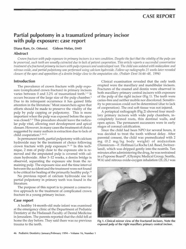
Clinical examination revealed that the only teetherupted were the maxillary and mandibular incisors.Fractures of the enamel and dentin were observed inboth maxillary primary central incisors with exposureof the pulp of the right incisor (Fig 1). The teeth werecaries-free and neither mobile nor discolored. Sensitiv-ity to percussion could not be determined (due to lackof cooperation). The oral soft tissue was not injured.
A periapical radiograph (Fig 2) showed four maxil-lary primary incisors with wide pulp chambers, in-completely formed roots, thin dentinal walls, andopen apices. The permanent successors were in earlystages of coronal calcification.
Since the child had been NPO for several hours, itwas decided to treat the tooth without delay. Afterparental consent, the child was premedicated with 2mg (0.2 mg/kg body weight) of midazolam(Dormicum—F. Hoffman La Roche Ltd. Basel, Switzer-land), which was dripped gently into the nostrils. Tenminutes after administering the drug, he was restrainedin a Papoose Board®, (Olympic Medical Group, Seattle,WA) and nitrous oxide-oxygen inhalation (2L:2L) was
Fig 1. Clinical mirror view of the fractured incisors. Note theexposed pulp of the right maxillary primary central incisor.
46 Pediatric Dentistry: January/February 1994 - Volume 16, Number 1
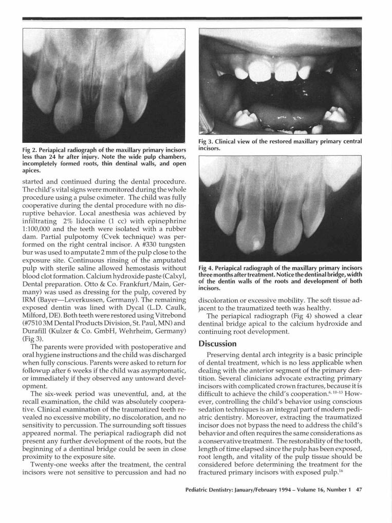
Fig 2. Periapical radiograph of the maxillary primary incisorsless than 24 hr after injury. Note the wide pulp chambers,incompletely formed roots, thin dentinal walls, and openapices.
started and continued during the dental procedure.The child's vital signs were monitored during the wholeprocedure using a pulse oximeter. The child was fullycooperative during the dental procedure with no dis-ruptive behavior. Local anesthesia was achieved byinfiltrating 2% lidocaine (1 cc) with epinephrine1:100,000 and the teeth were isolated with a rubberdam. Partial pulpotomy (Cvek technique) was per-formed on the right central incisor. A #330 tungstenbur was used to amputate 2 mm of the pulp close to theexposure site. Continuous rinsing of the amputatedpulp with sterile saline allowed hemostasis withoutblood clot formation. Calcium hydroxide paste (Calxyl,Dental preparation. Otto & Co. Frankfurt/Main, Ger-many) was used as dressing for the pulp, covered byIRM (Bayer—Leverkussen, Germany). The remainingexposed dentin was lined with Dycal (L.D. Caulk,Milford, DE). Both teeth were restored using Vitrebond(#7510 3M Dental Products Division, St. Paul, MN) andDurafill (Kulzer & Co. GmbH, Wehrheim, Germany)(Fig 3).
The parents were provided with postoperative andoral hygiene instructions and the child was dischargedwhen fully conscious. Parents were asked to return forfollowup after 6 weeks if the child was asymptomatic,or immediately if they observed any untoward devel-opment.
The six-week period was uneventful, and, at therecall examination, the child was absolutely coopera-tive. Clinical examination of the traumatized teeth re-vealed no excessive mobility, no discoloration, and nosensitivity to percussion. The surrounding soft tissuesappeared normal. The periapical radiograph did notpresent any further development of the roots, but thebeginning of a dentinal bridge could be seen in closeproximity to the exposure site.
Twenty-one weeks after the treatment, the centralincisors were not sensitive to percussion and had no
Fig 3. Clinical view of the restored maxillary primary centralincisors.
Fig 4. Periapical radiograph of the maxillary primary incisorsthree months after treatment. Notice the dentinal bridge, widthof the dentin walls of the roots and development of bothincisors.
discoloration or excessive mobility. The soft tissue ad-jacent to the traumatized teeth was healthy.
The periapical radiograph (Fig 4) showed a cleardentinal bridge apical to the calcium hydroxide andcontinuing root development.
DiscussionPreserving dental arch integrity is a basic principle
of dental treatment, which is no less applicable whendealing with the anterior segment of the primary den-tition. Several clinicians advocate extracting primaryincisors with complicated crown fractures, because it isdifficult to achieve the child's cooperation.6-10~'3 How-ever, controlling the child's behavior using conscioussedation techniques is an integral part of modern pedi-atric dentistry. Moreover, extracting the traumatizedincisor does not bypass the need to address the child'sbehavior and often requires the same considerations asa conservative treatment. The restorability of the tooth,length of time elapsed since the pulp has been exposed,root length, and vitality of the pulp tissue should beconsidered before determining the treatment for thefractured primary incisors with exposed pulp.16
Pediatric Dentistry: January/February 1994 - Volume 16, Number 1 47

Several alternative treatments for teeth with a com-plicated crown fracture have been suggested. McTigue17
advocated a formocresol pulpotomy for teeth in whichthe injury has exposed the pulp within the last severalhours and the pulp in the canal is judged to be vital.Formocresol is the suitable medication for fully devel-oped roots, but may not allow closure of the apex inyoung and undeveloped incisors. Hawes8 and Harrison9
advocated calcium hydroxide cervical pulpotomy incase the pulp was exposed before closure of the apex.However, this procedure seems impractical in imma-ture primary incisors due to the extremely thin dentinwalls at the cervical region of the teeth. A complete rootcanal treatment could be another choice, but the vitalpulp, thin dentinal walls of the root, and the open apexin this case were contraindications.
Calcium hydroxide is believed to lead to internalresorption when used for pulpotomy in primary teethwith carious exposure.18 This is probably due to aninflammatory response present in the radicular pulp.Cvek19 demonstrated inflammatory changes reachingthe depth of 2 mm in monkeys’ incisors followingmechanical exposure of the pulp. Since calcium hy-droxide failed to induce the formation of a dentin bar-rier under an exposed and inflamed pulp,2° using cal-cium hydroxide in primary teeth was recommendedonly for small mechanical or traumatic exposures whenconditions for a favorable response are optimalo21
Cvek14,19 suggested the partial pulpotomy in which
excision of 1-2 mm of the pulp tissue adjacent the expo-sure site results in removing the infected part of thepulp, allowing application of calcium hydroxide on thehealthy tissue. In addition, a better control of the surgi-cal wound and retention of the sealing material areachieved. Cvek14 reported 96% success of partialpulpotomy in permanent teeth, and found that successwas not dependent on the size of the exposure or thetime since the injury.
The present report describes a fractured young pri-mary incisor with pulp exposure treated with a proce-dure performed, to date, only on permanent teeth. Ra-diographic evidence observed after 21 weeks of theformation of a dentin bridge and continued root devel-opment with no clinical and radiographic pathology isan early sign of success. Signs of a calcified tissue closeto the calcium hydroxide layer, could be seen in theradiograph exposed as early as six weeks after treat-ment. More cases with longer follow-up periods shouldbe reported and studies conducted before assessing thesuccess rate of partial pulpotomy in traumatized pri-mary incisors. However, the present report should en-courage other clinicians to try a conservative approachin similar conditions.
Dr. Ram is senior resident and Dr. Holan is lecturer, Department ofPediatric Dentistry, The Hebrew University--Hadassah Faculty ofDental Medicine, Founded by the Alpha Omega Fraternity, Jerusa-lem, Israel.
1. Andreasen JO, Ravn JJ: Epidemiology of traumatic dental inju-ries to primary and permanent teeth in a Danish populationsample. Int J Oral Surg 1:235-39, 1972.
2. Zadik D: A survey of traumatized primary anterior teeth inJerusalem preschool children. Community Dent Oral Epidemiol4:149-51, 1976.
3. Garcia-Godoy F, Garcia-Godoy F, Olivo M: Injuries to primaryand permanent teeth treated in a private paedodontic practice.J Can Dent Assoc 6:281-84, 1979.
4. Ferguson FS, Ripa LW: Prevalence and type of traumatic inju-ries to the anterior teeth of preschool children. J Pedod 4:3-8,1979.
5. Garcia-Godoy F, Garcia-Godoy F, Garcia-Godoy FM: Primaryteeth traumatic injuries at a private pediatric dental center.Endod Dent Traumatol 3:126-29, 1987.
6. Bennet DT: Traumatised anterior teeth. Br Dent J 116:52-5,1964.
7. Hargreaves JA, Craig JW: The Management of TraumatizedAnterior Teeth of Children. London: E & S Livingstone, 1970,pp 113-25.
8. Hawes RR: Traumatized primary teeth. Dent Clin North Am10:391-404 1966.
9. Harrison LM Jr: Treatment of traumatized primary anteriorteeth. J Louis Dent Assoc 26:12-7, 1968.
10. Andreasen JO: Traumatic Injuries of the Teeth, 2nd Ed.Copenhagen: Munl~sgaard, 1981, pp 88.
11. Mackie IC, Warren VN: Dental trauma: 1. General aspects ofmanagement, and trauma to the primary dentition. Dent Up-date 15:155-56, 158-59, 1988.
12. Kenny DJ, Yacobi R: Management of trauma to the primarydentition Ontario Dent 65:27-9, 1988.
13. Johnson R: Traumatic dental injuries in children. Part I: Evalu-ation of traumatic dental injuries and treatment of injuries toprimary teeth. Update Pediatr Dent 2:1-4, 6-7, 1989.
14. Cvek M: A clinical report on partial pulpotomy and cappingwith calcium hydroxide in permanent incisors with compli-cated crown fracture. J Endod 4:232-37, 1978.
15. Klein H, Fuks A, Eidelman E, Chosack A: Partial pulpotomyfollowing complicated crown fracture in permanent incisors: Aclinical and radiographical study. J Pedod 9:142-49,1985.
16. Wright GZ, Friedman CM: Management of dental trauma. InTextbook of Pediatric Dentistry. 2nd Ed. Braham RL, MorrisME, EDS. Baltimore: Williams & Wilkins, 1985, pp 565-92.
17. McTigue DJ: Introduction to dental trauma: managing trau-matic injuries in the primary dentition. In Pediatric Dentistry:Infancy Through Adolescence. Pinkham JR, ED. Philadelphia:WB Saunders, pp 171-82, 1988.
18. Via W: Evaluation of deciduous molars treated by pulpotomyand calcium hydroxide. J Am Dent Assoc 50:34--43, 1955.
19. Cvek M, CleatonoJones PE, Austin JC, Andreasen JO: Pulpreactions to exposure after experimental crown fractures orgrinding in adult monkeys. J Endod 8:391-97, 1982.
20. Tronstad L, Mjor IA: Capping of the inflamed pulp. Oral SurgOral Med Oral Pathol 34:477-85, 1972.
21. Fuks A, Eidelman E: Pulp therapy in the primary dentition.Curr Opin Dent 1:556-63, 1991.
48 Pediatric Dentistry: January/February 1994- Volume 16, Number 1