particulate matter 2.5 damages skin cells by inducing
TRANSCRIPT

Vol.:(0123456789)1 3
Archives of Toxicology (2018) 92:2077–2091 https://doi.org/10.1007/s00204-018-2197-9
IN VITRO SYSTEMS
Particulate matter 2.5 damages skin cells by inducing oxidative stress, subcellular organelle dysfunction, and apoptosis
Mei Jing Piao1 · Mee Jung Ahn2 · Kyoung Ah Kang1 · Yea Seong Ryu1 · Yu Jae Hyun1 · Kristina Shilnikova1 · Ao Xuan Zhen1 · Jin Woo Jeong3 · Yung Hyun Choi3 · Hee Kyoung Kang1 · Young Sang Koh1 · Jin Won Hyun1
Received: 9 November 2017 / Accepted: 21 March 2018 / Published online: 26 March 2018 © The Author(s) 2018
AbstractThe skin is the largest organ of the human body and the one mostly exposed to outdoor contaminants. To evaluate the biological mechanisms underlying skin damage caused by fine particulate matter (PM2.5), we analyzed the effects of PM2.5 on cultured human keratinocytes and the skin of experimental animals. PM2.5 was applied to human HaCaT keratinocytes at 50 µg/mL for 24 h and to mouse skin at 100 µg/mL for 7 days. The results indicate that PM2.5 induced oxidative stress by generating reactive oxygen species both in vitro and in vivo, which led to DNA damage, lipid peroxidation, and protein carbonylation. As a result, PM2.5 induced endoplasmic reticulum stress, mitochondrial swelling, and autophagy, and caused apoptosis in HaCaT cells and mouse skin tissue. The PM2.5-induced cell damage was attenuated by antioxidant N-acetyl cysteine, confirming that PM2.5 cellular toxicity was due to oxidative stress. These findings contribute to understanding of the pathophysiological mechanisms triggered in the skin by PM2.5, among which oxidative stress may play a major role.
Keywords PM2.5 · Oxidative stress · Apoptosis · Endoplasmic reticulum stress · Mitochondrial damage · Autophagy
Introduction
Global air pollution has become a major threat to human health. This worldwide problem is especially relevant to the release of fine particulate matter (PM2.5), which has the aerodynamic diameter less than 2.5 µm and originates from incomplete coal combustion and diesel vehicle exhaust in Korea (Lee et al. 2005; Jung et al. 2017). In recent years, the relationship between PM2.5 production and public health hazards has attracted an increasing attention. Several toxi-cological and epidemiological studies have suggested that PM2.5 exerts negative biological effects on several major organs, including the lung (Liu et al. 2017), immune system
(Zhao et al. 2013), cardiovascular system (Du et al. 2016), and nervous system (Wang et al. 2017). Among the affected organs, the skin is the primary tissue exposed to ambient pollutants and, similar to the respiratory tract, presents an interface between the body and surrounding atmosphere.
PM2.5-carrying organic chemicals such as polycyclic aro-matic hydrocarbons (PAHs) are highly lipophilic and easily penetrate the skin (Krutmann et al. 2014). PAHs are potent activators of the aryl hydrocarbon receptor (AhR), which is a ligand-dependent transcription factor expressed in keratino-cytes and melanocytes (Fritsche et al. 2007; Jux et al. 2011). AhR activation by PAHs upregulates the expression of cytochrome P450 (CYP1A1) involved in the metabolism of xenobiotics (Vogel et al. 2016) and promotes generation of intracellular reactive oxygen species (ROS) (Costa et al. 2010). Accumulated evidence indicates that oxidative stress is a common mechanism of PM2.5-induced damage (Gual-tieri et al. 2012; Kouassi et al. 2010). Recently, the effect of PM2.5 on the skin has attracted attention of both clinical dermatologists and basic scientists (Han et al. 2016; Li et al. 2017), who recognized ambient PM2.5 as a crucial risk fac-tor in skin diseases. Thus, PM2.5 was shown to aggravate symptoms in children with allergic dermatitis and eczema (Song et al. 2011), and to promote inflammatory disorders,
* Jin Won Hyun [email protected]
1 Jeju National University School of Medicine and Jeju Research Center for Natural Medicine, Jeju 63243, Republic of Korea
2 Laboratory of Veterinary Anatomy, College of Veterinary Medicine, Jeju National University, Jeju 63243, Republic of Korea
3 Department of Biochemistry, College of Oriental Medicine, Dongeui University, Busan 47340, Republic of Korea

2078 Archives of Toxicology (2018) 92:2077–2091
1 3
aging, androgenetic alopecia, and cancers of the skin (Kim et al. 2016).
Mitochondria are unique double-membrane subcellular organelles that provide energy through oxidative phospho-rylation and participate in metabolic and genetic processes in the body. Once mitochondria are disrupted, their dysfunction leads to reduced generation of ATP and higher production of ROS. Mitochondria are targeted by environmental pollutants such as PM2.5 (Guo et al. 2017) and mitochondrial dam-age may be a critical part of the pathophysiological mecha-nisms induced by PM2.5 exposure. Oxidative stress has been shown to be an initiator and major contributor to both endo-plasmic reticulum (ER) stress (Hotamisligil 2010; Kaneto et al. 2005) and lysosome-mediated autophagy (Lee et al. 2012); however, the research on molecular pathways linking atmospheric PM2.5 and skin damage is limited. Although skin is the organ mostly exposed to PM2.5, the association of skin-damaging effects of PM2.5 with oxidative stress and dysfunction of subcellular organelles such as mitochondria, ER, and lysosomes is still not fully elucidated. In this study, we investigated the effects of PM2.5 on the induction of oxi-dative stress and structure of subcellular organelles using in vitro and in vivo models and explored the mechanisms underlying PM2.5 toxicity for the skin.
Materials and methods
Preparation of PM2.5
Diesel particulate matter NIST 1650b (PM2.5) was purchased from Sigma-Aldrich, Inc. (St. Louis, MO, USA). PM2.5 stock solution (25 mg/mL) was prepared in dimethyl sulfoxide (DMSO) and sonicated for 30 min to avoid agglomeration of the suspended PM2.5 particles. Experiments were performed within 1 h of stock preparation to avoid variability in PM2.5 composition in solution.
Cell culture
Human HaCaT keratinocytes (Amore Pacific Company, Gyeonggi-do, Republic of Korea) were maintained at 37 °C in an incubator with a humidified atmosphere of 5% CO2. Cells were cultured in Dulbecco-modified Eagle medium (DMEM) containing 10% heat-inactivated fetal bovine serum and antibiotic–antimycotic (100 units/mL penicillin, 100 µg/mL streptomycin, and 0.25 µg/mL amphotericin B) (Gibco, Life Technologies Co., Grand Island, NY, USA).
Animal experiment
In vivo experiments were conducted using HR-1 hairless male mice (OrientBio, Kyungki-do, Republic of Korea)
in accordance with the guidelines for the care and use of laboratory animals at Jeju National University (Jeju, Repub-lic of Korea) (permit number: 2017-0026). Mice were randomly divided into three groups (n = 4 each): control, and treated with PM2.5 or N-acetyl cysteine (NAC; Sigma-Aldrich) + PM2.5. PM2.5 was dispersed in propylene glycol to the concentration of 100 µg/mL, spread on a nonwoven poly-ethylene pad over a 1 cm2 area, and applied to the dorsal skin of mice for 7 consecutive days. At the end of the treatment, the exposed skin tissue was immediately dissected for his-tological and biochemical analysis as previously described (Lee et al. 2016).
ROS measurement
Cells were incubated with different concentrations of PM2.5 (25, 50, 75, and 100 µg/mL) for 24 h or treated with PM2.5 at a concentration of 50 µg/mL for different times (1, 2, 4, 8, 12, and 24 h). After staining with 25 µM 2′,7′-dichlorodi-hydrofluorescein diacetate (H2DCFDA; Molecular Probes, Eugene, OR, USA) dye for 10 min, H2DCF fluorescence was detected by flow cytometry (Becton Dickinson, Moun-tain View, CA, USA) and analyzed using the Cell Quest software. For imaging, cells were plated in a 4-well glass chamber slide, treated with 1 mM NAC and/or 50 µg/mL PM2.5, and analyzed for intracellular and mitochondrial ROS production after staining with H2DCFDA and dihy-drorhodamin 123 (DHR 123; Molecular Probes), respec-tively, for 30 min. Images of stained cells were generated by confocal microscopy as previously described (Kim and Yoo 2016; Soeur et al. 2017). To detect ROS in zebrafish treated with NAC and/or PM2.5, they were incubated with 10 µM H2DCFDA for 30 min in the dark at 28.5 °C. After anes-thesia with 1-phenoxy-2-propanol (1/500 dilution; Acros Organics, Morris Plains, NJ, USA), the stained zebrafish were observed under a fluorescence microscope (Zeiss AX10, Carl Zeiss, Göttingen, Germany) (Jeong et al. 2017).
Hoechst 33342/propidium iodide (PI) staining
Cells were treated with different concentrations of PM2.5 (25, 50, 75, and 100 µg/mL) for 24 h or pre-treated with 1 mM NAC for 1 h and then treated with 50 µg/mL of PM2.5 for 24 h and then stained with DNA-specific fluorescent dyes Hoechst 33342 (10 µM) and propidium iodide (PI; 5 µg/mL) (both from Sigma-Aldrich). Cells with fragmented nuclei stained with Hoechst 33342 were considered apoptotic and those stained with PI were considered necrotic. Cell staining was visualized under a fluorescence microscope equipped with a CoolSNAP-Pro color digital camera (Media Cyber-netics, Rockville, MD, USA) and the proportions of apop-totic and neurotic cell were quantified.
2079Archives of Toxicology (2018) 92:2077–2091
1 3
Trypan blue assay
Cells were seeded in 35-mm culture dishes at a concentra-tion of 1.0 × 10⁵ per mL, cultured for 16 h, and treated with different concentrations of PM2.5 (25, 50, 75, and 100 µg/mL) for 24 h. Then, 5 µL of 0.1% trypan blue solution was added to 0.1 mL cell suspension for 5 min at room tempera-ture, and the numbers of viable and dead cells were deter-mined under a microscope using 10× magnification. Cell viability (%) was calculated as: unstained cells/(unstained cells + stained cells) × 100%.
Detection of 8‑oxoguanine
Cellular DNA was isolated using the G-DEX™ IIc Genomic DNA Extraction Kit (iNtRON Biotechnology, Inc., Sung-nam, Kyungki-Do, Republic of Korea) and quantified by spectrophotometry. The amount of 8-hydroxy-2-deoxy-guanosine (8-OHdG, the deoxyriboside form of 8-oxoG) in DNA was determined using the BIOXYTECH® 8-OHdG-EIA™ kit (OXIS Health Products, Inc., Portland, OR, USA) according to the manufacturer’s instructions. The amount of 8-oxoG was also estimated by a fluorescence-binding assay: cells were fixed and permeabilized with ice-cold methanol for 15 min and 8-oxoG was visualized with avidin-TRITC conjugate (Sigma-Aldrich) under a confocal microscope (Piao et al. 2011).
Single cell gel electrophoresis (Comet assay)
Cell-coated slides were immersed in lysis buffer (2.5 M NaCl, 100 mM Na-EDTA, 10 mM Tris, 1% Trion X-100, and 10% DMSO, pH 10) for 1 h at 4 °C, subjected to elec-trophoresis, stained with ethidium bromide, and observed under a fluorescence microscope equipped with an image analysis software (Kinetic Imaging, Komet 5.5, UK) as pre-viously described (Park et al. 2017). The percentage of the total fluorescence in the comet tail and the length of the tail was recorded in 50 cells per slide.
Lipid peroxidation assay
Cells were stained with 5 µM of a fluorescent probe diphe-nyl-1-pyrenylphosphine (DPPP; Molecular Probes) as described (Morita et al. 2016) and analyzed using an Olym-pus FV1200 laser scanning microscope equipped with the FV10-ASW viewer 4.2 software. Mouse skin tissue was analyzed by immunohistochemistry using an antibody to 4-hydroxy-2-nonenal (4-HNE) (Cosmo Bio Co., Tokyo, Japan).
Protein carbonylation
Total cellular proteins were extracted with lysis buffer and quantified by spectrophotometry, and protein carbonyla-tion was determined using an OxiSelect™ protein carbonyl ELISA kit (Cell Biolabs, San Diego, CA, USA) according to the manufacturer’s instructions. In tissues, protein carbon-ylation was assessed using an immunohistochemical staining kit (Cosmo Bio Co.)
Western blotting
Cell and mouse skin lysates were subjected to SDS-PAGE, and the separated proteins were transferred to membranes and incubated with primary antibodies against phospho-H2A.X (Ser139), CHOP, phospho-PERK, beclin-1, LC3B, caspase-3, and caspase-9 (Cell Signaling Technology, Bev-erly, MA, USA), GRP78, Bax (Santa Cruz Biotechnology, Santa Cruz, CA, USA), phospho-IRE1 (Abcam, Cambridge, MA, USA), and actin (Sigma-Aldrich) followed by the incubation with a secondary antibody (Pierce, Rockford, IL, USA). Protein bands were detected using an Amersham Enhanced Chemiluminescence Plus Western Blotting Detec-tion system (GE Healthcare Life Sciences, Buckingham-shire, UK).
ER staining
Cells were reacted with an ER-tracker blue-white DPX dye (Molecular Probes) for 30 min, and images were taken under a confocal microscope (Li et al. 2015).
Quantification of Ca2+ level
Cells were loaded for 30 min with 10 µM fluo-4-acetoxy-methyl ester (Fluo-4-AM) or with Rhod-2 acetoxymethyl ester (Rhod-2 AM) (Molecular Probes) to detect intracellular and mitochondrial Ca2+, respectively, and fluorescence was measured by confocal microscopy (Wang et al. 2016).
Mitochondrial membrane potential (Δψm) measurement
Mitochondrial Δψ was analyzed by confocal micros-copy after staining with 5,5′,6,6′-tetrachloro-1,1′,3,3′-tetraethylbenzimidazolylcarbocyanine iodide (JC-1, Invitro-gen, Carlsbad, CA, USA), a lipophilic cationic fluorescence dye (Yao et al. 2016).
Acridine orange staining
To analyze autophagy, cells were reacted with acrid-ine orange (Invitrogen) for 15 min and fluorescence was

2080 Archives of Toxicology (2018) 92:2077–2091
1 3
measured using a fluorescence microscope (BH2-RFL-T3; Olympus, Tokyo, Japan) (Farah et al. 2016). Depending on the acidity, autophagic lysosomes appeared as orange/red fluorescent cytoplasmic vesicles, while the nuclei were stained green.
LC3 transfection and detection of punctate LC3‑positive structures
Membrane-bound microtubule-associated protein 1 light chain 3 (LC3) is present in the autophagic double-membrane structure, which is an important marker of autophagy (Fazeli and Wehman 2017). Cells were transfected with GFP-tagged LC3 using Lipofectamine reagent (Invitrogen) according to the manufacturer’s instructions and GFP-LC3 fluorescence was observed under a confocal microscope.
Histological analysis
Skin pieces were fixed in 4% paraformaldehyde, embed-ded in paraffin, and cut into 5 µm sections, which were then deparaffinized and stained with hematoxylin and eosin. The height of epidermis (from the stratum basale to the stratum corneum) was measured in ten randomly chosen fields from three representative sections per group by microscopy at 100× magnification using a digital camera. Immunohisto-chemistry was performed by incubating skin sections with primary anti-Bax antibody (1:400; Abcam, Cambridge, MA, USA) for 1 h and the reaction was visualized using an ABC Elite kit (Vector Labs, Burlingame, CA, USA). The sections were counterstained with hematoxylin before mounting. For the in situ detection of apoptotic cells in skin sections, the DeadEnd colorimetric TUNEL system (Promega, Wiscon-sin, WI, USA) was used according to the manufacturer’s recommendation.
Transmission electron microscopy (TEM)
Cells and tissues were fixed, dehydrated, incubated with increasing concentrations of propylene oxide dissolved in ethanol, and infiltrated with increasing concentrations of Eponate 812 resin. Samples were baked in a 65 °C oven overnight, sectioned in an ultramicrotome, and examined by TEM using a field electron emission unit (JEM-2100F, JEOL) at the Korean Basic Science Institute (Chuncheon, Republic of Korea).
Statistical analysis
Statistical significance of the difference between groups was determined by analysis of variance and Tukey’s tests using the SigmaStat version 3.5 software (Systat Software Inc., San Jose, CA, USA). All data are presented as the
mean ± standard error. p < 0.05 was considered to indicate statistically significant difference.
Results
PM2.5 induced oxidative stress both in vitro and in vivo
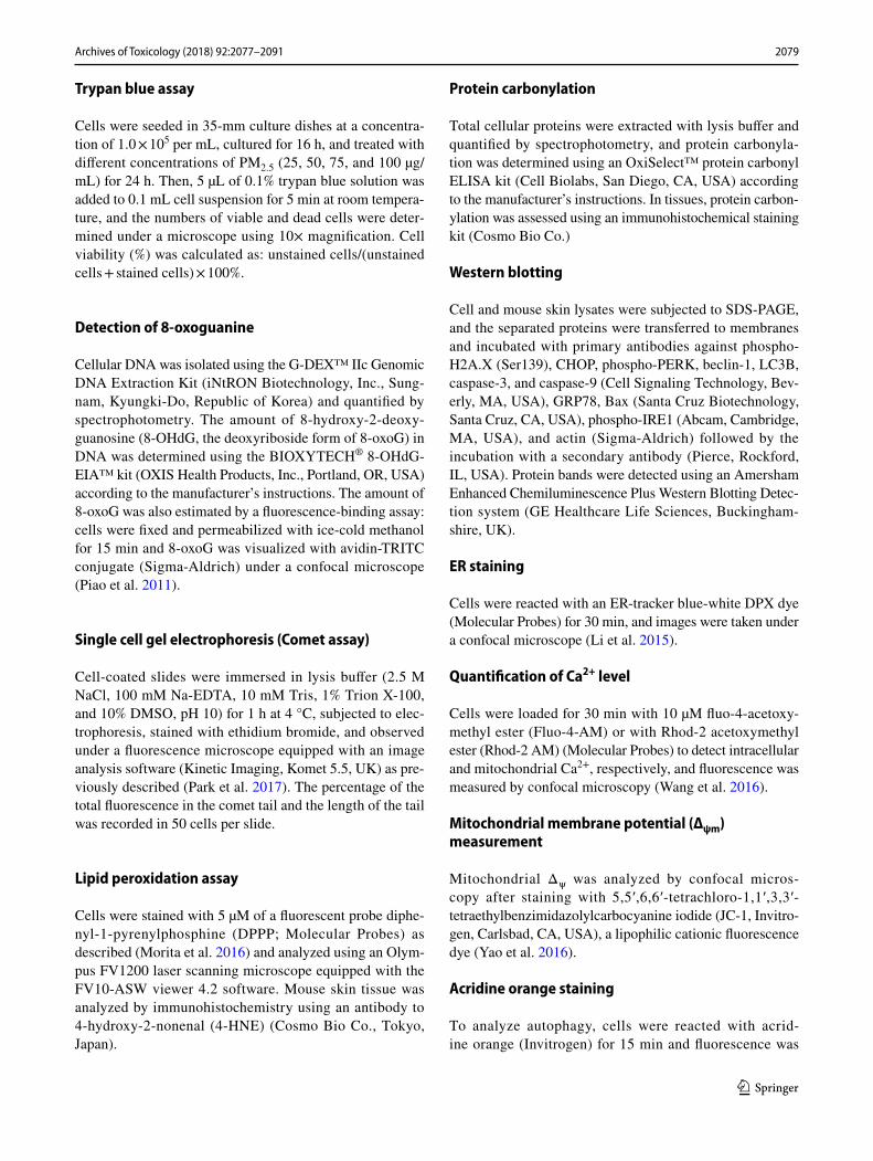
To investigate the potential role of oxidative stress induced by PM2.5, we measured ROS generation and cel-lular damage in PM2.5-treated human keratinocytes and mouse skin. Figure 1a shows that PM2.5 treatment pro-moted the production of ROS in a dose-dependent manner as evidenced by H2DCFDA staining. Analysis of Hoechst 33342/PI staining indicated that PM2.5 at a concentration of 50 µg/mL induced apoptosis (Hoechst 33342-stained cells); however, at concentrations above 75 µg/mL, PM2.5 induced necrosis (PI-stained cells) (Fig. 1b). We used 50 µg/mL PM2.5 as the optimal concentration in further experiments. ROS generation was increased starting from 1 h up to 24 h of treatment with 50 µg/mL PM2.5 (Fig. 1c). Next, we determined whether PM2.5 induced cytotoxicity via ROS generation. An antioxidant NAC was not toxic for HaCaT cells at concentrations up to 1 mM (MTT test; data not shown); therefore, 1 mM NAC was used in this study. Confocal microscopy indicated that green fluores-cence was increased in PM2.5-treated cells compared to control, but the effect was suppressed by 1 mM NAC (Fig. 1d), indicating that PM2.5 stimulated ROS produc-tion in keratinocytes. Furthermore, PM2.5 induced cyto-toxicity as evidenced by trypan blue exclusion; however, 1 mM NAC reduced it (Fig. 1e), suggesting that PM2.5 caused cytotoxicity via ROS. We next evaluated the dam-age of intracellular macromolecules by PM2.5. The level of 8-oxoG, a hallmark of oxidative DNA damage, was meas-ured based on 8-OHdG detection. The results indicated that PM2.5 promoted the generation of 8-oxoG in DNA in a time-dependent manner when used at 50 µg/mL (Fig. 1f) and in a dose-dependent manner when administered for 8 h (Fig. 1g). In addition, condensed 8-oxoG staining was observed in PM2.5-treated cells, whereas 1 mM NAC reduced it (Fig. 1h). The Comet assay assessing DNA breakage indicated that PM2.5 increased the presence of cellular DNA tails by 30% compared to control; however, NAC pre-treatment reduced it to 12% (Fig. 1i). Further-more, fluorescence intensity of DPPP oxide, an indicator of lipid peroxidation, was enhanced in PM2.5-incubated cells but significantly reduced by NAC pre-treatment (Fig. 1j). Similarly, the level of protein carbonylation, a biomarker of oxidative stress-induced protein damage, was increased in PM2.5-treated cells, whereas NAC could prevent PM2.5-induced carbonyl formation (Fig. 1k). To

2081Archives of Toxicology (2018) 92:2077–2091
1 3
a b
c
0
100
200
300
Intra
cellu
lar R
OS
[%]
PM2.5
0 1 2 4 8 12 24 h
* * * * *
*
0
200
400
600
Intra
cellu
lar R
OS
[%]
PM2.5 [μg/mL]
* *
* *
0 25 50 75 100
Control PM NAC+PMe
d
0
30
60
90
120
*
#
Control PM2.5 NAC+PM2.5
Cel
l via
bilit
y
0
10
20
30
Inde
x of
RO
S ge
nera
tion
Control PM2.5 NAC+PM2.5
*
#
Control PM2.5 NAC+PM2.5
DC
F-D
A
0
20
40
60
80
100
8-O
HdG
leve
l (ng
/ml
* * * * *
PM2.5
0 1 4 8 12 24 h
f
0 25 50 75 100
PM2.5 [μg/mL]
Inde
x of
apo
ptot
ic
& n
ecro
tic c
ells
0
10
20
30
40
50Apoptotic cellsNecrotic cells
PM2.5 [μg/mL]
0 25 50 75 100
*
** *
*
*
**
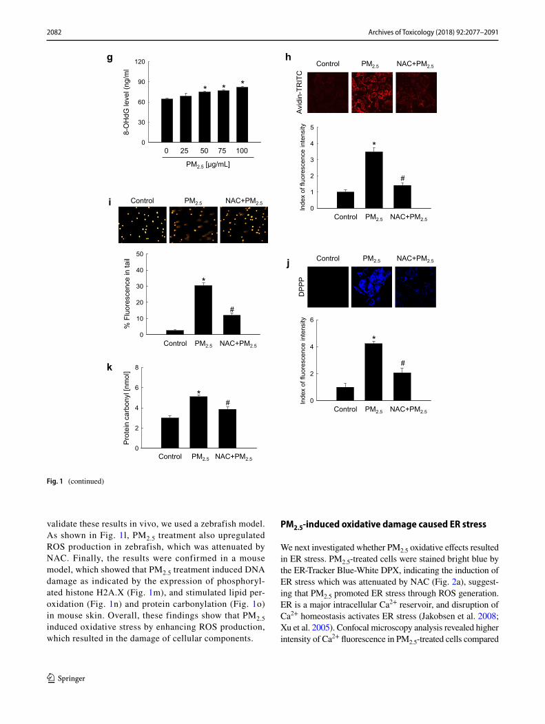
Fig. 1 PM2.5 induces ROS production leading to oxidative damage. Cells were treated with PM2.5 at the indicated concentrations for 24 h. a ROS generation was assessed by the H2DCFDA assay. b Apoptotic and necrotic cells were detected by Hoechst 33342 and PI nuclear staining, respectively. Arrows indi-cate apoptotic bodies and red color indicates necrotic cells. c Cells were treated with 50 µg/mL PM2.5 for the indicated times and ROS generation was assessed by the H2DCFDA assay. Cells were pre-treated with NAC (1 mM) for 1 h and then treated with PM2.5 (50 µg/mL) for 24 h. d ROS levels were assessed by confocal microscopy after H2DCFDA staining. e Cell viability was analyzed by trypan blue exclusion. Cells were treated with 50 µg/mL PM2.5 f for the indi-cated times or g with the indicated concentrations of PM2.5 for 8 h and ana-lyzed for the generation of 8-oxoG in DNA by avidin-TRITC binding using confocal microscopy. i DNA damage was evaluated by the Comet assay; rep-resentative images show comet tails and the graph shows quantification of cel-
lular DNA damage. j Lipid peroxidation was analyzed by confocal microscopy after DPPP staining. k Protein oxidation was assayed by measuring carbonyl formation. l Zebrafish was pre-treated or not with NAC and then treated with PM2.5 (50 µg/mL) for 24 h, and analyzed for ROS production by H2DCFDA staining; zebrafish treated with H2O2 was used as a positive control. Mouse dorsal skin was treated with PM2.5 (100 µg/mL) for 7 days. m Tissue lysates were analyzed for H2A.X expression by immunoblotting; actin was used as loading control. n Immunohistochemistry of mouse tissue to analyze 4-HNE levels as a marker of lipid peroxidation; signals were detected in a peroxidase reaction (brown), and slides were counterstained with hematoxylin; magnifi-cation: ×400. o Protein carbonylation in mouse tissue was assessed using an immunohistochemical staining kit for protein carbonyls. *p < 0.05 compared to control groups and #p < 0.05 compared to PM2.5-treated groups. (Color figure online)
2082 Archives of Toxicology (2018) 92:2077–2091
1 3
validate these results in vivo, we used a zebrafish model. As shown in Fig. 1l, PM2.5 treatment also upregulated ROS production in zebrafish, which was attenuated by NAC. Finally, the results were confirmed in a mouse model, which showed that PM2.5 treatment induced DNA damage as indicated by the expression of phosphoryl-ated histone H2A.X (Fig. 1m), and stimulated lipid per-oxidation (Fig. 1n) and protein carbonylation (Fig. 1o) in mouse skin. Overall, these findings show that PM2.5 induced oxidative stress by enhancing ROS production, which resulted in the damage of cellular components.
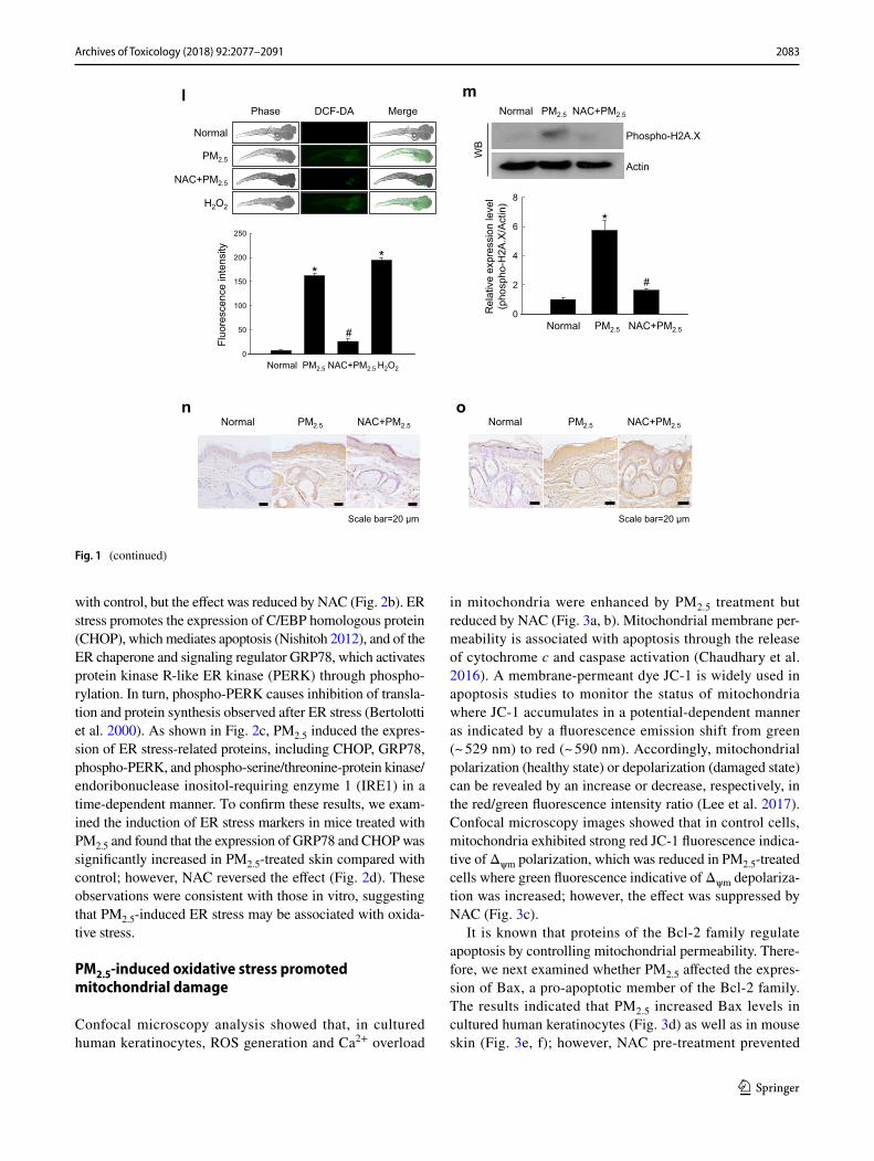
PM2.5‑induced oxidative damage caused ER stress
We next investigated whether PM2.5 oxidative effects resulted in ER stress. PM2.5-treated cells were stained bright blue by the ER-Tracker Blue-White DPX, indicating the induction of ER stress which was attenuated by NAC (Fig. 2a), suggest-ing that PM2.5 promoted ER stress through ROS generation. ER is a major intracellular Ca2+ reservoir, and disruption of Ca2+ homeostasis activates ER stress (Jakobsen et al. 2008; Xu et al. 2005). Confocal microscopy analysis revealed higher intensity of Ca2+ fluorescence in PM2.5-treated cells compared
g h
0
10
20
30
40
50
% F
luor
esce
nce
in ta
il
Control PM2.5 NAC+PM2.5
*
#
Control PM2.5 NAC+PM2.5 i
Avi
din-
TRIT
C Control PM2.5 NAC+PM2.5
0
30
60
90
120
8-O
HdG
leve
l (ng
/ml
* * *
PM2.5 [μg/mL]
0 25 50 75 100
0
1
2
3
4
5
Inde
x of
fluo
resc
ence
inte
nsity
Control PM2.5 NAC+PM2.5
*
#
j
DP
PP
Control PM2.5 NAC+PM2.5
0
2
4
6
Inde
x of
fluo
resc
ence
inte
nsity
Control PM2.5 NAC+PM2.5
*
# k
0
2
4
6
8
Control PM2.5 NAC+PM2.5
* #
Pro
tein
car
bony
l [nm
ol]
Fig. 1 (continued)
2083Archives of Toxicology (2018) 92:2077–2091
1 3
with control, but the effect was reduced by NAC (Fig. 2b). ER stress promotes the expression of C/EBP homologous protein (CHOP), which mediates apoptosis (Nishitoh 2012), and of the ER chaperone and signaling regulator GRP78, which activates protein kinase R-like ER kinase (PERK) through phospho-rylation. In turn, phospho-PERK causes inhibition of transla-tion and protein synthesis observed after ER stress (Bertolotti et al. 2000). As shown in Fig. 2c, PM2.5 induced the expres-sion of ER stress-related proteins, including CHOP, GRP78, phospho-PERK, and phospho-serine/threonine-protein kinase/endoribonuclease inositol-requiring enzyme 1 (IRE1) in a time-dependent manner. To confirm these results, we exam-ined the induction of ER stress markers in mice treated with PM2.5 and found that the expression of GRP78 and CHOP was significantly increased in PM2.5-treated skin compared with control; however, NAC reversed the effect (Fig. 2d). These observations were consistent with those in vitro, suggesting that PM2.5-induced ER stress may be associated with oxida-tive stress.
PM2.5‑induced oxidative stress promoted mitochondrial damage
Confocal microscopy analysis showed that, in cultured human keratinocytes, ROS generation and Ca2+ overload
in mitochondria were enhanced by PM2.5 treatment but reduced by NAC (Fig. 3a, b). Mitochondrial membrane per-meability is associated with apoptosis through the release of cytochrome c and caspase activation (Chaudhary et al. 2016). A membrane-permeant dye JC-1 is widely used in apoptosis studies to monitor the status of mitochondria where JC-1 accumulates in a potential-dependent manner as indicated by a fluorescence emission shift from green (~ 529 nm) to red (~ 590 nm). Accordingly, mitochondrial polarization (healthy state) or depolarization (damaged state) can be revealed by an increase or decrease, respectively, in the red/green fluorescence intensity ratio (Lee et al. 2017). Confocal microscopy images showed that in control cells, mitochondria exhibited strong red JC-1 fluorescence indica-tive of Δψm polarization, which was reduced in PM2.5-treated cells where green fluorescence indicative of Δψm depolariza-tion was increased; however, the effect was suppressed by NAC (Fig. 3c).
It is known that proteins of the Bcl-2 family regulate apoptosis by controlling mitochondrial permeability. There-fore, we next examined whether PM2.5 affected the expres-sion of Bax, a pro-apoptotic member of the Bcl-2 family. The results indicated that PM2.5 increased Bax levels in cultured human keratinocytes (Fig. 3d) as well as in mouse skin (Fig. 3e, f); however, NAC pre-treatment prevented
l MergePhase DCF-DA
Normal
PM2.5
NAC+PM2.5
H2O2
m
Phospho-H2A.X
Actin
Normal PM2.5 NAC+PM2.5
WB
n o
0
50
100
150
200
250
Fluo
resc
ence
inte
nsity
Normal PM2.5 NAC+PM2.5 H2O2
*
#
*
Normal PM2.5 NAC+PM2.5
Scale bar=20 μm Scale bar=20 μm
Normal PM2.5 NAC+PM2.5
0
2
4
6
8
Normal PM2.5 NAC+PM2.5
*
#
Rel
ativ
e ex
pres
sion
leve
l (p
hosp
ho-H
2A.X
/Act
in)
Fig. 1 (continued)

2084 Archives of Toxicology (2018) 92:2077–2091
1 3
PM2.5-induced upregulation of Bax expression (Fig. 3e, f). Cumulatively, these data indicate that PM2.5 increased oxida-tive stress in mitochondria by stimulating ROS production, which resulted in mitochondrial damage.
PM2.5‑induced oxidative stress caused autophagy
We next determined whether PM2.5-induced oxidative stress could promote autophagy. In cultured keratinocytes,
Fig. 2 PM2.5 induces ER stress via ROS generation. a, b Cells were pre-treated or not with NAC (1 mM) for 1 h and then with PM2.5 (50 µg/mL) for 24 h and analyzed by confocal microscopy for ER stress using a ER-Tracker Blue-White DPX staining and for intracellular Ca2+ levels using b Fluo-4-AM staining. Representative confo-cal images are shown. Lysates of c cells and d mouse skin tissue were analyzed for the expression of CHOP, GRP78, phospho-PERK, and phospho-IRE1 by western blotting. Actin was used as loading control. *p < 0.05 compared to control groups and #p < 0.05 compared to PM2.5-treated groups
Control PM2.5 NAC+PM2.5
ER
trac
ker
Control PM2.5 NAC+PM2.5
Fluo
-4
a
b
c
CHOP
PM2.5
0 3 6 12 24 h
GRP78
Phospho-IRE1
Actin
Phospho-PERKWB
Rel
ativ
e ex
pres
sion
leve
ls
0
1
2
3
4
*
CH
OP
/Act
in
0
2
4
6
*
GR
P78/
Actin
0
1
2
3
4
* *
Phos
pho-
PER
K/Ac
tin
0
1
2
3
*
PM2.5
0 3 6 12 24 h
Phos
pho-
IRE1
α/Ac
tin
0
1
2
3
4
Inde
x of
fluo
resc
ence
inte
nsity
Control PM2.5 NAC+PM2.5
*
#
0
4
8
12
16
Inde
x of
fluo
resc
ence
inte
nsity
Control PM2.5 NAC+PM2.5
* #
d CHOP
GRP78
Actin
Normal PM2.5 NAC+PM2.5
WB
0
1
2
3
4
*
CH
OP
/Act
in
#
0
2
4
6
8
10
Normal PM2.5 NAC+PM2.5
*
# GR
P78/
Actin
Rel
ativ
e ex
pres
sion
leve
ls

2085Archives of Toxicology (2018) 92:2077–2091
1 3
Fig. 3 PM2.5 induces mitochon-drial damage via ROS genera-tion. Cells were pre-treated or not with NAC (1 mM) for 1 h and then with PM2.5 (50 µg/mL) for 24 h and analyzed by confocal microscopy to assess a mitochondrial ROS (DHR123 staining), b mitochondrial Ca2+ levels (Rhod-2 AM staining), and c Δψm (JC-1 staining). Lysates of d cells and e mouse skin tissue were analyzed for the expression of Bax protein by western blotting. f Mouse skin tissue was analyzed for Bax expression by immunohisto-chemistry. Nuclei were stained with hematoxylin; arrows indi-cate Bax. *p < 0.05 compared to control groups and #p < 0.05 compared to PM2.5-treated groups
a b
DH
R 1
23
Control PM2.5 NAC+PM2.5
c d PM2.5
0 3 6 12 24 h
Bax
Actin
WB
0
2
4
6
8
Rel
ativ
e ex
pres
sion
le
vel (
Bax
/Act
in)
*
PM2.5
0 3 6 12 24 h
0
10
20
30
Inde
x of
fluo
resc
ence
inte
nsity
Control PM2.5 NAC+PM2.5
*
#
0.0
0.5
1.0
1.5
2.0
2.5
Red
/gre
en fl
uore
scen
ce ra
tio
Control PM2.5 NAC+PM2.5
*
#
Control PM2.5 NAC+PM2.5
Mer
ge
D
epol
ariz
ed
Pol
ariz
ed
Δψ
m
0
2
4
6
8
Inde
x of
fluo
resc
ence
inte
nsity
Control PM2.5 NAC+PM2.5
*
#
Rho
d-2
AM
Control PM2.5 NAC+PM2.5
e
0
1
2
3
Normal PM2.5 NAC+PM2.5
* #
Rel
ativ
e ex
pres
sion
leve
l (B
ax/A
ctin
)
Bax
Actin
WB
Normal PM2.5 NAC+PM2.5f
Normal PM2.5
NAC+PM2.5 Negative control
Scale bar=20 μm

2086 Archives of Toxicology (2018) 92:2077–2091
1 3
PM2.5 triggered accumulation of intracellular vacuoles indicative of autophagy, as evidenced by staining with a lysosome marker dye acridine orange (Fig. 4a). The two distinct steps of autophagy, autophagosome formation and autolysosome formation, can be discerned by the pres-ence of LC3-phospholipid conjugates (Tanida et al. 2005). PM2.5-treated GFP-LC3-transfected cells had increased levels of GFP-LC3-positive puncta (Fig. 4b). In addition, PM2.5 upregulated the expression of beclin-1, the protein initiating autophagosome formation during autophagy, and LC3B-II, the processed form of LC3, in a time-dependent manner (Fig. 4c). These in vitro results were confirmed in PM2.5-treated mouse skin (Fig. 4d). However, the effects of PM2.5 both in cultured cell and animals were reversed by NAC (Fig. 4a, b, d), suggesting that PM2.5 increased autophagy through oxidative stress.
PM2.5‑induced oxidative stress promoted apoptotic cell death
PM2.5 induced apoptosis both in cultured cells and mouse skin tissues, as shown by the formation of apoptotic bodies
and DNA fragmentation revealed by Hoechst 33342 staining and TUNEL assay, respectively; however, NAC pre-treat-ment diminished the effects (Fig. 5a, b). Another evidence that PM2.5 promoted apoptosis was time-dependent increase in the expression of cleaved caspase-9 and caspase-3 (Fig. 5c), which indicated caspase activation in response to mitochondrial membrane disruption. Similar results were obtained for the mouse skin, where cleaved forms of caspase-3 and caspase-9 were upregulated in response to PM2.5 treatment; however, the effect was attenuated by NAC (Fig. 5d), suggesting that PM2.5 induced apoptosis via oxida-tive stress.
PM2.5 internalization damaged the ultrastructure of mouse skin tissue
TEM analysis revealed PM2.5 internalization in HaCaT cells after 24 h of exposure to 50 µg/mL PM2.5 (Fig. 6a-2). In addition, to evaluate organelle ultrastructure in skin cells following PM2.5 exposure, we performed TEM analy-sis of mouse skin tissue after treatment with 100 µg/mL PM2.5. Compared to normal mice (Fig. 6b-1), skin tissue
Fig. 4 PM2.5 induces autophagy via ROS generation. Cells were pre-treated or not with NAC (1 mM) for 1 h, treated with PM2.5 (50 µg/mL) for 24 h. a Cells were stained with acridine orange and analyzed for autophagy by fluorescence microscopy. b Cells were transfected with the GFP-LC3 expression construct and observed under a fluorescence microscope. Lysates extracted from c cells and d mouse skin tissue were analyzed for the expression of beclin-1 and LC3B-II proteins by western blotting; actin was used as load-ing control. *p < 0.05 compared to control groups and #p < 0.05 compared to PM2.5-treated groups. (Color figure online)
a
c
b
AO
Control PM2.5 NAC+PM2.5
d
GFP
-LC
3
Control PM2.5 NAC+PM2.5
0.0
0.5
1.0
1.5
2.0
2.5
Bec
lin-1
/Act
in
* * *
0.0
0.5
1.0
1.5
2.0
2.5
3.0
LC3B
-II/A
ctin *
*
PM2.5
0 3 6 12 24 h
Rel
ativ
e ex
pres
sion
leve
ls
* *
0
1
2
3
* #
Bec
lin-1
/Act
in
Rel
ativ
e ex
pres
sion
leve
ls
0
1
2
3
4
* #
LC3B
-II/A
ctin
Normal PM2.5 NAC+PM2.5
Beclin-1
Actin
Normal PM2.5 NAC+PM2.5
WB LC3B-
PM2.5
0 3 6 12 24 h
Beclin-1
Actin
WB LC3B-

2087Archives of Toxicology (2018) 92:2077–2091
1 3
of PM2.5-treated mice showed increased swelling of mito-chondria (Fig. 6b-2) and ER (Fig. 6b-3), and autophago-some formation (Fig. 6b-4), indicating that PM2.5 dis-rupted intracellular network in the skin.
Discussion
According to the Air Korea site of Korea Environment Cor-poration (2015, 2016, 2017) of the National Environmental Research Institute of the Republic of Korea, the average
a
0
4
8
12
16
20
Inde
x of
apo
ptot
ic b
ody
Control PM2.5 NAC+PM2.5
*
#
Control PM2.5 NAC+PM2.5
Hoe
chst
333
42
c PM2.5
0 3 6 12 24 h
Actin
Cleaved caspase-3
Cleaved caspase-9
WB Cleaved caspase-3
Actin
Cleaved caspase-9
Normal PM2.5 NAC+PM2.5
WB
d
b Normal PM2.5 NAC+PM2.5
Scale bar=20 μm
0
1
2
3
* #
Cle
aved
ca
spas
e-9/
Actin
0
1
2
3
4
*
#
Cle
aved
ca
spas
e-3/
Actin
Normal PM2.5 NAC+PM2.5
Rel
ativ
e ex
pres
sion
leve
ls
0
1
2
3
4
5
Cle
aved
ca
spas
e-3/
Act
in
* * *
PM2.5
0 3 6 12 24 h
0
1
2
3
4
Cle
aved
ca
spas
e-9/
Act
in
*
Rel
ativ
e ex
pres
sion
leve
ls
Fig. 5 PM2.5 induces apoptosis via ROS generation. a Cells were pre-treated or not with NAC (1 mM) for 1 h, treated with PM2.5 (50 µg/mL) for 24 h, and analyzed for apoptotic body formation after Hoe-chst 33342 staining; apoptotic bodies are indicated by arrows. b Mouse skin treated with NAC and PM2.5 (100 µg/mL) for 7 days was
analyzed for apoptosis by TUNEL staining; TUNEL-positive cells are indicated by arrows. Lysates extracted from c cells and d mouse skin tissue were analyzed for the expression of caspase-3 and caspase-9 by western blotting. *p < 0.05 compared to control groups and #p < 0.05 compared to PM2.5-treated groups

2088 Archives of Toxicology (2018) 92:2077–2091
1 3
concentrations of PM2.5 in the air of seven major Korean cities from January to March were 31, 28, and 29 µg/m3 in 2015, 2016, and 2017, respectively, exceeding the national environmental standard of 25 µg/m3.
Skin keratinocytes present the first barrier for environ-mental pollutants, and it was shown that PM exposure could upregulate pro-inflammatory mediators and AhR expression, leading to increased ROS generation in keratinocytes (Choi et al. 2011). Wei et al. (2017) demonstrated that organic extracts containing PAHs with PM2.5 induced stronger oxi-dative stress compared to those without PM2.5. It was also shown that PM2.5 may penetrate the skin and have harmful effects on viable skin cells, including keratinocytes (Krut-mann et al. 2014; Li et al. 2017). A recent study demon-strated that PM2.5 could increase ROS production and inhibit the intracellular antioxidant system, which resulted in mor-phological changes and decreased viability of keratinocytes (Hu et al. 2017). Therefore, in the current study, we inves-tigated the effects of oxidative stress induced by PM2.5 on keratinocytes in vitro and in vivo. Our data indicate that PM2.5 treatment promoted ROS generation (Fig. 1a, c) and caused structural damage, including DNA oxidation, lipid peroxidation, and protein carbonylation (Fig. 1f–k). Recent studies have shown that ER stress is associated with oxida-tive stress and that ROS may act as messengers between these processes (Cao and Kaufman 2014; Laing et al. 2010). Excessive ER Ca2+ release and mitochondrial Ca2+ overload further amplify oxidative stress (Ly et al. 2017). Therefore, we hypothesized that ROS overproduction induced by PM2.5 affected the ER which plays an important role in cellular quality control and sensitivity to oxidative stress. Abnormal ER stress is associated with autophagy-induced protein deg-radation and activation of cytotoxic processes such as apop-tosis (Schrock et al. 2013), which may be a key mechanism underlying PM2.5 toxicity. GRP78 is a major ER chaperone critical for protein quality control in the ER and activation of ER transmembrane signaling molecules (Wang et al. 2009). GRP78 interacts with misfolded proteins and promotes their refolding, thereby playing an important role in regulating three ER transmembrane proteins: PERK, IRE-1α, and acti-vating transcription factor 6 (ATF6) (Mei et al. 2013). Our data show that PM2.5 could induce IRE-1 phosphorylation, upregulate GRP78 and CHOP expression, and activate the ER stress pathway in human keratinocytes (Fig. 2c, d). Fur-thermore, ER stress is known to be strongly associated with the disruption of cellular Ca2+ homeostasis, and our results revealed that PM2.5-induced ER stress increased intracellular Ca2+ levels, which was inhibited by NAC (Fig. 2b).
Mitochondria are considered the main source of intra-cellular ROS and mitochondrial dysfunction plays an important role in the pathogenesis and/or progression of various diseases. Our results demonstrate that PM2.5 induced structural alterations of mitochondria, including swelling, which can deregulate the functional activity of the mitochondrial respiratory chain and the production of ROS, and lead to mitochondrial damage, suggesting that
a
b Normal
1
PM2.5
2
PM2.5
3
PM2.5
4 Scale bar: 1 and 3=500 nm, 2 and 4=2 μm
Control PM2.5
1 2
2-1 Scale bar: 1 and 2=2 μm, 2-1=1 μm
Fig. 6 TEM analysis of HaCaT cell and mouse skin after PM2.5 treat-ment. a Cells were treated with 50 µg/mL PM2.5 for 24 h and ana-lyzed for PM2.5 internalization (white arrow); 1 control, 2 internal-ized PM2.5. Scale bars: 1 and 2, 2 µm; 2 − 1, 1 µm. b Mouse skin treated or not with 100 µg/mL PM2.5 for 7 days. Compared to 1 nor-mal untreated skin tissue, PM2.5-treated skin showed swelling of 2 mitochondria and 3 ER, and the presence of 4 autophagosomes. Scale bars, 1 and 3, 500 nm; 2 and 4, 2 µm

2089Archives of Toxicology (2018) 92:2077–2091
1 3
PM2.5 exposure promotes oxidative stress through destruc-tion of mitochondria.
Autophagy is a regulated process of degradation and recycling of dysfunctional organelles and proteins, which are sequestered into autophagosomes that subsequently fuse with lysosomes where the cargo is degraded by lyso-somal hydrolases (Ryter et al. 2013); however, excessive autophagy can directly cause cell death (Fulda and Kögel 2015). It has been reported that PM2.5-induced oxidative stress could trigger autophagy in various cell types (Deng et al. 2013, Su et al. 2017; Zhou et al. 2017). Consistent with these findings, we observed stimulation of autophagy in PM2.5-treated HaCaT keratinocytes in vitro and mouse keratinocytes in vivo.
In conclusion, our study shows that PM2.5 causes skin damage through induction of oxidative stress, which results in the destruction of complex macromolecules and cellular organelles, including the ER, mitochondria, and lysosomes, and promotes apoptotic cell death (Fig. 7). Thus, our results contribute to understanding of the mech-anisms underlying PM2.5-induced adverse effects on the skin.
Acknowledgements This work was supported by grant from the Basic Research Laboratory Program (NRF-2017R1A4A1014512) by the
National Research Foundation of Korea (NRF) Grant funded by the Korea government (MSIP).
Compliance with ethical standards
Conflict of interest The authors declare no conflicts of interest.
Open Access This article is distributed under the terms of the Crea-tive Commons Attribution 4.0 International License (http://creat iveco mmons .org/licen ses/by/4.0/), which permits unrestricted use, distribu-tion, and reproduction in any medium, provided you give appropriate credit to the original author(s) and the source, provide a link to the Creative Commons license, and indicate if changes were made.
References
Air Korea site of Korea Environment Corporation (2015) Atmospheric Environment Statistical Yearbook, Chap 2.2, Korea. http://www.airko rea.or.kr/detai lView Down. Accessed 30 Oct 2017
Air Korea site of Korea Environment Corporation (2016) Atmospheric Environment Statistical Yearbook, Chap 2.2, Korea. http://www.airko rea.or.kr/detai lView Down. Accessed 30 Oct 2017
Air Korea site of Korea Environment Corporation (2017) Atmospheric Environment Statistical Yearbook, Chap 3.1, Korea. http://www.airko rea.or.kr/detai lView Down. Accessed 30 Oct 2017
Bertolotti A, Zhang Y, Hendershot LM, Harding HP, Ron D (2000) Dynamic interaction of BiP and ER stress transducers in the
Fig. 7 PM2.5 causes skin injury by increasing apoptosis through oxidative stress and destruc-tion of cellular organelles. PM2.5-induced ROS generation promotes ER stress, mito-chondrial dysfunction, and autophagy, leading to apoptotic cell death and skin damage

2090 Archives of Toxicology (2018) 92:2077–2091
1 3
unfolded-protein response. Nat Cell Biol 2:326–332. https ://doi.org/10.1038/35014 014
Cao SS, Kaufman RJ (2014) Endoplasmic reticulum stress and oxida-tive stress in cell fate decision and human disease. Antioxid Redox Signal 21:396–413. https ://doi.org/10.1089/ars.2014.5851
Chaudhary AK, Yadav N, Bhat TA, O’Malley J, Kumar S, Chandra D (2016) A potential role of X-linked inhibitor of apoptosis protein in mitochondrial membrane permeabilization and its implica-tion in cancer therapy. Drug Discov Today 21:38–47. https ://doi.org/10.1016/j.drudi s.2015.07.014
Choi H, Shin DW, Kim W, Doh SJ, Lee SH, Noh M (2011) Asian dust storm particles induce a broad toxicological transcriptional pro-gram in human epidermal keratinocytes. Toxicol Lett 200:92–99. https ://doi.org/10.1016/j.toxle t.2010.10.019
Costa C, Catania S, De Pasquale R, Stancanelli R, Scribano GM, Mel-chini A (2010) Exposure of human skin to benzo[a]pyrene: role of CYP1A1 and aryl hydrocarbon receptor in oxidative stress generation. Toxicology 271:83–86. https ://doi.org/10.1016/j.tox.2010.02.014
Deng X, Zhang F, Rui W, Long F, Wang L, Feng Z, Chen D, Ding W (2013) PM2.5-induced oxidative stress triggers autophagy in human lung epithelial A549 cells. Toxicol In Vitro 27:1762–1770. https ://doi.org/10.1016/j.tiv.2013.05.004
Du Y, Xu X, Chu M, Guo Y, Wang J (2016) Air particulate matter and cardiovascular disease: the epidemiological, biomedical and clini-cal evidence. J Thorac Dis 8:E8-E19. https ://doi.org/10.3978/j.issn.2072-1439.2015.11.37
Farah MA, Ali MA, Chen SM, Li Y, Al-Hemaid FM, Abou-Tarboush FM, Al-Anazi KM, Lee J (2016) Silver nanoparticles synthesized from Adenium obesum leaf extract induced DNA damage, apopto-sis and autophagy via generation of reactive oxygen species. Col-loids Surf B Biointerfaces 141:158–169. https ://doi.org/10.1016/j.colsu rfb.2016.01.027
Fazeli G, Wehman AM (2017) Safely removing cell debris with LC3-associated phagocytosis. Biol Cell 109:355–363. https ://doi.org/10.1111/boc.20170 0028
Fritsche E, Schäfer C, Calles C, Bernsmann T, Bernshausen T, Wurm M, Hübenthal U, Cline JE, Hajimiragha H, Schroeder P, Klotz LO, Rannug A, Fürst P, Hanenberg H, Abel J, Krutmann J (2007) Lightening up the UV response by identification of the arylhy-drocarbon receptor as a cytoplasmatic target for ultraviolet B radiation. Proc Natl Acad Sci USA 104:8851–8856. https ://doi.org/10.1073/pnas.07017 64104
Fulda S, Kögel D (2015) Cell death by autophagy: emerging molecu-lar mechanisms and implications for cancer therapy. Oncogene 34:5105–5113. https ://doi.org/10.1038/onc.2014.458
Gualtieri M, Longhin E, Mattioli M, Mantecca P, Tinaglia V, Man-gano E, Proverbio MC, Bestetti G, Camatini M, Battaglia C (2012) Gene expression profiling of A549 cells exposed to Milan PM2.5. Toxicol Lett 209:136–145. https ://doi.org/10.1016/j.toxle t.2011.11.015
Guo Z, Hong Z, Dong W, Deng C, Zhao R, Xu J, Zhuang G, Zhang R (2017) PM2.5-induced oxidative stress and mitochondrial dam-age in the nasal mucosa of rats. Int J Environ Res Public Health 14:E134. https ://doi.org/10.3390/ijerp h1402 0134
Han X, Liang WL, Zhang Y, Sun LD, Liang WY (2016) Effect of atmospheric fine particles on epidermal growth factor receptor mRNA expression in mouse skin tissue. Genet Mol Res. https ://doi.org/10.4238/gmr.15017 188
Hotamisligil GS (2010) Endoplasmic reticulum stress and the inflam-matory basis of metabolic disease. Cell 140:900–917. https ://doi.org/10.1016/j.cell.2010.02.034
Hu R, Xie XY, Xu SK, Wang YN, Jiang M, Wen LR, Lai W, Guan L (2017) PM2.5 exposure elicits oxidative stress responses and mito-chondrial apoptosis pathway activation in HaCaT keratinocytes.
Chin Med J 130:2205–2214. https ://doi.org/10.4103/0366-6999.21294 2
Jakobsen CH, Størvold GL, Bremseth H, Follestad T, Sand K, Mack M, Olsen KS, Lundemo AG, Iversen JG, Krokan HE, Schønberg SA (2008) DHA induces ER stress and growth arrest in human colon cancer cells: associations with cholesterol and calcium homeostasis. J Lipid Res 49:2089–2100. https ://doi.org/10.1194/jlr.M7003 89-JLR20 0
Jeong JW, Cha HJ, Han MH, Hwang SJ, Lee DS, Yoo JS, Choi IW, Kim S, Kim HS, Kim GY, Hong SH, Park C, Lee HJ, Choi YH (2017) Spermidine protects against oxidative stress in inflamma-tion models using macrophages and zebrafish. Biomol Ther. https ://doi.org/10.4062/biomo lther .2016.272
Jung S, Lim J, Kwon S, Jeon S, Kim J, Lee J, Kim S (2017) Charac-terization of particulate matter from diesel passenger cars tested on chassis dynamometers. J Environ Sci 54:21–32. https ://doi.org/10.1016/j.jes.2016.01.035
Jux B, Kadow S, Luecke S, Rannug A, Krutmann J, Esser C (2011) The aryl hydrocarbon receptor mediates UVB radiation-induced skin tanning. J Investig Dermatol 131:203–210. https ://doi.org/10.1038/jid.2010.269
Kaneto H, Matsuoka TA, Nakatani Y, Kawamori D, Miyatsuka T, Mat-suhisa M, Yamasaki Y (2005) Oxidative stress, ER stress, and the JNK pathway in type 2 diabetes. J Mol Med 83:429–439. https ://doi.org/10.1007/s0010 9-005-0640-x
Kim HB, Yoo BS (2016) Propolis inhibits UVA-induced apoptosis of human keratinocyte HaCaT cells by scavenging ROS. Toxicol Res 32:345–351. https ://doi.org/10.5487/TR.2016.32.4.345
Kim KE, Cho D, Park HJ (2016) Air pollution and skin diseases: adverse effects of airborne particulate matter on various skin diseases. Life Sci 152:126–134. https ://doi.org/10.1016/j.lfs.2016.03.039
Kouassi KS, Billet S, Garçon G, Verdin A, Diouf A, Cazier F, Djaman J, Courcot D, Shirali P (2010) Oxidative damage induced in A549 cells by physically and chemically characterized air particulate matter (PM2.5) collected in Abidjan, Côte d’Ivoire. J Appl Toxi-col 30:310–320. https ://doi.org/10.1002/jat.1496
Krutmann J, Liu W, Li L, Pan X, Crawford M, Sore G, Seite S (2014) Pollution and skin: from epidemiological and mechanistic studies to clinical implications. J Dermatol Sci 76:163–168. https ://doi.org/10.1016/j.jderm sci.2014.08.008
Laing S, Wang G, Briazova T, Zhang C, Wang A, Zheng Z, Gow A, Chen AF, Rajagopalan S, Chen LC, Sun Q, Zhang K (2010) Air-borne particulate matter selectively activates endoplasmic reticu-lum stress response in the lung and liver tissues. Am J Physiol Cell Physiol 299:C736–C749. https ://doi.org/10.1152/ajpce ll.00529 .2009
Lee BK, Smith TJ, Garshick E, Natkin J, Reaser P, Lane K, Lee HK (2005) Exposure of trucking company workers to particulate mat-ter during the winter. Chemosphere 61:1677–1690. https ://doi.org/10.1016/j.chemo spher e.2005.03.091
Lee J, Giordano S, Zhang J (2012) Autophagy, mitochondria and oxida-tive stress: cross-talk and redox signalling. Biochem J 441:523–540. https ://doi.org/10.1042/BJ201 11451
Lee CW, Lin ZC, Hu SC, Chiang YC, Hsu LF, Lin YC, Lee IT, Tsai MH, Fang JY (2016) Urban particulate matter down-regulates filaggrin via COX2 expression/PGE2 production leading to skin barrier dysfunction. Sci Rep 6:27995. https ://doi.org/10.1038/srep2 7995
Lee YK, Kim SW, Park JY, Kang WC, Kang YJ, Khang D (2017) Suppression of human arthritis synovial fibroblasts inflammation using dexamethasone-carbon nanotubes via increasing caveolin-dependent endocytosis and recovering mitochondrial membrane potential. Int J Nanomed 12:5761–5779. https ://doi.org/10.2147/IJN.S1421 22

2091Archives of Toxicology (2018) 92:2077–2091
1 3
Li D, Li L, Li P, Li Y, Chen X (2015) Apoptosis of HeLa cells induced by a new targeting photosensitizer-based PDT via a mitochondrial pathway and ER stress. Onco Targets Ther 8:703–711. https ://doi.org/10.2147/OTT.S7637 0
Li Q, Kang Z, Jiang S, Zhao J, Yan S, Xu F, Xu J (2017) Effects of ambient fine particles PM2.5 on human HaCaT cells. Int J Envi-ron Res Public Health 14:E72. https ://doi.org/10.3390/ijerp h1401 0072
Liu Q, Xu C, Ji GX, Liu H, Shao WT, Zhang CL, Gu A, Zhao P (2017) Effect of exposure to ambient PM2.5 pollution on the risk of res-piratory tract diseases: a meta-analysis of cohort studies. J Biomed Res 31:130–142. https ://doi.org/10.7555/JBR.31.20160 071
Ly LD, Xu S, Choi SK, Ha CM, Thoudam T, Cha SK, Wiederkehr A, Wollheim CB, Lee IK, Park KS (2017) Oxidative stress and calcium dysregulation by palmitate in type 2 diabetes. Exp Mol Med 49:e291. https ://doi.org/10.1038/emm.2016.157
Mei Y, Thompson MD, Cohen RA, Tong X (2013) Endoplasmic reticulum stress and related pathological processes. J Pharmacol Biomed Anal 1:1000107
Morita M, Naito Y, Yoshikawa T, Niki E (2016) Plasma lipid oxi-dation induced by peroxynitrite, hypochlorite, lipoxygenase and peroxyl radicals and its inhibition by antioxidants as assessed by diphenyl-1-pyrenylphosphine. Redox Biol 8:127–135. https ://doi.org/10.1016/j.redox .2016.01.005
Nishitoh H (2012) CHOP is a multifunctional transcription factor in the ER stress response. J Biochem 151:217–219. https ://doi.org/10.1093/jb/mvr14 3
Park JE, Piao MJ, Kang KA, Shilnikova K, Hyun YJ, Oh SK, Jeong YJ, Chae S, Hyun JW (2017) A Benzylideneacetophenone deriva-tive induces apoptosis of radiation-resistant human breast cancer cells via oxidative stress. Biomol Ther 25:404–410. https ://doi.org/10.4062/biomo lther .2017.010
Piao MJ, Kim KC, Choi JY, Choi J, Hyun JW (2011) Silver nano-particles down-regulate Nrf2-mediated 8-oxoguanine DNA gly-cosylase 1 through inactivation of extracellular regulated kinase and protein kinase B in human Chang liver cells. Toxicol Lett 207:143–148. https ://doi.org/10.1016/j.toxle t.2011.09.002
Ryter SW, Cloonan SM, Choi AM (2013) Autophagy: a critical regula-tor of cellular metabolism and homeostasis. Mol Cells 36:7–16. https ://doi.org/10.1007/s1005 9-013-0140-8
Schrock JM, Spino CM, Longen CG, Stabler SM, Marino JC, Pasternak GW, Kim FJ (2013) Sequential cytoprotective responses to sigma1 ligand-induced endoplasmic reticulum stress. Mol Pharmacol 84:751–762. https ://doi.org/10.1124/mol.113.08780 9
Soeur J, Belaïdi JP, Chollet C, Denat L, Dimitrov A, Jones C, Perez P, Zanini M, Zobiri O, Mezzache S, Erdmann D, Lereaux G, Eilstein J, Marrot L (2017) Photo-pollution stress in skin: traces of pol-lutants (PAH and particulate matter) impair redox homeostasis in keratinocytes exposed to UVA1. J Dermatol Sci 86:162–169. https ://doi.org/10.1016/j.jderm sci.2017.01.007
Song S, Lee K, Lee YM, Lee JH, Lee SI, Yu SD, Paek D (2011) Acute health effects of urban fine and ultrafine particles on children with atopic dermatitis. Environ Res 111:394–399. https ://doi.org/10.1016/j.envre s.2010.10.010
Su R, Jin X, Zhang W, Li Z, Liu X, Ren J (2017) Particulate mat-ter exposure induces the autophagy of macrophages via oxida-tive stress-mediated PI3K/AKT/mTOR pathway. Chemosphere 167:444–453. https ://doi.org/10.1016/j.chemo spher e.2016.10.024
Tanida I, Minematsu-Ikeguchi N, Ueno T, Kominami E (2005) Lyso-somal turnover, but not a cellular level, of endogenous LC3 is a marker for autophagy. Autophagy 1:84–91
Vogel CF, Chang WL, Kado S, McCulloh K, Vogel H, Wu D, Haar-mann-Stemmann T, Yang G, Leung PS, Matsumura F, Gershwin ME (2016) Transgenic overexpression of aryl hydrocarbon recep-tor repressor (AhRR) and AhR-mediated induction of CYP1A1, cytokines, and acute toxicity. Environ Health Perspect 124:1071–1083. https ://doi.org/10.1289/ehp.15101 94
Wang M, Wey S, Zhang Y, Ye R, Lee AS (2009) Role of the unfolded protein response regulator GRP78/BiP in development, cancer, and neurological disorders. Antioxid Redox Signal 11:2307–2316. https ://doi.org/10.1089/ARS.2009.2485
Wang Z, Liu D, Varin A, Nicolas V, Courilleau D, Mateo P, Caubere C, Rouet P, Gomez AM, Vandecasteele G, Fischmeister R, Brenner C (2016) A cardiac mitochondrial cAMP signaling pathway regu-lates calcium accumulation, permeability transition and cell death. Cell Death Dis 7:e2198. https ://doi.org/10.1038/cddis .2016.106
Wang Y, Xiong L, Tang M (2017) Toxicity of inhaled particulate mat-ter on the central nervous system: neuroinflammation, neuropsy-chological effects and neurodegenerative disease. J Appl Toxicol 37:644–667. https ://doi.org/10.1002/jat.3451
Wei H, Feng Y, Liang F, Cheng W, Wu X, Zhou R, Wang Y (2017) Role of oxidative stress and DNA hydroxymethylation in the neu-rotoxicity of fine particulate matter. Toxicology 380:94–103. https ://doi.org/10.1016/j.tox.2017.01.017
Xu C, Bailly-Maitre B, Reed JC (2005) Endoplasmic reticulum stress: cell life and death decisions. J Clin Investig 115:2656–2664. https ://doi.org/10.1172/JCI26 373
Yao J, Jiao R, Liu C, Zhang Y, Yu W, Lu Y, Tan R (2016) Assess-ment of the cytotoxic and apoptotic effects of chaetominine in a human leukemia cell line. Biomol Ther 24:147–155. https ://doi.org/10.4062/biomo lther .2015.093
Zhao J, Gao Z, Tian Z, Xie Y, Xin F, Jiang R, Kan H, Song W (2013) The biological effects of individual-level PM(2.5) exposure on systemic immunity and inflammatory response in traffic police-men. Occup Environ Med 70:426–431. https ://doi.org/10.1136/oemed -2012-10086 4
Zhou ZX, Shao T, Qin MN, Miao XY, Chang Y, Sheng W, Wu FS, Yu YJ (2017) The effects of autophagy on vascular endothe-lial cells induced by airborne PM2.5. J Environ Sci. https ://doi.org/10.1016/j.jes.2017.05.019