passive water transport in biological pores · transport model based on macroscopic parameters can...
TRANSCRIPT
Passive Water Transport in Biological Pores
Thomas Zeuthen and Nanna MacAulay Institute of Medical Physiology, The Panum Institute, University of Copenhagen, DK 2200 Copenhagen N, Denmark
Three kinds of membrane proteins have been shown to have water channels properties: the aquaporins, the cotransporters, and the uniports. A molecular-kinetic description of water transport in pores is compared to analytical models based on macroscopic parameters such as pore diameter and length. The use and limitations of irreversible thermodynamics is discussed. Experimental data on water and solute permeability in aquaporins are reviewed. No unifying transport model based on macroscopic parameters can be set up; for example, there is no correlation between solute diameter and permeability. Instead, the influence of hydrogen bonds between solute and pore, and the pH dependence of permeability, point toward a model based upon chemical interactions. The atomic model for AQPl based on electron crystallographic data defines the dimensions and chemical nature of the aqueous pore. These structural data combined with quantum mechanical modeling and computer simulation might result in a realistic description of water transport. Data on water and solute permeability in cotransporters and uniports are reviewed. The function of these proteins as substrate transporters involves a series of conformational changes. The role of conformational equilibria on the water permeability will be discussed.
KEY WORDS: Aquaporins, Cotransporters, Uniports, Water permeability, Reflection coefficient. 0 2002, Elsevier Science (USA).
I. Introduction
Water is the most abundant molecule in organisms and one of the smallest. Passive transport of water is therefore of vital importance for all aspects of cellular volume homeostasis. Historically, passive water transport across cell membranes was con- sidered an osmotic transport problem in the lipid membrane only. Over the last few years, however, osmotic water transport in membrane proteins has become well established. This concerns the specific water channels (aquaporins) as well as pro- teins usually thought of in connection with other functions such as cotransporters and uniports.
Intematimd Review of Cymlogy, Vol. 2IS 0074-7696/M $35.00
203 Copyright 2002, Elsevier Science (USA). AU rights resewed.

204 ZEUTHENANDMACAULAY
It has proved difficult to set up an analytical physical model for water permeation in biological pores. For a wide pore, macroscopic concepts such as pore diameter, pore length, and hydrostatic pressures are sufficient to give an analytical descrip- tion of the transport. Unfortunately, these macroscopic concepts have meaning only for dimensions much larger than the water molecule. For the more biologi- cally relevant narrow pore with diameters similar to those of the water molecule, models would require a molecular-kinetic description, which is unavailable in an analytical version. To put it crudely, there is a good physical theory for pores that are biologically irrelevant; but there is no adequate theory for pores that are relevant. The first part of this chapter summarizes the state of the art in regard to physical descriptions of permeation in water channels.
A comparison among the permeability data obtained for aquaporins shows clearly the difficulties of setting up a unifying physical transport model. There is no correlation between the diameter of the osmolyte and the measured water permeability. Furthermore, some aquaporins are permeable to both water and other substances, say glycerol, and transport parameters may depend on external pH and the hydrogen bonding properties of the solutes. Such data point toward models in which the chemical bonds between water and the pore are of central importance. It is therefore a major incentive to obtain the amino acid composition and tertiary structure of the channel protein. What amino acid residues are responsible for the selectivity filter, what residues line the access pathways, and how is the channel protein stabilized and anchored in the membrane? Answers to such questions are now emerging for the aquaporin water channel AQPl (Murata et al., 2000; see
Engel and Stahlberg, this volume) and the related glycerol channel GlpF (Fu et al.,
2000; see Engel and Stahlberg, this volume) based on electron crystallographic data combined with amino acid sequence data and model building (see Engel and Stahlberg, this volume).
Cotransporters and uniports are usually associated with substrate transport of, for example, sugars and amino acids. It is now established that these proteins have a well-defined passive water permeability and may contribute significantly to pas- sive water transport across cell membranes. The understanding of the underlying mechanisms is subject to the same difficulties as those outlined for the aquaporins. Furthermore, the proteins occupy various conformational states during substrate transport, and the passive water permeability may not be the same among the various states. This is the subject of the final part of the chapter.
II. Physical Models
A. Molecular-Kinetic Description
In aqueous solutions, both water and solute molecules perform thermal motions in which particles exchange kinetic energy in a random fashion. As a result, each
PASSlVEWATERTRANSPORTINBlOLOGlCALPORES 205
particle continuously changes position and velocity. Given a physiological salt so- lution of concentration 110 mm01 L-‘, there will be about 500 water molecules per molecule of salt. In such dilute solutions, two solute molecules rarely interact, and the properties of the solution can be defined by the interaction between an individ- ual solute molecule and the water molecules. To understand how water permeates a membrane, it is necessary to have a physical model of the molecular-kinetic events in the solution, at the interphase between membrane and solution, and in the mem- brane itself. Unfortunately, there is no molecular-kinetic model of diffusion in aqueous solutions, let alone of interactions between water solute and a membrane. Dainty (1965) attempted a molecular-kinetic treatment but concluded that this would require knowledge of “the statistical mechanics of liquid flow, in which the transfer of momentum to individual molecules arising from the unbalanced jump- ing process at the pore mouth is considered in details.” Accordingly, researchers have been forced to apply macroscopic or continuum theories where parameters are averages, both temporarily and spatially, of molecular-kinetic events.
B. Diffusion and Osmosis
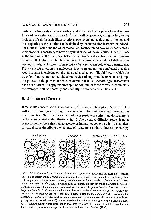
If the solute concentration is nonuniform, diffusion will take place. More particles will move from regions of high concentration into dilute ones and fewer in the other direction. Since the movement of each particle is entirely random, there is no force associated with diffusion (Fig. 1). The so-called diffusion force “is not a ponderomotive force that can accelerate or impart net velocities. It is a statistical or virtual force describing the increase of ‘randomness’ due to increasing entropy
diffusion osmosis diffusion + osmosis
FIG. 1 Molecular-kinetic descriptions of transport: Diffusion, osmosis, and diffusion plus osmosis. The smaller circles indicate water molecules and the membrane is considered to be infinitely thin. Diffusing solute molecules move randomly; each jump may take place either to the left (from 2 to 3) or to the right (from 2 to 3’). There is no net transfer of momentum between solute and water. In osmosis solutes cannot cross the membrane. Compared with diffusion, the jumps from 2 to 3 are not balanced by jumps from 2 to 3’. Consequently there must be a net transfer of momentum from the solutes to the water in the direction towards the concentrated bath. In case the membrane is partly permeable, the situation is intermediate between diffusion and osmosis. The solute molecule can either be reflected giving rise to an osmotic event (3) or jump into the dilute solution which gives rise to a diffusive event (3’). It follows that the water permeability measured by means of a permeable solute is smaller than that recorded by means of an impermeable solute. Redrawn from Zeuthen (1995).

206 ZEUTHENANDMACAULAY
of dilution” (Hille, 1992, p. 263; see also Einstein, 1956; Hartley and Crank, 1949; Sten-Knudsen, 1978). At present, there is no model that combines the microscopic interactions between solutes and solvent during diffusion with macroscopic or measurable properties. This has forced investigators to apply macroscopic or con- tinuum theories. Einstein (1956), for example, used Stokes’ relation to arrive at an expression for the diffusion coefficient.
In osmosis, a semipermeable membrane separates two solutions of different concentrations; the membrane is permeable to water but restricts solute move- ments. Two cases are usually distinguished: one where the membrane is purely lipid and one where it contains pores. In the lipid case, water crosses by a diffu- sional process charaterized by a relatively high Arrhenius activation energy, about 12 to 20 kcal mall’ . The process probably requires formation of sites for water molecules within the hydrocarbon part of the bilayer (Deamer and Nicols, 1989; Marrink et al., 1996; Nagle, 2000). The rate of transport is independent of the na- ture of the driving force for water, whether this be diffusive, osmotic, or hydraulic (for a review, see Finkelstein, 1987).
In the case of membrane pores, it is useful first to compare the case where the pore is infinitely short (i.e., the membrane infinitely thin) with the case of simple diffusion (see Fig. 1). In case the membrane is perfectly semipermeable, a solute molecule that has arrived at a position in front of the opening (position 2) can only move away if it returns toward the concentrated solution (position 3). In case of diffusion, however, the solute molecule might equally well have proceeded toward the dilute solution (from position 2 to 3’). In diffusion, therefore, water molecules experience an equal number of solute molecules moving to one side and to the other; consequently, there is no net transfer of momentum from the solute to the water molecules. In osmosis, the water molecules situated in the hole experience more solute molecules moving toward the bath with the high solute concentration. As a consequence, there is a net transfer of momentum to the water molecules toward this side. The mechanism for the osmotic transport must be sought in this asymmetry of momentum transfer.
In case the membrane is not perfectly semipermeable, the diameter of the pore is large enough to allow solute molecules to pass. A solute molecule that permeates the membrane (position 2 to 3’) will behave entirely diffusively, and the event will not lead to net movement of water. If, however, the solute molecule is reflected from the edge of the hole, osmotic forces will arise. This suggests that a molecular- kinetic description of the permeable pore must contain both diffusive and osmotic events and that macroscopic or measured parameters must be continuous functions of solute radius.
C. Pores with a Length
Pores that span plasma membranes have lengths much larger than the diameter of the water molecule and of the solute molecule. For an impermeable pore, all

PASSlVEWATERTRANSPORTINBlOLOGlCALPORES 207
solute molecules are reflected from the solute-facing end of the pore. Following the treatment above, a momentum will be transferred to the water molecules residing inside the pore with direction into the solute-containing compartment, and water transport will ensue. The situation is described in thermodynamic terms, chem- ical potentials, and hydrostatic pressures in the so-called Vegard-Mauro model (Vegard, 1908; Mauro, 1957; Hill, 1982; Finkelstein, 1987; see also Manning, 1968; Mauro, 1979; Hammel, 1979; Soodak and Iberall, 1979; Tomicki, 1985). The model postulates a gradient of hydrostatic pressure through the pore; at one end, the pressure equals the pressure of the solvent-free bath while it describes a sharp jump at the interface with the solute-containing bath. The unphysical abrupt change in the hydrostatic pressure must be seen as the temporal and spatial aver- age of the stochastic processes. The hydrodynamic nature of the driving force is supported by experiments (Mauro, 1957).
If, on the other hand, the pore is permeable to the solute, there is no agreement with regard to a model for permeation. One kind of model simply combines the mechanisms described for the impermeable pore with the assumption that the pore wall rejects the permeable solute, thus decreasing its concentration (Garby, 1957; Anderson and Malone, 1974; Tomicki, 1985; Finkelstein, 1987). This strategy has been criticized (Hill, 1982, 1989a,b) on the view that if a solute can enter the pore, the activity of the solute, at equilibrium, will be the same outside and inside the pore. As a consequence, there should be no difference in osmotic pressure and therefore no build up of hydrostatic pressure gradients. With no hydrostatic pressure differences, flows should be entirely diffusive and driven by the difference in the activities at the two sides of the membrane.
This conflict does not arise in a molecular-kinetic description. If a solute particle collides with the orifice of the pore and is reflected, forces are set up that move water from the pore into the bath. Such events are driven by a gradient of hydrostatic pressure as described above. If, on the other hand, the solute particle enters the pore, water moves from the pore into the bath by interdiffusion. In short, the permeable pore will be characterized by a mixture of hydrostatic and diffusive events. For the very wide pore, most solute molecules will enter the pore, and an entirely diffusive description will be adequate. In the other limit, the narrow impermeable pore, all solute molecules will be reflected, and the hydrodynamic description is applicable.
In conclusion: the molecular-kinetic description gives an intuitively correct description of osmosis in membrane pores, but it gives no analytical formulae. A thermodynamic model based on macroscopic concepts leads to analytical formulae but not to a unified physical picture.
D. Irreversible Thermodynamics
Irreversible thermodynamics is a useful framework in which to interpret water transport experiments. It defines a set of parameters and defines the conditions
208 ZEUTHENANDMACAULAY
under which they should be measured (see review by Katchalsky and Curran, 1965). Unfortunately, the physical meaning of the parameters is less clear and has been subject to various interpretations. Consider, for example, a membrane with water channels that are permeable to glycerol. The equations for the volume and solute fluxes and the coupling between them are:
Jv = -RTLp(ACi + o,,G~ACG~) (1)
JGI = RTPGIACGI + Jvcc~'(l - cf,GI> (2)
where Jv is the volume flow, Joi is the flux of glycerol, i.e., determined from tracer uptake, R is the gas constant, and T the absolute temperature. AC, is the transmembrane concentration difference of impermeable solutes such as mannitol; ACc, is the difference in glycerol concentration. Coi’ is the average concentration of glycerol in the aqueous pore. The coupling between the solute and volume fluxes in the pore can be characterized by the reflection coefficient C-F. The Lr in Eq. (1) is determined from osmotic challenges by impermeable osmolytes (i.e., mannitol), while gradients implemented by glycerol determine the reflection coefficient (T,,oi. The parameters of Eq. (2) are determined from ultrafiltration experiments. In these, Jo1 arises from two mechanisms: by diffusion (first term) and by the water flow in the pore itself (second term). This second term is a product of the volume flow Jv, the glycerol concentration Co,‘, and a term (1 - ar,ol), which contains a reflection coefficient. Traditionally, the two reflection coefficients of Eqs. (1) and (2) cs,oi and af,ol, are assumed to be equal, as would be the case if Onsager symmetry applied. In that case, co1 ( = ar,oi = oSs,ol) and Co,’ can be determined from
numerical solution of Eqs. (1) and (2) (see Macey and Karan, 1993). It has been argued, however, that for a leaky pore, gr and (T, are in fact differ-
ent since they arise from two different physical mechanisms for which Onsager symmetry does not apply (Hill, 1982). Only in the limits of either very narrow or wide pores will br and us be equal. Different bf and a, would make a precise determination of the parameters in Eqs. (1) and (2) difficult. Certainty about glyc- erol permeation in the aqueous pathway could only be obtained by testing whether osmotic flows increased or decreased the glycerol fluxes, above or below that given by the permeability alone. In such a case, the second term of Eq. (2) would be dif- ferent from zero. Experimentally, this could be difficult. It might require gradients of several hundred mosm L-’ to induce significant solute fluxes, gradients which may not be tolerated by cells. In addition, there is no way to estimate the glycerol concentration Coi’ inside the pore.
So far, published experimental data have been interpreted under the assumption that cr and us are equal. This means that L, and the reflection coefficient for a permeable solute, say, glycerol, goi, can be determined in osmotic experiments using mannitol and glycerol as osmolytes. A hoi smaller than 1 will be indicative of glycerol permeation:
OGI = 1 - RTpa(Ax/%v)fsw (3)

PASSlVEWATERTRANSPORTINBlOLOGlCALPORES 209
where Poi is the glycerol permeability, Ax/bow is a constant that gives the ratio of the membrane thickness to the volume fraction of water, and f,, is the friction between the solute (glycerol) and water in the pore. Equation (3) is adapted from Eq. (10-56) in Katchalsky and Curran (1965) with partial molar volume effects ignored. In our discussion of experimental data, we employ Eq. (3) bearing in mind the caveats outlined above. It appears that a smaller (T is associated with a larger permeability P.
E. Pores with Variable Diameters
Consider an aqueous pore with a wide and a narrow section in series (Fig. 2). If the Lr, is measured by means of an osmolyte with a diameter larger than the wide part of the pore, then the Lr will represent the water permeability of the whole pore (Fig. 2A). The L, may also be measured by an osmolyte that is small enough to enter the wide part of the pore in an osmotic active form (Fig. 2B). In that case, the measured Lr, will be larger, and given by the narrow part of the pore only. In the language of irreversible thermodynamics (Eq. l), it might appear as if 6, for the smaller osmolyte were larger than 1. In case the osmolyte is small enough to permeate throughout the pore (Fig. 2C), the interpretation becomes very
FIG. 2 Pores with a variable diameter. Consider a pore that has a wide and a narrow section in series. In A the water permeability Lp is measured by an osmolyte (filled circles) which is too large to enter the pore at all. In B Lr is measured by an osmolyte which is small enough to enter the wide part but not the narrow part. Under the assumption that the osmolyte remains osmotically active within the wide cavity of the pore, the Ir recorded will be larger than in A. If the osmolyte is small enough to enter both the narrow and the wide part of the protein (C) the water permeability can be recorded to be both smaller, larger or equal to that found in A.

210 ZEUTHENANDMACAULAY
complicated. The o will appear to increase on account of the partial permeation into the pore as described above; on the other hand, 0 will tend to be smaller than 1 on account of the permeation through the narrow section of the pore. All in all, the measured a may be larger, equal to, or smaller than 1 depending on the size and permeability properties of the probe molecule. A measured, (T equal to 1 might arise as a combination of these various mechanisms. Measurements of u in filtration experiments (cF~, Eq. 2) would alleviate the problem.
Recent data suggest that water pores have diameters that vary along the length of the aqueous pathway. This applies to aquaporins (Murata et al., 2000) (see Section 1II.A and Engel and Stahlberg, this volume), and to some cotransporters where larger Lps are obtained with smaller osmotic probes (see Section 1V.B). It should be emphasized that the concept of the wideness of a pore should be understood in a broad sense. It may include other phenomena such as the properties of surface water in the protein and whether the osmotic probe has access to this layer. Structural information is required to select among the various possibilities for physical mechanisms. This may be achieved in studies in pores of known structure such as gramicidin or from high-resolution atomic models of biological pores such as that described below.
III. Aquaporins
Aquaporins are membrane proteins whose major function is to mediate transmem- brane transport of water (for reviews see Borgnia et al., 1999; Ishibashi et al., 2000). So far, 10 mammalian aquaporins have been identified: AQPO to AQP9. Of these, AQPO, 1,2,4,5, and 8 are predominately water selective, while the others may support significant fluxes of other substances. Glycerol permeates through AQP3, AQP7, and AQP9, and these proteins have been named aquaglyceroporins. In addition, AQP9 is permeable to a broad spectrum of substances, among them urea and mannitol (Tsukaguchi et al., 1998, 1999). Some investigations, however, find that AQP9 is permeable only to urea and water (Ko et al., 1998; Ishibashi et al., 1998). AQP6 exhibits anion conductance (Yasui et al., 1999). Plants express numerous types of aquaporins (see Maurel, this volume) and so do yeasts (see Hohman, this volume).
A. Molecular Architecture of AQPI
AQPl was initially identified in red blood cells and renal proximal tubules and was shown to be an integral membrane protein (Denker et uE., 1988; Preston and Agre, 1991). Expression in Xenopus Zuevis oocytes revealed that the protein functioned as a water channel (Preston et al., 1992). Subsequent sequence analysis and expression studies applying functional epitope-scanning mutants have shown

PASSIVE WATER TRANSPORT IN BIOLOGICAL PORES 211
‘-W D COOH
‘V
extracellular
cytoplasm
FIG. 3 Structural organization of the AQPl The AQPl is made from six membrane spanning helices connected by five loops (top panel). In the functional monomer (lower panel), the hydrophilic loop B, which connects helix 2 and 3, and loop E, which connects helix 5 and 6, are bent back into the hole formed by the helices. The two loops meet in the middle to form the putative water-selective gate that contains two NPA motifs (Asn-Pro-Ala). For clarity, the helices 3, 1 and 2 are drawn separated from helices 5,4 and 6. The hydrogen-bonding properties of the side-groups of the two asparagine residues are thought to constitute the permeation barrier. In the membrane the six o-he&es form a right-handed twisted arrangement. Based on Murata er al. (2000) redrawn from Zeuthen (2001).
that AQPl consists of 269 amino acid residues which form two tandem repeats, each of which has three membrane-spanning a-helices. The carboxy- and amino termini are located to the cytoplasm&z side. Two of the loops that connect the (II- helices, the B and E loops, fold back and connect in what appears to be the aqueous pathway through the protein. This unique structure is known as the hourglass model (Jung et al., 1994) (Fig. 3). The two loops each contain an NPA motif (asparagine, proline, alanine, or Asn-Pro-Ala), which is well conserved among the aquaporins and believed to play a pivotal role for water selectivity.
Preparation of crystals from highly purified samples of human red blood cell AQPl has recently permitted the determination of the atomic structure at a high level of resolution (Walz et al., 1997; Cheng et al., 1997; Li and Jap, 1997; Murata et al., 2000). In the latter work, the order and spatial orientation of the constituent cy-helices have been determined. The six membrane-spanning cr-helices form a right-handed twisted arrangement (Fig. 3B). Seen from the extracellular side, the

212 ZEUTHENANDMACAULAY
helices have the clockwise order 2, 1, and 3. Helix 1 contains the amino terminal and connects to helix 2 via the extracellular A-loop that contains a glycosylation site (asparagine 42). The cytoplasmic end of helix 2 connects via the functionally important B-loop to helix 3. It is the B-loop that bends into and contributes to the properties of the aqueous pore. The helices of the other part of the protein (the second repeat) appear in the order 5,4, and 6. Helix 3 connects extracellularly to helix 4. Helix 5 connects extracellularly to helix 6 via the functionally important E-loop, which bends into the pore and combines with loop B. Helix 6 terminates the protein with a relatively short carboxy terminal. The structure achieves its stability from the large crossing angles of the helices, ridge-grove fittings, and by interactions of highly conserved glycines (Russ and Engelman, 2000) at the crossing sites. This applies to helices within each repeat and for the helices holding together the two halves of the AQP 1.
The B- and E-loops take on central importance, since it is the two asparagines (76 and 192) of the NPA motifs which form the permeability barrier for water. The B-loop and E-loop are shaped into short a-helices positioned at an angel of about 90” to each other (Murata et al., 2000) (Fig. 4). The N-terminals of the B- and E-loop helices are capped by the formation of hydrogen bonds between the carbonyl groups of the asparagines and NH groups of the main chain. The
I
(B) - -
- -
FIG. 4 The atomic model for water permeation in AQPl, The B and E loops which dip into the interior of the AQPl monomer form short a-helices. The N-termini of these short helices point into the aqueous pathway and create a positive electrostatic field which will orientate the water oxygen towards the two asparagines. These residues are located in a narrow portion of the pore (3 8, diameter) which prevent larger passage of larger molecules. The water molecules are able to form a hydrogen bonds with these polar amino acid residues. In A there is a water molecule connected with each of the two asparagines. In B one water molecule occupies both asparagine sites. As a consequence water molecules pass through the aqueous pathway by making and breaking two hydrogen bonds. Importantly, the string of hydrogen bonded water molecules extending through the pore is broken preventing transport of H+. The rest of the channel is mainly made up from hydrophobic residues allowing for fast transport rates. Based on Murata ef al. (2000), Fig. 5.

PASSIVEWATERTRANSPORTIN BIOLOGICALPORES 213
two loops are locked to each other by van der Waals interactions between the two prolines. In addition, the two helices are tied up with the rest of the protein via a number of ion pairs and hydrogen bonds.
AQPl exists in the membrane as a homotetramer (Van Hoek et al., 1991; Smith and Agre, 1991). The diameter of the hole formed between the four AQPls is about 3.5 A at its narrowest, and 8.5 A at its widest. It has been discussed whether this center, which is not occupied by the AQPs, could form a pathway for water perme- ation. Previous experimental studies have discredited the notion. and Murata et al. (2000) give a structural rationale for this. The hole is too small to accommodate a lipid molecule, and since the protein exposes hydrophobic side groups toward this cavity, it is considered unlikely that water is present.
B. Atomic Model for Water Permeation in AQPI
What is the molecular architecture of the aqueous pore that provides the high water permeability and selectivity? More than lo9 water molecules can pass through the channel per second (Zeidel et al., 1992b), while the protein has been found to be impermeable to molecules and ions, in particular H+. The molecular architecture shows that the pore has a narrow centrally located constriction of about 3 A, which is only slightly larger than the 2.8 A usually given for the diameter of the water molecule (Murata et al., 2000). Size alone then would block the passage of larger solutes. The selectivity of the constriction is determined by the two asparagines (76 and 192) which extend their amido groups into the lumen of the pathway. It is by forming hydrogen bonds with these groups that water, and water alone, slips through (Fig. 4). The portions of the pore which connect the constriction with the cellular and extracellular solutions are mainly lined with hydrophobic residues that ensure rapid transit of water. During transport, a water molecule is initially connected to neighboring water molecules by hydrogen bonds. As the water molecule approaches the constriction formed by the two asparagines, the oxygen atom of the water molecule orients toward the two residues. This is because the E and B helices are orientated with their N-termini pointing toward the lumen, giving a positive electrostatic field. The water molecule then breaks its hydrogen bonds with the adjacent water molecules and forms instead, first one, then two hydrogen bonds with the asparagines. The deserted water molecules cannot form other hydrogen bonds with the hydrophobic pore wall. As a consequence, water molecules are predicted to permeate with an energy barrier equivalent to making and breaking one hydrogen bond, about 3-4 kcal mol-‘, in good agreement with experimental data. Most importantly, the string of hydrogen bonds that would otherwise extend through the pore and constitute an efficient pathway for H+ transport is broken (Porn&s and Roux, 1996, 1998).
The atomic structure of the aquaporin homolog, the glycerol facilitator GlpF from Escherichia coli, has been determined to a resolution of 2.2 A (Fu et al., 2000). This protein is impermeable to water and ions but allows high rates of

214 ZEUTHENANDMACAULAY
diffusive transport of glycerol (Maurel et al., 1993; see Maurel, and Engel and Stahlberg, this volume). The tertiary structure is similar to that of AQPl with six major a-helices and two shorter a-helices in the pore. The selectivity filter of GlpF is different from that of AQPl and consists of a 28-A long, 3.4- to 3.8-A wide amphiphatic channel. During permeation, the alkyl backbone of the glycerol molecule is wedged against the hydrophobic part of the channel, while the -OH groups of the glycerol form successive hydrogen bonds with side groups from the hydrophilic part.
Several questions arise from the comparison between AQPl and GlpF. First, in bulk solution, water molecules share hydrogen bonds with up to four others. To permeate a channel in single file, a water molecule would have to reduce this number to two. Unless these bonds are replaced by other bonds to the pore wall, the activation energy would increase significantly, since 3 to 4 kcal mol-’ are involved per bond. Fu et aE. (2000) suggest that the energy barrier for this dehy- dration suffices to explain the impermeability of GlpF to water. This differs from the mechanism suggested for AQPl which involved the making and breaking of an additional hydrogen bond inside the pore (Fig. 4) (Murata et al., 2000). The
activation energy for water transport in AQPl is in the range 3 to 7 kcal mol-’ (Zeidel et al., 1992; Meinild et al., 1998). Clearly a precise evaluation of the ener- getics of hydrogen bond formation for water in both AQPl and GlpF is required. Second, the mechanisms suggested for the impermeability of AQPl and GlpF to H+ ions are different. In neither pore can a hydrated ion pass the narrow section, and the hydrophobic part of the wall prevents the costly dehydration of the ion. Fu et al. (2000) consider this sufficient to explain the impermeability of GlpF to H’. They do not invoke any breaking of the string of hydrogen bonds, as suggested for AQPl (Murata et al., 2000).
C. Transport Parameters of Aquaporins
Most data on transport parameters of aquaporins have been obtained from expres- sion studies in Xenopus oocytes. The water permeability L, has been found to be the same whether obtained from shrinkage or swelling experiments (Meinild et al, 1998b). The unit channel permeabilities have been obtained from counting the number of expressed channels by tagging (Yang and Verkman, 1997) or by freeze-fracture (Zampighi et al., 1995; Chandy et al., 1997). Unit permeabilities were (in units of cm3 see-’ 10-14) for AQPO: 0.25 (Yang and Verkman, 1997) 0.015 (Zampighi et al., 1995), 0.028 (Chandy et al., 1997); AQPl: 4.6 (Zeidel et al., 1992b), 1.4 (Zampighi et al., 1995), 1.2 (Chandy et al., 1997), and 6 (Yang and Verkman, 1997); AQP 2: 3.3 (Yang and Verkman, 1997); AQP3: 3.1 (Yang and Verkman, 1997); AQP4: 24 (Yang and Verkman, 1997), and for AQPS: 5.0 (Yang and Verkman, 1997). The variability among the data calls for further experiments (see Engel and Stahlberg, this volume).

PASSIVE WATER TRANSPORT IN BIOLOGICAL PORES
TABLE I Reflection Coefficients 0 for Aquaporins 0 to 5
215
Mannitol Urea Glycerol Acetamide Formamide
Mole weight 182 60 92 63 45
AQPO 1 1 1 0.8 0.6
AQPl 1 1 0.8 1 1
AQP2 1 1 0.8 1 1
AQP3 23°C 1 1 0.2 0.7 0.4
AQP3 13°C 1 1 0.4 1 0.5
AQP4 1 1 0.9 1 1
AQP5 1 1 1 1 0.8
From Meinild et al. (1998b).
The reflection coefficients for various test solutes (a, Eq. 1) have been measured by a highly accurate assay for aquaporins 0 to 5 expressed in oocytes (Table I). The values were obtained as the ratio of the cell shrinkage obtained by the test solute divided by the shrinkage obtained using mannitol. A 0 smaller than 1 is taken as an indication of permeation of the test solute in the aqueous channel; the smaller the G, the larger the permeability (see Eq. 3 and accompanying discussion). A comparison of L, and c shows the inadequacy of a purely hydraulic model based on size and length of the pore and the van der Waals dimensions of the solute. Consider the dimensions of the solutes employed (in A): mannitol(7.4 x 8.2 x 12), urea(3.6~5.2~5.4),glycerol(4.8~5.1 x7.8),acetamide(3.8~5.2~5.4),and formamide (3.4 x 4.4 x 5.4). For AQPl, AQP2, and AQP4 only for glycerol was (T lower than 1. Accordingly, glycerol should permeate, yet the glycerol molecule is not the smallest one tested. In AQP3, (T for glycerol is smaller than (T for formamide which is smaller than (T for acetamide. Yet, glycerol is the largest among the three. Furthermore, both glycerol and acetamide are larger than urea, which does not permeate. Thus a straightforward application of van der Waals dimensions is insufficient to explain. the sequences of observed US. Part of the explanation could be solute-pore interactions. If, for example, the --NH2 and the -OH of the solutes formed hydrogen bonds with the pore wall, the effective size of the solute would be smaller.
A similar conclusion is reached from a discussion of pore lengths. The cr for glycerol is close to 1 for all the aquaporins, except AQP3. If this is taken to mean
that the cross section of the pores in AQPs 1,2,4, and 5 are equal, it follows from the unit permeability that AQPl and AQP2 have the same length while AQP4 is 6 times shorter. From the US, it seems that AQP3 has the largest pore cross section, but since its single channel permeability is similar to AQPl and AQP2, the length
216 ZEUTHENANDMACAULAY
of its pore must be greater. These postulated differences in cross section and length are unlikely when the similarity in secondary structure among the aquaporins is considered.
D. Polyol Permeation in AQP3, Role of Hydrogen Bonds and Backbone Length
AQP3 acts as a channel for both water and glycerol. The fluxes are linearly increas- ing functions of their respective chemical driving forces (Echevarria et al., 1994, 1996; Ishibashi et al., 1994; Zampighi et al., 1995), and the activation energies for the fluxes are low (Ishibashi et al., 1994; Echevarrta et al., 1996). Furthermore, the two fluxes share the pathway in the protein, as indicated by the low 0s (Echevarrfa et al., 1996; Meinild et al., 1998b).
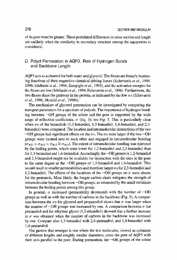
The mechanism of glycerol permeation can be investigated by comparing the transport parameters for a spectrum of polyols. The importance of hydrogen bond- ing between -OH groups of the solute and the pore is supported by the wide range of reflection coefficients, (T (Eq. 3); see Fig. 5. This is particularly clear when US of the butandiols (1,2-butandiol, 1,3-butandiol, 1,4-butandiol, and 2,3- butandiol) were compared. The location and intramolecular interactions of the two -OH groups had significant effects on the US. The os were larger if the two -OH groups were located next to each other and engaged in intramolecular bonding (on23 > (Tat2 > 0~13 g antd). The extent of intramolecular bonding was mirrored by the boiling points, which were lower for 1,2-butandiol and 2,3-butandiol than for 1,3-butandiol and 1 ,Cbutandiol. Accordingly, the -OH groups in 1,2-butandiol and 2,3-butandiol might not be available for interaction with the sites in the pore to the same degree as the -OH groups of 1,3-butandiol and 1,Cbutandiol. This would result in smaller permeabilities and therefore larger os for 2,3-butandiol and 1,2-butandiol. The effects of the locations of the -OH groups on r~ were absent for the pentanols. Most likely, the longer carbon chain mitigates the strength of intramolecular bonding between -OH groups, as witnessed by the small variations between the boiling points among this group.
In general, o increased (permeability decreased) with the number of -OH groups as well as with the number of carbons in the backbone (Fig. 5). A compar- ison between the US for glycerol and propanediol shows that cr was larger when the number of -OH groups was increased by one. A comparison between cr for pentandiol and for ethylene glycol (1,2-ethandiol) showed that a further increase in o was obtained when the number of carbons in the backbone was increased by one. Compare also 1,3-butandiol with 2,4-pentandiol, and 1,4-butandiol with 1 ,Cpentandiol.
The picture that emerges is one where the test molecules, viewed as cylinders of different lengths and roughly similar diameters, cross the pore of AQP3 with their axis parallel to the pore. During permeation, the -OH groups of the solute

PASSIVEWATERTRANSPORTIN BIOLOGICALPORES 217
0.6
# 0.4
0.2
“’ Ma EG PD GI 912 813 914 923 P12 P14 P15 P24
osmolyte
FIG. 5 Reflection coefficients of AQP3 for different straight chain polyols. The aquaporin was ex- pressed in Xenopus oocytes and the rate of oocyte shrinkage was measured by exposing the oocyte to solutions made 20 mOsm hyperosmolar by means of addition of various straight chain polyols: Ethylen glycol (EG), 1,Zpropanediol (PD), glycerol (Gl), 1,2-, 1,3-, 1,4-, and 2,3-Butandiol (B12, B13, B14, B23), and 1,2-, 1,3-, 1,4-, and 2,3-Pentandiol (P12, P14, P15, P24). The shrinkages produced were compared to those produced by mannitol, the ratio presented as c (a, of Eq. 1). The structures of the test solutes are given above. An open circle indicates an -OH group, and squares indicate backbone carbons. Each carbon has four bonds, but single hydrogen atoms and associated bonds are not shown. In general the data suggest that the shorter the backbone of the polyol and the fewer -OH bonds, the easier the polyol penetrates (small 6, Eq. 3). Interestingly, the two polyols B12 and B23 deviate from this trend having larger 0s (low permeabilities). This could be a result of intramolecular bonds between the two abutting -OH groups which are then not available to make contact with transport sites in the pore. Redrawn from (Zeuthen and Klaerke, 1999).
form a succession of single hydrogen bonds with the pore wall as indicated by the low activation energies of around 5 kcal mol-’ observed for permeation (Meinild et al., 1998b; Ishibashi et al., 1994; Echevam’a et al., 1996).
E. Effects of pH on AQPO, AQP3, and AQP6
The water permeability L, of AQPO is regulated by pH and Ca2+ (NCmeth-Cahalan and Hall, 2000). AQPO was expressed in the Xenopus oocytes; when external pH was reduced from 7.5 to 6.4, L, increased 3- to 4-fold. At further reductions in

218 ZEUTHENANDMACAULAY
external pH, to 5.5, Lp decreased toward the values obtained at pH of 7.5. It appears that the Lr is stimulated in a pH range of about 1.5 units centered around pH 6.4. If the external Ca2+ concentration was nominally reduced to zero, L, increased about 4-fold. Lowering of internal Ca*+ concentrations and calmodulin inhibitors produced the same effects. Importantly, the clamping of internal Ca2+ overrode the effect of changes in external Ca*+ concentration; possibly the changes in Ca2+ concentrations are sensed intracellularly. The pH and the Ca*+ effects were not strictly additive, which suggests that H+ and Ca2+ act at two different sites, which affects the same common pathway regulating Lr. This concept was supported by site-directed mutagenesis of a histidine, His4’, which is unique for AQPO. Histidine has a pK around 7, similar to physiological pH values. When His4’ was replaced by alanine, aspartic acid, or lysine, functional water channels with no pH sensitivity were produced. Similar effects were obtained by covalent modification of histidines. From the predicted membrane topology, His4’ is located extracellularly. This suggests a working model of the AQPO with the H+ sensor on the extracellular face of the channel and the Ca*+ sensor on the inside.
At a normal external pH of 7.4, AQP3 expressed in Xenopus oocytes conducts both water and glycerol (references above). Both fluxes, however, depended on external pH and were abolished at acid values (Zeuthen and Klaerke, 1999) (Fig. 6). The L, was characterized by a pK of 6.4 and a Hill coefficient of 3, the glycerol permeability Poi by a pK of 6.1 and a Hill coefficient of 6. The difference in pH
- i
i i E v
P 0
C
fP
External pH
FIG. 6 pH sensitivity of water and glycerol permeability of AQP3 expressed in Xenopus oocytes. The water permeability L,r, decreased with external pH with a pK of 6.4 and a Hill coefficient of 2.7, it was completely abolished at pH below 5.6. The glycerol permeability Pot was measured from uptake of [‘4C]glycerol. It had a pK of 6.1 and a Hill coefficient of 6.2. It was completely abolished for pH below 5.7. The findings are discussed in the text on the basis of a model in which water and glycerol permeates via the same pathway. Redrawn from (Zeuthen and Klaerke, 1999).

PASSIVEWATERTRANSPORTIN BIOLOGICALPORES 219
sensitivities for Lr, and Par shows that AQP3 is a water and glycerol channel at neutral pH, while it is predominantly a glycerol channel in the pH range 5.8 to 6.2. It follows that the reduction of Lr, by H+ does not arise from a physical closure of the putative pore, since the aquaporin is still open to glycerol transport while closed for water transport. The pH effects were reversible.
One way to view the data from AQP3 is to interpret them by means of an Eyring energy barrier model (Glasstone et al., 1941). In this model, the molecule permeates by a series of jumps, the energy barriers of which are determined by the chemical bonds between the molecule and specific sites in the pathway. For AQP3, the Arrhenius activation energy (EJ for Lp is low, around 5 kcal mol-’ (Meinild, et al., 1998b; Ishibashi et al, 1994; Echevarrfa et al., 1996), which suggests that the water molecule at neutral pH permeates by forming a succession of single H+-bonds. Titration of the sites at acid pH would abolish their H+-bonding capacity and render them effectively hydrophobic, thereby abolishing the Lr,. In analogy to the Lr,, Poi also had a low E, and a marked dependence on external pH. This would suggest that glycerol also permeates by forming successive H+-bonds.
Despite the differences in pH dependence, it is possible that the pathways for Hz0 and glycerol share some titratable groups. The relatively low pK for glycerol transport could result from a competitive interaction between H+ and glycerol at the titratable groups. Such competition has been described in intact human red blood cells (Carlsen and Wieth, 1976), where the glycerol transport has been suggested to be mediated by AQP3 (Roudier et al., 1998). For an opposing view on the role of AQP3 in red blood cell, see Yang et al. (2001) and Section V below. In these cells, pK for Pal was about 6.0 at external glycerol concentrations of 1 mm01 L-’ and 5 5 at external glycerol concentrations of 2 mol L-l, which was taken as evidence for competition between H+ and glycerol. In addition, the Hill coefficients were estimated to be larger than 2 (see also Stein, 1962). The values are in agreement with those of the present study where glycerol concentrations of 20 mm01 L-’ were employed. The finding suggests that glycerol, when close to the titratable site, to a certain extent displaces water molecules, an effect that would be enhanced by the confinement of the pore. The resulting lower molar fraction of water near the site would result in a lower local H+ concentration and consequently in a decrease of the effective pK.
AQPl, 2,4, and 5 did not exhibit any pH sensitivity (Zeuthen and Klaerke, 1999; Nemeth-Cahalan and Hall, 2000). AQPl has a small but significant permeability to glycerol (Meinild et al., 1998b; Abrami et al., 1995, 1996) and the CTS for the smaller polyols, ethylene glycol(l,2-ethandiol) and 1,2-pentanediol, were similar to that of glycerol (Zeuthen and Klaerke, 1999). This shows that although these polyols may interact with water in AQPl, some structural incompatibility, not found in AQP3, prevents them from permeating at any higher rate.
AQP6 is functionally distinct from other known aquaporins (Yasui et al., 1999). Expressed in Xenopus oocytes, AQP6 exhibits a relatively low water permeability L,. Application of Hg*+ increases L, up to IO-fold and lowers the activation

220 ZEUTHENANDMACAULAY
energy to values expected from osmotic water transport. This is surprising, since Hg2+ is a well-known inhibitor of most other aquaporins (AQP4 is an exception). Hg2+ also stimulated Cl- conductance in AQP6. The L, increased about 3-fold when extracellular pH was decreased from control values (7.5) to 4.0. The unique properties of AQP6 might be understood in conjunction with its localization to the membranes of intracellular vesicles of acid-secreting cells of renal collecting duct, where it is colocalized with H+-ATPase.
IV. Cotransporters
In addition to their primary function, many membrane proteins mediate a passive water flux, given a transmembrane difference in water chemical potential. They act as water channels with a well-defined water permeability Lp. This dual func- tion is found among the cotransporters such as those coupling Na+ with sugar or amino acid transport. The aquaglyceroporins described above, i.e., AQP3, also transport two substrates, glycerol and water. There is one fundamental difference, however, between cotransporters and aquaglyceroporins. The cotransporters un- dergo conformational changes during transport of the nonaqueous substrate. Each conformational state can in principle be described by its own Lr, and reflection coefficient (Eqs. 1 to 3). Interpretation of data will therefore require additional information about the temporal occupancy of the various states.
To complicate matters even further, water transport by cotransporters is bimodal. In addition to the passive water transport, the proteins also sustain an active mode of transport. In this mode, water is cotransported along with the nonaqueous substrates in a fixed ratio determined by the properties of the protein. (This mode of transport is dealt with in the chapter by Zeuthen and MacAulay, Cotransporters as Molecular Water Pumps, this volume.) In the present article, we restrict ourselves to the water channel properties where transport is induced by transmembrane osmotic gradients.
The hypothesis that a cotransporter could serve as a passive water channel was first suggested in connection with the Naf-coupled glucose transporter SGLTl (Fischbarg et al., 1993) and was confirmed by its expression in Xenopus laevis oocytes (Zampighi et al., 1995; Loike et al., 1996; Loo et al., 1996, 1999). The finding has been extended to other cotransporters: the Na+/GABA transporter GATI (Loo et al., 1999), the Na+/dicarboxylate transporter NaDCl (Meinild et al., 1999), and the Na+/glutamate transporter EAATl (MacAulay et al., 2001). The L, is characterized by a small unit water permeability. For the Nat/glucose transporter (SGLTl), it could be estimated to be about two orders of magnitude lower than that of AQPl (Zampighi et aZ., 1995). Even so, the contribution of cotransporters to the water permeability of a given cell may be significant due to the large number of cotransporters present; this would apply in particular to cell membranes without aquaporins.
PASSlVEWATERTRANSPORTINBlOLOGlCALPORES
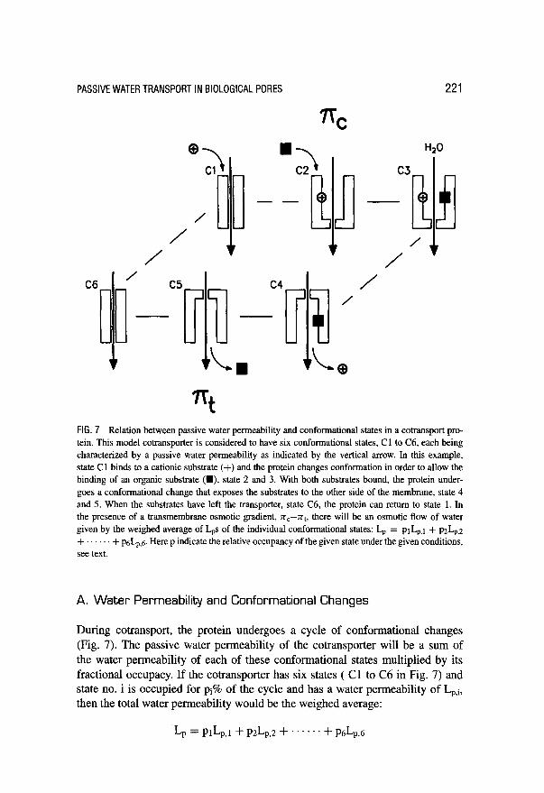
FIG. 7 Relation between passive water permeability and conformational states in a cotransport pro- tein. This model cotransporter is considered to have six conformational states, Cl to C6, each being characterized by a passive water permeability as indicated by the vertical arrow. In this example, state Cl binds to a cationic substrate (+) and the protein changes conformation in order to allow the binding of an organic substrate (M), state 2 and 3. With both substrates bound, the protein under- goes a conformational change that exposes the substrates to the other side of the membrane, state 4 and 5. When the substrates have left the transporter, state C6, the protein can return to state 1. In the presence of a transmembrane osmotic gradient, n,--nt, there will be an osmotic flow of water given by the weighed average of Las of the individual conformational states: Lr, = prI+t + p&,2 + + p&n,,5 Here p indicate the relative occupancy of the given state under the given conditions, see text.
A. Water Permeability and Conformational Changes
During cotransport, the protein undergoes a cycle of conformational changes (Fig. 7). The passive water permeability of the cotransporter will be a sum of the water permeability of each of these conformational states multiplied by its fractional occupacy. If the cotransporter has six states ( Cl to C6 in Fig. 7) and state no. i is occupied for pi% of the cycle and has a water permeability of Lp,i, then the total water permeability would be the weighed average:
I-T = PlLp,l+ PzL,z + . . . . . + plsI+

222 ZEUTHEN AND MACAULAY
Na+/glutamate (MT0
Na+/glucose (sGLT~)
/ 4.q / -glucose 4 //l/J///
/ /fi , +glucose /j /&-I////
I I I I I 0 2 4 6 8 10
L, [lO”cm set-‘1
FIG. 8 Water permeability of cotransporters in the presence and absence of the organic substrates, The human Na+/glutamate cotransporter EAATl (MacAulay er al., 2001) and the rabbit Na+/glucose cotransporter were expressed in Xenopus oocytes. The Lt, was measured by means of osmotic gradients implemented by manmtol in the absence (-) and presence (+) of the organic substrate. The water per- meability of the native oocyte membrane has been subtracted. It is seen that EAATl has a higher Lt,, and that the Lp of the SGLTl is unaffected by the presence of the substrate (Loo er al., 1999). The two trans- porters are electrogenic and the finding applies to both the voltage clamped and unclamped situation.
Since the states may have different water permeabilities, the total L, would be expected to be a complicated function of the external parameters, such as clamp voltage and substrate concentrations. This is exemplified by the effect of sub- strate on the Lr of the Na+/glutamate cotransporter which increased its total L, in the presence of glutamate (MacAulay et al., 2001) (Fig. 8). In contrast, the L, of the Na+/glucose transporter was unaffected by the presence of sugar.
The conformational states and their fractional occupancy have been studied in detail for the SGLTl (see Fig. 7) (Parent et al., 1992a,b; Loo et al., 1999). Under clamped conditions without Na+, 60% of the cotransporters are thought to be in state C 1 and40% in state C6; under clamped conditions in the presence of Naf, 80% are in C2,10% in Cl, and 10% in C3; in the presence of Na+ and sugar, 80% are in state C.5 and 20% in C6. Thus Cl, C2, C5, and C6 may each contribute to the L,. In the presence of the inhibitorphlorizin the L, was abolished (Loo et al., 1999). With phlorizin, SGLTl is locked in conformational state C3 (Fig. 7). One interpretation of the transport data is that this state has a low passive water permeability.

PASSIVEWATERTRANSPORTIN BIOLOGICALPORES 223
The activation energy E, of the L, of cotransporters was found to be around 5 kcal mol-‘, as would be expected from transport via an aqueous pathway (Loo et al., 1996, 1999; Meinild et al., 1998a; MacAulay et al, 2001).
B. Reflection Coefficients of Cotransporters
In order to define the reflection coefficient of a given solute ((TV, Eq. l), the shrink- age produced by equimolar addition of the test solute is determined. The reflection coefficient is then obtained as the ratio between this shrinkage and the one ob- tained with an inert large osmolyte, say, mannitol. Such experiments have been performed for the Na+/glutamate cotransporter (EAATl, MacAulay et al. unpub- lished) and the Na+/glucose cotransporter (SGLTl, Zeuthen unpublished). Five osmolytes were employed: mannitol, urea, glycerol, acetamide, and formamide. The cotransporters were expressed in Xenopus oocytes under clamped conditions but with no organic substrates present (glutamate or sugar). The ratio between the shrinkage (or water permeability) measured with a given test osmolyte (Lr,,) and that obtained with mannitol (Lr,J is shown in Fig. 9. Surprisingly, the shrinkages obtained with urea and glycerol for the EAATl were larger than those obtained with mannitol. A straightforward use of Eq. (1) would imply that CJ was larger than 1, which has no direct interpretation within the framework of irreversible thermodynamics set up by Eqs. (1) and (2). It would require the applications of the equations developed for composite membranes which involve a large number of partly inaccessible transport parameters (Kedem and Katchalsky, 1962, 1963). An intuitive interpretation of the data would be that urea and glycerol can enter into a vestibule of the aqueous pore in an osmotically active form. In this way, the transmembrane osmotic gradient would be applied across a shorter section of the pore, which would result in a larger L, being measured (see Fig. 2 and related text). The smallest test solutes, acetamide and formamide, may enter the whole channel, yielding a reduced Lrx/Lp,m due to the diffusive process in the aqueous pathway. In other words, the reflection coefficients of these substances are probably small for both sections of the pore. The SGLTl behaved differently from the EAATl with respect to urea and glycerol. The L, recorded with these os- molytes was nearly the same as that obtained with mannitol; the smaller osmolytes behaved similarly for the two cotransporters. The dimensions of the pore of the SGLTl are dependent on conformational changes in the protein. This is reflected by the temperature dependence of the L, measured by means of acetate and for- mamide. At 14°C the L,s obtained with these smaller osmolytes were similar to those obtained with mannitol. One interpretation is that the pore narrows at low temperatures. The data obtained with formamide, for example, indicate a dou- bling of L, for a 10°C change in temperature. This is equivalent to an E, of about 11 kcal mol-‘.

224 ZEUTHEN AND MACAULAY
A B rSGLT rSGLT
1.2 7 24-c 14-c
6 E ci
i . 2
C
1.41 EMT1 T
D Glut1
1.2
1 .o
0.8
0.6
0.4
0.2
0.0 man urea glyc ocet form man urea glyc ocet form
FIG. 9 Water permeabilities Lr, obtained with different sized solutes in cotransporters and uniports. The Lps obtained with urea (mw 60), glycerol (mw 92), acetate (mw 63) and formamide (mw 45) are given relative to that determined with mannitol (mw 182), that is as Lp,&,,,,,. Different transporters of organic substrates were tested, (A) the rabbit Na+/glucose transporter SGLTl at 24” C and (B) at 14” C, Zeuthen unpublished; (C) the human Na+/glutamate transporter EAATl, MacAulay and Zeuthen, unpublished; and (D) the glucose monoport GLUTl, Zeuthen, unpublished. The transporters were expressed in Xenopus oocytes and Lr was measured by a sensitive optical system (Zeuthen er al., 1997).

PASSIVEWATERTRANSPORTIN BIOLOGICALPORES
V. Uniports
225
A. Glucose Transporters [GLUT)
Many animal cells posses membrane proteins specifically designed for facilitated glucose transport. The proteins, which belong to the GLUT family, have been identified in the brain, skeletal muscle, hepatocytes, adipocytes, and fetal muscle. The transport of glucose is passive, driven by the transmembrane concentration difference for glucose and involves conformational changes of the protein. The GLUTS also support passive water transport. This was suggested from studies in macrophage cell lines 5774 (Fischbarg et al., 1989) and later confirmed by ex- pression studies in Xenopus oocytes (Fischbarg et al., 1990; Zhang et al., 1991). The water permeability per protein is low compared to that of aquaporins, esti- mates of 6 to 60% have been presented (Fischbarg et al., 1993). The Arrhenius activation energy E, is higher than that of passive water diffusion, between 10 and 13 kcal mol-’ (Zeidel et al., 1992a). This high E, may not reflect solely the mode of water transport through the pore, but could also reflect temperature-dependent conformational changes in the protein leading to a narrowing of the aqueous path- way.
Measurements of the reflection coefficients (a,, Eq. 1) of GLUT1 for urea, glyc- erol, acetamide, and formamide disclose a very low CJ for glycerol (Fig. 9; Zeuthen, unpublished). This would indicate a high permeability for glycerol (Eq. 3). The protein was expressed in Xenopus oocytes and the Lp and 0 were measured by challenging the oocyte with solutions made hyperosmolar with the respective test molecules (Meinild et al., 1998a). When compared to the ~7s for SGLTl (Fig. 9), it is seen that acetamide and formamide penetrate through both transporters, but the high glycerol permeability is exclusive for the GLUT. It would be interesting to test if GLUT constituted a significant pathway for glycerol in erythrocytes. Recent studies using double-knockout mice lacking AQP 1 and AQP3 showed no reduction in the phloritin sensitivity of the glycerol permeability (Yang et al., 2001). This may contradict transport models for erythrocytes in which AQP3 constitutes the pathway for glycerol. But the glycerol could well permeate through the GLUT1 transporter of these cells (see Fig. 9).
B. Urea Transporters
Uniports for urea have been identified in particular in the kidney. One of these, the UT3, has been shown to be an efficient water channel (Yang and Verkman, 1998). The unit water permeability was 0.14 lo-l4 cm3 set-‘, which is 40 times less than the L, for AQPl and about 20 times less than that of AQP3, but is of the order of the passive water permeability of the Na+/glucose transporters SGLTl. The activation energy was low, less than 4 kcal mol-‘, and the reflection coefficient for urea was

226 ZEUTHENANDMACAULAY
0.3. These are good indications that water and urea share an aqueous pathway in the protein. Urea and water permeation are also combined in AQP 9 (Tsukaguchi et al., 1998), in the Na+/glucose transporter SGLTl (Leung et al., 2000), and in the Na+/glutamate cotransporter EAATl (MacAulay et al., unpublished). The question of how water and urea may share an aqueous pathway is an interesting biophysical question that deserves more experiments.
VI. Conclusions
With the discovery of water channel proteins (aquaporins) and constitutive passive water permeability in proteins with other functions (cotransporters and uniports) the amount of data for passive water permeation has increased dramatically and so has the need for a physicochemical model of transport. The permeation of water through aqueous pores is determined by a wide spectrum of parameters. First, there are those that define the water molecule, the pore, and, in the case of osmosis, the solute molecule used for creating the driving force. Secondly, there are the param- eters that define the interactions between the participants. Due to this complexity, a realistic physicochemical model has so far been an elusive goal. Simple models based on macroscopic concepts such as pore length, pore diameter, and the physi- cal dimensions of the test solutes do not, in general, comply with the experimental data. Experiments on permeability have hinted at the importance of hydrogen bonding as an important mechanism of interaction between solutes and pore wall.
The advent of the atomic structures for AQPl (Murata et al., 2000) and for GlpF (Fu et al., 2000) is an important turning point for the description of transport in biological pores. Questions like those presented above can now be approached by quantum mechanical modeling and computer simulation, similar to the approaches to gramicidin A (Porn&s and Roux 1996, 1998). The atomic model for AQPl will provide a useful framework for questions regarding other aquaporins, some of which have unique properties. What makes AQPS, 7, and 9 permeable to both water and glycerol (Borgnia et al., 1999)? Which sites in AQPO are responsible for its Ca2+ sensitivity (Nemeth-Cahalan and Hall, 2000), and which sites are responsible for the pH sensitivity of the AQP3 (Zeuthen and Klaerke, 1999)? Which structures in AQP6 give rise to its anion conductance (Yasui et al., 1999)‘? How can AQP9 be permeable to a variety of neutral solutes (Tsukaguchi et al., 1998)?
Models for water transport in cotransporters and uniports face the same prob- lems as those for the aquaporins. In addition, these proteins undergo a series of conformational changes in order to fulfill their other transport functions. Each of these conformational states is characterized by a water permeability, so the total water permeability is a complicated function of the functional state of the protein. This, in turn, depends on external parameters such as substrate availability and electrochemical driving forces.
PASSlVEWATERTRANSPORTINBlOLOGlCALPORES 227
Note added in proofi In a recent paper, Curry et aE. (2001) show that single water channels of AQP-1 do not obey the Kedem-Katchalsky equations: Methyl- urea is impermeable, yet its reflection coefficient is smaller than one. [Curry, M. R., Shachar-Hill, B., and Hill, A. E. (2001). “Single water channels of aquaporin-1 do not obey the Kedem-Katchalsky equations.” J. Membr Bid. 181, 115-123.1
Acknowledgments
Useful discussions with Professor W. D. Stein, Dr. D. A. Klaerke, and Dr. A. E. Hill are gratefully acknowledged. Svend Christoffersen is thanked for the professional artwork.
References
Abrami, L., Berthonaud, V., Deen, P. M. T., Rousselte, G., Tacnet, F., and Riproche, P. (1996). Glycerol permeability of mutant aquaporin 1 and other AQP-MIP proteins: Inhibitions studies. Pfiigers Arch. 431,408-414.
Abrami, L., Tacnet, F., and Riproche, P (1995). Evidence for a glycerol pathway through aquaporin 1 (CHIP28) channels. P@gers Arch. 430,447-458.
Anderson, .I. L., and Malone, D. M. (1974). Mechanism of osmotic flow in porous membranes. Biophys. .I. 14,957-982.
Borgnia, M., Nielsen, S., Engel, A., and Agre, P. (1999). Cellular and molecular biology of the aquaporin water channels. Annu. Rev. Biochem. 68,425-458.
Carlsen, A., and Wieth, J. 0. (1976). Glycerol transport in human red cells. Acta Physiol. &and. 97, 501-513.
Chandy, G., Zampighi, G. G. A., Kreman, M., and Hall, J. E. (1997). Comparison of the water trans- porting properties of MIP and AQPl J. Membr. Biol. 159,29-39.
Cheng, A., Van Hoek, A. N., Yeager, M., Verkman, A. S., and Mitra, A. K. (1997). Three-dimensional organization of a human water channel. Nature 387,627-630.
Dainty, J. (1965). Osmotic flow. Syrup. Sot. Exp. Bio. 19,75-85. Deamer, D. W., and Nichols, J. W. (1989). Proton flux mechanisms in model and biological membranes.
J. Membl: Biol. 107,91-103. Denker, B. M., Smith, B. L., Kuhajda, F. P., and Agre, P. (1988). Identification, purification, and partial
characterization of a novel M, 28,000 integral membrane protein from erythrocytes and renal tubules. J. Biol. Chem. 263, 15,634-15,642.
Echevarrfa, M., Windhager, E. E., and Frindt, G. (1996). Selectivity of the renal collecting duct water channel Aquaporin-3. J. Biol. Chem. 271,25,079-25,082.
Echevarrfa, M., Windhager, E. E., Tate, S. S., and Frindt, G. (1994). Cloning and expression of AQP3, a water channel from the medullary collecting duct of rat kidney. Proc. Natl. Acad. Sci. USA 91, 10,997-11,001.
Einstein, A. (1956). “Investigations on the Theory of the Brownian Movements.” Dover, New York. Finkelstein, A. (1987). “Water Movement through Lipid Bilayers, Pores, and Plasma Membranes.”
John Wiley & Sons, New York. Fischbarg, J., Kuang, K., Hirsch, J., Lecuona, S., Rogozinski, L., Silverstein, S. C., and Loike, J. (1989).
Evidence that the glucose transporter serves as a water channel in 5774 macrophages. Proc. Natl. Acud. Sci. USA 86,8397-8401.
Fischbarg, J., Kuang, K., Li, J., Arant-Hickman, S., Vera, J. C., Silverstein, S. C., and Loike, J. D. (1993). Facilitative and sodium-dependent glucose transporters behave as water channels. In

228 ZEUTHENANDMACAULAY
“Isotonic Transport in Leaky Epithelia, Alfred Benzon Symposium 34” (H. H. Ussing, J. Fischbarg, 0. Sten-Knudsen,E. H. Larsen, and N. J. Willumsen, Eds.), pp. 432-446. Munksgaard, Copenhagen.
Fischbarg, J., Kuang, K., Vera, J. C., Arant, S., Silverstein, S. C., Loike, J., and Rosen, 0. M. (1990). Glucose transporters serve as water channels. Proc. Natl. Acad. Sci. USA 87,3244-3247.
Fu, D., Libson, A., Miercke, L. J. W., Weitzman, C., Nollert, P., Krucinski, J., and Stroud, R. M. (2000). Structure of a glycerol-conducting channel and the basis for its selectivity. Science 290,481-486.
Garby, L. (1957). Studies on transfer of matter across membranes with special reference to the isolated human amniotic membrane and the exchange of amniotic fluid. Acta Physiol. Stand. 40, l-84.
Glasstone, S., Laidler, K. J., and Eyring, H. (1941). “The Theory of Rate Processes.” McGraw-Hill Book Company, Inc., New York, London.
Hammel, H. T. (1979). Forum on osmosis. I. Osmosis: Diminished solvent activity or enhanced solvent tension? Am. J. Physiol. 237, R95-R107.
Hartley, G. S., and Crank, J. (1949). Some fundamental definitions and concepts in diffusion processes. Trans. Faraday Sot. 4.5,801-819.
Hill, A. (1982). Osmosis: A bimodal theory with implications for symmetry. Proc. R. Sot. Land. 215, 155-174.
Hill, A. E. (1989a). Osmosis in leaky pores: The role of pressure. Proc. R. Sot. Land. (B) 237,363-367. Hill, A. E. (1989b). Osmotic flow equations for leaky porous membranes. Pmt. R. Sot. Lund. 237,
369-377. Hill, A. E. (1994). Osmotic flow in membrane pores of molecular size. J. Membr: Biol, 137, 197-203. Hille, B. (1992). “Ionic Channels of Excitable Membranes.” Sinauer Associates Inc., Sunderland,
Massachusetts. Ishibashi, K., Kuwahara, M., Gu, Y.. Tanaka, Y., Marumo, F., and Sasaki, S. (1998). Cloning and
functional expression of a new aquaporin (AQP9) abundantly expressed in the peripheral leukocytes permeable to water and urea, but not to glycerol. &o&m. Biophys. Res. Commun. 244,268-274.
Ishibashi, K., Sasaki, S., Fushimi, K., Uchida, S., Kuwahara, M., Saito, H., Furukawa, T., Nakajima, K., Yamaguchi, M., Gojoboti, T., and Marumo, F. (1994). Molecular cloning and expression of a member of the aquaporin family with permeabililty to glycerol and urea in addition to water expressed at the basolateral membrane of kidney collecting duct cells. Proc. Natl. Acad Sci. USA 91,6369-6273.
Ishibashi, K., Kuwahara, M., and Sasaki, S. (2000). Molecular biology of aquaporins. Rev. Physiol. Biochem. Pharmacol. 14&l-32.
Jung, J. S., Preston, G. M., Smith, B. L., Guggino, W. B., and Agre, P (1994). Molecular structure of the water channel through aquaporin CHIP: The hourglass model. J. Biol. Gem. 269,14,648-14,654.
Katchalsky, A., and Curran, P F. (1965). “Nonequilibrium Thermodynamics in Biophysics.” Harvard Univ. Press, Cambridge, Massachusetts.
Kedem, 0.. and Katchalsky, A. (1962). Permeability of composite membranes. Trans. Faraday Sot. 59,1931-1940.
Kedem, O., and Katchalsky, A. (1963). Permeability of composite membranes. Trans. Faraday Sot. 59,1941-1953.
Ko, S. B., Uchida, S., Naruse, S., Kuwahara, M., Ishibashi, K., Marumo, F., Hayakawa, T., and Sasaki, S. (1998). Cloning and functional expression of rAQP9L a new member of the aquaporin family from rat liver. Biochem. Mol. Biol. Int. 47,309-3 18.
Leung, D. W., Loo, D. E, Hirayama, B. A., Zeuthen, T., and Wright, E. M. (2000). Urea transport by cotransporters. J. Physiol. 528(2), 251-257.
Li, H., and Jap, B. K. (1997). Molecular design of aquaporin-1 water channel as revealed by electron crystallography. Nat. Struct. Biol. 4,263-265.
Loike, J., Hickman, S., Kuang, K., Xu, M., Cao, L., Vera, J. C., Silverstein, S. C., and Fischbarg, J. ( 1996). Sodium-glucose cotransporters display sodium- and phlorizin-dependent water permeability. Am. J. Physiol. 271, C1774-C1779.
Loo, D. D. F., Zeuthen, T., Chandy, G., and Wright, E. M. (1996). Cotransport of water by the Na+/glucose cotransporter. Proc. Natl. Acad. Sci. USA 93,13,367-13,370.

PASSlVEWATERTRANSPORTINBlOLOGlCALPORES 229
Loo, D. F., Hiiayama, B. A., Meinild, A.-K., Chandy, G., Zeuthen, T., and Wright, E. (1999). Passive water and ion transport by cotransporters. J. Physiol. 195-202.
MacAulay, N., Gether, U., Klaerke, D. A., and Zeuthen, T. (2001). Water Transport by the Na+-coupled glutamate cotransporter. J. Physiol. 530,367-378.
Macey, R. I., and Karan, D. M. (1993). Independence of water and solute pathways in human red blood cells. J. Membl: Bid. 134,241-250.
Manning, G. S. (1968). Binary diffusion and bulk flow through a potential-energy profile: A kinetic basis for the thermodynamic equations of flow through membranes. J. Gem. Phys. 49,2668-2675.
Marrink, S. J., J&rig, F., and Berendsen, H. J. C. (1996). Proton transport across transient single-file water pores in a lipid membrane studied by molecular dynamics simulation. Biophys. J. 71,632-647.
Maurel, C., Reizer, J., Schroeder, J. I., and Chrispeels, M. J. (1993). The vacuolar membrane protein TIP creates water specific channels in Xenopus oocytes. EMBO J. 12,2241-2247.
Mauro, A. (1957). Nature of solvent transfer in osmosis. Science 126,252-253. Mauro, A. (1979). Forum on osmosis. III. Comments on Hammel and Scholander’s solvent tension
theory and its application to the phenomenon of osmotic flow. Am. J. Physiol. 237, R1104113. Meinild, A.-K., Klaerke, D. A., Loo, D. D. E, Wright, E. M., and Zeuthen, T. (1998a). The human
Na+/Glucose cotransporter is a molecular water pump. J. Physiol. 508(l), 15-21. Meinild, A.-K., Klaerke, D. A., and Zeuthen, T. (1998b). Bidirectional water fluxes and specificity for
small hydrophilic molecules in aquapotins 0 to 5. J. Bid. Chem. 273,32,446-32,45 1. Meinild, A.-K., Loo, D. F. F., Pajor, A., Zeuthen, T., and Wright, E. M. (2000). Water transport by the
renal Na+/dicarboxylate cotransporter. Am. J. Physiol. 278, F777-F783. Murata, K., Mitsouka, K., Hirai, T., Walz, T., Agre, l?, Heymann, J. B., Engel, A., and Fujiyoshi, Y.
(2000). Structural determinants of water permeation through aquaporin-1. Nufure 407,599-605. Nagle, J. F. (2000). Theory of passive proton conductance in lipid bilayers. J. Bioenerg. Biomembr: 19,
413-426. Nemeth-Cahalan, K. L., and Hall, J. E. (2000). pH and calcium regulate the water permeability of
aquaporin 0. J. Biol. Biochem. 275,6777-6782. Parent, L., Supplisson, S., Loo, D. D. F., and Wright, E. M. (1992a). Electrogenic properties of the
cloned Na’lglucose cotransporter: I. Voltage-clamp studies. J. Membr: Biol. 125,49-62. Parent, L., Supplisson, S., Loo, D. D. F., and Wright, E. M. (1992b). Electrogenic properties of the
cloned Na+/glucose cotransporter: II. A transport model under nonrapid equilibrium conditions. J. Membr. Biol. 125,63-79.
Porn&s, R., and Roux, B. (1996). Structure and dynamics of a proton wire: A theoretical study of H? Translocation along the single-file water chain in the gramicidin A channel. Biopbys. J. 71, 19-39.
Porn&s, R., and Roux, B. (1998). Free energy profiles for H+ conduction along hydrogen-bonded chains of water molecules. Biophys. J. 75,33-40.
Preston, G. M., and Agre, P. (199 1). Isolation of the cDNA for erytbrocyte integral membrane protein of 28 kilodaltons: Member of an ancient channel family. Proc. Natl. Acad. Sci. USA 88,ll ,110-l 1,114.
Preston, G. M., Carroll, T. P., Guggino, W. B., and Agre, P. (1992). Appearance of water channels in Xenopus oocytes expressing red cell CHIP28 protein. Science 256,385-389.
Roudier, N., Verbavatz, J.-M., Maurel, C., Riproche, P., and Tacnet, F. (1998). Evidence for the presence of aquaporin-3 in human red blood cells. J. Biol. Chem. 273,8407-8412.
Russ, W. P., and Engelman, M. (2000). The GxxxG motif: A framework for transmembrane helix-helix association. J. Mol. Bid. 296,911-919.
Smith, B. L., and Agre, P. (199 1). Erythrocyte Mr 28,000 transmembrane protein exists as a multisubunit oligomer similar to channel proteins. J. Biol. Chem. 266,6407-6415.
Soodak, H., and lberah, A. (1979). Forum on osmosis, IV. More on osmosis and diffusion. Am. J. Physiol. 237, R1144122.
Stein, W. D. (1962). Spontaneous and enzyme-induced dimer formation and its role in membrane permeability. II. The mechanism of movement of glycerol across the human erythrocyte membrane. Biochim. Biophys. Acta 59,47-65.

230 ZEUTHENANDMACAULAY
Sten-Knudsen, 0. (1978). Passive transport processes. In “Membrane Transport in Biology. Concepts and Models” (G. Giebish, D. C. Tosteson, and H. H. Ussing, Eds.), Vol I, pp. l-l 13. Springer-Verlag, Berlin.
Tomicki, B. (1985). The role of pressure in osmotic flow. J. Colloid Znterface Sci. 108,484-494. Tsukaguchi, H., Shayakul, C., Berfer, U. V., Mackenzie, B., Devidas, S., Guggino, W. B., VanHoek,
A. N., and Hediger, M. A. (1998). Molecular characterization of a broad selectivity neutral solute channel. J. Biol. Biochem. 273,24,737-24,743.
Tsukaguchi, H., Weremowicz, S., Morton, C. C., and Hediger, M. A. (1999). Functional and molecular characterization of the human neutral solute channel aquaporin-9. Am. J. Physiol. 277, F685-F696.
Van Hoek, A. N., etal. (1991). Functional unit of 30 kDa for proximal tubule water channels as revealed by radiation inactivation. J. Biol. Chem. 266, 16,633-16,635.
Vegard, L. (1908). On the free pressure in osmosis. Proc. Cumb. Phil. Sot. Math. Phys. Sci. 15,13-23. Verbavatz, J.-M., Brown, D., Sabolic, I., Valenti, G., Ausiello, D. A., Van Hoek, A. N., and Verkman,
A. S. (1993). Tetrameric assembly of CHIP28 water channels in liposomes and cell membranes: A freeze-fracture study. J. Cell Biol. 123,605-618.
Walz, T., Hirai, T., Murata, K., Heyman, J. B., Mitsuoka, K., Fujiyoshi, Y., Smith, B. L., Agre, P., and Engel, A. (1997). The three-dimensional structure of aquaporin-1. Nature 387,624-626.
Yang, B., and Verkman, A. S. (1997). Water and glycerol permeabilities of aquaporin l-5 and MIP determined quantitatively by expression of epitope-tagged constructs in Xenopus oocytes. J. Biol. Chem. 272, 16,140-14,146.
Yang, B., and Verkman, A. S. (1998). Urea transporter UT3 functions as an efficient water channel. J. Biol. Chem. 273,9369-9372.
Yang, B., Ma, T., and Verkman, A. S. (2001). Erytbrocyte water permeability and renal function in double knockout mice lacking aquaporin-1 and aquaporin-3. J. Biol. Chem. 276,624-628.
Yasui, M., Hazama, A., Kwon, T.-H., Nielsen, S., Guggino, W. B., and Agre, P (1999). Rapid gating and anion permeability of an intracellular aquaporin. Nature 402, 184-187.
Zampighi, G. A., Kreman, M., Boorer, K. J., Loo, D. D. F., Bezanilla, F., Chandy, G., Hall, J. E., and Wright, E. M. (1995). A method for determining the unitary functional capacity of cloned channels and transporters expressed in Xenopus laevis oocytes. J. Membl: Biol. 148,65-78.
Zeidel, M. L., Albalak, A., Grossman, E., and Carruthers, A. (1992a). Role of glucose carrier in human erythrocyte water permeability. Biochemistry 31,589-596.
Zeidel, M. L., Ambudkar, S. V., Smith, B. L., and Agre, P (1992b). Reconstitution of functional water channels in liposomes containing purified red cell CHIP28 protein. Biochemisfry 31,7436-7440.
Zeuthen, T. (1995). Molecular mechanisms for passive and active transport of water. ht. Rev. Cyt. 160, 99-161.
Zcuthen, T. (2001). How water pass through aquaporins. Trends Biochem. Sci. 26,77-79. Zeuthen, T., and Klaerke, D. A. (1999). Transport of water and glycerol in aquaporin 3 is gated by H+.
J. Biol. Chem. 274, 21,631-21,636. Zeuthen, T., Meinild, A.-K., Klaerke, D. A., Loo, D. D. F., Wright, E. M., Belhage, B., and Litman, T.
(1997). Water transport by the Na+/glucose cotransporter under isotonic conditions. Biol. Cell 89, 307-312.
Zhang, R., Alper, S. L., Thorens, B., and Verkman, A. S. (1991). Evidence from oocyte expression that the erythrocyte water channel is distinct from band 3 and the glucose transporter. J. Clin. Invesf. 88, 1553-1558.